Résumé
Les accidents vasculaires cérébraux ischémiques résultent de l’occusion d’une artère cérébrale par un phénomène embolique ou thrombotique. Cette obstruction déclenche une série d’évènements, cascade ischémique, qui vont conduire à la mort neuronale. Cette destruction est liée à des facteurs spatiaux et temporels. D’une part la reperméabilisation précoce de l’artère occluse va permettre une récupération fonctionnelle des cellules qui ont survécu à l’ischémie grâce au réseau de suppléances artérielles. En effet ce réseau permet de maintenir une perfusion sanguine suffisante dans la partie périphérique du territoire occlus (pénombre). Cette récupération sera meilleure si les mesures thérapiques sont appliquées rapidement. Le traitement de l’ischémie cérébrale vise doncà combattre tous ces facteurs nocifs et actuellement le facteur temps est devenu primordial en raison des traitements thrombolytiques. Des facteurs hémodynamiques jouent également un rôle dans la survenue d’un AVC. En présence d’une sténose d’une artère précérébrale ou cérébrale, toute modification de la tension artérielle peut engendrer une diminution de la perfusion dans le territoire où siège la sténose. Dans ce cas l’ischémie va léser la partie périphérique: icuts jonctionnel ou «des derniers près». La prise en charge d’un patient victime d’un AVC implique la mise en route d’un traitement le plus précocement possible et nécessite une recherche étiologique précise pour assurer le meilleur traitement préventif.
Zusammenfassung
Ischämische zerebrovaskuläre Ereignisse entstehen auf dem Boden eines embolischen oder thrombotischen Verschlusses einer der zerebralen Arterien. Ein solcher Verschluss führt zu einer Reihe von Vorgängen, der sogenannten «ischämischen Kaskade», die letztlich zum neuronalen Zelltod führt. Der Zelluntergang ist an gewisse räumliche und zeitliche Faktoren geknüpft. So kann die akute Wiedereröffnung der verschlossenen Arterie eine funktionelle Erholung der Nervenzellen zur Folge haben, insbesondere wenn die Zellen dank eines arteriellen Umgehungskreislaufs die Ischämie überleben können. Fast immer kann ein derartiger Umgehungskreislauf eine genügende Blutversorgung in der ischämischen Randzone des verschlossenen Gefässes gewähren. Somit kann die Erholung der Nervenzellen davon abhängen, wie schnell therapeutische Massnahmen in die Wege geleitet werden können. Die Behandlung der zerebralen Ischämie zielt deswegen auf die Beeinflussung solcher Faktoren hin, und insbesondere der Zeitfaktor hat dank der Thrombolyse entscheidende Bedeutung erlangt. Auch hämodynamische Faktoren spielen beim zerebrovaskulären Insult eine wichtige Rolle. Besteht eine Stenose der prä-zerebralen oder zerebralen Arterien, können Blutdruckänderungen zu einer Perfusionsverminderung im betreffenden Versorgungsgebiet führen. In diesem Fall wird die Ischämie die peripheren Versorgungsgebiete betreffen: ein «junktionaler Ictus» oder ein Insult der «letzten Wiese». Der therapeutische Zugang zu einem Patienten mit zerebrovaskulärem Insult bedingt ein so akutes Vorgehen wie möglich, indem eine genaue ätiologische Klärung erzwungen werden muss, wonach die bestmögliche Therapiestratifizierung gewählt werden kann.
Introduction
Two main mechanisms, namely occlusion or hemodynamic problems, are potential causes of ischemic stroke [
1]. Both cause a decrease in cerebral perfusion pressure and lead to neural death. However, several defence mechanisms protect the brain from ischemia up to a certain limit, the principal one being blood flow autoregulation. Cerebral blood flow (CBF) is normally maintained, despite blood pressure fluctuation; however, when the lower limit of autoregulation is reached, if the blood perfusion pressure decreases, the cerebral blood flow is insufficient to provide the metabolic needs of the brain cells, resulting in cell death.
Cerebral blood flow and autoregulation
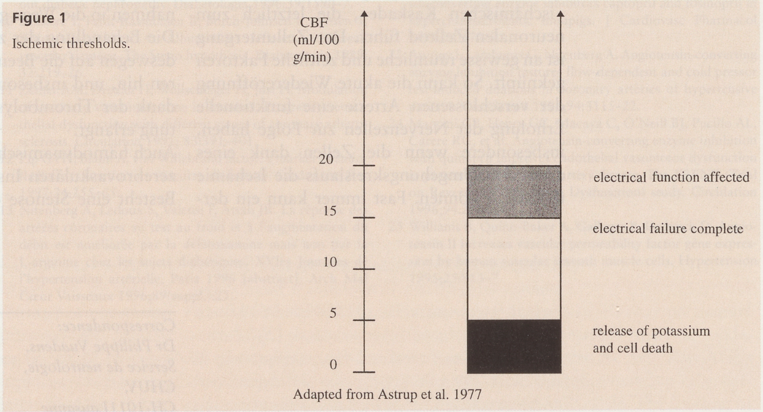
The CBF can be maintained by the autoregulatory properties of the cerebral arteries. In the normal adult, it is approximately 75 mL/100 g/min in the grey matter and less in the white matter. If it is reduced to 10–20 mL/100 g/min, electroencephalogram and evoked potential modifications appear; if reduced further, membrane damage and disruption of the NA/K pump occur, leading to neural death (
Figure 1) [
2]. The CBF varies with the metabolic needs of the brain, any conditions resulting in acidosis (hypoxia, hypercapnia, ischemia) causing cerebrovascular dilatation, thus increasing the CBF.
As measurement of the CBF is difficult, the cerebral perfusion pressure (CPP), the difference between the mean arterial pressure on entering the brain and the exit pressure, is a useful marker of cerebral circulation [
3]. In the supine patient, the mean CPP can be considered equal to the mean systemic arterial blood pressure minus the mean intracranial pressure, with the mean arterial blood pressure being equal to the diastolic value plus one third of the difference between the systolic and diastolic value. In normal patients, the CPP is 70–100 mm Hg, and ischemia does not occur until the CPP falls below 30–40 mm Hg.
In the absence of a brain lesion, the CBF remains constant over a wide range of perfusion pressures due to autoregulatory mechanisms [
4,
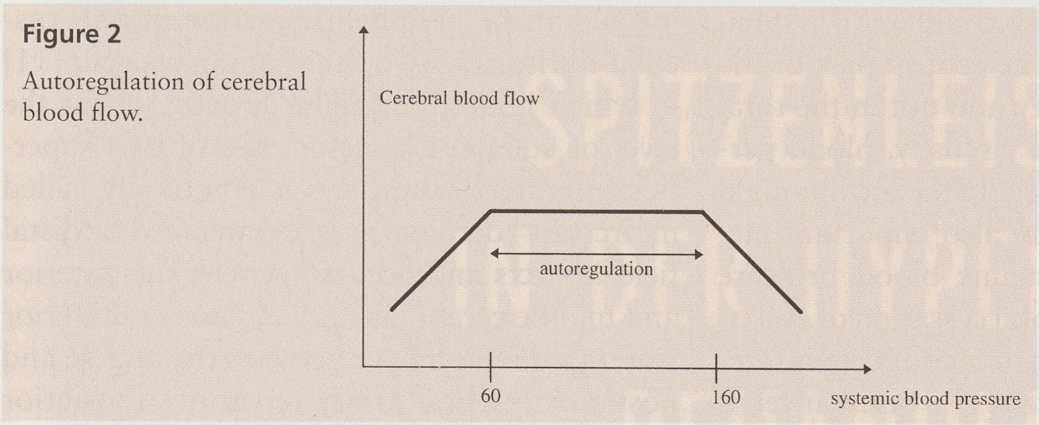
5]. This essential property of the cerebral vessels enables cerebral perfusion to remain stable if the systemic blood pressure changes. In adults, the CBF remains stable when the blood pressure is between 60 and 160 mm Hg (
Figure 2); these limits are exceeded in chronic hypertensive patients. The CBF also remains constant over a wide range of arterial oxygen pressures [
6]; overall, it is dependent on the arterial carbon dioxide pressure. For example, lowering the P
CO2 to 20 mm Hg causes the CBF to fall by about 40%, whereas increasing the P
CO2 to 80 mm Hg almost doubles the CBF. This ability of the CBF to change with P
CO2 explains why hyperventilation reduces the intracranial pressure.
Autoregulation is dependent on the myogenic control of arteriolar resistance, with the cerebral vessels dilating as the perfusion pressure falls and constricting with increasing perfusion pressure to maintain the CBF. Thus, the CBF is a function of both the blood pressure gradient and the vascular resistance, the latter depending on the viscosity of the blood and the length and diameter of the blood vessels.
During the acute phase of stroke, the autoregulatory process is perturbed because the blood vessels are confronted with hypoxia, hypercapnia, and acidosis, which stimulate vasodilatation [
4]. Moreover, the local blood pressure in the ischemic area is below the lower level for autoregulation and the vessels are therefore dilated to allow maximum oxygen extraction. Further reduction of arterial resistance to compensate for a decrease in systemic blood pressure is thus impossible and it is therefore necessary to maintain a high systemic blood pressure to shift the local perfusion pressure on to the autoregulatory curve, producing vasoconstriction. This is also primordial for the collateral arteries supplying the ischemic territory. These perturbations of autoregulation following stoke may persist for several days (up to 30) and explain the phenomenon of luxury perfusion, which corresponds to an increase in local blood flow over and above the oxygen needs of the tissue, leading to reduced oxygen extraction.
”Penumbra” and collateral system
A reduced oxygen supply results in reversible disturbances in the electrophysiological functions of cerebral cells before irreversible damage to cell structure is seen [
7]. The range of blood flows separating this functional change level and the structural damage threshold is 10–20 mL/100 g/min, with the lower limit corresponding to membrane failure, leading to neural death. Between these two levels, autoregulation is perturbed, but the CO
2 reactivity is still partially preserved. This critical zone of blood flow, termed the penumbra [
8], corresponds to that in which the functional modifications of the cells are still reversible without permanent damage and, consequently, is flow-dependent. In contrast, the level at which structural injury occurs is both flowand time-dependent.
The term “penumbra”, in analogy with the half-shaded region surrounding the centre of a solar eclipse, describes the ring-like area of reduced blood flow around the ischemic centre of the infarct, in which the functional activity of the neurons is disturbed or suppressed, but the structural integrity is always preserved, explaining the functional improvement seen following stroke. Indeed, the neurons surviving in this critical area of the infarct under reduced blood flow may begin to function as soon as blood flow and oxygen delivery are restored.
In the acute phase of stroke and during its evolution, the local perfusion also depends on the collateral circulation and the vascular reactivity of the surrounding intact tissue [
9], with extra- and intracranial collateral systems protecting the brain from focal ischemia (
Table 1). This collateral system can compensate for a decrease in CBF equivalent to the occlusion of up to three main precerebral arteries. At the intracranial level, the blood supply mainly depends on leptomeningeal anastomoses but this system is often not sufficient to compensate for the obstruction of a main intracerebral artery. Moreover, the arteries supplying the basal ganglia and brainstem are end-arteries and have no collaterals.
As the size of the infarcts depends on the number and vascular tone of the collateral vessels, in the presence of a preserved collateral system, infarction will occur in the central part of the occluded arterial territory and not in the total vascular territory. Blood viscosity, blood perfusion pressure and especially the intraluminal pressure of the vessels are also important factors, and when the systemic blood pressure falls below 50 mm Hg, total cessation of collateral flow occurs, leading to a complete infarction. This is very important if a vascular territory is already compromised by stenosis or occlusion (hemodynamic theory of stroke).
In the presence of a functional collateral system, a global reduction in blood perfusion pressure (e.g., hypovolemic shock) will result in flow impairment in those areas most distant from the arterial inflow, corresponding to “borderzone”, watershed or distal field infarcts [
10]. This situation corresponds to the law of the most distal field and is anatomically located at the peripheral junction of two vascular territories. Two situations may occur [
11] in which infarction might develop: (1) at the level of collateral anastomoses of two superficial pial territories; this is commonly called infarcts of “derniers prés”, watershed or distal field infarcts and occurs between the anterior and middle cerebral artery territories (anterior watershed infarcts), or between the middle and posterior cerebral artery territories (posterior watershed infarcts) or (2) at the limit between a superficial territory (pial arteries) and a deep territory (deep perforator arteries) without collateral anastomoses. In this case, the term “watershed” is inappropriate because no collaterals exist between the deep perforator arteries (which are end-arteries) and the pial branches of the middle cerebral artery. The terms subcortical junctional or border zone infarcts seem more appropriate.
Embolic occlusion
Embolism is the most frequent cause of ischemic stroke [
12] and can originate in the heart, aortic arch or cervico-encephalic arteries. Artery-to-artery embolisms are composed of fibrin platelet material, sometimes together with red blood cells or atheromatous substances, and are produced by disaggregation of atheromatous plaques. In contrast, cardiac embolisms are composed of thrombotic material. In addition to these two types, rare embolisms of fat, air, neoplasm, infected material or other foreign substance may occur. Artery-to-artery embolism is linked mainly to the development of extracranial atherosclerotic plaques [
13], the progression of which leads to arterial stenosis, formation of a wall thrombus and embolism. Moreover, in the presence of arterial occlusion, anterograde stagnation thrombus may progressively occlude the intracranial arteries or produce embolism.
The size and composition of the embolism and the capacity of the collateral system are the main factors determining infarct size [
14,
15]. Usually, small platelet embolisms are rapidly disaggregated and lead to transient ischemic accident by transient occlusion of the distal cerebral arteries, while fibrin-rich thrombotic embolisms are less readily dissociated and may cause more severe neurological problems [
16,
17].
An embolism may migrate to the piemerian branches and damage the cortical territory. If the occlusion of the cerebral artery is more proximal (e.g., the middle cerebral artery), the infarct is subcortical, because the anastomotic collaterals preserve the superficial territory. However, if the collateral system is not sufficiently developed, the infarct is larger, corresponding to the whole vascular territory.
Autopsy studies have clearly demonstrated that the main site for atherosclerotic plaques is the bifurcation of the internal carotid artery, followed by the carotid siphon, the proximal and distal vertebral arteries and the mid-basilar artery. Sometimes embolisms from the external or common carotid artery may occur. Even in young patients,’ atherosclerosis is a prominent cause of infarcts, accounting for about 30% of cases [
18,
21].
The onset of ischemic stroke is related to the appearance of, and dynamic changes in the atherosclerotic plaques [
14,
15]. The degree of stenosis is correlated with the risk of stroke, especially in the case of high-grade stenosis [
22,
23,
24,
25,
26]. It seems that lipid-laden plaques, or those with a greater metabolic activity, carry a higher risk of ischemic events [
17]. Plaque ulceration is also an important source of artery-to-artery embolisms and the exposure of the arterial media may accelerate the formation of wall thrombi and atheromatous material [
27,
28]. Moreover, with severe associated stenosis the risk of stroke is more than doubled. Intraplaque hemorrhages may occur in stenotic plaques but the risk of ischemic events is controversial. Acute intraplaque hemorrhages seem to be correlated with neurological symptoms, probably because intimal ulceration occurs over protruding mounds of intraplaque hemorrhages [
29,
30]. The risk of intraplaque hemorrhages has also been correlated with the degree of stenosis [
31]. Other studies, however, did not confirm the association between ischemic stroke and the presence, size or age of intraplaque hemorrhages [
32,
33]. However, the presence of hemorrhages in carotid plaque is a criterion of the severity of stenosis and plaque instability [
34].
Plaque size can change with time, with 51% remaining unchanged, 30% progressing and 19% showing spontaneous regression [
35,
36]. Thus the onset of ischemic symptoms is correlated with the stenotic progression, especially if it produces hemodynamic obstruction [
37]. Consequently, two main mechanisms of cerebral ischemia are due to extracranial atherosclerosis. Firstly, thromboembolism, due to the detachment of atheromatous or cholesterol material from plaques and especially fibrinoplatelet aggregates of wall thrombi, is the main pathophysiological cause of stroke. Secondly, the degree of stenosis determines the hemodynamic parameters. Moreover, plaque progression may lead to arterial occlusion. However, wall thrombus formation is not specifically related to the severity of stenosis. Occlusion results from the rupture of the fibrous lining of the artery over an atheroma, producing a tight stenosis and thrombosis [
38]. In fact, angiographic evidence of intraluminal thrombus is rare [
39].
Lacunar infarcts
Four main mechanisms are responsible for the pathogenesis of lacunar stroke: (a) occlusion of the small arteries by either lipohyalinosis or (b) athrosclerosis, (c) embolic occlusion of small arteries, and (d) hypoperfusion in small arterial territories [
40] (
Table 2). Usually, lacunar infarcts occur in the deep perforator territories as these arteries are terminal branches without any arterial network. For this reason, the occlusion of one of these small arteries is associated with a small infarct, termed “lacunar”. It is important to notice that lacunes may also be caused by nonischemic processes, such as small hemorrhage or nonischemic dilatation of the periarteriolar space.
Autopsy studies have demonstrated that about 81% of lacunar infarcts are produced by occlusion of the small penetrating artery supplying the territory of the infarct [
41,
42] (
Table 3). Hypertension seems to be the main factor in lipohyalinosis formation, in which pathological modification of the arterial wall causes progressive obstruction of the arterial lumen by fibrous connective tissue or fibrinoid of lipohyalinosis [
42]. Thrombosis in a lipohyalinotic microaneurism may also occur. It seems that the risk of occlusion is greater in arteries smaller than 300 mm, since, as the arterial diameter is larger than 300 mm, the capillary collateral circulation is sufficient to make infarction less likely when the small arteries are stenosed.
Atherothrombotic occlusion is the second cause of lacunar infarcts. The site of occlusion may be within the proximal part of the penetrating artery (microatheroma), at the junction with a larger artery (junctional atheroma), or in a larger artery occluding the origin of the penetrating artery (mural atheroma).
According to autopsy studies, a certain number of lacunar infarcts are produced by embolic occlusion resulting from cardiogenic embolism or artery-to-artery embolism [
43,
44].
Microatheromatous or lipohyalinotic material, related to chronic arterial hypertension, is the main cause of lacunar infarcts, especially in the case of very small lacunar infarcts (<0.3–0.5 cm), which are usually caused by occlusion of one deep perforator artery and are generally asymptomatic. In the presence of larger lacunar infarcts (0.5–1.5 cm or larger), small artery disease remains the main cause, but more than one third of these patients have a cardiac source of embolism or large-artery disease, even in the absence of concomitant hypertension. Moreover, hypertension is not the only factor associated with small-artery disease, diabetes should also be considered.
Hemodynamic factors also play a role in the pathogenesis of lacunar infarcts. Fischer has demonstrated that the arteries of lacunar infarcts caused by microatheroma or junctional atheroma are not occluded, but only stenosed [
45] and, therefore, hypoperfusion seems to be the cause of infarct in these cases. Usually, unstable clinical pattern is suggestive of critical state of low perfusion distal to a stenosed penetrating artery. In fact, about 30% of lacunar infarcts are preceded by transient stenosed penetrating arteries of reduced diaischemic accidents. However, the risk of lacu- meter, is decreased due to the presence of nar stroke, secondary to hypoperfusion in capillary collaterals.
Conclusion
Although the exact mechanisms of ischemic stroke are becoming increasingly better known due to recent advances in neurosciences and neuroradiology, in reality, the precise cause of stroke may be unknown or related to several pathophysiological mechanisms in certain cases. For example, it is not unusual to see lesions of embolic type associated with watershed infarcts or patients with both HTA and potential cardiac source embolism. However, it is to be hoped that a better understanding of atherosclerotic development will lead to more efficient treatment, not only for the acute phase of stroke, but also for primary or secondary prevention.