Next Article in Journal
New Trends in Treatment of Coronary Artery Disease
Previous Article in Journal
Pathogenesis of Atherosclerosis: A Possible Relation to Infection
 
 
Cardiovascular Medicine is published by MDPI from Volume 28 Issue 1 (2025). Previous articles were published by another publisher in Open Access under a CC-BY (or CC-BY-NC-ND) licence, and they are hosted by MDPI on mdpi.com as a courtesy and upon agreement with Editores Medicorum Helveticorum (EMH).
Proceeding Paper

Coronary Artery Disease: Pathophysiology

Cardiovascular Department, La Tour Hospital, CH-1217 Meyrin, Geneva, Switzerland
Cardiovasc. Med. 1998, 1(2), 125-129; https://doi.org/10.3390/cardiovascmed1020030
Published: 30 August 1998

Pathophysiologie der Koronaren Herzkrankheit

Unser Verständnis der Pathophysiologie der koronaren Herzkrankheit hat sich umfassend gewandelt dank der Interaktion von Grund-lagenforschung, Epidemiologie und klinischer Medizin. Das Aufbrechen der atheromatösen Plaque ist das primäre Ereignis, welches die Kaskade der lokalen Thrombose und der Verminderung oder sogar Unterbrechung des koronaren Flusses triggert. Bereits vor 140 Jahren hat Rudolf Virchow die Hypothese der vaskulären Thrombusbildung formuliert und die 3 Elemente genannt: die Zusammensetzung des Blutes, die Eigenschaften der Gefässwand und die Blurfluss-Charakteristika. Dieses Paradigma behält auch heute noch seinen Wert und erlaubt die systematische Erfassung der verschiedenen Elemente innerhalb der Pathophysiologie der koronaren Herzkrankheit.

Physiopathologie de la Maladie Coronarienne

Notre compréhension de la physiopathologie de la maladie coronarienne a évolué de façon considérable ces dernières années grâce à l’interaction de la recherche fondamentale, de l’épidémiologie et de la médecine clinique. La fissure de la plaque athéromateuse est l’événement primordial qui déclenche la cascade de la thrombose locale et de la diminution et/ou de l’interruption du flux coronaire. Il y a plus de 140 ans, Rudolf Virchow a émis l’hypothèse que la thrombose vasculaire devait nécessairement dépendre de 3 éléments: la composition du sang, la nature de la paroi vasculaire et le caractère du flux sanguin. Ce paradigme garde toute sa valeur aujourd’hui et permet de systématiser le rôle des différents éléments dans la physiopathologie de la maladie coronarienne.

Introduction

Although the first correct ante mortem diagnosis of coronary thrombosis was made less than 120 years ago by Adam Hammer [1], coronary artery disease is today the main single cause of death in the developed world. Elucidation of its physiopathology is a major ongoing effort that is based upon the constant interaction of basic research, epidemiology, and clinical medicine. One of the most important concepts to have been developed over the past 15 years is that plaque fissure is the critical step leading to luminal thrombosis and clinical manifestations. In fact, atherosclerosis can be seen as a very benign disease, most often clinically entirely silent, until such an event takes place and thrombus supervenes. In 1856, Virchow suggested his famous triad concept. According to this, luminal thrombosis is determined by the combination of three elements: blood composition, vessel wall, and flow. The paradigm remains entirely valid today and is a fitting framework to review the fundamental of coronary atherothrombosis.

Vessel Wall

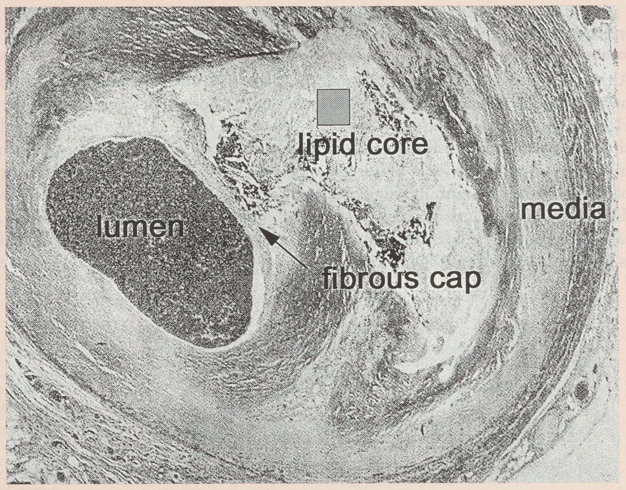
The current consensus is to consider atherosclerosis not so much as a degenerative disease brought about by age and accelerated by risk factors, but rather as a response to injury. Such injury could be derived from several sources, and among them, oxidised LDL [2], toxins, viruses (see review in this issue), immunological mechanisms and mechanical trauma have all received considerable attention. The first target for injury must of necessity be the endothelium [3], because of its strategic location at the interface of blood flow and vessel wall. The atherosclerotic process then typically becomes located within the intima of medium sized epicardial coronary vessels. The sequence of events, as it is understood today [4], involves early uptake of modified (oxidised) lipid into the intima, early expression of adhesion molecules at the surface of a morphologically intact endothelium (selectins, VCAM-1, ECAM-1) enabling later migration of monocytes into the intima. These monocytes/macrophages may then accumulate lipids, and become known as foam cells. Other cells also appear in the intima, such as T-lymphocytes [5]. The macroscopic equivalent to these early histological events is the fatty streak. This can be observed in a large number of arteries in otherwise healthy young adults. If the stimulus (“injury”) is maintained, the process continues to evolve with gradual plaque growth involving smooth muscle cell proliferation and matrix production [6]. This process leads to the formation of the atherosclerotic plaque. This is typically composed of a fibrous cap and a softer inner core, composed largely of lipids. The geometry of this plaque is one of the critical elements that leads to its vulnerability to fissure and thrombosis (see below) (Figure 1).
Not only does the development of a full atherosclerotic plaque carry the risk of inducing thrombosis and occlusion, it also appears to be capable of leading to major changes in vessel configuration. The concept of remodeling is relatively recent [7,8] but is one of the most useful contributions of recent years to help explain the frequent discrepancies between histological and clinical finding. Glagov et al. [8] were the first to report the ability of the left main stem to accommodate stenosis up to 40% without significantly reducing its lumen. This was related to the ability of the vessel to expand so as to accommodate the added volume of the plaque. Only when the stenosis was greater than 40% did the vessel wall’s ability to expand apparently cease, and the lumen began to diminish. The validity of this concept was later verified by other means, and Mintz [9] elegantly demonstrated by serial intravascular ultrasound measurements that remodeling is in fact a more important element than fibrointimal proliferation in the induction of restenosis following balloon coronary angioplasty.

Flow

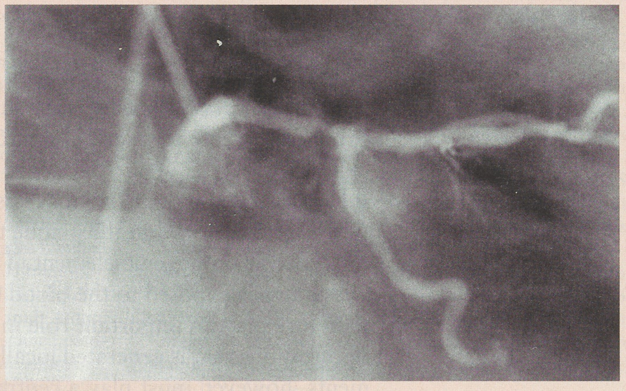
Although atherosclerosis is a diffused systemic disease, it is quite clear that different segments of the vascular tree respond in a different manner to what must be the same systemic insults. Within the coronaries, lesions are often focal and show a marked preference for angulated or bifurcated segments (Figure 2). This corresponds to areas of low shear stress and increased turbulence. Both of these factors increase the contact time between blood elements and the vessel wall. It has also been shown that turbulence and low shear stress are capable of inducing local expression of adhesion molecules. That flow is an important player in the progression of atherosclerotic plaque is further demonstrated by the fact that any decrease in translesional flow, whether turbulent or laminar, is likely to accelerate plaque progression and promote local thrombus deposition. This has been conclusively demonstrated following both PTCA [10] and by-pass surgery [11]. Finally, very slow flow linked for example with either distal plugging of the vessel or systemic low flow induced by cardiogenic shock is known to promote local thrombus deposition in the setting of acute reperfusion following myocardial infarction [12].

Blood Composition

Blood, as envisioned by Rudolf Virchow, must be the obligatory vector for the majority of injurious stimuli to the vessel wall. This includes lipids, possible infectious agents, as well as the main “acute” players such as catecholamines, fibrinogen, platelets, and other elements of the hemostatic system. Some of these will be reviewed in the next paragraph, since they related mostly to the acute clinical events. It should be stressed here that the local atherosclerotic plaque is also subjected to the influence of systemic events. A stable lesion may be induced to become unstable, and lead to an acute clinical manifestation when it is submitted to the influence of events such as systemic thrombocytosis, non-specific inflammation, early postoperative stress, etc.

Relation to Clinical Events

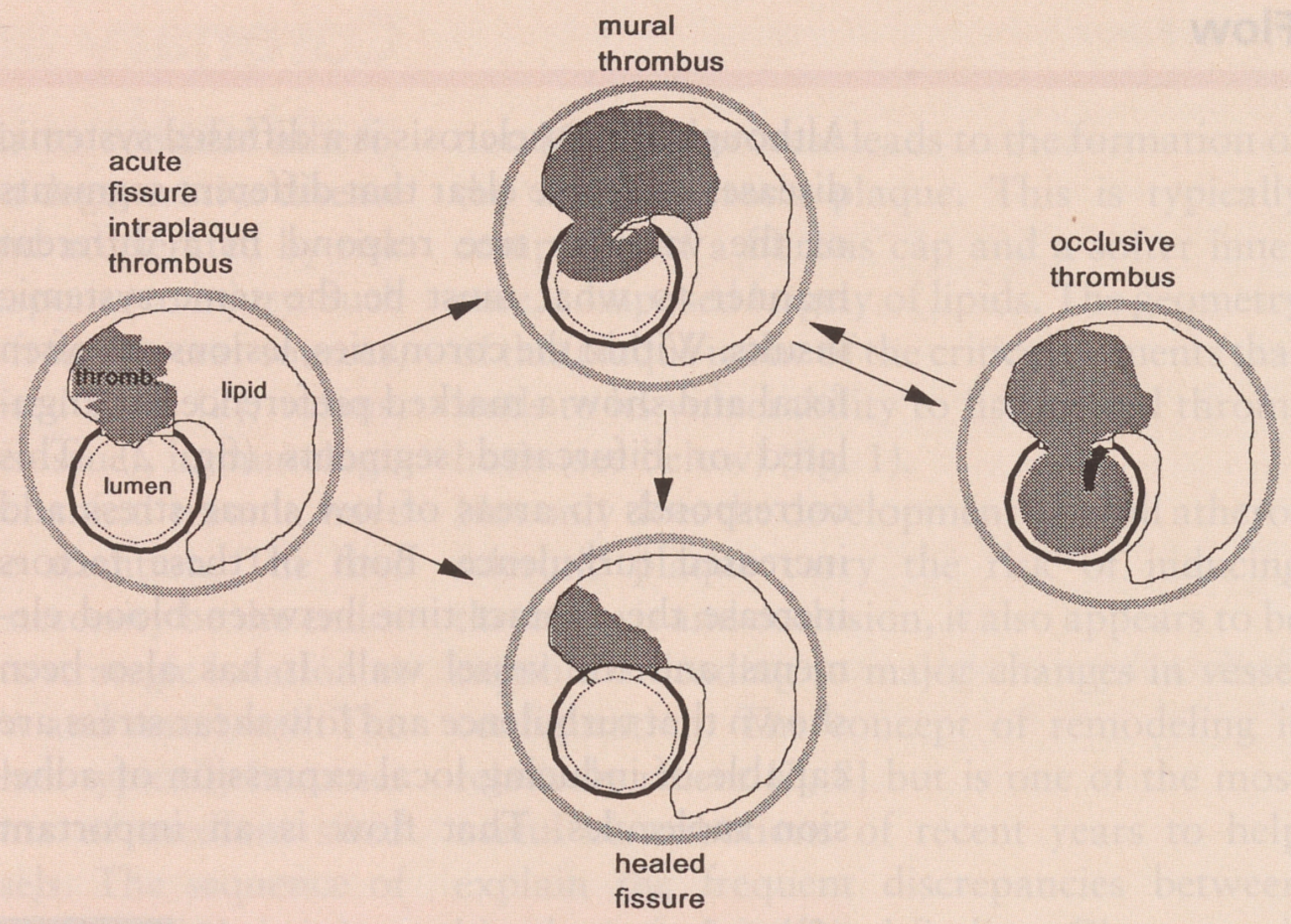
The clinical progression of atherosclerotic coronary artery disease is seldom continuous and gradual, but is mostly abrupt and step-wise. This underlines the critical role of rapid thrombus formation. A large body of evidence has built up over the past 15 years to implicate plaque fissure and/or rupture at the level of the fibrous cap, as the main initiating event leading to luminal thrombosis and decreased blood flow (Figure 3). Depending on a delicate balance of hemostatic and rheological factors, the thrombus may be small, encroach only little into the lumen, and be incorporated into the plaque without any acute clinical manifestations. The frequency of this type of event may have been very much underestimated until recently, and could be one of the important mechanism leading to gradual silent plaque growth [13,14]. When the thrombus is more important, it bulges into the lumen, and is capable of inducing acute blood flow reduction with ischaemic symptoms such as unstable angina. If thrombus growth is sufficient to induce complete occlusion with cessation of flow, transmural ischaemia may supervene and progress to acute myocardial infarction or sudden death.
It has been well shown [15] that it is the content of the lipid core within the plaque that is by far the most thrombogenic element. The degree to which this is exposed to the blood stream must therefore play an important role in the amount of thrombus that is generated locally. Other elements, however, must play a central role in the exact degree of thrombotic response that is induced by any individual plaque fissure. The complex balance of both local or intrinsic factors that define plaque vulnerability, and systemic elements that act upon it with more or less impact (triggers) is determining [16]. Plaque vulnerability is related both to the thickness of its fibrous cap and to the composition and importance of its lipid core [13,14,17,18]. The degree of local inflammation, mostly in the shoulder zone of the plaque, where the fibrous cap joins the more healthy media, is also an important element. In these vulnerable zones, the cell population contains a large proportion of macrophages and T-lymphocytes and less smooth muscle cells. The production of enzymes capable of digesting the interstitial matrix is also important in such areas. Casscells et al. [19], using heat probes, have elegantly demonstrated that local temperature is increased in vulnerable carotid plaques, suggesting an active local inflammatory process. Non-specific markers of inflammation, such as CRP, have also been conclusively shown to increase in parallel with unstable coronary syndromes [20]. This, again, points to a significant degree of inflammation concomitant with plaque activity and thrombus deposition.
Triggers that are capable of inducing plaque fissure include systemic inflammation, catecholamines [21], tilting of the hemostatic balance, and mechanical stimulation at local level, such as plaque “fatigue”, non-laminar flow, etc. Genetic factors most certainly also have a major impact on a majority of these elements [22].
The observation that those plaques that lead to acute clinical event such as myocardial infarction were often not very tight prior to the acute event has led to the hypothesis that non-significant stenosis are in fact more dangerous than their tighter counterparts [23,24]. Although it is likely that a non-significant stenosis evolving brutally into a total coronary occlusion will induce more severe ischaemia because of nonexistent or little developed collaterals, tighter lesions will almost always have a higher propensity to progress and lead to vessel occlusion. In other words, the risk “that” the vessel may occlude is greater for significant stenoses, but the risk “if” is more important for lesions that initially only encroached very little into the lumen.

Conclusions

The field of atherosclerotic coronary artery disease is the object of intensive study. Great progress has been made in relating the histological and cellular events to those observed by the clinicians. The role of thrombus has been clarified, and its pivotal nature recognised. This understanding has helped better relate what previously appeared to be a very chronic, slow and gradual “degenerative” disease and its often dramatic, explosive and unheralded clinical manifestations.

References

  1. Lie, J.T. Centenary of the first correct antemortem diagnosis of coronary thrombosis by Adam Hammer (1818-1878): English translation of the original report. Am J Cardiol 1978, 42, 849–852. [Google Scholar] [CrossRef] [Scilit] [PubMed]
  2. Chisolm, G.M.; Penn, M.S. Oxidized lipoproteins and atherosclerosis. In Atherosclerosis and Coronary Artery Disease; Fuster, V., Ross, R., Topol, E.J., Eds.; Lippincott-Raven: Philadelphia, 1996; pp. 129–147. [Google Scholar]
  3. Lüscher, T.; Noll, G. Endothelial function as an end-point in interventional trials: concepts, methods and current data. J Hypertens Suppl (England) 1996, 14, 5111–5119. [Google Scholar]
  4. Ross, R.; Fuster, V. The pathogenesis of atherosclerosis: a perspective for the 1990’s. Nature 1993, 362, 801–809. [Google Scholar] [CrossRef] [Scilit] [PubMed]
  5. Libby, P. Atheroma: more than mush. Lancet 1997, 348, 54–57. [Google Scholar] [CrossRef] [Scilit]
  6. Schmitt-Gräff, A.; Desmouli; re, A.; Gabbiani, G. Heterogeneity of myofibroblast phenotypic features: an example of fibroblastic cell plasticity. Virchows Archiv 1994, 425, 3–24. [Google Scholar] [CrossRef] [Scilit]
  7. Glagov, S.; Weisenberg, E.; Zarins, C.K.; et al. Compensatory enlargement of human atherosclerotic coronary arteries. N Engl J Med 1987, 316, 1371–1375. [Google Scholar] [CrossRef] [Scilit]
  8. Gibbons, G.H.; Dzau, V.J. The emerging concept of vascular remodeling. N Engl J Med 1994, 330, 1431–1438. [Google Scholar]
  9. Mintz, G.S.; Popma, J.J.; Pichard, A.D. Arterial remodeling after coronary angioplasty — a serial intravascular ultrasound study. Circulation 1996, 94, 35–43. [Google Scholar] [CrossRef] [Scilit]
  10. Urban, P.; Meier, B.; Finci, L.; De Bruyne, B.; Steffenino, G.; Rutishauser, W. Coronary wedge pressure: a predictor of restenosis after coronary balloon angioplasty. J Am Coll Cardiol 1987, 10, 504–509. [Google Scholar]
  11. Cashin, W.L.; Sanmarco, M.E.; Nessim, S.A. Accelerated progression of atherosclerosis in coronary vessels with minimal lesions that are bypassed. N Engl J Med 1984, 311, 824–828. [Google Scholar] [CrossRef] [Scilit] [PubMed]
  12. Ohman, E.M.; George, B.S.; White, C.J.; Kern, M.].; Gurbel, P.A.; Freedman, R.J.; et al. for the randomized IABP study group. Use of aortic counterpulsation to improve sustained coronary artery patency during acute myocardial infarction. Results of a randomized trial. Circulation 1994, 90, 792–799. [Google Scholar] [CrossRef] [Scilit]
  13. Falk, E. Plaque rupture with severe pre-existing stenosis precipitating coronary thrombosis. Characteristics of coronary atherosclerotic plaques underlying fatal occlusive thrombi. Br Heart J 1983, 50, 127–134. [Google Scholar] [CrossRef] [Scilit]
  14. Davies, M.J.; Thomas, A.C. Plaque fissuring — the cause of acute myocardial infarction, sudden ischaemic death, and crescendo angina. Br Heart J 1985, 53, 363–373. [Google Scholar] [CrossRef] [Scilit] [PubMed]
  15. Fernandez-Ortiz, F.; Badimon, J.J.; Falk, E.; et al. Characterization of the relative thrombogenicity of atherosclerotic plaque components: implications for consequences of plaque rupture. J Am Coli Cardiol 1994, 23, 1562–1569. [Google Scholar] [CrossRef] [Scilit]
  16. Muller, J.E.; Abela, G.S.; Nesto, R.W.; Tofler, G.H. Triggers, acute risk factors and vulnerable plaques: the lexicon of a new frontier. J Am Coll Cardiol 1994, 23, 809–813. [Google Scholar] [CrossRef] [Scilit] [PubMed]
  17. Davies, M.J.; Richardson, P.D.; Woolf, N. Risk of thrombosis in human atherosclerotic plaques: role of extracellular lipid, macrophage, and smooth muscle cell content. Br Heart J 1993, 69, 377–381. [Google Scholar] [CrossRef] [Scilit]
  18. Mann, J.M.; Davies, M.J. Vulnerable plaque — relation of characteristics to degree of stenosis in human coronary arteries. Circulation 1996, 94, 928–931. [Google Scholar] [CrossRef] [Scilit]
  19. Casscells, W.; Hathorn, B.; David, M.; Krabach, T.; Vaughn, W.K.; McAllister, H.A.; et al. Thermal detection of cellular infiltrates in living atherosclerotic plaques: possible implications for plaque rupture and thrombosis. Lancet 1996, 347, 1447–1449. [Google Scholar] [CrossRef] [Scilit] [PubMed]
  20. Liuzzo, G.; Biasucci, L.M.; Gallimore, R.; Grillo, R.L.; Rebuzzi, A.G.; Pepys, M.B.; Maseri, A. The prognostic value of C-reactive protein and serum amyloid a protein in severe unstable angina. N Engl J Med 1994, 331, 417–424. [Google Scholar] [CrossRef] [Scilit]
  21. Leor, J.; Poole, K.; Kloner, R.A. Sudden cardiac death triggered by an earthquake. N Engl J Med 1996, 334, 413–419. [Google Scholar] [CrossRef] [Scilit]
  22. Weiss, E.J.; Bray, P.F.; Tayback, M.; Schulman, S.P.; Kickler, T.S.; Becker, L.C.; et al. A polymorphism of a platelet glycoprotein receptor as an inherited risk factor for coronary thrombosis. N Engl J Med 1996, 334, 1090–1094. [Google Scholar] [CrossRef] [Scilit] [PubMed]
  23. Fischbein, M.C.; Siegel, R.J. How big are coronary atherosclerotic plaques that rupture? Circulation 1996, 94, 2662–2666. [Google Scholar] [CrossRef] [Scilit] [PubMed]
  24. Giroud, D.; Li, J.M.; Urban, P.; Meier, B.; Rutishauser, W. Relation of the site of acute myocardial infarction to the most severe coronary arterial stenosis at prior arteriography. Am J Cardiol 1992, 69, 729–732. [Google Scholar] [CrossRef] [Scilit] [PubMed]
Figure 1. Cross section of an atheromatous epicardial human coronary artery. The lumen is filled with contrast medium. The fibrous cap is thin and the lipid core abundant, indicating “vulnerability”.
Cardiovascmed 01 00125 g001
Figure 2. Sixty-eight-year old woman with NYHA grade Ill angina. Right anterior oblique view of the left coronary. Tight eccentric distal stenosis of the left main stem.
Cardiovascmed 01 00125 g002
Figure 3. Diagram of intracoronary thrombosis. On the left, a localised intra-plaque thrombus is depicted, with no significant decrease in the lumen size. In the middle bottom diagram, the intra-plaque thrombus has become organised, and the lumen is “normalized”. In the middle top diagram, the thromhus has expanded and encroaches into the lumen. This would be expected to induce anginal symptoms. On the right side diagram, the thrombus has become occlusive. This would be expected to generate transmural ischaemia and infarction unless sufficiency developed collaterals are present.
Cardiovascmed 01 00125 g003