Summary
Atherosclerosis with its complications represents an important health problem. In large epidemiological studies during the last decades, important risk factors such as smoking, hypertension, dyslipidaemia and diabetes mellitus have been identified which account for approximately 70 percent of the total risk of atherosclerosis. Histology of atherosclerotic lesions shows an inflammatory process within the vessel wall. Monocytes and T-lymphocytes can be demonstrated within arteriosclerotic plaques. In clinical trials, cardiovascular events such as myocardial infection and stroke were associated with increased plasma concentrations of acute phase proteins such as C-reactive protein which is also elevated during acute inflammatory processes. Several infectious agents such as viruses (cytomegalovirus, Herpes simplex virus, Coxsackie virus B6) or bacteria (Chlamydia pneumoniae, Helicobacter pylori) have been proposed to be involved in the pathogenesis of atherosclerosis. Presence of Chlamydia pneumoniae has been demonstrated within arteriosclerotic plaques and increased antibody titres have been found in patients with atherosclerosis. Pilot studies using antibiotics in patients after acute myocardial infarction revealed promising results concerning the risk of subsequent cardiovascular events. These findings should now be confirmed in ongoing large-scale clinical trials.
Zusammenfassung
Die Arteriosklerose mit ihren Komplikationen stellt ein wichtiges gesundheitspolitisches Problem dar. 1m Verlauf der letzten Jahrzehnte konnten aufgrund grosser epidemiologischer Studien Risikofaktoren wie Rauchen, Hypertonie, Dyslipidämie und Diabetes mellitus identifiziert werden, die etwa 70% des Gesamtrisikos erklären. Histologisch stellt die Arteriosklerose eine entzündliche Erkrankung der Gefässwand dar. In arteriosklerotischen Läsionen können Monozyten und T-Lymphozyten nachgewiesen werden. Klinische Studien haben gezeigt, dass kardiovaskuläre Ereignisse wie Myokardinfarkt und Hirnschlag wie auch entzündliche Erkrankungen mit einer Erhöhung von Akutphasenproteinen wie des Creaktiven Proteins assoziiert sind. Seit Jahren wird immer wieder postuliert, dass infektiöse Agentien wie Viren (Zytomegalievirus, Herpes-simplex-Virus, Coxsackie-Virus B6) Oder Bakterien (Chlamydia pneumoniae, Helicobacter pylori) für die Pathogenese mitverantwortlich sein könnten. So konnten Bestandteile von Chlamydien jn arteriosklerotischen Plaques und erhöhte Antikörptertiter bei Patienten mit Arteriosklerose nachgewiesen werden. Pilotstudien mit Antibiotika bei Patienten nach Myokardinfarkt haben vielversprechende Resultate bezüglich auftreten kardiovaskulärer Ereignisse ergeben. In grossen klinischen Studien sollen diese Befunde nun bestätigt werden.
Introduction
Atherosclerosis, primarily a chronic inflammatory process, is the major cause of disability and death in industrialised countries. The principal results are coronary artery disease, stroke, and peripheral vascular disorders. Atherosclerotic plaques form as a response to damage of arterial endothelium and smooth muscle cells. These plaques often ulcerate; as a protective response platelets then aggregate and adhere to the ulcerated surface resulting in a partial or complete thrombotic block of the narrowed artery. The result is ischemia.
Hypercholesterolaemia and other risks such as smoking, arterial hypertension and poorly controlled diabetes mellitus are important factors in the development of atherosclerosis but they do not account for the total incidence of either atherosclerosis itself or its consequences. Chronic infection may be an additional risk factor. This review summarises the functional pathology of atherosclerosis and provides an overview of presently available evidence for the possible role of infection as one of the mechanisms that result in atherosclerosis.
The Pathology of Atherosclerosis
Atherosclerotic Lesions
Atherosclerosis is initiated by the appearance of fatty streaks in the arterial wall, a common finding even in young healthy subjects [
1]. These can slowly, often over many years, form a fibrous surface layer over the fatty deposit, resulting in a fibrous plaque. As well as fibrous tissue, the lipoid rich plaque also contains modified smooth muscle cells, with an inflammatory response demonstrated by the presence of macrophages, monocytes, T-lymphocytes and inflammatory cytokines [
2]. Plaques often progress to calcification with a variable degree of necrosis.
Atherosclerosis, manifested by the appearance of such plaques, develops as a response to arterial wall injury. Endothelial dysfunction, precipitated by such factors as hyperlipidaemia and cigarette smoking, and possibly also by certain infections, results in localised initial foci containing macrophages (which ingest lipid to become foam cells), monocytes and lymphocytes. The acute lesions are closely similar to normal body protective mechanisms associated with repair of inflammatory lesions. Growth factors stimulate smooth muscle cell proliferation, the cells secreting extracellular matrix. Continued damage results in a chronic response within the arterial wall, and progressive lipid accumulation causes necrosis and finally calcification.
During the atherosclerotic process cytokines, growth factors and other substances including nitric oxide, which are also involved in coagulation, vasospastic and vasodilatory mechanisms, seem to modulate cell migration and proliferation as well as the control of lipid and protein synthesis [
1].
The Endothelium
As noted above, damage to the arterial endothelium is the precursor of the inflammatory response that leads to plaque development. The endothelium, protecting the arterial wall from circulating blood, acts both as a cellular boundary layer and also as the source of substances that control the contraction and relaxation of arterial smooth muscle; it also modulates platelet aggregation and adhesion, and blood coagulation [
3,
4]. Nitric oxide (NO) inhibits smooth muscle migration and proliferation, while NO and prostacyclin inhibit platelet aggregation and adhesion, and cause vasodilatation.
Endothelium derived relaxing factor (EDRF), now known to be NO, is formed from L-arginine by NO-synthase [
5], relaxing smooth muscle cells by activating guanylyl cyclase. Various forms of NO synthase occur in endothelial cells, vascular smooth muscle cells, platelets, macrophages, and also the brain [
5,
6]. In vivo, inhibition of NO formation increases vasoconstrictor responses, increasing arterial blood pressure [
7].
The endothelium also produces three vasoconstrictors, thromboxane A2, prostaglandin H2, and endothelin-1 (the most potent vasoconstrictor yet identified); of the three, endothelin-1 alone stimulates vascular smooth muscle proliferation but does not affect platelets.
In healthy subjects, the endothelium provides a non-thrombotic surface, prevents adhesion of blood cells, promotes vasodilatation and inhibits vascular smooth muscle proliferation. Endothelial dysfunction, occurring in atherosclerotic pathology, results in vasoconstriction, adhesion of platelets and monocytes, and smooth muscle cell proliferation [
3,
4].
Atherosclerosis and Endothelial Activity
Acetylcholine acts as a vasodilator in normal coronary arteries by releasing nitric oxide (NO) from the endothelium. But in atherosclerotic arteries acetylcholine mediates paradoxical vasoconstriction, inferring that atherosclerosis is associated with endothelial dysfunction [
7].
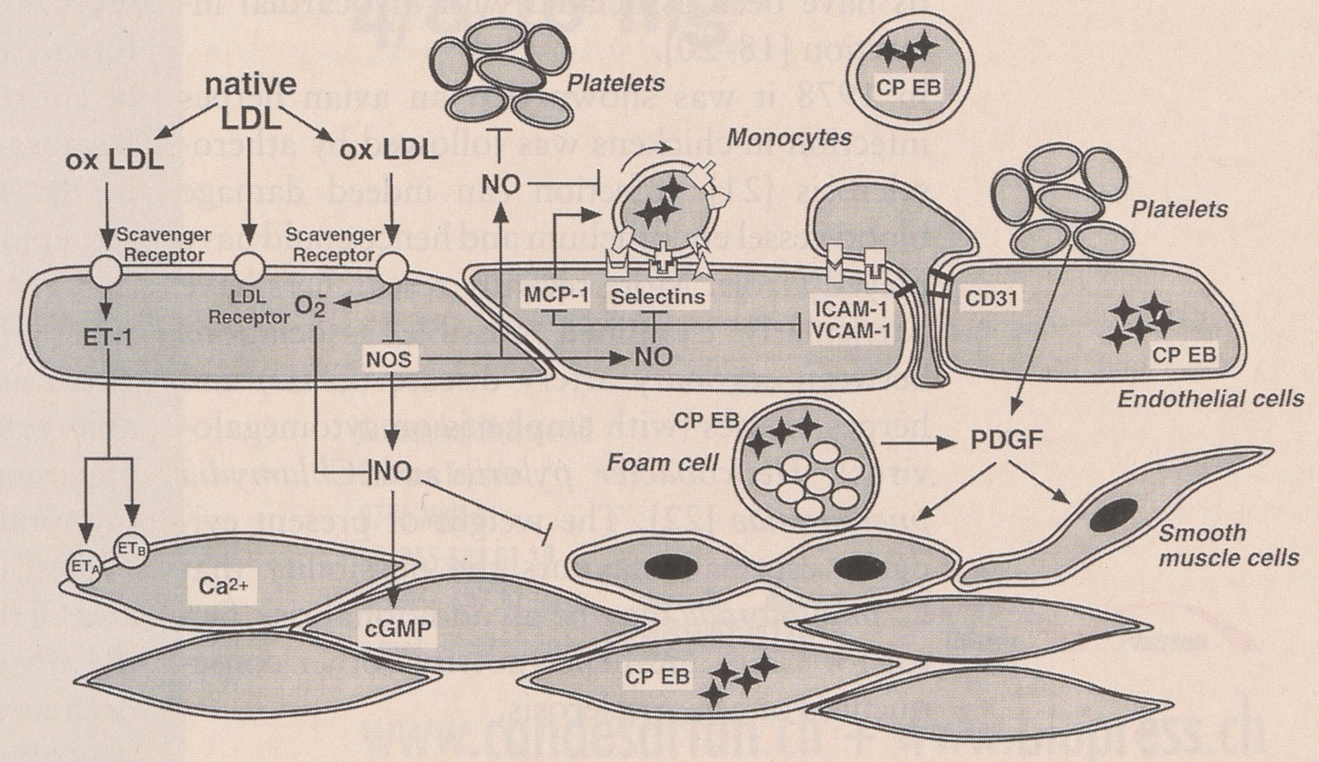
There is also evidence that NO has an important role in regulating the expression of endothelial chemoattractant and adhesive proteins which recruit monocytes into the vessel wall (
Figure 1 [
8]). NO thus has anti-inflammatory, antiatherogenic effects, and endothelial dysfunction can result in defective NO production with resultant vessel wall inflammatory and atherosclerotic changes.
Experimental work with cultured endothelial cells has shown that inhibition of NO production encourages release of monocyte chemoattractant protein and protein secretion, while the addition of NO decreased both [
9]. Additionally, interleukin-1 alpha simulated monocyte adhesion has been shown to be reduced by NO in a study of human saphenous vein endothelial cells and the expression of vascular cell adhesion molecule-1 (VCAM-1) induced by interleukin-1 alpha was inhibited dosedependently by NO. The endothelial expression of other leukocyte adhesion molecules, such as E-selectin, are also inhibited by NO. LN-monomethyl-arginine inhibits endogenous NO production, inducing expression of the vascular cell adhesion molecules.
Endothelial Function: Lipoproteins and Atherosclerosis
Hyperlipidaemia and atherosclerosis impair vascular smooth muscle relaxation mediated by the endothelium. It is now widely accepted that oxidised low density lipoprotein (ox-LDL), a component of atherosclerotic plaques, contributes significantly to endothelial injury; this is specific for ox-LDL since LDL itself does not have such an effect [
10]. As noted earlier. The enzyme NO-synthase acts on L-arginine to produce NO which mediates endotheliumdependent vasodilatation. L-arginine improves or restores reduced endothelium-dependent relaxation in the presence of ox-LDL, so a reduction in intracellular L-arginine availability may be an important component of endothelial injury [
11].
Endothelial damage includes dysfunction of the L-arginine NO pathway but in atherosclerotic vessels there may also be increased formation of vasoconstrictor substances since ox-LDL, thrombin, and hypoxia all stimulate endothelin production whose vasoconstrictor effect can encourage both cellular proliferation and ischemia [
3,
12].
The endothelial wall may be directly injured by ox-LDL which can also increase adherence of monocytes and T-lymphocytes into the arterial wall at least in part by stimulating production of adhesive surface cell glycoproteins which facilitate leukocyte recruitment into the vessel wall (
Figure 1 [
1]). Within the arterial intima atherogenic processes encourage the activation of monocytes into macrophages which take up ox-LDL to form foam cells (
Figure 1 [
1]). In addition macrophages can scavenge and oxidise LDL using mechanisms that include lipoxygenase enzymes.
Thus, ox-LDL is an active component of atherosclerotic plaques, inhibiting endotheliumdependent smooth muscle cell relaxation and promoting both endothelium-dependent and independent contractions. This results in vasoconstriction, setting the scene for thrombosis.
The Inflammatory Component of Atherosclerosis
Locally, atherosclerotic plaques contain a significant inflammatory component, demonstrated by the presence of white blood cells such as monocytes and T-lymphocytes.
Systemic evidence of an inflammatory response is provided by elevated C reactive protein and interleukin 6, and serum amyloid, proteins, indicating the presence of an active inflammatory process [
13,
14,
15,
16]. High CRP levels have been associated with poor prognosis in unstable angina patients who had significantly more ischemic episodes and a higher incidence of death and MI than those with low CRP levels [
14]. That this was not a consequence of myocardial ischemia is suggested by the fact that in patients with unstable angina the CRP levels were significantly higher (p<0.001) than in patients with variant angina and normal CRP levels even though this group had a longer total duration of angina [
17]. Plasma levels of interleukin 6, a pro-inflammatory cytokine regulating acute phase hepatic protein production, are elevated in unstable angina while remaining normal in stable angina and healthy volun.teers, again emphasising the importance of inflammation in unstable angina and by inference in atherosclerosis [
13].
Infection: A Possible Risk Factor for Atherosclerosis?
Several factors are clearly demonstrable as contributing to the risk of atherosclerotic progression. These include raised serum lipids, hypertension, cigarette smoking (where CO inhalation is a likely major component of risk), and poorly controlled diabetes. But established risk factors fail to account for the whole incidence of atherosclerotic cardiovascular disease.
The possibility of a link between infection and atherosclerosis was made by Osler early this century. There followed sporadic case reports and uncontrolled studies that seemingly supported such an association. Septicaemia and also milder, including respiratory, infections increase the risk of stroke, although this could be due to direct mediation of coagulation processes; dental infections and chronic bronchitis have been associated with myocardial infarction [
18,
19,
20].
In 1978 it was shown that an avian herpes infection in chickens was followed by atherosclerosis [
21]; infection can indeed damage blood vessel endothelium and hence could have played a role. More recently researchers have particularly examined possible associations between coronary artery disease (CHD) and herpes viruses (with emphasis on cytomegalovirus),
Helicobacter pylori, and
Chlamydia pneumoniae [
22]. The weight of present evidence especially favours the possibility that
C. pneumoniae may be an additional risk factor for CHD and hence possibly other consequences of atherosclerosis.
Cytomegalovirus (CMV) and Helicobacter pylori
Several epidemiological studies have indicated an odds ratio of at least two between antiCMV antibodies and CHD [
22]. Most of these studies were small, making adjustment for confounding factors difficult or impossible, but in some cases the odds ratios were higher with increasing antibody titres or in patients with more severe disease [
23,
24,
25,
26,
27,
28]. Most, although not all, of the studies were in heart transplant patients [
27,
28]. The evidence that CMV could be a factor in the accelerated atherosclerosis sometimes observed in heart transplant patients is no more than circumstantial; however, herpes simplex virus, and CMV antigens and DNA have been detected in such atherosclerotic lesions [
27,
28]. Present evidence for a causative role is weak but should not be entirely rejected.
Because of the likely causative role of
H. pylori in the development of peptic ulcer, several seroepidemiological studies on a total of some 2600 cases, have examined the association between
H. pylori antibodies and CHD, and stroke in one study [
22,
29]. Presently available evidence is weak, an especially evident apparent confounding factor being low socioeconomic class which is associated with the highest incidences of both
H. pylori infection and CHD [
22]. In addition, at this point in time the absence of any good evidence that the organism is present in atheromatous plaques contributes to the weakness of the hypothesis that
H. pylori may play a role in atherosclerosis development [
22].
Chlamydia pneumoniae
C. pneumoniae is the most promising candidate for an infective organism to be an additional risk factor for CHD and atherosclerosis development. Presence of the organism in atherosclerotic plaques has been demonstrated by electron microscopy, PCR, ELISA, and isolation tissue cultures [
30,
31,
32,
33]. During the past decade seroepidemiological studies have documented a possible etiological role for
H. pylori [
34,
35,
36,
37,
38,
39].
In 13 published biohistological studies, chlamydia DNA, antigens, or elementary bodies demonstrated the presence of
C. pneumoniae in only 5% of control [normal arterial wall) samples but in 52% of atheromatous lesions, the weighted odds ratio being approximately (95% CI 5-22). Review of 16 published studies of the prevalence of antibodies to
C. pneumoniae in CHD, and 2 studies in cerebrovascular disease, most have reported an odds ratio of two-fold or more for the association, and in some higher odds ratios occurred with increasing antibody titres [
22].
It would seem logical to assume that macrophages ingest
C. preumoniae organisms from lung tissue and then reach atheromatous or pre-atheromatous plaques via the bloodstream (
Figure 1). The rather strong evidence of an association between
C. pneumoniae and atheromatous cardiovascular disease does not in itself demonstrate causation [
22,
34,
35,
37]. The organism may persist in macrophages taken up into atheromatous plaques without contributing to their development. Alternatively,
C. Pneumoniae may even trigger the inflammatory processes that mediate the progressive development of atheromatous plaques.
The systemic and local inflammatory response related to the progressive development of atheromatous lesions is established. A role for infection can be justified by its ability to cause changes in serum lipids, increased cytokine production, and procoagulant effects. It is also known that damage caused by chronic infection can result in the production of fibrous tissue. In this context the effects of Chlamydia trachomatis, the causative organism of trachoma, are worthy of note. The original trachoma infection results in conjunctival infiltration with macrophages and lymphocytes; such infiltrations also occur in atheromatous lesions. Patients with trachoma develop fibrotic conjunctival scarring, often after many years, leading to blindness. It is tempting to compare this with the fibrous cap formation over atheromatous lesions. C. pneumoniae may induce a chronic immune reaction mediated by cytokines, contributing to chronic endothelial cell damage, stimulating the synthesis of acute phase reactants such as fibrinogen and C reactive protein. Such chronic infection could also increase the risk of thrombosis by stimulating the expression of monocyte-derived procoagulants.
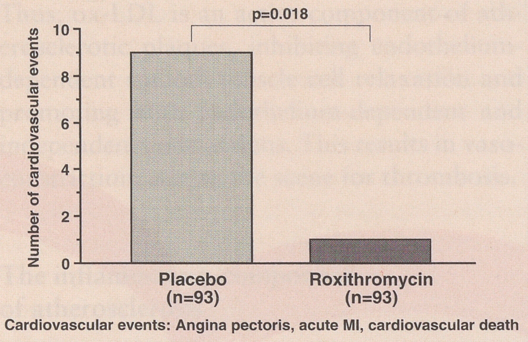
Recently two pilot-studies have been published in which patients with CHD have been treated with antibiotics [
40,
41]. In the Argentinean trial patients after acute non-Q-wave infarction treated with roxithromycin experienced significantly less cardiovascular events (angina pectoris, reinfarction, cardiac death) than placebo treated patients during the follow-up of 6 month (
Figure 2 [
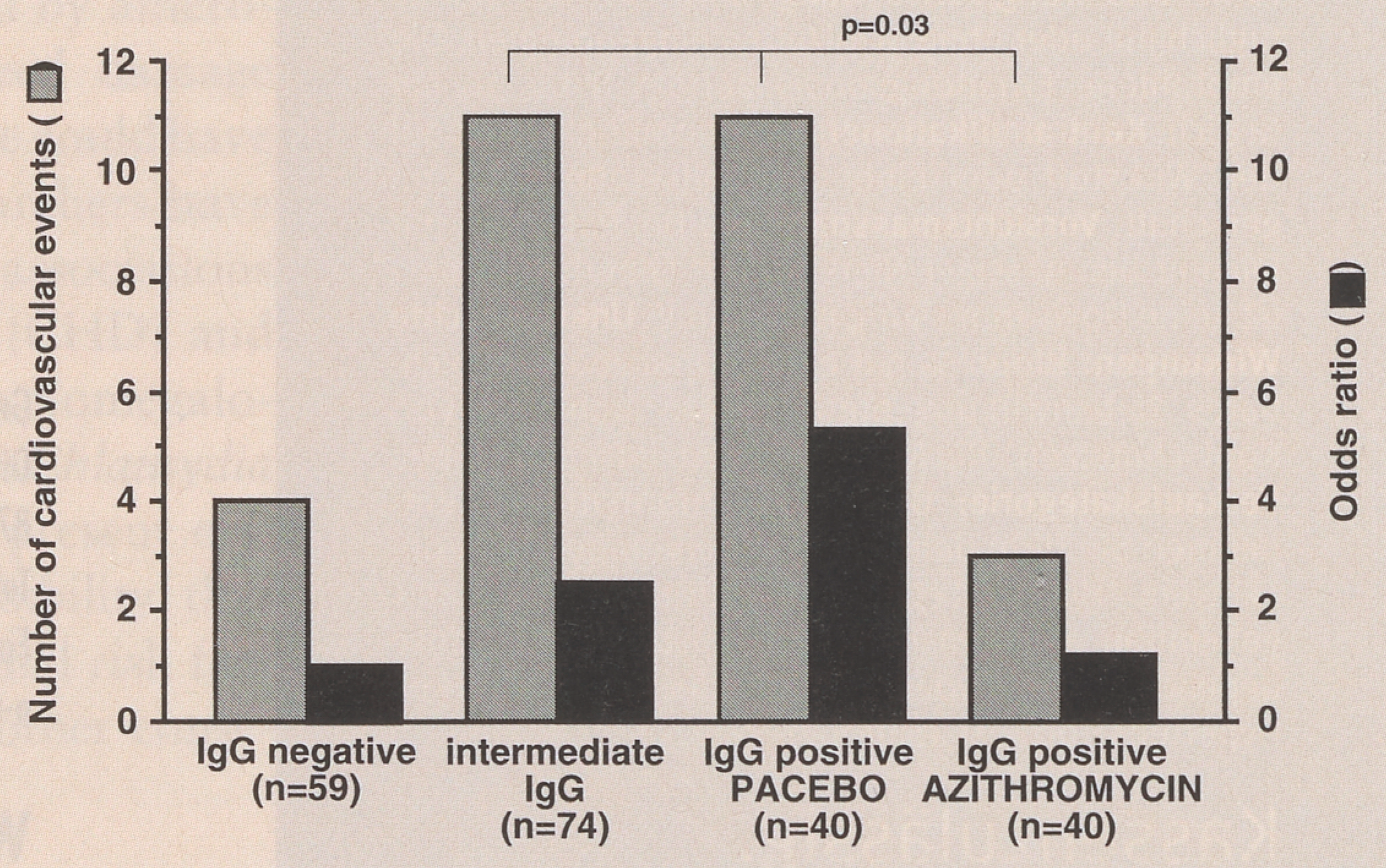
40]). In another trial it has been demonstrated that in patients after myocardial infarction the risk for subsequent cardiovascular events correlates with seroprevalence for
Chlamydia pneumoniae (
Figure 3 [
41]). IgG positive patients (antibody titers >1:64) were randomised either to be treated with azithromycin or placebo. During the follow-up of 18 months, the risk of cardiovascular events (unstable angina, reinfarction, PTCA or CABG) was significantly lower in treated patients (
Figure 3 [
41]). Based on these promising results largescale clinical trials have been started.
Conclusions
Inflammation is an important component in the initiation, development, and perpetuation of atheromatous plaques. There is evidence for a possible causative role for infection in the process and the strongest candidate at this time is Chlamydia pneumoniae. Although controversial, the possibility of prevention or treatment of cardiovascular disease using appropriate antimicrobial therapy has prompted the initiation of clinical trials examining cardiovascular end points. The results will hopefully increase our knowledge of possible causative roles that infection may play in the causation of atheroma progression with its major consequences of disability and premature death.