Zusammenfassung
Blutplättchen spielen sowohl im kranken als auch gesunden Menschen eine wesentliche Rolle bei der Aufrechterhaltung der vaskulären Integrität. Zusätzlich scheinen sie zahlreiche andere Funktionen zu erfüllen und könnten an der Entstehung oder am Verlauf verschiedener Krankheiten beteiligt sein. Blutplättchenaktivierung ist für die physiologische Funktion unerlässlich, die empfindliche Balance jedoch, welche die Aktivierung bzw. Deaktivierung reguliert, kann durch zahlreiche Faktoren gestört werden. Ein besseres Verständnis dieser Faktoren und die Möglichkeit, die Blutplättchenaktivität mittels einer Reihe jüngst entwickelter Inhibitoren zu kontrollieren, sollte die mit Blutplättchen in Verbindung stehenden Erkrankungen positiv beeinflussen können. Über die Rolle der Blutplättchen bei der Entstehung von kardiovaskulären Erkrankungen bleiben jedoch noch viele Fragen offen.
The role of platelets in many aspects of disease, in particular cardiovascular problems, has long been suspected but it is really only in the last two decades that hard evidence for this has become available. One of the main factors in determining this change of attitude was the demonstration that aspirin, one of the only pharmacologically active drugs then available with high specificity for platelets, given prophylactically, had a protective effect on the incidence and survival rate in ischaemic heart disease and stroke [
1]. Since then there has been a great deal of interest both in exploring better methods of inhibiting platelet activity more efficiently as well as in investigating a possible involvement of platelets in other disorders.
Several platelet specific inhibitors have been developed and are either in clinical use, in clinical trials, or in development (
Table 1). These include ReoPro, a humanised monoclonal antibody against α
IIbβ
3, now in widespread use for treating acute clinical situations [
2]; ticlopidine and clopidogrel, two metabolically-activated drugs inactivating the platelet ADP receptor linked to activation of α
IIbβ
3, and used for prophylaxis against cardiovascular disorders, particularly stroke [
3,
4]. In addition, several small molecular mass α
IIbβ
3 inhibitors such as integrilin [
5] and lamifiban [
6] have been shown to reduce mortality and myocardial infarction associated with unstable angina. Orally available versions of small molecular mass α
IIbβ
3 inhibitors are currently being tested for use in prophylaxis.
Clinical problems involving platelet activation can fall into two categories, where this function is either deficient, in principle leading to bleeding disorders, or excessive, leading to thrombotic disorders. The normal balance of platelet reactivity and its control is maintained by an extremely complex system of positive and negative feed back mechanisms. Because of this, platelets in the circulating blood are normally able to bind immediately to denuded areas of subendothelium or to the surface of wounds in the vessel wall to prevent blood loss and to start the processes of repair leading to the reinstallation of a non-thrombotic surface. These are processes which occur continuously on a minor scale in a normal healthy body due to the wear and tear of daily life, even without obvious external injury, and this critical function becomes apparent only when platelet numbers drop to a level (approximately 1–2 × 10
10/1) where they are unable to perform this job. Platelets may also perform poorly for a variety of reasons linked to details of their function for both hereditary and acquired reasons. There are several well-defined hereditary bleeding disorders such as Glanzmann’s thrombasthenia [
7], Bernard-Soulier syndrome [
8] and Gray platelet syndrome [
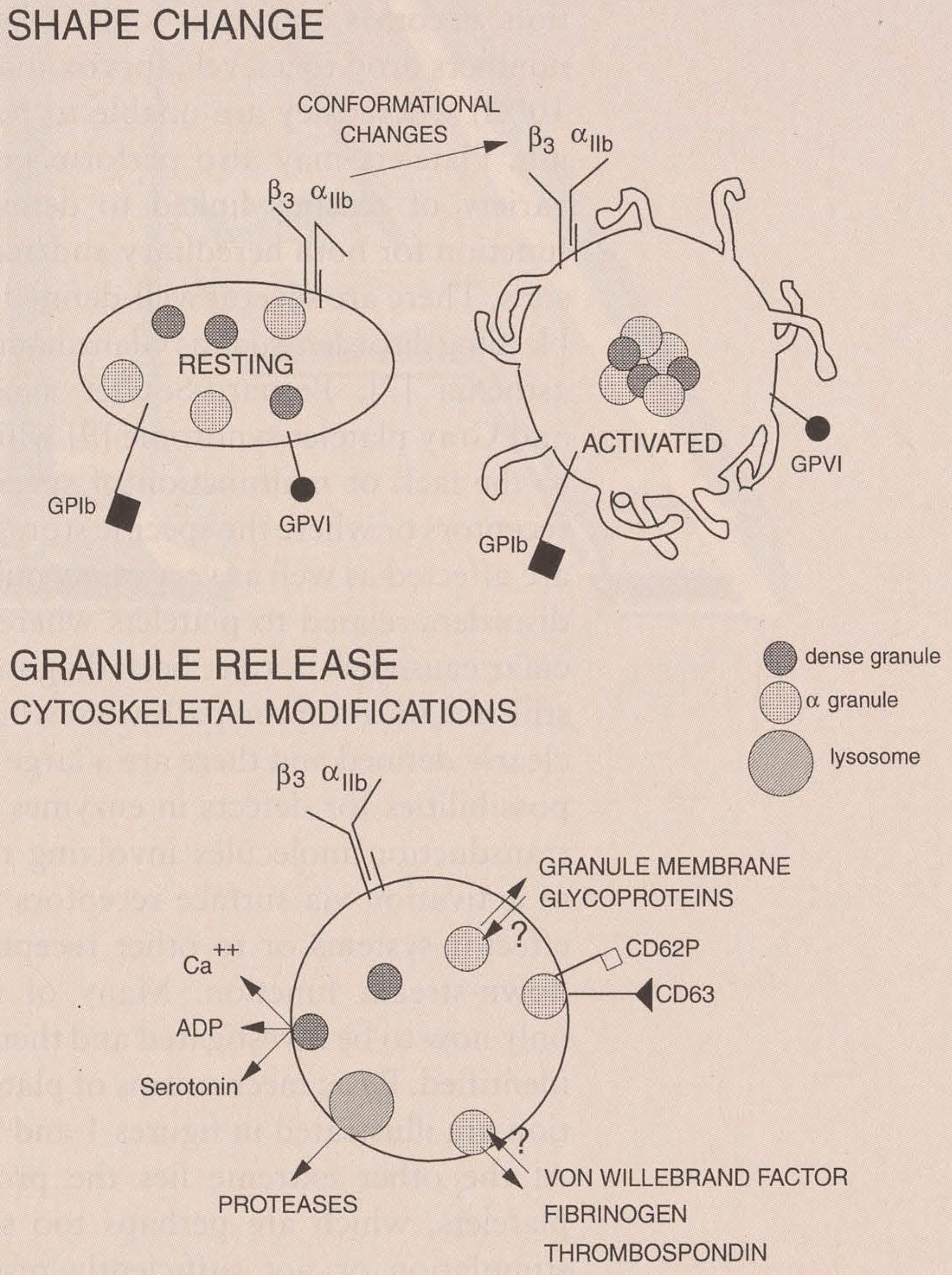
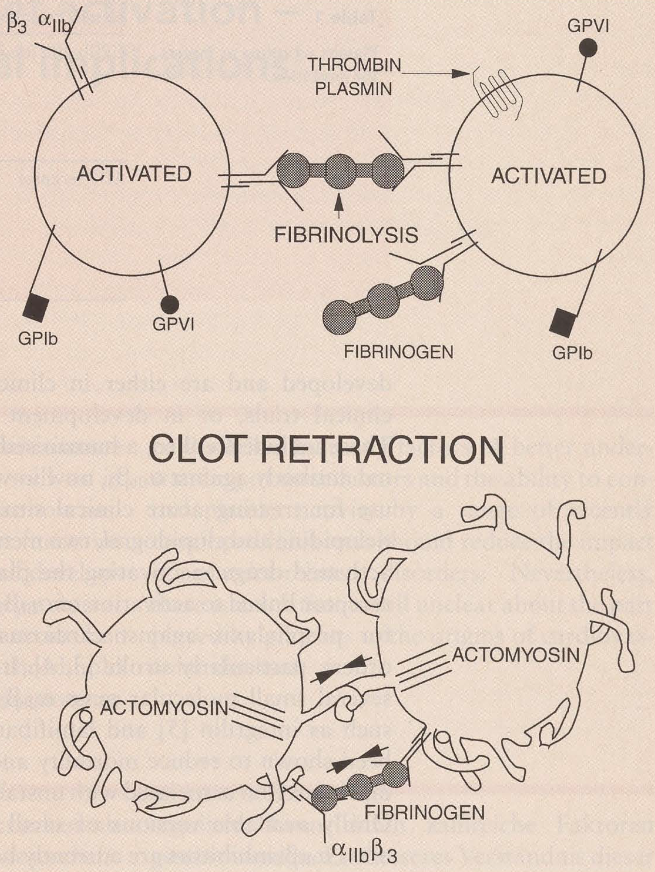
9] which are due to the lack or malfunction of specific surface receptors or where the specific storage granules are affected as well as very many mild bleeding disorders related to platelets where the molecular cause has not yet been diagnosed. There still remain receptor pathways which are not clearly defined and there are a large number of possibilities for defects in enzymes and signal transduction molecules involving the linkage of activation via surface receptors to internal effector systems or to other receptors with a down-stream function. Many of these start only now to be investigated and their functions identified. Basic mechanisms of platelet activation are illustrated in
Figure 1 and
Figure 2.
At the other extreme lies the problem that platelets, which are perhaps too sensitive to stimulation or not sufficiently responsive to negative feed-back mechanisms, nitric oxide or prostacyclin, may over-react to such events and, forming large platelet aggregates which do not dissociate when the requirement for them has passed and becoming stabilised, lay the basis for thrombosis. It is clear that platelets, and the changes which occur in them over the years due to lifestyle and/or disease are by no means the only factors involved. Nevertheless, as extremely reactive and flexible cells, participating at various levels in vascular biology, they present intriguing targets for intervention and for investigation of the effect of such intervention on disease development and progress.
Platelets have a large number of different surface receptors involved in various aspects of their function. Some of these such as GPIb-V-IX [
10], the integrin α
2β
1, and the immunological-like, collagen receptor GPVI [
11,
12,
13] appear to have major roles in the adhesion process of stopping and binding platelets to damaged vessel wall under the high shear stress conditions often encountered in smaller vessels in flowing blood. Recent evidence also points to a role, for GPIb-V-IX at least, in the further process of capturing freshly arriving, resting platelets to a growing thrombus [
14]. On the other hand the major role of the α
IIbβ
3 integrin [
15] is linking activated platelets together via fibrinogen to form aggregates and in the later stage of clot retraction which is so important in drawing together the sides of a wound in the healing progress. Other integrins and adhesive receptors such as α
5β
1 and α
6β
1 may be involved in adhesion under less stringent conditions whereas still other receptors and adhesive proteins have an important role to play in stabilisation of the platelet aggregate and in determining whether or not it will be able to redissociate into the bloodstream or remain associated with the subendothelium as part of the reshaping of the injury site. Still other receptors on the platelet surface belong for the most part to the seven transmembrane, G-protein coupled class [
16] and have roles at least at two levels, as feed-back receptors, both positive and negative, for components released both from the storage granules of activated platelets or generated from other cells within the vicinity of the injury site, such as ADP from erythrocytes (positive, activating). Endothelial cells normally bathe the passing platelets in NO [
17] and prostacyclin [
18], and remove traces of ADP from plasma via the recently described ectoADPase, CD39 [
19], to maintain them in a generally passive state. These pacifying effects are also affected in the vicinity of an injury and may remain perturbed for some time. Arachidonic acid liberated from platelet membranes exposed to activated phospholipases can feed back, becoming converted to thromboxane A2, a powerful inflammatory agonist for several types of cell including platelets, via the celebrated platelet cyclooxygenase that is inhibitable by aspirin [
20]. Thrombin is a major agonist for platelets via a proteolytically activated seven transmembrane receptor [
21]. Activated platelets and derived microparticles also provide important procoagulant surfaces for the generation of thrombin from prothrombin providing rapid amplification of this response if it is not quickly damped by negative feed-back. Another factor affecting the platelet activation balance is epinephrin which is released under conditions of stress and, as well as causing vascular retraction, influences the platelet response to other agonists.
There are many environmental and dietary factors which can affect this overall balance of responses and the possible mechanisms are very poorly understood.
There is still a strong tendency to regard elevated cholesterol levels as a major, if not the major environmental factor, predisposing patients to cardiovascular disease. More recently this has been less emphasised following lengthy speculations about Mediterranean diets as well as the “French paradox”. A significant suggestion has been that it may be the oxidation status of the fat consumed and the level of antioxidants in the plasma which may be crucial in maintaining a healthy cardiovascular system. Recently, several major constituents of red wine, such as resveratrol [
22], have been shown to have anti-oxidant and enzyme inhibitory properties. These suggestions impinge upon the role of platelets as well, apart from the composition of the membranes and the well-researched effects of fatty acid composition on thromboxane production. CD36, which has had a number of roles bestowed upon it since its discovery, has been shown to function as a receptor for oxidised LDL [
23] and platelets are activated by oxidised LDL binding. Ethanol also blocks several platelet signal transduction pathways and has been shown to inhibit platelet activation by various agonists. High blood pressure is a major factor leading to platelet involvement. The pathophysiology is not completely clear but one possible mechanism has been proposed to be the increased shear stress to which both endothelial cells as well as platelets are exposed. In the case of platelets this may lead to increased interactions between the GPIb-V-IX complex and vWf [
24] which can activate platelets in the circulation, causing release and changes in the platelet function, as well as the direct formation of microthrombi with all the consequences that may have for the more distant micro-vasculature.
Major surgery is equivalent to a large wound with the massive activation of platelets in the haemostatic processes which follow. These can lead to thrombotic complications by a sort of overshoot phenomenon affecting both platelets and coagulation factors and it is therefore essential to keep these in balance by providing inhibitors of platelet activation or of thrombin, such as low molecular weight heparin. The prevention of restenosis following angioplasty is also a major area where platelet inhibitors such as ReoPro [
2] have an important role to play. These are acute situations where the role of platelets is fairly clear, however, a role in chronic situations developing over many years is also suspected and platelet inhibitors would also be expected to help.
The possible importance of excessive, in vivo platelet activation in the origins and development of disease states [
25] has lead to a considerable interest in suitable diagnostic approaches. Unfortunately, over the years almost none of these has proved to be as simple and straightforward as was originally suggested. The first methods to be tested, some of which still have applications today, were based upon the fact that strongly activated platelets release the contents of their storage granules, including α-granules. This can be used as a diagnostic method because several of these are synthesised only in the megakaryocyte and are hence platelet specific. Platelet factor 4 and β-thromboglobulin [
26] have been the major peptides used for this over the years. The use of platelet factor 4 in vivo is severely restricted by the fact that it binds tightly to heparin-like substances found on the surface of endothelial cells and that, therefore, the plasma levels do not reflect any on-going platelet activation process. On the other hand β-thromboglobulin and related molecules, although they do not bind nearly as tightly and therefore do not have this disadvantage, are nevertheless rapidly removed from the circulation by the kidneys and are therefore only valuable as markers for a short time after any platelet activation has occurred. Attention therefore turned to alternative markers as soon as these became available. The major markers used have remained granule membrane proteins which are expressed on the plasma membrane following activation. These include P-selectin (CD62P) and CD63 [
27,
28]. Several interesting and significant studies have been carried out with these showing that platelet activation does indeed accompany vascular disease states. Although, initially it was thought that these markers have only advantages over the older ones, they too suffer from some problems. Thus, CD62P, once exposed, was thought to remain on the platelet surface. This is now known not to be the case and it also appears to be rapidly lost from circulating platelets, either by cleavage or by recycling to the α-granules. Thus, again, only events occurring within the past hour or so will be detected and in patients with episodic disease there is the danger of not picking up important events. Other markers that have been used or suggested suffer from the same basic problem but may have advantages in given situations. These include the use of antibodies which recognise specific activation conformations such as to the fibrinogen binding conformation of GPIIb-IIIa or the detection of shape-changed platelets by light scattering methods. These do have the advantages that they detect more sensitive changes but they suffer from its disadvantages also and are rapidly reversible. Generally flow cytometry is used as a convenient and rapid method for the quantitation of platelet activation, detecting the expression of these typical markers and their distribution on a population of platelets. This approach allows the analysis of several markers at the same time by using different fluorescent-labelled antibodies. More recently methods have been proposed based upon in
vivo proteolysis of platelet proteins and its detection. Such proteolysis might be the consequence of platelet activation and/or destruction, since calpain is a major platelet component, or it might come from the effects of proteases released from α-granules or lysosomes after platelet activation, or from granulocytes during pathological processes. Such changes would have the advantage, if no shedding or recycling occurred, that they would steadily accumulate at a rate, related to the disease process, until the platelet was withdrawn from circulation. Whether such damage determines the circulating age of a platelet or if other factors are more important is still not known [
29]. Although several assays have been developed that measure the soluble protein released into the circulation, in particular measuring glycocalin, derived from GPIba, there have not yet been assays for the remnant fragments on human platelets. Both GPIbα and GPV are good candidates for this with several sites which are sensitive to proteases. The precise sites of cleavage are still unknown preventing the preparation of specific reagents. Such an approach has been tested in rats treated with thrombin using a monoclonal antibody which recognises the released side of the GPV thrombin-cleavage site [
30]. There may still be some surprises with this complex since it is associated with the membrane cytoskeleton and there is a reserve of receptors present in the surface connected canalicular system possibly permitting the exchange of degraded receptor. All of these methods (with the possible exception of the latter which have not yet been thoroughly tested) suffer from one major disadvantage, namely, the necessity to draw blood, with the inherent risk of activating the platelets, in order to do these studies. Although a great deal of progress has been made in this direction so that a blood sample can be drawn without activating the platelets it contains, it remains nearly as much an art as a science. Appropriate needles, inhibitory solutions and bleeding protocols while keeping the donor relaxed, are essential.
Among major clinical disorders where platelet activation clearly plays a dominant role are immune thrombocytopenia and allied disorders and diabetes. Immune thrombocytopenia is an autoimmune disease in which autoantibodies to platelet antigens, generally membrane glycoproteins, are produced. In itself this would not necessarily lead to problems, were it not that the antibodies complexed to the platelet surface by their Fab domains, also interact with platelet Fe receptors via their Fe domains and thus cluster them and induce signal transduction leading to platelet aggregation. This in turn leads to aggregate removal in the spleen eventually causing thrombocytopenia. There have been a few interesting cases of this disorder where despite (perhaps because of) the demonstrated presence of high affinity antibodies, affecting platelet function, no platelet activation was observed and, in consequence, there was no thrombocytopenia [
31]. In these rare situation it is possible that the autoantibody is virtually monoclonal and, when bound to its platelet antigen, it is in a conformation where it can not simultaneously attach to the Fe receptor and cause it to cluster. Approaches which have been used to treat these disorders have all been aimed at the immunological background of this disorder but a targeted approach to preventing platelet activation via the Fe receptors might reduce some of the worse aspects of this disease. In most cases the antibodies have only minor direct effects on platelet function via the receptors to which they bind. The major problem is due to the low platelet counts.
Drug-induced thrombocytopenia is caused by an immune reponse to a combination of self antigen and drug being recognised as foreign. It remains quite rare and therefore there must be additional reasons why one person reacts in this way and another does not which are not yet apparent. A wide variety of drugs have been implicated. Because of their widespread use heparin and quinine (quinidine) are associated with the most common forms of this disorder. In particular heparin can cause dangerous thrombotic situations if conditions are suitable [
32]. Thus, unfractionated heparin or, in a few cases even low molecular weight heparin, given to a previously exposed individual can boost an earlier incipient immune response to a complex of heparin and the platelet antigen PF4. The antibodies formed bind to platelets, possibly via PF4/surface glycosaminoglycan complexes, activate platelets and induce thrombocytopenia. Heparin is often immediately withdrawn at this point and anti-coagulant therapy used which if there are inbalances in coagulation cascade components (e.g., antithrombin III, protein C) may by itself cause thrombosis. Currently, anti-thrombin agents such as hirudin are recommended as replacement but anti-platelet drugs are also being tested for use in future.
In diabetes, problems related to platelet activation are seen in the thromboses of the lower limbs and in blindness due to blockage of vessels irrigating the retina [
33] that often accompany relatively late developments of the disease. Although the pathophysiology is again complex and the disease is treated classically by maintaining the insulin balance as well as possible the question remains open whether anti-platelet treatment might show positive effects on such patients. This is one of several areas which will remain to be explored using such treatment when beneficial effects can be demonstrated in more conservative areas. Other areas where platelets may have a role in disease which might benefit from altering the balance of platelet reactivity include asthma and cancer. In asthma, platelets may be simply innocent bystanders in allergic reactions involving cells of the immune system. It is known, however, that platelet α-granules contain several chemokines of both the CXC and CC families [
34] as well as the two mentioned earlier, PF4 and β-thromboglobulin. In particular they have been shown to contain RANTES which is thought to play an important role in asthma by attracting eosinophil granulocytes. Thus, activation of platelets within the lung vasculature can also provide a positive feed-back mechanism. Problems with platelet activation have also been reported in hereditary diseases affecting erythrocytes such as β-thalassemia and sickle-cell anaemia and are thought to be due to the exposure of procoagulant phospholipids by damaged cells.
Several tumour lines have been identified in animal models where the cells were surrounded by platelet aggregates [
35]. In general, in such cases, more detailed analysis has shown that the tumour cells secrete a component such as ADP or thrombin capable of activating platelets. The frequency with which such cases are encountered suggests that this is a strategy selected for in rapidly growing tumours which provides protection against destruction by the immune system. Tumour cells have also been described which bind to platelets to facilitate the metastatic process. Inflammation is yet another area where platelets have a poorly defined role. Vascularised areas come into contact with areas of inflammation and, in their α-granules, platelets contain a range of factors and proteins which could participate in these reactions. In particular, platelets, as well as granulocytes, contain secretory Type II PLA2 [
36] which is often found associated with sites of inflammation. Platelets are also postulated to have a role in defence against parasitic disorders, accounting for the remnants of phagocytic mechanisms retained by platelets such as lysosomes and IgE receptors [
37]. The problem of platelet activation during blood drawing was mentioned earlier. Platelet replacement therapy is becoming more common in various diseases and before major surgical intervention in patients with deficiency-related bleeding problems. The quality of these platelets and their ability to function correctly and have a reasonable life-span in their new host are important challenges to the medical profession. Platelets become activated during preparation and also on storage leading to a progressive loss of function. The recent tendency to cool blood donor units which are later used for platelet preparation has poorly-understood deleterious effects on the life time and function of the platelets which may be related to changes in platelet cytoskeleton. This is an important area where improved preparation and storage techniques could provide large benefits to patients.
The problem of balancing platelet reactivity which was mentioned right at the start lies at the centre of therapeutic strategies involving anti-platelet treatment. However, the mode of action of the antagonist may well be important in determining the width of the therapeutic window between insufficient effect and overdosing, leading to bleeding complications. A better understanding of platelet functions and their modulation as well as a wider palette of pharmacological tools are a prerequisite for progress in this area.