1. Introduction
Inflammatory bowel diseases (IBDs), such as Crohn’s disease and ulcerative colitis, are chronic idiopathic disorders characterized by persistent inflammation of the gastrointestinal tract [
1]. In recent decades, the incidence of IBDs has increased markedly in East Asia, including Japan. Epidemiological analyses indicate that the global age-standardized incidence rate of IBDs rose from 4.22 to 4.45 per 100,000 population between 1990 and 2021 [
2]. Although North America, Europe, and Australia continue to have the highest incidence rates, substantial increases have been reported in Asia and Africa, reflecting the global expansion of IBDs beyond traditional high-prevalence regions [
2]. Ulcerative colitis, a chronic inflammatory disease of the colon that primarily affects the mucosal layer and leads to erosions and ulcerations, currently affects more than 5 million individuals worldwide [
3]. In Japan, its prevalence has steadily increased since 2017, with an estimated 147,000 patients in 2023, making it the second most common intractable disease after Parkinson’s [
4]. Because the pathogenesis of ulcerative colitis remains incompletely understood, a variety of chemically induced animal models have been developed to investigate disease mechanisms and potential interventions, among which the dextran sulfate sodium (DSS)-induced colitis model is the most widely used [
5,
6,
7,
8,
9,
10,
11].
Litsea cubeba, commonly known as May Chang, is an aromatic plant belonging to the Lauraceae family and is widely distributed throughout East and Southeast Asia, including China, Taiwan, Indonesia, and Japan, particularly in the northern Kyushu region [
12]. Traditionally, various parts of
L. cubeba have been used in folk medicine to treat digestive disorders, inflammation, and skin conditions, with particular emphasis on essential oils derived from its fruit [
13,
14]. In recent years,
L. cubeba has attracted increasing scientific interest because of its diverse pharmacological properties, including anti-inflammatory, antimicrobial, antioxidant, and immunomodulatory activities, which are largely attributed to its major phytochemical constituents, such as citral (geranial and neral), limonene, and linalool [
13,
14]. Experimental studies have demonstrated that the essential oil of
L. cubeba decreases the production of inflammatory cytokines including tumor necrosis factor-α (TNF-α) and interleukin-12 (IL-12) in lipopolysaccharide (LPS)-stimulated dendritic cells [
15] and exerts anti-inflammatory effects by reducing TNF-α, IL-6, and IL-1β mRNA levels in the liver and intestine of LPS-treated mice [
16]. Ethanol extracts of
L. cubeba leaves have been reported to inhibit NOD-like receptor family pyrin domain-containing 3 inflammasome activation and significantly ameliorate DSS-induced ulcerative colitis in mice [
17]. These findings indicate that
L. cubeba leaves may exert therapeutic potential in experimental ulcerative colitis; however, most previous studies have focused on extract-based interventions, and the effects of dietary supplementation with
L. cubeba leaves remain poorly understood.
The intestinal tract maintains homeostasis through multiple barrier function layers, including a biological barrier composed of antimicrobial peptides, immunoglobulin A (IgA), and immune cells; a physical barrier formed by epithelial tight junctions and a mucus layer rich in mucins; and an environmental barrier maintained by the gut microbiota [
18,
19,
20]. Disruption of epithelial tight junctions, together with the impairment of mucus secretion and mucosal immunity, is a hallmark of DSS-induced colitis and plays a central role in disease onset and progression. Although
L. cubeba leaf extracts have been shown to alleviate experimental colitis primarily by modulating inflammatory signaling pathways [
17], it remains unclear whether dietary supplementation with
L. cubeba leaves influences colonic barrier integrity, particularly key functional components of the biological and physical barriers. Therefore, the present study aimed to investigate the effects of dietary supplementation with
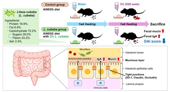
L. cubeba leaves on key functional components of the colonic barrier by assessing fecal IgA and mucin content as functional markers, together with the mRNA expression of tight junction-related factors, in a mouse model of DSS-induced ulcerative colitis.
2. Materials and Methods
2.1. Preparation and Characterization of L. cubeba Leaves
The collected
L. cubeba leaves were washed with tap water, blanched in a steam convection oven, dried, and ground into a powder at the Industrial Technology Center of Saga (Saga, Japan). The nutritional composition of the dried and powdered
L. cubeba leaves was determined by the Institute of Food Hygiene, Nagasaki Food Hygiene Association, and Food and Environment Research Center (Nagasaki, Japan) according to standard analytical procedures (
Table 1). DSS is commercially available from multiple manufacturers. A previous study by Bamba et al. compared three different types of DSS in terms of their chemical properties, cytotoxic effects, and colitis severity, and found that DSS from MP Biomedicals (Irvine, CA, USA) induced the most severe colitis [
21]. Therefore, DSS from MP Biomedicals was used in the present study.
To further characterize the phytochemical properties of L. cubeba leaves, the total polyphenol content and phytochemical profiles were analyzed. For determination of total polyphenol content, L. cubeba leaf powder (4 g) was extracted with 400 mL of 50% aqueous ethanol by shaking for 3 h. The extract was centrifuged at 10,000× g for 30 min at 4 °C, and the supernatant was collected. The residue was re-extracted with an additional 400 mL of 50% aqueous ethanol by shaking for 2.5 h, followed by centrifugation under the same conditions. The combined supernatants were concentrated, freeze-dried, and re-dissolved in 50% aqueous ethanol. The total polyphenol content was determined using the Folin–Ciocalteu method with slight modification. Briefly, 20 μL of the sample was mixed with 100 μL of Folin–Ciocalteu reagent (FUJIFILM Wako Pure Chemical Co., Osaka, Japan) in a 96-well plate and incubated for 3 min at room temperature. Subsequently, 80 μL of 7.5% sodium carbonate solution was added. After incubation for 1 h, the absorbance was measured at 765 nm using a microplate reader (Infinite M200 PRO, Tecan, Männedorf, Switzerland). Gallic acid was used as a standard, and the total polyphenol content was expressed as mg gallic acid equivalents per g dry weight. All measurements were performed in triplicate.
2.2. Experimental Diets
The experimental diets were prepared according to the AIN-93G formula [
22] with minor modifications.
L. cubeba leaf powder was added to the experimental diet at a final concentration of 2% (
w/
w, 20 g/kg diet) (
Table 2). The supplementation level of
L. cubeba leaf powder (2%
w/
w) was determined based on preliminary experiments that confirmed no adverse effects on food intake. To achieve iso-nitrogenous and iso-energetic conditions, the protein, fat, dietary fiber, and other components (sugars, ash, and moisture) in the
L. cubeba leaf-supplemented diet were adjusted using casein, soybean oil, cellulose, and β-cornstarch, respectively, to match the control diet (
Table 2). The amounts of these components were reduced to compensate for the corresponding nutrients provided by the
L. cubeba leaf powder.
2.3. Animals and Experimental Design
All experiments were conducted in accordance with the Guidelines for Animal Experiments of University of Nagasaki, Siebold, Japanese Law No. 105 and Notification No. 6, and ARRIVE guidelines 2.0 [
23]. The animal experimental protocol used in this study was approved by the Animal Experiment Committee of University of Nagasaki, Siebold (Approval No. R04-03).
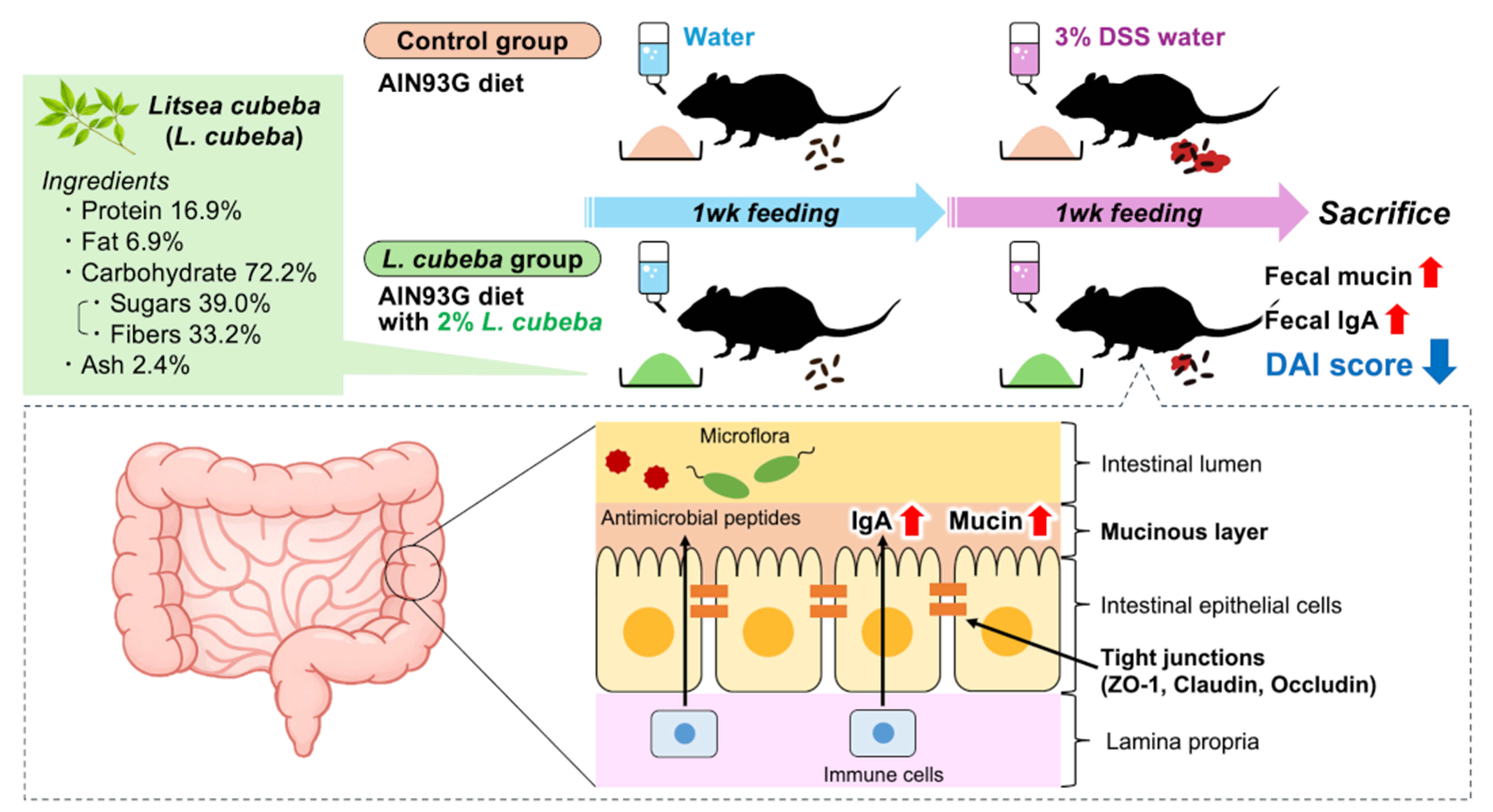
Eight-week-old male C57BL/6J mice (C57BL/6JJcl) were obtained from CLEA Japan, Inc. (Osaka, Japan). The mice were housed individually in plastic cages in a temperature-controlled room maintained at 22 ± 1 °C with 55 ± 5% relative humidity under a 12 h light/dark cycle. After a 4-day acclimation period with free access to the control diet and distilled water, the mice were randomly assigned to three groups with comparable mean body weights: the Normal (
n = 6), Control (
n = 7), and
L. cubeba (
n = 7) groups. Mice in the Normal and Control groups were fed the control diet, whereas those in the
L. cubeba group were fed the
L. cubeba-supplemented diet for 2 weeks. Experimental diets were replaced daily with fresh diets. During both acclimation and experimental periods, the Normal group was allowed free access to distilled water. In contrast, the Control and
L. cubeba groups were provided distilled water during the acclimation period and the first 7 days of the experimental period, while the drinking water was replaced with 3% (
w/
v) DSS solution from day 8 to day 14 to induce ulcerative colitis (
Figure 1). Fecal samples were collected daily from days 8 to 11 of the experimental period to measure the fecal IgA and mucin levels. During the DSS treatment period (days 8–14), DSS-induced colitis symptoms were evaluated daily using the Disease Activity Index (DAI), as described by Cooper et al. [
24]. The DAI consists of three parameters: body weight loss, stool consistency, and severity of fecal bleeding. The scores for each parameter were summed to yield a total DAI score ranging from 0 to 12 (
Table 3). At the end of the experimental period, mice were fasted for 6 h and euthanized by exsanguination via cardiac puncture under isoflurane anesthesia. Blood was collected in ethylenediaminetetraacetic acid disodium salt-containing tubes, and plasma was obtained by centrifugation at 1200×
g for 20 min at 4 °C. The liver, spleen, kidneys, epididymal white adipose tissue (WAT), cecum (with contents removed), and colon (with contents removed) were excised and weighed. Immediately after excision, the length of the colon was measured using a digital caliper (BDC300; AS ONE Corporation, Osaka, Japan). All collected samples were stored at −80 °C until further analysis.
2.4. Measurement of Intestinal Barrier-Related Parameters
The collected fecal samples were vacuum-dried at room temperature for 2 days and then powdered using a mortar and pestle. The fecal IgA content was measured using a commercial enzyme-linked immunosorbent assay (ELISA) kit (Mouse IgA ELISA Kit; Bethyl Laboratories, Inc., Montgomery, TX, USA). Fecal mucin content was measured using a commercial assay kit (Fecal Mucin Assay Kit; Cosmo Bio Co., Ltd., Tokyo, Japan) according to the manufacturer’s instructions.
Total RNA was extracted from approximately 0.1 g of colon tissue using RNAzol
® RT Reagent (Molecular Research Center, Inc., Cincinnati, OH, USA). The extracted RNA was reverse transcribed into cDNA as described previously [
25]. Quantitative reverse transcription PCR (RT-qPCR) was performed using SYBR Green (THUNDERBIRD
® Next SYBR™ qPCR Mix; Toyobo Co., Ltd., Osaka, Japan). The primer sequences used in this study are listed in
Table S2. The target genes analyzed included toll-like receptor 4 (
Tlr4), claudin-1 (
Cldn1), occludin (
Ocln), tight junction protein 1 (
Tjp1), mucin 2 (
Muc2), C-C motif chemokine ligand 2 (
Ccl2), IL-1β (
Il1b), IL-6 (
Il6), and tumor necrosis factor-α (
Tnf). For normalization of gene expression, the stability of three candidate housekeeping genes [β-actin (
Actb), ribosomal protein L13a (
Rpl13a), and ribosomal protein L32 (
Rpl32)] was evaluated, and
Rpl13a was used as the reference gene. The relative mRNA expression levels were calculated using the Pfaffl method [
26].
2.5. Measurement of Inflammatory Markers
Plasma levels of IL-6 and monocyte chemoattractant protein-1 (MCP-1) were measured using commercial mouse ELISA kits (Mouse IL-6 SimpleStep ELISA Kit; Abcam plc, Cambridge, UK, and LEGEND MAX Mouse MCP-1 ELISA Kit; BioLegend, Inc., San Diego, CA, USA), according to the manufacturer’s instructions.
2.6. Statistical Analysis
A few observations were unavailable because of the death of two mice in the Control group during the experimental period; however, all available data were included in the statistical analysis, and no data were excluded from this study. Data from the Normal group are presented as reference values and were not included in the between-group statistical comparisons. The equality of variance between the Control and
L. cubeba groups was assessed using an
F-test. Statistical analyses of continuous data were performed using Student’s
t-test or Welch’s
t-test for equal or unequal variances, respectively. Continuous data are presented as the mean ± standard error of the mean. The DAI score, which is an ordinal variable, was analyzed using the Mann–Whitney
U test, and the results are presented as box-and-whisker plots showing the interquartile range with minimum and maximum values. Survival time was analyzed using the log-rank test. All statistical analyses were performed using EZR (version 1.68), which is a graphical user interface for R (R Foundation for Statistical Computing, Vienna, Austria) [
27]. Results with
p < 0.05 were considered statistically significant; values with 0.05 ≤
p < 0.1 were considered indicative of a trend.
3. Results and Discussion
3.1. L. cubeba Leaf Supplementation Attenuates DSS-Induced Ulcerative Colitis
As shown in
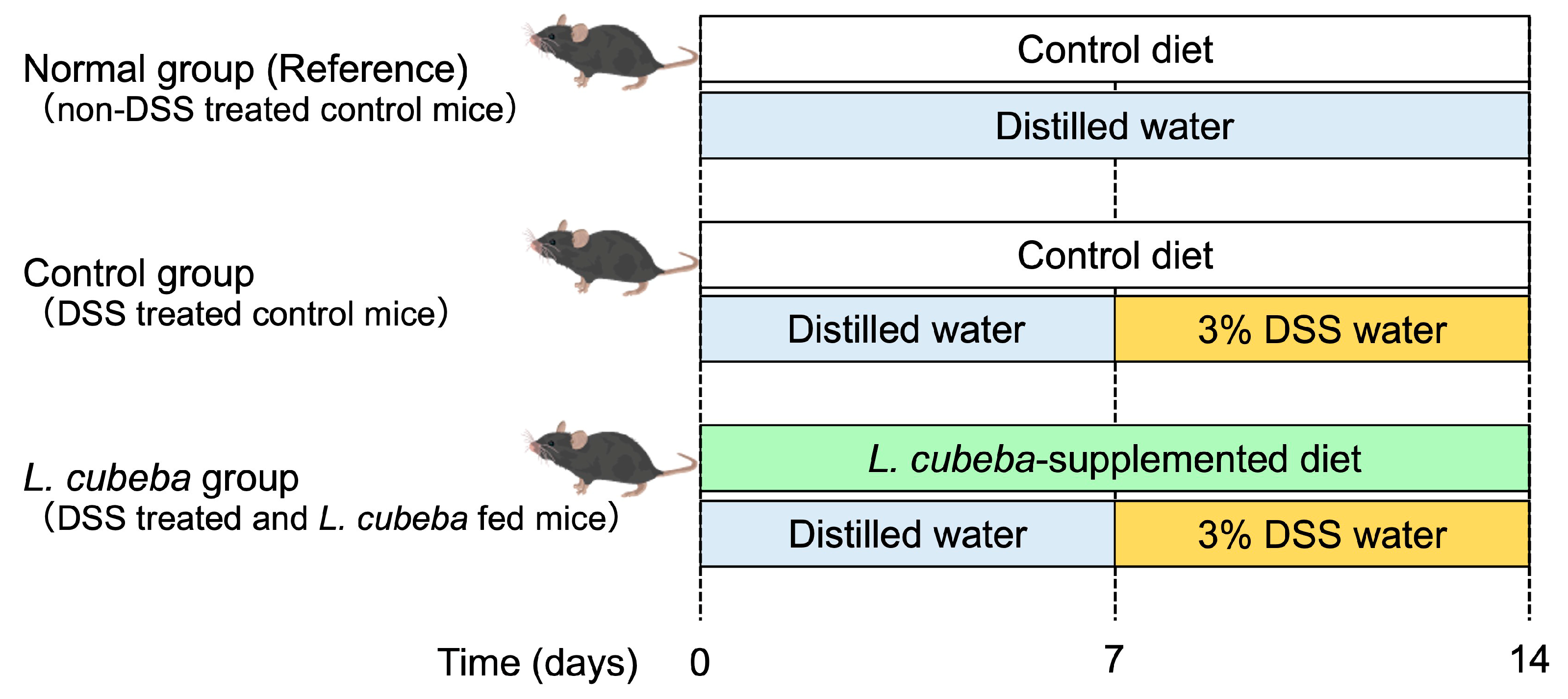
Figure 1, DSS administration was initiated on day 7 and continued for another 7 days. During the DSS treatment period (days 8–14), two out of seven Control group mice died before the study end on day 14, whereas no deaths were observed in the
L. cubeba group. However, the survival analysis using the log-rank test did not reveal a statistically significant difference between the groups (
Figure 2a).
DSS administration induced marked body weight loss in both the Control and
L. cubeba groups, reflecting the development of ulcerative colitis. Body weight loss became more pronounced in the Control group from day 12 (5 days after DSS initiation). In contrast, mice fed the
L. cubeba-supplemented diet exhibited a significant attenuation of body weight loss compared to that of the Control group on days 11, 12, and 13 (
Figure 2b). Throughout the experimental period, no significant differences were observed between the Control and
L. cubeba groups in terms of food intake or water consumption, indicating that the observed effects were not attributable to differences in DSS exposure (
Table 4).
At necropsy, no significant differences were detected between the Control and
L. cubeba groups in the weights of the liver, spleen, kidneys, epididymal WAT, or cecum (
Table 4). In contrast, the colonic weight was significantly higher in the
L. cubeba group than in the Control group (
Table 4). DSS administration caused marked shortening of the colon relative to the Normal group. Although colon shortening tended to be attenuated in the
L. cubeba group, the difference was not statistically significant (
Figure 2c and
Table 4).
Consistent with the changes in body weight and colonic morphology, DSS administration induced a progressive increase in the DAI in both DSS-treated groups, indicating the development of ulcerative colitis. In contrast, the Normal group maintained a DAI score of zero throughout the experimental period (
Figure 2d). Notably, the DAI score was significantly lower in the
L. cubeba group than in the Control group from day 10 onward (3 days after DSS initiation) (
Figure 2d), demonstrating a marked attenuation of disease severity. Because the DAI integrates clinical symptoms such as body weight loss, stool consistency, and fecal bleeding, these results indicate that dietary supplementation with
L. cubeba leaves alleviates the overall clinical manifestations of DSS-induced ulcerative colitis rather than merely delaying disease onset.
3.2. Effects of L. cubeba Leaf Supplementation on Functional Components of the Colonic Barrier
The intestinal epithelial barrier plays a critical role in preventing luminal antigens and microorganisms from penetrating the mucosa, and its disruption is a hallmark of DSS-induced ulcerative colitis [
5,
6,
7,
8,
9,
10,
11]. Intestinal barrier function is generally classified into three major components: (i) a biological barrier composed of antimicrobial peptides, IgA, and immune cells; (ii) a physical barrier formed by epithelial tight junctions and a mucin-rich mucus layer; and (iii) an environmental barrier maintained by the gut microbiota [
18,
19,
20]. In the present study, we focused on the colonic barrier function in a DSS-induced colitis model. To investigate whether dietary supplementation with
L. cubeba leaves influences functional components of the colonic barrier, we first examined the expression of genes related to epithelial tight junctions and mucus production in the colon. The mRNA expression levels of tight junction-related factors, including
Cldn1 (encoding claudin 1),
Ocln (encoding occludin), and
Tjp1 [encoding Zonula occludens-1 (ZO-1)], as well as
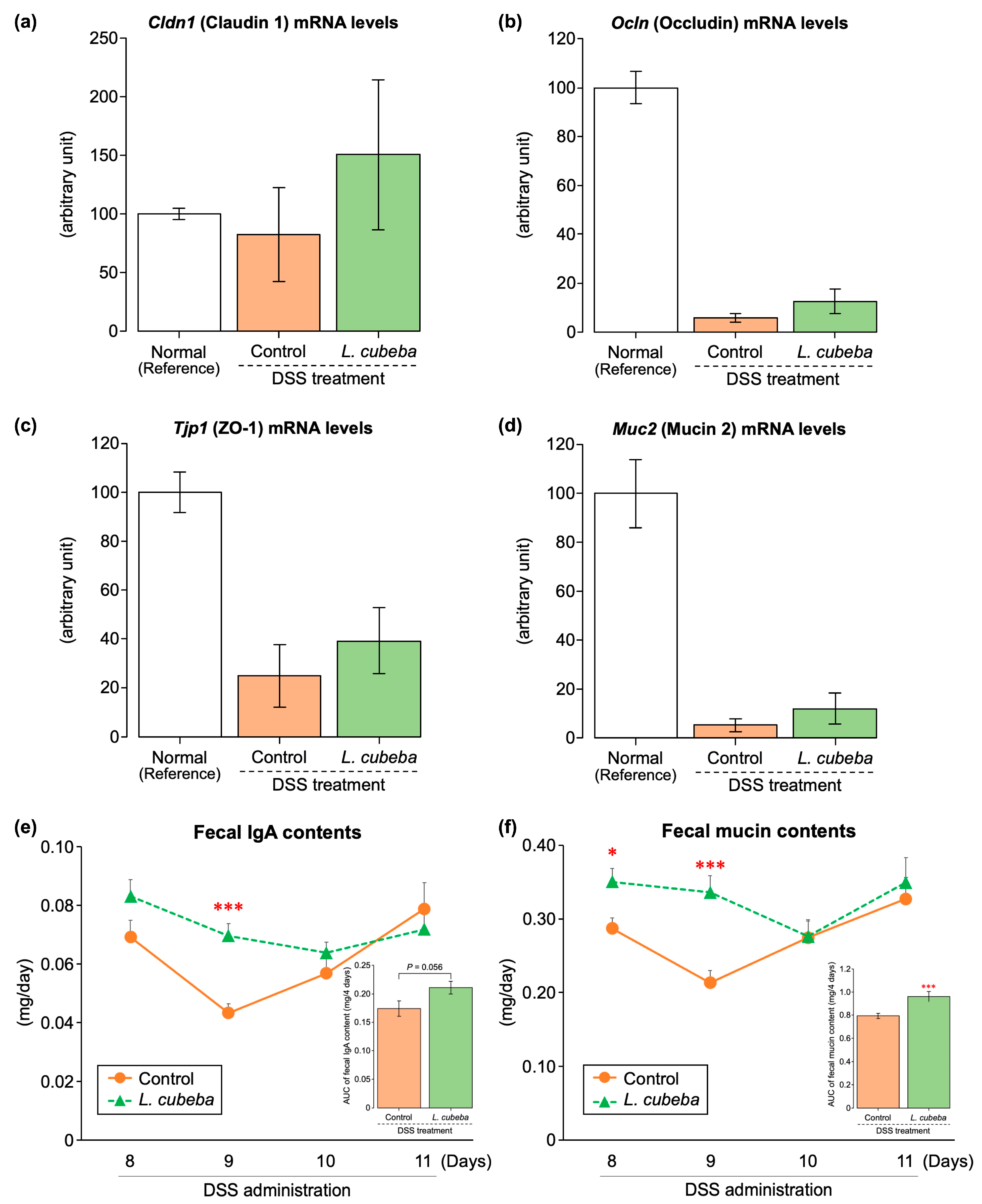
Muc2 (encoding mucin 2), were analyzed using RT-qPCR. DSS-treated mice exhibited a marked reduction in the mRNA expression levels of these genes relative to the Normal group. However, no significant differences were detected between the Control and
L. cubeba groups (
Figure 3a–d). These results suggest that dietary supplementation with
L. cubeba leaves does not markedly affect the transcriptional regulation of the major tight junction components or
Muc2 in the colon under the present experimental conditions.
Next, we examined functional markers of the colonic barrier, focusing on fecal IgA and mucin content, measured from days 8 to 11 of the experimental period (
Figure 3e,f). Fecal IgA content was significantly higher in the
L. cubeba group than in the Control group on day 9. When evaluated as the area under the curve (AUC) over a 4-day period (days 8–11), the fecal IgA content increased more in the
L. cubeba group than in the Control group (
p = 0.056) (
Figure 3e). In contrast, the fecal mucin content was significantly higher in the
L. cubeba group than in the Control group on days 8 and 9. Furthermore, the 4-day AUC of fecal mucin content (days 8–11) was significantly higher in the
L. cubeba group than in the control group (
Figure 3f). These results suggest that dietary supplementation with
L. cubeba leaves favorably modulates functional components of the colonic barrier during the early phase of DSS-induced colitis, with a greater impact on mucus secretion than on IgA production. Notably, although no statistically significant differences were observed in the transcriptional levels of tight junction-related genes and
Muc2, the mean
Cldn1 mRNA level tended to be higher in the
L. cubeba group than in the Control group; however, this finding should be interpreted with caution given the variability and limited sample size. Taken together, these observations suggest that
L. cubeba leaf supplementation preferentially influences the functional components of the colonic barrier evaluated in the present study, although the underlying mechanisms remain to be elucidated. However, because histological evaluation of the mucus layer and direct assessment of barrier permeability were not performed, the effects of
L. cubeba leaf supplementation on overall colonic barrier integrity remain to be confirmed.
3.3. Effects of L. cubeba Leaf Supplementation on Systemic and Colonic Inflammatory Responses
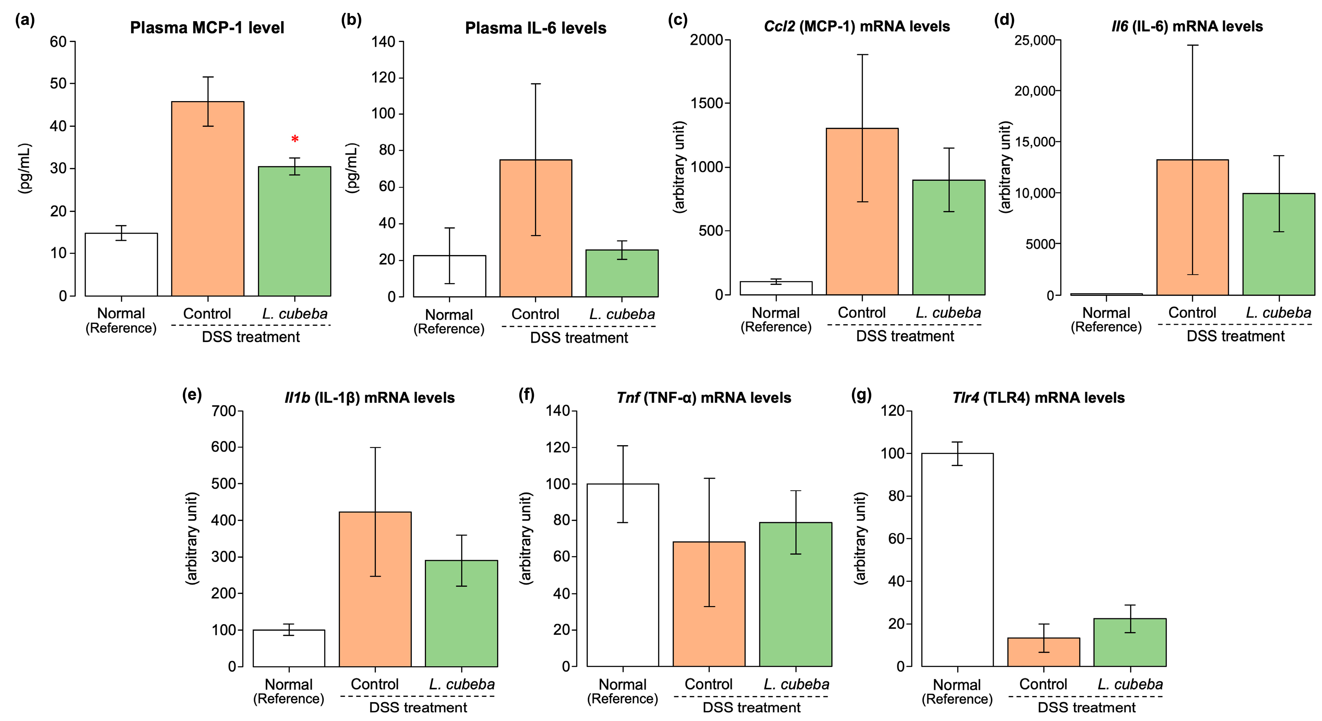
Given that disruption of the colonic barrier is closely associated with inflammatory responses during DSS-induced colitis, we examined systemic and colonic inflammatory markers. DSS administration markedly increased the plasma concentrations of MCP-1 and IL-6 relative to those in the Normal group. The plasma MCP-1 levels were significantly lower in the
L. cubeba group than in the Control group (
Figure 4a). Although the difference did not reach statistical significance, plasma IL-6 levels were 66% lower in the
L. cubeba group than in the Control group (
Figure 4b). In the colon, the mRNA expression levels of inflammation-related genes, including
Ccl2 (encoding MCP-1),
Il6 (encoding IL-6),
Il1b (encoding IL-1β), and
Tnf (encoding TNF-α), were markedly increased by DSS treatment relative to the Normal group levels; however, no significant differences were detected between the Control and
L. cubeba groups (
Figure 4c–f). In addition,
Tlr4 mRNA levels did not differ between the groups (
Figure 4g). Notably, these findings differ from those of previous reports showing that
L. cubeba leaf extracts directly suppress inflammatory signaling pathways in experimental colitis models [
17], suggesting that dietary
L. cubeba leaf supplementation attenuates DSS-induced colitis, at least in part, through modulation of functional components of the colonic barrier rather than the direct inhibition of colonic inflammatory gene expression. Despite the limited effects on the transcriptional levels of pro-inflammatory cytokines in colonic tissue,
L. cubeba leaf supplementation significantly enhanced functional components of the colonic barrier, including fecal IgA and mucin contents, which were accompanied by a marked reduction in circulating MCP-1 levels. Increased fecal mucin levels may reflect alterations in mucus dynamics, whereas increased fecal IgA levels may enhance immune exclusion of luminal antigens. Together, these changes may contribute to the maintenance of key functional components of the colonic barrier and thereby attenuate systemic inflammatory responses, as reflected by the reduction in circulating MCP-1 levels. This dissociation between local transcriptional responses and systemic inflammatory markers may be associated with changes in functional components of the colonic barrier; however, the underlying mechanisms, including the potential involvement of luminal antigen translocation, remain to be elucidated. In addition, direct assessments of intestinal permeability and endotoxin levels were not performed in the present study; therefore, the proposed mechanisms require further investigation.
3.4. Phytochemical Characterization of L. cubeba Leaves
To provide preliminary insight into the phytochemical characteristics of L. cubeba leaves, the total polyphenol content and LC-MS/MS-based profiling were performed. The total polyphenol content was determined to be 51.4 ± 0.7 mg gallic acid equivalents/g dry weight (mean ± SEM, measured in triplicate).
LC-MS/MS analysis revealed the presence of several polyphenolic compounds. Among these, kaempferitrin (kaempferol 3,7-O-dirhamnoside) and afzelin (kaempferol 3-O-rhamnoside) were identified using authentic standards, while several other peaks were tentatively assigned based on mass spectral data (
Supplementary Figure S1 and Table S1).
These findings provide preliminary information on the chemical composition of L. cubeba leaves; however, the specific compounds responsible for the observed biological effects remain to be elucidated.
3.5. Integrated Interpretation of the Effects of L. cubeba Leaf Supplementation on Colonic Barrier Components, Systemic Inflammation, and Liver Injury
Taken together, the results of the present study indicate that dietary supplementation with
L. cubeba leaves may attenuate DSS-induced ulcerative colitis through the coordinated effects on colonic barrier integrity and systemic inflammation. The enhancement of key functional components of the colonic barrier was accompanied by the attenuation of systemic inflammatory responses, particularly a significant reduction in circulating MCP-1 levels. In addition to intestinal inflammation, inflammatory bowel diseases are frequently associated with extraintestinal manifestations, including secondary liver injury [
28,
29]. In this context, plasma alanine aminotransferase (ALT) activity, a marker of hepatocellular injury, was significantly lower in the
L. cubeba group than in the Control group (
Table S3), suggesting the potential attenuation of DSS-associated liver dysfunction. However, because only a single hepatic enzyme was evaluated in the present study, these findings should be interpreted with caution. Given that systemic inflammation has been implicated in colitis-associated liver injury, the reduction in circulating MCP-1 levels observed in the
L. cubeba group may be associated with reduced plasma ALT activity. Collectively, these findings suggest that dietary
L. cubeba leaf supplementation attenuates DSS-induced ulcerative colitis by reinforcing colonic barrier integrity, thereby attenuating systemic inflammatory signaling and secondary liver injury. This mode of action may be distinct from previously reported extract-based interventions that directly target inflammatory signaling pathways, highlighting the potential of
L. cubeba leaves as a dietary strategy for the management of ulcerative colitis.
3.6. Limitations
The present study has several limitations. First, although dietary supplementation with L. cubeba leaves markedly enhanced key functional components of the colonic barrier, only limited phytochemical characterization, including total polyphenol analysis and LC-MS/MS-based profiling, was performed, and the specific bioactive components responsible for these effects have not been fully identified. Second, the present study primarily evaluated transcriptional responses in colonic tissues; therefore, changes at the protein level or post-translational regulation of barrier- and inflammation-related factors cannot be excluded. Third, direct histological assessments of the colonic barrier, including evaluation of goblet cell abundance, mucus layer thickness, and mucosal architecture, as well as direct measurements of intestinal permeability, were not performed. Therefore, the effects of L. cubeba leaf supplementation on overall colonic barrier integrity remain to be confirmed. Fourth, alterations in the gut microbiota composition, which may contribute to colonic barrier integrity and systemic inflammation, were not assessed and warrant further investigation. Fifth, the relatively limited sample size, particularly following the loss of animals in the Control group, may have reduced the statistical power to detect differences in some parameters; therefore, the negative findings should be interpreted with caution. Sixth, only a single dose of L. cubeba leaf supplementation was examined, and dose–response relationships were not evaluated. Finally, although the DSS-induced colitis model is widely used to study ulcerative colitis-like pathology, extrapolation of the present findings to human disease should be performed with caution. Future studies addressing these points are essential to elucidate the mechanisms underlying the protective effects of L. cubeba leaf supplementation against DSS-induced colitis.




 ,
,