Review

Vanillin and Its Derivatives: A Critical Review of Their Anti-Inflammatory, Anti-Infective, Wound-Healing, Neuroprotective, and Anti-Cancer Health-Promoting Benefits

by
Magdalini Kafali
,
Marios Argyrios Finos
and
Alexandros Tsoupras
*
Hephaestus Laboratory, School of Chemistry, Faculty of Sciences, Democritus University of Thrace, Kavala University Campus, St Lukas, 65404 Kavala, Greece
*
Author to whom correspondence should be addressed.
Nutraceuticals 2024, 4(4), 522-561; https://doi.org/10.3390/nutraceuticals4040030
Submission received: 4 August 2024 / Revised: 26 August 2024 / Accepted: 9 October 2024 / Published: 11 October 2024
(This article belongs to the Special Issue Nutraceuticals and Their Anti-inflammatory Effects)

Abstract

:
Inflammation and thrombosis are implicated in several non-communicable chronic disorders, including cardiovascular diseases, diabetes, renal and neurodegenerative disorders, skin diseases, and especially in cancer. Natural bioactives and especially phytochemicals like phenolic compounds have been proposed to reduce the inflammatory burden with several health benefits against these disorders. Vanillin is a phenolic compound found in the seeds of various species of vanilla plants. It has been known since ancient times for its aromatic and soothing properties; however, recent outcomes have outlined several other pleiotropic actions for this phenolic bioactive compound. Within this article, the potent anti-inflammatory activities of vanillin and its derivatives are thoroughly reviewed, with emphasis on their anti-cancer, anti-infective, wound-healing, and neuroprotective health-promoting properties. The mechanisms of their action(s), along with recent outcomes from in vitro and in vivo studies and clinical trials, on the benefits of these vanillin-based phenolic bioactives against each of these disorders, and especially against specific types of cancer, are also outlined. Limitations and future perspectives of their use solely as bioactive ingredients, as ingredients in several functional products—such as functional foods, supplements, nutraceuticals, or even cosmetics and drugs—and even as adjuvant therapies are also discussed.

1. Introduction

Inflammation is a physiological mechanism for maintaining the body’s homeostasis against any disturbance and includes three primary stages: (1) initiation, (2) regulation, and (3) resolution [1]. Every phase is influenced by both immune and non-immune cells, including fibroblasts, endothelium, and epithelial cells, as well as macrophages, dendritic cells, mast cells, neutrophils, and lymphocytes [1,2]. Depending on the type of the stimulus, the structure of different cell types and tissues, the control of gene expression, as well as the signaling of the involved biochemical reactions are affected, mainly through substances such as interleukins (IL-1β, IL-6, IL-8), tumor necrosis factor alpha (TNF-α), reactive oxygen species (ROS), and nitric oxide (NO) [3,4].
Failure to terminate inflammation can be due to environmental or non-environmental factors. This leads to systemic chronic inflammation, which involves inactivation of the immune response, activation of various immune components, modification of normal cell, tissue, and organ function, and ultimately the development of chronic diseases [5]. According to the World Health Organization, chronic inflammatory diseases, such as type 2 diabetes, obesity, cardiovascular diseases, cancer, neurodegenerative diseases, etc., are the leading cause of death, with three out of five people dying from them. It is still challenging to explain the distinct mechanisms underlying these inflammation-related diseases; nevertheless, it is widely accepted that systematic inflammation results from an inability to effectively counteract the initial stimulus [6]. The challenge is even greater when the original stimulus is unknown [7].
Excessive exposure to resistant microbes, allergens, pollutants, drugs, and processed foods, combined with sedentary lifestyles and psychological factors, seems to disrupt the immune system’s normal functioning, leading to chronic inflammation and all its aftereffects [8]. Currently, non-steroidal medicines are widely used to combat inflammation, inevitably causing patient side effects like gastrointestinal complications, and several cardiovascular complications [9]. An anti-inflammatory diet contains natural foods that retain their antioxidant and anti-inflammatory characters without the addition of additives that affect their toxicity and biocompatibility [10]. Anti-inflammatory dieting is considered key in not only replacing non-steroidal drugs in the treatment of inflammatory diseases, but also in preventing inflammation-linked illnesses through a balanced diet [3,11,12]. Among various natural anti-inflammatory substances, the products and by-products of bioactive and phenolic compounds, found in large quantities in health promoting plants (like kiwi, avocado, wine-grape, apple, etc.) [13], are of special interest [3,11,12].
Vanillin (4-hydroxy-3-methoxybenzaldehyde) is a phenolic aldehyde that is found in high concentration mainly in the seeds of various vanilla species and is the most stable cleavage product of curcumin [14]. Although it has been known since ancient times for its aromatic and soothing properties, the bioactive qualities of vanillin, such as anti-inflammatory, neuroprotective, antibacterial, and anti-cancer activity, have only recently come to light [4]. The vanillin molecule consists of a benzene ring joined to an ether group, an aldehyde group, and a hydroxyl group [15]. It is this chemical structure that provides it with its characteristic bioactive properties but also the ability to be modified or combined with other substances in order to produce several derivatives [16], lessening adverse effects while aiding in the bioactive substance’s targeted transport and release, dose management, enhanced stability, and bioavailability. Moreover, vanillin and several derivatives also exist naturally in several natural sources, including fruits and their products and by-products [17,18].
In essence, vanillin derivatives can be broadly classified into two groups depending on their functionality when enclosed with other molecules or in complexes. In the first category of derivatives, vanillin is either bound or enclosed by another substance, which facilitates its transport into biological systems and its targeted action. In the second class, vanillin in combination with various other compounds forms complexes that act as transporters of other drugs or bioactive substances [19].
In vitro and in vivo studies demonstrate that the anti-inflammatory nature of vanillin and its derivatives is mainly due to its involvement in the control of the expression of various genes, which contribute to the reduced secretion of pro-inflammatory cytokines (IL-1β, IL-8, IL-6, TNF-a), in the reduced activity of the COX-2 and inducible nitric oxide synthase (i-NOS) enzymes, with the subsequent reduction of the produced NO and prostaglandins, and finally in the increased secretion of anti-inflammatory cytokines (IL-4, IL-10, TGF-B). As long as chronic inflammation, neurodegeneration, and cancer are interrelated concepts, the mechanisms above not only have neuroprotective effects but are also crucial to the fight against cancer cells.
The anti-cancer activity of vanillin and its derivatives is demonstrated by its use as an anti-cancer agent in various types of cancer. Moreover, vanillin disrupts the normal growth and reproduction of bacteria, which makes both it and its derivatives effective antibacterial agents [20,21], with promising applications in wound healing. Existing research provides strong evidence for the benefits of vanillin; however, due to its low bioavailability, human clinical trials are limited [22]. This report aims to analyze the promising bioactive properties of vanillin and its derivatives, including anti-inflammatory, neuroprotective, antibacterial, and wound-healing properties. Particular emphasis is placed on the use of vanillin and its derivatives as anti-cancer agents against melanoma, colorectal, breast, hepatic, and lung cancer.

2. Method

The Scopus database was utilized for finding relevant literature. The following keywords were used: “Vanillin”, “vanillin analogues”, “vanillin derivatives”, “vanillin metal complex”, “bioactive”, “bioactivity”, “activity” “action”, “effect”, “health benefits”, “antioxidant”, “antiaging”, “anti-inflammatory”, “skin protection”, “wound healing”, “anti-infective”, “antimicrobial”, “anti-bacterial”, “neuroprotective”, “cardio-protective”, “anti-tumor”, “anti-cancer”, “anti-melanoma”, “inflammation”, “oxidative stress”, “infection”, “in vitro”, “in vivo”, “cardiovascular diseases”, “diabetes”, “cancer”, “melanoma”, “colorectal cancer”, “breast cancer”, “neurodegenerative disorders”, “applications”, “functional foods”, “supplements”, “nutraceuticals”, “cosmetic”, “nutricosmetics”, “cosmeceuticals”, “pharmaceuticals”, and “drug”, with the use of combinations of these keywords by using the AND and/or OR terms. This query was applied to the titles, abstracts, and keywords of searched articles, and the search process was conducted during March–May 2024, encompassing material from the last 15 years.

2.1. Inclusion Criteria

The selection criteria were determined by considering the metadata available from Scopus, with the eligible studies meeting the following criteria: (i) be exclusively research articles; (ii) be written in English; and (iii) be published between 2010 and 2024. A limited amount of important information from review papers was also used, and a few articles before 2010 were also included, as they were not previously reviewed thoroughly.

2.2. Exclusion Criteria

Conference papers, books, and short surveys, as well as publications written in languages other than English, were excluded.

2.3. Quality Assessment

To evaluate the articles’ quality and relevance, we first reviewed their titles and abstracts, excluding those unrelated to the topic. Subsequently, the remaining articles were thoroughly read to determine whether they met the predefined inclusion criteria and provided pertinent information for this review.

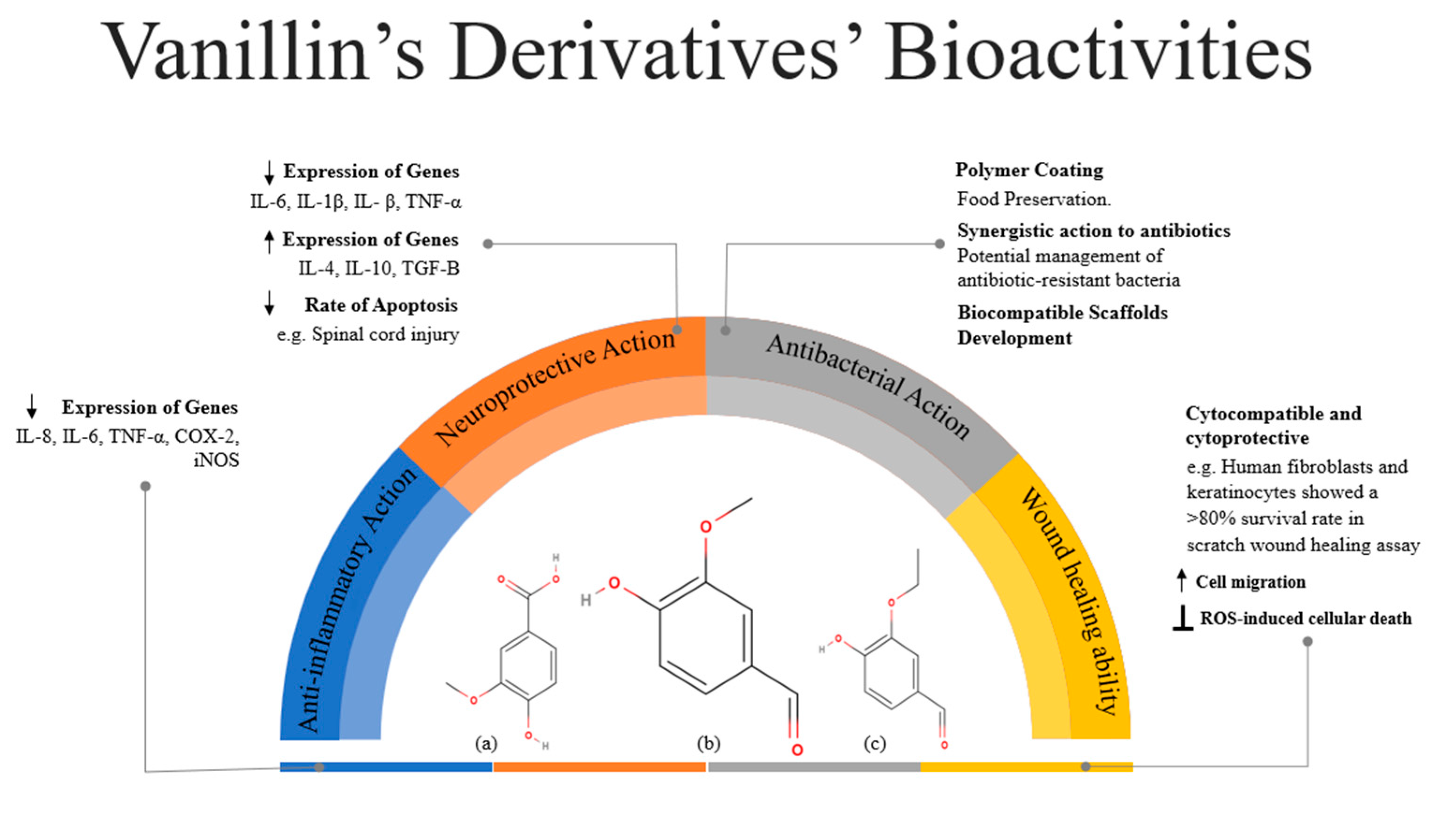
3. Bioactive Actions of Vanillin

3.1. Anti-Inflammatory Action

Experiments conducted on many animal cell types reveal that vanillin possesses anti-inflammatory characteristics, mostly due to its ability to reduce the expression of genes that promote inflammation, inflammatory mediators, and enzymes that are involved in the inflammatory response [14]. Regardless of the test model, administration of vanillin led to a decrease in the expression of genes such as IL-8, IL-6, and TNF-α and consequently a decrease in the production of the corresponding pro-inflammatory cytokines. Furthermore, a number of long-term inflammatory conditions, such as periodontitis, are linked to the overexpression of the enzymes cyclooxygenase 2 (COX-2) and nitric oxide synthase (iNOS), which results in an excess of prostaglandins and nitric oxide (NO). Numerous studies show that administering vanillin regulates the expression of these genes [3]. Indicatively, in rats with acute liver injury induced by carbon tetrachloride, vanillin indeed appears to reduce the expression of TNF-α, IL-1β, IL-6, and COX-2 genes [23], while treatment with vanillin prior to the administration of CCl(4) reduced the decrease of protein synthesis and the increase in plasma alanine (ALT) and aspartate (AST) aminotransferases [24]. Another study found that vanillin suppressed nitric oxide synthase (iNOS), with consequent reduction of NO levels [19]. As a potential NF-κB inhibitor, vanillin has shown promising anti-inflammatory properties in various tissues and cells such as the liver, colon, and macrophages [25]. For example, the effect of vanillin was evaluated in mice with trinitrobenzene sulfonic acid (TNBS)-induced colitis, and results showed that vanillin improved the features of TNBS-induced colitis in a dose-dependent manner. Vanillin not only prevented TNBS-induced colitis but also suppressed in vivo NF-κB activities through the inhibition of p65 translocation, IκB-α phosphorylation, and IκB kinase activation [26]. Furthermore, vanillin exhibited anti-inflammatory action in an acute lung injury (ALI) in vivo model via the inhibition of the expression levels of pro-inflammatory cytokines by inhibiting the phosphorylation activities of ERK1/2, p38, AKT, and NF-κB p65 [27]. By decreasing the oxidative stress and inflammation, vanillin seems to assist the function of the liver, kidneys, and muscles in fructose-STZ-induced diabetic rats, suggesting that it could be a potential agent against type 2 diabetes and its subsequent complications [28] (Figure 1).
The treatment of several of these tissues using vanillin as well as the underlying mechanisms are thoroughly investigated via in vitro or in vivo models which are listed in Table 1, along with the main findings and specific benefits of each study.

3.2. Neuroprotective Action

The progressive loss of neurons in the central nervous system (CNS) and their subsequent under-functioning is the cause of many neurodegenerative diseases, including Alzheimer’s and Parkinson’s disease, traumatic brain and spinal cord injury, stroke, multiple sclerosis, various neuropsychiatric disorders, etc. [30]. Astrocytic and microglial cells are the main immunological factors in the brain, and the interaction between the brain and immune systems can be beneficial or harmful [30,31]. Immune mediators serve two purposes: first, they aid in the removal of debris from age-related breakdown, neutralization of antigens, and tissue repair, and support normal development of the central nervous system (CNS). Secondly, they induce chronic inflammation in the CNS [30,31]. When homeostasis is disrupted, these cells are activated by the release of pro-inflammatory substances like cytokines (e.g., IL-1, IL-6) as well as by the decrease of anti-inflammatory cytokines (e.g., IL-10) [31]. The accumulation of immune cells in the brain is responsible for the over-secretion of cytokines and other substances in order to activate more and more immune cells. The result is chronic inflammation, which seems to cause gradual neurodegeneration [30,31].
Although the neuroprotective role of vanillin has not yet been fully elucidated, it nevertheless appears to be due to its simultaneous antioxidant and anti-inflammatory activity [32,33]. Oxidative stress appears to enhance the chronic secretion of pro-inflammatory cytokines, which among others are responsible for various neurodegenerative diseases [33]. Through its antioxidant action, the activity of active radicals (ROS and RNS) that appear to be involved in diseases such as Parkinson’s (PD), Alzheimer’s (AD), and Huntington’s (HD) diseases is inhibited, while antioxidant enzymes are activated. The combination of all the above actions works protectively toward the brain [34]. In addition, the anti-inflammatory neuroprotective effect of vanillin again lies in controlling the expression of various genes, with the main ones being those related to pro-inflammatory and anti-inflammatory cytokines. In particular, vanillin reduces the production of pro-inflammatory cytokines secreted by activated microglial cells (e.g., interleukin-1β, interleukin-6, interferon-β, and tumor necrosis factor-α), while also activating the production of anti-inflammatory cytokines in tissues (e.g., interleukin-4, interleukin-10, and TGF-B) and reducing the rate of cell apoptosis in case of spinal cord injury [33,34,35,36].
Indicatively, when tested on MPP+/MPTP-intoxicated human neuroblastoma (SH-SY5Y) cells and a mouse model of Parkinson’s disease, vanillin exhibited potential neuroprotective action via the amelioration of MPP+-induced dysregulations in protein expression of tyrosine hydroxylase (TH) and mRNA expressions of GSK-3β, PARP1, p53, Bcl-2, Bax, and Caspase-3 genes in SH-SY5Y cells [37]. Due to the phenolic structure and the interactions with human transferrin (hTf), vanillin could be an effective agent against AD via its antioxidant properties and the maintenance of iron balance [38]. Moreover, the levels of AChE, beta secretase, caspase-3, and Abeta plaques were reduced, while the levels of brain-derived neurotrophic factor (BDNF) in cortical and hippocampal regions were enhanced, when vanillin was administrated on healthy Swiss albino mice, and on an aluminum chloride and D-galactose-induced AD model in mice [39].
Table 2 demonstrates several in vitro and in vivo studies that prove the neuroprotective action of vanillin and its derivatives.

3.3. Antibacterial Action

The mode of action of antimicrobials is mainly (a) interaction with the cell membrane, (b) inactivation of important enzymes, or (c) destruction or inactivation of genetic material. Phenolic compounds, like vanillin, due to their hydrophobicity, interact with bilayer lipids as well as membrane proteins, leading to plasma membrane destabilization [21]. The in vitro proven antimicrobial activity of vanillin against bacteria, yeast, and molds, combined with its lower cytotoxicity compared to other phenolic compounds [20], favor its use in various applications. Vanillin’s inhibitory action was found to be bacteriostatic rather than bactericidal. We cannot say for sure that vanillin has a greater antibacterial effect against Gram-negative or Gram-positive bacteria. In the literature, it appears effective for both Gram-positive bacteria and Gram-negative bacteria, with the range of antibacterial activity varying from bacteria to bacteria [21]. Indicatively, when coating polyethylene with vanillin nanoparticles for the purpose of manufacturing food packaging films, the presence of vanillin particles appears to have drastically reduced the concentration of E. coli (Gram-negative bacteria), while the concentration of S. aureus (Gram-positive bacteria) was not significantly affected. It is worth noting that vanillin being a volatile compound can neutralize bacteria not only when it comes in contact with them, but also through its vapors [41].
The combination of vanillin with other particles or even its modification through organic reactions leads to the synthesis of derivatives, which in most cases show improved antibacterial properties compared to their individual components or the original reagents, respectively. For example, vanillin seems to work synergistically with certain antibiotics against common bacteria, which may prove extremely useful since bacteria are becoming increasingly resistant to antibiotics [42]. The combination of antibiotics and anti-virulence therapy seems to be promising as far as the inhibition of pathogenic bacteria is concerned. Among several plant extracts, vanillin could serve as a potential anti-virulence therapy mainly by controlling the quorum sensing (QS) as an inhibitor of the pqs QS, probably via the PqsR receptor. It is worth noting that the minimal inhibitory concentration (MIC) of colistin—an antimicrobial peptide—was significantly reduced by the simultaneous administration of vanillin and colistin against P. aeruginosa. Thus, the combinatorial treatment of vanillin, as an anti-virulence agent, and antibiotics could be useful against bacterial infections [43]. Another study concluded that P. aeruginosa was effectively inhibited using vanillin-capped gold nanoparticles (VAuNPs) as a novel antibacterial agent. VAuNPs suppressed the expression of several efflux pump genes better than vanillin, while enhancing the antibacterial action of last-line antibiotics such as meropenem and trimethoprim, addressing the problem of antimicrobial resistance [44]. Vanillin-derived 1,4-disbustituted 1,2,3-triazoles also seem to be effective against resistant bacteria and have a higher inhibitory effect than ciprofloxacin [45].
Moreover, the use of vanillin combined with natural polymers such as chitosan is being investigated as an effective non-toxic way of preserving foods and preventing the growth of harmful bacteria on them [46]. The antibacterial properties of vanillin’s derivatives can be applied to biocompatible implants, as in the case of manufacturing antimicrobial scaffolds that reduce the possible infections and implant rejections [47]. Table 3 lists the most characteristic examples of in vitro and in vivo studies regarding the antibacterial action of vanillin and its derivatives.

3.4. Wound-Healing Ability

Hydrogels (HGs) are flexible and moist materials made of 3D cross-linked macromolecular polymer chains that include a large number of water molecules in their network. They mimic human tissues and, for this reason, it is possible to create biocompatible hydrogels for tissue engineering scaffolds and wound dressings by combining polymers with natural crosslinkers [52]. Chitosan hydrogels have been made using vanillin as a natural crosslinker and have been proven to promote wound healing [53], while combined with polyvinyl alcohol (PVA) the produced hydrogels also have a potent scavenging effect on DPPH, ABTS+, and PTIO free radical [54]. PVA, vanillin, and carboxymethyl chitosan also form hydrogels that effectively inhibit Staphylococcus aureus (S. aureus) and Escherichia coli (E. coli) [55]. Indicatively, novel hydrogels, that were synthesized by crosslinking chitosan with a vanillin isomer, 5-methoxysalicylaldehyde, not only showed promising physicochemical properties but also effectively inhibited the fungi Candida albicans, indicating their great potential for the cure of several skin lesions [56]. Vanillin also showed cytocompatibility and cytoprotective properties against cellular damage brought on by ROS. In response to vanillin (10–500 µM), human fibroblast and keratinocyte cells showed a >80% survival rate. It has been demonstrated that vanillin increases cell migration and inhibits ROS-induced cellular death, which in turn promotes wound healing [57]. Furthermore, ethyl vanillin (EV) is an excellent option for the manufacture of hydrogels because of its phenolic functional group’s involvement in the development of hydrogels and its capacity to be coupled with gelatin by the homogenous Schiff base reaction. The antibacterial qualities of the complex, which inhibit Staphylococcus aureus and Escherichia coli, along with its non-toxic nature, suggest that new polymeric wound dressings consisting of gelatin, polyvinyl alcohol, and ethyl vanillin can be effectively employed in wound-healing applications [52]. By examining the healing ability of a novel vanillin-derived zinc complex [Zn(phen)(van)2] (ZPV) on diabetic rats, decreased levels of TNF-α and IL-1β, increased expression of VEGF and TGF-β genes, decreased wound size, re-epithelialization, angiogenic stimulation, and collagen deposition were revealed. Thus, in diabetic rats, the tested complex improved the healing process [58]. Concordantly, Figure 2 and Table 4 describes some in vitro and in vivo studies that prove the wound-healing ability of vanillin and its derivatives.

4. Anti-Cancer Action

4.1. Melanoma

Research conducted both in vivo and in vitro suggest that vanillin and its derivatives have promising potential as anti-cancer and anti-metastatic agents against melanoma. Hypoxia-inducible factor-1 (HIF-1) is a critical transcription factor in the adaptation of cells to low-oxygen circumstances, since a hypoxic microenvironment is a fundamental characteristic of several forms of solid tumors, including melanoma. Vanillin suppresses the accumulation of HIF-1α protein and the transcripts of HIF-1α target genes linked to cancer metastasis, such as fibronectin 1 (FN1), lysyl oxidase-like 2 (LOXL2), and urokinase plasminogen activator receptor (uPAR) [60]. Moreover, vanillin was found to inhibit HIF-1α mRNA expression, de novo HIF-1α protein production, as well as STAT3 phosphorylation. Additionally, an in vitro migration assay demonstrated that vanillin administration reduced the motility of melanoma cells triggered by hypoxia. Hence, by activating the STAT3–HIF-1α signaling pathway, vanillin is a potentially anti-metastatic substance that inhibits metastatic activity and the expression of metastatic genes under hypoxic conditions. Furthermore, the growth of allografted B16BL6 melanoma was decreased by the administration of vanillic acid, also mainly through the inhibition of STAT3 phosphorylation [61].
STAT3, one of the proteins of the STAT family, enters the nucleus to function as a transcription factor after becoming phosphorylated in response to cytokines and growth factors (GFs) [10]. Over-activated STAT3 controls transcription pathways that prevent apoptosis and increase the ability of cancer cells to survive [16]. The melanoma cells’ death and autophagy were promoted by the vanillic-acid-inhibited STAT3 pathway. Although it is proven that vanillin is not an efficient inhibitor of the NF-ĸB transcriptional activity, neither when stimulated by hypoxia [60] nor by doxorubicin [62], o-vanillin drastically inhibited doxorubicin-mediated induction of NF-ĸB activity, suppressing constitutive (basal) NF-ĸB activity in A375 cells [62]. The transcription factor NF-κB significantly regulates immune and inflammatory responses [63], and the NF-κB-inhibitory action of vanillin seems to be dose-dependent. Vanillin treatment dramatically decreased the viability of B16F10 cells, particularly when the cells were exposed to doses of 2 and 5 µg/mL of vanillin. The treatment with vanillin considerably reduced the tumor’s weight and volume, especially daily doses of 50 mg/kg and 100 mg/kg. When comparing the 100 mg/kg/day group to the control group, there was a significant drop in NF-κB expression; however, concentrations below that did not have a significant impact on NF-κB expression [64]. Both the synthesis of melanin and tyrosinase activity decreased in a concentration-dependent manner upon treatment with vanillic acid. These results suggest a connection between the downregulation of the NO-mediated cGMP/PKG signaling pathway and vanillic acid’s anti-melanogenic action [65].
Additionally, substances that result from combining inorganic metals with vanillin appear to have considerable promise as anti-cancer medications. Flow cytometry assays against the 786-0 cell line clearly demonstrated that [Cu(phen)(van)2], a vanillin-based copper(II) metal complex, outperformed cisplatin. Additionally, the complex caused a consequent G1 phase arrest, indicating that it induces DNA damage and makes it difficult for the cells to be driven to S and G2/M phases. A rise in sub-G1 cells was seen at doses greater than 20 mmol L−1, suggesting that the compound induced sufficient damage to cause a significant percentage of cells to undergo apoptosis [66]. In vivo studies proved that the administration of either vanillin or its derivatives caused the tumor volume and tumor weight to be reduced [62,64].
Of special interest are some novel metal-based complexes containing vanillin or vanillin derivatives that have shown anti-inflammatory benefits against cancer [67]. For example, chromium (III), iron (III), and ruthenium (III) complexes containing derivatives of ortho-vanillin showed potent inhibition of edema tumor formation induced by carrageenan (1% w/v) in the leg of Wistar rats, in comparison with the well-established anti-inflammatory drug sodium diclofenac and DMSO as the control, which were also administered at the same concentration [68].
Table 5 summarizes different in vitro and in vivo studies that examine the anti-cancer action of vanillin and its derivatives against melanoma. The hypothesis, study design, results, and main benefits of each study are also included.

4.2. Colorectal Cancer

Vanillin is not so cytolytic; rather, it is cytostatic. The cytostatic and cytolytic properties of vanillin, respectively, at low and high concentrations, indicate that vanillin is a potentially effective treatment for colorectal cancer. A number of cancer types share vanillin targets, the most likely being the suppression of the AP-1 transcription factor complex. Vanillin causes apoptosis and suppresses cell proliferation in a dose-dependent manner. For instance, vanillin was found to cause G0/G1 arrest at a dosage of 200 mg/mL, whereas G2/M arrest happens at a concentration of 1000 mg/mL [70]. Vanillin significantly reduced the spontaneous mutations on HCT116 colon cancer in a dose-dependent way and induced DNA damage that elicits recombinational DNA repair and, consequently, reduces spontaneous mutation [71]. Furthermore, vanillin slowed the growth of colon cancer in mice that was caused by AOM/DSS. The improvement caused by vanillin may be linked to the downregulation of MAPK, NF-κB, and proteasome pathways [72]. Indeed, vanillin specifically decreased the phosphorylation of mitogen-activated protein kinases (MAPK) and statistically significantly decreased the quantity of granulocytes, proliferating cells, and p65-positive cells in colon tissues. Vanillin’s intervention in these pathways is confirmed in many other types of cancer like cervical, hepatic, etc. [73,74]. Vanillin further decreased the tumors in mice with AOM/DSS-produced colitis-associated colon cancer. Plant extracts that are primarily composed of vanillin appear to have anti-cancer properties as well. Extracted vanillin (1 and 2) compounds inhibited the progression of the cell cycle by increasing the number of apoptotic cells in the sub G0 phase and inhibiting cells at the G0/G1 phase in a concentration-dependent manner [75].
The primary factor contributing to colorectal cancer’s (CRC) poor prognosis is chemoresistance. By blocking NNMT expression, vanillin was able to reverse the effects of nicotinamide N-methyltransferase (NNMT), which included increased cell proliferation, decreased cell apoptosis, and resistance to 5-Fu. Given that vanillin is a widely used flavoring and aromatic ingredient in food, it is thought to be a good candidate for anti-cancer treatment since it inhibits NNMT and may lessen NNMT-induced resistance to 5-Fu in human CRC therapy with minimal adverse effects [76]. Vn16 (6-shogaol), one of the 20 non-cytotoxic vanilloids examined, was found to have the strongest cytotoxic effects on human colorectal cancer cells (HT-29), according to [77]. When examined under a phase contrast microscope, Vn16 was shown to have decreased the quantity of HT-29 cells more than 5 fluorouracil did. Vn16 caused HT-29 cells to undergo apoptosis. A total of 43 apoptotic-related indicators were found to be regulated by Vn16 in HT-29. According to the findings, there was either upregulation or downregulation of eight apoptotic markers: caspase 8, BAD, Bax, second mitochondrial-derived activator, caspase 3, survivin, bcl–2, and cIAP–2 [77]. As was previously reported, in a variety of human cancer cell lines, vanillic acid strongly suppresses HIF-1α expression generated by hypoxia. The steady-state HIF-1α mRNA levels and the rate of HIF-1α protein degradation were unaffected by vanillic acid, although it did decrease the synthesis of HIF-1α proteins. By inhibiting the mammalian target of rapamycin/p70 ribosomal protein S6 kinase/eukaryotic initiation factor 4E-binding protein-1 and Raf/extracellular signal-regulated kinase (ERK) kinase (MEK)/ERK pathways, vanillic acid reduced the production of HIF-1α. The expression of the VEGF and EPO proteins was dose-dependently reduced, and tube formation was hampered by vanillic acid. Vanillic acid dramatically triggered G1 phase arrest and suppressed the growth of human colon cancer HCT116 cells, according to flow cytometry studies. Vanillic acid therapy significantly inhibited tumor growth in a xenografted tumor model, as demonstrated by in vivo tests. In human colon cancer HCT116 cells, vanillic acid inhibits the mTOR/p70S6K/4E-BP1 and Raf/MEK/ERK pathways, thereby reducing the creation of HIF-1α protein. Furthermore, angiogenesis and cell proliferation—both necessary for cancer cells to adjust to the hypoxic microenvironment and advance tumors—were suppressed by vanillic acid [78].
Making anti-colorectal-cancer drugs frequently involves binding metals to vanillin or its derivatives. For example, various types of carcinoma cells, such as human colon (HCT-116 cell line), breast (MCF-7 cell line), and hepatic (HepG-2), were more severely affected by the metal–imine complexes consisting of 2-amino-3-hydroxypyridine and o-vanillin with trivalent metal (M = Cr (III), Fe (III), and Ru (III)) salt [68]. It is observed that the biological potency is affected by the metal kind and various coordination places. The anti-inflammatory potency of Ru (III)–imine complex was superior to that of Cr (III) and Fe (III)–imine complexes. The positive charge of the metal may have contributed to the increase in anti-cancer effectiveness. This, in turn, raised the acidity of the protons-bearing imine, which resulted in stronger hydrogen bonds and enhanced biological potency [68]. Increased cytotoxicity against HCT116 was demonstrated by mononuclear and dinuclear nickel(II) and cobalt(II) complexes (LA: a Schiff base ligand, synthesized from ortho-vanillin and ortho-phenylenediamine), with the sequence of Ni2(LA) > Ni(LA) > LA > Co2(LA) > Co(LA). The ligand (LA) can connect with the DNA in HCT116 even in the absence of any metal, which will slow down the cell’s growth. The cobalt(II) complexes have lower anti-cancer activities than their parent ligand; however, the Ni2(LA) complex showed the strongest anti-cancer activity. The two dinuclear complexes exhibited superior performance compared to their mononuclear counterparts. This finding suggests that these chemicals’ anti-cancer efficacy is also, to some degree, metal-dependent. The most effective bioactive substance against the human colorectal cancer HCT116 cell line was dinuclear Ni2(LA) [79]. Table 6 depicts several in vivo and in vitro experiments regarding the anti-cancer action of vanillin and its derivatives against colorectal cancer

4.3. Breast Cancer

The effectiveness of vanillin and its novel derivatives against breast cancer is being investigated through both in vitro and in vivo experiments. Vanillin was shown to have growth-inhibiting properties, induce apoptosis, activate caspase-9, change the Bax:Bcl-2 ratio at the mRNA level, and have an anti-metastatic effect in MCF-7 cells [82]. In fact, because of its antioxidant and apoptotic effects, vanillin demonstrated anti-neoplastic action in EAC tumor-bearing mice, not only on its own but also synergistically with doxorubicin, a well-known chemotherapy medication [82]. The vanillin-induced apoptosis is the common anti-cancer mechanism not only for vanillin but also for several derivatives. By inducing apoptotic cell death through the activation of a caspase-8-mediated pathway, vanillin-derived compounds (3b, 3e, and 3f) inhibited the survival and proliferation of MCF-7 cells in a time- and dose-dependent manner [83], while another vanillin-derived product called VALD-3, by inhibiting Wnt/β-catenin signaling, also caused apoptosis and cell cycle arrest in human breast cancer cells, suggesting that it may one day be used as a medication to treat breast cancer [84]. The induction of dose-dependent apoptosis combined with the antioxidant nature seems to be the mechanism of anti-cancer action against breast cancer cells, for several derivatives [85]. An oxidovanadium (IV) complex (VOL2) generated from ortho-vanillin exhibited selective activity, primarily on the triple-negative breast cancer cell line [86]. When compared to the free ligand and the free metal ion separately, the metal complexation showed higher anti-cancer activity. In comparison to the untreated cell, the chemical reduced MDA-MB-231 cell motility and induced ROS generation, indicating that antioxidant action plays a vital role in the compound’s anti-cancer mechanism. Moreover, a series of synthesized vanillin-substituted indolin-2 derivatives was found to be also selectively effective against MCF-7 breast cancer cell line, and the most promising derivative of them all induced the inhibition of estrogenic activity in vitro and in vivo [87].
Vanillin, among other natural products, has been shown to be a potent CDK6 inhibitor, and has also been shown to effectively inhibit MARK4 and CAMKIV [88]. However, it also lowers CDK6 expression and causes cancer cells to undergo apoptosis. As a result, vanillin reduces colonization and cell viability, and its anti-cancer effects stem from its function as a kinase inhibitor [89]. The anti-cancer effect of standard vanillin products appears to be enhanced when combined with metals. Due to its direct contact with DNA and ability to induce apoptosis, (Pd(L1)2) seems to be more effective than cisplatin among four different palladium complexes supported with ligands that derive from ortho-vanillin [90]. Vanillin’s anti-cancer properties against breast cancer can be enhanced via the combination of vanillin with natural polymers such as hyaluronic acid [91]. Vanillin, among other o-methyl catechols, was tested regarding its synergistic action with tamoxifen (TAM), a synthetic non-steroidal anti-estrogen used to treat patients with breast cancer. Vanillin exhibited the highest cytotoxic effect, suggesting that it could be effectively used in combination with other anti-cancer medication in order to treat cancer [92].
It is noteworthy that vanillin derivatives have the potential to function as bioactive compounds or agents that improve the effectiveness of conventional anti-cancer medications. Chitosan–vanillin with calcium ferrite nanoparticles (CFNP), for example, can function as a hybrid carrier for the delivery of curcumin. The cytotoxicity of curcumin-loaded chitosan–vanillin with CFNP was higher than that of the pure vanillin ligand, but a little lower than that of pure curcumin. Improved cell toxicity against MCF-7 breast cancer cell lines is produced by the synergistic interaction of CFNP, curcumin, and hydrophobically modified chitosan–vanillin, which has a long-lasting effect on curcumin release [93]. Table 7 demonstrates in vitro and in vivo studies that prove the anti-cancer action of vanillin and its derivatives against breast cancer (Figure 3).

5. Limitations and Future Perspectives

Vanillin’s partially hydrophobic structure and bioavailability negatively impact its pharmacokinetics and bioactive efficiency, which prevents its promising qualities from being fully utilized. In particular, vanillin derivatives have been discussed recently as a way to increase its solubility; these derivatives often exhibit better biological qualities for usage in the biomedical and pharmaceutical industries. Derivatives of vanillin lessen adverse effects while aiding in the bioactive substance’s targeted transport and release, dose management, enhanced stability, and bioavailability. Vanillin derivatives can be broadly classified into two groups. In the first category of derivatives, vanillin is either bound or enclosed by another substance, which facilitates its transport into biological systems and its targeted action. For example, chitosan nanoparticles are proven to constitute an effective carrier for vanillin, with their synergistic antioxidant action potentially being utilized against hepatotoxicity [94]. In the second class, vanillin in combination with various other compounds forms complexes that act as transporters of other drugs or bioactive substances, due to the presence of several distinctive groups on its molecule [19].
Vanillin is therefore made to constitute a molecule that may be readily changed to create a wide variety of derivatives because of its several distinctive groups [16]. The original molecule is altered when the hydroxyl or aldehyde groups participate in common organic chemical reactions, such as hydroxyl group alkylation. On the other hand, complex derivatives like vanillin–hydrazone derivatives, Schiff-base and Mannich-base derivatives, vanillin-based pyrazoline derivatives, triazole-based vanillin derivatives, and vanillin hybrids are created when the substance interacts with other substances [95]. Vanillin can be grafted into an already existing biopolymer, resulting in the creation of promising vanillin-based biopolymers [96].
Depending on the purpose of the material, different chemicals can be combined with vanillin to create novel composites that are anti-inflammatory and anti-cancer. Many substances have already undergone successful testing, including metal particles, metal oxides, paramagnetic nanoparticles, phenolic compounds, plant extracts, biopolymers, medications, etc. Even provided that the synthesized materials have low toxicity and are biocompatible, the combinations that can be made are countless. It is for this reason that derivatives of natural compounds and in particular vanillin are expected to play an important role in the field of pharmaceuticals, biomedicine, etc.
Nevertheless, it should also be stressed that in the majority of the studies on the benefits of vanillin and its derivatives, cell lines and/or rat models are used, with few to no clinical trials being conducted. Thus, further research in functional products (drug release systems or adjuvants) must be conducted, along with the much-needed clinical trials, to actually shed light on their promising health-promoting properties.

6. Conclusions

Both in vitro and in vivo research suggests that vanillin’s anti-inflammatory effects are mainly due to its control over gene expression. This control results in a reduction in the secretion of pro-inflammatory cytokines (IL-1β, IL-8, IL-6, and TNF-a), a decrease in the activity of the enzymes i-NOS and COX-2, which in turn reduces the amount of NO and prostaglandins produced, and finally an increase in the secretion of anti-inflammatory cytokines (IL-4, IL-10, and TGF-B). Considering the links between neurodegeneration, cancer, and chronic inflammation, the processes discussed above both defend against cancer cells and have a neuroprotective effect. Furthermore, vanillin’s inhibitory action was found to be bacteriostatic rather than bactericidal and effective against both Gram-positive and Gram-negative bacteria. The antibacterial nature of vanillin and its derivatives combined with the antioxidant and cell migration properties promote the wound-healing process as well. As far as the anti-cancer action is concerned, vanillin and its derivatives seem to effectively participate in biochemical pathways, and thus suppress the proliferation of several cancer cells and induce apoptosis. The kinase-inhibitory nature of vanillin and its derived products make them suitable as anti-cancer agents. The bioactive actions of vanillin are multidimensional, but its use is limited due to its poor bioavailability. The chemical structure of vanillin allows the synthesis of novel vanillin derivatives with additional groups, which enhance the pharmacological benefits and the bioavailability of the produced derivatives. These derivatives may seem promising; however, their safety and effectiveness ought to be confirmed by human clinical trials.

Author Contributions

Conceptualization, A.T.; methodology, A.T.; software, all authors; validation, A.T.; investigation, all authors; writing—original draft preparation, all authors; writing—review and editing, A.T.; visualization, A.T.; supervision, A.T.; project administration, A.T. All authors have read and agreed to the published version of the manuscript.

Funding

This research received no external funding.

Acknowledgments

The authors would like to thank the School of Chemistry of the Faculty of Sciences of the Democritus University of Thrace for its continuous support.

Conflicts of Interest

The authors declare no conflicts of interest.

References

  1. Ahmed, A.U. An Overview of Inflammation: Mechanism and Consequences. Front. Biol. 2011, 6, 274. [Google Scholar] [CrossRef]
  2. Bosma-den Boer, M.M.; Van Wetten, M.-L.; Pruimboom, L. Chronic Inflammatory Diseases Are Stimulated by Current Lifestyle: How Diet, Stress Levels and Medication Prevent Our Body from Recovering. Nutr. Metab. 2012, 9, 32. [Google Scholar] [CrossRef] [PubMed]
  3. Costantini, E.; Sinjari, B.; Falasca, K.; Reale, M.; Caputi, S.; Jagarlapodii, S.; Murmura, G. Assessment of the Vanillin Anti-Inflammatory and Regenerative Potentials in Inflamed Primary Human Gingival Fibroblast. Mediat. Inflamm. 2021, 2021, 5562340. [Google Scholar] [CrossRef]
  4. Kim, M.E.; Na, J.Y.; Park, Y.-D.; Lee, J.S. Anti-Neuroinflammatory Effects of Vanillin Through the Regulation of Inflammatory Factors and NF-κB Signaling in LPS-Stimulated Microglia. Appl. Biochem. Biotechnol. 2019, 187, 884–893. [Google Scholar] [CrossRef]
  5. Furman, D.; Campisi, J.; Verdin, E.; Carrera-Bastos, P.; Targ, S.; Franceschi, C.; Ferrucci, L.; Gilroy, D.W.; Fasano, A.; Miller, G.W.; et al. Chronic Inflammation in the Etiology of Disease across the Life Span. Nat. Med. 2019, 25, 1822–1832. [Google Scholar] [CrossRef]
  6. Tsoupras, A.; Lordan, R.; Zabetakis, I. Inflammation, Not Cholesterol, Is a Cause of Chronic Disease. Nutrients 2018, 10, 604. [Google Scholar] [CrossRef] [PubMed]
  7. Serhan, C.N.; Savill, J. Resolution of Inflammation: The Beginning Programs the End. Nat. Immunol. 2005, 6, 1191–1197. [Google Scholar] [CrossRef]
  8. Ehlers, S.; Kaufmann, S.H.E. Infection, Inflammation, and Chronic Diseases: Consequences of a Modern Lifestyle. Trends Immunol. 2010, 31, 184–190. [Google Scholar] [CrossRef]
  9. Brune, K.; Patrignani, P. New Insights into the Use of Currently Available Non-Steroidal Anti-Inflammatory Drugs. J. Pain Res. 2015, 8, 105–118. [Google Scholar] [CrossRef]
  10. Mignogna, C.; Costanzo, S.; Di Castelnuovo, A.; Ruggiero, E.; Shivappa, N.; Hebert, J.R.; Esposito, S.; De Curtis, A.; Persichillo, M.; Cerletti, C.; et al. The Inflammatory Potential of the Diet as a Link between Food Processing and Low-Grade Inflammation: An Analysis on 21,315 Participants to the Moli-Sani Study. Clin. Nutr. 2022, 41, 2226–2234. [Google Scholar] [CrossRef]
  11. Lee, H.; Lee, I.S.; Choue, R. Obesity, Inflammation and Diet. Pediatr. Gastroenterol. Hepatol. Nutr. 2013, 16, 143. [Google Scholar] [CrossRef] [PubMed]
  12. Ambriz-Pérez, D.L.; Leyva-López, N.; Gutierrez-Grijalva, E.P.; Heredia, J.B. Phenolic Compounds: Natural Alternative in Inflammation Treatment. A Review. Cogent Food Agric. 2016, 2, 1131412. [Google Scholar] [CrossRef]
  13. Kontaxi, N.-I.; Panoutsopoulou, E.; Ofrydopolou, A.; Tsoupras, A. Anti-Inflammatory Benefits of Grape Pomace and Tomato Bioactives as Ingredients in Sun Oils against UV Radiation for Skin Protection. Appl. Sci. 2024, 14, 6236. [Google Scholar] [CrossRef]
  14. Zhao, D.; Jiang, Y.; Sun, J.; Li, H.; Huang, M.; Sun, X.; Zhao, M. Elucidation of The Anti-Inflammatory Effect of Vanillin In Lps-Activated THP-1 Cells. J. Food Sci. 2019, 84, 1920–1928. [Google Scholar] [CrossRef] [PubMed]
  15. Rakoczy, K.; Szlasa, W.; Saczko, J.; Kulbacka, J. Therapeutic Role of Vanillin Receptors in Cancer. Adv. Clin. Exp. Med. 2021, 30, 1293–1301. [Google Scholar] [CrossRef]
  16. Gendron, D. Vanillin: A Promising Biosourced Building Block for the Preparation of Various Heterocycles. Front. Chem. 2022, 10, 949355. [Google Scholar] [CrossRef]
  17. Tsoupras, A.; Ni, V.L.J.; O’Mahony, É.; Karali, M. Winemaking: “With One Stone, Two Birds”? A Holistic Review of the Bio-Functional Compounds, Applications and Health Benefits of Wine and Wineries’ By-Products. Fermentation 2023, 9, 838. [Google Scholar] [CrossRef]
  18. Meghwar, P.; Ghufran Saeed, S.M.; Tsoupras, A.; Aadil, R.M.; Boateng, I.D. Effect of Electrolyzed Watermelon Rind Flour on Unleavened Flatbread Quality, Techno-Functional Properties, Minerals, Bioactivities, and Consumer Preferences. Food Biosci. 2024, 61, 104564. [Google Scholar] [CrossRef]
  19. Arya, S.S.; Rookes, J.E.; Cahill, D.M.; Lenka, S.K. Vanillin: A Review on the Therapeutic Prospects of a Popular Flavouring Molecule. Adv. Tradit. Med. (ADTM) 2021, 21, 1–17. [Google Scholar] [CrossRef]
  20. Chen, P.; Liu, Y.; Li, C.; Hua, S.; Sun, C.; Huang, L. Antibacterial Mechanism of Vanillin against Escherichia coli O157: H7. Heliyon 2023, 9, e19280. [Google Scholar] [CrossRef]
  21. Fitzgerald, D.J.; Stratford, M.; Gasson, M.J.; Ueckert, J.; Bos, A.; Narbad, A. Mode of Antimicrobial Action of Vanillin against Escherichia coli, Lactobacillus Plantarum and Listeria Innocua. J. Appl. Microbiol. 2004, 97, 104–113. [Google Scholar] [CrossRef] [PubMed]
  22. Olatunde, A.; Mohammed, A.; Ibrahim, M.A.; Tajuddeen, N.; Shuaibu, M.N. Vanillin: A Food Additive with Multiple Biological Activities. Eur. J. Med. Chem. Rep. 2022, 5, 100055. [Google Scholar] [CrossRef]
  23. Cheng, H.-M.; Chen, F.-Y.; Li, C.-C.; Lo, H.-Y.; Liao, Y.-F.; Ho, T.-Y.; Hsiang, C.-Y. Oral Administration of Vanillin Improves Imiquimod-Induced Psoriatic Skin Inflammation in Mice. J. Agric. Food Chem. 2017, 65, 10233–10242. [Google Scholar] [CrossRef]
  24. Makni, M.; Chtourou, Y.; Fetoui, H.; Garoui, E.M.; Boudawara, T.; Zeghal, N. Evaluation of the Antioxidant, Anti-Inflammatory and Hepatoprotective Properties of Vanillin in Carbon Tetrachloride-Treated Rats. Eur. J. Pharmacol. 2011, 668, 133–139. [Google Scholar] [CrossRef]
  25. Yan, X.; Liu, D.-F.; Zhang, X.-Y.; Liu, D.; Xu, S.-Y.; Chen, G.-X.; Huang, B.-X.; Ren, W.-Z.; Wang, W.; Fu, S.-P.; et al. Vanillin Protects Dopaminergic Neurons against Inflammation-Mediated Cell Death by Inhibiting ERK1/2, P38 and the NF-κB Signaling Pathway. Int. J. Mol. Sci. 2017, 18, 389. [Google Scholar] [CrossRef] [PubMed]
  26. Wu, S.-L.; Chen, J.-C.; Li, C.-C.; Lo, H.-Y.; Ho, T.-Y.; Hsiang, C.-Y. Vanillin Improves and Prevents Trinitrobenzene Sulfonic Acid-Induced Colitis in Mice. J. Pharmacol. Exp. Ther. 2009, 330, 370–376. [Google Scholar] [CrossRef]
  27. Guo, T.; Su, Z.; Wang, Q.; Hou, W.; Li, J.; Zhang, L.; Zhang, J. Vanillin Protects Lipopolysaccharide-Induced Acute Lung Injury by Inhibiting ERK1/2, P38 and NF-κB Pathway. Future Med. Chem. 2019, 11, 2081–2094. [Google Scholar] [CrossRef]
  28. Salau, V.F.; Erukainure, O.L.; Olofinsan, K.O.; Msomi, N.Z.; Ijomone, O.M.; Islam, M.S. Vanillin Improves Glucose Homeostasis and Modulates Metabolic Activities Linked to Type 2 Diabetes in Fructose–Streptozotocin Induced Diabetic Rats. Arch. Physiol. Biochem. 2024, 130, 169–182. [Google Scholar] [CrossRef]
  29. Guo, W.; Liu, B.; Hu, G.; Kan, X.; Li, Y.; Gong, Q.; Xu, D.; Ma, H.; Cao, Y.; Huang, B.; et al. Vanillin Protects the Blood–Milk Barrier and Inhibits the Inflammatory Response in LPS-Induced Mastitis in Mice. Toxicol. Appl. Pharmacol. 2019, 365, 9–18. [Google Scholar] [CrossRef]
  30. Amor, S.; Peferoen, L.A.N.; Vogel, D.Y.S.; Breur, M.; Van Der Valk, P.; Baker, D.; Van Noort, J.M. Inflammation in Neurodegenerative Diseases—An Update. Immunology 2014, 142, 151–166. [Google Scholar] [CrossRef]
  31. Campbell, A. Inflammation, Neurodegenerative Diseases, and Environmental Exposures. Ann. N. Y. Acad. Sci. 2004, 1035, 117–132. [Google Scholar] [CrossRef] [PubMed]
  32. Lan, X.-B.; Wang, Q.; Yang, J.-M.; Ma, L.; Zhang, W.-J.; Zheng, P.; Sun, T.; Niu, J.-G.; Liu, N.; Yu, J.-Q. Neuroprotective Effect of Vanillin on Hypoxic-Ischemic Brain Damage in Neonatal Rats. Biomed. Pharmacother. 2019, 118, 109196. [Google Scholar] [CrossRef] [PubMed]
  33. Li, X.; Lou, X.; Xu, S.; Du, J.; Wu, J. Hypoxia inducible factor-1 (HIF-1α) reduced inflammation in spinal cord injury via miR-380-3p/NLRP3 by Circ 0001723. Biol. Res. 2020, 53, 35. [Google Scholar] [CrossRef]
  34. Dhanalakshmi, C.; Manivasagam, T.; Nataraj, J.; Justin Thenmozhi, A.; Essa, M.M. Neurosupportive Role of Vanillin, a Natural Phenolic Compound, on Rotenone Induced Neurotoxicity in SH-SY5Y Neuroblastoma Cells. Evid. -Based Complement. Altern. Med. 2015, 2015, 626028. [Google Scholar] [CrossRef]
  35. Khoshnam, S.E.; Sarkaki, A.; Rashno, M.; Farbood, Y. Memory Deficits and Hippocampal Inflammation in Cerebral Hypoperfusion and Reperfusion in Male Rats: Neuroprotective Role of Vanillic Acid. Life Sci. 2018, 211, 126–132. [Google Scholar] [CrossRef]
  36. Iannuzzi, C.; Liccardo, M.; Sirangelo, I. Overview of the Role of Vanillin in Neurodegenerative Diseases and Neuropathophysiological Conditions. Int. J. Mol. Sci. 2023, 24, 1817. [Google Scholar] [CrossRef]
  37. Rani, L.; Mondal, A.C. Vanillin Mitigates the MPTP-Induced α-Synucleinopathy in a Mouse Model of Parkinson’s Disease: Insights into the Involvement of Wnt/β-Catenin Signaling. J. Integr. Neurosci. 2024, 23, 175. [Google Scholar] [CrossRef] [PubMed]
  38. Alrouji, M.; Yasmin, S.; Alhumaydhi, F.A.; Sharaf, S.E.; Shahwan, M.; Furkan, M.; Khan, R.H.; Shamsi, A. Comprehensive Spectroscopic and Computational Insight into the Binding of Vanillin with Human Transferrin: Targeting Neuroinflammation in Alzheimer’s Disease Therapeutics. Front. Pharmacol. 2024, 15, 1397332. [Google Scholar] [CrossRef]
  39. Anand, A.; Khurana, N.; Kaur, S.; Ali, N.; AlAsmari, A.F.; Waseem, M.; Iqbal, M.; Alzahrani, F.M.; Sharma, N. The Multifactorial Role of Vanillin in Amelioration of Aluminium Chloride and D-Galactose Induced Alzheimer’s Disease in Mice. Eur. J. Pharmacol. 2023, 954, 175832. [Google Scholar] [CrossRef] [PubMed]
  40. Ben Saad, H.; Kharrat, N.; Driss, D.; Gargouri, M.; Marrakchi, R.; Jammoussi, K.; Magné, C.; Boudawara, T.; Ellouz Chaabouni, S.; Zeghal, K.M.; et al. Effects of Vanillin on Potassium Bromate-Induced Neurotoxicity in Adult Mice: Impact on Behavior, Oxidative Stress, Genes Expression, Inflammation and Fatty Acid Composition. Arch. Physiol. Biochem. 2017, 123, 165–174. [Google Scholar] [CrossRef]
  41. Buslovich, A.; Horev, B.; Rodov, V.; Gedanken, A.; Poverenov, E. One-Step Surface Grafting of Organic Nanoparticles: In Situ Deposition of Antimicrobial Agents Vanillin and Chitosan on Polyethylene Packaging Films. J. Mater. Chem. B 2017, 5, 2655–2661. [Google Scholar] [CrossRef]
  42. Bezerra, C.F.; Camilo, C.J.; Do Nascimento Silva, M.K.; De Freitas, T.S.; Ribeiro-Filho, J.; Coutinho, H.D.M. Vanillin Selectively Modulates the Action of Antibiotics against Resistant Bacteria. Microb. Pathog. 2017, 113, 265–268. [Google Scholar] [CrossRef]
  43. Mok, N.; Chan, S.Y.; Liu, S.Y.; Chua, S.L. Vanillin Inhibits PqsR-Mediated Virulence in Pseudomonas aeruginosa. Food Funct. 2020, 11, 6496–6508. [Google Scholar] [CrossRef]
  44. Arya, S.S.; Sharma, M.M.; Das, R.K.; Rookes, J.; Cahill, D.; Lenka, S.K. Vanillin Mediated Green Synthesis and Application of Gold Nanoparticles for Reversal of Antimicrobial Resistance in Pseudomonas aeruginosa Clinical Isolates. Heliyon 2019, 5, e02021. [Google Scholar] [CrossRef]
  45. Hussain, M.; Qadri, T.; Hussain, Z.; Saeed, A.; Channar, P.A.; Shehzadi, S.A.; Hassan, M.; Larik, F.A.; Mahmood, T.; Malik, A. Synthesis, Antibacterial Activity and Molecular Docking Study of Vanillin Derived 1,4-Disubstituted 1,2,3-Triazoles as Inhibitors of Bacterial DNA Synthesis. Heliyon 2019, 5, e02812. [Google Scholar] [CrossRef]
  46. Narasagoudr, S.S.; Hegde, V.G.; Vanjeri, V.N.; Chougale, R.B.; Masti, S.P. Ethyl Vanillin Incorporated Chitosan/Poly(Vinyl Alcohol) Active Films for Food Packaging Applications. Carbohydr. Polym. 2020, 236, 116049. [Google Scholar] [CrossRef]
  47. Polo, L.; Díaz De Greñu, B.; Della Bella, E.; Pagani, S.; Torricelli, P.; Vivancos, J.L.; Ruiz-Rico, M.; Barat, J.M.; Aznar, E.; Martínez-Máñez, R.; et al. Antimicrobial Activity of Commercial Calcium Phosphate Based Materials Functionalized with Vanillin. Acta Biomater. 2018, 81, 293–303. [Google Scholar] [CrossRef]
  48. Celik, S.; Ozkok, F.; Ozel, A.E.; Cakir, E.; Akyuz, S. Synthesis, FT-IR and NMR Characterization, Antibacterial and Antioxidant Activities, and DNA Docking Analysis of a New Vanillin-Derived Imine Compound. J. Mol. Struct. 2021, 1236, 130288. [Google Scholar] [CrossRef]
  49. Salahuddin, N.; Awad, S.; Elfiky, M. Vanillin-Crosslinked Chitosan/ZnO Nanocomposites as a Drug Delivery System for 5-Fluorouracil: Study on the Release Behavior via Mesoporous ZrO2–Co3O4 Nanoparticles Modified Sensor and Antitumor Activity. RSC Adv. 2022, 12, 21422–21439. [Google Scholar] [CrossRef]
  50. Kim, S.A.; Rhee, M.S. Highly Enhanced Bactericidal Effects of Medium Chain Fatty Acids (Caprylic, Capric, and Lauric Acid) Combined with Edible Plant Essential Oils (Carvacrol, Eugenol, β-Resorcylic Acid, Trans-Cinnamaldehyde, Thymol, and Vanillin) against Escherichia coli O157:H7. Food Control 2016, 60, 447–454. [Google Scholar] [CrossRef]
  51. Wu, Q.; Cai, H.; Yuan, T.; Li, S.; Gan, X.; Song, B. Novel Vanillin Derivatives Containing a 1,3,4-Thiadiazole Moiety as Potential Antibacterial Agents. Bioorganic Med. Chem. Lett. 2020, 30, 127113. [Google Scholar] [CrossRef] [PubMed]
  52. Hafezi Moghaddam, R.; Dadfarnia, S.; Haji Shabani, A.M.; Shirmardi, S.P.; Hafezi Moghaddam, Z. Fabrication of Two Hydrogels Composites through the Coupling of Gelatin with Ethyl Vanillin/Polyvinyl Alcohol Using Electron Beam Irradiation for Ciprofloxacin Delivery. Polym. Bull. 2023, 80, 8407–8429. [Google Scholar] [CrossRef]
  53. Xu, C.; Zhan, W.; Tang, X.; Mo, F.; Fu, L.; Lin, B. Self-Healing Chitosan/Vanillin Hydrogels Based on Schiff-Base Bond/Hydrogen Bond Hybrid Linkages. Polym. Test. 2018, 66, 155–163. [Google Scholar] [CrossRef]
  54. Xiong, S.; Li, R.; Ye, S.; Ni, P.; Shan, J.; Yuan, T.; Liang, J.; Fan, Y.; Zhang, X. Vanillin Enhances the Antibacterial and Antioxidant Properties of Polyvinyl Alcohol-Chitosan Hydrogel Dressings. Int. J. Biol. Macromol. 2022, 220, 109–116. [Google Scholar] [CrossRef]
  55. Suneetha, M.; Hemalatha, D.; Kim, H.; Rao, K.S.V.K.; Han, S.S. Vanillin/Fungal-Derived Carboxy Methyl Chitosan/Polyvinyl Alcohol Hydrogels Prepared by Freeze-Thawing for Wound Dressing Applications. Int. J. Biol. Macromol. 2024, 266, 130910. [Google Scholar] [CrossRef] [PubMed]
  56. Iftime, M.-M.; Rosca, I.; Sandu, A.-I.; Marin, L. Chitosan Crosslinking with a Vanillin Isomer toward Self-Healing Hydrogels with Antifungal Activity. Int. J. Biol. Macromol. 2022, 205, 574–586. [Google Scholar] [CrossRef]
  57. Sinsuebpol, C.; Burapapadh, K.; Chowjaroen, V.; Changsan, N. The Radical Scavenging Activity of Vanillin and Its Impact on the Healing Properties of Wounds. J. Adv. Pharm. Technol. Res. 2023, 14, 99. [Google Scholar] [CrossRef]
  58. De Aragão Tavares, E.; De Medeiros, W.M.T.Q.; De Assis Pontes, T.P.; Barbosa, M.M.; De Araújo, A.A.; De Araújo, R.F.; Figueiredo, J.G.; Leitão, R.C.; Da Silva Martins, C.; Da Silva, F.O.N.; et al. Chitosan Membrane Modified With a New Zinc(II)-Vanillin Complex Improves Skin Wound Healing in Diabetic Rats. Front. Pharmacol. 2019, 9, 1511. [Google Scholar] [CrossRef]
  59. Zhou, G.; A, R.; Ge, H.; Wang, L.; Liu, M.; Wang, B.; Su, H.; Yan, M.; Xi, Y.; Fan, Y. Research on a Novel Poly (Vinyl Alcohol)/Lysine/Vanillin Wound Dressing: Biocompatibility, Bioactivity and Antimicrobial Activity. Burns 2014, 40, 1668–1678. [Google Scholar] [CrossRef]
  60. Park, E.-J.; Lee, Y.-M.; Oh, T.-I.; Kim, B.; Lim, B.-O.; Lim, J.-H. Vanillin Suppresses Cell Motility by Inhibiting STAT3-Mediated HIF-1α mRNA Expression in Malignant Melanoma Cells. Int. J. Mol. Sci. 2017, 18, 532. [Google Scholar] [CrossRef]
  61. Park, J.; Cho, S.Y.; Kang, J.; Park, W.Y.; Lee, S.; Jung, Y.; Kang, M.-W.; Kwak, H.J.; Um, J.-Y. Vanillic Acid Improves Comorbidity of Cancer and Obesity through STAT3 Regulation in High-Fat-Diet-Induced Obese and B16BL6 Melanoma-Injected Mice. Biomolecules 2020, 10, 1098. [Google Scholar] [CrossRef] [PubMed]
  62. Marton, A.; Kúsz, E.; Kolozsi, C.; Tubak, V.; Zagotto, G.; Buzás, K.; Quintieri, L.; Vizler, C. Vanillin Analogues O-Vanillin and 2,4,6-Trihydroxybenzaldehyde Inhibit NFĸB Activation and Suppress Growth of A375 Human Melanoma. Anticancer Res. 2016, 36, 5743–5750. [Google Scholar] [CrossRef] [PubMed]
  63. Giridharan, S.; Srinivasan, M. Mechanisms of NFκB P65 and Strategies for Therapeutic Manipulation. J. Inflamm. Res. 2018, 11, 407–419. [Google Scholar] [CrossRef] [PubMed]
  64. Pourhadi, M.; Ghasemi, A.; Abediny, R.; Haghjooy Javanmard, S.; Vaseghi, G. The Inhibitory Effects of Vanillin on the Growth of Melanoma by Reducing Nuclear Factor-κB Activation. Adv. Biomed. Res. 2022, 11, 68. [Google Scholar] [CrossRef] [PubMed]
  65. Liu, J.; Xu, X.; Jiang, R.; Sun, L.; Zhao, D. Vanillic Acid in Panax Ginseng Root Extract Inhibits Melanogenesis in B16F10 Cells via Inhibition of the NO/PKG Signaling Pathway. Biosci. Biotechnol. Biochem. 2019, 83, 1205–1215. [Google Scholar] [CrossRef]
  66. De Medeiros, W.M.T.Q.; De Medeiros, M.J.C.; Carvalho, E.M.; De Lima, J.A.; Da S. Oliveira, V.; De B. Pontes, A.C.F.; Da Silva, F.O.N.; Ellena, J.A.; De O. Rocha, H.A.; De Sousa, E.H.S.; et al. A Vanillin-Based Copper(ii) Metal Complex with a DNA-Mediated Apoptotic Activity. RSC Adv. 2018, 8, 16873–16886. [Google Scholar] [CrossRef]
  67. Tsoupras, A.; Pafli, S.; Stylianoudakis, C.; Ladomenou, K.; Demopoulos, C.A.; Philippopoulos, A. Anti-Inflammatory and Antithrombotic Potential of Metal-Based Complexes and Porphyrins. Compounds 2024, 4, 376–400. [Google Scholar] [CrossRef]
  68. Abdel-Rahman, L.H.; El-Khatib, R.M.; Abdel-Fatah, S.M.; Moustafa, H.; Alsalme, A.M.; Nafady, A. Novel Cr (III), Fe (III) and Ru (III) Vanillin Based Metallo-Pharmaceuticals for Cancer and Inflammation Treatment: Experimental and Theoretical Studies. Appl. Organom. Chemis 2019, 33, e5177. [Google Scholar] [CrossRef]
  69. Chou, T.; Ding, H.; Hung, W.J.; Liang, C. Antioxidative Characteristics and Inhibition of A-melanocyte-stimulating Hormone-stimulated Melanogenesis of Vanillin and Vanillic Acid from Origanum vulgare. Exp. Dermatol. 2010, 19, 742–750. [Google Scholar] [CrossRef]
  70. Ho, K.; Yazan, L.S.; Ismail, N.; Ismail, M. Apoptosis and Cell Cycle Arrest of Human Colorectal Cancer Cell Line HT-29 Induced by Vanillin. Cancer Epidemiol. 2009, 33, 155–160. [Google Scholar] [CrossRef]
  71. King, A.A.; Shaughnessy, D.T.; Mure, K.; Leszczynska, J.; Ward, W.O.; Umbach, D.M.; Xu, Z.; Ducharme, D.; Taylor, J.A.; DeMarini, D.M.; et al. Antimutagenicity of Cinnamaldehyde and Vanillin in Human Cells: Global Gene Expression and Possible Role of DNA Damage and Repair. Mutat. Res./Fundam. Mol. Mech. Mutagen. 2007, 616, 60–69. [Google Scholar] [CrossRef] [PubMed]
  72. Li, J.-M.; Lee, Y.-C.; Li, C.-C.; Lo, H.-Y.; Chen, F.-Y.; Chen, Y.-S.; Hsiang, C.-Y.; Ho, T.-Y. Vanillin-Ameliorated Development of Azoxymethane/Dextran Sodium Sulfate-Induced Murine Colorectal Cancer: The Involvement of Proteasome/Nuclear Factor-κB/Mitogen-Activated Protein Kinase Pathways. J. Agric. Food Chem. 2018, 66, 5563–5573. [Google Scholar] [CrossRef] [PubMed]
  73. Lirdprapamongkol, K.; Sakurai, H.; Kawasaki, N.; Choo, M.-K.; Saitoh, Y.; Aozuka, Y.; Singhirunnusorn, P.; Ruchirawat, S.; Svasti, J.; Saiki, I. Vanillin Suppresses in Vitro Invasion and in Vivo Metastasis of Mouse Breast Cancer Cells. Eur. J. Pharm. Sci. 2005, 25, 57–65. [Google Scholar] [CrossRef] [PubMed]
  74. Liang, J.-A.; Wu, S.-L.; Lo, H.-Y.; Hsiang, C.-Y.; Ho, T.-Y. Vanillin Inhibits Matrix Metalloproteinase-9 Expression through Down-Regulation of Nuclear Factor-κB Signaling Pathway in Human Hepatocellular Carcinoma Cells. Mol. Pharmacol. 2009, 75, 151–157. [Google Scholar] [CrossRef]
  75. Ramadoss, D.P.; Sivalingam, N. Vanillin Extracted from Proso and Barnyard Millets Induce Apoptotic Cell Death in HT-29 Human Colon Cancer Cell Line. Nutr. Cancer 2020, 72, 1422–1437. [Google Scholar] [CrossRef]
  76. Li, G.; Kong, B.; Tong, Q.; Li, Y.; Chen, L.; Zeng, J.; Yu, H.; Xie, X.; Zhang, J. Vanillin Downregulates NNMT and Attenuates NNMT-related Resistance to 5-fluorouracil via ROS-induced Cell Apoptosis in Colorectal Cancer Cells. Oncol. Rep. 2021, 45, 110. [Google Scholar] [CrossRef]
  77. Mai, C.; Kang, Y.; Nadarajah, V.D.; Hamzah, A.S.; Pichika, M.R. Drug-like Dietary Vanilloids Induce Anticancer Activity through Proliferation Inhibition and Regulation of Bcl-related Apoptotic Proteins. Phytother. Res. 2018, 32, 1108–1118. [Google Scholar] [CrossRef]
  78. Gong, J.; Zhou, S.; Yang, S. Vanillic Acid Suppresses HIF-1α Expression via Inhibition of mTOR/p70S6K/4E-BP1 and Raf/MEK/ERK Pathways in Human Colon Cancer HCT116 Cells. Int. J. Mol. Sci. 2019, 20, 465. [Google Scholar] [CrossRef]
  79. Bahron, H.; Khaidir, S.S.; Tajuddin, A.M.; Ramasamy, K.; Yamin, B.M. Synthesis, Characterization and Anticancer Activity of Mono- and Dinuclear Ni(II) and Co(II) Complexes of a Schiff Base Derived from o-Vanillin. Polyhedron 2019, 161, 84–92. [Google Scholar] [CrossRef]
  80. Ma, W.; Li, X.; Song, P.; Zhang, Q.; Wu, Z.; Wang, J.; Li, X.; Xu, R.; Zhao, W.; Liu, Y.; et al. A Vanillin Derivative Suppresses the Growth of HT29 Cells through the Wnt/β-Catenin Signaling Pathway. Eur. J. Pharmacol. 2019, 849, 43–49. [Google Scholar] [CrossRef]
  81. Ma, W.; Zhang, Q.; Li, X.; Ma, Y.; Liu, Y.; Hu, S.; Zhou, Z.; Zhang, R.; Du, K.; Syed, A.; et al. IPM712, a Vanillin Derivative as Potential Antitumor Agents, Displays Better Antitumor Activity in Colorectal Cancers Cell Lines. Eur. J. Pharm. Sci. 2020, 152, 105464. [Google Scholar] [CrossRef] [PubMed]
  82. Elsherbiny, N.M.; Younis, N.N.; Shaheen, M.A.; Elseweidy, M.M. The Synergistic Effect between Vanillin and Doxorubicin in Ehrlich Ascites Carcinoma Solid Tumor and MCF-7 Human Breast Cancer Cell Line. Pathol. Res. Pract. 2016, 212, 767–777. [Google Scholar] [CrossRef] [PubMed]
  83. Sahoo, C.R.; Paidesetty, S.K.; Sarathbabu, S.; Dehury, B.; Senthil Kumar, N.; Padhy, R.N. Molecular Dynamics Simulation, Synthesis and Topoisomerase Inhibitory Actions of Vanillin Derivatives: A Systematic Computational Structural Integument. J. Biomol. Struct. Dyn. 2022, 40, 11653–11663. [Google Scholar] [CrossRef] [PubMed]
  84. Li, H.; Dang, C.; Tai, X.; Xue, L.; Meng, Y.; Ma, S.; Zhang, J. VALD-3, a Schiff Base Ligand Synthesized from o-Vanillin Derivatives, Induces Cell Cycle Arrest and Apoptosis in Breast Cancer Cells by Inhibiting the Wnt/β-Catenin Pathway. Sci. Rep. 2021, 11, 14985. [Google Scholar] [CrossRef]
  85. Bezerra, D.P.; Soares, A.K.N.; De Sousa, D.P. Overview of the Role of Vanillin on Redox Status and Cancer Development. Oxidative Med. Cell. Longev. 2016, 2016, 9734816. [Google Scholar] [CrossRef]
  86. Rodríguez, M.R.; Balsa, L.M.; Del Plá, J.; García-Tojal, J.; Pis-Diez, R.; Parajón-Costa, B.S.; León, I.E.; González-Baró, A.C. Synthesis, Characterization, DFT Calculations and Anticancer Activity of a New Oxidovanadium(iv) Complex with a Ligand Derived from o-Vanillin and Thiophene. New J. Chem. 2019, 43, 11784–11794. [Google Scholar] [CrossRef]
  87. Bender, O.; Celik, I.; Dogan, R.; Atalay, A.; Shoman, M.E.; Ali, T.F.S.; Beshr, E.A.M.; Mohamed, M.; Alaaeldin, E.; Shawky, A.M.; et al. Vanillin-Based Indolin-2-One Derivative Bearing a Pyridyl Moiety as a Promising Anti-Breast Cancer Agent via Anti-Estrogenic Activity. ACS Omega 2023, 8, 6968–6981. [Google Scholar] [CrossRef] [PubMed]
  88. Mohammad, T.; Khan, F.I.; Lobb, K.A.; Islam, A.; Ahmad, F.; Hassan, M.I. Identification and Evaluation of Bioactive Natural Products as Potential Inhibitors of Human Microtubule Affinity-Regulating Kinase 4 (MARK4). J. Biomol. Struct. Dyn. 2019, 37, 1813–1829. [Google Scholar] [CrossRef]
  89. Yousuf, M.; Shamsi, A.; Queen, A.; Shahbaaz, M.; Khan, P.; Hussain, A.; Alajmi, M.F.; Rizwanul Haque, Q.M.; Imtaiyaz Hassan, M. Targeting Cyclin-dependent Kinase 6 by Vanillin Inhibits Proliferation of Breast and Lung Cancer Cells: Combined Computational and Biochemical Studies. J. Cell. Biochem. 2021, 122, 897–910. [Google Scholar] [CrossRef]
  90. Faghih, Z.; Neshat, A.; Wojtczak, A.; Faghih, Z.; Mohammadi, Z.; Varestan, S. Palladium (II) Complexes Based on Schiff Base Ligands Derived from Ortho-Vanillin; Synthesis, Characterization and Cytotoxic Studies. Inorganica Chim. Acta 2018, 471, 404–412. [Google Scholar] [CrossRef]
  91. Abla, M.; Waked, A.; Trombotto, S.; Maalouf, M. Synthesis and Evaluation of Anticancer Activity of Hyaluronic Acid/Vanillin Conjugates. Macro Chem. Phys. 2022, 223, 2200190. [Google Scholar] [CrossRef]
  92. Pedroso, L.S.; Fávero, G.M.; De Camargo, L.E.A.; Mainardes, R.M.; Khalil, N.M. Effect of the O-Methyl Catechols Apocynin, Curcumin and Vanillin on the Cytotoxicity Activity of Tamoxifen. J. Enzym. Inhib. Med. Chem. 2013, 28, 734–740. [Google Scholar] [CrossRef] [PubMed]
  93. Kamaraj, S.; Palanisamy, U.M.; Kadhar Mohamed, M.S.B.; Gangasalam, A.; Maria, G.A.; Kandasamy, R. Curcumin Drug Delivery by Vanillin-Chitosan Coated with Calcium Ferrite Hybrid Nanoparticles as Carrier. Eur. J. Pharm. Sci. 2018, 116, 48–60. [Google Scholar] [CrossRef]
  94. Al-Baqami, N.; Hamza, R. Synergistic Antioxidant Capacities of Vanillin and Chitosan Nanoparticles against Reactive Oxygen Species, Hepatotoxicity, and Genotoxicity Induced by Aging in Male Wistar Rats. Hum. Exp. Toxicol. 2021, 40, 183–202. [Google Scholar] [CrossRef] [PubMed]
  95. Senthil Kumar, R.; Naveena, S.; Praveen, S.; Yogadharshini, N. Therapeutic Aspects of Biologically Potent Vanillin Derivatives: A Critical Review. J. Drug Deliv. Ther. 2023, 13, 177–189. [Google Scholar] [CrossRef]
  96. Fache, M.; Boutevin, B.; Caillol, S. Vanillin, a Key-Intermediate of Biobased Polymers. Eur. Polym. J. 2015, 68, 488–502. [Google Scholar] [CrossRef]
Figure 1. Non-exhaustive visual representation of the anti-inflammatory actions and signaling interactions of vanillin and its derivatives that exhibit in various murine models, including a trinitrobenzene sulfonic acid (TNBS)-induced colitis model, an acute lung injury (ALI) model, and a fructose–streptozotocin-induced diabetic model. Abbreviations: IL-1β: interleukin-1 beta; IL-6: interleukin-6; TNF-α: tumor necrosis factor alpha; COX-2: cyclooxygenase-2; iNOS: inducible nitric oxide synthase; NO: nitric oxide; ERK1/2: extracellular signal-regulated kinase 1/2; AKT: protein kinase B; p38: mitogen-activated protein kinases; p65: nuclear factor NF-kappa-B p65 subunit; IκB kinase: inhibitor of nuclear factor-κB kinase; IκB-α: nuclear factor of kappa light polypeptide gene enhancer in B-cells inhibitor alpha.
Nutraceuticals 04 00030 g001
Figure 2. Non-exhaustive portrayal of vanillin’s derivatives’ bioactivities (vanillic acid (a), vanillin (b), and ethyl vanillin (c)) and their distinct mechanism(s) of action behind their role as anti-inflammatory, neuroprotective, antibacterial, and wound-healing agents. Abbreviations: IL-8: interleukin-8; TNF-α: tumor necrosis factor alpha; COX-2: cyclooxygenase-2; iNOS: inducible nitric oxide synthase; TGF-B: transforming growth factor beta; ROS: reactive oxygen species.
Nutraceuticals 04 00030 g002
Figure 3. Non-exhaustive portrayal of Vanillin’s derivatives’ (vanillin, vanillic acid, ethyl vanillin, etc.) anti-cancer actions and their distinct mechanism(s) of action behind their role as anti-metastatic agents or adjuvants to chemotherapeutic drugs. Abbreviations: STAT3-HIF-1α: signal transducer and activator of transcription 3-hypoxia-inducible factor 1-alpha; FN1: fibronectin 1; LOXL2: lysyl oxidase-like 2; Upar: urokinase plasminogen activator receptor; AOM/DSS: azoxymethane/dextran sodium sulfate model; MAPK: mitogen-activated protein kinases; mTOR/p70S6K/4E-BP1: mammalian target of rapamycin/p70 ribosomal protein S6 kinase/eukaryotic initiation factor 4E-binding protein-1; Raf/MEK/ERK: rapidly accelerated fibrosarcoma/mitogen-activated protein kinase/extracellular signal-regulated kinase; HT-29: human colorectal cancer cell line; CDK6: cyclin-dependent kinase 6; MARK4: microtubule affinity-regulating kinase 4; CAMKIV: calmodulin-dependent protein kinase IV; bcl-2: B-cell leukemia/lymphoma 2 protein; Bax: Bcl-2-like protein 4.
Nutraceuticals 04 00030 g003
Table 1. In vitro and in vivo studies of the anti-inflammatory action of vanillin and its derivatives.
HypothesisStudy DesignMain FindingsSpecifc BenefitsYear of StudyReference
Vanillin may hasten wound healing by lowering inflammation and, in particular, cytokine production, which would facilitate oral tissue regeneration.
  • In vitro study:
    Primary HGF cells stimulated with IL-1β were pretreated with vanillin. After 24 h, the wound healing and the gene expression and production of IL-6, TNF-α, IL-8, COX-2, iNOS, and NO were assessed.
  • Pre-incubation with vanillin decreased NO release and the production of IL-6, IL-8, COX-2, and iNOS in IL-1β-primed cells as compared to untreated IL-1β-primed cells.
  • The elevated expression of nAChRα7 suggests that vanillin plays a part in the cholinergic anti-inflammatory pathway’s activation.
  • Vanillin reduces inflammation and promotes tissue repair in IL-1β-primed HGF.
  • Consequently, vanillin is a promising anti-inflammatory agent that could be used in dental health and periodontal regeneration.
2021[3]
The anti-inflammatory action of vanillin was tested on LPS-activated THP-1 cells, which served as an inflammation model.
  • In vitro study:
    The anti-inflammatory properties of vanillin were assessed in LPS-induced THP-1 cells using ELISA, RT-PCR, and Western blot.
    Vanillin’s potential reaction to LPS was ruled out based on the findings of the HS-SPME–GC–MS quantification.
  • Vanillin inhibited the expression of mediators (NO, iNOS, PGE2, and COX-2), inflammatory cytokines (TNF-α, IL-1β, IL-6, and IL-8), as well as the NLRP3 inflammasome (NLRP3, ASC, and caspase-1).
  • It prevented the LPS-induced activation of the NF-κB/IκBα/AP-1 signaling pathway and triggered the Nrf2/HO-1 signaling pathway’s gene expression.
  • Vanillin has the ability to modulate the expression of intracellular signaling pathways in THP-1 cells, thereby successfully attenuating the inflammatory response generated by LPS.
2019[14]
Assessment of the vanillin’s action on the blood–milk barrier and the inflammatory response in mastitis caused by LPS.
  • In vitro study:
    Four groups of mouse mammary epithelial cells (mMECs) were cultured:
    NT (no treatment),
    NT + vanillin 1000 nM,
    LPS (1 μg/mL),
    LPS + vanillin 200 nM,
    LPS + vanillin 400 nM,
    LPS + vanillin 600 nM,
    LPS + vanillin 800 nM, and LPS + vanillin 1000 nM. Vanillin was added after three hours, and LPS was added one hour after that. After three hours, mMECs were used to extract protein or RNA. Cell viability was assessed using the CCK-8 assay. RNA and proteins were further analyzed with Western blot analysis and RT-PCR, respectively.
  • In vivo study:
    Eight lactating mice were split into five groups: control, LPS, and 6, 12, and 24 mg/kg vanillin pre-injection followed by LPS. Twenty-four hours after LPS injection, the mice were anaesthetized, and the mammary glands were collected and further analyzed.
  • By (a) lowering myeloperoxidase activity and (b) reducing the synthesis of pro-inflammatory mediators such as COX-2, iNOS, IL-6, and IL-1β, vanillin inhibited the inflammatory response.
  • In vitro experiments revealed that vanillin can prevent inflammation brought on by LPS. Additional research has demonstrated that vanillin reduces inflammation by blocking the signaling pathways of NF-κB and mitogen-activated protein kinases (MAPKs).
  • Vanillin improved the blood–milk barrier and reduced inflammation, thus serving as a protective agent against LPS-induced mastitis.
2019[29]
Vanillin’s action on IMQ-induced psoriatic skin was studied.
  • In vivo study:
    IMQ was applied topically to the back skin of the mice, and for seven days straight, different dosages of vanillin were given orally. Further analysis of the mechanisms underlying vanillin’s treatment of psoriasis was conducted using cytokine 103 ELISA, gene expression profiles, and immunohistochemistry labeling.
  • Vanillin inhibited the expression of IMQ-upregulated genes such as TNF-α genes in the skin as well as IL-23, IL-17A, IL-17B, IL-17C, IL-17E, and 335 IL-17F.
  • Moreover, vanillin downregulated the expressions of the IL-4 and IL-10 genes that IMQ stimulated. Anti-inflammatory cytokines such as IL-4 and IL-10 reduce pathological inflammation in tissues by preventing the generation of pro-inflammatory cytokines.
  • This improvement of IMQ-induced psoriatic skin is possibly caused by the downregulation of the IL-23/IL-17 axis.
2017[23]
Abbreviations: HGF: human gingival fibroblast; IL-6: interleukin-6; COX-2: cyclooxygenase-2; iNOS: inducible nitric oxide synthase; nAChRα7: α7 nicotinic acetylcholine receptor; NF-κB: nuclear factor kappa-light-chain-enhancer of activated B cells; LPS: lipopolysaccharide; RT-PCR: real-time polymer chain reaction; MAPKs: mitogen-activated protein kinases; IMQ: imiquimod; ELISA: enzyme-linked immunosorbent assay.
Table 2. In vitro and in vivo studies of the neuroprotective action of vanillin and its derivatives.
HypothesisStudy DesignMain FindingsSpecific BenefitsYear of StudyReference
Investigation of vanillin’s anti-neuroinflammatory action against LPS-stimulated BV-2 microglial cells.
  • In vitro study:
    Western blot and mRNA expression analysis were performed. TNF-α, IL-6, and IL-1β production was assesed.
  • Nitric oxide, TNF-α, IL-6, IL-1β iNOS, and COX-2 were reduced by vanillin.
  • Vanillin’s anti-neuroinflammatory action is possibly caused by the inhibition of the MAPK/NF-κB signaling pathway.
2019[4]
Examination of the neuroprotective and anti-inflammatory action of vanillin in LPS-induced in vivo and in vitro Parkinson’s disease models.
  • In vitro study:
    After vanillin treatment of murine microglial BV-2 cells before LPS incubation, the inflammatory reactions and associated signaling pathways were examined.
  • In vivo study:
    A rat model of Parkinson’s disease was produced by unilaterally injecting LPS into the substantia nigra. Then, vanillin’s effects on motor impairment, microglial activation, and dopaminergic cell degradation were examined.
  • According to in vivo findings, vanillin significantly reduced motor dysfunction, stopped dopaminergic neuron degeneration, and prevented the LPS-induced over-activation of microglia.
  • Through controlling ERK1/2, p38, and NF-κB signaling, vanillin has been shown in in vitro studies to decrease LPS-induced expression of iNOS, COX-2, IL-1β, and IL-6.
  • All of these findings suggested that vanillin protects dopaminergic neurons by preventing the activation of inflammation.
2017[25]
Investigation of vanillin’s neuroprotective mechanism in rotenone-induced neurotoxicity.
  • In vitro study:
    For 24 h, SHSY5Y cells were exposed to several doses of rotenone (5–200 nM) to determine the vitality of the cells. Through pretreatment of vanillin at different doses (5–200 nM) and subsequent incubation with rotenone (100 nM), the therapeutic efficacy of vanillin against rotenone was determined.
  • Vanillin pretreatment reduced the generation of intracellular ROS, boosted cell viability, and decreased apoptotic cell death compared to rotenone treatment. The rotenone-treated group showed a decrease in the levels of Bcl-2 and cyt-c in mitochondria and an increase in the expression of Bax, caspase-3, caspase-8, and caspase-9. Vanillin pretreatment balanced these proteins’ expression levels.
  • Adding rotenone after pretreatment with vanillin drastically lowered the levels of p-JNK, p-P38, and p-ERK.
  • Vanillin pretreatment reduced oxidative stress, apoptosis, and mitochondrial dysfunction induced by rotenone.
  • Vanillin could therefore be an effective therapeutic agent for the treatment of neurodegenerative illnesses like Parkinson’s disease.
2015[34]
Determination of vanillin’s therapeutic efficacy against oxidative stress and depression-like behavior in mice induced by KBrO3.
  • In vivo study:
    Four groups of twelve mice each were randomly assigned to the following conditions:
    -vanillin (100 mg/kg body weight by intraperitoneal injection);
    -KBrO3+ vanillin (2 g/L by their drinking water);
    -control.
    The effects on neurobehavior were investigated, while the course of the treatments lasted for fifteen days. Biochemical and histological studies are included. Among them, RT-PCR, cDNA synthesis, and total RNA extraction were carried out.
  • Findings showed that the fatty acid content of the mice treated with KBrO3 had changed significantly. Furthermore, KBrO3 caused a notable decrease in gene expressions and enzymatic activity of the Na+, K+, and Mg2+ ATPases as well as acetylcholinesterase and butylcholinesterase.
  • The cerebrum of the KBrO3-treated group showed a substantial rise in the gene expression of COX2, IL-6, IL-1β, and TNF-a.
  • Vanillin co-treatment dramatically reduced oxidative stress and inflammation induced by KBrO3.
  • Vanillin has a neuroprotective impact on KBrO3-induced depression that is mediated by its anti-oxidant and anti-inflammatory properties.
2017[40]
Examination of the neuroprotective properties of vanillin in a spinal cord injury (SCI) model in rats.
  • In vivo study:
    Rats were divided into three groups at random: a sham group that had surgery, and two groups that underwent I/R-induced SCI and were given either saline or vanillin (286 mg/kg, intraperitoneally). Concentrations of oxidative stress, inflammatory cytokines, and mitochondrial proteins in spinal tissue homogenates were measured.
  • When compared to SCI rats, it was discovered that the vanillin-treated group’s motor dysfunction had dramatically improved.
  • In addition, the SCI rats had changed expressions of mitochondrial proteins, inflammatory cytokines, and oxidative stress levels, all of which were improved by the vanillin treatment.
  • Vanillin demonstrated neuroprotective effects in the SCI rat model by lowering apoptosis and suppressing HIF-1 expression in spinal tissues.
2019[33]
Abbreviations: LPS: lipopolysaccharide; TNF-α: tumor necrosis factor alpha; IL-6: interleukin-6; iNOS: inducible nitric oxide synthase; COX-2: cyclooxygenase-2; MAPK: mitogen-activated protein kinases; NF-κB: nuclear factor kappa-light-chain-enhancer of activated B cells; ERK1/2: extracellular signal-regulated kinase 1/2; KBrO3: potassium bromate; I/R-induced SCI: ischemia/reperfusion-induced spinal cord injury; HIF-1: hypoxia-inducible factor-1.
Table 3. In vitro studies of the antibacterial action of vanillin and its derivatives.
HypothesisStudy DesignMain FindingsSpecific BenefitsYear of StudyReference
The objective of this study was to assess vanillin’s antibacterial activity and examine its modulator activity in relation to traditional antibiotics.
  • Standard strains of Escherichia coli (E. coli), Staphylococcus aureus (S. aureus), and Pseudomonas aeruginosa (Pa), as well as multiresistant strains of Escherichia coli 06, Staphylococcus aureus 10, and Pseudomonas aeruginosa 24, were used to assess the antimicrobial activity of vanillin. Combining vanillin with norfloxacin, imipenem, gentamicin, erythromycin, and tetracycline against the multiresistant strains allowed for the analysis of the antibiotic modifying action.
  • Since vanillin’s CIMs were ≥1024 µg/mL, the antibacterial action is negligible. Vanillin with antibiotics, however, had either antagonistic or synergistic effects on both Gram-positive and Gram-negative bacteria.
  • The effectiveness of antibiotics against multiresistant bacteria was modulated by vanillin, suggesting that it could be helpful in the creation of novel treatments for resistant microbes.
2017[42]
Assessment of the antibacterial action of a novel vanillin derivative, [5-((1E,15E)-16-(3-methoxy-4-hydroxyphenyl)hexadeca-1,15-diimine)-2-methoxyphenol].
  • Gram-positive bacteria Staphylococcus aureus ATCC 29213, Bacillus subtilis ATCC 6633, and Bacillus cereus ATCC 11778 and the Gram-negative bacteria Escherichia coli ATCC 25922, Klebsiella pneumonia ATCC 43816, and Pseudomonas aeruginosa ATCC 27853 were used for the determination of the antibacterial action of the derivative.
  • The compound exhibited the least amount of antibacterial activity against Escherichia coli and the greatest amount against S. aureus.
  • Salmonella, Escherichia coli O157 H7, and Listeria showed no signs of resistance to the antimicrobial agent. It was shown that the functional group of vanillin increased the antioxidant capability.
  • The examined vanillin-imine-derived product is a promising agent against Gram-positive bacteria.
2021[48]
5-FU was effectively encapsulated using vanillin-crosslinked chitosan nanocomposites with different concentrations of ZnO nanoparticles. To ascertain the compound’s potential for medication delivery, its antibacterial activity was investigated.
  • Using the disc diffusion method, the antibacterial activity of the various samples was investigated against Gram-positive (Staphylococcus aureus) and Gram-negative (Escherichia coli and Pseudomonas aeruginosa) bacteria. The MTT assay was used to examine the samples’ cytotoxicity against the following normal cell lines: WI-38, WISH, HCT116, MCF-7, HEPG-2.
  • Chitosan showed enhanced antibacterial activity as a result of vanillin. As the weight percentage of ZnO nanoparticles increased, the antibacterial activity against both Gram-positive and Gram-negative bacteria was strengthened.
  • These novel compounds are promising agents for drug delivery applications, due to their low toxicity and antimicrobial properties.
2022[49]
The current study set out to create EV-incorporated CS/PVA active films and assess how different EV cross-linking agents affected the CS/PVA matrix ratios. To determine their suitability for use in active food packaging, researchers examined the produced mix films’ mechanical, structural, food compatibility, and antibacterial properties.
  • Using the agar-well diffusion method, the antibacterial properties of CS/PVA/EV mix films were tested against two types of bacteria: Staphylococcus aureus, a Gram-positive bacteria, and Escherichia coli a Gram-negative bacteria.
  • When compared to Staphylococcus aureus bacteria, the composite more effectively suppressed the growth of Escherichia coli. CPEV-3 mix films demonstrated strong antibacterial efficacy against Escherichia coli and Staphylococcus aureus germs.
  • CS/PVA/EV blends are promising for their applications in safe food packaging.
2020[46]
Investigation of the bactericidal effects of different combinations of EOs (CAR, EUG, RA, TC, TM, and vanillin) and MCFAs (CA, CPA, and LRA).
  • Treatments for Escherichia coli O157:H7 included:
    (1) 2% ethanol as a control;
    (2) MCFA alone;
    (3) EO alone; and
    (4) various combinations of MCFAs and EOs at 37 °C for five and ten minutes.
    Combining treatments produced synergistic bactericidal effects that were observed.
  • The antibacterial activity was highest at the largest ratio of vanillin to capric acid.
  • In contrast to the combination of acids with other chemicals like carvacrol, eugenol, etc., the combination of acid and vanillin particles had the lowest antibacterial activity.
  • When coupled with MCFAs, the order of EOs exhibiting the strongest bacterial killing activity was typically RA > CAR > TM > EUG > TC > vanillin.
  • Foodborne bacteria might be effectively eradicated by the combination of treatments. Thus the safety of food is enhanced.
2016[50]
The antibacterial properties of thirty-four newly discovered vanillin derivatives with a 1,3,4-thiadiazole structure were assessed.
  • The evaluation of the antibacterial properties was conducted on Xanthomonas oryzae pv. oryzae (Xoo) and Xanthomonas oryzae pv. oryzicola (Xoc).
  • Xoo and Xoc were inhibited by the title compounds. Compound 29 was found to have greater antibacterial activity compared to thiodiazole copper and bismerthiazol.
  • Derivatives with a 1,3,4-thiadiazole-structure showed great antibacterial properties that can be employed to limit bacterial infections on rice.
2020[51]
Abbreviations: WI-38: human lung fibroblast cell line; WISH: human amnion cell line; HCT116: human colorectal carcinoma cell line, MCF-7: mammary gland breast cancer cell line; HEPG-2: hepatocellular carcinoma cell line; EV: ethyl vanillin; CS/PVA: chitosan/polyvinyl alcohol: EOs: essential oils; CAR: carvacrol; EUG: eugenol; RA: b-resorcylic acid; TC: trans-cinnamaldehyde; TM: thymol; MCFAs: medium-chain fatty acids; CA: caprylic acid; CPA: capric acid; LRA: lauric acid.
Table 4. In vitro and in vivo studies of the wound-healing ability of vanillin and its derivatives.
HypothesisStudy DesignMain FindingsSpecific BenefitsYear of StudyReference
  • The healing ability of a novel zinc complex, [Zn(phen)(van)2] (ZPV), was assessed on diabetic rats. ZPV was synthesized, described, and linked to chitosan (CS) membranes, and by using the Schiff base reaction, chitosan membranes were altered.
  • In vivo study:
    A 40 mg/kg (i.v.) dose of streptozotocin was used to induce diabetes in Wistar rats. A 1.0 cm circular skin excision was made on the seventh day following diabetes induction. Both the pure chitosan membrane and the membrane linked to the zinc–vanillin complex were applied to the lesions in two distinct dosages. Skin samples underwent reverse transcriptase polymerase chain reaction (TGF-β and VEGF) assays, macroscopic and histological examinations, and cytokine quantification (TNF-α, IL-1β, and IL-10).
  • The results revealed an increase in VEGF and TGF-β gene expression, and a decrease in wound size, re-epithelialization, angiogenic stimulation, and collagen deposition, along with decreased levels of TNF-α and IL-1β.
  • Therefore, the tested complex enhanced the healing process in diabetic rats.
  • The two tested concentrations of CS/ZPV may be useful in the management of chronic wounds.
2019[58]
  • By employing electron beam irradiation to combine gelatin with EV and PVA, two hydrogels containing ciprofloxacin were created for a topical wound-dressing system. The purpose of this effort was to improve the hydrogel’s mechanical stability for use as an antibacterial wound dressing.
  • In vitro study:
    The MTT assay was used in order to assess the indirect cytotoxicity of the Cip-loaded HG films using HDF.
  • In vivo study:
    The Cip-loaded HG films’ antibacterial properties were evaluated using Gram-negative (ATCC 25922) and Gram-positive (ATCC 25923) strains of Escherichia coli and Staphylococcus aureus.
  • The antibacterial activity test verified the antibacterial characteristics of Cip-loaded EV/Gel/PVA composite HGs against Escherichia coli and Staphylococcus aureus.
  • Furthermore, the MTT test demonstrated the drug’s good non-toxicity in combination with HGs. These results suggest that EV-gel and its composite with PVA HGs may be useful in the creation of novel polymeric wound dressings.
  • These findings suggest that the synthesized hydrogels could be valuable in the production of antimicrobial polymeric wound dressings that can promote slow drug release.
2022[52]
  • The cytocompatibility, cytoprotective effects, and in vitro wound-healing capacities of vanillin in primary dermal fibroblast cells and human keratinocytes (HaCaT) are assessed.
  • MTT assay was used to test the vitality of HaCaT keratinocyte or skin fibroblast cells. The propidium iodide (PI)/AO double-staining technique was utilized to evaluate the apoptosis of HaCaT keratinocytes or skin fibroblast cells subsequent to vanillin exposure. Using the scratch-wound-healing assay, the impact of vanillin on the stimulation of HaCaT keratinocyte or primary skin fibroblast migration was investigated.
  • Vanillin demonstrated cytocompatibility and cytoprotective qualities against ROS-induced cellular damage. When subjected to vanillin (10–500 µM), human keratinocytes and fibroblast cells demonstrated >80% survival rate. The absence of necrosis or apoptosis in both cells was verified by the use of propidium iodide/acridine orange stain.
  • Vanillin showed promise in preventing ROS-induced cellular death. Vanillin may also be able to mend wounds in vitro, as evidenced by a notable increase in cell migration.
  • The wound-healing and antioxidant properties of vanillin encourage the creation of coverings for wounds or other potential uses for wound healing.
2023[57]
  • Using the freezing–thawing method, a novel composite hydrogel based on PVA containing lysine and vanillin was investigated for its potential to promote wound healing.
  • In vivo study:
    Bacteriolytic plates were used to investigate the antibacterial properties. The target organisms were Escherichia coli and Staphylococcus aureus. For the wound-healing assessment, a hot probe was used to inflict pain on rats that had shaved their dorsum for three seconds. After that, the wounds were covered with dressings so that the cicatrization process could be seen. Every day, the dressings were changed. All rats were sacrificed on the seventh day. Samples of skin and wound tissue were used for histological analysis.
  • Due to the Schiff base, this hydrogel demonstrated remarkable bactericidal activity against Gram-positive (Staphylococcus aureus) and Gram-negative (Escherichia coli) bacteria. Additionally, one antibacterial activity outperformed another when it came to Gram-negative bacteria.
  • On day 7, regenerating epidermis covered 95–100% of the burns treated with PVA/lysine/vanillin composite. Additionally, following therapy, new tissue and capillaries developed around the incisions.
  • Patients with burns and other skin lesions will benefit from PVA/lysine/vanillin composite hydrogel treatment.
2014[59]
Abbreviations: TGF-β: transforming growth factor beta; VEGF: vascular endothelial growth factor; TNF-α: tumor necrosis factor alpha; IL-1β: interleukin-1β; EV: ethyl vanillin; PVA: polyvinyl alcohol; HG: hydrogels: MTT assay: 3-(4,5-dimethylthiazolyl-2)-2, 5-diphenyltetrazolium bromide; HDF: human dermal fibroblasts; Cip: ciprofloxacin; Gel: gelatin.
Table 5. Anti-cancer action of vanillin and its derivatives against melanoma.
HypothesisStudy DesignMain FindingsSpecific BenefitsYear of StudyReference
Vanillin and ten other aromatic aldehydes were examined for their cytotoxic and NF-κB-inhibitory qualities either by themselves or in conjunction with doxorubicin, an NF-κB-activating medication.
  • In vitro study:
    A375cells were used to measure NF-κB activity. Doxorubicin was applied to the cells either with or without 250 μM of each of the chosen aldehydes. A cell viability assay was performed.
  • In vivo study:
    PBS, TBA (60 mg/kg), or o-vanillin (60 mg/kg) were given orally once a day to healthy male Balb/c mice for five days in a row, with a two-day wash-out after the administration. Every day, the mice were checked for indicators of illness. Following a three-week course of therapy, the animals were killed, and their primary organs were examined. Ιnjections of A375 cell suspension were administered to NSG mice. The mice were divided into four groups: control group; o-vanillin (60 mg/kg); cyclophosphamide (80 mg/kg); o-vanillin (60 mg/kg) with cyclophosphamide (80 mg/kg).
  • Vanillin was not efficient in blocking the induction of NF-κB transcriptional activity or reducing A375 cells’ constitutive (basal) NF-κB activity, but o-vanillin was the most effective (65%).
  • When given orally, either alone or in conjunction with cyclophosphamide, o-vanillin inhibited the growth of A375 human melanoma xenografts in immunocompromised NSG mice by 32%, which was equivalent to the anti-cancer impact of cyclophosphamid.
  • o-vanillin is promising as an anti-cancer medication due to its inhibitory effect on tumor growth and reduction of NF-κB signaling.
2016[62]
Vanillin’s possible anti-cancer and anti-metastatic properties by blocking HIF-1α in human malignant melanoma cells A2058 and A375 were examined.
  • In vitro study:
    Cell viability assay, cytosolic and nuclear extract preparation, immunoblotting and quantitative RT-PCR analysis, chromatin immunoprecipitation, in vitro migration assay, and luciferase activity assay were among the techniques employed in this in vitro investigation.
  • Vanillin was found to downregulate HIF-1α protein accumulation and the transcripts of HIF-1α target genes linked to cancer metastasis, such as fibronectin 1 (FN1), lysyl oxidase-like 2 (LOXL2), and urokinase plasminogen activator receptor (uPAR).
  • HIF-1α mRNA expression and de novo HIF-1α protein synthesis were both suppressed by vanillin. Vanillin inhibits the promoter occupancy of HIF1A by signal transducer and activator of STAT3, but not by NF-κB.
  • An in vitro migration study showed that vanillin treatment reduced the mobility of melanoma cells driven by hypoxia.
  • Under hypoxic conditions, vanillin suppresses the expression of metastatic genes and migratory activity by activating the STAT3–HIF-1α signaling pathway. As such, it can be utilized to develop anti-metastatic drugs or functional diets that aid in the prevention of malignant melanoma.
2017[60]
A melanoma COC model was created, and research was conducted on the impact of VA on the STAT3 pathway.
  • In vitro study:
    For the cell growth assay, B16BL6 melanoma cells were used. Cytotoxicity assay and immunofluorescence staining are also included.
  • In vivo study:
    To create obesity, male C57BL6/J mice were fed a high-fat diet for five weeks. The mice were split into three groups at random, and the right leg of two of the groups received subcutaneous injections containing one million B16BL6 melanoma cells. One of the tumor-injected groups received VA (10 mg/kg/day) by oral gavage every other day for two weeks, starting two days after the tumor was inoculated. Every group was kept up to date on the high-fat diet management. The mice were slaughtered at the end of the experiment. The tissues and cells underwent adipocyte differentiation, protein extraction and Western Blot analysis, hematoxylin and eosin staining, and conditional media harvesting.
  • By causing browning and lipolysis in iWAT and AMPK activation in eWAT, VA improved obesity measures such as body weight increase, WAT weight, and lipid droplet size.
  • Moreover, VA therapy inhibited the growth of allografted B16BL6 melanoma. Because VA-inhibited STAT3 phosphorylation led to enhanced apoptosis and the autophagy signaling pathway, this was the cause.
  • Findings from an in vitro COC model using B16BL6 cells treated with CM as an adipocyte validated the impact of VA on autophagy and STAT3.
  • The findings demonstrated that while improving obesity-related parameters, VA therapy dramatically shrank the size of the malignancy. This demonstrates unequivocally that VA could help COC by reducing weight and cancer symptoms.
  • In melanoma cells, VA therapy effectively inhibited STAT3 activity, which in turn triggered the apoptotic and autophagic pathways.
2020[61]
A novel copper metal complex called [Cu(phen)(van)2] was synthesized by combining copper with vanillin, as a promising anti-cancer agent.
  • In vitro study:
    HUH-7 cells (JCRB-0403),
    B16–F10 cells (ATCC CRL-6475), and
    786-0 cells (ATCC CRL-1932) were used. MTT assay was used to measure the cytotoxicity of the copper(II) complex. For the annexin V-FITC/PI double labeling and flow cytometry analysis, only 786-0 cells were utilized. The examination of the cell cycle was done using flow cytometry.
  • The substance supported an oxidative nuclease pathway by demonstrating a high ability to break DNA only when co-activated with glutathione and hydrogen peroxide, two redox active agents.
  • The IC50 values for three distinct cancer cell types were significantly lower than those for CuCl2, vanillin, or the ligands 1,10-phenanthroline. Flow cytometry experiments against the 786-0 cell line demonstrated that [Cu(phen)(van)2] outperformed cisplatin. Because of the DNA damage caused by the complex, it is challenging to drive the cells into the S and G2/M stages. An expressive increase in sub-G1 cells was seen at doses greater than 20 mmol L−1, suggesting that the compound induced sufficient damage to cause a significant percentage of cells to undergo apoptosis.
  • According to the results, this molecule is very interesting as a possible antitumor chemotherapeutic drug that merits more biological research.
2018[66]
The antioxidant properties of Origanum vulgare’s vanillin and vanillic acid are examined in this work. These compounds may operate as a scavenger of oxidative stressors and contribute to the antimelanogenesis.
  • In vitro study:
    DPPH radical scavenging activities of vanillin and vanillic acid were examined. The reducing power and the radical scavenging ability of vanillin and vanillic acid against the ABTS+ radical were ascertained. Additionally, a measurement of the degree of lipid peroxidation was made. The generation of ROS within cells was assessed. The MTT assay was used to examine B16F0 and Hs68 cells for the cell viability test. B16F0 cells were also used to measure the cellular tyrosinase activity assay, cellular DOPA oxidase activity, cellular melanin levels, and the expression of MC1R, MITF, tyrosinase, TRP-1, and TRP-2.
  • In vivo study:
    Seven-week-old male ICR mice were used to determine the amounts of thiobarbituric-acid-reactive compounds (TBARS). TBARS values were used to track tissue lipid peroxidations.
  • More so than vanillin, vanillic acid had an impact on DPPH, ABTS+, and free radical scavenging.
  • According to the TBA assay, treatment with vanillic acid strongly inhibited the development of TBARS in the liver and brain tissue of mice, and its effects were greater than those of vanillin.
  • The MTT assay revealed the non-toxicity of vanillin and vanillic acid.
  • Melanin synthesis was decreased when vanillic acid was added to cells, particularly when the concentration was 20 lg⁄ml.
  • Vanillic acid decreased the expressions of tyrosinase, TRP-2, TRP-1, MC1R, and MITF in cells below the levels of control; MC1R and MITF expressions were notably downregulated.
  • The findings imply that vanillic acid decreased melanin formation and inhibited the activity of tyrosinase and DOPA oxidase. These effects may be connected to the decline in protein levels of MC1R, MITF, tyrosinase, TRP-2, and TRP-1.
2010[69]
In this study, it was examined whether vanillin could shrink melanoma tumors by blocking the expression of NF-κB.
  • In vitro study:
    The MTT test was used to examine the impact of vanillin on B16F10 viability in the cell viability experiment.
  • In vivo study:
    The B16F10 cells were cultured in PBS until a density of 1 × 10⁶ cells/100 µL was attained. Male C57BL6 mice (20–28 g, 6–8 weeks old) had subcutaneous injections of the cell suspensions (100 µL/mouse). Mice were split into three groups on the seventh day following injection (6–8 mice in each group). Two groups received intraperitoneal injections of vanillin at doses of 50 and 100 mg/kg/day, respectively. The vehicle for the control group was 5% DMSO mixed with regular saline. After receiving vanillin treatment for ten days, the mice were killed, and the tumors were not included in the analysis. The weight and volume of the tumor were measured in order to examine the antitumor effect. Utilizing immunohistochemistry, it was possible to ascertain NF-κB expression.
  • The viability of B16F10 cells was considerably decreased upon treatment with vanillin, particularly at dosages of 2 and 5 µg/mL.
  • The injection of vanillin resulted in a considerable decrease in tumor volume and weight. Tumor weight and volume were decreased by both the 50 mg/kg and 100 mg/kg daily dosages.
  • When compared to the control group, the 100 mg/kg/day group’s NF-κB expression was significantly lower, but concentrations below that had no discernible impact on NF-κB expression.
  • Vanillin treatment of C57BL/6 mice demonstrated that vanillin could inhibit the expression of NF-κB.
2022[64]
The most prevalent component of the ethyl acetate extract from ginseng root, vanillic acid, was examined in this work for its anti-melanogenic effects on the NO/PKG signaling pathway in B16F10 cells. This research sheds light on vanillic acid’s ability to inhibit melanogenesis.
  • In vitro study:
    B16F10 cells were used to measure the amount of melanin present. Tyrosinase activity, flow cytometric analysis, and NOS activity assay were conducted. The levels of cGMP and the activity of PKG and GC in B16F10 cells were assessed. Vanillic acid’s impact on the expression of MITF, TYR, TYRP-1, and TYRP-2 in melanocytes was ascertained by immunofluorescence staining and Western blot analysis.
  • Vanillic acid inhibited tyrosinase and melanin. As a result, vanillic acid suppresses melanogenesis when α-MSH is stimulated. Vanillic acid treatment reduced the αMSH-induced rise in MITF and TYR, TYRP1, and TYRP2 levels and expression. Vanillic acid lowered the amount of NO and NOS activity in B16F10 cells.
  • Tyrosinase activity and melanin formation were both markedly decreased by vanillic acid treatment, with the latter occurring in a concentration-dependent fashion.
  • These results suggest that downregulation of the NO-mediated cGMP/PKG signaling pathway is linked to vanillic acid’s anti-melanogenic action.
2019[65]
Abbreviations: NF-κB: nuclear factor kappa-light-chain-enhancer of activated B cells; HIF-1α: hypoxia-inducible factor-1α; RT-PCR: real-time polymer chain reaction; STAT3: signal transducer and activator of transcription 3; COC: cancer–obesity comorbidity model; MTT assay: 3-(4,5-dimethylthiazolyl-2)-2, 5-diphenyltetrazolium bromide; HUH-7: human hepatocarcinoma cell line (JCRB-0403); B16–F10: murine melanoma cell line (ATCC CRL-6475); 786-0: human renal adenocarcinoma cell line (ATCC CRL-1932); MC1R: melanocortin 1 receptor; MITF: microphthalmia-associated transcription factor; TRP-1: tyrosinase-related protein 1; TRP-2: tyrosinase-related protein 2; TBARS: thiobarbituric-acid-reactive compounds; NO/PKG: nitric oxide–protein kinase G; NOS: nitric oxide synthase; cGMP/PKG: cyclic GMP–dependent protein kinase.
Table 6. Anti-cancer action of vanillin and its derivatives against colorectal cancer.
HypothesisStudy DesignMain FindingsSpecific BenefitsYear of StudyReference
This study’s primary goal was to ascertain vanillin’s cytolytic and cytostatic effects on the HT-29 cell line.
  • In vitro study:
    HT-29 and NIH/3T3 cells were cultivated for the cell viability assay. Following trypan blue staining, the number and viability of the cells were assessed using a hemocytometer. Apoptosis testing using annexin V–propidium iodide (AnnV–PI), cell cycle analysis using a flow cytometer, and a BrdU–labeling cell proliferation experiment were also carried out.
  • Vanillic acid did not exhibit cytolytic or cytostatic effects on HT-29, although vanillin did. Compared to normal NIH/3T3 cells, malignant HT-29 cells showed enhanced cytolytic activity in response to vanillin. Vanillin most likely targets the AP-1 transcription factor complex.
  • The ethidium bromide–acridine orange dual staining experiment demonstrated that vanillin causes apoptosis and decreases cell proliferation.
  • Different amounts of vanillin halt the cell cycle. G2/M arrest happens at higher concentrations of vanillin, while G0/G1 arrest at lower concentrations.
  • Vanillin is a prospective treatment for colorectal cancer due to its cytostatic (at low concentrations) and cytolytic (at high concentrations) properties.
2009[70]
The goal of this work is to investigate IPM711’s anti-colorectal cancer activities in HT-29 and HCT116 cells.
  • In vitro study:
    Western blot assay, docking, and assays for cell invasion and migration were performed. Human colon cancer HT-29 cells, colorectal cancer cell lines HCT116, and normal cell lines NCM460 were used in the in vitro investigations.
  • The results indicated that IPM711 had a greater inhibitory effect on HCT116 cells than it did on HT-29 cells compared to 5-FU. E-cadherin expression was elevated. Following IPM711 treatment in both cell lines, there was a contrary tendency in the β-catenin protein expression in distinct cell lines. In HT-29 cells, there was a reduction in the expression of the cancer-associated protein c-Myc. However, in HCT116 cells, c-Myc expression remained unchanged. IPM711 can stably bind to the active pocket of FZD4-CRD, according to docking findings. Strong binding affinities are caused by both the hydrophobic contacts between FZD4-CRD and IPM711 as well as van der Waals interactions. The way that IPM711 causes cytotoxicity in HCT116 cells might not apply to HT-29 cells.
  • Following IPM711 treatment, HT-29 and HCT116 cell growth, invasion, and migration were suppressed. Using the Wnt/β-catenin signaling pathway, IPM11 inhibits the growth, invasion, and migration of HT-29 cells, as demonstrated by Western blot and molecular docking analyses.
  • These findings suggest that IPM711 is a promising anti-cancer agent for the treatment of colorectal cancer in humans.
2019[80]
The goal of this study was to investigate IPM712’s acute toxicity in vivo as well as its anti-cancer activity in vitro.
  • In vitro study:
    HCT116, HT-29, SW480, NCM460, and CT26 cells were used to demonstrate the MTT assay. HCT116 and SW480 were used in the migration assay, and the number of moving cells was then determined via microscopy. HCT116 cells were used for the colony formation test, while HCT116 and SW480 cells were used for the cell cycle analysis and cell apoptosis study. Additionally included are Western blot experiments and molecular docking.
  • In vivo study:
    Seven groups of Balb/c mice were created: control, accumulation (50 mg/kg), 5, 50, 500, and 1000 mg/kg, and solvent group. IPM712 was given orally in single dosages to Balb/c mice of 5, 50, 500, and 1000 mg/kg, as well as to the solvent group and control group. For 15 days, the accumulation group’s Balb/c mice received 50 mg/kg of IPM712. Following treatment, the Balb/c mice were observed once a day for the following fourteen days. At the end, every mouse was killed, and tissues were removed for pathological sectioning.
  • IPM712 decreased HCT116 and SW480 cell viability in a concentration-dependent manner. The anti-cancer efficacy of IPM712 is superior to that of 5-Fu.
  • It is demonstrated by the Transwell assay that IPM712 inhibits CRC cell line migration in a concentration-dependent manner.
  • IPM712 had no effect on the HCT116 or SW480 cell cycle. The annexin V-FITC/PI double-labeling experiment revealed that IPM712 caused the HCT116 and SW480 cells to undergo apoptosis.
  • Based on docking data, IPM712 has the ability to bind to PI3K. After IPM712 treatment, Western blot analysis showed that the expression of PI3KCA and AKT proteins had been downregulated in HCT116 and SW480 cell lines. IPM712 may have an impact on the PI3K/AKT signal pathway’s functionality. An acute toxicity study conducted on mice demonstrated the safety of oral administration of IPM712.
  • Because of its combination of non-toxic and inhibitory actions, IPM712 is a promising anti-cancer medication for the treatment of colorectal cancer in humans.
  • IPM712 can alter the expression of PI3K-related proteins, which can impede cell migration and proliferation, and trigger death.
2020[81]
Vanillin’s inhibitory action was studied in vitro as well as on a mouse model of CRC caused by colitis.
  • In vitro study:
    In vitro investigation: HCT116 cells were cultivated and incubated for the proteasome activity assay. Cells were incubated for 16 h before being cleaned and subjected to different concentrations of vanillin.
  • In vivo study:
    60 BALB/c mice were split up into six groups of ten mice each, and colon cancer linked to colitis was developed in groups:
    (1) mock,
    (2) sham,
    (3) 5-aminosalicylic acid (5-ASA),
    (4) V10,
    (5) V50, and
    (6) V100.
    On the first day, mice received intraperitoneal injections of either 12.5 mg/kg of diluted AOM in PBS (sham group) or phosphate-PBS (mock group). Then, 2.5% DSS was administered to mice who had received the AOM injection on day 8. The DSS therapy was administered for three further cycles. For 13 weeks in a row, mice in the 5-ASA group were administered 75 mg/kg of 5-ASA orally three times a week. Mice in the vanillin groups received 10, 50, and 100 mg/kg of vanillin orally for 13 weeks in a row. Day 91 saw the sacrifice of mice, whose tissues were then analyzed and blood samples collected.
  • Vanillin was found to downregulate the expression levels of proteasome genes in colon tissues, according to gene expression analysis. Furthermore, vanillin at 10 mM dramatically reduced the activity of proteasomes in HCT116 cells. Moreover, vanillin statistically decreased the phosphorylation of MAPK and decreased the quantity of granulocytes, proliferating cells, and p65-positive cells in colon tissues. Vanillin further decreased the tumors in mice with colitis-associated colon cancer caused by AOM/DSS.
  • The inhibition of NF-κB, MAPK, and proteasome pathways may be linked to the anti-colorectal cancer properties of vanillin.
2018[72]
Μetal (M = Cr (III), Fe (III), and Ru (III)) salt is combined with 2-amino-3-hydroxypyridine and o-vanillin to create metal–imine complexes that are investigated for their antibacterial, antifungal, anti-inflammatory, and anti-cancer properties.
  • In vitro study:
    The anti-cancer properties were assessed against HepG-2, MCF-7, and HCT-116 cell lines for colon, breast, and hepatic cellular carcinomas. The SRB colorimetric assay was used to gauge the tumor cell lines’ sensitivity, while an estimate of the cell survival proportion was also made.
  • In vivo study:
    Six rats were randomly assigned to each of five groups. Test groups received a dose of 20 mg/kg of the complex solutions orally. The control group received oral DMSO, while the reference group received oral diclofenac sodium at a dose of 20 mg/kg. Τhe right hind paw was injected with carrageenan suspension (1% w/v). At 0.5, 1, 2, 3, and 4 h following carrageenan injection, the paw volume was assessed. The proportion of inflammatory inhibition was eventually calculated.
  • It was discovered that the DNA was being cleaved. Εvery metal–imine complex showed significant cytotoxicity towards HCT116 cancer cells and demonstrated considerable activity against MCF-7 and HepG-2 cancer cells.
  • It is observed that the biological potency is affected by the metal kind and various coordination places. The anti-inflammatory potency of Ru (III)–imine complex was superior to that of Cr (III) and Fe (III)–imine complexes, while the Cr (III) complex showed a better binding ability to CT-DNA.
  • All the complexes demonstrated a great degree of cytotoxicity. The positive charge of the metal may have contributed to the increase in anti-cancer activity. This increased the acidity of the protons-bearing imine, which resulted in stronger hydrogen bonds.
2019[68]
The ingredient isolated from proso and barnyard millets is thought to prevent colon cancer. Chemically, the extracted bioactive components from these millets resembled the phenolic aldehyde–vanillin [4-hydroxy-3-methoxybenzaldehyde] in structure.
  • In vitro study:
    The MTT test was used to measure cell viability. Assaying the reduction of MTT to formazan allowed for the determination of cell proliferation. The assay for lactate dehydrogenase reveals non-apoptotic cell death. Apoptosis was detected morphologically using phase and inverted contrast microscopy. Apoptosis in cells was identified using flow cytometry and the annexin V-FITC/PI labeling technique; DNA fragmentation assay verified the apoptotic cell death.
  • At low concentrations (250 mg/mL), vanillin isolated from PM and BM suppresses the growth of cells and efficiently induces HT-29 apoptosis. The number of apoptotic colon cancer cells was higher at 1000 mg/mL than at 250 mg/mL, suggesting that apoptotic death is concentration-dependent. Extracted vanillin (1 and 2) raised the number of apoptotic cells in the sub-G0 phase and reduced cells at the G0/G1.
  • After 48 h of treatment, the bioactive component vanillin, which was isolated from PM and BM, efficiently suppresses cell growth and induces non-cytotoxic cell death in the colon cancer cell line HT-29 in dose-dependant manner.
2019[75]
The effects of a Schiff base ligand (LA) generated from ortho-vanillin and ortho-phenylenediamine, as well as its mononuclear and dinuclear complexes of nickel(II) and cobalt(II) (Ni(LA), Ni2(LA), Co(LA), and Co2(LA)), on human colorectal carcinoma HCT116 are investigated.
  • In vitro study:
    The MTT test was used to evaluate the compounds’ cytotoxicity.
  • Ni2(LA) > Ni(LA) > LA > Co2(LA) > Co(LA) is the order in which LA and its complexes are cytotoxic to HCT116. The ligand (LA) can bind with the DNA in HCT116 even in the absence of any metal, which will slow down the cell’s growth. Cobalt(II) complexes have weaker anti-cancer activities than the ligand alone; however, the Ni2(LA) complex showed the strongest anti-cancer activity. The two dinuclear complexes were more efficient than their mononuclear equivalents. Consequently, the drug’s anti-cancer effect is possibly related to the used metal.
  • The most effective compound against the human colorectal cancer HCT116 cell line was dinuclear Ni2(LA).
2019[79]
Investigation of how vanillic acid affects the activation of HIF-1α.
  • In vitro study:
    The transcriptional activity of HIF-1α was validated by the luciferase reporter assay in an in vitro study. The MTT assay was used to assess cell viability. Additionally, immunofluorescence assay and Western blot analysis were performed, while RT-PCR was carried out using total RNA that had been isolated from HCT116 cells.
  • In vivo study:
    Three groups of five male Balb/c individuals were formed. A suspension of HCT116 cells was injected into the mice together with phosphate-buffered saline for the purpose of implanting tumor cells. Mice were given vanillic acid (10 and 30 mg/kg) three times a week for five days. Up to 50 days following injection, the tumor size was measured every 5 days. Solid tumors were excised from the animals and subjected to additional analysis.
  • Vanillic acid strongly suppresses HIF-1α expression that is triggered by hypoxia. The steady-state HIF-1α mRNA levels and the rate of HIF-1α protein degradation were unaffected by vanillic acid, although it did decrease the synthesis of HIF-1α proteins.
  • By inhibiting the mTOR/p70S6K/4E-BP1 and Raf/MEK/ERK pathways, vanillic acid reduced the production of HIF-1α.
  • The expression of the VEGF and EPO proteins was dose-dependently reduced. The findings imply that vanillic acid successfully prevents angiogenesis.
  • Vanillic acid dramatically triggered G1 phase arrest and suppressed the growth of human colon cancer HCT116 cells, according to flow cytometry studies. Vanillic acid therapy significantly inhibited tumor growth, as demonstrated by in vivo tests.
  • In human colon cancer HCT116 cells, vanillic acid inhibits the mTOR/p70S6K/4E-BP1 and Raf/MEK/ERK pathways, thereby reducing the creation of HIF-1α protein. Furthermore, angiogenesis and cell proliferation were suppressed.
2019[78]
In this study, a series of 20 structurally similar vanilloids (Vn1–Vn20) were tested for their antiproliferative effects against 12 human cancer cells, among them human colorectal (HT-29 and HCT116) cancer cells.
  • In vitro study:
    The methyl thiazolyl tetrazolium cell viability assay was utilized for the cell proliferation assay. Vanilloids were added to the cells and cultured for 48 h. Diluted dosages of vanilloids (1–100 μM) were used in experiments where they lowered cell viability by more than 50% at 100 μM. The study was also conducted using human epithelial non-cancerous colon cells (CCD 841 CoN) in order to determine the cytotoxic effect of vanilloid’s selectivity. Quantitative sandwich enzyme immunoassay (ELISA) was used to measure the mechanism of cancer cell death to the most potent vanilloid (6-shogaol, Vn16) and 5-fluorouracil on HT-29 cells.
  • Vn2, Vn7, Vn9, Vn11, Vn12, Vn13, Vn14, Vn15, Vn16, and Vn17 are effective against human colorectal cancer cells, but Vn16 (6-shogaol) demonstrated the strongest cytotoxic effects and decreased the number of HT-29 cells more than 5-fluorouracil.
  • Apoptosis was markedly increased by Vn16. Among the pro-apoptotic markers most highly increased in HT-29 cells treated with Vn16 was caspase 8. Additionally, there was an upregulation of BAD, Bax, and SMAC. Conversely, Vn16 inhibited the expression of both survivin and BIRC3.
  • According to the findings, there was either upregulation or downregulation of seven apoptotic markers: caspase 8, BAD, Bax, SMAC, survivin, bcl–2, and cIAP–2.
  • These findings provide more evidence for the chemopreventive benefits of diets high in vanilloids.
2018[77]
Abbreviations: HT-29: human colorectal cancer cell line; NIH/3T3: murine fibroblast cell line that was isolated from an NIH/Swiss embryo; BrdU: bromo-2-deoxyuridine; HCT116: human colorectal carcinoma cell line; MTT assay: 3-(4,5-dimethylthiazolyl-2)-2, 5-diphenyltetrazolium bromide; SW480: human colorectal cancer cell line; NCM460: normal human colon mucosal epithelial cell line; CT26: N-nitroso-N-methylurethane-(NNMU) induced, undifferentiated colon carcinoma cell line; PI3K/AKT: phosphatidylinositol 3-kinase/protein kinase B; MAPK: mitogen-activated protein kinases; MCF-7: human breast cancer cell line with estrogen, progesterone, and glucocorticoid receptors; HepG-2: human hepatoblastoma cell line; HIF-1α: hypoxia-inducible factor-1α; RT-PCR: real-time polymer chain reaction; mTOR/p70S6K/4E-BP1: mammalian target of rapamycin/p70 ribosomal protein S6 kinase/eukaryotic initiation factor 4E-binding protein-1; Raf/MEK/ERK: rapidly accelerated fibrosarcoma/mitogen-activated protein kinase/extracellular signal-regulated kinase; VEGF: vascular endothelial growth factor; EPO: erythropoietin; bcl-2: B-cell leukemia/lymphoma 2 protein; BAD: Bcl-2 antagonist of cell death; Bax: Bcl-2-like protein 4; ELISA: enzyme-linked immunosorbent assay; SMAC: second mitochondrial-derived activator of caspase; Survivin: baculoviral inhibitor of apoptosis protein repeat-containing protein 5; BIRC3: baculoviral inhibitor of apoptosis protein repeat-containing protein 3; cIAP-2: inhibitor of apoptosis protein-2.
Table 7. Anti-cancer action of vanillin and its derivatives against breast cancer.
HypothesisStudy DesignMain FindingsSpecific BenefitsYear of StudyReference
Investigations were conducted into the anti-cancer qualities of vanillin and its strong synergistic effect with DOX in relation to breast cancer.
  • In vitro study:
    Vanillin (1 and 2 mM), DOX (100 µM), or their combination were applied to the MCF-7 human breast cancer cell line in order to perform cell viability and cytotoxicity experiments. The MTT assay was used to assess cell viability, and the release of lactate dehydrogenase was used to measure cell death in the culture.
  • In vivo study:
    On day 1, EAC-bearing mice were divided into four groups and given intraperitoneal treatments for 21 days:
    - 0.5% CMC (control),
    - DOX (2 mg/kg/day),
    - Vanillin (100 mg/kg/day), or a combination of DOX and Van.
    The tumor development was measured every five days. On day 21, six mice from each group were killed, and the tumor mass was excised, weighed, conserved, and used for immunohistochemical and histological analyses. Blood was also drawn.
  • In vitro, vanillin demonstrated anti-metastatic activity in MCF-7 cells, inhibited growth, caused apoptosis and caspase-9 activation, and altered the Bax:Bcl-2 ratio at mRNA levels. Because of apoptosis and antioxidant activity, tumor-bearing animals in EAC showed anti-neoplastic effects.
  • Vanillin and DOX were shown to have a synergistic impact both in vivo and in vitro, and vanillin shielded rats from the nephrotoxicity that DOX caused.
  • When used alone or in conjunction with DOX, vanillin has great potential against breast cancer.
2016[82]
The current study includes examination of the vanillin–CDK6 binding process and how it is related to vanillin’s anti-cancer nature.
  • In vitro study:
    HEK293, A549, and MCF-7 cells were used. Vanillin’s mechanism of interaction with CDK6 was elucidated through the application of structural modeling, molecular docking, and solvent dynamics simulations. The inhibitory effect of vanillin on CDK6 was examined by kinase inhibition assay and fluorescence measurements. Cell viability was assessed by MTT assay, while the apoptotic potential of vanillin was measured by annexin-V/PI staining.
  • Vanillin binding stabilizes the CDK6 structure. Vanillin inhibits CDK6.
  • Furthermore, vanillin has been shown to reduce colonization characteristics and cell survival in human cancer cell lines (MCF-7 and A549).
  • Vanillin causes apoptosis in cancer cells and decreases CDK6 expression.
  • Vanillin has been proven to be a kinase inhibitor. Vanillin effectively inhibited CDK6, as well as MARK4 and CAMKIV.
2021[89]
The cytotoxic and apoptotic effects of VALD-3, a Schiff base ligand produced from derivatives of o-vanillin, were examined.
  • In vitro study:
    MTT assay was used to assess the impact of VALD-3 on the proliferation of MCF-7 and MDA-MB-231 cells. PI single staining was used to analyze the effects of VALD-3 on the cell cycle, and flow cytometry was used to calculate the percentage of apoptotic cells. Following VALD-3 (0–20 mg/L) therapy, the expression of the bcl-2, Bax, Wnt/β-catenin signaling pathway, as well as its downstream target genes, c-Myc and CyclinD1, was evaluated by RT-PCR.
  • In vivo study:
    BALB/c mice were injected with a suspension of MCF-7 tumor cells. Every three days, tumor development was measured. While there were two groups in the survival experiment (control, 20 mg/kg/d VALD-3, n = 6), there were five groups in the time course experiment (negative control, control, 5 mg/kg/3d cisplatin, 20 mg/kg/d VALD-3, and 5 mg/kg/3d cisplatin + 20 mg/kg/d VALD-3, n = 8). On day 13, mice were euthanized in the time course experiment, and blood and tumor samples were taken for additional examination. The mice in the survival experiment were given treatments until they passed away, and the survival analysis curve was plotted after each mouse’s time of death was noted.
  • VALD-3-induced cell cycle arrest and apoptosis. VLD-3 upregulated anti-apoptotic proteins (Bcl-2, Bcl-xl, survivin, and XIAP) and downregulated pro-apoptotic proteins (Bad and Bax), hence increasing the production of cleaved caspase-3, cleaved caspase-8, Cyto-c, and cleaved PARP.
  • Furthermore, in breast cancer cells, VALD-3 regulated the Wnt/β-catenin signaling pathway. VALD-3 showed minimal harm to vital organs and a considerable reduction in tumor cell proliferation.
  • By inhibiting Wnt/β-catenin signaling, VALD-3 causes apoptosis and cell cycle arrest in human breast cancer cells.
2021[84]
An oxidovanadium(IV) complex (VOL2) was produced by the interaction of the vanadyl ion with a ligand (HL), which was synthesized via the condensation reaction of o-vanillin and 2-thiophenemethylamine. The study examined the potential anti-cancer mechanisms and the cytotoxic activity of the complex ([VO(oVATPNH2)2]) against three tumoral cell lines (MG-63, MCF7, MDA-MB-231) and one normal cell line (L929).
  • In vitro study:
    MTT test was used to determine the cell viability of MCF-7 breast cancer cells and MDA-MB-231 triple-negative breast cells. Oxidative stress in MDA-MB-231 cells was assessed by measuring the intracellular generation of ROS during a 24 h incubation period at 37 °C of the cell monolayers with varying doses of [VO(oVATPNH2)2]. Prior to analysis, cells were cultured for 48 h after being treated with 0, 2.5, 5, 10, and 25 mM of [VO(oVATPNH2)2] for the apoptosis investigation. Annexin V-FITC and PI labeling were used to identify cells that were in both the early and late phases of apoptosis.
  • The MCF7 and MG-63 cells’ ability to survive was compromised by the complex.
  • The triple-negative breast adenocarcinoma cell line was the primary target of the complex’s selective action against breast cancer cells.
  • Furthermore, when compared to the free ligand and the free metal ion separately, the results show the positive impact of metal complexation on the anti-cancer activity.
  • One of the anti-cancer mechanisms of [VO(oVATPNH2)2] is ROS generation.
  • [VO(oVATPNH2)2] triggered apoptosis.
  • The anti-cancer mechanisms of [VOL2] in a triple-negative breast cancer cell line (MDA-MB-231) are possibly the ROS production and apoptosis, making this compound a possible anti-cancer agent against breast cancer.
2019[86]
Schiff base ligands (L1–L4) were combined with ortho-vanillin in order to form four palladium complexes. Palladium was selected due to its similar structure to platinum.
  • In vitro study:
    SKOV3, A549, and MCF-7 cells were used, and the cytotoxicity of the four palladium complexes was determined by MTT assay. As a positive control, various concentrations of cisplatin were also employed. Pd(L1)2 was shown to induce apoptosis in the MCF-7 cell line.
  • Pd(L1)2 not only showed the strongest cytotoxic effect on nearly all three cell lines, but also worked better than cisplatin.
  • Pd(L1)2 induced apoptosis and, as shown by the electrophoresis mobility shift experiment, interacted with DNA. Based on these findings, it appears that Pd(L1)2’s cytotoxic effects are partially mediated by direct interactions with DNA.
  • On the three malignant cell lines under investigation, Pd(L1)2 showed the strongest antiproliferative activity—even greater than cisplatin. As Pd(L1)2 binds to DNA efficiently and induces apoptosis, it may be used as a novel potential medication to treat cancer, especially breast carcinoma.
2018[90]
Creation of vanillin (V) derivatives (3a3r) as strong small molecules that work as nuclear receptor inhibitors.
  • In vitro study:
    The MTT test was used to assess the cytotoxic effects of compounds 3b, 3e, and 3f. The colorimetric assay kit of caspase was used to measure the caspase-3, 8, and 9 assays.
  • The vitality and proliferation of MCF-7 cells were markedly decreased in a time- and dose-dependent manner by the derivatives 3b, 3e, and 3f.
  • The compounds induced apoptotic cell death in breast cancer cell lines by activating the caspase-8 mediated pathway.
  • The most promising derivatives (3b, 3e) could opperate as novel nuclear receptor inhibitors, which could have therapeutic uses in the management of cancer.
2021[83]
Vanillin–chitosan hybrid nanoparticles loaded with curcumin and coated with super paramagnetic calcium ferrite were tested on the MCF-7 breast cancer cell line to determine their cytotoxic and anti-cancer properties.
  • In vitro study:
    MTT cell viability assay was used to assess the effects of pure curcumin, CFNP, chitosan, chitosan–vanillin, curcumin-loaded chitosan, curcumin-loaded chitosan–vanillin, curcumin-loaded chitosan with CFNP, and curcumin-loaded chitosan–vanillin with CFNP on L929 cell lines for 24 and 72 h. MCF-7 breast cancer cells were subjected to a cytotoxicity assay for CFNP, curcumin, vanillin, and curcumin-loaded chitosan–vanillin with CFNP.
  • Curcumin’s cytotoxicity is improved by the conjunction of CFNP and modified chitosan–vanillin, and CFNP enhances the biocompatibility of the chitosan–vanillin hybrid carrier for the delivery of curcumin. The cytotoxicity of the synthesized compound was lower than that of pure curcumin and greater than that of the vanillin ligand alone.
  • The hydrophobic interaction between chitosan and curcumin was enhanced by the presence of vanillin. Thus, not only the drug release ability but also the cytotoxic effect of curcumin against MCF-7 breast cancer cell lines was improved by the drug delivery agent.
2018[93]
Abbreviations: DOX: doxorubicin; MTT assay: 3-(4,5-dimethylthiazolyl-2)-2, 5-diphenyltetrazolium bromide; MCF-7: human breast cancer cell line with estrogen, progesterone, and glucocorticoid receptors; CDK6: cyclin-dependent kinase 6; HEK293: human embryonic kidney cell line; A549: human lung cancer cell line; MARK4: microtubule affinity-regulating kinase 4; CAMKIV: calmodulin-dependent protein kinase IV; MDA MB 231: breast cancer cell line; bcl-2: B-cell leukemia/lymphoma 2 protein; Bax: Bcl-2-like protein 4; Wnt/β-catenin: wingless-related integration site/β-catenin; RT-PCR: real-time polymer chain reaction; MG-63: human osteosarcoma cell line; L929: murine fibroblast cell line; SKOV3: human ovarian cancer cell line; CNFP: calcium ferrite nanoparticles.
Disclaimer/Publisher’s Note: The statements, opinions and data contained in all publications are solely those of the individual author(s) and contributor(s) and not of MDPI and/or the editor(s). MDPI and/or the editor(s) disclaim responsibility for any injury to people or property resulting from any ideas, methods, instructions or products referred to in the content.

