Article

Enhanced Bioavailability and Immune Benefits of Liposome-Encapsulated Vitamin C: A Combination of the Effects of Ascorbic Acid and Phospholipid Membranes

1
NIS Labs, 807 St. George St., Port Dover, ON N0A 1N0, Canada
2
NIS Labs, 1437 Esplanade, Klamath Falls, OR 97601, USA
*
Author to whom correspondence should be addressed.
Nutraceuticals 2024, 4(4), 626-642; https://doi.org/10.3390/nutraceuticals4040034
Submission received: 31 July 2024 / Revised: 6 November 2024 / Accepted: 7 November 2024 / Published: 12 November 2024
(This article belongs to the Special Issue Nutraceuticals and Their Anti-inflammatory Effects)

Abstract

:
The bioavailability of vitamin C, or ascorbic acid, depends on limiting transport mechanisms that may be bypassed by liposome-encapsulation. The goal for this study was to evaluate the uptake, antioxidant, and immune-modulating effects of liposome-encapsulated vitamin C (LEC) using Lypo-Spheric® technology, compared to three controls: ascorbic acid (AA), the phospholipid fraction composing the liposome, and placebo. A double-blinded placebo-controlled cross-over study design involved twelve healthy participants attending four clinic visits. At each visit, a baseline blood draw was performed, followed by consumption of 1 g LEC, 1 g AA, the phospholipid component of LEC, or placebo. Additional blood draws were performed at 2, 4, and 6 h. Consuming LEC and AA increased blood levels of vitamin C; the levels were significantly higher after consuming LEC at all timepoints when compared to AA (p < 0.01). LEC consumption increased serum antioxidant capacity (p < 0.01 at 2 h) and protection. Consuming LEC increased IFN-γ levels at 6 h, while consuming the phospholipid fraction rapidly decreased inflammatory cytokines IL-6, MCP-1, and MIP-1α at 2 h. Consuming LEC provided enhanced vitamin C bioavailability and antioxidant protection compared to AA. Consuming the phospholipids had anti-inflammatory effects. The results suggest that LEC provides antioxidant and immune benefits above AA, useful in preventive medicine.

1. Introduction

Vitamin C, or ascorbic acid, is a water-soluble essential nutrient that plays many pivotal roles in human health. Humans lack the biosynthetic machinery to produce vitamin C and therefore must obtain it from external sources, such as fruits, vegetables, organ meat, and supplements [1], with the challenge that its absorption depends on rate-limiting transport mechanisms. This has led to an increased interest in mechanisms to enhance the absorbance of this vitamin by delivering the vitamin encapsulated in liposomes.
Vitamin C acts as an antioxidant, scavenging free radicals and mitigating oxidative damage to cellular components [2]. Our body is constantly challenged by oxidative stress from our normal metabolic pathways, as well as exogenous oxidative stressors in the form of pathogens, pollutants, and UV radiation from the sun. Exogenous antioxidants, like vitamin C, aid endogenous free radical scavengers and help maintain redox balance within cells, mitochondria [3], and in the tissue microenvironment, which is fundamental for preventing damage to cellular components.
The antioxidant properties of vitamin C contribute to neuroprotection by scavenging damaging free radicals generated during synaptic activity and neuronal metabolism [4,5]. Studies in mice have demonstrated its support of cognitive function and memory when administered prior to experimental neuroinflammation induced by a bacterial toxin [6] and that it is an effective tool in supporting brain health by reducing damage after injury [7,8]. While research is limited in humans, vitamin C administration was shown to reduce the length of stay in intensive care and risk of mortality in traumatic-brain-injury patients [9].
Multiple enzymes involved in immune health, as well as for collagen synthesis, which is essential for the structural integrity and stability of the extracellular matrix [10], rely on vitamin C as a cofactor. This translates to many additional benefits of this vitamin in tissue integrity [11], delayed aging [12], and resistance to tissue disruption by wounding [13], infectious agents, and transformed cells [14,15].
Bioavailability is relevant when considering the effects of vitamin C since it cannot be produced endogenously. It is a water-soluble substance, and excess unabsorbed material is efficiently and quickly excreted [16]. Therefore, the dose and timing are critical when considering vitamin C uptake [17]. When vitamin C is consumed, several transport mechanisms facilitate the uptake from the gut lumen across the gut epithelium into blood plasma, where it then circulates throughout the body, available for cellular uptake. Conventional ascorbic acid depends on sodium-coupled transporters for entry into the cell [18,19]. The cells in different organs and tissues express different levels of these transporters, leading to highly differentiated distribution of vitamin C in different anatomical locations in the body, with the brain maintaining the highest absorption and retention of vitamin C [20].
The cell membrane of all living cells consists of specific types of phospholipids, which are a class of molecules with a water-soluble ‘head’ attached to lipid ‘tails’. The phospholipids naturally arrange themselves into bilayers, forming a barrier to protect and control the intracellular environment. The cell membrane is selectively permeable and houses several types of proteins, including the vitamin C transporter, which facilitates entry into the cell for nutrients that cannot cross the membrane [18]. Different types of phospholipids can influence membrane fluidity and therefore cellular communication [21,22,23]. Phosphatidylcholine, a type of phospholipid, is the major component of mammalian cell membranes, helps modulate inflammation [24], and plays additional roles in energy storage [25], nerve insulation [26,27], and cell communication [28]. Phosphatidylcholine has beneficial effects on cardiovascular health by helping to maintain membrane cholesterol homeostasis and prevent cholesterol over-accumulation [29]. It also supports brain health and cognition [30] through support of neuronal regeneration [31].
Liposomes are small vesicles composed of phospholipids and occur naturally as extracellular vesicles, playing key roles in communication throughout the body [32]. Liposomal delivery of encapsulated nutrients into living cells may happen in several ways [33]. The most straightforward mechanism involves the fusion of the liposomal encapsulation material with the cell membrane, leading to delivery of the encapsulated nutrients into the cell’s cytoplasm [34]. This mechanism is important in the case of liposome-encapsulated vitamin C because the beneficial phospholipids become integrated into the cell membrane. Another mechanism involves endocytosis, in which a cell swallows the liposome in a similar process as phagocytosis [35]. In that case, an intracellular degradation or digestion of the liposome results in both the encapsulated nutrient and the lipids being made available in the cell’s cytoplasm.
Liposomes can be engineered synthetically using specific health-supporting phospholipids to encapsulate nutrients that benefit from enhanced bioavailability at the cellular level. Liposome-mediated delivery of vitamin C offers an alternative method with potential advantages over the rate-limiting constraints of sodium-coupled transporters described above [35]. Liposomes have been used extensively in drug delivery for their bioavailability-enhancing properties, ease of site-specific targeting, and reduced immunogenetic capabilities [36,37]. In drug delivery, the size, lipid composition, electrical charge, and molecules incorporated on the exterior can affect the targeting and nature of a liposome-encapsulated intervention [38,39]. Nutraceutical applications, as in the context of vitamin C, typically do not utilize site-specific targeting and instead focus on enhancing the cellular uptake [34]. Liposome-encapsulated nutraceuticals have been evaluated for the enhanced bioavailability of glutathione [40], curcumin [41], and omega-3 fatty acids [42].
The objective of the clinical trial presented here was to compare the uptake and immediate downstream biological effects of liposomal vitamin C in healthy adults, using an established randomized, double-blinded placebo-controlled cross-over study design for evaluating acute effects of nutraceutical products, in which each participant served as their own control [43,44]. The phospholipid material used for liposomal encapsulation was tested to document its effects and to help evaluate its contributing effects. The study focused on changes to vitamin C levels, antioxidant capacity and protection of the blood serum, and changes to specific immune-modulating cytokines.

2. Materials and Methods
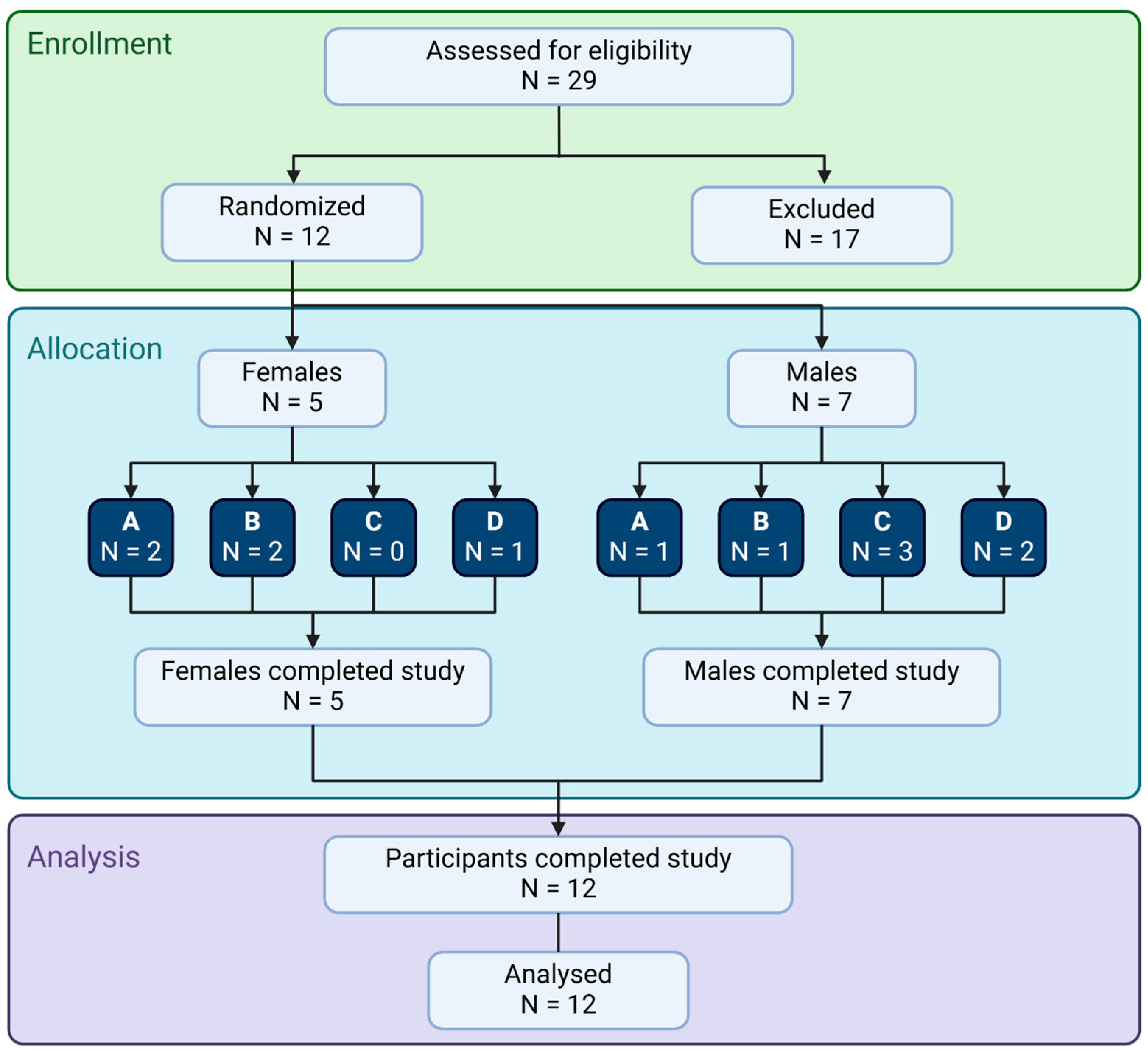
2.1. Study Design

This clinical trial was conducted using a randomized, double-blinded, cross-over study design (NCT04463030); conducted in accordance with the Declaration of Helsinki; and approved by the Argus Independent Review Board, Tucson, AZ, USA. Clinic and lab work was performed at NIS Labs in Klamath Falls, OR, USA. Individuals from the study site’s database, who had previously expressed interest in participating in clinical studies were contacted to update their health information, and those demonstrating interest were then invited for a screening process. Out of twenty-nine individuals screened, nine did not meet the inclusion/exclusion profile, and eight were not interested in participation. Twelve participants were enrolled after providing written consent and completed the study (Figure 1, Table 1).
The screening involved an interview to gather information on age, BMI, medical history, diet, lifestyle, current health, medications, and supplement use. Inclusion criteria were healthy adults aged 18–75 (inclusive), BMI between 18.0 and 34.9 (inclusive), accessible veins, and a willingness to comply with study requirements, including maintaining a consistent diet and lifestyle; bland breakfasts on clinic visit days; abstaining from exercise and supplements on the morning of clinic visits; avoiding coffee, tea, soft drinks, for at least one hour before a clinic visit; and refraining from music, candy, gum, and electronic device use during visits.
The following exclusion criteria were used: previous major gastrointestinal surgery; taking anti-inflammatory medications on a daily basis; currently in intensive athletic training (such as marathon runners); cancer during the past 12 months; chemotherapy during the past 12 months; currently treated with immune suppressant medication; diagnosed with autoimmune disorders, e.g., systemic lupus erythematosus, hemolytic anemia; donation of blood during the study or within the 4 weeks prior to study start; received a cortisone shot within the past 12 weeks; immunization during the last month; currently taking antipsychotic, hypnotic, or antidepressant prescription medication; ongoing acute infections; participation in another clinical trial involving an investigational product or lifestyle change during this study; unusual sleep routine; unwilling to maintain a constant intake of supplements over the duration of the study; anxiety about having blood drawn; pregnant, nursing, or trying to become pregnant; known food allergies related to ingredients in the active test—vitamin C and soy—as the phospholipids used for the liposome-encapsulation were derived from soy, with 50% of the phospholipids being phosphatidylcholine. Subjects who met the inclusion and exclusion criteria were informed that they qualified, scheduled for clinic visits upon providing written informed consent, and enrolled into the study.
All participants received all four test products on different clinic visits. The participants were randomized to receive the test products in different sequences of liposome-encapsulated vitamin C (LEC), a matching dose of conventional ascorbic acid (AA), phospholipids (the liposome-encapsulation material), or placebo one week apart (Figure 2). The principal investigator generated the random allocation sequence without knowing which participant would be assigned to which sequence. Clinic staff enrolled participants; the scheduling of each person determined the allocation to sequence. All clinic staff were blinded.
Participants in the study were scheduled for four clinic visits spaced one week apart. Each participant’s visits were consistently scheduled with the same arrival time on each clinic day to reduce the impact of circadian fluctuations. One visit involved consuming a placebo (45 mL rice milk) and served as control for natural variations in cytokine levels and immune surveillance for each participant, one visit involved consuming the test product liposome-encapsulated vitamin C in 45 mL rice milk, one visit involved consuming a matching dose of conventional ascorbic acid in 45 mL rice milk, and one visit involved consuming the phospholipid fraction in 45 mL rice milk to evaluate their independent effects. The test products were added to rice milk shortly before consumption, and the rice milk helped camouflage the tastes of both the ascorbic acid and the phospholipid fraction. Upon arrival, participants completed a questionnaire to monitor exceptional circumstances that may have had an effect on their health that day upon arrival at the clinic; none were identified, and no visits had to be rescheduled. After completing the questionnaire, participants were instructed to remain in the clinic for the next 6.5 h. The baseline blood sample was drawn, and, immediately after the baseline sample, a test product was provided and consumed in the presence of the clinic staff. Blood samples were drawn at 2, 4, and 6 h after consumption of the test products or placebo. After the 2 h blood draw, participants consumed a fixed number of crackers and a small amount of cheese. After the 4 h blood draw, participants were provided with a bland lunch, ensuring each participant consumed the same type of lunch at each visit.
Below is a simplified diagram illustrating the involvement of each participant, in which all study participants were tested on four different clinic days separated by a 1-week wash-out period (Figure 3).

2.2. Consumable Test Products

The three active test products were provided by the manufacturer, LivOn Labs (Henderson, NV, USA). The Lypo-Spheric®/Altrient® Vitamin C product is a nutritional supplement consisting of cold-processed liposome-encapsulated vitamin C (LEC). The liposomal material without vitamin C (phospholipids) and vitamin C without the liposomal material (ascorbic acid (AA)) were provided in parallel. All three products have distinct tastes that were camouflaged by adding them into 45 mL (1.5 oz) plain rice milk immediately prior to consumption. Therefore, plain rice milk was served as the placebo.

2.3. Blood Draws

During each blood draw, three vacutainer tubes were collected: one heparin tube, one serum separator tube, and one EDTA tube. The heparin tube was immediately placed in an ice bath, and the plasma was harvested and frozen at −80 °C until sending to a clinical diagnostic laboratory for testing of vitamin C levels. The EDTA tube was kept cold, the plasma was harvested within an hour, and the plasma was banked at −80 °C. The serum separator tube was allowed to sit at room temperature for 30–60 min, after which the tube was centrifuged, and the serum was transferred to a conical 15 mL tube, centrifuged cold, and the serum was aliquoted and frozen at −80 °C until tested in the FRAP assay, the CAP-e assay, and the 8OHdG assay for DNA/RNA oxidation.

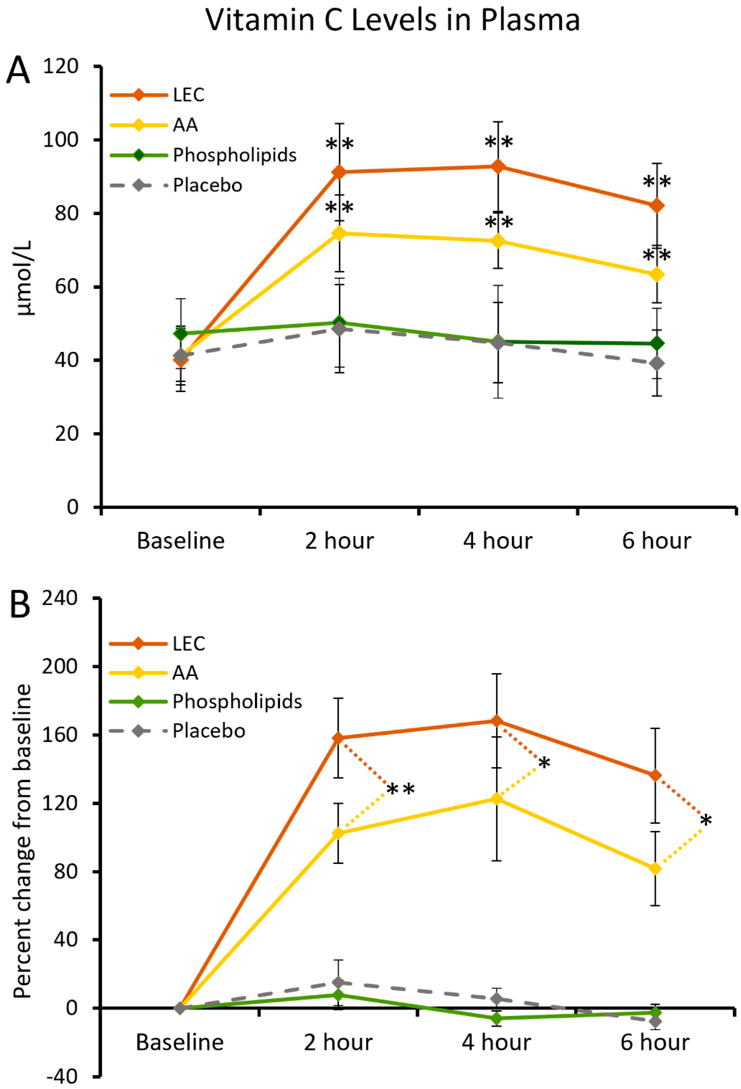
2.4. Vitamin C Levels in Plasma

Plasma samples were collected in heparinized vacutainer tubes that were immediately placed in an ice bath. The tubes were centrifuged at 4 °C immediately, transferred to transport tubes, and frozen at −80 °C. The tubes were transported to the testing lab in a frozen condition. The vitamin C levels in the blood plasma were evaluated using Quantitative High Performance Liquid Chromatography-Tandem Mass Spectrometry at a clinical diagnostic laboratory (Arup Laboratories, Salt Lake City, UT, USA).

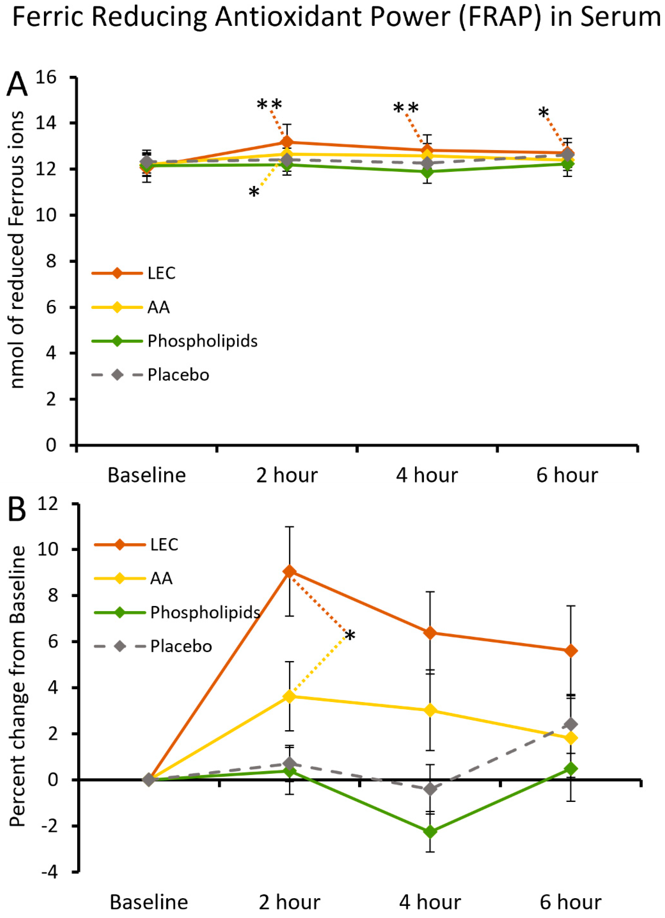
2.5. Ferric-Reducing Antioxidant Power of Serum

The ferric-reducing antioxidant power (FRAP) assay is a widely used method wherein Fe3+ is reduced to Fe2+ using antioxidants as reductants in a redox-linked colorimetric reaction [45]. The FRAP assay was performed using a commercially available kit (BioVision (K515-200)) following the manufacturer’s instructions. In brief, duplicate samples of 10 µL serum from each blood draw were mixed with 190 µL reaction mix, and the colorimetric absorbance was measured at 593 nanometer every 5 min for 1 h. Data were plotted onto a standard curve for reduced ferrous ions, and the results were provided as nanomole reduced ferrous ions.

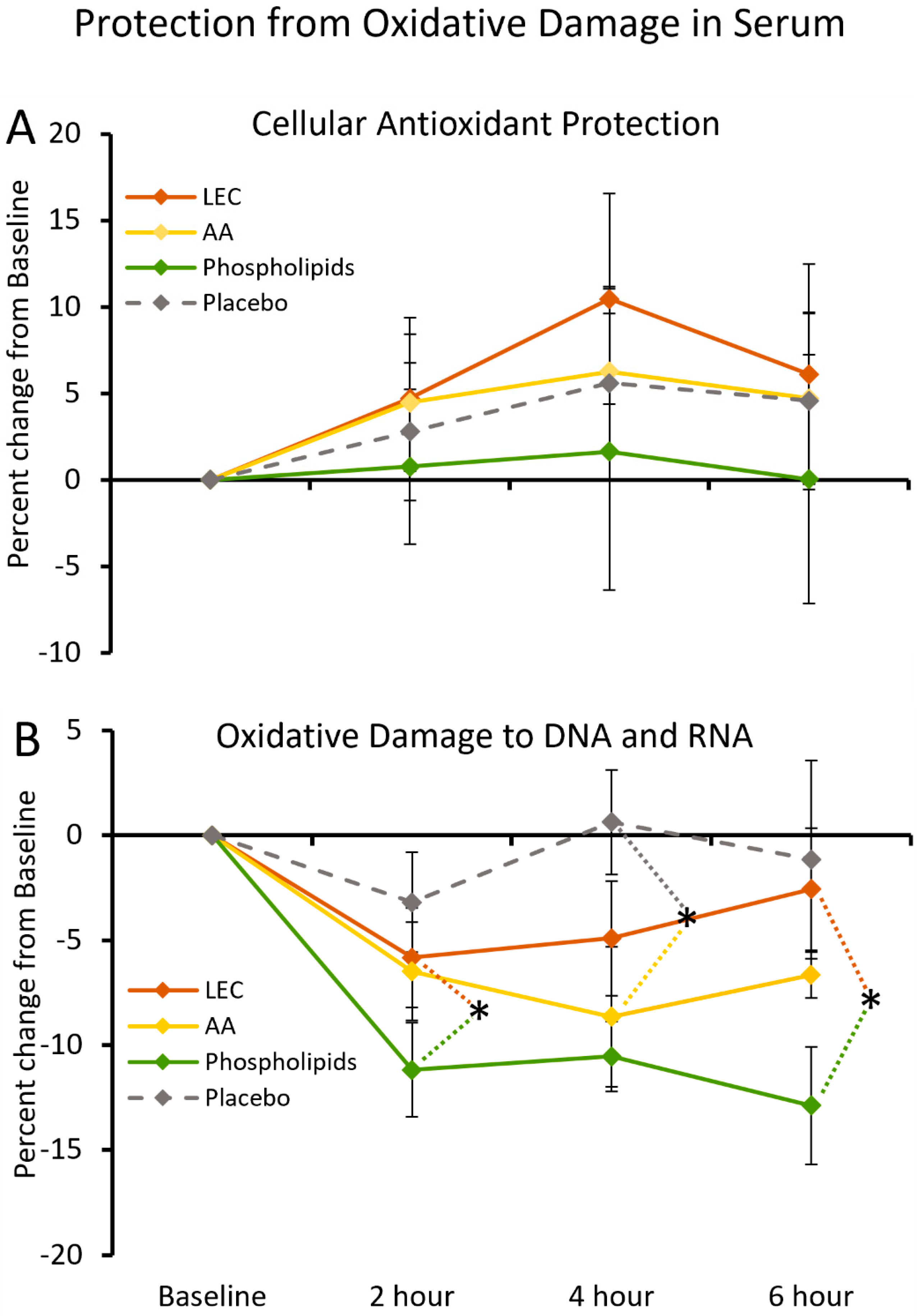
2.6. Cellular Antioxidant Protection by Serum

The Cellular Antioxidant Protection (CAP-e) assay was performed according to the method published by Honzel et al. [46], using an accelerated and more sensitive microplate-based protocol. Briefly, a 1% suspension of erythrocytes was prepared for the CAP-e bioassay by adding 0.1 mL packed red blood cells to 10 mL physiological saline (pH 7.4) and adding 100 µL erythrocyte suspension per well in V-bottom 96-well microplates. Wells that did not receive serum but that instead received the same volume of PBS served as negative controls (no induced oxidative damage). Wells that received oxidizing agent but no serum as a source of antioxidants served as positive controls (maximum oxidative damage in the absence of antioxidants). Gallic acid was used as a standard reference antioxidant compound. For assessment of the cellular antioxidant protection provided by, the serum samples, obtained before and after consuming active products versus placebo, were tested in quadruplicate. The cells were incubated for 20 min with serum to allow antioxidants in the serum samples to penetrate into the cells. After the incubation, antioxidants that were not absorbed into the erythrocyte cells were removed by two washes in PBS. A non-fluorescent precursor dye DCF-DA was added to the wells for 15 min, after which oxidative damage was induced by the addition of AAPH and incubation for 1 h. The oxidation leads to the transformation of the DCF-DA precursor dye to a fluorescent marker, where the fluorescence intensity is a measure of the level of oxidative damage. The green fluorescence intensity was recorded at 488 nm using a Tecan Spectrafluor plate reader (Durham, NC, USA). Cellular antioxidant protection was calculated as the inhibition of oxidative damage reflected by the reduced fluorescence intensity in the wells where cells were pretreated with serum, compared with the baseline (negative controls) and maximum oxidative damage (positive controls).

2.7. DNA and RNA Oxidation in Serum

Testing for levels of oxidized DNA and RNA in serum was performed using a commercially available DNA/RNA oxidative damage (high-sensitivity) ELISA kit from Cayman Chemical (Ann Arbor, MI, USA), following the manufacturer’s instructions. In brief, serum samples were thawed, had precipitates removed by a brief centrifugation, and were diluted 25-fold with assay buffer. Each serum sample was tested in triplicate by adding 250 µL of diluted serum per well on ELISA plates pre-coated with antibodies towards 8-hydroxy-2′-deoxyguanosine, 8-hydroxyguanosine from RNA, and 8-hydroxyguanine from either DNA or RNA. Monoclonal antibody and acetylcholine esterase tracer were added. After an overnight incubation, plates were washed and developed using Ellman’s Reagent. The color development was measured as the optical density at 412 nm on a PowerWave microplate reader (BioTek Instruments, Winooski, VT, USA). The color intensity is proportional to the amount of DNA/RNA oxidative damage tracer bound to the well, which is inversely correlated to the amount of free 8OHdG present in each well. Data were plotted onto a standard curve, and the results were provided as nanogram 8OHdG per mL sample (ng/mL). The changes after consuming products or placebo were tested in triplicate and plotted as the average percent change from baseline.

2.8. Levels of Cytokines, Chemokines, and Growth Factors

Serum samples from all blood draws were used for evaluation of changes to blood levels of 27 cytokines and chemokines, quantified using Bio-Plex protein arrays (Bio-Rad Laboratories Inc., Hercules, CA, USA) and utilizing xMAP technology (Luminex, Austin, TX, USA). The following markers were tested: IL-1β, IL-1ra, IL-2, IL-4, IL-5, IL-6, IL-7, IL-8, IL-9, IL-10, IL-12 (p70), IL-13, IL-15, IL-17, Eotaxin, Basic FGF, G-CSF, GM-CSF, IFN-γ, IP-10, MCP-1 (MCAF), MIP-1α, MIP-1β, PDGF-BB, RANTES, TNF-α, and VEGF. In brief, during the manufacturing process, twenty-seven types of magnetic beads (one type for each analyte) were dyed internally to produce a specific fluorescence signature for each bead type, pre-coated with capture antibodies towards analytes, and the twenty-seven types of beads were mixed, allowing for simultaneous quantification of the twenty-seven analytes. The testing of cytokine levels was performed in 96-well plates by adding magnetic beads to the serum samples and incubating for 1 h, after which the beads were washed on a magnet to remove unbound proteins. Biotinylated detection antibodies were added, and samples were incubated for 45 min, after which they were washed to remove unbound detection antibodies. Streptavidin-PE was added and incubated for 10 min, followed by washing to remove unbound streptavidin-PE. The fluorescence intensity of the beads was documented using a MagPix microplate reader, in which the mean fluorescence intensity of the analytes in each sample was calculated using the xPonent software (Version 4.2, Luminex, Austin, TX, USA).

2.9. Statistical Analysis

The average and standard deviation was calculated for each data set using Microsoft Excel (Microsoft Corporation, Redmond, WA, USA). The differences between two means were evaluated for each timepoint. This allowed evaluation of changes on the day a person consumed the active product, in the context of the person’s changes on the day they consumed another test product or placebo, benefiting from the high statistical power of a cross-over trial [47]. The evaluation used within-subject analysis [47] and the two-tailed paired t-test, [48], where statistical significance was set at p < 0.05 and a high level of significance at p < 0.01.

3. Results

3.1. Serum Vitamin C Levels

Consumption of liposome-encapsulated vitamin C (LEC) led to a significant increase in blood plasma levels of vitamin C at 2 h (Figure 4) with a high level of significance (p < 0.01, Figure 4A). This significant increase was sustained at the 4 h and 6 h timepoints. Consumption of ascorbic acid (AA) also resulted in a significant increase in plasma vitamin C levels, at the 2, 4, and 6 h timepoints (p < 0.01, Figure 4A). The increase seen after consuming LEC was more robust, with the increased plasma vitamin C levels at 2 h being highly significant when compared to those observed after AA ingestion at the 2 h timepoint (p < 0.01, Figure 4B) and significant at the 4 h and 6 h timepoints (p < 0.05, Figure 4B). Neither consumption of placebo nor the phospholipid fraction resulted in increased plasma vitamin C levels. Neither consumption of LEC nor AA resulted in elevated levels at baseline for the subsequent clinic visit one week later.

3.2. Ferric-Reducing Antioxidant Power of Serum

The ferric-reducing antioxidant power (FRAP) showed rapid increases after consumption of both LEC and AA (Figure 5). The antioxidant power was higher after consuming LEC; this increase was highly significant at the 2 and 4 h timepoints (p < 0.01, Figure 5A) and significant at the 6 h timepoint (p < 0.05, Figure 5A) when compared to baseline. Consumption of AA also significantly increased the antioxidant capacity but only at the 2 h timepoint (p < 0.05, Figure 5B). At 2 h, the higher antioxidant power seen after consuming LEC was significantly higher than after consuming AA. Neither consumption of phospholipids nor placebo were associated with changes to the serum antioxidant power in the FRAP assay.

3.3. Antioxidant Protection of Cells and Nucleic Acids

Two assays were performed to address functional antioxidant protection by serum samples (Figure 6).
For evaluation of the capacity of serum samples to provide cellular protection, an assay for cellular antioxidant protection using erythrocytes (CAP-e assay) was used (Figure 6A). Serum collected after consuming LEC showed increased cellular antioxidant protection compared to baseline, peaking at 4 h (Figure 6A), verifying that the liposome-encapsulated vitamin C was at least as capable as regular ascorbic acid to enter into cells. A mild increase was seen after consuming both AA and placebo, whereas no changes were detected after consuming phospholipids. The changes in cellular antioxidant protection after consuming LEC or AA did not reach statistical significance but still suggest a clinically meaningful cellular protection.
For evaluation of the capacity of serum to provide protection against oxidative damage to nucleic acids, the levels of oxidatively damaged DNA and RNA in blood serum were measured using the 8-OHdG oxidation marker, quantifying levels of 8-hydroxy-2′-deoxyguanosine from degraded DNA, 8-hydroxyguanosine from degraded RNA, and 8-hydroxyguanine from either DNA or RNA. Oxidative damage to DNA/RNA was reduced following the consumption of phospholipids compared to LEC (Figure 6B), reaching statistical significance at the 2 h and 6 h timepoints (p < 0.05). There was a statistically significant, but less robust, decrease in oxidative damage to DNA/RNA following the consumption of AA at the 4 h timepoint. The consumption of placebo was not associated with any significant changes to oxidative damage to DNA/RNA. The reduction in blood levels of oxidatively damaged DNA/RNA was most pronounced after consuming phospholipids. The differences between phospholipids when compared to AA did not reach statistical significance.

3.4. Modulation of Cytokine Levels

Several changes to serum cytokines were observed (Figure 7). The heatmap in Figure 7A showed rapid anti-inflammatory effects at 2 h after consuming LEC, with reduced levels of multiple chemokines MCP-1, MIP-1α, MIP-1β, and RANTES. This was followed by a different wave of immune-modulating effects involving increased levels of a different group of cytokines at 6 h, including IFN-γ, IL-1β, IL-5, IL-6, IL-12 (p70), IL-13, and IL-17A. Consuming the phospholipids triggered rapid reductions for almost all cytokines at 2 h; only a few reached significance when compared to baseline, but some reached significance when compared to other test products (Figure 7B). After consuming LEC, IFN-γ levels increased by 6% at the 6 h timepoint, in contrast to a decrease after consuming phospholipids (Figure 7B). The difference in serum IFN-γ levels after consuming LEC versus phospholipids was statistically significant (p < 0.05, Figure 7B). The difference in serum IFN-γ levels after consuming LEC versus AA reached a statistical trend (p < 0.01, Figure 7B). Rapidly reduced levels of multiple inflammatory cytokines were seen at 2 h after consumption of phospholipids (Figure 7A). The reduced level of IP-10 after consuming phospholipids was statistically significant when compared to AA (p < 0.05) (Figure 7B). After consuming LEC, there were reduced levels of all five pro-inflammatory cytokines, where the reduced levels of IL-6 were significantly lower than after consuming AA, and the levels of MIP-1α reached a statistical trend when compared to AA (Figure 7B). In contrast, after consuming AA, there was an increase in the serum levels of IP-10 and TNF-α. There were no changes to the serum level of IL-6 or MIP-1α after consuming AA. The only cytokine that showed reduced levels after consuming AA was MCP-1, in which the magnitude of the reduction was similar to that seen in both LEC and phospholipids.

4. Discussion

Vitamin C is a water-soluble nutrient, abundant in fruits, vegetables, and organ meat. Vitamin C acts as an epigenetic regulator [49], where DNA and histone methylation regulate gene expression [50,51,52], and exerts specific epigenetic influences on various components of the immune system via the activation of kinases [53], which enhances immunogenic activity [54]. In addition, its involvement in gene regulation plays pivotal roles in stem cell biology and the protection of stemness [55] (Table 2).
Despite its prevalence in our diet, the absorption of vitamin C across biological membranes is restricted by its dependence on specific transport mechanisms for crossing the gut epithelial barrier, uptake into the blood circulation, and entering living cells in the body’s tissue. The clinical results presented here document the differences between conventional ascorbic acid (AA) and liposome-encapsulated vitamin C (LEC), showing an advantage of the Lypo-Spheric® technology in enhancing vitamin C bioavailability. This advanced encapsulation technique supports a more efficient uptake of vitamin C, and further evaluation is warranted for the delivery of other water-soluble nutrients. The elevated blood plasma levels of vitamin C at 2 h after consuming LEC were highly significant when compared to the levels at 2 h after consuming AA; the increase persisted over the 6 h duration of the study, indicating greater bioavailability and retention.
The increased blood levels of vitamin C after consuming LEC translated into increased antioxidant protection. The increase in serum antioxidant capacity, as measured using the ferric-reducing antioxidant power (FRAP) assay, was also rapid, increasing within 2 h after consumption and persisting throughout the 6 h duration of the study. While the increase in serum antioxidant capacity after consuming AA was also rapid, it did not reach the same magnitude and did not persist as long as that of LEC. Since the FRAP assay measures the total antioxidant power in serum samples, it is possible that the FRAP results were not only influenced by increased serum vitamin C levels but could also be influenced by several other metabolites affected by vitamin C and a contributing effect from the phospholipid material reducing free radical levels.
The increased uptake of vitamin C seen with liposomal delivery also translated to two functional observations pertaining to protection of cells and nucleic acids from oxidative stress. Cellular antioxidant protection, as measured by the CAP-e bioassay, showed that serum from participants after consumption of LEC was able to provide stronger cellular antioxidant protection ex vivo than serum from the same participants after they consumed AA. The increased cellular protection ex vivo after consuming LEC peaked at 4 h but did not reach significance when compared to AA. This was expected due to the highly dynamic and complex pharmacokinetics of vitamin C. The presence of vitamin C in the blood stream is transient, as it disappears from the blood when absorbed by erythrocytes and tissue cells, with the brain maintaining the highest levels of vitamin C [20].
The phosphatidylcholine-rich phospholipid fraction, used for the liposome encapsulation in LEC, presented beneficial effects on its own. After consumption of the phospholipid fraction, blood serum showed reduced levels of oxidatively damaged DNA and RNA, indicating that the phospholipids contributed to protection from free radical damage in ways that seemed independent of a direct antioxidant capacity, and which may be associated with other, more complex, protective properties, possibly related to cell membrane fluidity and regulation of cellular signaling (Table 3) [21,22]. Consumption of the phospholipid fraction alone demonstrated immune-modulating effects, including a rapid decrease in multiple inflammatory cytokines at 2 h, which was most robust for IL-6, MCP-1, and MIP-1α. Therefore, this liposomal encapsulation material, rich in phosphatidylcholine, serves as a valuable nutraceutical support in itself. This observation may be specific to the Lypo-Spheric® technology and should not be extended to other types of liposomal encapsulation materials without similar clinical documentation.
Consumption of LEC triggered a gradual increase in IFN-γ, known for its critical role in orchestrating innate and adaptive immunity, increasing natural killer-cell activity, and inhibiting viral replication in infected target cells [56,57]. This effect was only seen for LEC and not for either of its components, AA or phospholipids, and suggests a synergistic effect of the two ingredients. Multiple other immune-activating cytokines were increased at 6 h after consumption of LEC but did not reach statistical significance when compared to baseline. These immune-activating properties of LEC differed from much milder and insignificant changes after consuming AA. This may be due, in part, to more efficient cellular delivery of vitamin C and, in part, to the stress-reducing properties of phosphatidylcholine [58]. Oxidative stress is an inherent part of metabolic activities, and, over time, aging cells lose some of their ability to maintain an appropriate redox balance, leading to more oxidative damage, increasing the risk of developing oxidative damage-related health conditions [59]. Physical and psychological stress levels [60,61], and environmental factors such as UV radiation [62] and pollution [63,64,65], can exacerbate these issues and put more pressure on endogenous antioxidant systems, which could create a need for more effective exogenous antioxidants. Phosphatidylcholine has been shown to support our intracellular antioxidant protective systems, including glutathione, glutathione peroxidase, and superoxide dismutase activity within the nervous system. The rapid benefits documented here are intriguing in the context of preparedness for pathogenic challenges across all age ranges, as well as general health and wellness in an aging population. Further research is needed and should include long-term studies across a broad age range with evaluation of physical, mental, and cognitive health.

5. Conclusions

The results reported here documented that vitamin C delivered in a liposome-encapsulated form (LEC) was absorbed better and quicker than pure ascorbic acid (AA), which translated into enhanced antioxidant protection and capacity. The phospholipid fraction supported modulation by reducing pro-inflammatory cytokines in serum. With these synergistic antioxidant and anti-inflammatory effects, we suggest that LEC can be an effective antioxidant and immune-modulating nutraceutical. Further work is warranted to explore the long-term epigenetic and anti-inflammatory effects of LEC consumption in the context of various stressors, both at the cellular level and in clinical trials.

Author Contributions

Conceptualization, G.S.J.; methodology, G.S.J.; formal analysis, S.V.M. and I.I.; data curation, G.S.J.; writing—original draft preparation, S.V.M., D.C., I.I. and G.S.J.; writing—review and editing, S.V.M., D.C., I.I. and G.S.J.; visualization, S.V.M., D.C., I.I. and G.S.J.; project administration, G.S.J. All authors have read and agreed to the published version of the manuscript.

Funding

The study was sponsored by LivOn Laboratories, a producer of commercially available liposome-encapsulated nutrients.

Institutional Review Board Statement

The study was conducted in accordance with the Declaration of Helsinki and approved by the Argus Institutional Review Board (protocol code 161-005).

Informed Consent Statement

Informed consent was obtained from all subjects involved in the study.

Data Availability Statement

The data presented in this study are available upon reasonable request from the corresponding author.

Conflicts of Interest

The authors declare no conflicts of interest. The funder was not involved in the study design, collection, analysis, interpretation of data, or the writing of this article.

References

  1. Drouin, G.; Godin, J.R.; Pagé, B. The genetics of vitamin C loss in vertebrates. Curr. Genom. 2011, 12, 371–378. [Google Scholar] [CrossRef] [PubMed]
  2. Njus, D.; Kelley, P.M.; Tu, Y.J.; Schlegel, H.B. Ascorbic acid: The chemistry underlying its antioxidant properties. Free Radic. Biol. Med. 2020, 159, 37–43. [Google Scholar] [CrossRef] [PubMed]
  3. Pan, Y.; Mansfield, K.D.; Bertozzi, C.C.; Rudenko, V.; Chan, D.A.; Giaccia, A.J.; Simon, M.C. Multiple factors affecting cellular redox status and energy metabolism modulate hypoxia-inducible factor prolyl hydroxylase activity in vivo and in vitro. Mol. Cell. Biol. 2007, 27, 912–925. [Google Scholar] [CrossRef] [PubMed]
  4. Covarrubias-Pinto, A.; Acuña, A.I.; Beltrán, F.A.; Torres-Díaz, L.; Castro, M.A. Old Things New View: Ascorbic Acid Protects the Brain in Neurodegenerative Disorders. Int. J. Mol. Sci. 2015, 16, 28194–28217. [Google Scholar] [CrossRef]
  5. May, J.M. Vitamin C transport and its role in the central nervous system. In Water Soluble Vitamins; Subcellular Biochemistry; Springer: Dordrecht, The Netherlands, 2012; Volume 56, pp. 85–103. [Google Scholar]
  6. Zhang, X.Y.; Xu, Z.P.; Wang, W.; Cao, J.B.; Fu, Q.; Zhao, W.X.; Li, Y.; Huo, X.L.; Zhang, L.M.; Li, Y.F.; et al. Vitamin C alleviates LPS-induced cognitive impairment in mice by suppressing neuroinflammation and oxidative stress. Int. Immunopharmacol. 2018, 65, 438–447. [Google Scholar] [CrossRef]
  7. Lin, J.L.; Huang, Y.H.; Shen, Y.C.; Huang, H.C.; Liu, P.H. Ascorbic acid prevents blood-brain barrier disruption and sensory deficit caused by sustained compression of primary somatosensory cortex. J. Cereb. Blood Flow Metab. 2010, 30, 1121–1136. [Google Scholar] [CrossRef]
  8. Maekawa, T.; Uchida, T.; Nakata-Horiuchi, Y.; Kobayashi, H.; Kawauchi, S.; Kinoshita, M.; Saitoh, D.; Sato, S. Oral ascorbic acid 2-glucoside prevents coordination disorder induced via laser-induced shock waves in rat brain. PLoS ONE 2020, 15, e0230774. [Google Scholar] [CrossRef]
  9. Khalili, H.; Abdollahifard, S.; Niakan, A.; Aryaie, M. The effect of Vitamins C and E on clinical outcomes of patients with severe traumatic brain injury: A propensity score matching study. Surg. Neurol. Int. 2022, 13, 548. [Google Scholar] [CrossRef]
  10. Li, Y.R.; Zhu, H. Vitamin C for sepsis intervention: From redox biochemistry to clinical medicine. Mol. Cell. Biochem. 2021, 476, 4449–4460. [Google Scholar] [CrossRef]
  11. Traber, M.G.; Stevens, J.F. Vitamins C and E: Beneficial effects from a mechanistic perspective. Free Radic. Biol. Med. 2011, 51, 1000–1013. [Google Scholar] [CrossRef]
  12. Boo, Y.C. Ascorbic Acid (Vitamin C) as a Cosmeceutical to Increase Dermal Collagen for Skin Antiaging Purposes: Emerging Combination Therapies. Antioxidants 2022, 11, 1663. [Google Scholar] [CrossRef] [PubMed]
  13. Chen, J.; Lan, J.; Liu, D.; Backman, L.J.; Zhang, W.; Zhou, Q.; Danielson, P. Ascorbic Acid Promotes the Stemness of Corneal Epithelial Stem/Progenitor Cells and Accelerates Epithelial Wound Healing in the Cornea. Stem Cells Transl. Med. 2017, 6, 1356–1365. [Google Scholar] [CrossRef] [PubMed]
  14. Cha, J.; Roomi, M.W.; Ivanov, V.; Kalinovsky, T.; Niedzwiecki, A.; Rath, M. Ascorbate supplementation inhibits growth and metastasis of B16FO melanoma and 4T1 breast cancer cells in vitamin C-deficient mice. Int. J. Oncol. 2013, 42, 55–64. [Google Scholar] [CrossRef] [PubMed]
  15. Maekawa, T.; Miyake, T.; Tani, M.; Uemoto, S. Diverse antitumor effects of ascorbic acid on cancer cells and the tumor microenvironment. Front. Oncol. 2022, 12, 981547. [Google Scholar] [CrossRef]
  16. Levine, M.; Conry-Cantilena, C.; Wang, Y.; Welch, R.W.; Washko, P.W.; Dhariwal, K.R.; Park, J.B.; Lazarev, A.; Graumlich, J.F.; King, J.; et al. Vitamin C pharmacokinetics in healthy volunteers: Evidence for a recommended dietary allowance. Proc. Natl. Acad. Sci. USA 1996, 93, 3704–3709. [Google Scholar] [CrossRef]
  17. Doseděl, M.; Jirkovský, E.; Macáková, K.; Krčmová, L.K.; Javorská, L.; Pourová, J.; Mercolini, L.; Remião, F.; Nováková, L.; Mladěnka, P.; et al. Vitamin C-Sources, Physiological Role, Kinetics, Deficiency, Use, Toxicity, and Determination. Nutrients 2021, 13, 615. [Google Scholar] [CrossRef]
  18. Bürzle, M.; Hediger, M.A. Functional and physiological role of vitamin C transporters. Curr. Top. Membr. 2012, 70, 357–375. [Google Scholar]
  19. Rivas, C.I.; Zúñiga, F.A.; Salas-Burgos, A.; Mardones, L.; Ormazabal, V.; Vera, J.C. Vitamin C transporters. J. Physiol. Biochem. 2008, 64, 357–375. [Google Scholar] [CrossRef]
  20. Lykkesfeldt, J.; Tveden-Nyborg, P. The Pharmacokinetics of Vitamin C. Nutrients 2019, 11, 2412. [Google Scholar] [CrossRef]
  21. Sunshine, H.; Iruela-Arispe, M.L. Membrane lipids and cell signaling. Curr. Opin. Lipidol. 2017, 28, 408–413. [Google Scholar] [CrossRef]
  22. Varshney, P.; Yadav, V.; Saini, N. Lipid rafts in immune signalling: Current progress and future perspective. Immunology 2016, 149, 13–24. [Google Scholar] [CrossRef] [PubMed]
  23. Fajardo, V.A.; McMeekin, L.; LeBlanc, P.J. Influence of phospholipid species on membrane fluidity: A meta-analysis for a novel phospholipid fluidity index. J. Membr. Biol. 2011, 244, 97–103. [Google Scholar] [CrossRef] [PubMed]
  24. Treede, I.; Braun, A.; Sparla, R.; Kühnel, M.; Giese, T.; Turner, J.R.; Anes, E.; Kulaksiz, H.; Füllekrug, J.; Stremmel, W.; et al. Anti-inflammatory effects of phosphatidylcholine. J. Biol. Chem. 2007, 282, 27155–27164. [Google Scholar] [CrossRef] [PubMed]
  25. van der Veen, J.N.; Kennelly, J.P.; Wan, S.; Vance, J.E.; Vance, D.E.; Jacobs, R.L. The critical role of phosphatidylcholine and phosphatidylethanolamine metabolism in health anddisease. Biochim. Biophys. Acta Biomembr. 2017, 1859 Pt B, 1558–1572. [Google Scholar] [CrossRef]
  26. Ruskamo, S.; Raasakka, A.; Pedersen, J.S.; Martel, A.; Škubník, K.; Darwish, T.; Porcar, L.; Kursula, P. Human myelin proteolipid protein structure and lipid bilayer stacking. Cell. Mol. Life Sci. 2022, 79, 419. [Google Scholar] [CrossRef] [PubMed]
  27. Kister, A.; Kister, I. Overview of myelin, major myelin lipids, and myelin-associated proteins. Front. Chem. 2023, 10, 1041961. [Google Scholar] [CrossRef]
  28. O’Donnell, V.B.; Rossjohn, J.; Wakelam, M.J. Phospholipid signaling in innate immune cells. J. Clin. Investig. 2018, 128, 2670–2679. [Google Scholar] [CrossRef]
  29. Lagace, T.A. Phosphatidylcholine: Greasing the Cholesterol Transport Machinery. Lipid Insights 2016, 8 (Suppl. S1), 65–73. [Google Scholar] [CrossRef]
  30. Kim, M.; Nevado-Holgado, A.; Whiley, L.; Snowden, S.G.; Soininen, H.; Kloszewska, I.; Mecocci, P.; Tsolaki, M.; Vellas, B.; Thambisetty, M.; et al. Association between Plasma Ceramides and Phosphatidylcholines and Hippocampal Brain Volume in Late Onset Alzheimer’s Disease. J. Alzheimers Dis. 2017, 60, 809–817. [Google Scholar] [CrossRef]
  31. Marcucci, H.; Paoletti, L.; Jackowski, S.; Banchio, C. Phosphatidylcholine biosynthesis during neuronal differentiation and its role in cell fate determination. J. Biol. Chem. 2010, 285, 25382–25393. [Google Scholar] [CrossRef]
  32. Gurunathan, S.; Kang, M.H.; Kim, J.H. A Comprehensive Review on Factors Influences Biogenesis, Functions, Therapeutic and Clinical Implications of Exosomes. Int. J. Nanomed. 2021, 16, 1281–1312. [Google Scholar] [CrossRef] [PubMed]
  33. Sheikholeslami, B.; Lam, N.W.; Dua, K.; Haghi, M. Exploring the impact of physicochemical properties of liposomal formulations on their in vivo fate. Life Sci. 2022, 300, 120574. [Google Scholar] [CrossRef] [PubMed]
  34. Shade, C.W. Liposomes as Advanced Delivery Systems for Nutraceuticals. Integr. Med. 2016, 15, 33–36. [Google Scholar]
  35. Akbarzadeh, A.; Rezaei-Sadabady, R.; Davaran, S.; Joo, S.W.; Zarghami, N.; Hanifehpour, Y.; Samiei, M.; Kouhi, M.; Nejati-Koshki, K. Liposome: Classification, preparation, and applications. Nanoscale Res. Lett. 2013, 8, 102. [Google Scholar] [CrossRef]
  36. Wang-Gillam, A.; Hubner, R.A.; Siveke, J.T.; Von Hoff, D.D.; Belanger, B.; de Jong, F.A.; Mirakhur, B.; Chen, L.T. NAPOLI-1 phase 3 study of liposomal irinotecan in metastatic pancreatic cancer: Final overall survival analysis and characteristics of long-term survivors. Eur. J. Cancer 2019, 108, 78–87. [Google Scholar] [CrossRef]
  37. Nightingale, S.D.; Saletan, S.L.; Swenson, C.E.; Lawrence, A.J.; Watson, D.A.; Pilkiewicz, F.G.; Silverman, E.G.; Cal, S.X. Liposome-encapsulated gentamicin treatment of Mycobacterium avium-Mycobacterium intracellulare complex bacteremia in AIDS patients. Antimicrob. Agents Chemother. 1993, 37, 1869–1872. [Google Scholar] [CrossRef]
  38. Calvagno, M.G.; Celia, C.; Paolino, D.; Cosco, D.; Iannone, M.; Castelli, F.; Doldo, P.; Frest, M. Effects of lipid composition and preparation conditions on physical-chemical properties, technological parameters and in vitro biological activity of gemcitabine-loaded liposomes. Curr. Drug Deliv. 2007, 4, 89–101. [Google Scholar] [CrossRef]
  39. Nsairat, H.; Khater, D.; Sayed, U.; Odeh, F.; Al Bawab, A.; Alshaer, W. Liposomes: Structure, composition, types, and clinical applications. Heliyon 2022, 8, e09394. [Google Scholar] [CrossRef]
  40. Sinha, R.; Sinha, I.; Calcagnotto, A.; Trushin, N.; Haley, J.S.; Schell, T.D.; Richie, J.P., Jr. Oral supplementation with liposomal glutathione elevates body stores of glutathione and markers of immune function. Eur. J. Clin. Nutr. 2018, 72, 105–111. [Google Scholar] [CrossRef]
  41. Flory, S.; Sus, N.; Haas, K.; Jehle, S.; Kienhöfer, E.; Waehler, R.; Adler, G.; Venturelli, S.; Frank, J. Increasing Post-Digestive Solubility of Curcumin Is the Most Successful Strategy to Improve its Oral Bioavailability: A Randomized Cross-Over Trial in Healthy Adults and In Vitro Bioaccessibility Experiments. Mol. Nutr. Food Res. 2021, 65, e2100613. [Google Scholar] [CrossRef]
  42. Jenski, L.J.; Zerouga, M.; Stillwell, W. Omega-3 fatty acid-containing liposomes in cancer therapy. Proc. Soc. Exp. Biol. Med. 1995, 210, 227–233. [Google Scholar] [CrossRef] [PubMed]
  43. Drapeau, C.; Benson, K.F.; Jensen, G.S. Rapid and selective mobilization of specific stem cell types after consumption of a polyphenol-rich extract from sea buckthorn berries (Hippophae) in healthy human subjects. Clin. Interv. Aging 2019, 4, 253–263. [Google Scholar] [CrossRef] [PubMed]
  44. Yu, L.; McGarry, S.; Cruickshank, D.; Jensen, G.S. Rapid increase in immune surveillance and expression of NKT and γδT cell activation markers after consuming a nutraceutical supplement containing Aloe vera gel, extracts of Poria cocos and rosemary. A randomized placebo-controlled cross-over trial. PLoS ONE 2023, 18, e0291254. [Google Scholar] [CrossRef] [PubMed]
  45. Benzie, I.F.; Strain, J.J. The ferric reducing ability of plasma (FRAP) as a measure of “antioxidant power”: The FRAP assay. Anal. Biochem. 1996, 239, 70–76. [Google Scholar] [CrossRef] [PubMed]
  46. Honzel, D.; Carter, S.G.; Redman, K.A.; Schauss, A.G.; Endres, J.R.; Jensen, G.S. Comparison of chemical and cell-based antioxidant methods for evaluation of foods and natural products: Generating multifaceted data by parallel testing using erythrocytes and polymorphonuclear cells. J. Agric. Food Chem. 2008, 56, 8319–8325. [Google Scholar] [CrossRef]
  47. Lim, C.Y.; In, J. Considerations for crossover design in clinical study. Korean J. Anesthesiol. 2021, 74, 293–299. [Google Scholar] [CrossRef]
  48. Genser, B.; Cooper, P.J.; Yazdanbakhsh, M.; Barreto, M.L.; Rodrigues, L.C. A guide to modern statistical analysis of immunological data. BMC Immunol. 2007, 8, 27. [Google Scholar] [CrossRef]
  49. Kietzmann, T. Vitamin C: From nutrition to oxygen sensing and epigenetics. Redox Biol. 2023, 63, 102753. [Google Scholar] [CrossRef]
  50. Liu, X.; Khan, A.; Li, H.; Wang, S.; Chen, X.; Huang, H. Ascorbic Acid in Epigenetic Reprogramming. Curr. Stem Cell Res. Ther. 2022, 17, 13–25. [Google Scholar] [CrossRef]
  51. Camarena, V.; Wang, G. The epigenetic role of vitamin C in health and disease. Cell. Mol. Life Sci. 2016, 73, 1645–1658. [Google Scholar] [CrossRef]
  52. Brabson, J.P.; Leesang, T.; Mohammad, S.; Cimmino, L. Epigenetic Regulation of Genomic Stability by Vitamin C. Front. Genet. 2021, 12, 675780. [Google Scholar] [CrossRef] [PubMed]
  53. Bowie, A.G.; O’Neill, L.A. Vitamin C inhibits NF-kappa B activation by TNF via the activation of p38 mitogen-activated protein kinase. J. Immunol. 2000, 165, 7180–7188. [Google Scholar] [CrossRef] [PubMed]
  54. Morante-Palacios, O.; Godoy-Tena, G.; Calafell-Segura, J.; Ciudad, L.; Martínez-Cáceres, E.M.; Sardina, J.L.; Ballestar, E. Vitamin C enhances NF-κB-driven epigenomic reprogramming and boosts the immunogenic properties of dendritic cells. Nucleic Acids Res. 2022, 50, 10981–10994. [Google Scholar] [CrossRef] [PubMed]
  55. Esteban, M.A.; Wang, T.; Qin, B.; Yang, J.; Qin, D.; Cai, J.; Li, W.; Weng, Z.; Chen, J.; Ni, S.; et al. Vitamin C enhances the generation of mouse and human induced pluripotent stem cells. Cell Stem Cell 2010, 6, 71–79. [Google Scholar] [CrossRef]
  56. Saha, B.; Jyothi Prasanna, S.; Chandrasekar, B.; Nandi, D. Gene modulation and immunoregulatory roles of interferon gamma. Cytokine 2010, 50, 1–14. [Google Scholar] [CrossRef]
  57. Schoenborn, J.R.; Wilson, C.B. Regulation of interferon-gamma during innate and adaptive immune responses. Adv. Immunol. 2007, 96, 41–101. [Google Scholar]
  58. Kim, S.T.; Kyung, E.J.; Suh, J.S.; Lee, H.S.; Lee, J.H.; Chae, S.I.; Park, E.S.; Chung, Y.H.; Bae, J.; Lee, T.J.; et al. Phosphatidylcholine attenuated docetaxel-induced peripheral neurotoxicity in rats. Drug Chem. Toxicol. 2018, 41, 476–485. [Google Scholar] [CrossRef]
  59. Halliwell, B. Understanding mechanisms of antioxidant action in health and disease. Nat. Rev. Mol. Cell. Biol. 2024, 25, 13–33. [Google Scholar] [CrossRef]
  60. Liu, Y.Z.; Wang, Y.X.; Jiang, C.L. Inflammation: The Common Pathway of Stress-Related Diseases. Front. Hum. Neurosci. 2017, 11, 316. [Google Scholar] [CrossRef]
  61. Frost, D.M.; Meyer, I.H. Minority stress theory: Application, critique, and continued relevance. Curr. Opin. Psychol. 2023, 51, 101579. [Google Scholar] [CrossRef]
  62. Tyrrell, R.M. Ultraviolet radiation and free radical damage to skin. Biochem. Soc. Symp. 1995, 61, 47–53. [Google Scholar] [PubMed]
  63. Pan, B.; Li, H.; Lang, D.; Xing, B. Environmentally persistent free radicals: Occurrence, formation mechanisms and implications. Environ. Pollut. 2019, 248, 320–331. [Google Scholar] [CrossRef] [PubMed]
  64. Huang, X.; Zalma, R.; Pezerat, H. Chemical reactivity of the carbon-centered free radicals and ferrous iron in coals: Role of bioavailable Fe2+ in coal workers pneumoconiosis. Free Radic. Res. 1999, 30, 439–451. [Google Scholar] [CrossRef] [PubMed]
  65. Pohjoismäki, J.L.O.; Goffart, S. Adaptive and Pathological Outcomes of Radiation Stress-Induced Redox Signaling. Antioxid. Redox Signal. 2022, 37, 336–348. [Google Scholar] [CrossRef] [PubMed]
Figure 1. CONSORT flow chart for study. Allocation of the sequence in which a participant consumed the 4 test products (A–D) are described in Figure 2.
Nutraceuticals 04 00034 g001
Figure 2. The randomization to Group A, B, C, and D dictated the allocation to a sequence of consumable test products. The trial involved three groups of four participants, and the goal for the sequence was to ensure that on each clinic visit four different test products were consumed by the four participants attending that clinic day. The participants were randomized to receive either liposome-encapsulated vitamin C, a matching dose of conventional ascorbic acid, phospholipids (the liposome-encapsulation material), or placebo one week apart. The allocation is also shown in Figure 1 (CONSORT flow chart). LEC: liposome-encapsulated vitamin C in rice milk (1 g ascorbic acid, 1 g phospholipids). AA: ascorbic acid in rice milk (1 g ascorbic acid). Phospholipids: the liposomal encapsulation material, matched to the dose in LEC (1 g phospholipids). Placebo: rice milk, used for the oral delivery of all test products to camouflage taste, color and consistency, was also the choice for placebo (45 mL plain rice milk).
Nutraceuticals 04 00034 g002
Figure 3. Diagram showing the involvement of each participant. Study participants were tested on 4 different clinic days. The liposome-encapsulated vitamin C is made with bioactive phospholipids. A matching dose of phospholipids (without vitamin C) was tested on 1 of the 4 clinic days. All test products were served to study participants in 45 mL (1.5 fluid oz) of rice milk. Therefore, the placebo control involved a similar serving of rice milk alone. The sequence of test products shown here is an example only since the sequence was randomized.
Nutraceuticals 04 00034 g003
Figure 4. Vitamin C levels in plasma. Data are shown as the averages ± standard error of the mean at baseline, 2 h, 4 h, and 6 h after consuming either liposome-encapsulated vitamin C (LEC), ascorbic acid (AA), the phospholipids used for the liposomal material, or placebo. Levels of statistical significance when compared to baseline levels (A) or to another test product at the same timepoint (B) are shown on the graphs, where p < 0.05: * and p < 0.01: **. (A) Vitamin C levels in micromole/liter (µM/L) blood plasma: Vitamin C levels increased after consuming both LEC and AA. The increased levels were highly significant following the consumption of both LEC and AA at all timepoints, compared to baseline. In contrast, the plasma levels of vitamin C did not change after consuming phospholipids or placebo. (B) The percent change from baseline for plasma vitamin C levels. The increase in blood plasma levels of vitamin C was higher after consuming LEC compared to AA. The higher level of plasma vitamin C after consuming LEC was highly significant at 2 h when compared to AA and statistically significant when compared to AA at both 4 and 6 h.
Nutraceuticals 04 00034 g004
Figure 5. Serum antioxidant capacity measured by the ferric-reducing antioxidant power (FRAP) assay. Data are shown as the averages ± standard error of the mean at baseline, 2 h, 4 h, and 6 h after consuming either liposome-encapsulated vitamin C (LEC), ascorbic acid (AA), the phospholipids used for the liposomal material, or placebo. Levels of statistical significance when compared to baseline levels (A) or to another test product at the same timepoint (B) are shown on the graphs, where p < 0.05: * and p < 0.01: **. (A) Antioxidant capacity in nanomole of reduced Ferrous (Fe2+) ions. Antioxidant capacity increased after consuming both LEC and AA. The increased levels were highly significant following the consumption of LEC at all timepoints compared to baseline and significant following the consumption of AA at 2 h compared to baseline. Consumption of placebo and phospholipids did not change antioxidant capacity. (B) The percent change from baseline for antioxidant capacity. The increase in antioxidant capacity was higher after consuming LEC compared to AA, and the difference was significant at 2 h.
Nutraceuticals 04 00034 g005
Figure 6. Functional antioxidant protection by serum. Levels of statistical significance when compared to another test product at the same timepoint are shown on the graphs, where p < 0.05: *. (A) Cellular antioxidant protection measured by the Cellular Antioxidant Protection of erythrocytes (CAP-e) assay. Data are shown as the averages ± standard error of the mean at baseline, 2 h, 4 h, and 6 h after consuming either liposome-encapsulated vitamin C (LEC), ascorbic acid (AA), the phospholipids used for the liposomal material, or placebo. The percent change from baseline is shown for cellular antioxidant protection. The increase in cellular antioxidant protection was highest after consuming LEC, peaking at 4 h. The increase in cellular antioxidant protection after consuming LEC was not statistically significant when compared to the other test product. (B) Oxidative damage to serum DNA/RNA measured using the 8-OHdG marker. Oxidative damage to DNA/RNA decreased after consuming phospholipids, LEC, and AA. The decrease in DNA/RNA oxidative damage was significantly more robust after consuming phospholipids than LEC at 2 and 6 h. Oxidative damage was not different from baseline after consuming placebo. Levels of statistical significance are shown in the graphs and were p < 0.05: *.
Nutraceuticals 04 00034 g006
Figure 7. Serum cytokine levels. (A): Heat maps showing the percent changes from baseline after consuming liposome-encapsulated vitamin C (LEC), ascorbic acid (AA), or the phospholipids used for the liposomal material. The left panel shows the levels of statistical significance, and the right panel shows the magnitudes of the increased versus decreased levels of cytokines. (B): The percent change is shown as the averages ± standard error of the mean at 2 h for IL-6, IP-10, MCP-1, MIP-1α, and TNF-α, and 6 h for IFN-y, after consuming either liposome-encapsulated vitamin C (LEC), ascorbic acid (AA), the phospholipids used for the liposomal material, or placebo. Levels of statistical significance when comparing the changes after consuming the different test products are shown on the graphs, where p < 0.1: (*) and p < 0.05: *.
Nutraceuticals 04 00034 g007
Table 1. Demographics of the study population.
GenderNAge
Average a
Age
Range
BMI
Average a
BMI
Range
Vitamin C
Average b
Vitamin C
Range b
Females564.3 ± 6.457.3–71.429.7 ± 3.425.1–34.438.6 ± 29.211.0–87.0
Males756.7 ± 18.929.7–7526 ± 5.519.6–34.744.0 ± 34.716.0–118.0
a The average ± standard deviation is shown. b The vitamin C levels (µmol/L) at study entry are shown. Two participants had higher levels at study start and throughout the study. The range of vitamin C levels at baseline without these two participants were for females: 11–39 µmol/L, males: 16–49 µmol/L.
Table 2. Vitamin C mechanisms of action.
RoleEffectsReferences
AntioxidantProtection from free radical damage:[2,3,4,5,6,7,8,9]
Mitochondrial energy production
Neuronal health during myelin formation and synaptic activity
Free radicals produced during active immune defense activity
Enzyme cofactorResilience against tissue invasion and wounding:[10,11,12,13,14,15]
Collagen synthesis
Tissue integrity
Immune responses
Anti-aging, tissue rejuvenation
Epigenetic regulatorRegulation of gene expression:[49,50,51,52,53,54,55]
Gene regulation in hypoxia, oxygen-sensing
Stem cell biology, maintaining or inducing stemness
Immune cell regulation of inflammation via NF-κB, Nrf2
Table 3. Phosphatidylcholine mechanisms of action.
RoleEffectsReferences
Building blocks for cell membranesCell membrane mechanics:[21,22,23]
Membrane fluidity for efficient cell signaling
Membrane cholesterol homeostasis
Prevention of cholesterol over-accumulation
CognitionNerve system support:[24,25,26,27,28,29]
Nerve insulation through myelin
Neuronal regeneration
Maintaining brain health
Memory
Disclaimer/Publisher’s Note: The statements, opinions and data contained in all publications are solely those of the individual author(s) and contributor(s) and not of MDPI and/or the editor(s). MDPI and/or the editor(s) disclaim responsibility for any injury to people or property resulting from any ideas, methods, instructions or products referred to in the content.

