Article

Exploring the In Vitro Protective Effects of Green-Lipped Mussel (GLM) Oil Extract against Biomarkers of Glucose Metabolism and Inflammation in Chondrocyte Cells

1
School of Science, Faculty of Health and Environmental Sciences, Auckland University of Technology, Auckland 1142, New Zealand
2
Auckland Bioengineering Institute, University of Auckland, Private Bag 92019, Auckland 1142, New Zealand
3
School of Health Science and Engineering, University of Shanghai for Science and Technology, Shanghai 200093, China
4
Department of Food and Agriculture Technology, Yangtze Delta Region Institute of Tsinghua University, Jiaxing 314006, China
*
Author to whom correspondence should be addressed.
Nutraceuticals 2024, 4(1), 1-22; https://doi.org/10.3390/nutraceuticals4010001
Submission received: 2 November 2023 / Revised: 4 December 2023 / Accepted: 12 December 2023 / Published: 19 December 2023
(This article belongs to the Special Issue Nutraceuticals and Their Anti-inflammatory Effects)

Abstract

:
Chondrocyte behavior is critical in osteoarthritis (OA) progression and cartilage deterioration. Recent studies have shown that green-lipped mussel (GLM) oil extracts obtained through solvent extraction can regulate cytokine secretion in cell lines, potentially reducing inflammation and immune cell activation. This study investigates the effects of these extracts on human chondrocyte cell lines to understand their impact on osteoarthritis development. Chondrocyte cultures exposed to osteoarthritis-related inflammatory factors were treated with various concentrations of GLM oil. The results revealed that mussel oils, particularly a commercial mussel powder extract (SOLV.COM), enhance glucose uptake and protect chondrocyte cells. SOLV.COM effectively manages the release of inflammatory markers like interleukin-6 (IL-6) and matrix metalloproteinases-3 (MMP-3), showing increased deoxyglucose uptake. This study highlights the significant influence of extract choice on managing interleukin-6 (IL-6) secretion and cellular activation pathways, demonstrating the potential of SOLV.COM in managing osteoarthritis by controlling reactive oxygen production, regulating glucose metabolism, and inflammatory markers.

1. Introduction

Osteoarthritis (OA) is a joint disorder that affects all joint tissues, not just cartilage. Changes in subchondral bone, like increased porosity and remodeling, occur simultaneously with cartilage damage. The synovium also undergoes significant changes showing inflammation, which plays a role in the condition’s initiation and progression [1]. Furthermore, alterations can be detected in the infrapatellar fat pad and meniscus during disease development, not just in the cartilage [2]. Further investigation is required to understand the complex crosstalk among different joint tissues and their roles in various stages of OA. The pathogenesis of this disease is intricately linked to alterations in chondrocyte growth, production of inflammatory cytokines, and response to signaling molecules. All these factors collectively contribute to the breakdown of cartilage. Targeting these changes could lead to therapeutic interventions that could slow the progression of OA and protect articular cartilage [3]. Chondrocytes, essential in maintaining extracellular matrix (ECM) equilibrium, can encounter disruption due to an imbalance between catabolic and synthetic activities [4]. In response, forming “wear” particles triggered by inflammation or physical trauma activates a surveillance system. Influenced by autocrine and paracrine stimulation, chondrocytes generate cytokines and proteases through fragmented matrix proteins initiated by enzymatic activity [4,5].
Consequently, molecules arising from collagen and proteoglycan breakdown, internalized by synovial macrophages, trigger the release of proinflammatory cytokines like tumor necrosis factor-alpha (TNF-α), interleukin-1 (IL-1), and interleukin-6 (IL-6) [5]. Elevated proinflammatory cytokine levels within the cartilage, synovial membrane, and subchondral bone closely correlate with structural changes in OA-affected joints [6]. Proinflammatory cytokines contribute to disease development, with IL-1β playing a central role in manifesting symptoms [4,7]. IL-1β cytokine disrupts cartilage equilibrium, inhibits type II collagen and proteoglycan expression, and triggers catabolic MMP enzymes, causing osteoarthritis [4]. While immune cells initially counteract these changes, phagocytes may fail to neutralize generated mediators and byproducts, potentially leading to inflammation mediation [4,5].
A study showed inflammation and metabolic dysregulation may be exacerbated during chondrocyte damage in osteoarthritis [7]. The control of glucose uptake by chondrocytes is believed to impact their response to cytokine activation [8]. Wu et al. (2023) posit that an increase in glucose transport under specific conditions may be linked to the development or progression of osteoarthritis [9]. Moreover, the cells’ metabolic profile can cause impaired mitochondrial function, producing an environment with high levels of oxidative stress [9]. Prolonged stress can have detrimental effects on the structure and function of mitochondria. This can lead to an imbalance in the regulation of reactive oxygen species (ROS) production, which is central to controlling cellular responses to cytokines and growth factors [10]. In pathological conditions, a lack of oxygen and cytokine exposure can disrupt the delicate balance of biochemical responses in the cartilage. This, in turn, can lead to the synthesis of new cartilage extracellular matrix and the deterioration of the cartilage’s structural integrity [11].
Even with the cascade of cytokines triggered in the intraarticular inflammatory process, the main symptom reported by patients is persistent pain and reduced physical activity. Symptoms are often the initial concern for patients, leading to medical intervention and medication prescription. While joint damage is related to symptoms, it is not the primary cause [12]. The pain associated with OA presents multifaceted factors, including peripheral pain mechanisms, sensitization, central pain pathways, innate immune reactions, and structural links. However, the precise interactions through which inflammation contributes to concurrent pain remain elusive [13,14]. Significantly, mechanical factors heavily influence OA, exacerbated by excessive joint strain. The concept of “mechanoinflammation” suggests that mechanical signals can intensify OA pain by triggering inflammatory responses. This emphasizes the interplay between mechanical stress and inflammatory pathways in disease development and pain onset [12,14]. It is paramount to attain a comprehensive understanding of the mediators engaged in osteoarthritis, with a specific emphasis on the functions of cytokines in the disease process. Such knowledge is fundamental in unravelling the underlying mechanisms of the ailment and formulating precise interventions that can effectively target the disrupted cytokine network. In this way, symptoms can be alleviated, and disease progression can be prevented [6].
Healthcare providers have recently been actively researching effective OA management methods [15]. The principal approach to managing the disease involves using pain-relieving medications wisely to provide temporary relief and incorporating core interventions [16,17]. These interventions empower patients to manage their health and conditions by making lifestyle adjustments [16]. However, there is an increase in the usage of alternative and complementary methods, such as physical activity and nutraceuticals, to reduce symptoms and improve patient wellbeing. [18,19,20,21].Extensive research is currently dedicated to investigating new treatment approaches for osteoarthritis. Among these innovative treatments is using polyunsaturated fatty acids (PUFAs) to replace traditional corticosteroids and anti-inflammatory drugs. PUFAs are believed to have the potential to significantly improve the symptoms of osteoarthritis and reduce the need for conventional medications. Although statistical significance was only sometimes achieved, the results are promising [22,23,24]. Some studies suggest that n-3 fatty acids are essential for maintaining health and preventing diseases. They serve as fundamental building blocks for lipid molecules, regulating inflammation and potentially aiding in mitigating autoimmune-induced inflammation [25].
Studies conducted on cell lines and animal models have extensively examined the impact of various fatty acids on chondrocytes. These studies focus on the influence of fatty acids on the release and presence of inflammatory agents like interleukins, TNF, matrix metalloproteinases (MMPs), and prostaglandins. Findings from these investigations highlight the ability of PUFAs, particularly eicosapentaenoic acid (EPA), and docosahexaenoic acid (DHA), to relieve pain associated with inflammation and protect against the disease’s progression by delaying IL-1-induced cell death [14,15,16,17,18,26,27,28,29,30]. Studies have also shown that preincubation with n-3 PUFAs, especially EPA, reduces the expression of inflammatory factors such as a disintegrin and metalloproteinase with thrombospondin motifs (ADAMTS-4, ADAMTS-5), cyclooxygenase-2 (COX-2), matrix metalloproteinases-3 (MMP-3), IL-1β, IL-1γ, and TNF-α. Notably, the presence of the n-6 PUFA arachidonic acid had a minimal impact on the levels of the investigated inflammatory markers [28,30,31]. In the past few years, numerous clinical trials have explored the effect of green-lipped mussel products on the management of OA [32,33,34,35,36,37,38,39]. These trials examined whole, green-lipped mussel extract, crude oil, and powder forms. The results of these studies have shown significant improvements in pain symptoms and overall quality of life for patients. These benefits were observed even when a daily dosage of 3000 mg was sustained over six months, and no adverse effects were reported during these trials [32,33,34,35,36,37,38,39]. Although GLM extracts are generally known to have clinical benefits, it should be noted that not all extracts exhibit these benefits. Nevertheless, it was observed that the usage of paracetamol was reduced after intervention in the groups receiving GLM extracts, compared to the placebo group [38]. Further research is required to determine these supplements’ long-term efficacy and safety. Still, current findings emphasize their potential to reduce inflammation and alleviate symptoms of osteoarthritis [40]. To continually enhance natural remedies for inflammation, other lipid extract options derived from green-lipped mussels were examined for their anti-inflammatory properties. Despite the availability of current extracts, the unique composition, bioavailability, and bioactivity of each formulation necessitates the exploration of new options.
Samples of green-lipped mussel oil were generated utilizing lyophilized powder. This powder was obtained from two distinct sources—commercial suppliers and a freshly prepared batch within a laboratory setting. The oil extract samples were meticulously produced through solvent extraction, adhering to the established protocol outlined by Salem et al. (2017) [41]. The data derived from the experiments suggest that these samples can effectively modulate the secretion of cytokines in mouse macrophages and regulate the inflammatory response in human immune system cells when exposed to an inflammatory agent in vitro. The main goal of this study was to evaluate their impact on chondrocytes that were stimulated to generate an inflammatory environment. The cells were then treated with GLM oil extract samples. This study investigated the impact of the oil extract samples on the levels of inflammatory markers and the degradation of extracellular matrix components. These markers are closely correlated to the advancement of osteoarthritis. Furthermore, this study measured the alterations in glucose metabolism of the cells and the management of ROS production following stimulation with IL-1β.

2. Materials and Methods

2.1. Mussel Crude Oils Treatment and Cell Viability

The previous study investigated the chemical composition and biological properties of GLM oil extracts obtained through organic solvent and supercritical CO2 extraction from two different GLM raw materials. The organic solvent samples consistently regulated cytokine expression in LPS-stimulated macrophages and activated lymphocyte cells, indicating their potential effectiveness in modulating inflammatory responses. The analysis data suggests that GLM oil extracts obtained through organic solvents have the potential to be effective therapeutic agents for managing diseases related to immune system imbalances. The oil extract was produced using the water-based extraction method (methyl tert-butyl ether: methanol). A prepared and sold powder from Nelson Greenshell Mussel Farm and a laboratory-prepared powder were used as raw materials. The green-lipped mussels used for the fresh sample were purchased from a local market without considering the gender or season. The preparation involved manually opening the shells, removing the meat, and freezing it immediately with liquid nitrogen. The meat was then lyophilized, finely ground, and stored in a freezing environment to avoid oxidation. Finally, it was stored at a temperature of −80 °C.
The solvent crude oils contain sterols, phospholipids, triglycerides, and several free fatty acids. The samples were namely according to the raw material used as SOLV.COM to the extract generated by the commercial mussel powder and SOLV.FR to the fresh powder preparation. Table S1 (Supplementary Materials) describes the oil content of both samples. Both samples contain a high percentage of DHA, EPA, and other omega-3 PUFA, like α-linolenic acid (ALA). The samples have a w3:w6 proportion of 7:1 and are characterized by a high content of phospholipids. The effectiveness of the extracts was evaluated through a murine macrophage assay, which determined the IC50 value for each sample. This assay measures the concentration needed to produce a 50% response, aiding in the precise dose titration determination for the intended therapeutic outcome while carefully examining any potential in vitro toxicity effects. In this way, the dilutions were prepared in 2% ethanol in 12.5, 25, 50, and 100 μg/mL concentrations for both samples.
Human chondrocyte cell line, CHON-001 (ATCC—American Type Culture Collection, Manassas, VA, USA), was maintained in DMEM (Life Technologies, New York, NY, USA), 0.1 mg/mL G-418 supplemented with 10% (vol/vol) fetal bovine serum (FBS). The cells were cultured in a humidified environment at 37 °C and 5% CO2. In 96-well plates, 50,000 cells/mL were treated with sample dilutions. A quantity of 1 μg/mL of dexamethasone (CLT) was used to demonstrate cellular recovery after stimulation. The cell viability rate assessed the number of viable cells through the MTT ((3-(4,5-Dimethylthiazol-2-yl)-2,5-Diphenyltetrazolium Bromide)) cell proliferation assay for each concentration, according to the Sigma-Aldrich MTT assay protocol. The experiments were prepared in triplicate.

2.2. Cellular Proliferation Assay

Proliferation assays were performed with RealTime-Glo MT Cell Viability Assay (Promega, Madison, WI, USA). The Real-time-Glo MT Cell Viability Assay is commonly used to determine the number of viable cells in culture by measuring the reduction potential of cells and thus metabolism. Briefly, cells were plated into 96 wells at a density of 100,000 cells/well overnight and incubated with RealTime-GloTM reagent according to manufacture protocol. The viability of cells during stimulation with IL-1β was luminescence measured every 24 h using a SPARK 10M microplate reader (Tecan Trading AG, Männedorf, Switzerland) over 72 h in a continuous read format. Due to the natural variance in seeding cell count per well, the foldchange for SOLV.COM and SOLV.FR conditions were calculated by dividing luminescence readings from every 24 h to the initial 0 h. This fold change was compared to the fold change observed from cells without drug incubation and reported as % viability. The experiment was conducted in triplicate, and a subsequent assay was performed independently.

2.3. Cellular ROS Assay

A DCFDA cellular ROS detection assay (#ab113851, Abcam PLC, Cambridge, MA, USA) was used to measure the cell’s hydroxyl, peroxyl, and other ROS activity. The chondrocyte cells were harvested and seeded in a 96-well microplate overnight with 25,000 cells per well, to construct an in vitro osteoarthritic chondrocyte model. Then, they were stimulated with IL-1β (10 ng/mL) for 24 and 72 h in DMEM media without phenol red and then treated with the sample’s concentrations overnight. Following the manufacturer’s protocol, cells were incubated with the diluted dichlorodihydrofluorescein diacetate (DCFDA) Solution for 30 min at 37 °C in the dark. The fluorescence was measured at Ex/Em = 485/535 nm in endpoint mode in the fluorescence plate reader (SPARK 10M—Tecan Trading AG, Switzerland).

2.4. Glucose Uptake Assay

The Glucose Uptake-Glo TM assay (Promega Corporation, Madison, WI, USA) was used to measure glucose uptake by detecting 2-deoxyglucose-6-phosphate (2DG6P). CHON-001 cell line was seeded in a 96-well plate with a density of 5000 cells per well and cultured as described above after 24 and 72 h of stimulation with or without IL-1β under normal glucose conditions. The cells were treated with four concentrations (12.5, 25, 50, and 100 μg/mL) of solvent-extracted oils. Cells were washed in 100 μL of phosphate-buffered saline (PBS); then, 50 µL of the prepared 1 mM 2DG was added per well and incubated for 10 min at room temperature. This was followed by adding 25 µL stop buffer, 25 µL neutralization buffer and 100 µL 2DG6P detection reagent. The plate was incubated for 1 h at room temperature, and the luminescence was recorded using a 1 s integration on a luminometer (SPARK 10M—Tecan Trading AG, Switzerland). Two separate experiments were performed in triplicate.

2.5. Chondrocyte Cell Line Stimulation

In the context of the inflammatory model, the objective was to trigger catabolic transformations in chondrocytes. The CHON-001 cells were activated with IL-1β (Merck, Rahway, NJ, USA, cat no. SRP6169) at a 10 ng/mL concentration to simulate osteoarthritis’s degradative and proinflammatory conditions in a laboratory environment for 24 and 72 h. The study was performed thrice in duplicate, utilizing 24-well plates.
In a second approach, an experiment involving continuous stimulation was prepared, following the hypothesis that Lorenz et al. (2013) postulated that bacterial metabolites may be important causative agents for OA as the IL-1 β stimulation [42]. In this assay, an experiment involving continuous stimulation, two groups of cells were prepared. A total of 20,000 cells were incubated for 21 days, with the medium changed every 3–4 days, in T25 Falcon cell culture flasks containing 7 mL of medium supplemented with either 10 μg/mL LPS (Group 1) and 5 ng/mL IL-1β (Group 2) to establish an inflammation model [43]. The experiment was carried out twice, ensuring duplication of the results.
In both in vitro inflammation models, the cells were treated with SOLV.FR and SOLV.COM samples at different dilutions (12.5, 25, 50, and 100 μg/mL) for 24 h after the desired stimulation period. The collected supernatants were stored at −80 °C until quantification.

2.6. Enzyme-Linked Immunosorbent Assay (ELISA) of Proinflammatory Cytokines

IL-6 (IL-6 Human Uncoated ELISA Kit with Plates) and MMP-3 (Matrix Metalloproteinase-3 Human ELISA Kit) production by CHON-001 cell lines under several stimulation conditions was measured using ELISA kits (Thermo Fisher Scientific, Waltham, MA, USA). ELISA was performed as described by the manufacturer.

2.7. Statistical Analysis

Unless otherwise indicated, data are expressed as the mean ± standard deviation (SD) values from at least three experiments for each test. All studies were repeated at least three times. Statistical differences were evaluated using one-way ANOVA (analysis of variance) with a post-hoc Tukey HSD (honestly significant difference) Test. The independent Student test was used to compare the means of the two groups. p value < 0.05 was considered significant.

3. Results

3.1. Cellular Proliferation Assay

To assess the cytotoxic activity of SOLV.COM and SOLV.FR on the CHON-001 cell line, the MTT assay (3-(4,5-Dimethylthiazol-2-yl)-2,5-Diphenyltetrazolium Bromide) was utilized. These lipid extracts were tested on chondrocyte cells at varying concentrations (12.5 to 100 μg/mL) without stimulation to evaluate potential cytotoxic effects. The results showed that adding 2% ethanol to dilute the samples did not affect cell viability. The results showed that samples not inducing toxicity at the tested concentrations kept cell viability around 90%.
The RealTime-Glo MT cell viability assay determined which IL-1β stimulation protocol resulted in the most viable cells. Two stimulation protocols were tested: the cells were incubated with IL-1β (10 ng/mL) for 24 or 72 h. After that, they were treated with SOLV.COM and SOLV.FR until 72 h after the stimulation. The data presented in Figure 1 demonstrated a decrease in cell viability after 72 h of incubation, with no recovery in viability observed even after 48 h of treatment for lipid extract samples, with a reduction of up to 30% for SOLV.COM and SOLV.FR, including the cells treated with dexamethasone at a concentration of 1 ug/mL. Based on the results, 72 h IL-1β stimulation was excluded for further assays.
Conversely, the cells incubated for 24 h with IL-1β showed increased cell viability after 48 h of treatment for lipid extract samples. SOLV.FR sample in a concentration of 100 μg/mL showed a viability range of 74.17 ± 4.45% (p < 0.001), with an increase of approximately 30%. The same trend was observed in low concentrations with less evidence of recovery. The cell number also increased for the 25 and 50 μg/mL concentrations (59.80 ± 0.8%; 66.20 ± 4.45%, respectively, p < 0.001). A similar effect was noted in the SOLV.COM sample. The sample concentrations 50 and 100 μg/mL, showed a recovery in cell viability, with statistical significance for the highest concentration (59.11 ± 3.8%, p < 0.001). Notably, the 1 μg/mL dexamethasone treatment (CLT) did not increase cell viability. The results suggest that SOLV.FR may protect cells, increasing cell viability above the positive control.

3.2. ROS Production Following Sample Treatment

To detect the generation of hydrogen peroxide before the formation of stable organic peroxides, DCFH-DA was used. CHON-001 cells were incubated with IL-1β (10 ng/mL) for 24 h before analysis. As previously determined, cell samples were treated with varying concentrations of SOLV.FR and SOLV.COM samples and analyzed via fluorescence to detect reactive oxygen species production. The effect of dexamethasone treatment in cells stimulated with IL-1β can be observed by comparing the range of ROS generation in treated cells (65.54 ± 1.93%) to untreated cells (42.52 ± 15.35%). The results presented in Figure 2 indicate a significant difference in the control of ROS generation between cells treated with dexamethasone and those stimulated with IL-1 β (p < 0.001). It is important to note that the level of ROS production in untreated cells was similar to that of regular cellular activity, suggesting that the cells maintained their signaling functions during the assay. As depicted in Figure 3, it can be observed that neither of the samples exhibited a decrease in ROS generation following stimulation. The treatment with SOLV.FR (Figure 3a) at low concentrations (12.5 and 25 μg/mL) slightly decreased ROS, producing a significantly lowered fluorescence intensity with a significant difference to the stimulated cells (p < 0.005), while the 100 μg/mL dilution showed an increase in the captured fluorescence. The SOLV.COM sample (Figure 3b) demonstrated a low regulation of ROS generation for all dilutions, with no dose dependence. Despite the dose dilution, the SOLV.COM decreased ROS generation by around 10%. In comparing the concentrations, no statistically significant difference was found in the efficacy of reducing ROS generation. Analyzing the effectiveness of managing ROS generation through dexamethasone treatment, it is possible to assume that the mussel oils do not effectively decrease hydrogen peroxide generation under IL-1β stimulation.

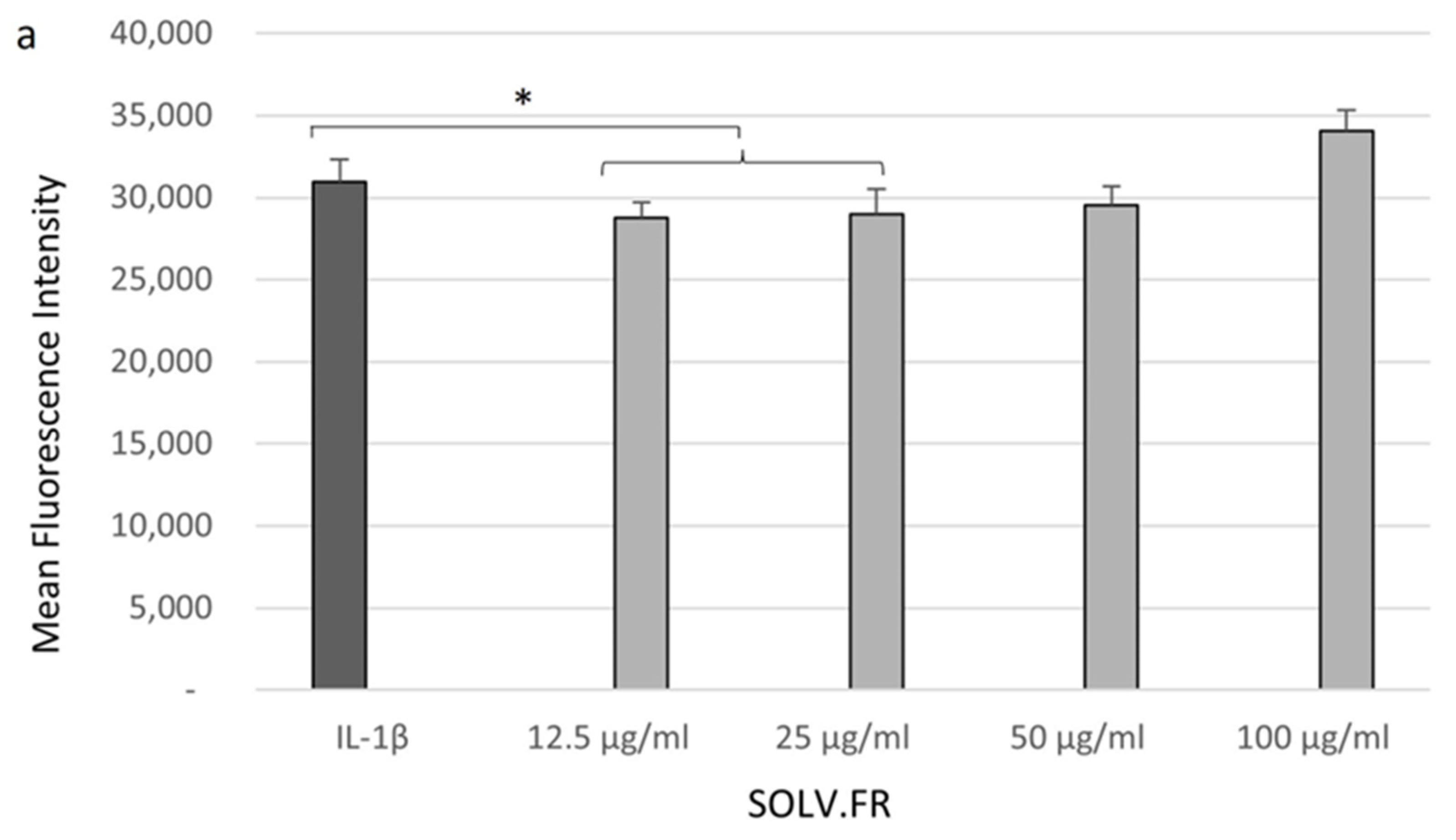
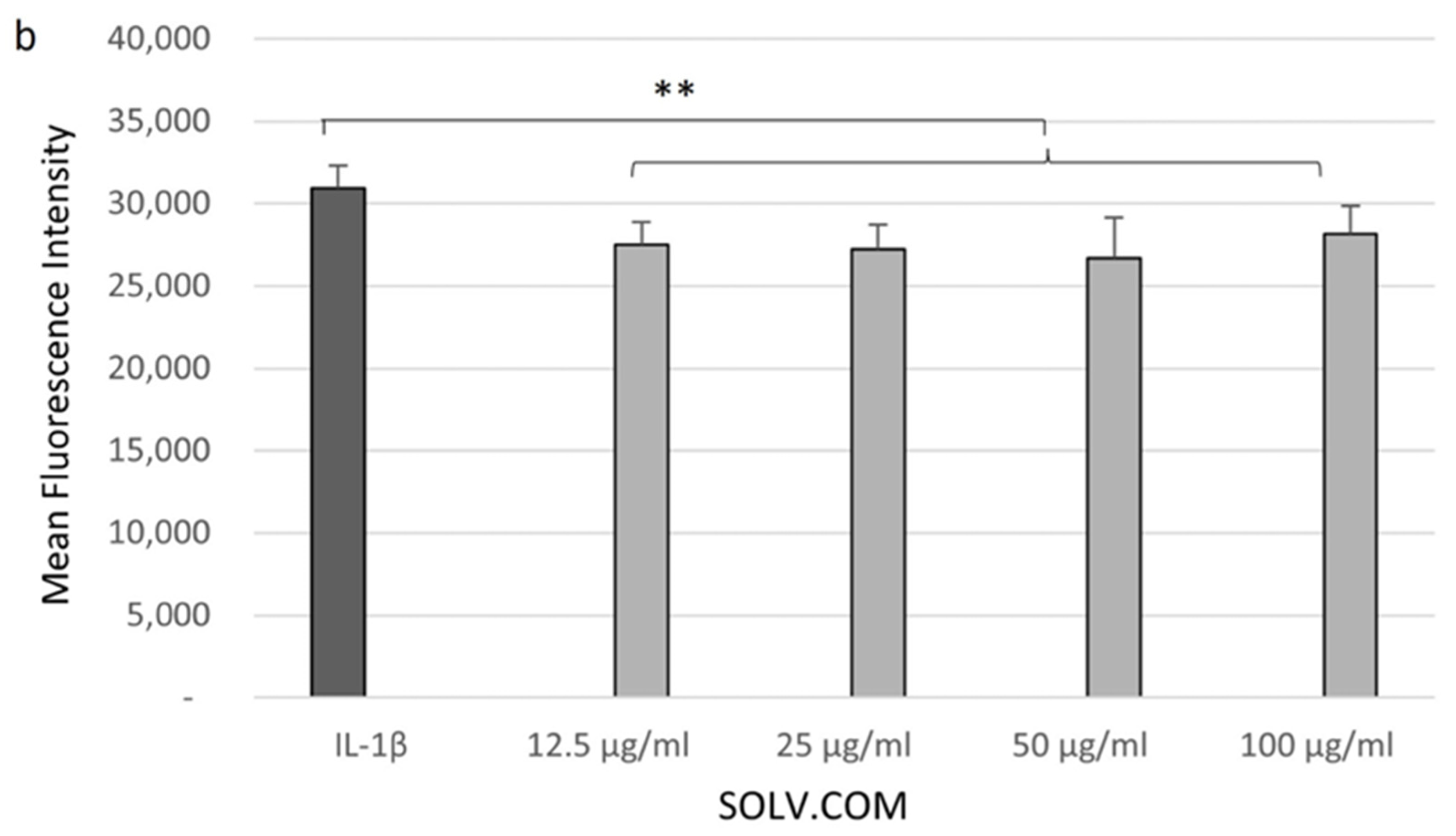
3.3. Cell Glucose Levels Recovered after Sample Treatment

Samples were administered without external stimulation to assess the effects of mussel oil extract on CHON-001 cells. The cells were maintained in their original DMEM media formulation with 1 g/L of glucose, and no supplementary glucose support was provided. The procedure allowed for a clear understanding of the extract’s impact on the cells without interference from external factors. Figure 4 illustrates a comparison of glucose uptake standards under different chondrocyte conditions. Normal chondrocyte cells exhibit lower metabolic activity than cells stimulated with IL-1β for 24 h (p < 0.00005). The results confirmed the expected higher demand for glucose in inflammatory conditions. The cells stimulated with IL-1β and treated with dexamethasone showed an increase in glucose uptake of around 30%. The data demonstrated that dexamethasone does not suppress glucose uptake, which increases glucose concentration in cells (p < 0.001).
To evaluate the protective effect of mussel extract on CHON-001, SOLV.FR and SOLV.COM were administered in cells without any stimulation and incubated for 24 h. The data presented in Figure 5a indicates that SOLV.FR significantly impacted glucose uptake, particularly at a concentration of 12.5 μg/mL (with a p-value of 0.03). It was observed that there was a discernible reduction in glucose levels at the most concentrated levels of SOLV.FR (25, 50, and 100 µg/mL). The reduction was equivalent to the basal state in untreated cells. In contrast, SOLV.COM demonstrated superior efficiency in transporting glucose, evidenced by a slight increase in luminescence at 50 and 100 μg/mL (p < 0.05).
Following a 24 h stimulation with IL-1β, the cells were treated with SOLV.FR and SOLV.COM. The anti-inflammatory response was measured using dexamethasone as the standard. This approach provided a reliable means of quantifying the efficacy of an anti-inflammatory agent in response to stimulation. The cells exposed to SOLV.FR showed a similar result profile to non-stimulated cells that received the same treatment. Glucose uptake increased at 12.5 μg/mL concentration (p < 0.05) and then declined at higher concentrations. In contrast, SOLV.COM resulted in a significant rise in glucose uptake at 25 and 50 μg/mL concentrations (p < 0.01), while concentrations of 12.5 and 100 μg/mL showed a similar range to dexamethasone treatment (Figure 5b). These results indicate that SOLV.COM promotes tissue homeostasis and accelerates cell recovery.

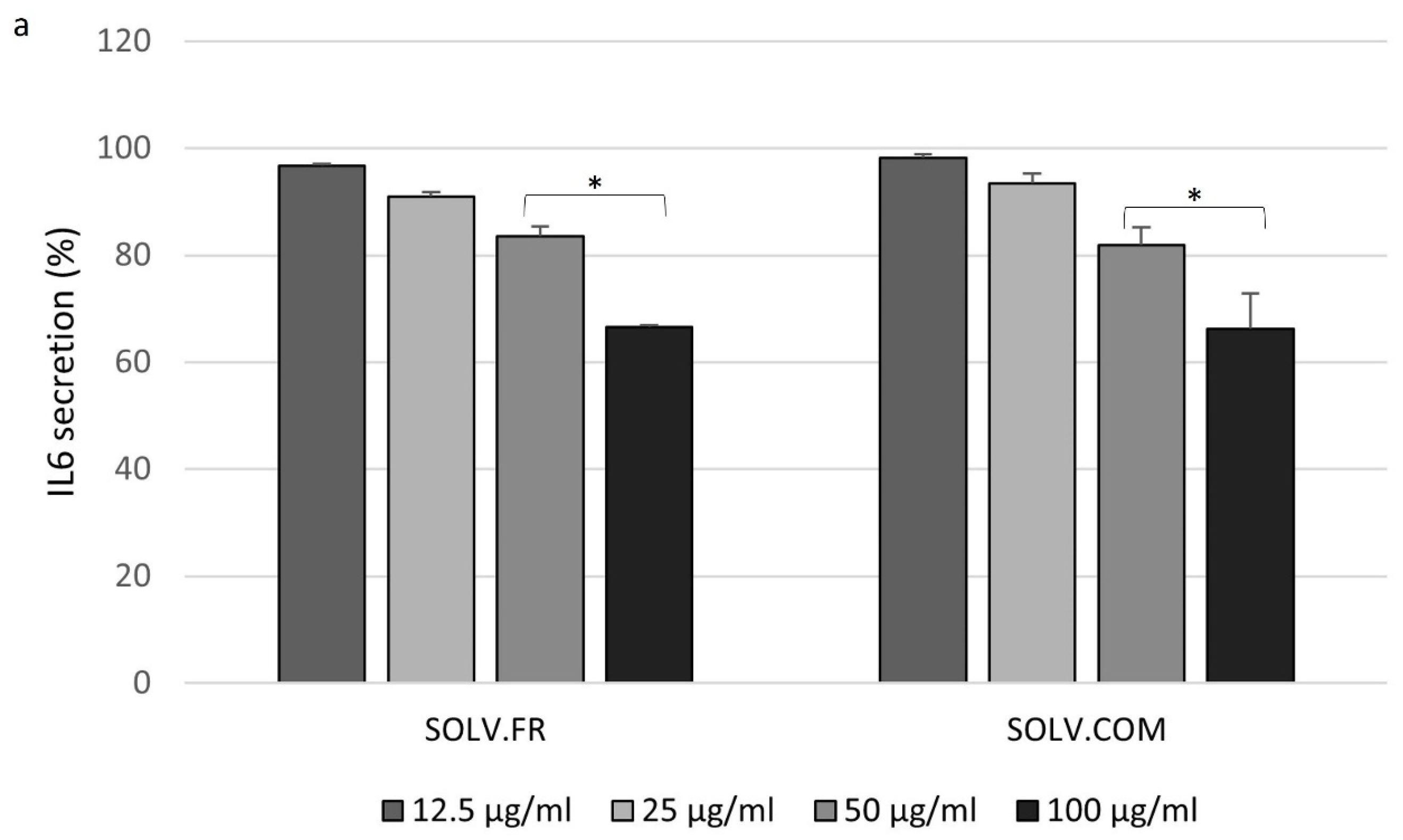
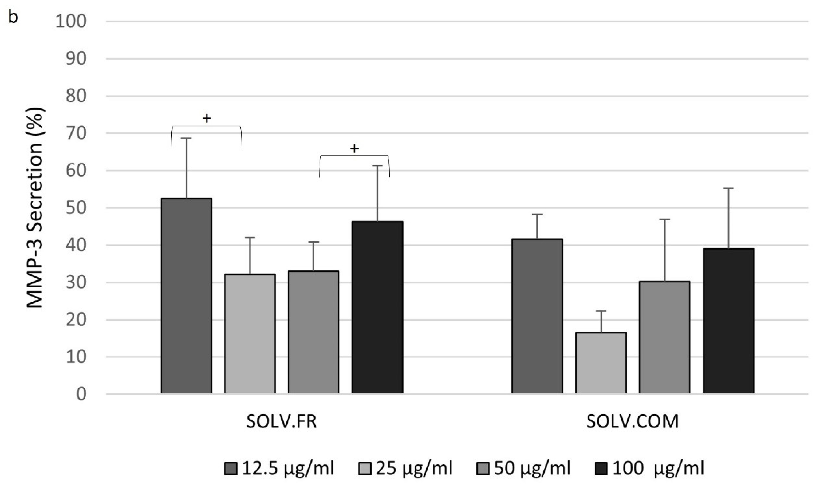
3.4. Modulation of IL-6 and MMP-3 Secretion in Chondrocyte Cells Following Treatment with Mussel Oil Extracts

To assess the effectiveness of the inflammatory model in chondrocyte cells activated by IL-1β, the secretion of IL-6 and MMP-3 was measured. The objective of administering SOLV.FR and SOLV.COM was to examine the extracts’ capacity to regulate the impact of the proinflammatory cytokine in the cells. The results were normalized based on the protein secretion by cells after stimulation. It was assumed that the IL-1β or LPS stimulation generated the highest cytokine release for each group. This approach minimizes relative variations across conditions, enabling an accurate assessment of the impact of stimulation on cytokine release.
The levels of target proteins (IL-6 and MMP-3) in the cell supernatant were determined using immunoassay. The analysis of IL-6 data indicates that both SOLV.FR and SOLV.COM can significantly reduce cytokine secretion dose-dependently. Upon dilution to 12.5 μg/mL, both SOLV.FR and SOLV.COM decreased IL-6 secretion to levels comparable to those achieved with dexamethasone treatment (99.17 ± 0.56%). SOLV.FR reduced secretion to 96.65 ± 0.34%, and SOLV.COM to 98.2 ± 0.7% (Figure 6a). As the concentration increased, IL-6 secretion decreased. At a concentration of 50 μg/mL, SOLV.FR achieved a release rate of 83.49 ± 1.94%, followed by 66.53 ± 0.4% at 100 μg/mL concentration (p < 0.01). Similarly, SOLV.COM reached 81.83 ± 3.28% at 50 μg/mL and 66.32 ± 6.5% at 100 μg/mL (p < 0.01).
Treatment with dexamethasone reduced the range of 56.12 ± 6.8% in the release of MMP-3. SOLV.FR and SOLV.COM regulated enzymes responsible for degrading the extracellular matrix. Figure 6b shows that SOLV.FR at a concentration of 12.5 ug/mL was able to reduce the MMP-3 release by 52.53 ± 16.10%, reaching the rate of 32.15 ± 9.93% at 25 ug/mL (p < 0.05). However, with extract concentration increased to 100 ug/mL, secretion was slightly increased (46.25 ± 14.95%). A similar pattern was observed in SOLV.COM, with a reduction of 41.64 ± 6.6% in MMP-3 secretion at a dilution of 12.5 ug/mL and a more pronounced decrease at 25 ug/mL (16.5 ± 5.8%). However, the analysis revealed that this extract, in the highest concentration, managed to decrease the MMP-3 release to close to 30% (30.3 ± 16.55% at 50 ug/mL and 39.01 ± 16.25% at 100 ug/mL).
Initially, the effect of continuum stimulation on CHON-001 cells was analyzed by observing their morphological changes. The cells were stimulated with IL-1β and LPS, and alterations in their shape, nuclear size or shape, and cytoplasmic granulation or granularity were observed under a microscope (Figure 7). The differences between the stimulated cells and the untreated ones were visible. The stimulated cells exhibited changes in morphology, such as transitioning from a typical rounded or polygonal shape to a more elongated or irregular morphology, indicating that the stimulation was successful.
In the second approach, an immunoassay was conducted to measure the levels of IL-6 in the supernatant collected from two groups of chondrocyte cells. CHON-001 cells were exposed to IL-1β (5 ng/mL) and LPS (10 μg/mL) for 21 days in T25 Falcon cell culture grow flasks. The effectiveness of the treatment was analyzed after treatment with SOLV.FR, SOLV.COM, and dexamethasone for 24 h. It was checked whether the control of cytokine secretion was equally suppressed by dexamethasone in both groups. Group 1, stimulated with IL-1β, presented a rate of 96.55 ± 0.8% in the IL-6 release, while group 2, stimulated with LPS, showed a significant reduction, reaching 26.66 ± 0.45% (Figure 8a,b). Preliminary analysis suggests that the pathways for stimulating chondrocyte cells with these substances could differ.
Group 1 and group 2 showed differences in the control of cytokine secretion upon administration of SOLV.FR and SOLV.COM dilutions. Following the stimulation of cells with IL-1β, SOLV.FR treatment was found to have a slight effect on the management of cytokine release at a dilution of 100 μg/mL, achieving a rate of 90.98 ± 16.26%. At a dilution of 50 μg/mL, it achieved a rate of 87.63 ± 10.57%, lower than the rate for cells treated with dexamethasone in the same group, although not statistically significant (Figure 8a). The other dilutions did not demonstrate significant control in the release of IL-6. In group 1, SOLV.COM did not appear to affect the levels of IL-6 and maintained similar ranges across all dilutions (Figure 8a). After a comprehensive analysis, it was determined that there is no statistically significant difference present in any of the compared variables.
In group 2, the results showed a potential control of the proinflammatory target. SOLV.FR effectively controlled the secretion of IL-6 at dilutions of 12.5 and 25 μg/mL (58.80 ± 5.33% and 50.03 ± 5.65%, respectively, with p < 0.05). However, at higher concentrations, it was reduced, with a rate of 77.24 ± 7.95% for 100 μg/mL and 74.1 ± 1.8% for 50 μg/mL (Figure 8b). In contrast, SOLV.COM successfully managed secretion levels in a dose-dependent manner. At a concentration of 100 μg/mL (33.86 ± 5.1%), the effectiveness of secretion management was almost equal to that of administering dexamethasone to the same group. Meanwhile, at a concentration of 50 μg/mL, the effectiveness of secretion management was measured at 47.16 ± 8.62% (as shown in Figure 8b). The lower dilutions showed a release rate similar to SOLV.FR dilutions at the same concentrations (54.54 ± 10.5% at 12.5 μg/mL and 58.68 ± 13.5% at 25 μg/mL). The data analysis indicates a significant difference in cytokine secretion control between SOLV.FR and SOLV.COM at 100 ug/mL concentration (74.1 ± 1.8% and 33.86 ± 5.1%, respectively, with p < 0.001).

4. Discussion

This study sought to promote screening of mussel lipid extract as an agent to treat OA and improve disease symptomatology due to their beneficial free fatty acid composition. An earlier analysis using gas chromatography–mass spectrometry was conducted to determine the fatty acid content of GLM extracts. The results showed the presence of mono-, poly-, and unsaturated fatty acids. Based on the analysis, SOLV.COM has higher ALA content (8.3 ± 0.07 to 4.15 ± 1.71 mg/g, p < 0.05) than SOLV.FR. However, SOLV.FR has the highest content of EPA (52.03 ± 0.006 mg/g, p = 0.03) and DHA (78.46 ± 2.19 mg/g, p = 0.002) (Table S1—Supplementary Materials). Although SOLV.FR contains more monounsaturated and saturated fatty acids than SOLV.COM, statistical analysis showed no significant difference. Notably, the amount of palmitic acid (67.4 ± 3.95 mg/g, p < 0.05) in SOLV.FR composition is approximately twice that of SOLV.COM.
This study utilized an in vitro model to screen the extract that generated a more robust response in stimulated cells. However, using chondrocyte monolayer cell cultures to study inflammatory processes has certain limitations that must be considered. It does not accurately represent all the components involved in disease development, such as cell–cell interactions, mechanical loading, and the native extracellular matrix (ECM). However, utilizing monolayer cell cultures to search for the most effective anti-inflammatory nutraceutical agent is a practical experimental approach. This method offers simplicity, reproducibility, and high-throughput capabilities, making it ideal for controlled experiments.

4.1. Chondrocytes Cell Viability after IL-1β Stimulation

The development of osteoarthritis is characterized by a gradual deterioration of the articular cartilage, which a range of primary and secondary factors can cause. One common factor in this process is the production of IL-1β, which can trigger inflammation [44]. IL-1β triggers cartilage degradation by increasing inflammatory mediators like cytokines and MMPs. During OA development, more proinflammatory cytokines, like IL-6, can be released, further degrading the cartilage matrix and ultimately causing damage to surrounding tissues and structures [45]. IL-6 promotes inflammation, destroys joint tissues, and causes cartilage loss and joint damage. IL-6 also affects chondrocyte functions and regulates MMP activity, stimulating MMP production and affecting matrix synthesis and degradation [46,47,48]. Studies indicate that overexpression of IL-1β can impact a variety of pathways within the body, such as cell cycle, autophagy, cytokine signaling, NF-κB, and Toll-like receptor pathways. In vitro models of articular inflammation by IL-1β-treated chondrocyte cells can help study the mechanisms and treatments for this condition [49].
This study analyzed the effect of IL-1β on chondrocyte cell viability and investigated the protective effects of mussel oil extract against cytokine exposure. The cells were exposed to IL-1β for 24 and 72 h, and metabolic activity was used to analyze the effects on total live cells in culture. This study found that incubation with 10 ng/mL of IL-1β for 24 h resulted in acceptable cell viability, and treatment with the samples for 24 h presented a protective effect, enabling an increase in cell viability. After 72 h of stimulation, the samples did not exhibit the same level of protective effect observed in the group stimulated for 24 h. A 2020 study attempted to replicate an OA cell model using IL-1β to induce inflammation [50]. The researchers found that administering IL-1β significantly increased cell apoptosis and the expression of MMPs, leading to decreased chondrocyte viability and extracellular matrix synthesis. In Li and colleagues’ study, the examined sample was found to have effectively countered the harmful impact of IL-1β [50]. Our study produced similar results, indicating that mussel oil can potentially reduce the negative impact of IL-1β. It may be helpful to conduct further research in this area to understand its effectiveness fully.

4.2. Green-Lipped Mussel Extract in the Control in ROS Generation

Reactive oxygen species (ROS) are metabolites that have undergone partial reduction and possess powerful oxidative capabilities. Elevated levels of ROS can harm cellular health. However, normal physiological concentrations play complex roles in governing various cellular phenomena, including growth regulation, cellular adhesion, differentiation, senescence, and programmed cell death [51]. ROS production is increased in OA due to chondrocytes responding to inflammatory cytokine stimulation, like that by IL-1β. This highly stimulated environment contributes to cellular damage and inflammation by forming molecules that exacerbate oxidative stress [14,52]. The mussel oil extracts do not effectively decrease hydrogen peroxide generation under IL-1β stimulation at all concentrations. Compared to the samples, SOLV.COM presented a more effective potential to manage hydrogen peroxide than SOLV.FR. The findings corroborate our previous study, where antioxidant DPPH examination was undertaken to evaluate the samples’ efficacy in reducing the hydrogen providers or free radicals. Sato and colleagues (2013) reported instability of omega-3 PUFAs for supplementation when exposed to oxygen, heat, or daylight [53]. Their discovery may explain that the mussel powder preparation’s solvent extraction samples exhibit low antioxidant activity. However, it has been proven that adding α-cyclodextrin can enhance the stability of green-lipped mussel (GLM) oil against oxidation [53].
Numerous publications have documented the antioxidant potential of omega-3 EPA and DHA in vitro models [26,54,55,56,57,58,59]. The studies utilized omega-3 samples originating from different commercial brands. It is worth noting that certain brands have included vitamin E and olive oil in their formulations, which are known for their exceptional antioxidant qualities. The observed antioxidant activity in these studies may be attributed to the inclusion of stabilizing or other compounds aimed at enhancing the sample’s capacity to manage hydrogen peroxide and free radical generation.
Heshmati and colleagues (2019) conducted a systematic review and meta-analysis to summarize the findings of randomized clinical trials examining the effects of omega-3 FAs on OS markers. Data analysis suggested that omega-3 fatty acids can improve the body’s defense against ROS [60]. Based on the evidence presented, it can be inferred that SOLV.COM can mitigate oxidative stress and safeguard cells from damage triggered by IL-1β stimulation.

4.3. Increased Glucose Transport in Response to Green-Lipped Mussel Extract in Chondrocytes Cells

The absence of vascular and lymphatic networks in adult cartilage requires diffusion from synovial fluid for nutrient distribution to chondrocytes. Adequate glucose supply is crucial to sustain primarily anaerobic metabolism and synthesize extracellular matrix (ECM) macromolecules [61]. The cells primarily use glucose as energy. Still, lipids, cholesterol, and fatty acids are crucial for energy, structural support, and signaling to maintain healthy cartilage [62,63]. The glucose uptake was measured after subjecting chondrocyte cells to incubation with mussel oil extract (SOLV.COM) for 24 h. There was a noticeable increase in glucose uptake without surpassing the level observed in cells stimulated with IL-1β, indicating that the sample could aid nutrition in healthy cells. In OA, the lack of blood vessels in cartilage tissue prevents cells from obtaining sufficient glucose, leading to compromised cell function and disrupting the balance between ECM synthesis and degradation.
Limited glucose supply significantly impairs the ability of chondrocytes to repair the matrix during OA progression [61]. Some studies have examined the regulation of glucose transport in human articular chondrocytes by proinflammatory cytokines. They concluded that stimulation of glucose transport represents a component of the chondrocyte response to IL-1β stimulation [8,61,64]. In the present study, it was possible to observe the regulation since the cells’ stimulation with IL-1β increased glucose uptake compared with untreated cells. The cells treated with GLM samples presented an enhancement in glucose uptake, as observed in the control treatment. However, the sample SOLV.COM enhanced the cell glucose supply more efficiently than the sample SOLV.FR. Our previous study found traces of cis-16:1n-7 in the solvent samples. Although the detected amount was not significantly high, it still comprised 8.2% of SOLV.COM and 5.2% of SOLV.FR. Research conducted by Cruz and colleagues (2018) suggests that this MUFA can improve the metabolic and oxidative capacity of 3T3-L1 adipocytes [65]. Therefore, it is conceivable that the variation in metabolism and oxygen uptake between commercial and fresh samples may be attributed to the difference in cis-16:1n-7 levels. Nonetheless, additional research is needed to grasp this FFA’s role in chondrocyte function fully.
Several articles have discussed the role of omega-3 PUFAs in cell metabolism. They agree that omega-3 daily supplements can enhance glucose absorption and regulation in different cells by activating signaling pathways [66,67,68,69,70,71]. SOLV.COM may have the potential to regulate uptake in chondrocyte cells. However, glucose uptake did not correspond in a dose-dependent manner to both sample types. This indicates that more studies are needed to understand the influence of mussel oil extracts on the regulation mechanisms of glucose transporters. These events lead to the hypothesis that the extracts could have protective effects on the chondrocyte once they improve glucose uptake, allowing for the cells’ nutrition and recovery.

4.4. Exploring the Interplay of Inflammatory Cytokines and Green-Lipped Mussel Oil Extracts in Stimulated Chondrocyte Cells

Inflammatory cytokines play a significant role in the development of OA. IL-6 contributes to both constructive and destructive mechanisms in cartilage physiology, disrupting tissue homeostasis at high levels [46,47]. IL-6 manages matrix creation and breakdown and negatively impacts other chondrocyte functions, such as slowing growth and raising oxidative stress [47].
The efficacy of mussel oil extracts in regulating IL-6 secretion was observed through reduced cytokine release in a dosage-dependent manner over 24 h of treatment with IL-1β. In an experiment to monitor the impact of cell stimulation on secretion rates, the experimental cell group stimulated with IL-1β did not show a significant decrease in IL-6 secretion rates over a long time. Notably, the positive control (1 ug/mL of dexamethasone) failed to suppress IL-6 release. Conversely, LPS-stimulated cells showed a dose-dependent decrease in the SOLV.COM treated group, with the highest concentration demonstrating a rate similar to that of the positive control; this suggests that the pathways activated by chondrocyte cells via IL-1β and LPS differ.
LPS can trigger the activation of immune system cells, such as macrophages and neutrophils, leading to tissue inflammation. This property of LPS has been utilized in the induction of OA in animal models owing to its disease-related characteristics [72]. Haglund et al. (2008) reported that rat chondrocytes express TLR4 as a primary receptor type. Upon exposure to LPS, a TLR4 stimulant, the cells generate MMPs and elements linked to the innate immune response. Notably, there was no detection of TNF-α or IL-1 in the culture media post-LPS exposure [73]. In this study, the release of IL-6 was successfully detected in the supernatant. Although, in the LPS-stimulated group, IL-1 and TNF-α were not detectable in the media supernatant.
Chondrocyte morphology changes were observed after IL-1β and LPS treatment, as shown in Figure 6. Zainal and colleagues (2009) reported that IL-1-treated cells exhibited spindle-like, sharp, or stellate forms with increased cellular debris and death after 48 h. However, cells preincubated with EPA maintained a rounded morphology and were protected against the detrimental effects of IL-1 [30].
OA development is influenced by the exact immune-triggering mechanisms that contribute to LPS. The TLR4 activation leads to elevated NF-κB in joint cells, triggering the activation of the inflammatory cascade and creating an environment conducive to in-stalling cell damage [72]. Conversely, cell activation via IL-1β follows four cellular signal-ling pathways; three are related to MAPK pathways, and the other is to the NF-κB pathway [74]. It is also thought that IL-1β stimulates chondrocytes in multiple ways, resulting in multiple cell clusters with variably expressed genes, modified functions, and different proportions of genes [75]. It is plausible to hypothesize that several molecular pathways are linked to the activation of chondrocytes, which subsequently results in the release of cytokines. The data analysis shows that mussel oil extracts have exhibited different levels of effectiveness when managing IL-6 secretion. The effectiveness of the extracts depends on the pathway of cell activation during chondrocyte stimulation caused by IL-1β and LPS. Further research on mussel oil extracts is necessary to understand their potential benefits.
As a glucocorticoid, dexamethasone suppresses NF-κB, a key transcription factor. When activated, the receptor represses these factors through various mechanisms and modifies the proinflammatory action of NF-κB [76]. Therefore, it is possible to assume that the difference in glucocorticoid response among groups (group 1 and group 2) was related to the complex activation of this condition, involving several signal/transduction pathways and affecting many genes. According to the results of the present study, the effectiveness of glucocorticoids in managing inflammatory cytokines is significantly impacted by the type of OA model used. The model impact was also mentioned in a review article about the chondroprotective effects of corticosteroids [77].
The interplay between IL-6 and MMP-3 underscores the complex mechanisms through which cells and their microenvironment interact to shape tissue remodeling processes [47]. The increased concentration of IL-6 collaborates to reduce type II collagen production and elevate MMP enzyme production, irrespective of IL-1β stimulation [47,78,79,80,81].
The mussel oil extracts appeared to manage IL-6 and MMP-3 synergistically. Both SOLV.FR and SOLV.COM demonstrated noteworthy decreases in MMP-3 secretion in cells stimulated with IL-1β. The reduction rate in the sample SOLV.COM was higher than in the positive control, but both samples did not follow a dosage-dependent decrease. Based on the available data, it can be inferred that SOLV.COM is highly proficient in regulating the release of IL-6 and MMP-3. This characteristic makes it a potential contender for preserving the homeostasis of chondrocytes. Nonetheless, it is imperative to conduct further research to gain a comprehensive understanding of its anti-inflammatory properties.
Indeed, it is crucial to consider that omega-3 undergoes conversion into specialized pro-resolving mediators (SPMs), exhibiting potent anti-inflammatory characteristics. Nevertheless, it is essential to highlight that bioactive lipids and fatty acids, such as saturated fatty acids, monounsaturated fatty acids, and omega-6, can function as both proinflammatory and anti-inflammatory agents within the framework of OA [82]. The biological impacts of fatty acids depend on their length and level of saturation, with saturated fatty acids being recognized as proinflammatory lipids [31]. The molecular mechanisms that underlie the interplay between dietary intake of omega-3 FA and the progression of osteoarthritis involve intricate adjustments in protein expression profiles that dictate disease evolution [30]. Several preclinical studies described in a narrative review suggest that omega-3 polyunsaturated fatty acids (PUFAs) may protect against cartilage deterioration in OA and reduce inflammatory markers [40]. Furthermore, studies conducted on in vivo models have provided evidence that the extract derived from GLM exhibits the potential to mitigate symptoms of joint disease and regulate immune cells that contribute to the advancement of the ailment [26,83]. The differences in the assay results observed in SOLV.FR and SOLV.COM may be linked to the diverse spectrum of bioactive lipids in each extract. Consequently, it cannot be stated that the significance of the inflammatory response solely relies on the amounts of EPA and DHA in a lipid extract.
In summary, GLM extracts, particularly SOLV.FR, exhibit promising protective effects on cell viability. Administering SOLV.COM after 24-h IL-1β stimulation increased cell viability, with SOLV.FR at 100 μg/mL showing significant protection. Regarding hydrogen peroxide generation, mussel oil extracts (SOLV.FR and SOLV.COM) have limited impact. However, dexamethasone effectively controls ROS production. Additionally, SOLV.FR reduces glucose levels, while SOLV.COM enhances glucose transport efficiency, particularly under IL-1β stimulation. The two extracts show a dose-dependent decrease in IL-6 secretion, comparable to dexamethasone, after 24 h of IL-1 β stimulation. The extracts effectively regulate MMP-3 release in chondrocyte cells, demonstrating their potential anti-inflammatory properties. In conclusion, this study suggests that SOLV.COM can regulate various physiological processes linked with osteoarthritis, including glucose metabolism and levels of inflammatory markers. These potential therapeutic functions may effectively aid in managing the symptoms of osteoarthritis and improving the overall quality of life for those suffering from this condition. Nonetheless, further investigation is required to comprehensively understand the GLM extract’s mechanism of action and the implications of prolonged use.

Supplementary Materials

The following supporting information can be downloaded at: https://www.mdpi.com/article/10.3390/nutraceuticals4010001/s1, Table S1: Fatty acid composition of oil extracts from GLM obtained in fresh and commercial powder.

Author Contributions

R.C.L. contributed to the conception, design, and statistics and drafted the manuscript; B.E. contributed to the conception, design and revision of the manuscript; X.G. contributed to the validation and revision of the manuscript; Y.L. and J.L. contributed to the conception, supervision and critically revised the manuscript. All authors have read and agreed to the published version of the manuscript.

Funding

This research was funded by New Zealand National Science Challenge; High Value Nutrition Fund, grant number HVN-1905; and China Ministry of Science and Technology 2023 National Foreign Experts Program, DL2023013002.

Institutional Review Board Statement

Not applicable.

Informed Consent Statement

Not applicable.

Data Availability Statement

The datasets generated for this study will be made available via reasonable request to the corresponding author due to space constraints.

Acknowledgments

Auckland University of Technology Doctoral Scholarships for Lessa, R.C.

Conflicts of Interest

The authors declare no conflict of interest.

Abbreviations

2DG2-Deoxy-D-glucose
2DG6P2-deoxy-D-glucose-6-phosphate
ADAMTSA disintegrin and metalloproteinase with thrombospondin motifs
ALAAlpha-linolenic acid
CO2Carbonate dioxide
COXCyclooxygenase
DCFH-DADiacetyldichlorofluorescein
DHADocosahexaenoic acid
ECMExtracellular matrix
EPAEicosapentaenoic acid
GLMGreen-lipped mussel
ILInterleukin
LPSLipopolysaccharides
MAPKMitogen-activated protein kinases
MMPsMatrix metalloproteinases
MTBE: MeMethyl tert-butyl ether: methanol
MTT3-(4,5-Dimethylthiazol-2-yl)-2,5-Diphenyltetrazolium Bromide
MUFAMonounsaturated fatty acids
n-3Omega-3
NF-κBNuclear factor kappa B
OAOsteoarthritis
PBSPhosphate-buffered saline
PUFAsPolyunsaturated fatty acids
ROSReactive oxygen species
TNF-αTumor necrosis factor-alpha

References

  1. Yao, Q.; Wu, X.; Tao, C.; Gong, W.; Chen, M.; Qu, M.; Zhong, Y.; He, T.; Chen, S.; Xiao, G. Osteoarthritis: Pathogenic signaling pathways and therapeutic targets. Signal Transduct. Target. Ther. 2023, 8, 56. [Google Scholar] [CrossRef] [PubMed]
  2. Fontanella, C.G.; Belluzzi, E.; Pozzuoli, A.; Scioni, M.; Olivotto, E.; Reale, D.; Ruggieri, P.; De Caro, R.; Ramonda, R.; Carniel, E.L. Exploring Anatomo-Morphometric Characteristics of Infrapatellar, Suprapatellar Fat Pad, and Knee Ligaments in Osteoarthritis Compared to Post-Traumatic Lesions. Biomedicines 2022, 10, 1369. [Google Scholar] [CrossRef] [PubMed]
  3. He, Y.; Li, Z.; Alexander, P.G.; Ocasio-Nieves, B.D.; Yocum, L.; Lin, H.; Tuan, R.S. Pathogenesis of osteoarthritis: Risk factors, regulatory pathways in chondrocytes, and experimental models. Biology 2020, 9, 194. [Google Scholar] [CrossRef] [PubMed]
  4. Ege, F. Pathogenesis, Pathology and Genetics of Osteoarthritis. In Rheum. Arthritis; IntechOpen: London, UK, 2021. [Google Scholar]
  5. Man, G.; Mologhianu, G. Osteoarthritis pathogenesis–a complex process that involves the entire joint. J. Med. Life 2014, 7, 37. [Google Scholar] [PubMed]
  6. Kapoor, M.; Martel-Pelletier, J.; Lajeunesse, D.; Pelletier, J.-P.; Fahmi, H. Role of proinflammatory cytokines in the pathophysiology of osteoarthritis. Nat. Rev. Rheumatol. 2011, 7, 33–42. [Google Scholar] [CrossRef]
  7. Defois, A.; Bon, N.; Charpentier, A.; Georget, M.; Gaigeard, N.; Blanchard, F.; Hamel, A.; Waast, D.; Armengaud, J.; Renoult, O. Osteoarthritic chondrocytes undergo a glycolysis-related metabolic switch upon exposure to IL-1b or TNF. Cell Commun. Signal. 2023, 21, 137. [Google Scholar] [CrossRef]
  8. Shikhman, A.R.; Brinson, D.C.; Valbracht, J.; Lotz, M.K. Cytokine regulation of facilitated glucose transport in human articular chondrocytes. J. Immunol. 2001, 167, 7001–7008. [Google Scholar] [CrossRef]
  9. Wu, X.; Liyanage, C.; Plan, M.; Stark, T.; McCubbin, T.; Barrero, R.; Batra, J.; Crawford, R.; Xiao, Y.; Prasadam, I. Dysregulated energy metabolism impairs chondrocyte function in osteoarthritis. Osteoarthr. Cartil. 2023, 31, 613–626. [Google Scholar] [CrossRef]
  10. Zhou, J.; Fang, C.; Rong, C.; Luo, T.; Liu, J.; Zhang, K. Reactive oxygen species-sensitive materials: A promising strategy for regulating inflammation and favoring tissue regeneration. Smart Mater. Med. 2023, 4, 427–446. [Google Scholar] [CrossRef]
  11. Zahan, O.-M.; Serban, O.; Gherman, C.; Fodor, D. The evaluation of oxidative stress in osteoarthritis. Med. Pharm. Rep. 2020, 93, 12. [Google Scholar] [CrossRef]
  12. Wood, M.J.; Miller, R.E.; Malfait, A.-M. The genesis of pain in osteoarthritis: Inflammation as a mediator of osteoarthritis pain. Clin. Geriatr. Med. 2022, 38, 221–238. [Google Scholar] [CrossRef]
  13. Hunter, D.J.; McDougall, J.J.; Keefe, F.J. The symptoms of osteoarthritis and the genesis of pain. Rheum. Dis. Clin. N. Am. 2008, 34, 623–643. [Google Scholar] [CrossRef] [PubMed]
  14. Tong, Z.; Yang, X.; Li, J. Research progress on the mechanism of interleukin-1β on epiphyseal plate chondrocytes. Eur. J. Med. Res. 2022, 27, 313. [Google Scholar] [CrossRef] [PubMed]
  15. Lan, Z.; Yang, Y.; Wang, H.; Guo, X.; Wu, Z.; Jin, Q. Impact of prevention strategies on quality of life in patients with osteoarthritis: A systematic review and meta-analysis of randomized controlled trials. World Neurosurg. 2023; Online ahead of print. [Google Scholar] [CrossRef]
  16. Bowden, J.L.; Hunter, D.J.; Deveza, L.A.; Duong, V.; Dziedzic, K.S.; Allen, K.D.; Chan, P.-K.; Eyles, J.P. Core and adjunctive interventions for osteoarthritis: Efficacy and models for implementation. Nat. Rev. Rheumatol. 2020, 16, 434–447. [Google Scholar] [CrossRef] [PubMed]
  17. Orlowsky, E.W.; Kraus, V.B. The role of innate immunity in osteoarthritis: When our first line of defense goes on the offensive. J. Rheumatol. 2015, 42, 363–371. [Google Scholar] [CrossRef] [PubMed]
  18. Smith, J.L.; Innes, A.Q.; Burns, D.S.; Deniszczyc, D.; Selfe, J.; MacConville, S.; Deighton, K.; Kelly, B.M. A scalable 12-week exercise and education programme reduces symptoms and improves function and wellbeing in people with hip and knee osteoarthritis. Front. Rehabil. Sci. 2023, 4, 1147938. [Google Scholar] [CrossRef] [PubMed]
  19. Dantas, L.O.; de Fátima Salvini, T.; McAlindon, T.E. Knee osteoarthritis: Key treatments and implications for physical therapy. Braz. J. Phys. Ther. 2021, 25, 135–146. [Google Scholar] [CrossRef]
  20. Valsamidou, E.; Amerikanou, C.; Tzavara, C.; Skarpas, G.; Mariolis-Sapsakos, T.D.; Zoumpoulakis, P.; Kaliora, A.C. A standardized nutraceutical supplement contributes to pain relief, improves quality of life and regulates inflammation in knee osteoarthritis patients; A randomized clinical trial. Heliyon 2023, 9, e20143. [Google Scholar] [CrossRef]
  21. Gahivad, M.D.P.; Gahivad, M.R.P.; Deo, S.D.; Daphal, G.T.; Borse, M.H.D.; Ahmed, M.N.; Mahajan, M.B. The trends of herbal nutraceuticals used in chronic disease. Int. J. Prog. Res. Eng. Manag. Sci. (IJPREMS) 2023, 11, 68–80. [Google Scholar]
  22. Lordan, S.; Ross, R.P.; Stanton, C. Marine bioactives as functional food ingredients: Potential to reduce the incidence of chronic diseases. Mar. Drugs 2011, 9, 1056–1100. [Google Scholar] [CrossRef]
  23. Lunn, J.; Theobald, H. The health effects of dietary unsaturated fatty acids. Nutr. Bull. 2006, 31, 178–224. [Google Scholar] [CrossRef]
  24. Senthilkumar, K.; Kim, S.-K. Marine invertebrate natural products for anti-inflammatory and chronic diseases. Evid.-Based Complement. Altern. Med. 2013, 2013, 572859. [Google Scholar] [CrossRef] [PubMed]
  25. Kostoglou-Athanassiou, I.; Athanassiou, L.; Athanassiou, P. The effect of omega-3 fatty acids on rheumatoid arthritis. Mediterr. J. Rheumatol. 2020, 31, 190. [Google Scholar] [CrossRef] [PubMed]
  26. Jhun, J.; Na, H.S.; Cho, K.-H.; Kim, J.; Moon, Y.-M.; Lee, S.Y.; Lee, J.S.; Lee, A.R.; Kim, S.J.; Cho, M.-L. A green-lipped mussel reduces pain behavior and chondrocyte inflammation and attenuated experimental osteoarthritis progression. PLoS ONE 2021, 16, e0259130. [Google Scholar] [CrossRef] [PubMed]
  27. Knott, L.; Avery, N.; Hollander, A.; Tarlton, J. Regulation of osteoarthritis by omega-3 (n-3) polyunsaturated fatty acids in a naturally occurring model of disease. Osteoarthr. Cartil. 2011, 19, 1150–1157. [Google Scholar] [CrossRef] [PubMed]
  28. Lu, J.; Feng, X.; Zhang, H.; Wei, Y.; Yang, Y.; Tian, Y.; Bai, L. Maresin-1 suppresses IL-1β-induced MMP-13 secretion by activating the PI3K/AKT pathway and inhibiting the NF-κB pathway in synovioblasts of an osteoarthritis rat model with treadmill exercise. Connect. Tissue Res. 2021, 62, 508–518. [Google Scholar] [CrossRef] [PubMed]
  29. Xie, Y.; Zhou, W.; Zhong, Z.; Yu, H.; Zhang, P.; Shen, H. Docosahexaenoic acid inhibits bone remodeling and vessel formation in the osteochondral unit in a rat model. Biomed. Pharmacother. 2019, 114, 108811. [Google Scholar] [CrossRef]
  30. Zainal, Z.; Longman, A.J.; Hurst, S.; Duggan, K.; Caterson, B.; Hughes, C.E.; Harwood, J.L. Relative efficacies of omega-3 polyunsaturated fatty acids in reducing expression of key proteins in a model system for studying osteoarthritis. Osteoarthr. Cartil. 2009, 17, 896–905. [Google Scholar] [CrossRef]
  31. Loef, M.; Schoones, J.W.; Kloppenburg, M.; Ioan-Facsinay, A. Fatty acids and osteoarthritis: Different types, different effects. Jt. Bone Spine 2019, 86, 451–458. [Google Scholar] [CrossRef]
  32. Cho, S.H.; Jung, Y.B.; Seong, S.C.; Park, H.B.; Byun, K.Y.; Lee, D.C.; Song, E.K.; Son, J.H. Clinical efficacy and safety of Lyprinol, a patented extract from New Zealand green-lipped mussel (Perna canaliculus) in patients with osteoarthritis of the hip and knee: A multicenter 2-month clinical trial. Eur. Ann. Allergy Clin. Immunol. 2003, 35, 212–216. [Google Scholar]
  33. Coulson, S.; Vecchio, P.; Gramotnev, H.; Vitetta, L. Green-lipped mussel (Perna canaliculus) extract efficacy in knee osteoarthritis and improvement in gastrointestinal dysfunction: A pilot study. Inflammopharmacology 2012, 20, 71–76. [Google Scholar] [CrossRef] [PubMed]
  34. Coulson, S.; Butt, H.; Vecchio, P.; Gramotnev, H.; Vitetta, L. Green-lipped mussel extract (Perna canaliculus) and glucosamine sulphate in patients with knee osteoarthritis: Therapeutic efficacy and effects on gastrointestinal microbiota profiles. Inflammopharmacology 2013, 21, 79–90. [Google Scholar] [CrossRef] [PubMed]
  35. Coulson, S.; Palacios, T.; Vitetta, L. Perna canaliculus (Green-Lipped Mussel): Bioactive components and therapeutic evaluation for chronic health conditions. Prog. Drug Res. 2015, 70, 91–132. [Google Scholar] [PubMed]
  36. Lau, C.; Chiu, P.; Chu, E.; Cheng, I.; Tang, W.; Man, R.; Halpern, G. Treatment of knee osteoarthritis with Lyprinol®, lipid extract of the green-lipped mussel—A double-blind placebo-controlled study. Prog. Nutr. 2004, 6, 17–31. [Google Scholar]
  37. Mickleborough, T.D.; Sinex, J.A.; Platt, D.; Chapman, R.F.; Hirt, M. The effects PCSO-524®, a patented marine oil lipid and omega-3 PUFA blend derived from the New Zealand green lipped mussel (Perna canaliculus), on indirect markers of muscle damage and inflammation after muscle damaging exercise in untrained men: A randomized, placebo controlled trial. J. Int. Soc. Sports Nutr. 2015, 12, 10. [Google Scholar]
  38. Stebbings, S.; Gray, A.; Schneiders, A.G.; Sansom, A. A randomized double-blind placebo-controlled trial to investigate the effectiveness and safety of a novel green-lipped mussel extract-BioLex®-for managing pain in moderate to severe osteoarthritis of the hip and knee. BMC Complement. Altern. Med. 2017, 17, 416. [Google Scholar] [CrossRef]
  39. Zawadzki, M.; Janosch, C.; Szechinski, J. Perna canaliculus lipid complex PCSO-524™ demonstrated pain relief for osteoarthritis patients benchmarked against fish oil, a randomized trial, without placebo control. Mar. Drugs 2013, 11, 1920–1935. [Google Scholar] [CrossRef]
  40. Cordingley, D.M.; Cornish, S.M. Omega-3 fatty acids for the management of osteoarthritis: A narrative review. Nutrients 2022, 14, 3362. [Google Scholar] [CrossRef]
  41. Salem, M.; Bernach, M.; Bajdzienko, K.; Giavalisco, P. A simple fractionated extraction method for the comprehensive analysis of metabolites, lipids, and proteins from a single sample. JoVE (J. Vis. Exp.) 2017, e55802. [Google Scholar]
  42. Lorenz, W.; Buhrmann, C.; Mobasheri, A.; Lueders, C.; Shakibaei, M. Bacterial lipopolysaccharides form procollagen-endotoxin complexes that trigger cartilage inflammation and degeneration: Implications for the development of rheumatoid arthritis. Arthritis Res. Ther. 2013, 15, R111. [Google Scholar] [CrossRef]
  43. Zhuang, J.C.; Wogan, G.N. Growth and viability of macrophages continuously stimulated to produce nitric oxide. Proc. Natl. Acad. Sci. USA 1997, 94, 11875–11880. [Google Scholar] [CrossRef] [PubMed]
  44. Haseeb, A.; Haqqi, T.M. Immunopathogenesis of osteoarthritis. Clin. Immunol. 2013, 146, 185–196. [Google Scholar] [CrossRef] [PubMed]
  45. Liao, Y.; Ren, Y.; Luo, X.; Mirando, A.J.; Long, J.T.; Leinroth, A.; Ji, R.-R.; Hilton, M.J. Interleukin-6 signaling mediates cartilage degradation and pain in posttraumatic osteoarthritis in a sex-specific manner. Sci. Signal. 2022, 15, eabn7082. [Google Scholar] [CrossRef] [PubMed]
  46. Haseeb, A.; Ansari, M.Y.; Haqqi, T.M. Harpagoside suppresses IL-6 expression in primary human osteoarthritis chondrocytes. J. Orthop. Res. 2017, 35, 311–320. [Google Scholar] [CrossRef] [PubMed]
  47. Wiegertjes, R.; van de Loo, F.A.; Blaney Davidson, E.N. A roadmap to target interleukin-6 in osteoarthritis. Rheumatology 2020, 59, 2681–2694. [Google Scholar] [CrossRef]
  48. Wan, J.; Zhang, G.; Li, X.; Qiu, X.; Ouyang, J.; Dai, J.; Min, S. Matrix metalloproteinase 3: A promoting and destabilizing factor in the pathogenesis of disease and cell differentiation. Front. Physiol. 2021, 12, 663978. [Google Scholar] [CrossRef]
  49. Lv, M.; Zhou, Y.; Polson, S.W.; Wan, L.Q.; Wang, M.; Han, L.; Wang, L.; Lu, X.L. Identification of chondrocyte genes and signaling pathways in response to acute joint inflammation. Sci. Rep. 2019, 9, 93. [Google Scholar] [CrossRef]
  50. Li, Z.; Cheng, J.; Liu, J. Baicalin protects human OA chondrocytes against IL-1β-induced apoptosis and ECM degradation by activating autophagy via MiR-766-3p/AIFM1 axis. Drug Des. Dev. Ther. 2020, 14, 2645–2655. [Google Scholar] [CrossRef]
  51. Mittal, M.; Siddiqui, M.R.; Tran, K.; Reddy, S.P.; Malik, A.B. Reactive oxygen species in inflammation and tissue injury. Antioxid. Redox Signal. 2014, 20, 1126–1167. [Google Scholar] [CrossRef]
  52. Ansari, M.Y.; Ahmad, N.; Haqqi, T.M. Oxidative stress and inflammation in osteoarthritis pathogenesis: Role of polyphenols. Biomed. Pharmacother. 2020, 129, 110452. [Google Scholar] [CrossRef]
  53. Sato, K.; Ishida, Y.; Ikuta, N.; Uekaji, Y.; Nakata, D.; Terao, K. Stabilization of unstable functional food ingredients by complexation with cyclodextrin. Oleo Sci. 2013, 13, 123–130. [Google Scholar]
  54. Chen, J.; Bao, C.; Cho, S.H.; Lee, H.J. Green lipped mussel oil complex suppresses lipopolysaccharide stimulated inflammation via regulating nuclear factor-κB and mitogen activated protein kinases signaling in RAW264. 7 murine macrophages. Food Sci. Biotechnol. 2017, 26, 815–822. [Google Scholar] [CrossRef] [PubMed]
  55. Clementi, M.E.; Lazzarino, G.; Sampaolese, B.; Brancato, A.; Tringali, G. DHA protects PC12 cells against oxidative stress and apoptotic signals through the activation of the NFE2L2/HO-1 axis. Int. J. Mol. Med. 2019, 43, 2523–2531. [Google Scholar] [CrossRef] [PubMed]
  56. Garcia-Garcia, L.; Recalde, S.; Fernandez-Robredo, P.; Hernandez, M.; Ispizua-Madariaga, N.; Reiter, N.; Albiasu, E.; Bezunartea, J.; Alonso, E.; Layana, A.G. Effect of EPA: DHA formulations on cell viability, proliferation and reactive oxygen species generation under inflamatory and oxidative stress conditions. Investig. Ophthalmol. Vis. Sci. 2015, 56, 16. [Google Scholar]
  57. Sakai, C.; Ishida, M.; Ohba, H.; Yamashita, H.; Uchida, H.; Yoshizumi, M.; Ishida, T. Fish oil omega-3 polyunsaturated fatty acids attenuate oxidative stress-induced DNA damage in vascular endothelial cells. PLoS ONE 2017, 12, e0187934. [Google Scholar] [CrossRef] [PubMed]
  58. Sakata, S.; Hayashi, S.; Fujishiro, T.; Kawakita, K.; Kanzaki, N.; Hashimoto, S.; Iwasa, K.; Chinzei, N.; Kihara, S.; Haneda, M. Oxidative stress-induced apoptosis and matrix loss of chondrocytes is inhibited by eicosapentaenoic acid. J. Orthop. Res. 2015, 33, 359–365. [Google Scholar] [CrossRef]
  59. Schönfeld, P.; Wojtczak, L. Fatty acids decrease mitochondrial generation of reactive oxygen species at the reverse electron transport but increase it at the forward transport. Biochim. Biophys. Acta (BBA)-Bioenerg. 2007, 1767, 1032–1040. [Google Scholar] [CrossRef]
  60. Heshmati, J.; Morvaridzadeh, M.; Maroufizadeh, S.; Akbari, A.; Yavari, M.; Amirinejad, A.; Maleki-Hajiagha, A.; Sepidarkish, M. Omega-3 fatty acids supplementation and oxidative stress parameters: A systematic review and meta-analysis of clinical trials. Pharmacol. Res. 2019, 149, 104462. [Google Scholar] [CrossRef]
  61. Rotter Sopasakis, V.; Wickelgren, R.; Sukonina, V.; Brantsing, C.; Svala, E.; Hansson, E.; Enerbäck, S.; Lindahl, A.; Skiöldebrand, E. Elevated glucose levels preserve glucose uptake, hyaluronan production, and low glutamate release following interleukin-1β stimulation of differentiated chondrocytes. Cartilage 2019, 10, 491–503. [Google Scholar] [CrossRef]
  62. Su, Z.; Zong, Z.; Deng, J.; Huang, J.; Liu, G.; Wei, B.; Cui, L.; Li, G.; Zhong, H.; Lin, S. Lipid metabolism in cartilage development, degeneration, and regeneration. Nutrients 2022, 14, 3984. [Google Scholar] [CrossRef]
  63. Villalvilla, A.; Gómez, R.; Largo, R.; Herrero-Beaumont, G. Lipid transport and metabolism in healthy and osteoarthritic cartilage. Int. J. Mol. Sci. 2013, 14, 20793–20808. [Google Scholar] [CrossRef] [PubMed]
  64. Mobasheri, A.; Vannucci, S.; Bondy, C.; Carter, S.; Innes, J.; Arteaga, M.; Trujillo, E.; Ferraz, I.; Shakibaei, M.; Martín Vasallo, P. Glucose transport and metabolism in chondrocytes: A key to understanding chondrogenesis, skeletal development and cartilage degradation in osteoarthritis. Histol. Histopathol. 2002, 17, 1239–1267. [Google Scholar] [PubMed]
  65. Cruz, M.M.; Lopes, A.B.; Crisma, A.R.; de Sá, R.C.; Kuwabara, W.M.; Curi, R.; de Andrade, P.B.; Alonso-Vale, M.I. Palmitoleic acid (16: 1n7) increases oxygen consumption, fatty acid oxidation and ATP content in white adipocytes. Lipids Health Dis. 2018, 17, 55. [Google Scholar] [CrossRef] [PubMed]
  66. Egalini, F.; Guardamagna, O.; Gaggero, G.; Varaldo, E.; Giannone, B.; Beccuti, G.; Benso, A.; Broglio, F. The Effects of Omega 3 and Omega 6 Fatty Acids on Glucose Metabolism: An Updated Review. Nutrients 2023, 15, 2672. [Google Scholar] [CrossRef] [PubMed]
  67. Jeromson, S.; Mackenzie, I.; Doherty, M.K.; Whitfield, P.D.; Bell, G.; Dick, J.; Shaw, A.; Rao, F.V.; Ashcroft, S.P.; Philp, A. Lipid remodeling and an altered membrane-associated proteome may drive the differential effects of EPA and DHA treatment on skeletal muscle glucose uptake and protein accretion. Am. J. Physiol. Endocrinol. Metab. 2018, 314, E605–E619. [Google Scholar] [CrossRef]
  68. Kim, N.; Kang, M.S.; Nam, M.; Kim, S.A.; Hwang, G.-S.; Kim, H.S. Eicosapentaenoic acid (EPA) modulates glucose metabolism by targeting AMP-activated protein kinase (AMPK) pathway. Int. J. Mol. Sci. 2019, 20, 4751. [Google Scholar] [CrossRef]
  69. Mone, P.; Varzideh, F.; Kansakar, U.; Infante, C.; Lombardi, A.; de Donato, A.; Frullone, S.; Santulli, G. Omega-3 fatty acids coordinate glucose and lipid metabolism in diabetic patients. Lipids Health Dis. 2022, 21, 31. [Google Scholar] [CrossRef]
  70. Pifferi, F.; Jouin, M.; Roux, F.; Perrière, N.; Alessandri, J.; Lavialle, M.; Guesnet, P. Omega-3 fatty acids and brain energy metabolism: Impact on the expression of glucose transporters and glucose transport activity in endothelial cells in culture. Oléagineux Corps Gras Lipides 2007, 14, 235. [Google Scholar] [CrossRef]
  71. Pifferi, F.; Cunnane, S.C.; Guesnet, P. Evidence of the role of Omega-3 polyunsaturated fatty acids in brain glucose metabolism. Nutrients 2020, 12, 1382. [Google Scholar] [CrossRef]
  72. Huang, Z.; Kraus, V.B. Does lipopolysaccharide-mediated inflammation have a role in OA? Nat. Rev. Rheumatol. 2016, 12, 123–129. [Google Scholar] [CrossRef]
  73. Haglund, L.; Bernier, S.M.; Önnerfjord, P.; Recklies, A.D. Proteomic analysis of the LPS-induced stress response in rat chondrocytes reveals induction of innate immune response components in articular cartilage. Matrix Biol. 2008, 27, 107–118. [Google Scholar] [CrossRef] [PubMed]
  74. Fan, Z.; Söder, S.; Oehler, S.; Fundel, K.; Aigner, T. Activation of interleukin-1 signaling cascades in normal and osteoarthritic articular cartilage. Am. J. Pathol. 2007, 171, 938–946. [Google Scholar] [CrossRef]
  75. Gao, C.; Pu, H.; Zhou, Q.; Tao, T.; Liu, H.; Sun, X.; He, X.; Xiao, J. Two reactive behaviors of chondrocytes in an IL-1β-induced inflammatory environment revealed by the single-cell RNA sequencing. Aging 2021, 13, 11646. [Google Scholar] [CrossRef] [PubMed]
  76. Cruz-Topete, D.; Cidlowski, J.A. One hormone, two actions: Anti-and pro-inflammatory effects of glucocorticoids. Neuroimmunomodulation 2014, 22, 20–32. [Google Scholar] [CrossRef] [PubMed]
  77. Black, R.; Grodzinsky, A.J. Dexamethasone: Chondroprotective corticosteroid or catabolic killer? Eur. Cells Mater. 2019, 38, 246. [Google Scholar] [CrossRef] [PubMed]
  78. Makki, M.S.; Haqqi, T.M. Histone deacetylase inhibitor vorinostat (SAHA) suppresses IL-1β–induced matrix metallopeptidase-13 expression by inhibiting IL-6 in osteoarthritis chondrocyte. Am. J. Pathol. 2016, 186, 2701–2708. [Google Scholar] [CrossRef] [PubMed]
  79. Ryu, J.H.; Yang, S.; Shin, Y.; Rhee, J.; Chun, C.H.; Chun, J.S. Interleukin-6 plays an essential role in hypoxia-inducible factor 2α–induced experimental osteoarthritic cartilage destruction in mice. Arthritis Rheum. 2011, 63, 2732–2743. [Google Scholar] [CrossRef]
  80. Sahu, N.; Viljoen, H.J.; Subramanian, A. Continuous low-intensity ultrasound attenuates IL-6 and TNFα-induced catabolic effects and repairs chondral fissures in bovine osteochondral explants. BMC Musculoskelet. Disord. 2019, 20, 193. [Google Scholar] [CrossRef]
  81. Wojdasiewicz, P.; Poniatowski, Ł.A.; Szukiewicz, D. The role of inflammatory and anti-inflammatory cytokines in the pathogenesis of osteoarthritis. Mediat. Inflamm. 2014, 2014, 561459. [Google Scholar] [CrossRef]
  82. Ioan-Facsinay, A.; Kloppenburg, M. Bioactive lipids in osteoarthritis: Risk or benefit? Curr. Opin. Rheumatol. 2018, 30, 108–113. [Google Scholar] [CrossRef]
  83. Yang, S.; Min, H.K.; Park, J.-S.; Na, H.S.; Cho, M.-L.; Park, S.-H. A green-lipped mussel prevents rheumatoid arthritis via regulation of inflammatory response and osteoclastogenesis. PLoS ONE 2023, 18, e0280601. [Google Scholar] [CrossRef] [PubMed]
Figure 1. The viability of CHON-001 cells was tested by stimulating them with IL-1β for 24 and 72 h, treating them with varying concentrations of GLM oil extracts for 48 h, and subjecting them to the RealTime-Glo MT cell viability assay. Dexamethasone was used as positive control (CLT). (a) The effect of SOLV.FR on cell viability after 24 h of IL-1β incubation and 48 h of sample treatment. (b) The effect of SOLV.COM on cell viability after 24 h of IL-1β incubation and 48 h of sample treatment. (c) The effect of SOLV.COM on cell viability after 72 h of IL-1β incubation and 48 h of sample treatment. (d) The effect of SOLV.COM on cell viability after 72 h of IL-1β incubation and 48 h of sample treatment. Data represents percentage means ± SD. * p < 0.001, based on ANOVA (p < 0.05).
Nutraceuticals 04 00001 g001
Figure 2. Effectivity control ROS generation by the positive control (1 ug/mL dexamethasone). Comparison of mean values was performed using an ANOVA test (IL-1β stimulation group is significantly different from the control group and untreated cells with *** p < 0.001). Values are mean ± SD of two independent experiments in triplicate.
Nutraceuticals 04 00001 g002
Figure 3. Intracellular ROS expression in CHON-001 cell line. Measured after treating stimulated cells for 24 h with sample concentrations (12.5, 25, 50, and 100 μg/mL). A decrease in mean fluorescence intensity (MFI) was observed, indicating the sample’s effect on controlling intracellular ROS. The impact of SOLV.FR (a) and SOLV.COM (b) on cells was measured, showing a significant decrease in MFI of intracellular ROS (ANOVA comparison * p < 0.05 and ** p < 0.005, respectively, compared to stimulated cells without treatment).
Nutraceuticals 04 00001 g003aNutraceuticals 04 00001 g003b
Figure 4. Glucose uptake-Glo TM assay in CHON-001 cell line. Comparison of glucose uptake standards in the experimental controls: cells with no treatment and no stimulation (untreated); cells with 24 h IL-1β (10 ng/mL) stimulation (IL-1β); cells treated with 1 ug of dexamethasone for 24 h (CLT). (*** p < 0.00005 and +++ p < 0.001 compared with IL-1β stimulation in Student’s t-test).
Nutraceuticals 04 00001 g004
Figure 5. Glucose uptake-Glo TM assay in CHON-001 cell line. (a) Untreated cells presented a basal level of glucose uptake. The cells did not receive stimulation and were incubated with SOLV.FR and SOLV.COM for 24 h with four concentrations (12.5, 25, 50, and 100 μg/mL) each (* p < 0.05, respectively, compared with cells that did not receive treatment in Student’s t-test). (b) The cells were stimulated with IL-1β (10 ng/mL) for 24 h following treatment with SOLV.FR and SOLV.COM concentrations (+ p < 0.01 and +++ p < 0.0001, respectively, compared to dexamethasone-treated cells in Student’s t-test). Data are presented as mean ± SD of triplicates of two independent experiments.
Nutraceuticals 04 00001 g005
Figure 6. IL-6 and MMP-3 secretion induced by IL-1β and suppression by mussel oil extract. ELISA levels of IL-6 (a) and MMP-3 (b) secreted in culture supernatants from CHON-001. The cell line was treated with SOLV.FR and SOLV.COM at different concentrations (12.5, 25, 50, and 100 μg/mL) for 24 h. The mean ± SD values are shown, and statistical significance is indicated by * p < 0.01 and + p < 0.05 (ANOVA with Tukey’s multiple comparison test) for each sample analyzed with two independent assays in duplicate.
Nutraceuticals 04 00001 g006aNutraceuticals 04 00001 g006b
Figure 7. Representative photo of CHON-001 cell line. The cells were cultured in a T25 Falcon cell culture flasks and subculture until they reached 80% confluency. (a) Cells without stimulation, (b) cells stimulated with 5 ng/mL of IL-1β, and (c) stimulated with 10 μg/mL of LPS. All groups were cultured for 21 days.
Nutraceuticals 04 00001 g007
Figure 8. Comparison of the effects of mussel oil extract on chondrocyte inflammation in two different models—ELISA was used to measure the levels of IL-6 in cell culture supernatants from CHON-001 cells stimulated with IL-1β ((a)—Group 1) and LPS ((b)—Group 2). Following 21 days of stimulation, the cell line was treated with SOLV.FR and SOLV.COM at varying concentrations (12.5, 25, 50, and 100 μg/mL) for 24 h. Mean ± SD values are shown, and statistical significance is indicated by * p < 0.05 and ++ p < 0.001 (ANOVA with Tukey’s multiple comparison test) for each sample analyzed in duplicate.
Nutraceuticals 04 00001 g008aNutraceuticals 04 00001 g008b
Disclaimer/Publisher’s Note: The statements, opinions and data contained in all publications are solely those of the individual author(s) and contributor(s) and not of MDPI and/or the editor(s). MDPI and/or the editor(s) disclaim responsibility for any injury to people or property resulting from any ideas, methods, instructions or products referred to in the content.

