A Cheek Nodule in a Child: Be Aware of Idiopathic Facial Aseptic Granuloma and Its Differential Diagnosis


 and
and
Abstract
:1. Introduction
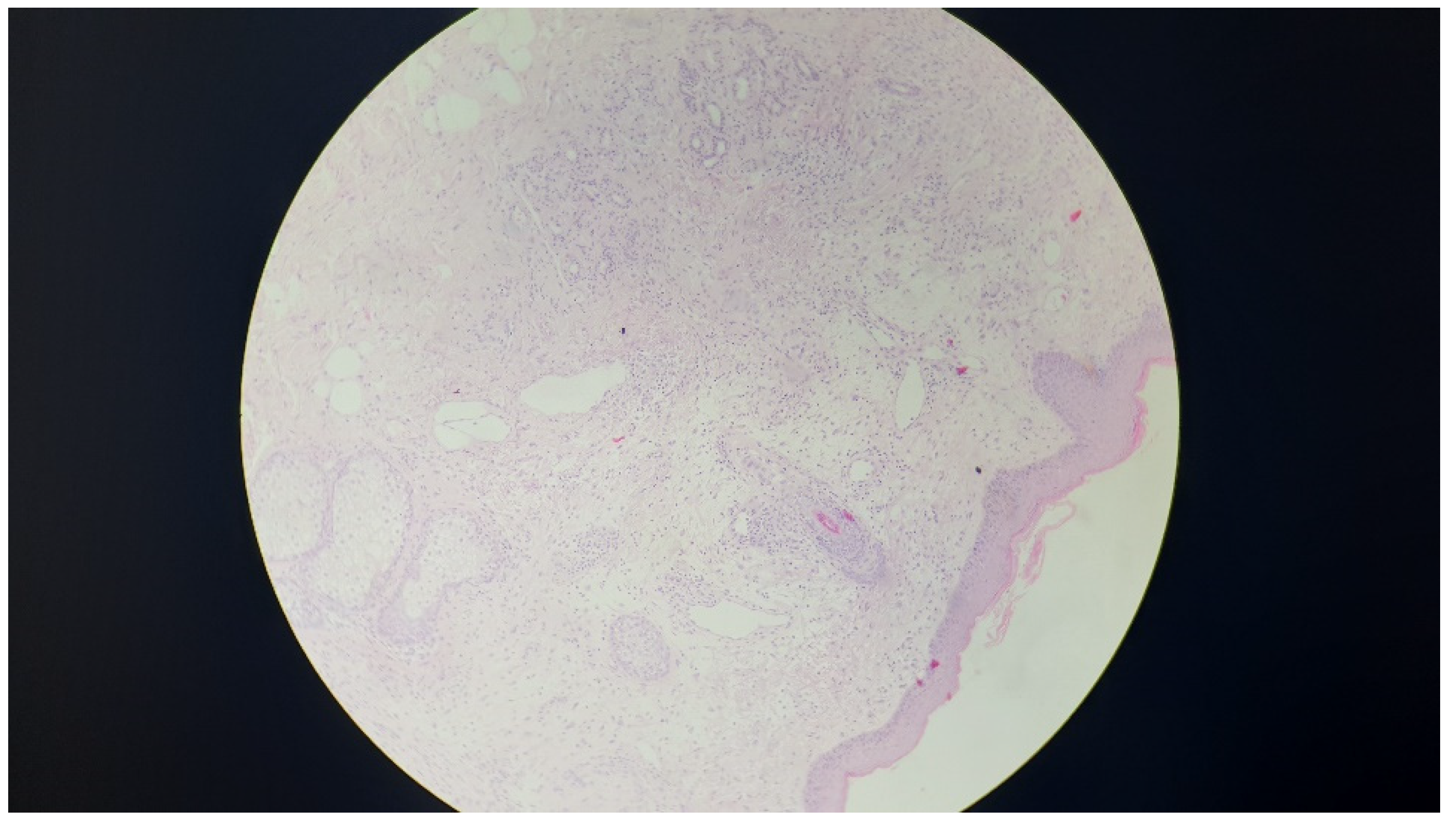
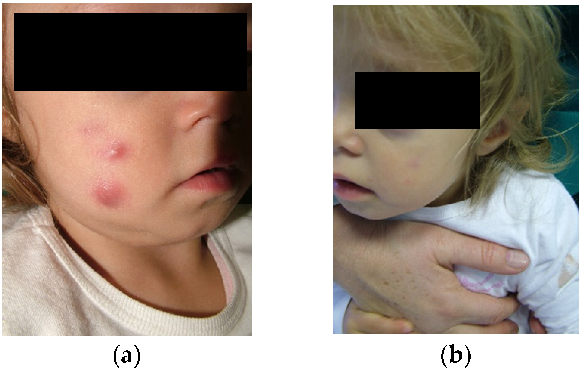
2. Case Report
3. Discussion
4. Conclusions
Author Contributions
Funding
Conflicts of Interest
References
- Boralevi, F.; Léauté-Labrèze, C.; Lepreux, S.; Barbarot, S.; Mazereeuw-Hautier, J.; Eschard, C.; Taïeb, A.; Groupe de Recherche Clinique en Dermatologie Pédiatrique. Idiopathic facial aseptic granuloma: A multicentre prospective study of 30 cases. Br. J. Dermatol. 2007, 156, 705–708. [Google Scholar] [CrossRef]
- Knöpfel, N.; Gómez-Zubiaur, A.; Noguera-Morel, L.; Torrelo, A.; Hernandez-Martin, A. Ultrasound findings in idiopathic facial aseptic granuloma: Case series and literature review. Pediatr. Dermatol. 2018, 35, 397–400. [Google Scholar] [CrossRef]
- González Rodríguez, A.J.; Jordá Cuevas, E. Idiopathic facial aseptic granuloma. Clin. Exp. Dermatol. 2015, 40, 298–300. [Google Scholar] [CrossRef]
- Neri, I.; Raone, B.; Dondi, A.; Misciali, C.; Patrizi, A. Should idiopathic facial aseptic granuloma be considered granulomatous rosacea? Report of three pediatric cases. Pediatr. Dermatol. 2013, 30, 109–111. [Google Scholar] [CrossRef] [PubMed]
- Prey, S.; Ezzedine, K.; Mazereeuw-Hautier, J.; Eschard, C.; Barbarot, S.; Boralevi, F.; Taïeb, A.; Léaute-Labrèze, C.; Groupe de Recherche Clinique en Dermatologie Pédiatrique. IFAG and childhood rosacea: A possible link? Pediatr. Dermatol. 2013, 30, 429–432. [Google Scholar] [CrossRef] [PubMed]
- Chamaillard, M.; Mortemousque, B.; Boralevi, F.; Marques da Costa, C.; Aitali, F.; Taïeb, A.; Léauté-Labrèze, C. Cutaneous and ocular signs of childhood rosacea. Arch. Dermatol. 2008, 144, 167–171. [Google Scholar] [CrossRef] [PubMed]
- Orion, C.; Sfecci, A.; Tisseau, L.; Darrieux, L.; Safa, G. Idiopathic facial aseptic granuloma in a 13-year-old boy dramatically improved with oral doxycycline and topical metronidazole: Evidence for a link with childhood rosacea. Case Rep. Dermatol. 2016, 8, 197–201. [Google Scholar] [CrossRef] [PubMed]
- Roul, S.; Léauté-Labrèze, C.; Boralevi, F.; Bioulac-Sage, P.; Maleville, J.; Taïeb, A. Idiopathic aseptic facial granuloma (pyodermite froide du visage): A pediatric entity? Arch. Dermatol. 2001, 137, 1253–1255. [Google Scholar] [PubMed]
- Jordan, G.A.; Beier, K. Chalazion; StatPearls Publishing: Treasure Island, FL, USA, 2019. [Google Scholar]
- Jansen, T.; Burgdorf, W.H.C.; Pledwig, G. Pathogenesis and treatment of acne in childhood. Pediatr. Dermatol. 1997, 14, 17–21. [Google Scholar] [CrossRef] [PubMed]
- Wollina, U.; Langner, D.; França, K.; Gianfaldoni, S.; Lotti, T.; Tchernev, G. Pyogenic granuloma-A common benign vascular tumor with variable clinical presentation: New findings and treatment options. Open Access Maced. J. Med. Sci. 2017, 5, 423–426. [Google Scholar] [CrossRef] [PubMed]
- Collie, J.S.; Fillman, E.P. Xanthogranuloma, Juvenile (Nevoxanthoendothelioma, JXG); StatPearls Publishing: Treasure Island, FL, USA, 2019. [Google Scholar]
- Baroni, A.; Russo, T.; Faccenda, F.; Piccolo, V. Idiopathic facial aseptic granuloma in a child: A possible expression of childhood rosacea. Pediatr. Dermatol. 2013, 30, 394–395. [Google Scholar] [CrossRef] [PubMed]
- Two, A.M.; Wu, W.; Gallo, R.L.; Hata, T.R. Rosacea: Part II. Topical and systemic therapies in the treatment of rosacea. J. Am. Acad. Dermatol. 2015, 72, 761–770. [Google Scholar] [CrossRef] [PubMed]
- Ozer, P.A.; Gurkan, A.; Kurtul, B.E.; Kabatas, E.U.; Beken, S. Comparative clinical outcomes of pediatric patients presenting with eyelid nodules of idiopathic facial aseptic granuloma, hordeola, and chalazia. J. Pediatr. Ophthalmol. Strabismus 2016, 53, 206–211. [Google Scholar] [CrossRef] [PubMed]


| Skin Lesion | Site | Clinic | Aetiology |
|---|---|---|---|
| IFAG | Nodules located on the cheeks or eyelids. | One or more nontender, erythematous to violaceous nodules. | Still unclear. |
| Chalazion | Conjunctival portion of the lid. | Red and painless. | Cyst because of a blocked oil gland. |
| Nodular infantile acne | Face and trunk. | Several superficial inflammatory papules and blackheads. | Chronic-inflammatory disorder of the hair follicle and sebaceous glands. |
| Pyodermas | Usually lower limbs or trunk. | Recurrent appearance of large skin ulcers. | Unknown, probably depends on an abnormal immuno-mediated response. |
| Pyogenic granulomas | Gums, nasal septum, or other sites on the skin. | Reddish exophytic vascular nodules that can grow rapidly. | Benign vascular tumor that is composed of capillaries and venules. |
| Xantogranulomas | Head, neck, or trunk. | One or multiple brown-yellow nodules. | Can be associated with glaucoma, uveitis or iritis, and Von Recklinghausen’s disease. |
| Vascular malformations | Anywhere along the blood vessels. | Variable, the flat angioma presents itself as a congenital macula, pale pink to vinous red, and variable extension and shape. | Abnormal development of capillaries, arteries, veins, and lymphatic vessels. |
| Pilomatricomas | Face and neck, in particular the preauricular area, cheek, forehead, upper eyelid, and eyebrow. | Solitary neoformed nodules asymptomatous of stony hardness of irregular shape, angled and faceted. | Keratin originating from the hair bulb derivated from the Trichocytes. |
| Dermoid and epidermoid cysts | Head, neck, or face. | Mass that normally becomes visible at birth or in early childhood as a small painless lump. | Congenital defect that is created during the development of the embryo for a defective growth of the skin layers. |
© 2019 by the authors. Licensee MDPI, Basel, Switzerland. This article is an open access article distributed under the terms and conditions of the Creative Commons Attribution (CC BY) license (http://creativecommons.org/licenses/by/4.0/).
Share and Cite
Miconi, F.; Principi, N.; Cassiani, L.; Celi, F.; Crispoldi, R.; Russo, A.; Esposito, S.; Papini, M. A Cheek Nodule in a Child: Be Aware of Idiopathic Facial Aseptic Granuloma and Its Differential Diagnosis. Int. J. Environ. Res. Public Health 2019, 16, 2471. https://doi.org/10.3390/ijerph16142471
Miconi F, Principi N, Cassiani L, Celi F, Crispoldi R, Russo A, Esposito S, Papini M. A Cheek Nodule in a Child: Be Aware of Idiopathic Facial Aseptic Granuloma and Its Differential Diagnosis. International Journal of Environmental Research and Public Health. 2019; 16(14):2471. https://doi.org/10.3390/ijerph16142471
Chicago/Turabian StyleMiconi, Francesco, Nicola Principi, Lorenzo Cassiani, Federica Celi, Roberta Crispoldi, Ada Russo, Susanna Esposito, and Manuela Papini. 2019. "A Cheek Nodule in a Child: Be Aware of Idiopathic Facial Aseptic Granuloma and Its Differential Diagnosis" International Journal of Environmental Research and Public Health 16, no. 14: 2471. https://doi.org/10.3390/ijerph16142471
APA StyleMiconi, F., Principi, N., Cassiani, L., Celi, F., Crispoldi, R., Russo, A., Esposito, S., & Papini, M. (2019). A Cheek Nodule in a Child: Be Aware of Idiopathic Facial Aseptic Granuloma and Its Differential Diagnosis. International Journal of Environmental Research and Public Health, 16(14), 2471. https://doi.org/10.3390/ijerph16142471