Article

Synthesis and Bioactivities of Marine Pyran-Isoindolone Derivatives as Potential Antithrombotic Agents

1
College of Food Science and Technology, Shanghai Ocean University, Shanghai 201306, China
2
Shanghai Engineering Research Center of Hadal Science and Technology, College of Marine Sciences, Shanghai Ocean University, Shanghai 201306, China
3
CQM-Centro de Química da Madeira, Campus da Penteada, Universidade da Madeira, 9000-390 Funchal, Portugal
4
School of Pharmacy, Jiangxi Science and Technology Normal University, Nanchang 330013, China
5
School of Pharmacy, Nanjing Medical University, Nanjing 211166, China
6
Shanghai Engineering Research Center of Aquatic-Product Processing & Preservation, Shanghai 201306, China
7
Laboratory of Quality and Safety Risk Assessment for Aquatic Products on Storage and Preservation (Shanghai), Ministry of Agriculture, Shanghai 201306, China
*
Authors to whom correspondence should be addressed.
These two authors contributed equally to this work.
Mar. Drugs 2021, 19(4), 218; https://doi.org/10.3390/md19040218
Submission received: 15 March 2021 / Revised: 9 April 2021 / Accepted: 13 April 2021 / Published: 15 April 2021

Abstract

:
2,5-Bis-[8-(4,8-dimethyl-nona-3,7-dienyl)-5,7-dihydroxy-8-methyl-3-keto-1,2,7,8-teraahydro-6H-pyran[a]isoindol-2-yl]-pentanoic acid (FGFC1) is a marine pyran-isoindolone derivative isolated from a rare marine microorganism Stachybotrys longispora FG216, which showed moderate antithrombotic(fibrinolytic) activity. To further enhance its antithrombotic effect, a series of new FGFC1 derivatives (F1F7) were synthesized via chemical modification at C-2 and C-2′ phenol groups moieties and C-1″ carboxyl group. Their fibrinolytic activities in vitro were evaluated. Among the derivatives, F1F4 and F6 showed significant fibrinolytic activities with EC50 of 59.7, 87.1, 66.6, 82.8, and 42.3 μM, respectively, via enhancement of urokinase activity. Notably, derivative F6 presented the most remarkable fibrinolytic activity (2.72-fold than that of FGFC1). Furthermore, the cytotoxicity of derivative F6 was tested as well as expression of Fas/Apo-1 and IL-1 on HeLa cells. The results showed that, compared to FGFC1, derivative F6 possessed moderate cytotoxicity and apoptotic effect on HeLa cells (statistical significance p > 0.1), making F6 a potential antithrombotic agent towards clinical application.

1. Introduction

Nowadays, non-transmissible chronic diseases have become fundamental medical problems [1] including cardiovascular, neurological, rheumatologic, diabetic, etc. Among them, cardiovascular disease is one leading cause of death in non-transmissible disease. The World Health Organization reported that approximately 18 million people died from cardiovascular disease each year [2]. Moreover, cardiovascular disease originated mostly from thrombosis [3], such as arterial and venous thrombosis, atherosclerosis, heart attacks, strokes and peripheral vascular diseases, whose death rate was close to that of cancer in recent years [4]. It is estimated that more than 1 million people die from cardiovascular disease every year in the United States [5]. Due to genetics and environmental influence, thrombosis has become recurrent and the age of onset has become younger [6], which seriously threaten human beings’ health worldwide [7,8].
To date, there are three generations of drugs that have been used for the treatments of thrombosis. The first generation of thrombolytic agent is an antigenic bacterial product with non-specificity and thrombolytic activities, including streptokinase (SK) [9] and urokinase (UK) [10]. They can transform plasminogen into active plasmin, which is combined with fibrin for promoting the dissolution of the thrombus. However, they result in bleeding diathesis [11]. The second-generation features improved fibrin specificity, containing tissue plasminogen activator (t-PA) [12], single-chain urokinase plasminogen activator (rscu-PA) [13] and acetylated plasminogen streptokinase activator complex (APSAC) [14]. The drugs can activate fibrin-associated plasminogen, causing plasminogen or α2-antiplasmindepletion or systemic fibrinogen disintegration. Nevertheless, it is limited due to its short half-life and serious anaphylactic reaction [15]. The third generation can lengthen half-life, increase resistance to plasma protease inhibitors, and bind to fibrin, more selectively including TNK-PA [16], STAR, r-PA [17], etc. [18]. However, they also have the drawback of bleeding risks [19]. Therefore, the exploration of small-molecule drugs with antithrombotic activities has received increasing attention owing to their great potential and security in the safety of thrombus. Currently, many small-molecule antithrombotic agents have been reported, including warfarin, apixaban, edoxaba, rivaroxaban, dabigatran, the vitamin K antagonists, etc. [20,21,22,23].
At present, the discovery of most drugs originates from chemical modifications of natural products [24,25,26,27,28,29]. Thus, chemists are devoted to synthesizing various new compounds using diverse natural products as substrates. Moreover, they found that those compounds have better biological activity than natural products [30,31].
Previously, a marine pyran-isoindolone derivative, 2,5-bis-[8-(4,8-dimethyl-nona-3,7-dienyl)-5,7-dihydroxy-8-methyl-3-keto-1,2,7,8-tetrahydro-6H-pyran [a]isoindol-2-yl]-pentanoic acid (FGFC1, Scheme 1), was isolated from a rare marine microorganism Stachybotrys longispora FG216 (CCTCC No M2012272) in our laboratory [32]. In our previous report, FGFC1 exhibited fibrinolytic activity in vitro and in vivo [33]. However, fibrinolytic activity and stability of FGFC1 have limited its further investigation as a thrombolytic candidate for therapeutic effects [34,35,36,37]. Phenolic hydroxyl moieties (C2-OH/C2′-OH) and carboxyl fragment (C1″-COOH) are important functional groups in FGFC1. Our previous works showed that liphatic and benzyl derivatives possessed significant potency [38,39]. Therefore, pyran-isoindolone derivatives F1F7 were synthesized using compound FGFC1 as a starting material via chemical modification at C-2 and C-2′ phenol groups moieties and C-1″ carboxyl group (Scheme 1). Furthermore, their fibrinolytic activities were also evaluated. The results indicated that F6 possessed the most remarkable fibrinolytic activity. Then, its cytotoxicity was tested as well as Fas/Apo-1 and IL-1 on HeLa cells with satisfactory results.

2. Results and Discussion
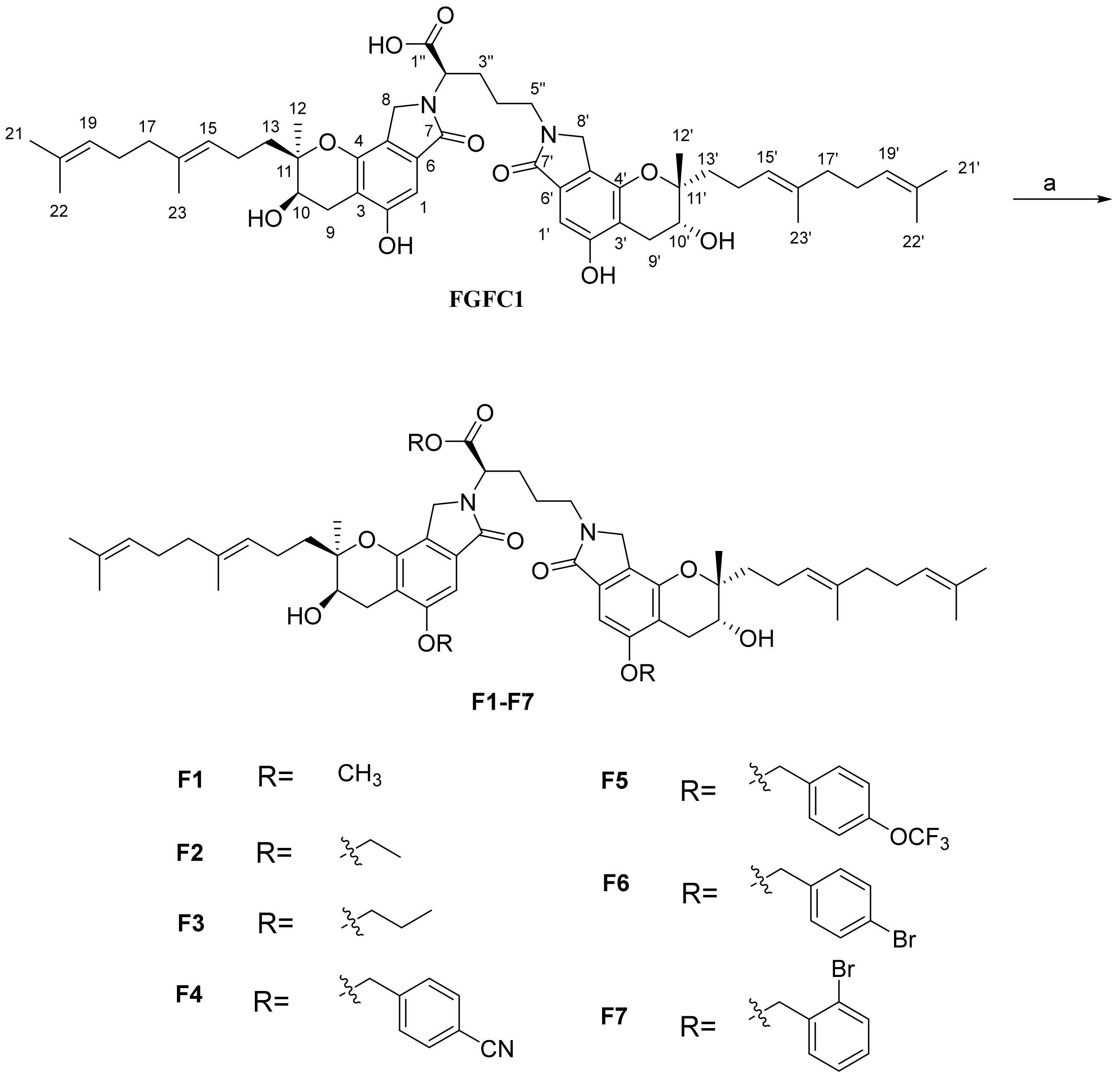
2.1. Chemistry

FGFC1 had two types of acidic functional groups: phenolic phenol groups moieties (C2-OH/C2′-OH) and carboxyl fragment (C1″-COOH). Whether FGFC1 was oral or injected, its acidic groups would cause unnecessary irritation at the site of administration. Thus, the acidity of FGFC1 should be alleviated but its stable antithrombotic activity needs to be maintained, which could be completed through chemical modification including esterification and etherification. Nucleophilic oxygen anions at C-2, C2′ and C-1″ in FGFC1 reacted with various substituted halo-(iodo or bromo) hydrocarbon compounds in the presence of K2CO3 to prepare new FGFC1 derivatives, with phenolic ether groups at C-2 and C2′, and aliphatic ester groups at C-1″. The hydrophobic modification reaction of FGFC1 performed on C-2 and C-2′ phenol groups moieties and C-1″carboxyl group is shown in Scheme 1. FGFC1 was treated with iodomethane, bromoethane, and bromopropane in the presence of K2CO3 in acetone to yield derivatives F1F3, with the isolated yields ranging from 62% to 75%. Moreover, FGFC1 was treated with 4-bromobenzonitrile, 4-(trifluoromethoxy)benzyl bromide, 4-bromobenzyl bromide, and 2-bromobenzyl bromide in the presence of K2CO3 in N,N-dimethylacetamide to yield derivatives F4F7, with the isolated yields ranging from 50% to 70%. Finally, molecular structures of the synthesized FGFC1 derivatives (F1F7) were fully characterized in the Supplementary Materials.

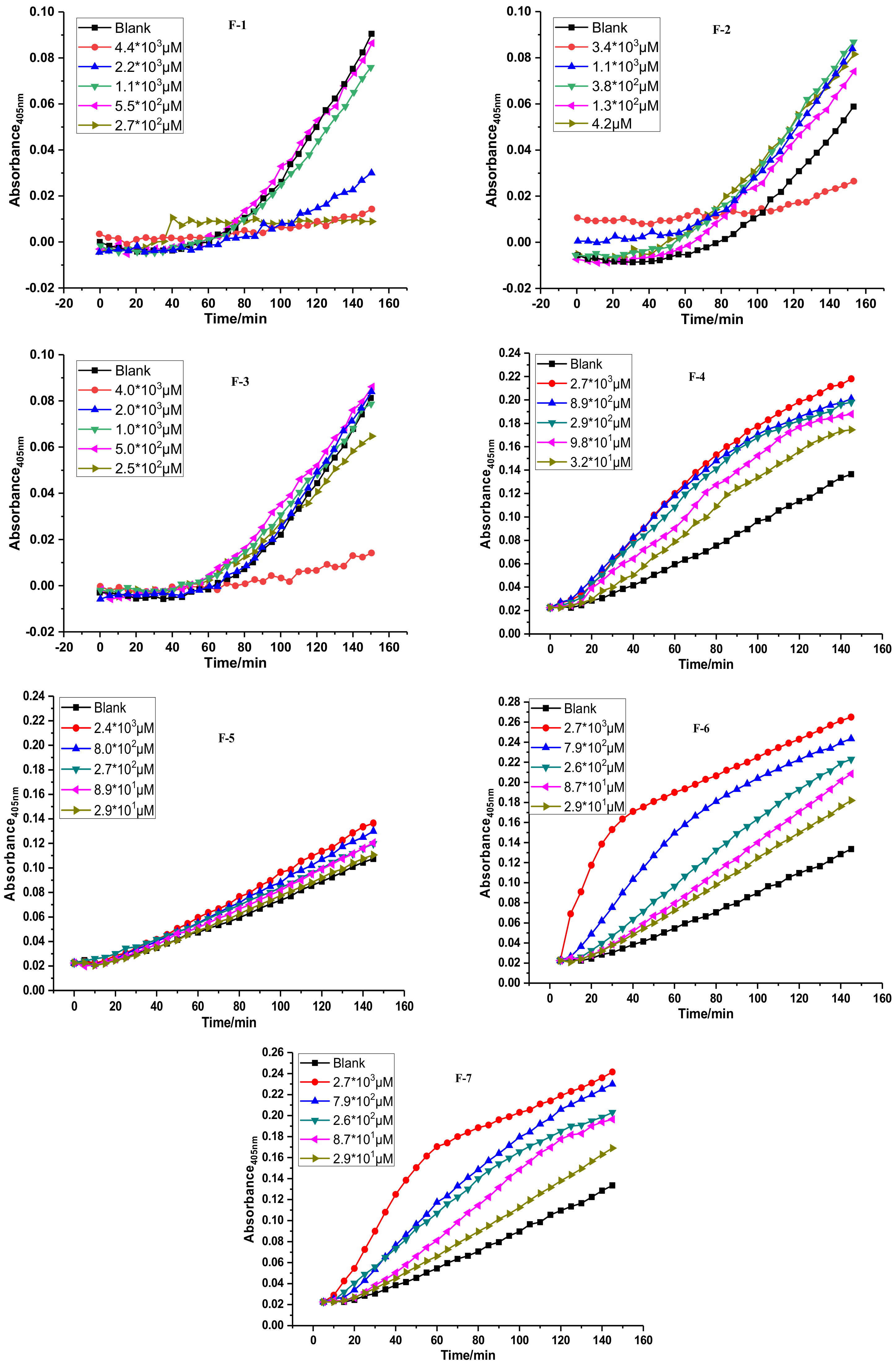
2.2. Fibrinolytic Activities of FGFC1 and F1–F7 In Vitro

To evaluate the antithrombotic effect, all the synthesized FGFC1 derivatives were tested for their fibrinolytic activities in vitro. Fibrinolytic activity of each compound was expressed as 50% effective concentration (EC50), and FGFC1 was used as a positive control. The reciprocal activation of pro-uPA catalyzed plasminogen, which was measured based on urokinase activity by a chromogenic substrate S-2444. EC50 was calculated with the slope of kinetic curve of enzymatic reaction based on the reciprocal activation of pro-uPA and plasminogen (Figure 1 and Figure 2) [40]. The results are shown in Table 1. The effect of introducing different hydrophobic moieties/groups to C-2, C-2′ and C-1′′ positions of FGFC1 was investigated by replacement of the protons on C2-OH/C2′-OH moieties and C1′′-COOH position, with methyl, ethyl, propyl, and bromo-substituted benzyl halide groups to yield derivatives F1F7 (purity > 98% by HPLC analysis), which presented different urokinase activities and reciprocal activation of Pro-uPA-catalyzed plasminogen in vitro.
The synthesized FGFC1 derivatives included aliphatic F1F3 and benzyl F4F7 compounds. For derivatives F1F3, aliphatic (methyl, ethyl and n-propyl) groups were introduced to C-2, C-2′ and C-1″ positions in FGFC1. The EC50 values showed that the fibrinolytic activity of derivatives F1F3 had a tendency as follows: with the increasing of aliphatic chain length, the EC50 gliding down initially from 59.7 μM (F1) to 87.1 μM (F2) and then climbing slowly to 66.6 μM (F3). Interestingly, all of the aliphatic derivatives F1F3 were more active (1.3–2.0 fold) than FGFC1. This result suggested that substituted C-2, C-2′ and C-1″ alkyl moieties could confer increased fibrinolytic activity. Methyl-substituted derivatives F1 had the most potent fibrinolytic activity with EC50 value of 59.7 μM. However, the kinetic curves of derivatives F1F3 (Figure 2) indicated the fibrinolytic activity in vitro was not an absolutely dose-dependent. Especially at high concentration (4.0 mg/mL), it presented a relatively flat trend, indicating the unsatisfactory performance of aliphatic substituted derivatives F1F3 at 4.0 mg/mL.
For benzyl derivatives F4F7, F4 (EC50 = 82.8 μM) and F6 (EC50 = 42.3 μM) at the 4-position of benzyl scaffold were beneficial to fibrinolytic activity. 4-Br-containing F6 possessed excellent fibrinolytic activity with an EC50 value of 42.3 μM (2.72-fold than FGFC1). Derivative F5, where a trifluoromethoxy group was introduced into the para position of benzene ring, markedly decreased the fibrinolytic activity. Shifting Br atom from para to ortho position to produce 3-Br-containing F7 resulted in retained fibrinolytic activity (EC50 = 119.6 μM for F7 vs. EC50 = 115.0 μM for FGFC1). The remarkably different EC50 of F6 (42.3 μM) and F7 (119.6 μM) implied that the bromo groups at para and ortho positions may bring them different urokinase-activating efficiencies. The kinetic curves of derivatives F1F7 showed that benzyl derivatives F4F7 possessed a dose- and time-dependent manner (Figure 1) and better fibrinolytic activity than aliphatic derivatives F1F3. The curves of aliphatic derivatives F1F3 were concave functions; moreover, no distinct fluctuation was observed in the first 60 min. However, those of benzyl derivatives F4F7 were convex, with a rapid upward trend in the same span of time. These results showed that, by introducing aromatic rings, the fibrinolytic activity could be increased rapidly in the early stage and the fibrinolytic activity could be adjusted by changing the substitution groups, especially on F6 and F7. Notably, F6 possessed more effective and faster fibrinolytic activity than FGFC1, which was worthy of in-depth research and exploration.

2.3. Cytotoxicity, Expression of Fas/Apo-1 and IL-1 of F6 on HeLa Cells

The cytotoxicity of FGFC1 and derivative F6 on HeLa cell lines was screened by MTT assay [41,42]. Figure 2A showed that FGFC1 and derivative F6 possessed moderate cytotoxicity on HeLa cell lines. The introduction of substituted benzyl halide to the C-2, C-2′ and C-1″ position of FGFC1 decreased cytotoxicity, which increased with increasing concentration (0.25, 0.5, 1.0 mg/mL). Moreover, to evaluate the possible apoptosis of HeLa cells induced by F6, Fas/APO-1 (a type of cell-surface NGF/TNF receptor in cancer cell) assay was carried out (Figure 2B) [43,44,45]. To evaluate the immunogenic (inflammation) property, interleukin (IL-1) levels of derivative F6 was also tested by the IL-1 assays (Figure 2C) [46]. The expression level of Fas was remarkably higher than that of the blank, but it was less than that of FGFC1 (Figure 2B). This meant that the ability of F6 to induce apoptosis was weaker than that of FGFC1. The expression level of IL-1 was obviously higher than that of blank, but it was less than that of FGFC1 (Figure 2C). This suggested that the inflammatory response of F6 was weaker than that of FGFC1. According to Figure 2B and Figure 2C, the negative effect of F6 on tissue cell was weaker than that of FGFC1.

3. Experiment

3.1. Materials

All chemicals were analytical grade. Reagents and materials were obtained from commercial suppliers and used without further purification. FGFC1 was isolated from S. longispora FG216. Silica gel (200–300 mesh) for column chromatography was purchased from Qingdao Makall Group Co., Ltd. Pro-uPA, BSA (bovine serum albumin), plasminogen and plasmin were purchased from Sigma Aldrich (China). The chromogenic pyro-glutamyl-glycyl-l-arginine-p-nitroanilide S-2444 was purchased from BioMed. Tris-HCl buffer (100 mmol/L, NaCl, pH 7.4) and an enzyme-labeled (microplate reader) instrument (SH-1000, CORONA, Ibarakiken, Japan) were used throughout the fibrinolytic activity in vitro. Fas/Apo-1 Elisa kit and IL-1 Elisa kit were purchased from Shanghai Fusheng Industrial Co., Ltd. (China). Human cervical cancer cells (HeLa) were purchased from Shanghai Cell Bank of Chinese Academy of Sciences.

3.2. Chemistry

The syntheses of the derivatives were described in Supplementary Materials. Column chromatography (CC): silica gel (200–300 mesh; Qingdao Makall Group Co., Ltd.; Qingdao, China). All reactions were monitored using thin-layer chromatography (TLC) on silica gel plates.
Nuclear magnetic resonance spectra were recorded on a Bruker DRX 500 MHz NMR spectrometer. Mass spectra (MS) were recorded on an Advantage Max LCQ Thermo-Finnigan mass spectrometer. General procedure for preparation and original spectra of derivatives F1F7 are available in Supporting Information.

3.3. Fibrinolytic Activity in Vitro

The fibrinolytic activities of compounds FGFC1 and derivatives F1F7 were evaluated by plasmin method. Tris-HCl buffer solution (100 mmol/L, NaCl, pH 7.4), plasminogen (plg), BSA, and S-2444 were used as substrates. Derivatives F1F7 dissolved in 0.05 mol/L Tris-HCl buffer containing NaCl (100 mmol/L) at pH 7.4 and the BSA used as a substrate.The concentrations of plg, BSA and S-2444 were 1.5 μmol/L, 5 μmol/L and 4 nmol/L, respectively. They were prepared in a 96-well microplate. After the predetermined sample solutions (compounds FGFC1 and F1F7) were added, pro-uPA (20 μmol/L) was added into the 96-well microplate. The microplate was then cultivated at 37 °C for 60 min. The continuous variation trend of absorbance was determined for evaluation of fibrinolytic activity on the slope of the plots of A405 nm within 150 min. In the blank group, an equal volume of Tris-HCl solution was used as a blank control. All sample solutions were prepared by adding a small amount of DMSO (<5%, v/v) solution to dissolve the derivatives.

3.4. Cytotoxicity, Expression of Fas/Apo-1 and IL-1 of Derivative F6 on HeLa Cells

3.4.1. Cell Lines

HeLa (human cervical carcinoma) cells were incubated in medium (10% fetal bovine serum, 1% penicillin, 1% streptomycin) at 37 °C with 95% air and 5% CO2. Cells were passaged every 2–3 d and select exponential growth cells were used for further experiments.

3.4.2. MTT Assay

The cell growth inhibitory activity of FGFC1 and derivative F6 on HeLa cell lines were determined by the MTT (3-(4,5-dimethylthiazol-2yl)-2,5-diphenyl tetrazolium bromide) assay as we previously reported [41,42]. In addition, an equal volume of DMEM medium was added to the blank group.

3.4.3. Fas/Apo-1 Assay on HeLa Cells

Fas/Apo-1 assay was carried out according to the manufacturer’s protocol [43,44,45]. A blank control group was set up. It was not added with the samples (FGFC1 and derivative F6) and the enzyme standard reagent, and the other steps were the same. The standard curve group and the sample group were tested. On the 96-well plate, the standard curve group was supplemented with 50 μL of the standard. The sample group tested was firstly added with a sample (FGFC1 and derivative F6) dilution of 40 μL, and then 10 μL of the sample was added (the final dilution of the sample was 5 times). After that, the plate was incubated at 37 °C for 30 min. Then, 50 μL chromogenic reagent A and 50 μL chromogenic reagent B per well were added, and the chromogenic reaction took place. Ultimately, the reaction was terminated by the addition of 50 μL stop solution per well and mixed thoroughly. After the addition of stop solution to reaction wells, the absorbance was recorded at OD 450 nm on an Enzyme-Labeled (microplate reader) Instrument after 15 min.

3.4.4. IL-1 Assay on HeLa Cells

The cell culture method on the 96-well plate is in accordance with that in the MTT assay. The Elisa kit assay based on IL-1 of HeLa cells was consistent with the Fas/Apo-1 Elisa kit assay [46].

4. Conclusions

In conclusion, a series of new FGFC1 derivatives (F1F7) were synthesized by chemical modification at C-2 and C-2′ phenol groups moieties and C-1″ carboxyl group. Derivatives F1F4 and F6 displayed significant fibrinolytic activities with EC50 of 59.7, 87.1, 66.6, 82.8 and 42.3 μM respectively, via enhancement of urokinase activity. Among them, derivative F6 presented the most remarkable fibrinolytic activity (2.72-fold than that of FGFC1). The evaluation data showed that derivative F6 possessed moderate cytotoxicity and apoptotic effect on HeLa cells in the Fas/Apo-1 assay and did not cause obvious inflammation (statistical significance p > 0.1), making derivative F6 a potential antithrombotic agent towards clinical application. Moreover, the results suggest a simple and efficient method to enhance/optimize the anti-thrombotic activity of FGFC-1 via “one-pot” hydrophobic modification.

Supplementary Materials

The following are available online at https://www.mdpi.com/article/10.3390/md19040218/s1. Part 1: General procedure for preparation of derivatives F1–F7 [1,2]; Part 2: NRM Spectra.

Author Contributions

R.G. designed the experiment and prepared the manuscript. W.W. and J.F. proposed many instructions on the experiment. Y.W. and Z.F. synthesized new compounds and collected experimental data. R.S. and H.C. revised the manuscript carefully. R.G. and Q.T. gave final approval of the version to be submitted. All authors have read and agreed to the published version of the manuscript.

Funding

The work was supported by the National Natural Science Foundation of China (No. 81502955), the Doctoral Scientific Research Foundation of Shanghai Ocean University (No. A2030214300077), the Young Teachers Training Program of Shanghai (No. A12056160002), the Plan of Innovation Action in Shanghai (No. 14431906000), the Natural Science Foundation of Jiangxi Province (No. 20161BAB204189), the Science and Technology Research Project of Jiangxi Provincial Department of Education (No. GJJ201115), the Doctoral Scientific Research Foundation of Jiangxi Science Technology Normal University (No. 3000990116). Ruilong Sheng appreciate ARDITI-Agência Regional para o Desenvolvimento da Investigação Tecnologia e Inovação, through the project M1420-01-0145-FEDER-000005-Centro de Química da Madeira-CQM+(Madeira 14-20) and ARDITI-2017-ISG-003, FCT-Fundação para a Ciência e a Tecnologia (project PEst-OE/QUI/UI0674/2019, CQM, Portuguese Government funds), and project PROEQUI PRAM-Reforço do Investimento em Equipamentos e Infraestruturas Científicas na RAM (M1420-01-0145-FEDER-000008) for sponsorship.

Institutional Review Board Statement

Not applicable.

Data Availability Statement

All data is contained within this article and Supplementary Materials.

Acknowledgments

The authors acknowledge the financial support from the National Natural Science Foundation of China, the Doctoral Scientific Research Foundation of Shanghai Ocean University, the Young Teachers Training Program of Shanghai, the Plan of Innovation Action in Shanghai and the Project Funded by Jiangsu Key Laboratory of Marine Pharmaceutical Compound Screening.

Conflicts of Interest

The authors declare no conflict of interest.

References

  1. Yan, Y.K.; Mi, J. Noncommunicable chronic disease prevention should start from childhood. Pediatric. Investig. 2021, 5, 3–5. [Google Scholar] [CrossRef] [PubMed]
  2. The World Health Oragnization. Disease Burden and Mortality Estimates; The World Health Organization: Geneva, Switzerland, 2020. [Google Scholar]
  3. Alagarsamy, K.N.; Mathan, S.; Yan, W.; Rafieerad, A.; Sekaran, S.; Manego, H.; Dhingra, S. Carbon nanomaterials for cardiovascular theranostics: Promises and challenges. Bioact. Mater. 2021, 6, 2261–2280. [Google Scholar] [CrossRef] [PubMed]
  4. Xie, Z.L.; Tian, Y.B.; Lv, X.; Xiao, X.; Zhan, M.M.; Cheng, K.; Li, S.Y.; Liao, C.Z. The selectivity and bioavailability improvement of novel oral anticoagulants: An overview. Eur. J. Med. Chem. 2018, 146, 299–317. [Google Scholar] [CrossRef] [PubMed]
  5. Zenych, A.; Fournier, L.; Chauvierre, C. Nanomedicine progress in thrombolytic therapy. Biomaterials 2020, 258, 120297. [Google Scholar] [CrossRef]
  6. Beaglehole, R.; Bonita, R. Global public health: A scorecard. Lancet 2008, 372, 1988–1996. [Google Scholar] [CrossRef]
  7. Mathers, C.D.; Dejan, L. Projections of global mortality and burden of disease from 2002 to 2030. PLoS Med. 2006, 3, 442. [Google Scholar] [CrossRef] [Green Version]
  8. Pranav, S.; Bindra, V.K. Newer antithrombotic drugs. Indian J. Crit. Care Med. 2010, 14, 188–195. [Google Scholar]
  9. Gemmill, J.D.; Hogg, K.J.; Dunn, F.G. Pre-dosing antibody levels and efficacy of thrombolytic drugs containing streptokinase. Heart 1994, 72, 222–225. [Google Scholar] [CrossRef] [Green Version]
  10. Verstraete, M. Thrombolytic drugs in non-coronary disorders. Drugs 1989, 38, 801–821. [Google Scholar] [CrossRef]
  11. Anderson, J.L.; Karagounis, L.A.; Califf, R.M. Meta-analysis of five reported studies on the relation of early coronary patency grades with mortality and outcomes after acute myocardial infarction. Am. J. Cardiol. 1996, 78, 1–8. [Google Scholar] [CrossRef]
  12. Peter, S.; Frans, V.W. Drug variables in thrombolytic therapy. J. Vasc. Interv. Radiol. 1995, 6, 62–65. [Google Scholar]
  13. Verstraete, M. Third-generation thrombolytic drugs. Am. J. Med. 2000, 109, 52–58. [Google Scholar] [CrossRef]
  14. Marc, V.; Désiré, C. Pharmacology of thrombolytic drugs. J. Am. Coll. Cardiol. 1986, 8, 33–40. [Google Scholar]
  15. Felicita, A.; Biasucci, L.M.; Silvano, B.; Giovanna, C.; Antonio, G.R. A randomized study to investigate the hemostatic effect of tenecteplase (TNK-t-PA) versus alteplase (rt-PA) and streptokinase (SK) in acute myocardial infarction. J. Am. Coll. Cardiol. 2002, 39, 279. [Google Scholar]
  16. Thomas, O.M.; Paul, D.; Jack, C.; Barbara, Q.; Kenric, A. Bleeding complications associated with the use of rt-PA versus r-PA for peripheral arterial and venous thromboembolic occlusions. Tech. Vasc. Interv. Radiol. 2001, 4, 92–98. [Google Scholar]
  17. Adnan, I.; Qureshi, M.D.; Amir, M.; Siddiqui, M.D.; Fareed, K.; Suri, M.D.; Stanley, H.; Kim, M.D.; Zulfiqar, A.M.D.; Abutaher, M.; et al. Aggressive mechanical clot disruption and low-dose Intra-arterial third-generation thrombolytic agent for ischemic stroke: A prospective study. Neurosurgery 2002, 51, 1319–1329. [Google Scholar]
  18. Chan, N.C.; Eikelboom, J.W.; Weitz, J.I. Evolving treatments for arterial and venous thrombosis: Role of the direct oral anticoagulants. Circ. Res. 2016, 118, 1409–1424. [Google Scholar] [CrossRef] [Green Version]
  19. Johannes, R.; Katus, H.A. New antithrombotic drugs on the horizon. Expert Opin. Investig. Drugs 2003, 12, 781–797. [Google Scholar]
  20. Manckoundia, P.; Rosay, C.; Menu, D.; Nuss, V.; Mihai, A.M.; Vovelle, J.; Nuemi, G.; Athis, P.; Putot, A.; Barben, J. The prescription of vitamin K antagonists in a very old population: A cross-sectional study of 8696 a mbulatory subjects aged over 85 years. Int. J. Environ. Res. Public Health 2020, 17, 6685. [Google Scholar] [CrossRef]
  21. Josefina, C.H. Rivaroxaban versus vitamin K antagonist in antiphospholipid syndrome. Ann. Intern. Med. 2020, 73, 505–506. [Google Scholar]
  22. Kapur, N.K.; Shenoy, C.; Yunis, A.A.; Mohammad, N.N.; Wilson, S.; Paruchuri, V.; Mackey, E.E.; Qiao, X.; Shah, A.; Esposito, M.L.; et al. Distinct effects of unfractionated heparin versus bivalirudin on circulating angiogenic peptides. PLoS ONE 2012, 7, e34344. [Google Scholar] [CrossRef] [PubMed] [Green Version]
  23. Rupprecht, H.J.; Blank, R. Clinical pharmacology of direct and indirect factor Xa inhibitors. Drugs 2010, 70, 2153–2170. [Google Scholar] [CrossRef]
  24. Cragg, G.M.; Newman, D.J.; Snader, K.M. Natural Products in Drug Discovery and Development. J. Nat. Prod. 1997, 60, 52–60. [Google Scholar] [CrossRef]
  25. Newman, D.J.; Cragg, G.M.; Snader, K.M. Natural Products as Sources of New Drugs over the Period, 1981–2002. J. Nat. Prod. 2003, 66, 1022–1037. [Google Scholar] [CrossRef]
  26. Newman, D.J.; Cragg, G.M.; Snader, K.M. Natural Products as Sources of New Drugs over the Last 25 Years. J. Nat. Prod. 2007, 70, 461–477. [Google Scholar] [CrossRef] [Green Version]
  27. Wang, J.W.; Li, X.Y.; Zhang, C.Y. Recent Advances on Bioactivity of Seaweed Polysaccharides. Med. Res. 2019, 3, 200003–200007. [Google Scholar] [CrossRef]
  28. Li, B.L.; Wu, W.H. Review on Adaptation between Biomaterials Function of Chitosan and Its Structure. Med. Res. 2019, 3, 190013–190017. [Google Scholar] [CrossRef]
  29. Wang, L.L.; Xie, J.Y.; Wu, W.H.; Li, B.L.; Ou, J. Excellent Microbial Cultivation for Astaxanthin Production and Its Extraction by Rhodotorula benthica. Med. Res. 2018, 2, 180015–180018. [Google Scholar]
  30. Alves, K.M.A.; Cardoso, F.J.B.; Honorio, K.M.; Molfetta, F.A. Design of inhibitors for glyceraldehyde-3-phosphate dehydrogenase (GAPDH) enzyme of leishmania Mexicana. Med. Chem. 2020, 16, 784–795. [Google Scholar] [CrossRef]
  31. De Sousa Luis, J.A.; da Silva Costa, N.A.; Luis, C.C.S.; Lira, B.F.; Athayde-Filho, P.F.; de Souza Lima, T.K.; da Camara Rocha, J.; Scotti, L.; Scotti, M.T. Synthesis of new cyclic lmides derived from safrole, structure- and ligand-based approaches to evaluate potential new multitarget agents against species of leishmania. Med. Chem. 2020, 16, 39–51. [Google Scholar] [CrossRef]
  32. Wang, G.; Wu, W.H.; Zhu, Q.G.; Fu, S.Q.; Wang, X.Y.; Hong, S.T.; Guo, R.H.; Bao, B. Identification and fibrinolytic evaluation of an isoindolone derivative isolated from a rare marine fungus Stachybotrys longispora FG216. Chin. J. Chem. 2015, 33, 1089–1095. [Google Scholar] [CrossRef]
  33. Yan, T.; Wu, W.H.; Su, T.W.; Chen, J.J.; Zhu, Q.G.; Zhang, C.Y.; Wang, X.Y.; Bao, B. Effects of a novel marine natural product: Pyrano indolone alkaloid fibrinolytic compound on thrombolysis and hemorrhagic activities in vitro and in vivo. Arch. Pharm. Res. 2014, 38, 1530–1540. [Google Scholar] [CrossRef] [PubMed]
  34. Guo, R.H.; Duan, D.; Hong, S.T.; Zhou, Y.; Wang, F.; Wang, S.J.; Wu, W.H.; Bao, B. A marine fibrinolytic compound FGFC1 stimulating enzymatic kinetic parameters of a reciprocal activation system based on a single chain urokinase-type plasminogen activator and plasminogen. Process Biochem. 2018, 68, 190–196. [Google Scholar] [CrossRef]
  35. Wang, M.X.; He, H.; Na, K.; Cai, M.H.; Zhou, X.S.; Zhao, W.J.; Zhang, Y.X. Designing novel glucose/ornithine replenishment strategies by biosynthetic and bioprocess analysis to improve fibrinolytic FGFC1 production by the marine fungus Stachybotrys longispora. Process Biochem. 2015, 50, 2012–2018. [Google Scholar] [CrossRef]
  36. Su, T.W.; Wu, W.H.; Yan, T.; Zhang, C.Y.; Zhu, Q.G.; Bao, B. Pharmacokinetics and tissue distribution of a novel marine fibrinolytic compound in Wistar rat following intravenous administrations. J. Chromatogr. B 2013, 942, 77–82. [Google Scholar] [CrossRef]
  37. Ma, Z.B.; Guo, R.H.; Elango, J.; Bao, B.; Wu, W.H. Evaluation of marine diindolinonepyrane in vitro and in vivo: Permeability characterization in Caco-2 cells monolayer and pharmacokinetic properties in Beagle dogs. Mar. Drugs 2019, 17, 651. [Google Scholar] [CrossRef] [Green Version]
  38. Wang, Y.N.; Wang, J.M.; Fu, Z.; Sheng, R.L.; Wu, W.H.; Fan, J.T.; Guo, R.H. Syntheses and evaluation of daphnetin derivatives as novel G protein-coupled receptor inhibitors and activators. Bioorg. Chem. 2020, 104, 104342–104350. [Google Scholar] [CrossRef]
  39. Wang, J.M.; Bian, C.H.; Wang, Y.N.; Shen, Q.; Bao, B.; Fan, J.T.; Zuo, A.X.; Wu, W.H.; Guo, R.H. Syntheses and bioactivities of Songorine derivatives as novel G protein-coupled receptor antagonists. Bioorg. Med. Chem. 2019, 27, 1903–1910. [Google Scholar] [CrossRef]
  40. Florea, A.M.; Büsselberg, D. Cisplatin as an anti-tumor drug: Cellular mechanisms of activity, drug resistance and induced side effects. Cancers 2011, 3, 1351–1371. [Google Scholar] [CrossRef]
  41. Jane, A.P. Cell sensitivity assays: The MTT assay. Methods Mol. Med. 1999, 80, 165–169. [Google Scholar]
  42. Manikandan, A.; Arumugam, S. Molecular explorations of substituted 2-(4-phenylquinolin-2-yl) phenols as phosphoinositide 3-kinase inhibitors and anticancer agents. Cancer Chemother. Pharmacol. 2017, 79, 389–397. [Google Scholar]
  43. Park, I.J.; Kim, M.J.; Ock, J.P.; Myoung, G.P.; Joohun, H. Cryptotanshinone sensitizes DU145 prostate cancer cells to Fas (APO1/CD95)-mediated apoptosis through Bcl-2 and MAPK regulation. Cancer Lett. 2010, 298, 88–98. [Google Scholar] [CrossRef] [PubMed]
  44. Laurie, B.O.S.; Zhang, W.; James, C.C.; Laura, S.A.; Sybil, M.S.; Toshiyoshi, F.; Jack, A.R.; Albert, B.D.; Zhang, W.W.; Ewa, K.; et al. Wild-type human p53 and a temperature-sensitive mutant induce Fas/APO-1 expression. Mol. Cell. Biol. 1995, 15, 3032–3040. [Google Scholar]
  45. Mark, P.B.; Tanya, M.G.; Yury, V.G.; David, W. Involvement of MACH, a novel MORT1/FADD-interacting protease, in Fas/APO-1- and TNF receptor–induced cell death. Cell 1996, 85, 803–815. [Google Scholar]
  46. Philip, L.S.; Jeffrey, T.L.; John, C.L. A modified assay for interleukin-1 (IL-1). J. Immunol. Methods 1985, 85, 85–94. [Google Scholar]
Scheme 1. Synthesis of compounds F1F7. Reagents and conditions: (a) RX (X = I, Br), K2CO3, acetone or N,N-dimethylacetamide, reflux, 2–3 h, yield 50–75%.
Marinedrugs 19 00218 sch001
Figure 1. The effect of derivatives F1F7 on the fibrinolytic activities of the reciprocal activation of Pro-uPA-catalyzed plasminogen.
Marinedrugs 19 00218 g001
Figure 2. The biological effect of F6 on HeLa cells, cytotoxicity (A), expression of Fas/Apo-1 (B), and expression of IL-1 (C) were determined in plasma, with FGFC1 as the reference.
Marinedrugs 19 00218 g002
Table 1. Fibrinolytic activities of FGFC1 and derivatives F1F7 in vitro a.
Marinedrugs 19 00218 i001
CompoundsREC50/μM
FGFC1H115.0
F1CH359.7
F2 Marinedrugs 19 00218 i00287.1
F3 Marinedrugs 19 00218 i00366.6
F4 Marinedrugs 19 00218 i00482.8
F5 Marinedrugs 19 00218 i005133.3
F6 Marinedrugs 19 00218 i00642.3
F7 Marinedrugs 19 00218 i007119.6
a All values are the mean of two independent experiments.
Publisher’s Note: MDPI stays neutral with regard to jurisdictional claims in published maps and institutional affiliations.

