Communication

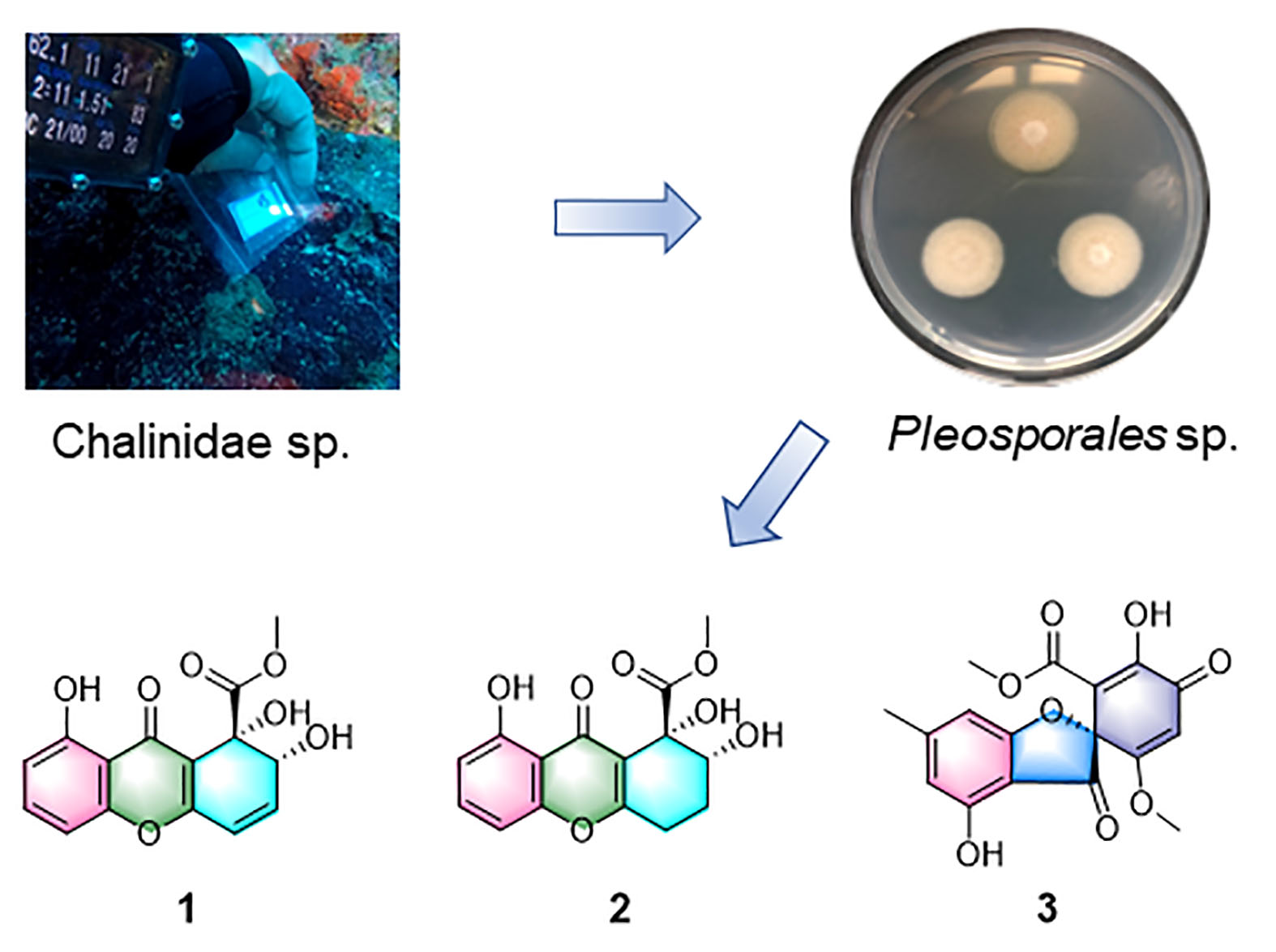
Cytotoxic Polyketide Metabolites from a Marine Mesophotic Zone Chalinidae Sponge-Associated Fungus Pleosporales sp. NBUF144

1
Department of Marine Pharmacy, Li Dak Sum Marine Biopharmaceutical Research Center, College of Food and Pharmaceutical Sciences, Ningbo University, Ningbo, Zhejiang 315800, China
2
State Key Laboratory of Pharmaceutical Biotechnology, School of Life Sciences, Nanjing University, Nanjing 210023, China
3
Key Laboratory of Marine Biogenetic Resources, Third Institute of Oceanography, Ministry of Natural Resources, Xiamen 361005, China
4
State Key Laboratory of Oncogene and Related Genes, Department of Pharmacy, Research Center for Marine Drugs, Ren Ji Hospital, School of Medicine, Shanghai Jiao Tong University, Shanghai 200127, China
5
National Institute of Molecular Biology and Biotechnology, University of the Philippines Diliman, Quezon 1101, Philippines
*
Authors to whom correspondence should be addressed.
Mar. Drugs 2021, 19(4), 186; https://doi.org/10.3390/md19040186
Submission received: 26 February 2021 / Revised: 20 March 2021 / Accepted: 23 March 2021 / Published: 26 March 2021
(This article belongs to the Special Issue Marine Fungal Metabolites: Structures, Activities and Biosynthesis)

Abstract

:
Two new polyketide natural products, globosuxanthone F (1), and 2′-hydroxy bisdechlorogeodin (2), were isolated from the fungus Pleosporales sp. NBUF144, which was derived from a 62 m deep Chalinidae family sponge together with four known metabolites, 3,4-dihydroglobosuxanthone A (3), 8-hydroxy-3-methylxanthone-1-carboxylate (4), crosphaeropsone C (5), and 4-megastigmen-3,9-dione (6). The structures of these compounds were elucidated on the basis of extensive spectroscopic analysis, including 1D and 2D NMR and high-resolution electrospray ionization mass spectra (HRESIMS) data. The absolute configuration of 1 was further established by single-crystal X-ray diffraction studies. Compounds 15 were evaluated for cytotoxicity towards CCRF-CEM human acute lymphatic leukemia cells, and it was found that 1 had an IC50 value of 0.46 µM.

Graphical Abstract

1. Introduction

Marine organisms, especially sponges and microbes associated with them, are prolific producers of structurally diverse and bioactive natural products [1,2,3,4,5]. As sessile filter-feeding animals, sponges provide habitats for diverse and specific microbial communities [6]. The term “holobiont” is used to describe multilevel assemblages of organisms composed, for example, of a sponge and associated microbiota (up to 40% of sponge volume), including fungi, yeast, and bacteria [7,8,9,10]. Sponge-associated fungi have produced many intriguing molecules reported with conspicuous pharmacological and biological properties, exemplified by antimicrobial [11,12,13], cytotoxic [13,14], anti-viral [15,16], antifungal [17], anti-inflammatory [18], and α-glucosidase inhibitory [19] activities. However, a vast quantity of sponge-derived fungal biodiversity remains uninvestigated, suggesting that many natural product metabolites also remain to be discovered.
Mesophotic coral ecosystems (MCEs or “Twilight zone” coral reefs) range from 30 to 150 m deep and represent approximately 80% of potential coral reef habitats worldwide, but little is known about them compared with shallow reefs [20,21]. In an ongoing investigation of fungal metabolites [22,23], rare or unexplored sponge-derived fungi from MCEs have attracted the attention of these authors and others. Several fungi with novel metabolite-producing potential were prioritized for further study after applying the combined strategy of LC-MS/MS molecular networking [24] with the OSMAC (One Strain MAny Compounds) approach [25] of crude extracts from 80 sponge-associated fungi (40 from shallow water and 40 from MCEs; Supplementary Material Figure S1). For example, in the previous work, the organism first prioritized was a Cymostachys fungus that yielded a series of new polyketide compounds, cymopolyphenols A-F [26]. Among them, cymopolyphenols D-F are low-order polymers of cymopolyphenols A that have novel chemical scaffolds. As a continuation of this work of mining new or novel fungal metabolites from MCEs, a second organism was prioritized from the same strain library after the previously reported molecular network [26] was adapted to highlight the secondary metabolite-producing activity of a Pleosporales sp. NBUF144 fungus, which was derived from a 62 m deep Chalinidae family sponge (Figure S1). The chemical investigation of this organism yielded compounds 16 (Figure 1), and the details of these experiments and associated biological testing results are reported herein.

2. Results and Discussion

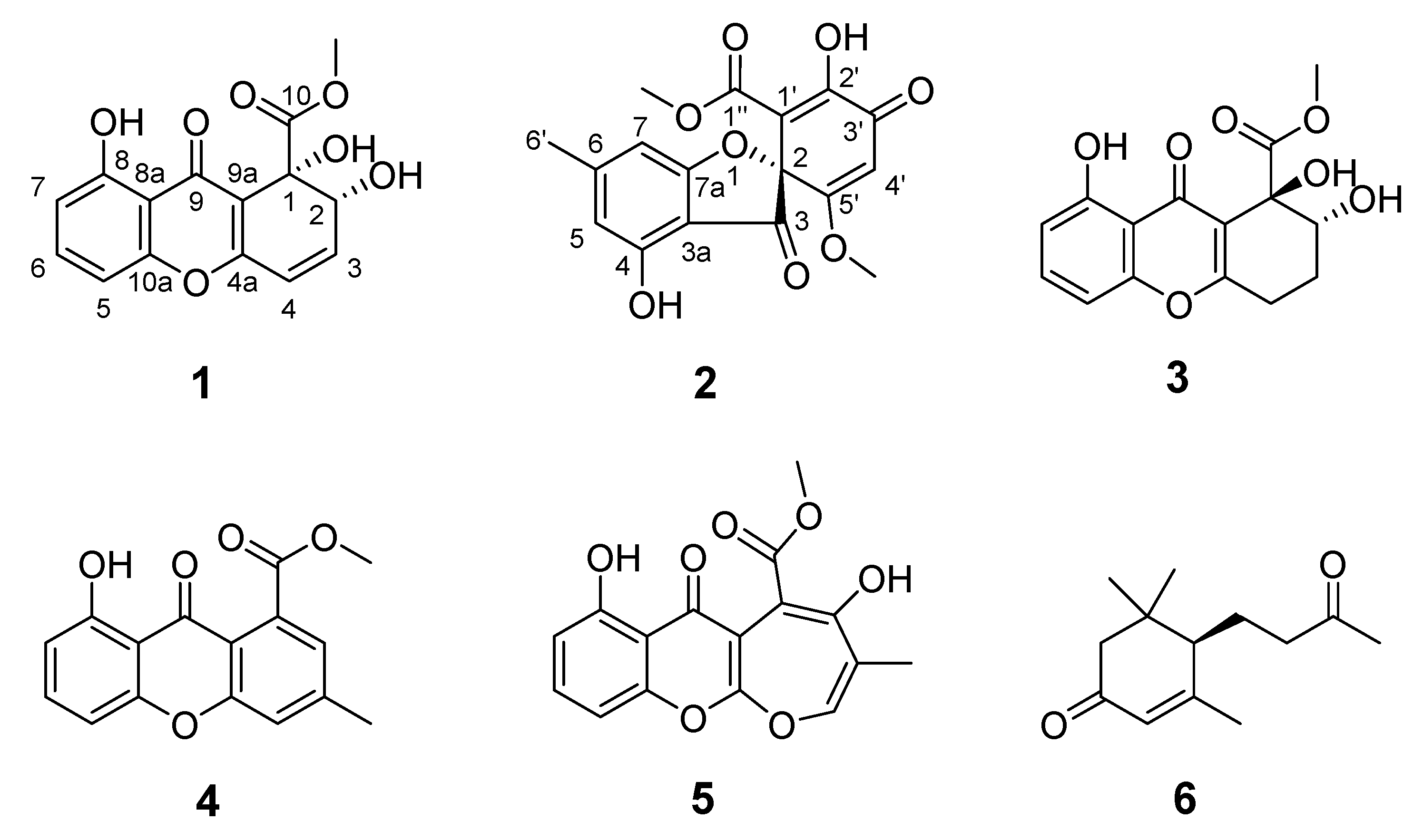
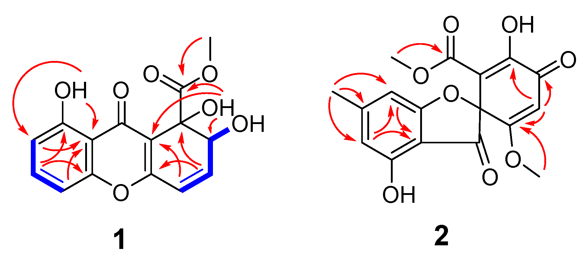
2.1. Structure Elucidation and Identification of Compounds

Compound 1 was determined to have the molecular formula of C15H12O7, based on a sodiated molecular ion peak at m/z 327.0481 [M + Na]+ (calcd for C15H12O7Na, 327.0475) in the high-resolution electrospray ionization mass spectra (HRESIMS), indicating 10 degrees of unsaturation. The 1H and 13C NMR spectra of 1 indicated the presence of one methoxy (δH 3.92, δC 54.4; 10-OCH3), three hydroxy (δH 4.42; 1-OH, δH 2.93; 2-OH, δH 12.13; 8-OH), five protonated olefins (δH 6.50, δC 143.8; CH-3, δH 6.32, δC 120.5; CH-4, δH 6.90, δC 107.3; CH-5, δH 7.52, δC 135.8; CH-6, δH 6.80, δC 112.1; CH-7), two carbonyls (δC 180.7; C-9, δC 174.7; C-10), and non-protonated carbons (δC 73.2; C-1, δC 160.9; C-4a, δC 160.8; C-8, δC 111.0; C-8a, δC 180.7; C-9, δC 113.7; C-9a; δC 155.5; C-10a) (Table 1). Cross peaks detected in the 1H-1H COSY spectrum led to the generation of two isolated spin systems, H-5−H-6−H-7 and 2-OH−H-2−H-3−H-4. HMBC correlations from H-5/H-7 to C-8a, H-6 to C-10a/C-8, and 8-OH to C-8a/C-7 revealed one o,m-disubstituted phenolic group (Figure 2). Further HMBC signals observed from H-3 to C-1/C-4a, H-4 to C-9a, 1-OH to C-2/C-9a/C-10, and 10-OCH3 to C-10 in HMBC established a methyl 1,6-dihydroxy-2,4-cyclohexadiene-1-carboxylate moiety (Figure 2). The co-existence of one unassigned carbonyl (δC 180.7; C-9) and the deshielded shifts of two non-protonated sp2 carbons that suggested linkage to an oxygen atom (C-4a at δC 160.9 and C-10a at δC 155.5) indicated the presence of a pyran-4-one moiety connecting the two identified spin systems (Figure 2), which together satisfied the remaining requirement of two degrees of unsaturation. The methoxy group had an HMBC correlation with C-10, conclusively assigning its position of attachment to the scaffold. Therefore, the planar structure of 1 was determined to be methyl 1,2,8-trihydroxy-9-oxo-2,9-dihydro-1H-xanthene-1-carboxylate, which matched that of globosuxanthone A [27,28,29]. Fortunately, compound 1 was able to be crystalized in a mixed organic solvents system (MeOH/DCM/H2O, 40:20:1), and it was further studied by single-crystal X-ray diffraction. This allowed the (1S,2R) configuration to be assigned to 1 (Figure 3) with a Flack parameter of −0.13(12) [30]. The 1-OH and 2-OH groups in 1 were determined to be cis oriented, while the corresponding groups in globosuxanthone A are trans. Therefore, compound 1 was assigned as a new natural product representing the C-1 epimer of globosuxanthone A, here named globosuxanthone F.
Compound 2 was afforded as colorless prisms. The molecular formula was determined to be C17H14O8 based on the sodium adduct peak at m/z 369.0587 [M + Na]+ (calcd. for C17H14O8Na, 369.0581) in the HRESIMS, requiring 11 degrees of unsaturation. The 1H and 13C NMR spectra of 2 revealed the occurrence of one methyl (δH 2.39, δC 23.2; C-6′), two methoxy (δH 3.44, δC 51.4; C-1″-O-CH3, δH 3.69, δC 57.1; C-5′-OCH3), three olefins (δH 6.40, δC 104.6; CH-5, δH 6.35, δC 109.0; CH-7, δH 5.82, δC 102.5; CH-4′), eight non-protonated carbons (δC 93.4; C-2, δC 108.2; C-3a, δC 171.8; C-4, δC 152.7; C-6, δC 155.8; C-7a, δC 149.6; C-1′, δC 148.3; C-2′, δC 171.2; C-5′), one unconjugated and one conjugated carbonyl (δC 198.1; C-3, δC 180.9; C-3′), and one ester (δC 167.7; C-1”). HMBC correlations were observed from H-5 to C-3a, H-7 to C-5/3a, and H-6′-CH3 to C-5/C-7 that established an m-cresol moiety (Figure 2). Further correlations from H-1”-O-CH3 to C-1”, H-4′ to C-2′/C-3′/C-5′, and H-5′-O-CH3 to C-5′ in HMBC suggested the hallmarks of methyl 2-hydroxy-5-methoxy-3-oxocyclohexa-1,4-diene-1-carboxylate motif as is present in the known fungal metabolite bisdechlorogeodin [31]. From this substructure, one carbonyl and an oxygen atom remained unassigned, together with two double bond equivalents. However, this was determined to indicate a furan-3(2H)-one moiety in 2 as shown in Figure 1 rather than pyran-4-one motifs as in 1, because only the deshielded quaternary carbon C-2 could be assigned to both this central ring system and the cyclohexadienone moiety. A molecule with the same carbon skeleton as 2 was previously reported as bisdechlorogeodin [31]. However, comparison of the NMR data of 2 with that of bisdechlorogeodin highlighted that an olefinic proton at δH/C-2′ 7.11/137.0 in bisdechlorogeodin was absent in 2, and instead an oxygenated sp2 carbon was observed at δC 148.3 in 2, reflecting the 2′-hydroxy group in this new molecule. Single-crystal X-ray diffraction analysis was attempted to establish the C-2 configuration of 2 (Figure 3), but since a centrosymmetric space group (P-1) was obtained with the crystals, only confirmation of the planar structure of 2 was achieved. However, on the basis of biosynthetic logic, it was suggested that 2 should share the 2R configuration of bisdechlorogeodin [31]. Since the additional 2′-OH group in 2 compared to bisdechlorogeodin is understood to impact less the optical rotation (OR) of the molecule than inverted configuration would, the absolute configuration of C-2 in 2 was assigned as R based on the comparable OR data of (−)-bisdechlorogeodin{[α]d-107 (c = 0.1, in EtOH) [32]; [α]d-154 ± 12} [33] and compound 2 {[α]25d-160 (c = 0.1, in MeOH)}. Furthermore, the crystal structure observed for 2 leaves no chance that the additional hydroxy group in this compound would participate in intra-molecular hydrogen bonding that could lead to an ambiguous OR phenomenon that was recognized recently in some analogs of (+)-egenine [34]. Accordingly, the new natural product analog of bisdechlorogeodin, 2, was given the trivial name 2′-hydroxy bisdechlorogeodin.
Compounds 36 were identified as 3,4-dihydroglobosuxanthone A (3) [35], 8-hydroxy-3-methylxanthone-1-carboxylate (4) [36], microsphaeropsone C (5) [35], and 4-megastigmen-3,9-dione (6) [37] by comparison of obtained experimental NMR, MS, and OR data with literature values.

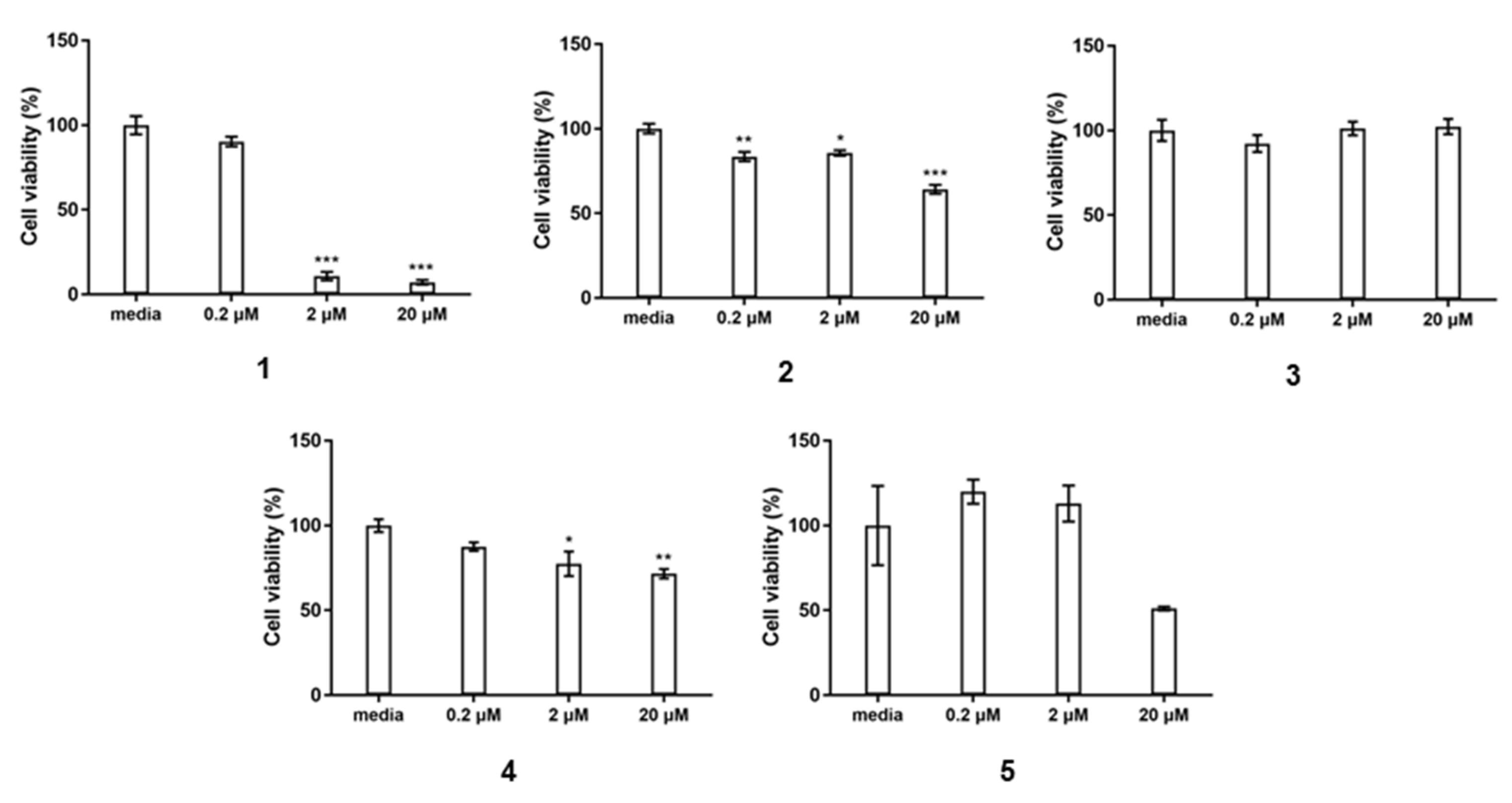
2.2. Bioactivity Assay

Globosuxanthone A has been reported to yield significant cytotoxicity towards eight human solid tumor cell lines, including NCI-H460, MCF-7, SF-268, PC-3, PC-3M, LNCaP, DU-145, and HCT-15, as well as T-cell leukemia Jurkat cells [27,29]. These data inspired in vitro cytotoxicity testing of the compounds isolated here. Compound 1 exhibited potent cytotoxicity in vitro against CCRF-CEM T-cell leukemia cells with a IC50 value of 0.46 μM. Compounds 35 each showed no pronounced cytotoxicity at 20 μM (Figure 4), despite sharing almost identical scaffolds with globosuxanthone A and 1, suggesting the importance of the Δ3,4 unsaturation (comparing 1 to 3) and the presence of hydroxy groups at C-1 and C-2 in this scaffold (comparing 1 to 4) for yielding cancer cell cytotoxicity. Compound 6 was not obtained in sufficient quantity for testing in this study.

3. Materials and Methods

3.1. General

Optical rotations were measured with a JASCO P-2000 automatic polarimeter in MeOH at 20 °C. The CD spectra were recorded on a JASCO P-1500 spectropolarimeter. NMR spectra were recorded in CDCl3 using residual solvents as internal standards with a Bruker AVANCE NEO 600 spectrometer with a 5 mm inverse detection triple resonance (H-C/N/D) cryoprobe having z-gradients. The chemical shift values are given in parts per million (ppm) relative to TMS at 0.0 ppm, and the coupling constants are in Hertz. High-resolution electrospray ionization mass spectra (HRESIMS) were measured on an Agilent (Santa Clara, CA, USA) 6545 Q-TOF instrument. Reversed-phase HPLC purification was carried out on a Waters HPLC equipped with a 1525 binary pump, and a Thermo Scientific (Waltham, MA, USA) ODS-2 Hypersil column (5 µm, 250 × 10 mm). Normal phase column chromatography and thin-layer chromatography were performed using silica gel (200–300 mesh) and GF254 (10–20 mm) (Qingdao Marine Chemical Company, Qingdao, China). YMC*GEL ODS-A (AA12S50; YMC Co., Ltd., Japan) was used for reverse phase column chromatography. Sephadex LH-20 was a product from GE Biotechnology, USA.

3.2. Fungal Strains

The fungal strain was isolated from a Chalinidae family sponge collected in 2018 by scientific-technical SCUBA diving at the depth of 62 m near Apo Island, Negros Oriental, Philippines (9°04′40.6″ N 123°15′57.3″ E and 9°04′33.0″ N 123°15′59.1″ E). The inner tissue of the sponge was sliced into squares (0.5 × 0.5 × 0.5 cm3) and incubated on plates containing modified fungal mediums described elsewhere [22]. The sponge sample loaded petri-dishes were then incubated at 28 °C for two weeks. The fungal colonies were picked and sub-cultivated on PDA (Potato Dextrose Agar; potato 200.0 g, glucose 20.0 g, sea salt 35.0 g, agar powder 20.0 g, and H2O up to a total volume of 1 L) media.
The fungal strain further grown and evaluated in this study was identified as a Pleosporales sp. based on morphological traits and sequence analysis of the ITS region (GenBank accession no. MW386403).

3.3. Cultivation of the Fungi

For chemical investigation, the Pleosporales sp. was cultured in 250 × 1 L Erlenmeyer flasks, each containing 400 mL PDB medium (80 g potato, 8 g glucose, 14.0 g sea salt, 400 mL H2O), for 15 days at 28 °C with agitation (120 rpm). The culture broth was filtered through cheesecloth to separate it into filtrate and mycelia (retained in the cheese), and both of them were extracted with EtOAc (v/v, 1:1) for 5 times and MeOH and DCM (v/v, 1:1) for 3 times, respectively. The separated extracts of the filtrate and mycelia were combined for their similar spots in TLC analysis to afford a total crude organic extract (280 g).

3.4. Extraction and Isolation

The crude extract was subjected to column chromatography (CC) over silica gel (PE/EtOAc, v/v, 100:0→0:100 then EtOAc/MeOH v/v, 100:0→0:100) to afford six fractions (Fr.1–Fr.6). Fr.1 was separated with Sephadex LH-20 in MeOH to give Fr.1.1–Fr.1.3. Of these, Fr.1.1 was further purified by semi-preparative reverse phase (SP-RP) HPLC with CH3CN/H2O (v/v 35:65, 2.0 mL/min) to yield compounds 1 (tR = 15.0 min, 8 mg), 3 (tR = 25.0 min, 1.1 mg). Compound 1 was able to be crystalized in a mixed organic solvents system (MeOH/DCM/H2O, 40:20:1) to afford colorless prisms. Fr.1.2 was subjected to reversed-phase medium-pressure liquid chromatography (MPLC) with MeOH/H2O (v/v 3:7 to 9:1) to offer five sub-fractions (Fr.1.2.1~Fr.1.2.5). Fr.1.2.2 was purified by SP-RP HPLC with CH3CN/H2O (v/v 33:67, 2.0 mL/min) to yield compounds 6 (tR = 30.0 min, 0.8 mg), 4 (tR = 45.0 min, 1.1 mg). Subfraction Fr.1.2.3 afforded compounds 2 (tR = 30.0 min, 10.3 mg) and 5 (tR = 78.0 min, 1.7 mg) after purification by SP-RP HPLC with CH3CN/H2O (v/v 40:60, 2.0 mL/min).
Compound 1: Colorless prisms, UV (MeOH) λmax (log ε) = 208 (4.86) nm, 262 (3.95) nm, 333(3.32) nm, [α]20d-63 (c = 0.1, MeOH). For 1H and 13C NMR spectroscopic data, see Table 1; HR-ESI-MS m/z 327.0481 [M + Na]+ (calcd for C15H12O7Na, 327.0475)
Compound 2: Colorless prisms, UV (MeOH) λmax (log ε) = 204 (3.53) nm, 227 (3.23) nm, 275 (3.31) nm, {[α]25d-160.0 (c = 0.1, MeOH). For 1H and 13C NMR spectroscopic data, see Table 1; HR-ESI-MS m/z 369.0587 [M + Na]+ (calcd. for C17H14O8Na, 369.0581).

3.5. Single-Crystal X-ray Diffraction Analysis

The crystals obtained for 1 and 2 were tested on a Bruker APEX-II CCD diffractometer through Ga Kα (λ = 1.34139 Å). The structures were solved by direct methods (SHELXT-2014) and refined via full-matrix least-squares difference Fourier techniques using SHELXL-2018/3. Crystallographic data for the structures have been deposited with the Cambridge Crystallographic Data Centre. Copies of the data can be obtained, free of charge, on application to the Director, CCDC, 12 Union Road, Cambridge CB2 1EZ, UK (fax: +44-(0)1223-336033 or e-mail: [email protected]).
Crystallographic data for 1: C15H12O7, Mr = 304.25, prism from MeOH/DCM/H2O (40:20:1), space group P21, a = 7.7645(5) Å, b = 6.7905(5) Å, c = 12.6077(9) Å, V = 663.16(8) Å3, Z = 2, µ = 0.669 mm−1, F(000) = 316.0; crystal size: 0.200 × 0.180 × 0.170 mm3; 2369 unique reflections with 2300 obeying the I > 2σ(I); R = 0.0288(2300), wR2 = 0.0781(2369), S = 1.060; supplementary publication no. CCDC-2027079.
Crystallographic data for 2: C17H14O8, Mr = 346.28, prism from MeOH/H2O (40:1), space group P-1, a = 7.6247(9) Å, b = 8.3365(10) Å, c = 14.4259(17) Å, V = 881.17(18) Å3, Z = 2, µ = 0.573 mm−1, F(000) = 360.0; crystal size: 0.220 × 0.160 × 0.190 mm3; 3092 unique reflections with 1293 obeying the I > 2σ(I); R = 0.0709(1293), wR2 = 0.1679(3092), S = 1.011; supplementary publication no. CCDC-2051826.

3.6. Cytotoxicity Assay

CCRF-CEM human T lymphoblast cells were obtained from the National Collection of Authenticated Cell Cultures, Shanghai. Compounds 15 were diluted serially from 10 uM mother solution in DMSO to make the final concentration from 0 to 20 μM and the final DMSO concentration was ≤0.1% in the reaction mixture. CCRF-CEM cells (5 × 10⁴ in 100 μL) and the tested compounds were co-incubated in a 96-well plate and the absorbance at 490 nm were recorded after published protocols [38,39]. The IC50 values were calculated using GraphPad Prism 7 software.

4. Conclusions

The chemical investigation of the MCE sponge-associated fungus Pleosporales sp. NBUF144, after prioritization by a combination of the OSMAC approach and LC-MS/MS molecular networking, led to the isolation of two new polyketide natural products (12). Compounds 15 were screened for in vitro cytotoxicity towards CCRF-CEM cells. Compound 1 showed strong cytotoxic activity with an IC50 value of 0.46 μM, while 3 was not active at concentrations up to 20 μM, indicating the importance of the Δ3,4 olefin moiety in the scaffold of these compounds for this biological activity. Since compound 4 was also not active, in comparison with 1, it is suggested that the C-1 and C-2 hydroxy groups are also important for cytotoxicity. Together, this extends the known potential of this chemical class, as drugs leading to the development of new anticancer agents, as well as restricting the flexibility for modification according to the preliminary natural structure-activity relationship (SAR). Further natural product research of sponge-derived fungi, especially those obtained from understudied MCEs, is expected to continue to yield new chemistry and associated biological activity for evaluation in drug development and ecological research.

Supplementary Materials

LC-MS/MS molecular networking figure showing prioritization of the title strain, 1H NMR, 13C NMR, DEPT135, 1H-1H COSY, HSQC, HMBC, and NOESY spectra for compounds 12, X-ray crystallographic data of 1 and 2 (CIF) and supplementary materials for ECD calculation are available online at https://www.mdpi.com/article/10.3390/md19040186/s1.

Author Contributions

S.H., T.W., X.Y., and H.L. afforded the idea and planned the research. J.Z. and H.Z. carried out the fungus fermentation, metabolites isolation, and structure elucidation with assistance and instruction from C.B.N. and T.W., J.Y. and X.W. performed the cytotoxicity assay. W.W. carried out the chemical calculation. T.W. wrote the manuscript and C.B.N. and J.E.H.L. revised it. All authors have read and agreed to the published version of the manuscript.

Funding

This study was supported by the National Key Research and Development Program of China, funded through MOST (the Ministry of Science and Technology of China; grant 2018YFC0310900 to H.L., X.Y., C.N., and S.H.), the NSFC (the National Natural Science Foundation of China; grant 41906093 to T.W., 41776168 to S.H.), the Natural Science Foundation of Zhejiang Province (grant LGF21D060003 to T.W.), Research Fund for Science in Ningbo University (XYL20021 to T.W.), and the National 111 Project of China (D16013), the Li Dak Sum Yip Yio Chin Kenneth Li Marine Biopharmaceutical Development Fund of Ningbo University.

Institutional Review Board Statement

Not applicable.

Informed Consent Statement

Not applicable.

Data Availability Statement

The datasets generated for this study can be found in the Cambridge Structural Database https://www.ccdc.cam.ac.uk/structures.

Acknowledgments

We are grateful to Ting Han from Blue Flag Diving Club, for technical diving and the collection of the sponges used for mycology in this research; we are indebted to Gong Lin at the Institute of Oceanology, Chinese Academy of Sciences, for the sponge identification.

Conflicts of Interest

The authors declare no conflict of interest.

References

  1. Sahidin, I.; Sabandar, C.W.; Wahyuni; Hamsidi, R.; Mardikasari, S.A.; Zubaydah, W.O.S.; Sadarun, B.; Musnina, W.O.S.; Darmawan, A.; Sundowo, A. Investigation of Compounds and Biological Activity of Selected Indonesian Marine Sponges. Nat. Prod. J. 2020, 10, 312–321. [Google Scholar] [CrossRef]
  2. Ibrahim, S.R.M.; Haidari, R.A.A.; Mohamed, G.A. Megaspinoxide A: New Norterpene Cyclic Peroxide from the Sponge Diacarnus megaspinorhabdosa. Nat. Prod. J. 2014, 4, 38–42. [Google Scholar]
  3. Rangnekar, S.S.; Khan, T. Novel Anti-inflammatory Drugs from Marine Microbes. Nat. Prod. J. 2015, 5, 206–218. [Google Scholar] [CrossRef]
  4. Mayer, A.M.; Rodríguez, A.D.; Berlinck, R.G.; Fusetani, N. Marine pharmacology in 2007–8: Marine compounds with antibacterial, anticoagulant, antifungal, anti-inflammatory, antimalarial, antiprotozoal, antituberculosis, and antiviral activities; affecting the immune and nervous system, and other miscellaneous mechanisms of action. Comp. Biochem. Physiol. Part C Toxicol. Pharmacol. 2011, 153, 191–222. [Google Scholar]
  5. Mayer, A.M.S.; Rodríguez, A.D.; Berlinck, R.G.S.; Hamann, M.T. Marine pharmacology in 2005–6: Marine compounds with anthelmintic, antibacterial, anticoagulant, antifungal, anti-inflammatory, antimalarial, antiprotozoal, antituberculosis, and antiviral activities; affecting the cardiovascular, immune and nervous systems, and other miscellaneous mechanisms of action. Biochim. Biophys. Acta (BBA) Gen. Subj. 2009, 1790, 283–308. [Google Scholar]
  6. Kiran, G.S.; Sekar, S.; Ramasamy, P.; Thinesh, T.; Hassan, S.; Lipton, A.N.; Ninawe, A.S.; Selvin, J. Marine sponge microbial association: Towards disclosing unique symbiotic interactions. Mar. Environ. Res. 2018, 140, 169–179. [Google Scholar] [CrossRef] [PubMed]
  7. Webster, N.S.; Thomas, T. The sponge hologenome. mBio 2016, 7, e00135-16. [Google Scholar] [CrossRef] [Green Version]
  8. Vacelet, J. Etude en microscopie electronique de l’association entre bacteries et spongiaires du genre Verongia (Dictyoceratida). J. Microsc. Biol. Cell 1975, 23, 271–288. [Google Scholar]
  9. Easson, C.G.; Thacker, R.W. Phylogenetic signal in the community structure of host-specific microbiomes of tropical marine sponges. Front. Microbiol. 2014, 5, 532. [Google Scholar] [CrossRef] [Green Version]
  10. Tian, R.M.; Wang, Y.; Bougouffa, S.; Gao, Z.M.; Cai, L.; Bajic, V.; Qian, P.Y. Genomic analysis reveals versatile heterotrophic capacity of a potentially symbiotic sulfur-oxidizing bacterium in sponge. Environ. Microbiol. 2014, 16, 3548–3561. [Google Scholar] [CrossRef]
  11. Li, W.; Jiao, F.W.; Wang, J.Q.; Shi, J.; Wang, T.T.; Khan, S.; Jiao, R.H.; Tan, R.X.; Ge, H.M.; Unguisin, G. A new kynurenine-containing cyclic heptapeptide from the spong-associated fungus Aspergillus candidus NF2412. Tetrahedron Lett. 2020, 61, 152322. [Google Scholar] [CrossRef]
  12. Tian, Y.Q.; Lin, S.T.; Kumaravel, K.; Zhou, H.; Wang, S.Y.; Liu, Y.H. Polyketide-derived metabolites from the sponge-derived fungus Aspergillus sp. F40. Phytochem. Lett. 2018, 27, 74–77. [Google Scholar] [CrossRef]
  13. Özkaya, F.C.; Ebrahim, W.; El-Neketi, M.; Tanrıkul, T.T.; Kalscheuer, R.; Müller, W.E.G.; Guo, Z.Y.; Zou, K.; Liu, Z.; Proksch, P. Induction of new metabolites from sponge-associated fungus Aspergillus carneus by OSMAC approach. Fitoterapia 2018, 131, 9–14. [Google Scholar] [CrossRef]
  14. Huang, L.M.; Ding, L.J.; Li, X.H.; Wang, N.; Yan, Y.S.; Yang, M.X.; Cui, W.; Naman, C.B.; Cheng, K.J.; Zhang, W.Y.; et al. A new lateral root growth inhibitor from the sponge-derived fungus Aspergillus sp. LS45. Bioorg. Med. Chem. Lett. 2019, 29, 1593–1596. [Google Scholar] [CrossRef] [PubMed]
  15. Liu, N.Z.; Peng, S.; Yang, J.; Cong, Z.W.; Lin, X.P.; Liao, S.R.; Yang, B.; Zhou, X.F.; Zhou, X.J.; Liu, Y.H.; et al. Structurally diverse sesquiterpenoids and polyketides from a sponge-associated fungus Aspergillus sydowii SCSIO41301. Fitoterapia 2019, 135, 27–32. [Google Scholar] [CrossRef] [PubMed]
  16. Zhao, Y.; Liu, D.; Proksch, P.; Zhou, D.; Lin, W.H. Truncateols, O-V, further isoprenylated cyclohexanols from the sponge-associated fungus Truncatella angustata with antiviral activities. Phytochemistry 2018, 155, 61–68. [Google Scholar] [CrossRef]
  17. Lei, H.; Lin, X.P.; Han, L.; Ma, J.; Dong, K.L.; Wang, X.B.; Zhong, J.L.; Mu, Y.; Liu, Y.H.; Huang, X.S. Polyketide derivatives from a marine-sponge-associated fungus Pestalotiopsis heterocornis. Phytochemistry 2017, 142, 51–59. [Google Scholar] [CrossRef] [PubMed]
  18. Liu, Z.; Wu, S.; Chen, Y.; Han, X.; Gu, Q.; Yin, Y.; Ma, Z. The microtubule end-binding protein FgEB1 regulates polar growth and fungicide sensitivity via different interactors in Fusarium graminearum. Environ. Microbial. 2017, 19, 1791–1807. [Google Scholar] [CrossRef] [PubMed]
  19. Fang, F.; Zhao, J.; Ding, L.J.; Huang, C.; Naman, C.B.; He, S.; Wu, B.; Zhu, P.; Luo, Q.J.; Gerwick, W.H.; et al. 5-Hydroxycyclopenicillone, a new β-amyloid fibrillization inhibitor from a sponge-derived fungus Trichoderma sp. HPQJ-34. Mar. Drugs 2017, 15, 260. [Google Scholar] [CrossRef] [Green Version]
  20. Weiss, K.R. Into the twilight zone. Science 2017, 355, 900–904. [Google Scholar] [CrossRef]
  21. Pyle, R.L.; Copus, J.M.; Loya, Y.; Puglise, K.A.; Bridge, T. Mesophotic Coral Ecosystems: Introduction and Overview. In Mesophotic Coral Ecosystems; Springer: New York, NY, USA, 2019; pp. 3–27. [Google Scholar]
  22. Wang, T.T.; Wei, Y.J.; Ge, H.M.; Jiao, R.H.; Tan, R.X. Acaulide, an osteogenic macrodiolide from Acaulium sp. H-JQSF, an isopod-associated Fungus. Org. Lett. 2018, 20, 1007–1010. [Google Scholar] [CrossRef]
  23. Wang, T.T.; Wei, Y.J.; Ge, H.M.; Jiao, R.H.; Tan, R.X. Acaulins A and B, trimeric macrodiolides from Acaulium sp. H-JQSF. Org. Lett. 2018, 20, 2490–2493. [Google Scholar] [CrossRef]
  24. Höller, U.; Wright, A.D.; Matthee, G.F.; Konig, G.M.; Draeger, S.; Aust, H.J.; Schulz, B. Fungi from marine sponges: Diversity, biological activity and secondary metabolites. Mycol. Res. 2000, 104, 1354–1365. [Google Scholar] [CrossRef]
  25. Zheng, H.; Zhang, Z.; Liu, D.Z.; Yu, Z.F. Memnoniella sinensis sp. nov. A new species from China and a key to species of the genus. Int. J. Syst. Evol. Microbiol. 2019, 69, 3161–3169. [Google Scholar] [CrossRef]
  26. Wang, T.T.; Zhou, J.; Zou, J.B.; Shi, Y.T.; Zhou, W.L.; Shao, P.; Yu, T.Z.; Cui, W.; Li, X.H.; Wu, X.X.; et al. Discovery of cymopolyphenols A-F from a marine mesophotic zone Aaptos sponge-associated fungus Cymostachys sp. NBUF082. Front. Microbiol. 2021, 12, 289. [Google Scholar] [CrossRef] [PubMed]
  27. Wijeratne, E.M.K.; Turbyville, T.J.; Fritz, A.; Whitesellb, L.; Gunatilaka, A.A.L. A new dihydroxanthenone from a plant-associated strain of the fungus Chaetomium globosum demonstrates anticancer activity. Bioorg. Med. Chem. 2006, 14, 7917–7923. [Google Scholar] [CrossRef] [PubMed]
  28. Hussain, H.; Krohn, K.; Floerke, U.; Schulz, B.; Draeger, S.; Pescitelli, G.; Antus, S.; Kurtán, T. Absolute configurations of globosuxanthone A and secondary metabolites from Microdiplodia sp.—A novel solid-state CD/TDDFT approach. Eur. J. Org. Chem. 2007, 292–295. [Google Scholar] [CrossRef]
  29. Yamazaki, H.; Rotinsulu, H.; Kaneko, T.; Murakami, K.; Fujiwara, H.; Ukai, K.; Namikoshi, M. A new dibenz [b, e] oxepine derivative, 1-hydroxy-10-methoxy-dibenz [b, e] oxepin-6, 11-dione, from a marine-derived fungus, Beauveria bassiana TPU942. Mar. Drugs 2012, 10, 2691–2697. [Google Scholar] [CrossRef] [Green Version]
  30. Flack, H.D.; Bernardinelli, G. Absolute structure and absolute configuration. Acta Cryst. 1999, 55, 908–915. [Google Scholar] [CrossRef] [Green Version]
  31. Mahmoodian, A.; Stickings, C.E. Studies in the biochemistry of micro-organisms. Biochem. J. 1964, 92, 369–378. [Google Scholar] [CrossRef] [Green Version]
  32. Tanaka, Y.; Yoshitake; Matsuzaki, K.; Zhong, C.L.; Yoshida, H.; Kawakubo, T.; Masuma, R.; Tanaka, H.; Omura, S. Dechlorogeodin and its new dihydro derivatives, fungal metabolites with herbicidal activity. J. Antibiot. 1996, 49, 1056–1059. [Google Scholar] [CrossRef] [PubMed] [Green Version]
  33. NordlÖv, H.; Gatenbeck, S. Enzymatic synthesis of (+)- and (−)-bisdechlorogeodin with sulochrin oxidase from Penicillium frequentans and Oospora sulphurea-ochracea. Arch. Microbiol. 1982, 131, 208–211. [Google Scholar] [CrossRef]
  34. Zhang, C.-L.; Huang, Q.-L.; Chen, J.; Zhang, W.-J.; Jin, H.-X.; Wang, H.-B.; Naman, C.B.; Cao, Z.-Y. Phthalideisoquinoline hemiacetal alkaloids from Corydalis decumbens that inhibit spontaneous calcium oscillations, including alkyl derivatives of (+)-egenine that are strikingly levorotatory. J. Nat. Prod. 2019, 82, 2713–2720. [Google Scholar] [CrossRef]
  35. Krohn, K.; Kouam, S.F.; Kuigoua, G.M.; Hussain, H.; Cludius-Brandt, S.; Flörke, U.; Kurtán, T.; Pescitelli, G.; Di Bari, L.; Draeger, S.; et al. Xanthones and oxepino[2, 3-b]chromones from three endophytic fungi. Chem. Eur. J. 2010, 15, 12121–12132. [Google Scholar] [CrossRef] [PubMed]
  36. Li, C.Y.; Zhang, J.; Shao, C.L.; Ding, W.J.; She, Z.G.; Lin, Y.C. A new xanthone derivative from the co-culture broth of two marine fungi (strain No. E33 and K38). Chem. Nat. Comp. 2011, 47, 382–384. [Google Scholar] [CrossRef]
  37. Xiao, W.L.; Chen, W.H.; Zhang, J.Y.; Song, X.P.; Chen, G.Y.; Han, C.R. Ionone-type sesquiterpenoids from the stems of Ficus Pumila. Chem. Nat. Comp. 2011, 52, 531–533. [Google Scholar] [CrossRef]
  38. Williams, R.B.; Martin, S.M.; Lawrence, J.A.; Norman, V.L.; O’Neil-Johnson, M.; Eldridge, G.R.; Starks, C.M. Isolation and identification of the novel tubulin polymerization inhibitor bifidenon. J. Nat. Prod. 2017, 80, 616–624. [Google Scholar] [CrossRef] [PubMed]
  39. Boudreau, P.D.; Byrum, T.; Liu, W.T.; Dorrestein, P.C.; Gerwick, W.H. Viequeamide A, a cytotoxic member of the kulolide superfamily of cyclic depsipeptides from a marine button Cyanobacterium. J. Nat. Prod. 2012, 75, 1560–1570. [Google Scholar] [CrossRef] [Green Version]
Figure 1. Structures of compounds 16.
Marinedrugs 19 00186 g001
Figure 2. Selected correlations used to determine the planar structures of compounds 12. Red single-sided arrows represent cross-peaks from the 1H-13C HMBC spectrum. Blue double-sided arrows show protons correlated in the 1H-1H COSY spectrum.
Marinedrugs 19 00186 g002
Figure 3. X-ray ORTEP drawings of compounds 1 (left) and 2 (right).
Marinedrugs 19 00186 g003
Figure 4. Cell viability of CCRF-CEM cells treated with compounds 15. The indicated cell lines were treated for 24 h with tested compounds at gradient concentrations 0.2, 2.0, 20.0 μM, and medium (control). Bars on the bar graphs represent the mean ± SEM versus the control group, * p < 0.5, ** p < 0.1, *** p < 0.01.
Marinedrugs 19 00186 g004
Table 1. 1H and 13C NMR spectroscopic data of 1 and 2(600, 150 MHz, CDCl3).
Pos.1Pos.2
δC, multδH, Mult (J in Hz)δC, multδH, Mult (J in Hz)
173.2, C 1
271.5, CH4.90 d (9.7)293.4, C
3143.8, CH6.50 d (9.8)3198.1, C
4120.5, CH6.32 d (9.8)3a108.2, C
4a160.9, C 4171.8, C
5107.3, CH6.90 d (8.3)5104.6, CH6.40, s
6135.8, CH7.52 t (8.3)6152.7, C
7112.1, CH6.80 d (8.2)7109.0, CH6.35, s
8160.8, C 7a155.8, C
8a111.0, C 1′149.6, C
9180.7, C 2′148.3, C
9a113.7, C 3′180.9, C
10174.6, C 4′102.5, CH5.82, s
10a155.5, C 5′171.2, C
1-OH 4.42 s6′23.2, CH32.39, s
2-OH 2.93 s1″167.7, C
8-OH 12.13 s1″-OCH351.4, CH33.44, s
10-OCH354.4, CH33.92 s5′-OCH357.1, CH33.69, s
Publisher’s Note: MDPI stays neutral with regard to jurisdictional claims in published maps and institutional affiliations.

