Review

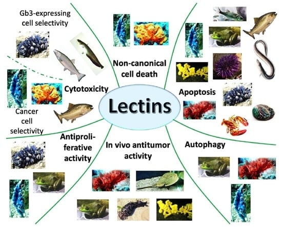
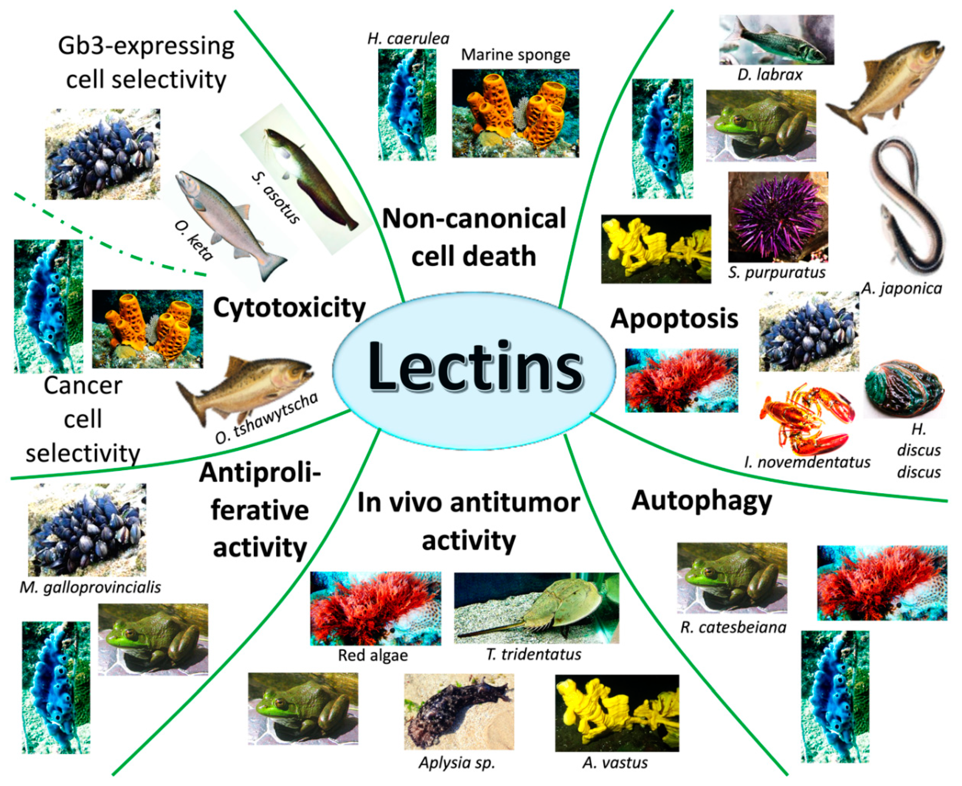
Antitumor Potential of Marine and Freshwater Lectins

1
Department for Life Quality Studies, Alma Mater Studiorum-Università di Bologna, Corso d’Augusto 237, 47921 Rimini, Italy
2
Lake Erie College of Osteopathic Medicine, Bradenton, FL 34211, USA
*
Authors to whom correspondence should be addressed.
Mar. Drugs 2020, 18(1), 11; https://doi.org/10.3390/md18010011
Submission received: 30 November 2019 / Revised: 18 December 2019 / Accepted: 18 December 2019 / Published: 21 December 2019
(This article belongs to the Special Issue Marine Glycoconjugates: Trends and Perspectives)

Abstract

:
Often, even the most effective antineoplastic drugs currently used in clinic do not efficiently allow complete healing due to the related toxicity. The reason for the toxicity lies in the lack of selectivity for cancer cells of the vast majority of anticancer agents. Thus, the need for new potent anticancer compounds characterized by a better toxicological profile is compelling. Lectins belong to a particular class of non-immunogenic glycoproteins and have the characteristics to selectively bind specific sugar sequences on the surface of cells. This property is exploited to exclusively bind cancer cells and exert antitumor activity through the induction of different forms of regulated cell death and the inhibition of cancer cell proliferation. Thanks to the extraordinary biodiversity, marine environments represent a unique source of active natural compounds with anticancer potential. Several marine and freshwater organisms, ranging from the simplest alga to the most complex vertebrate, are amazingly enriched in these proteins. Remarkably, all studies gathered in this review show the impressive anticancer effect of each studied marine lectin combined with irrelevant toxicity in vitro and in vivo and pave the way to design clinical trials to assess the real antineoplastic potential of these promising proteins. It provides a concise and precise description of the experimental results, their interpretation as well as the experimental conclusions that can be drawn.

Graphical Abstract

1. Introduction

Water covers 71% of the surface of our planet and hosts vibrant biodiversity. More than 2 million different species inhabit the seawater, many of which still await discovery [1]. Terrestrial natural compounds have always represented a source of biologically active molecules, while the marine environment is still a step behind. So far, the World Register of Marine Species checked and classified only 232,297 different species [2]. This number could be considered modest, but it mirrors how late oceans have started to be studied compared to land environments. Indeed, the spread of terrestrial natural history began during the 17th century, and escalated through the scientific travels of 18th and 19th centuries—including Charles Darwin’s expeditions—until nowadays. On the contrary, it was not until the 20th century that ocean exploration started. It should be enough to underline that before the last century, technology was not sufficient to explore deep waters [3]. However, the number of discoveries is now exponentially increasing [4].
From the perspective of a pharmacologist, the marine biodiversity represents the ideal environment to quest for new substances with therapeutic potential. Indeed, since oceans give a home to many sessile or limited-mobility species, it is logical to assume that they synthesize metabolites to protect themselves and that those metabolites can be exploited for therapeutic uses [5]. Especially in anticancer research, the marine environment is an actual gold mine. By 2017, Food and Drug Administration of the United States approved seven marine-derived pharmaceutical drugs as anticancer agents, while many other compounds are currently being studied in different phases of clinical trials in oncology and hematology fields (4 in phase III, 6 in phase II, 8 in phase I) [6]. It has been estimated that 594,232 novel compounds of marine origin are waiting to be studied, assuming that between 55 and 214 could be approved as new anticancer drugs [4].
Nonetheless, if the natural world is so rich in bioactive molecules, why is the cure for cancer one of the hardest challenges scientists are facing for the last two centuries? First of all, it is incorrect to refer to cancer as a single disease. At least 200 different types of tumors exist. For instance, the organ where the tumor originates gives rise to specific neoplastic lesions; thus, breast cancer is different from lung cancer. Likewise, the type of cells from which cancer derives is crucial, and carcinomas derived from epithelial cells are different from lymphomas that derive from cells of the immune system. Furthermore, cancer is not quiescent, and the possibility to evolve and acquiring resistance to a full-blown effective therapy is very high [7]. Thus, it is pretty unlikely finding a single cure for all cancer types. Moreover, most of the anticancer agents used in clinic cause unacceptable toxicity that limits their compliance and effectiveness, hampering the positive outcome of cancer treatment. Since the cytotoxic effect of the antineoplastic drug also affects healthy cells, the primary cause of toxicity is the lack of selectivity towards tumors cells [8,9,10]. Thus, an interesting antitumor strategy would foresee a drug able to eradicate tumor cells distinguishing between them and healthy cells.
Therefore, the next question is: how do cancerous cells differ from normal ones? What all neoplastic cells have in common and that distinguish them from normal cells is the acquisition of different abilities. These abilities allow them to proliferate persistently, resist to cell death, circumvent growth restraint stimuli, reprogram energy metabolism, and escape the immune system recognition [11]. All the pathways involved in the acquisition of these capabilities make tumor cells unique and ideally can be exploited to obtain a therapy that selectively kills cancer cells. Targeting fast proliferating cells was one of the first strategies designed for this aim. However, some healthy tissues have a high physiological rate of proliferation and damage to normal cells cannot be avoided [12]. More, to survive in hostile environments, cancer cells deregulate cellular energetics. Indeed, reactive oxygen species (ROS) production is increased while the antioxidant machinery is often impaired. Thus, the basal oxidative stress level of cancer cells is higher than that of normal cells. By the consequence, enhancing ROS levels in cancer cells most likely provokes the selective eradication of these cells rather than normal cells [8,10,13]. In addition to oxidative stress, many other pathways, such as increased glycolysis (the Warburg effect), hypoxia and acidity have been targeted [9,11]. Nonetheless, hitting any one of these targets does not lead to a complete selectivity of action. Conversely, an effective strategy to identify and selectively hit cancer cells could be aiming at the phenotypic changes that come along with the malignancy transformation, such as the glycosylation pattern at the cell surface. In normal conditions, most proteins and lipids of eukaryotic and prokaryotic cells are coated by a sugar structure called glycocalyx. Glycocalyx plays a pivotal role in cell-cell recognition, communication, and intercellular adhesion. It is composed by mono-, oligo- or polysaccharides and changes in a predictable fashion throughout normal cell development. Glycosyltransferase enzymes, located in the Golgi apparatus, catalyze the construction of this carbohydrate structure. During malignant transformation, alterations in glycosyltransferases and their gene expression occur [14], and glycosylation profile of cells is irreversibly altered, conferring unique phenotypes to cancer cells [15]. For instance, protein fucosylation and sialylation, i.e., the addition of fucose moieties or sialic acid to proteins, is a common event in many cancer types [16]. The modifications of cell surface sugar patterns and in particular glycoproteins can be exploited to target cancer cells exclusively. Lectins are non-immunogenic glycoproteins that specifically bind unique carbohydrates residues on cell surfaces. Thus, they represent an attractive tool to detect and kill cancer cells [17].
Several studies have shown that plant and animal lectins exert different biological activities. These compounds inhibit bacterial [18] and fungal growth [19], and have immunomodulatory properties [20,21]. On cancer cell lines, they exhibit antiproliferative properties [20,22], promote apoptosis and autophagy, and block angiogenesis [20,22]. Moreover, their ability to bind to carbohydrates could make them useful for the detection of subtle modifications of carbohydrate composition, which appears during cancer transformation [23]. However, although anticancer activity of terrestrial lectins has been summarized in several reviews [20,24,25], to our knowledge no comprehensive report highlighting uniquely the antineoplastic activity of marine lectins has been written.
This review will explore the antitumor activity of lectins from both marine and freshwater organisms. A short overview of lectins will be followed by the presentation of the most studied lectins that showed overt anticancer potential. Diagnostic applications of lectins will not be discussed since it goes beyond the aim of this review.

2. Lectins

Lectins are expressed by animals, plants, fungi, and bacteria. They contain a domain for carbohydrate recognition that allows them to bind with carbohydrates without modification, i.e., they do not exhibit enzymatic activity on carbohydrate molecules. Since they reversibly and specifically bind carbohydrates, whether simple sugars or complex carbohydrates, lectins are capable of precipitating polysaccharides and glycoproteins or agglutinating cells [17].
Lectins can be classified using different criteria, including cellular localization, structural and evolutionary sequence similarities, taxonomic origin, carbohydrate recognition, function, and structure [26] (Figure 1).
Lectins play a pivotal role in cell-cell and host-pathogen interaction and communication, tissue development, sugar storage, and other mechanisms of cell survival and immune system stimulation. All endogenous lectins can be involved in both physiologic and pathologic processes [25]. Based on their characteristics, they can induce apoptosis and autophagy and inhibit angiogenesis, thus representing an interesting anticancer strategy [25]. Their mechanism of action consists of recognizing and reversibly binding specific carbohydrate moieties on cell surfaces. In particular, lectins bind to a specific sequence of monosaccharide moieties within glycosylated proteins, lipids, and glycans that are on cell surface. In humans, several types of cells express these proteins, from epithelial cells to antigen presenting ones and, depending on the type of lectin, they can be exposed on the cellular membrane or secreted into the extracellular matrix [27]. Each lectin usually has more than one binding site for the sugar units. As a consequence, they are able to bind different carbohydrates exposed on the surface of different cells, thus enabling cell-cell interaction [22]. Unique glycosylated-proteins expressed in tumor cells become the target of lectins that bind the characteristic glycan-chains. This process is the key to discriminate abnormal glycosylation moieties on the surface of cancer cell membranes [28]. Not all cancer types show the same glycosylation pattern, but they share common features. In particular, during the tumorigenesis, specific subsets of glycans on the cancer cell surface face different modifications, such as enrichment or decrease in their main components. The most recurrent changes on cell surface glycosylation patterns are enrichment in sialylation, branched-glycan structures, and the generation of the so-called “fucosylation core” [29,30]. For instance, the tetrasaccharide carbohydrate moiety sialyl-LewisX and sialyl-Tn antigen represent well-known markers of malignancies, since they are found in almost all cancer types [31]. Besides, O-linked and N-linked glycosylation create the most common branches on the tumor cell surface glycocalyx. For example, O-linked glycosylation is promoted by the entry of a GalNAc sequence, which can then be expanded with the addition of other different structures. Simultaneously, different blocks of sugars can be attached to the main branch through an amide group of an asparagine residue resulting in N-linked glycosylation. Among others, ovarian, skin, breast, and liver cancers show these types of modifications [30,32]. To complete the picture, core fucosylation, i.e., the enrichment in α1,6-fucose of the innermost GlcNAc residue of N-glycans, is a specific trait in many cancers, such as lung and breast cancer [29,33].
The recognition of glycosylated proteins on the outer cellular membrane represents only the first step necessary to trigger cancer cell death. After the binding with the carbohydrate moiety, lectins can be internalized, and there, in the intracellular environment, they trigger specific pathways that lead to cell death. Furthermore, taking these notions into consideration, exogenously induced expression of lectin inside the tumor cells could be another effective strategy to promote tumor eradication. For this reason, vaccinia viruses (VVs) have been exploited as replicating vectors harboring the genes needed for lectin expression and thus as an efficient way to transport lectins into cancer cells [34].
Marine and freshwater lectins show anticancer properties in vitro and in vivo thanks to the binding of cancer cell membranes, causing cytotoxicity, apoptosis, other forms of regulated cell death, and inhibition of tumor growth. A literature review was performed to identify publications on the antitumor potential of lectins isolated from water organisms such as invertebrates, vertebrates and tunicates, fish, and algae. The following paragraphs will present their activity as direct antitumor agents or through their delivery via VV.

3. Lectins from Marine and Freshwater Algae

Aquatic algae are a group of simple organisms containing chlorophyll. To sustain themselves, they almost only need light to carry out photosynthesis. They inhabit seas, rivers, lakes, soils, and grow on animal and plants as symbiotic partners. The complexity of these organisms ranges from the simplest unicellular to pluricellular entities that reorganize themselves to form simple tissues [28]. At least 44,000 species of algae have been identified and named [29], and among them 250 have been reported to contain lectins [3]. However, only a few of them have been adequately studied to investigate their antitumor potential (Table 1). For some of them, the only cytotoxic potential has been evaluated.
The most characterized lectin of algae origin, the Eucheuma serra agglutinin (ESA), was extracted from the homonymous red macroalga. ESA is a mannose-binding lectin able to promote apoptosis on different cell lines and animal tumor models (Table 1). ESA amino acid structure is composed by four tandemly repeated motifs, each of them representing one binding site for mannose sequence. Specifically, each repeated motif binds specifically high mannose N-glycans with a minimum dimension of tetra- or penta-saccharide, such as Man(alpha1-3)Man(alpha1-6)Man(beta1-4)GlcNAc(beta1-4) GlcNAc [43].
ESA promotes cell death of many cancer cell lines, such as Colo201 (human colon adenocarcinoma), Colon26 (murine colon-carcinoma), HeLa (human cervix adenocarcinoma), MCF-7 (human breast adenocarcinoma), OST (human osteosarcoma), LM8 (murine osteosarcoma) [36,37,38,39]. In each of these cell lines, the mechanism of cell death is apoptosis, as demonstrated by DNA fragmentation, exposition of phosphatidylserine, and activation of caspase-3. The activity of ESA is tumor-type dependent and, comparing distinct studies, cervix adenocarcinoma and colon adenocarcinoma came back the most sensitive followed by osteosarcomas and breast cancer, which respond to a higher concentration of this lectin [36,37,38]. Since lectins, in general, have precise targets, this behavior probably reflects the different glycosylation pattern of different types of tumor. Certainly, the different glycosylation pattern between normal and cancer cells is behind the lack of ESA activity on non-transformed cells. Indeed, ESA did not affect the viability of normal fibroblasts and the non-tumorigenic epithelial MCF10-2A cell line [37,38]. Furthermore, their selectivity for tumor cells translates into lack of toxicity in vivo. For instance, ESA delayed the growth of Colon26 cells injected on BALB/c mice without affecting the body weight nor causing direct death, thus showing promising in vivo tolerability [36].
The ability of lectins to selectively target cancer cells can be exploited not merely to kill tumors, but also to deliver antitumor drugs on cancer cells. Furthermore, the antitumor activity of lectins could theoretically sum to that of the antitumor drug. With this aim, the development of a selective drug delivery system (DDS) was designed, and lipid vesicles resembling a microcapsule were tagged with ESA [38]. The microcapsules were made by sorbitan monooleate (Span80) with or without poly(ethylene glycol) (PEG) [37,39]. PEG was added in order to prolong the half-life of the vesicles compared to normal liposomes since PEGylation should decrease the reticuloendothelial uptake. First of all, it was demonstrated that both ESA-labelled DDSs target the exact same carbohydrate-sequence of free ESA, and that the drug transportation system does not abolish its cytotoxic activity, nor selectivity towards tumor cells [37,44]. ESA-immobilized lipid vesicles reached and bonded Colo201, HB4C5, OST, while no interaction was recorded with normal human fibroblasts and MCF10-2A [37,38,39] (Table 1). Indeed, all microcapsules directly exhibited pro-apoptotic activity on Colo201 and OST cells [37,39], while little effect was observed in normal MCF10-2A [39]. In vivo, the injection of the vesicles delayed tumor growth in nude mice bearing Colo201-derived tumors [39] (Table 1). No study directly compared the antitumor activity of the PEGylated vs. the not-PEGylated ESA-vesicles, while only in vivo bioavailability experiments were performed, showing no difference between the two systems [37,39]. In fact, after the injection of one or the other radioactive-labeled microcapsules in nude mice bearing Colo201 tumor cells, no difference in tumor accumulation nor uptake was recorded [39]. Finally, to understand if the DDS was able to transfer the encapsulated material into tumor cells, two different research groups [38,39] loaded them with the fluorophore fluorescein isothiocyanate (FITC) and treated cells with this complex. They demonstrated that FITC is internalized into tumor cells cytoplasm, both in vitro and in vivo, and showed the interesting potential of ESA-vesicles as a carrier of antitumor drugs as well as direct antineoplastic agents [38,39].
If ESA was not particularly active on MCF-7, two isolectins isolated from Solieria filiformis (Sfl) proved their ability to fight breast cancer (Table 1). Sfl-1 and Sfl-2 are mannopentose-binding lectins differing from each other for only 18% of the aminoacidic sequence. SfL-1 and SfL-2 consist of 268 amino acids in the form of four identical tandem-repeat protein domains. They both are constituted by two β-barrel-like domains made of five antiparallel β-strands, which are connected by a short peptide linker [41]. Despite the different biological activity of Sfls and ESA, both Sfl isoforms share at least 50% of their amino acid sequence with it [45] (Figure 2A). Thus, we can hypothesize that the similar structure allows them to both bind the same glycan structure, while the remaining part is responsible for the different cytotoxic activity.
Both Sfls isoforms promote MCF-7 cell death to an equal extent, while they were not able to entirely kill primary human dermal fibroblasts. However, at a higher concentration than those promoting cell death, Sfls induce fibroblast proliferation [41]. Keeping these data in mind, the difference in the sensitivity of cancer and normal cells allows the identification of an interval of concentration toxic for the tumor ones. The mechanism of anticancer action of these lectins lies in the induction of apoptosis. Normally, apoptosis can be promoted by triggering the intrinsic and/or extrinsic pathway. The first one involves the B-cell lymphoma 2 (Bcl-2) protein family, mitochondrial permeability, and the creation of the apoptosome. The extrinsic pathway involves death receptors, death-inducing signaling complex and caspase activation. The two pathways share effector caspases. Due to congenital or acquired genetic alterations of cancer cells, one or, in the worst case, both pathways are often compromised [46]. Thus, the ability to activate both pathways maximizes the probability to promote programmed cell death successfully and minimizes the one to acquire resistance. Sfls upregulate the gene expression of the effector caspase-3 and trigger both the intrinsic [upregulation of the proapoptotic gene Bcl-2 associated X protein (Bax); downregulation of the anti-apoptotic gene Bcl-2; increased expression of caspase-9] and the extrinsic (increased expression of caspase-8) apoptotic pathways, qualifying themselves as very interesting anticancer agents. Further studies will be needed to understand if and how the 18% difference between the two isoforms affects their biological properties [41].
Ulva pertusa lectin 1 (UPL1) is an N-acetyl d-glucosamine-binding lectin that interacts with several intracellular pathways involved in proliferation and cell survival (Table 1). UPL1 primary structure does not share amino acid sequence similarity with any known plant or animal lectin. cDNA sequencing analysis showed that UPL1 is 1084 base-pair long and encodes for a premature protein of 203 amino acids. During post-translational modifications, 53 amino acids are lost and the N-terminal sequence of UPL1 starts at amino acid number 54 [47]. Intracellular delivery of UPL1 through a flag adenovirus does not exert an antiproliferative effect on the human papillomavirus-related endocervical adenocarcinoma BEL-7404, and hepatocyte-derived cellular carcinoma Huh7 cells. UPL1 mediates both the activation of the extracellular-signal-regulated kinase (ERK)-mitogen-activated protein kinase (MAPK), also known as MAPK/ERK kinase (MEK), signal and the phosphorylation of p38 MAPK. Since constitutive activation of ERK promotes cell survival and drug-resistance [47] and p38 MAPK can act as a survival factor, this could be the cause of UPL1 inability to stop cancer growth [42]. Indeed, the inhibition of each one of these pathways enhanced the antiproliferation activity of exogenous UPL1. Even if the cellular functions of tubulin in cancer is not clear [48], the ability of UPL1 to modulate MAPK pathways lies on its binding with β-tubulin. Probably, this critical protein is glycosylated in a specific manner providing recognition sites for the lectin. What is certain is that when the binding UPL1-tubulin was impeded, nor ERK-MEK or p38 MAPK were modulated, and UPL1 was able to kill cancer cells [42]. Moreover, only on Huh7, UPL1 triggered autophagy. If autophagy-associated cell death is considered a tumor suppressor characteristic, it can also enhance the survival of neoplastic cells exposed to metabolic stress and foster metastasis [49]. As a consequence, attention must be paid to assessing UPL1 real antitumor potential, alone or combined with survival signaling inhibitors, such as MAPK inhibitors [42].
Since lectins are such attractive pharmaceutical agents, the identification of new organisms able to produce them is compelling. However, the qualitative analysis of extracts could be time- and resource-consuming. Affinity electrophoresis or photometric assays are two tricky techniques used to carry out this task [48]. Additionally, an easy and fast method for understanding if a substance contains lectins is to evaluate its hemagglutination activity. Anam et al. [35] studied different crude extracts of red macroalgae (Nitophylium punctatum, Acanthophora spicifera, Acrocystis nana, Helminthora divaricata and Gloiocladia repens) and showed different degrees of hemagglutination activity. This activity, however, did not reflect the potency of their antitumor potential. All five crude extracts were able to kill MCF-7 and HeLa cells to a different extent (Table 1). They were all tested at a concentration of 100 µg/mL. Acrocystis nana extract was the second extract for hemagglutination activity, but the most potent in killing HeLa cells, inhibiting almost 50% of cell growth after 24 h. For MCF-7 cells, the extract more enriched in lectins (Gloiocladia repens) was the most cytotoxic with inhibition of almost 30% cell growth. All other extracts affected cell growth in an interval ranging between 2 and 15% [35]. These data, although not outstanding, may be deemed a starting point for the complete purification of the extracts in order to isolate the lectins. In general, consequential chromatographic steps are needed to purify lectins from crude extracts, such as affinity chromatography, ion-exchange chromatography, hydrophobic chromatography, and gel filtration chromatography [17]. In this case, the complete purification of the lectins from the crude extracts will clarify if lectins alone still have anticancer potential and whether that potential is higher than that of the extract. Anam et al. [35], who authored the study, suggest that extracts could contain impurities that dilute the samples and that purification should allow increasing the antitumor activity [35]. However, very often, extracts are more potent than the single molecules alone [49]. By the consequences, it will be interesting to see how these aspects pan out.

4. Lectins from Marine and Freshwater Invertebrates

Animals belonging to marine invertebrates have been exploited for their biological activity since the times of ancient Greece. Hippocrates and Galen, two pioneers of modern medicine, extensively narrated about dietary and pharmaceutical uses of shellfish, sponges or cephalopods, and their prescriptions containing marine invertebrates along with other ingredients have been found. Marine invertebrates consist of a large variety of organisms and have been categorized into over 30 different phyla [50]. They do not have an innate immune system nor develop an adaptive immune response against pathogens. Thus, as an immune defense they constitutively express small peptides that are induced upon danger is sensed. Lectins are among these proteins [51], and interestingly, they showed impressive antitumor activity both in vivo and in vitro (Table 2).

4.1. Phylum Mollusca and Arthropoda

The phylum Mollusca represents the second biggest phylum on earth. Approximately 90% of mollusks fall into the Gastropoda class, followed by Bivalves and Cephalopods [70]. Shelled mollusks are the most widely used in traditional medicine and together with the crustaceous (phylum Arthropoda) fall into the shellfish family [71]. Several studies revealed that the richness in hepato-pancreas mass, the characteristic open circulatory system, the filtering abilities and the shell arrangement make bivalves and crustaceans a remarkable source of molecules [72], such as the unique lectin proteins that can be exploited as therapeutic agents.
Crenomytilus grayanus lectin (CGL) is an intriguing lectin isolated from the homonymous bivalve belonging to the Mytilidae family. It consists of three highly similar tandem sequences of amino acids for a total of 150 residues. Secondary structure envisages a predominance of β-structure that sometimes alternates with α-ones [73]. CGL has a particular structure that makes it able to specifically recognize the globotriaosylceramide (Gb3) resulting in Gb3-dependent cytotoxicity. Gb3 is a globoside, and thus a non-protein cluster of differentiation composed by a galactose-lactosylceramide sequence [74]. It is overexpressed in several human tumors with intrinsic or acquired multidrug resistance [75]. CGL blocks cell proliferation and promotes cell death on Gb3-expressing tumor cells, such as Raji cells (Burkitt’s lymphoma) and in a lesser extent MCF-7 (breast carcinoma) (Table 2). No effect was recorded on K562, a human myelogenous leukemia cell line not expressing Gb3 [53,54]. Furthermore, on Raji cells, the CGL cytotoxicity was completely hindered when Gb3 was enzymatically degraded [54]. On the same cell line, CGL promoted a G2/M cell-cycle arrest that led to apoptosis, as demonstrated by phosphatidylserine externalization, activation of caspases-3 and caspase-9, and poly (ADP-ribose) polymerase (PARP) cleavage [54]. Even though further studies are needed to confirm the following assumption, the high expression of Gb3 on some cancer cells and the very specific action of CGL for these cells bodes well for a selective activity of this lectin towards neoplastic lesions and let us presume that it would bring to lack of toxicity in vivo.
Mytilus galloprovincialis, better known as Mediterranean mussel, contains a lectin, Mytilec, that shares a similar behavior with CGL. No wonder, they share 50% in amino acid sequence and bind the same glycan moiety [45] (Figure 2B; Table 2). Mytilec is incorporated into cells through the interaction with Gb3 and, thanks to that, it promotes cytotoxic effects. As CGL, it exhibits antitumor activity only on Gb3-expressing cell lines, such as Raji’s and Ramos, another Burkitt’s lymphoma cell line (Table 2), while no effect was induced on K562 [35,36]. The mechanism of action of Mytilec has been investigated in Ramos cells, where it promotes apoptosis and activates all MAPK pathways. MAPK system consists of three sequentially activated protein kinases that play a central role on different transduction pathways. These pathways are involved in processes, such as cell proliferation, differentiation, and cell death in eukaryotes [76,77]. Precise extracellular stimuli elicit the phosphorylation cascade and provoke the activation of a MAPK by the consecutive activation of a MAPK kinase kinase (MAPKKK) and a MAPK kinase (MAPKK). Briefly, after the stimulus is inferred, MAPKKK phosphorylates and triggers MAPKK, which, in turn, activates MAPK. Here, MAPK phosphorylates different substrates in the nucleus and cytoplasm, inducing variations in protein function and gene expression that deliver the proper biological response. Depending on which stimulus triggered the MAPK system in the first place and the intended biological effect, three leading families of MAPK can be activated. Growth factors and mitogens activate ERK, which controls cell survival, growth, differentiation and development; stress, inflammatory cytokines and, once more, growth factors prompt c-Jun N-terminal kinases (JNKs) and p38. These two latter MAPKs, in turn, deal with inflammatory response, apoptosis, cell growth, and differentiation [78]. Mytilec activates all these three pathways, but only in Gb3-expressing cell lines. The activation of the ERK pathway increased the levels of tumor necrosis factor-α (TNF-α) and the cell-cycle inhibitor p21, but it was excluded to be the cause of apoptosis induction. The role of JNK and p38 on the pro-apoptotic properties of Mytilic has still to be verified [60].
The gonads of the adult giant marine Aplysia, a sea slug belonging to Gastropoda class, contains the corresponding lectin: Aplysia gonad lectin (AGT). It is a Ca2+-dependent lectin and is composed of two 32-33 kDa subunits [79]. Although the majority of studies about AGT describe its chemical characteristics, this galacturonic acid-galactose-lactose-binding lectin has been injected on mice and it was found able to abolish tumor appearance when tested at 30 µg and decrease lung tumor burden of 60% when tested at 5 µg, compared to untreated animals [79,80]. However, no information is available on treatment time and route of administration [80].
iNol is a very big lectin physiologically synthesized by the Arthropoda slipper lobster (Ibacus novemdentatus) probably to destroy pathogens. iNol consists of five subunits, which are composed by 70-, 40-, or 30-kDa polypeptides that are held together by disulfide bonds. Under physiological conditions, it has a polygonal structure. Despite the high molecular weight, this lectin is incorporated into mammalian cells, such as HeLa, through endocytosis thanks to the binding with N-acetylated glycan moieties on the cell surface. iNol kills HeLa, MCF-7, T47D (breast cancer) and Caco-2 (colon cancer) cells (Table 2). HeLa cells were the most sensitive to iNol activity, so they were used to understand its apoptotic potential. iNol promotes DNA fragmentation and activation of caspase-3 and caspase-9 only if the lectin is able to bind its carbohydrate-ligand, demonstrating that its antitumor potential is strictly linked to its lectin nature [58].
Chinese or Japanese horseshoe crab or tri-spine horseshoe crab are the common names of Tachypleus tridentatus, an Arthropoda resembling a crab, but more closely related to spiders and scorpions. Despite the not gracious description, Tachypleus tridentatus represents a source of several lectins, among which the rhamnose-binding Tachypleus tridentatus lectin (TTL). TTL characterization has not been completely clarified. Different studies agree on the fact that this lectin is a multimer, but it is not clear if it is composted by hexamers and octamers [81] or tetramers [82]. The antitumor activity of TTL was assessed in vivo, after its genomic insertion into an oncolytic VV (oncoVV-TTL). Balb/c nude mice were subcutaneously engrafted with MHCC97-H liver cancer cells and then treated with oncoVV-TTL. OncoVV-TTL was able to replicate inside tumor cells and significantly reduced tumor growth compared to onvoVV-only treated mice [55] (Table 2).
Another replication-deficient adenovirus was modified to encode the gene of Haliotis discus discus sialic acid-binding lectin (Ad.FLAG-HddSBL). HddSBL is an another Ca2+-dependent binding lectin. It has 151 amino acid residues, but so far the glycosylation site has not been identified yet [83].
Ad.FLAG-HddSBL promoted cell death of hepatocellular carcinoma (Hep3B), lung cancer (A549 and H1299), and colorectal carcinoma (SW480) in a tumor-dependent manner, as the different sensitivity of these cell lines to this compound suggests [56]. Hep3B resulted in the most sensitive cell line to Ad.FLAG-HddSBL, followed by A549, H1299 and last SW480 (Table 2). Yang et al. [56], who authored the study, suggested that the different intracellular metabolism of sialic acids is the cause of the different activity of Ad.FLAG-HddSBL. On Hep3B, Ad.FLAG-HddSBL’s mechanism of action has been investigated. It promotes a regulated form of cell death, since it triggered the mobilization of annexin-V to the outer cellular membrane and decreased the expression of the anti-apoptotic factors Bcl-2 and X-linked inhibitor of apoptosis protein (XIAP). Curiously, it did not affect PARP expression, and thereby it does not activate caspases at the analyzed time point [56]. Then, two possibilities arise: either it activates PARP at a different time point, or it promoted one of the so-called non-canonical cell deaths, which are caspase-independent forms of regulated cell death.

4.2. Phylum Porifera

The name Porifera means “pore bearer”, and it is the most characteristic feature of sponges. Indeed, they are invertebrates sessile characterized by the lack of digestive, nervous, circulatory, and immune system. To get food, oxygen, and to reject all discards, they maintain a continuous water circulation in their body. Sponges do not have a physical barrier that protects them from predators, and the only way to survive is to produce secondary toxic metabolites. Furthermore, Porifera phylum is characterized by a unique biodiversity that results in a vast diversity of metabolites. Several studies have shown the chemopreventive and antitumor properties of many of them [5].
Two lectins originated from different species of the genus Haliclona showed an outstanding antitumor potential. Haliclona cratera lectin (HCL) was extracted, purified and tested for antitumor activity on human cervical and melanoma cell lines. It displayed cytotoxicity on both tumor models, which had almost the same sensitivity (half maximal inhibitory concentration 9 µg/mL vs. 11 µg/mL, respectively) [67] (Table 2). Another species, Haliclona caerulea, produces a peculiar lectin called halilectin-3 (H3). Its primary structure shows no similarity to any other animal lectin and consists of 251 amino acids, of which 145 arrange themselves into an α-chain and 106 into two β-chains. Alongside, quaternary structure has a heterotrimer conformation stabilized by disulfide bonds [84], which differs from the dimeric [67], trimeric [85], tetrameric [86] or multimeric [87] structure of all other lectin originated from sponges characterized so far. H3 is able to promote cancer cell death exploiting different mechanisms. On MCF-7 cells, H3 triggers both intrinsic and extrinsic pathways of apoptosis promoting an early upregulation of p53 and inhibits cell proliferation causing an accumulation of cells in the G1 phase. In addition to apoptosis, autophagy-associated cell death has been recorded after a few hours of MCF-7 treatment with H3. In particular, H3 upregulated the microtubule-associated protein 1A/1B-light chain 3 (LC3) expression, promoting the accumulation of LC3-II (Table 2). Contextually, autophagosome vesicles have been identified microscopically [66]. The authors of the study [40] hypothesize that both apoptosis and autophagy can be triggered thanks to Bcl-2 downregulation or p53 upregulation. To complete the picture, H3 has been found able to promote anoikis, a particular form of cell death that happens when cells are stimulated to detach from the extracellular matrix [66]. In normal conditions, cells need to adhere to the tissue where they grow since the essential growth factors and other survival signals are provided by the extracellular matrix and proximal cells [88]. H3 reduced the expression of integrin α6β1 and interacted with integrin α5β1, the fibronectin receptor, thus impairing MCF-7 adhesion and promoting cell death [66]. The involvement of apoptosis, autophagy, and anoikis in the cytotoxicity of H3 makes it a very promising antitumor lectin, able to fight cancer on several fronts and thus reduce drug resistance.
A lactose-binding lectin isolated from the sponge Cinachyrella apion (Cal) exhibited antitumor activity on human cervical (HeLa) and prostate adenocarcinoma (PC-3) cells, while milder cytotoxicity was recorded on non-transformed 3T3 mouse fibroblasts (Table 2). Cal primary structure consists of eight subunits of 15.5 kDa assembled by hydrophobic interactions and does not need Ca2+, Mg2+, and Mn2+ ions to bind sugars [87]. On HeLa, it promoted the typic phenotype changes of early apoptosis and modulated some of the pro-apoptotic proteins of the intrinsic pathway [64]. However, the inhibition of caspase activation did not significantly reduce its cytotoxicity. This suggests that apoptosis is not the leading mechanism through which Cal promotes cytotoxicity. Alternatively, the inhibition of the apoptotic pathway could activate a different pattern of regulated cell death [89]. Further studies are needed to untangle this knot.
Hol-18 is an N-acetylhexosamine-binding lectin isolated from the Japanese black sponge Halicondria okadai. It is a 72 kDa tetrameric lectin organized into four non-covalently bonded 18 kDa subunits [68]. Few studies demonstrated its newsworthy antitumor potential on Jurkat (acute T cell leukemia), K562 [68], HeLa, MCF-7 and T47D cells (Table 2). On Hela, MCF-7, and T47D, Hol-18 promoted apoptosis and activated the MAPK-ERK signal [69], showing once again how this pathway is often involved in marine lectin mechanism of action. No effect was recorded on Caco-2 cells since these cells were not able to internalize Hol-18 at all [69].
The recombinant oncoVV-AVL is the result of the insertion of the Aphrocallistes vastus lectin (AVL) into an oncolytic VV. AVL is a 34 kDa glycoprotein with a 24 kDa proteinaceous core i.e., the lectin core. It counts 191 amino acids and is a Ca2+-dependent lectin. It is characterized by vast hydrophobic sequences that allow it to bind the target cell membranes and facilitates the interaction with sugars [90]. The insertion of this lectin in the oncolityc VV allowed its exogenous expression into infected cells. It promoted cell death on several tumor cell types (Table 2), such colorectal cancer, glioma, and hepatocellular carcinoma cell lines. The mechanisms of cytotoxic activity of OncoVV-AVL have been investigated only on HCT116 colorectal carcinoma cells. It promoted regulated cell death but without the cleavage of caspase-8 nor the modulation of the pro-apoptotic Bax protein. Digging deeper into oncoVV-AVL mechanism of action, it has been demonstrated that ERK activation is necessary to allow virus replication. Since the virus replication is necessary to express the AVL lectin, it can be postulated that ERK is essential for oncoVV-AVL activity. OncoVV-AVL was tested also on Balb/c nude mice bearing BEL-7404- or HCT116-derived tumors. In both cases, oncoVV-AVL efficiently inhibited tumor growth [63] (Table 2).
Queiroz et al. [65] described the cytotoxic activity of the Cliona varians lectin (CVL) on different tumor cell lines and healthy cells. CVL is a Ca2+-dependent glycoprotein. It is a tetramer of 114 kDa composed by different subunits of 28 kDa linked by disulphide bridges [91]. CVL killed K562 and Jurkat cells, while all solid tumor cell models (melanoma, renal carcinoma, and prostate tumor) and normal human peripheral blood lymphocytes were found to be insensitive to CVL (Table 2). On K562, it increased p21 and repressed the expression of pRb (retinoblastoma protein), suggesting the ability to block the cell cycle. More, CVL promoted morphological changes typical of apoptosis while cytofluorimetric analysis showed a scenario composed by the presence of both primary necrotic and late apoptotic cells. No caspase-3, caspase-8 or caspase-9 activation has been recorded, but CVL stimulated the translocation of cathepsins B (CTPB) from the vesicular compartment to the cytoplasm [65]. The role of the lysosomal cysteine protease CTPB in cell death is intriguing but not yet fully understood. CTPB modulates both necrosis and regulated cell-death processes in a tumor-type fashion. In some cell lines, it acts as an initiator of the intrinsic apoptotic pathway, modulates Bcl-2 family proteins such as Bax, Bcl-2, and Bid, and activates the caspases cascade [92,93]. In others, rather than activate effector caspases, it represents a caspase mediator, triggering some of the morphological changes of apoptosis [93]. For example, the release of CTPB enhances the release of cytochrome c and the caspase activation in hepatocytes treated with TNF [93], while on fibrosarcoma cells exposed to the same stimulus CTPB operates as a downstream mediator of the caspase apoptosis cascade [94]. CTPB is also involved in a caspase-independent form of regulated cell death, i.e., necroptosis. Necroptosis shares with necrosis the morphological features but, in contrast to the latter phenomenon, defined molecular pathways drive it. Necroptosis is triggered through the activation of death receptors such as tumor necrosis factor receptor 1 (TNFR1). TNFR1 promotes the formation of the so-called necrosome, a complex formed by receptor-interacting protein kinase 1 (RIP1)–RIP3–mixed lineage kinase domain-like protein (MLKL) [95]. The complex activates downstream events such as CTPB mobilization, which actively promotes programmed necrosis [96]. In CVL-mediated activity, the ablation of CTPB restored almost entirely cell viability, while TNFR1 was upregulated and the subunit p65 of the nuclear factor kB (Nf-kB) was downregulated. The authors of the study [65] suggest that this latter event could be the cause of the increase in both the pro- and anti-apoptotic Bax and Bcl-2 proteins. Even if the Bax/Bcl-2 protein ratio is not known, the authors still suggest that it could be the cause of what they call “caspase-independent apoptosis” [65]. Taken together, these data suggest the ability of CVL to promote a CTPB-mediated programmed form of cell death. Annexin V exposure—a hallmark of the early stages of regulated cell death—the upregulation of TNFR1, and the complete absence of caspase activation recall necroptosis, but further studies are needed to assess if apoptosis, regulated necrosis, or both events trigger CVL-mediated cell death.

4.3. Phylum Chordata

Chordata is another marine filter-feeding phylum. They have tubular openings through which water goes in and out. They are sessile and very often fixed to rocks or similar surfaces [97]. Tunicates, in particular, represent a source of didemins, such as aplidine and trabectin, molecules with anticancer, antiviral, anti-inflammatory, and immunosuppressive potential [98,99,100]. Furthermore, they contain lectins.
The most compelling tunicates’ lectin is the N-acetyl-D-glucosamine-binding Didemnum ternatanum lectin (DTL). DTL is a homotrimer which activity is not dependent by Ca2+ and Mg2+. It contains relatively high amounts of Gly, Ala, Asx, Glx Leu, Val residues and, as for the other lectin isolated from marine sponges, it is composed in part by carbohydrate (around 1.3%) [101]. Since one of the most critical functions of lectins is the cell-matrix interaction, the antitumor potential of DTL on HeLa cells has been studied in different microenvironment conditions. DTL showed different behavior on tumor cells depending on anchorage status (Table 2). In anchorage-independent conditions (cells cultivated in soft agar), DTL promotes cell growth, while in normal adhesion conditions (cells cultivated in adhesion plates) it inhibits cell proliferation and triggers cell differentiation in a way that promotes cell attachment [62]. Thus, cell microenvironment plays a key role in the DTL’s activity. Taking this in mind, an interesting approach to better predict the anticancer potential of lectins could be using tumor 3D cultures built with specific bioreactors. Standard in vitro models do not capture the complex tumor biology and do not consider cell-to-cell and cell-to-matrix interactions. Perfusion-based bioreactor systems create heterogeneous cell populations, and an optimal physiological cell-cell and cell-extracellular matrix interactions, perfectly miming tumor microenvironment [102,103].

5. Lectins from Marine and Freshwater Vertebrates

5.1. Amphibians

Amphibians are ectothermic, tetrapod vertebrates that count several species worldwide, except Antarctica [104]. Behavioral as well as biochemical, physiological, and morphological adaptations make them able to survive in such different habitats. Driven by the knowledge that amphibians produce several metabolites mainly as an essential self-defense strategy, recently drug discovery dip into them. Many peptides with anticancer activities have been found in amphibian skin or oocyte cells and eggs (Table 3) [105].
A sialic acid-binding lectin (SBLc), better known as leczyme, is a multifunctional protein isolated from the oocytes of Rana catesbeiana and characterized by a dual nature: a lectin and RNAse activity. This nature is the key to its very interesting antitumor properties (Figure 3). This lectin is unique and not homologous to any other known protein. It is a single subunit of 111 residues of amino acids and does not contain any covalently bound carbohydrate. The amino terminus is a pyroglutamyl residue and all the half-cystinyl residues are in the form of oxidized disulfide bridges [129]. SBLs lectin-glycan binding has not been completely elucidated. Nitta et al. [130] showed that Lectin hemagglutination activity was inhibited blocking amino groups and in particular t-amino ones, but not tyrosine residues, letting presume that those residues are responsible for the binding with the sugar.
The role of the lectin part of SBLc is to select cancer cells through the binding of specific sialic acid carbohydrates residues; then, specific receptors allow it to penetrate the cells, where the RNAse activity promotes the cleavage of RNA. Different structural changes on SBLc demonstrated that both intracellular incorporation and RNAse activity play a pivotal role for its anticancer activity. Indeed, increased incorporation with moderately increased RNAse activity is more effective than the increase in total RNAse activity [106,109]. For example, a less efficient internalization system does not affect or affects to a lesser extent the cytotoxicity of SBLc on estrogen negative cell lines, such as SK-BR3 [111,116]. On the other hand, a significant decrease in RNAse activity compromises SBLc’s antitumor activity [111]. In other words, as Aristotle would suggest, in medio stat virtus.
When inside the cell, SBLc activates the intrinsic apoptotic pathway on more than 30 tumor cell lines representing leukemia [107,108,109,118,120,131,132], Burkitt’s lymphoma [108,109], cervical [133], hepatocellular [120,121,133], and estrogen-positive and -negative breast carcinomas [113,114,116,117,120], glioblastoma [119], and malignant mesothelioma [110,113] (Table 3).
Just as in a jigsaw puzzle, many studies have tried to elucidate the proper molecular mechanism of action of SBLc. The pieces of the puzzles are a tumor-specific mechanism of action, the activation of the intrinsic apoptotic pathway with the involvement of heat shock protein 70 (HSP70) and the phosphorylation of p38 MAPK [108,110,116,120,134,135].
On MCF-7 estrogen receptor (ER)-positive breast cancer cells and H28 malignant mesothelioma cells, SBLc induced Bcl-2-mediated cell death [116]. On Jurkat cells, it promoted endoplasmic reticulum stress that may or may not be involved in its antitumor activity [134]. On p388 mouse lymphoma cells, HSP70 was linked to its ability to cause apoptosis. Even though the mechanism through which HSP70 assists SBLc cytotoxicity is not clear, it does not involve the binding between SBLc and cell membrane [109,135]. Finally, even if mitochondrial disruption is often part of SBLc mechanism of action, this lectin promoted a caspase-independent modality of cell death in SK-Hep1 hepatocellular carcinoma cells [120].
The activation of p38 MAPK pathway by SBLc has been recorded in leukemia, mesothelioma, and several breast cancer cells [108,111]. In these cell lines, the phosphorylation of p38 MAPK plays a key role in the pro-apoptotic activity of SBLc since the ablation of this pathway significantly decreases SBLc-mediated activation of caspase-3 and caspase-7 and cleavage of PARP and reduced cell death [111,114].
Special reference needs to be made to understand SBLc activity on breast cancer. The expression of ER, progesterone receptor (PR) and the human epidermal growth factor receptor 2 (HER2) represents the groundwork for the classification of this type of cancer and is useful for prognostic prediction and therapy selection. Usually, the worst prognosis refers to triple negative cancer (ER, PR and HER negative) and to those tumors overexpressing HER2 together with the epidermal growth factor (EGFR) [113]. Different studies about SBLc antitumor ability showed different results on the same tumor types. This is probably due to the different experimental procedures used in the studies, such as the presence of hormones, growth factor or fetal bovine serum in the medium. All studies agree on the fact that SBLc is very active on ER-positive cells, such as MCF-7 and ZR-75-1 [111,114,116], while contradictory data are published on ER-negative cell lines. For Tseng and colleagues [116], ERs are essential for cell binding of SBLc and thus its internalization. Accordingly, they reported a very mild activity of SBLc on MDA-MB-231 and ZR-75-30 ER-negative cell lines. On the contrary, Tatsuta et al. [114] and Kariya et al. [111] showed that both ER-negative (MDA-MB-231) and HER2-positive are sensitive to SBLc. Furthermore, Tatsuta et al. [114] showed a comprehensive screening of SBCc cytotoxic on several breast cancer cell lines with different phenotypes, such as ER-positive, PR-positive, HER2-positive, and triple-negative. As above mentioned, they demonstrated the involvement of p38 MAPK signal in its cytotoxic activity together with the modulation of the anti-apoptotic proteins of the Bcl-2 family on all cell lines. On MCF-7 and ZR-75-1, SBLc downregulated ERα, PR and HER2, while no modulation of EGFR was observed. This result is particularly interesting since hormonal therapies, and in particular receptor antagonism, represent efficient and usually well-tolerated treatments for breast cancer [136]. The cause of ER and PR downregulation could be linked to the activation of kinases, such as p38, drove by the ubiquitin proteasome system. Since SBLc promotes the phosphorylation of this kinase, Tatsua et al. [114] suggest that SBLc-promoted p38 phosphorylation can be linked to the destruction of hormone receptors. Likewise, the ubiquitin proteasome system may be involved in HER2 downregulation [114]. The hypothesis is based on the observation that SBLc modulates HSP70 [109,135]. Indeed, HSP70, together with HSP90, is responsible for the delivery of proteins, such as HER, to the ubiquitin-proteasome system in order to be degraded [114].
To avoid inflammation and promote dying-cell clearance, many types of specialized cells can phagocyte them. Cancer cells are not the first kind of professional phagocytes that come to mind; however, in rare situations, they are able to do that. It was already demonstrated that MCF-7 cells can play that role [137]. SBLc itself stimulated morphologic and phenotypic changes that allow viable MCF-7 cells phagocyting SBLc-triggered dying cells [115], promoting a very efficient auto-clearance. This unusual behavior represents an added value to the already promising anticancer activity of SBLc.
For many types of cancer, combination therapy is the best way to increase therapeutic efficacy, avoid drug resistance, and lessen the cytotoxicity of each single compound used in the therapy [138]. For example, malignant mesothelioma is treated with a combination of pemetrexed and cisplatin. SBLc alone was effective on cisplatin- and pemetrexate-resistant MESO-1 and MESO-4 cells (Table 3). Moreover, it synergistically increased the antitumor activity of pemetrexate in H28 [138] and MSTO cells [113] (Table 3) with an even stronger effect than that of pemetrexed plus cisplatin. The association of pemetrexed plus SBLs was particularly effective, because the first one blocks the proliferation of cells, which will be then easily killed thanks to the cytotoxic effect of the second one [113]. On the contrary, antagonistic effects were recorded when cisplatin was used together with SBLc. The antagonism is probably due to the opposite modulation of the tumor suppressor p21 by the two compounds. If cisplatin sharply increases p21 expression, SBLc antagonizes it, resulting in a lack of synergy [112]. In the case of SBLc plus TNF-related apoptosis-inducing ligand, a synergistic effect on H28 cells has been recorded, probably caused by an enhancement in the cleavage of the proapoptotic protein Bid that reinforces caspase activation [110]. SBLc also synergizes the antitumor cytotoxicity of interferon-γ (IFN-γ) in a cell-type-dependent way on different cell lines. The most marked effect occurred on MCF-7 cells. For the other tested cell lines, the degree of cell differentiation seems to play a role in this synergism: less degree of cell differentiation, more synergistic activity of SBLc plus IFN-γ. Then, an increasing trend of potency accompanied the well-differentiated HepG2 cells, the intermediate-differentiated J5 cells, and the poorly differentiated SK-hep1 cells, respectively. No evidence of synergism has been reported on HL-60 cells [120]. Considering that IFN-γ promotes differentiation of HL-60, this explanation perfectly fits with the abovementioned remarks [120]. Even if the vast majority of studies ascribe the SBLc’s selectivity to its lectin nature (i.e., to its ability to bind tumor cells because of their membrane glycosylation pattern), a correlation with the degree of differentiation of target cells and the ability of SBLc to recognize them is probably involved here as well. Indeed, the antitumor activity of SBLc decreases with the increase of differentiation of both hepatoma [121] and leukemia cells [118]. SBLc does not affect any of the several non-transformed cell lines tested that are all characterized by a high degree of differentiation: fibroblasts [109,120,121,133,139], epidermal melanocytes [109], keratinocytes [109], and mesothelial cells [110].
The effective and safe profile of SBLc reported above was also observed in animal models. Already in 1994, Nitta et al. [107] demonstrated the ability of SBLc to counteract sarcoma 180 cells inoculated on mice (Table 3) with doses at least 150 times lower than the lethal ones. More, SBLc suppressed mesothelioma (H2452 or MSTO cells) growth in xenografted nude mice, without causing toxicity nor body weight changes [113]. For the record, pemetrexed, which is one of the drugs of choice currently used for the cure of mesothelioma, failed to eradicate murine MSTO-xenografted tumors on the same experimental conditions [113]. In addition, SBLc effectively inhibited the growth of glioblastoma tumors in nude mice once again without causing any side effect [119].

5.2. Fish

The economic value of fish is not entirely due to the food industry. On the contrary, many pieces of research are focusing on these animals since they produce many bioactive compounds, such as the human calcitonin from salmon used for the treatment of postmenopausal osteoporosis [140]. As for amphibians, fishes produce many types of lectin that have been isolated from eggs, skin mucus, plasma, and serum [141,142,143,144]. The biologic function of fish lectin is not crystal clear, even if evidence shows some implication on fertilization and morphogenesis and a defense activity versus microorganisms [145], whether for some of them, an antitumor activity has been proven (Table 3).
Aristichthys nobilis, commonly known as bighead carp, belongs to the Cyprinidae, from the same family as zebrafish. It produces a lectin, GANL, which causes tumor-type-dependent cell death. GANL is a homo-multimeric glycoprotein that does not require Ca2+ ions to perform its functions. The carbohydrate content is approximately 13.4%, while the protein part is enriched in Asp, Glu, Leu, Val, and Lys. These amino acids organize themselves to form α-helices, unordered structures, β-turns, and β-sheets [124,146]. GANL blocked HeLa, SKOV3 (ovarian cancer cells), and HepG2 proliferation in a concentration-dependent manner while no effect was recorded on SMMC-7721 carcinoma cells and BGC803 gastric cancer cells (Table 3). Yao et al. [124] suggested that this behavior could be a consequence of the different glycosylation pattern that characterizes the different cells since it is not known whether the binding site of the lectin is the same for all cell lines and how each cell line expresses it [124].
The α-galoctoside-binding lectins isolated from the eggs of the catfish Silurus asotus (SAL) and the chum salmon eggs (CSEL) recognize specifically Gb3 sugar chain. Thanks to Gb3, SAL and CSEL bind cells, are internalized, and exhibit antitumor activity. Accordingly, SAL is not active on the Gb3-devoid K562 cells, while several studies showed its antitumor activity on Burkitt’s lymphoma cells [127,128,147] (Table 3). Similarly, CSEL promotes apoptosis on Gb3-positive Caco-2 cells, but it does not exert any effect on Gb3-negative DLD-1 colorectal adenocarcinoma cells [125]. On Raji cells, SAL induces the typic phenotypic changes of apoptosis, such as phosphatidylserine exposition and cell shrinkage [127], but its antitumor activity is more probably linked to its ability to inhibit the cell cycle [128]. A family of kinase proteins regulates rate and progression of the cell cycle. These enzymes consist of a regulating subunit (which takes the name of cyclin) and a catalytic subunit that takes the name of kinase-cyclin-dependent proteins (CDK). CDKs are inactive in the absence of the cyclin subunit and become active only when cyclins bind to the catalytic subunit. CDKs act during particular moments of the cell cycle via phosphorylation activity, stimulating or inactivating, the specific proteins that modulate cell-cycle advancement. For their part, synthesis and deactivation of cyclins are strictly regulated within the different phases of the cell cycle in order to allow this process running correctly [148]. After binding Gb3, SAL activated the GTPase Raf and the two kinases MEK and ERK. In turn, they promoted the synthesis of p21 [128], which physiological function is to regulate cell-cycle progression at G1 and S phase [149]. Moreover, SAL increased the expression of CDK4, c-myc, and cyclin D3. The overall effect is a G0-G1 cell-cycle arrest [128].
The Chinook salmon, also known as king salmon for its massive dimensions, is the source of another antitumor lectin, called rhamnose-binding chinook salmon roe lectin (CSRL). CSRL was reported to inhibit MCF-7 and HepG2 cell proliferation. HepG2 resulted in being more sensitive to CSRL than MCF-7 with a double potency (Table 3). As for all lectins mentioned in this review, CSRL as well did not exert any toxic effect on WRL68 non-transformed liver cell line at the same concentration used for MCF-7 and HepG2 cells [126] (Table 3).
Protein arginine methyltransferases (AMTs) are proteins involved in several processes, such as cell proliferation, cell differentiation, and also tumorigenesis. They catalyze the methylation of specific arginines on several nuclear and cytoplasmic substrates. AMT-5’ main targets are specific histones, and the result of their methylation is the silencing of different genes [150]. Recently, the transcription factor E2F-1 has been identified as a specific substrate for symmetric methylation, behaving as AMTs downstream element. E2F-1 modulates both apoptosis and cell-cycle progression, depending on which member of AMT family activates it. In particular, E2F-1 methylation by AMT-5 promotes cell proliferation, while if AMT-1 activates it, apoptosis is triggered [150]. p53 and p73 (the proline 73 polymorphic variant of p53) represent the links between EF2-1 and apoptosis [151,152]. Two different lectins are able to trigger this pathway and promote apoptosis in Hep3b cells. Dicentrarchus labrax fucose-binding lectin (DlFBL) and Anguilla japonica lectin 1 (AJL1) were harbored in a replication-defective adenovirus, generating respectively DIFBL-FLAG and AJL1-FLAG. DIFBL-FLAG and AJL1-FLAG elicited cytotoxicity in several human liver and lung cancer cell lines [61,123] (Table 3). DIFBL is a Ca2+-independent non-glycosylated lectin. It is a dimeric protein composed by two protein fractions, and, if they are separated, only one of them has lectin activity, while in physiological conditions they are stabilized by disulfide bonds [153]. Additionally, AJL1 has beta-galactoside specific activity in a Ca2+-independent manner. This lectin is composed of 142 amino acid residues having no half-cysteinyl residues, and exists in the form of homodimer without any covalent bonds [144]. On Hep3B cells, both lectins promoted apoptosis through the modulation of the AMT-5 pathway. In particular, the involvement of this enzyme provoked the downregulation of E2F-1 that in turn called Bcl-2 apoptotic protein family into play. Indeed, exogenous DIFBL and AJL1 downregulated Bcl-2 and XIAP proteins [61,123]. AJL1 also downregulated p38 MAPK and ERK protein levels, but it is not clear if this event is linked to its ability to promote apoptosis [123].
Despite the remarkable biological activity of lectins transported by viral vectors, some of them struggle to significantly infect cells, due to the lack of specific receptors such as coxsackie-adenovirus receptor (CAR). To facilitate this process, it is possible to target cell membrane receptors with other oncolytic adenoviruses that conveys a CAR-ligand-expression cassette in its genome. In this way, after infection and replication, the expression of CAR ligand will help the following adenovirus infection, triggering a positive feedback mechanism [57]. For this reason, a CAR-DIFBL was built and used to infect K562/adr doxorubicin-resistant leukemia and U87MG glioblastoma cells. CAR-DIFBL was able to favor cell infection by DIFBL-FLAG and also to synergize its cytotoxic activity [57] (Table 3). However, attention must be paid because another CAR-lectin, CAR-HddSBL, did help DIFBL-FLAG to infect glioblastoma cells, but it counteracted its cytotoxicity probably upregulating E2F-1 transcription levels [57].

6. Conclusions

The role of marine and freshwater lectins as anticancer agents has been discussed in this review and sustained by the reports summarized here. Marine and freshwater lectins exploit their antitumor action triggering apoptosis and other forms of programmed cell death, inhibiting cell cycle and blocking neoangiogenesis (Figure 4).
Some of them are also able to improve the antineoplastic activity of standard antitumor drugs. Moreover, all the lectins described in this review disclosed distinctive features against different types of tumors and, in the majority of the times, their ability to distinguish between normal and transformed cells. Of note, all lectins tested on animals showed optimal antitumor activity matched by negligible toxicity. Thus, selectivity and cytotoxicity are the two main characteristics that promote lectins to the rank of ideal antitumor agents. Indeed, toxicity of lectins of plant origin has been recorded only after direct ingestion of significant amount of specific lectins, such as phytohemagglutinin, which is particularly enriched in raw kidney beans. Gastric symptoms, such as nausea, vomiting, and diarrhea, are the most common ones after oral acute exposure, while immune system impairment, lung hypertrophy, and other systemic effects are very rare. On the contrary, no significant adverse reactions have been documented if lectins were administered as drug through alternative ways [154].
Another aspect that this review unearthed is that marine lectins are often tumor-type specific. This is not surprising because, going back to tumor heterogenicity, the glycosylation pattern is not the same for all cancer cells. This evidence does not represent a limiting factor for lectins. Indeed, marine biodiversity comes to our aid, and if one tumor is not sensitive to one specific lectin, most likely it would be to another one.
So far, the most concerning issue about exploiting marine compounds for a pharmacological purpose is that lectins, as all marine metabolites, are not produced in vast quantities. To obtain a reasonable amount of them, a large number of organisms has to be harvested. Thus, a significant limiting factor that stands between lectins and their exploitation as therapeutic agents is their availability. In other words, one of the hardest challenges about lectins is moving from laboratory scale to bulk production. However, despite the peculiarity of lectins that does not allow an easy identification of the aminoacidic bone structure and a cheap synthesis de novo, aquaculture or mariculture could bridge the gap. Additionally, the easiest and most efficient way to produce lectins is to exploit heterologous systems, such as bacteria and yeast. Indeed, different synthesis strategies exploit genetic engineering to transfer the genes encoding the lectin of interest to microorganisms, which can be grown in vast quantities, or to virus vectors in order to allow the synthesis of the lectins directly inside tumor cells [26,155,156]. As presented above, the latter approach has been exploited adequately for several lectins and with considerable results.
Despite the outstanding potential of marine lectins demonstrated by preclinical studies, so far, no clinical trials are translating this knowledge to cancer patients. In this context, terrestrial lectins are paving the way in that direction. For instance, mistletoe lectin extract has been subjected to many phase-I clinical trials, with promising, although not conclusive, results [157]. However, bearing in mind that the drug development process can last up to decades, preclinical studies represent the foundation to draw clinical trials.
Taking all this data together, it follows that aquatic organisms represent an important source of lectins and that those proteins represent a bright future for anticancer research. Certainly, further studies are needed to understand the actual antitumor potential of lectins on humans full. To reach this aim, clinical studies are essential to progress cancer research in employing lectins for antineoplastic care, but the premises for outstanding outcomes are all there.

Funding

This research received no external funding.

Conflicts of Interest

The authors declare no conflict of interest.

References

  1. Fondation Tara Océan. Available online: https://oceans.taraexpeditions.org/en/m/agenda/tara-museum-national-histoire-naturelle/ (accessed on 18 August 2019).
  2. World Register of Marine Species (WORMS). Available online: http://www.marinespecies.org (accessed on 18 August 2019).
  3. Vermeulen, N. From Darwin to the census of marine life: marine biology as big science. PLoS ONE 2013, 8, e54284. [Google Scholar] [CrossRef] [PubMed] [Green Version]
  4. Erwin, P.M.; López-Legentil, S.; Schuhmann, P.W. The pharmaceutical value of marine biodiversity for anti-cancer drug discovery. Ecol. Econ. 2010, 70, 445–451. [Google Scholar] [CrossRef]
  5. Calcabrini, C.; Catanzaro, E.; Bishayee, A.; Turrini, E.; Fimognari, C. Marine sponge natural products with anticancer potential: An updated review. Mar. Drugs 2017, 15, 310. [Google Scholar] [CrossRef] [PubMed] [Green Version]
  6. Dyshlovoy, S.A.; Honecker, F. Marine Compounds and Cancer: 2017 Updates. Mar. Drugs 2018, 16, 41. [Google Scholar] [CrossRef] [Green Version]
  7. Holohan, C.; Van Schaeybroeck, S.; Longley, D.B.; Johnston, P.G. Cancer drug resistance: An evolving paradigm. Nat. Rev. Cancer 2013, 13, 714–726. [Google Scholar] [CrossRef]
  8. Postovit, L.; Widmann, C.; Huang, P.; Gibson, S.B. Harnessing oxidative stress as an innovative target for cancer therapy. Oxid. Med. Cell. Longev 2018, 2018, 6135739. [Google Scholar] [CrossRef] [Green Version]
  9. Molecular and Chemical Targets for Tumor-Selective Cancer Treatment. Available online: https://www.frontiersin.org/research-topics/2268/molecular-and-chemical-targets-for-tumor-selective-cancer-treatment (accessed on 20 August 2019).
  10. Akhtar, M.J.; Ahamed, M.; Alhadlaq, H.A. Therapeutic targets in the selective killing of cancer cells by nanomaterials. Clin. Chim. Acta 2017, 469, 53–62. [Google Scholar] [CrossRef]
  11. Hanahan, D.; Weinberg, R.A. Hallmarks of cancer: the next generation. Cell 2011, 144, 646–674. [Google Scholar] [CrossRef] [Green Version]
  12. Feitelson, M.A.; Arzumanyan, A.; Kulathinal, R.J.; Blain, S.W.; Holcombe, R.F.; Mahajna, J.; Marino, M.; Martinez-Chantar, M.L.; Nawroth, R.; Sanchez-Garcia, I.; et al. Sustained proliferation in cancer: Mechanisms and novel therapeutic targets. Semin. Cancer Biol. 2015, 35, S25–S54. [Google Scholar] [CrossRef]
  13. Widodo, N.; Priyandoko, D.; Shah, N.; Wadhwa, R.; Kaul, S.C. Selective killing of cancer cells by Ashwagandha leaf extract and its component Withanone involves ROS signaling. PLoS ONE 2010, 5, e13536. [Google Scholar] [CrossRef] [Green Version]
  14. Kaltner, H.; Toegel, S.; Caballero, G.G.; Manning, J.C.; Ledeen, R.W.; Gabius, H.-J. Galectins: Their network and roles in immunity/tumor growth control. Histochem. Cell Biol. 2017, 147, 239–256. [Google Scholar] [CrossRef] [PubMed]
  15. Taniguchi, N.; Kizuka, Y. Glycans and cancer: role of N-glycans in cancer biomarker, progression and metastasis, and therapeutics. Adv. Cancer Res. 2015, 126, 11–51. [Google Scholar] [PubMed]
  16. Vajaria, B.N.; Patel, P.S. Glycosylation: A hallmark of cancer? Glycoconj. J. 2017, 34, 147–156. [Google Scholar] [CrossRef] [PubMed]
  17. Nascimento, K.S.; Cunha, A.I.; Nascimento, K.S.; Cavada, B.S.; Azevedo, A.M.; Aires-Barros, M.R. An overview of lectins purification strategies. J. Mol. Recognit. 2012, 25, 527–541. [Google Scholar] [CrossRef] [PubMed]
  18. De Araújo, R.M.S.; da Ferreira, R.S.; Napoleão, T.H.; das Carneiro-da-Cunha, M.G.; Coelho, L.C.B.B.; dos Correia, M.T.S.; Oliva, M.L.V.; Paiva, P.M.G. Crataeva tapia bark lectin is an affinity adsorbent and insecticidal agent. Plant Sci. 2012, 183, 20–26. [Google Scholar] [CrossRef] [PubMed]
  19. Francis, F.; Jaber, K.; Colinet, F.; Portetelle, D.; Haubruge, E. Purification of a new fungal mannose-specific lectin from Penicillium chrysogenum and its aphicidal properties. Fungal Biol. 2011, 115, 1093–1099. [Google Scholar] [CrossRef] [Green Version]
  20. Bhutia, S.K.; Panda, P.K.; Sinha, N.; Praharaj, P.P.; Bhol, C.S.; Panigrahi, D.P.; Mahapatra, K.K.; Saha, S.; Patra, S.; Mishra, S.R.; et al. Plant lectins in cancer therapeutics: Targeting apoptosis and autophagy-dependent cell death. Pharmacol. Res. 2019, 144, 8–18. [Google Scholar] [CrossRef]
  21. Wang, H.; Ng, T.B. A lectin with some unique characteristics from the samta tomato. Plant Physiol. Biochem. 2006, 44, 181–185. [Google Scholar] [CrossRef]
  22. Santos, A.F.S.; da Silva, M.D.C.; Napoleão, T.H.; Paiva, P.M.G.; Correia, M.T.S.; Coelho, L.C.B.B. Lectins: Function, structure, biological properties and potential applications. Curr. Top. Pep. Protein Res. 2014, 15, 41–62. [Google Scholar]
  23. Višnjar, T.; Romih, R.; Zupančič, D. Lectins as possible tools for improved urinary bladder cancer management. Glycobiology 2019, 29, 355–365. [Google Scholar] [CrossRef]
  24. Shi, Z.; Li, W.; Tang, Y.; Cheng, L. A novel molecular model of plant lectin-induced programmed cell death in cancer. Biol. Pharm. Bull. 2017, 40, 1625–1629. [Google Scholar] [CrossRef] [PubMed] [Green Version]
  25. Yau, T.; Dan, X.; Ng, C.C.W.; Ng, T.B. Lectins with potential for anti-cancer therapy. Molecules 2015, 20, 3791–3810. [Google Scholar] [CrossRef] [PubMed] [Green Version]
  26. Martínez-Alarcón, D.; Blanco-Labra, A.; García-Gasca, T. Expression of lectins in heterologous systems. Int. J. Mol. Sci. 2018, 19, 616. [Google Scholar] [CrossRef] [Green Version]
  27. Drickamer, K. Two distinct classes of carbohydrate-recognition domains in animal lectins. J. Biol. Chem. 1988, 263, 9557–9560. [Google Scholar] [PubMed]
  28. De Oliveira Figueiroa, E.; Albuquerque da Cunha, C.R.; Albuquerque, P.B.S.; de Paula, R.A.; Aranda-Souza, M.A.; Alves, M.S.; Zagmignan, A.; Carneiro-da-Cunha, M.G.; Nascimento da Silva, L.C.; Dos Santos Correia, M.T. Lectin-carbohydrate interactions: implications for the development of new anticancer agents. Curr. Med. Chem. 2017, 24, 3667–3680. [Google Scholar] [CrossRef]
  29. Pinho, S.S.; Reis, C.A. Glycosylation in cancer: mechanisms and clinical implications. Nat. Rev. Cancer 2015, 15, 540–555. [Google Scholar] [CrossRef]
  30. Munkley, J.; Elliott, D.J. Hallmarks of glycosylation in cancer. Oncotarget 2016, 7, 35478–35489. [Google Scholar] [CrossRef] [Green Version]
  31. Häuselmann, I.; Borsig, L. Altered tumor-cell glycosylation promotes metastasis. Front. Oncol. 2014, 4, 28. [Google Scholar] [CrossRef] [Green Version]
  32. Schwarz, F.; Aebi, M. Mechanisms and principles of N-linked protein glycosylation. Curr. Opin. Struct. Biol. 2011, 21, 576–582. [Google Scholar] [CrossRef]
  33. Liu, Y.-C.; Yen, H.-Y.; Chen, C.-Y.; Chen, C.-H.; Cheng, P.-F.; Juan, Y.-H.; Chen, C.-H.; Khoo, K.-H.; Yu, C.-J.; Yang, P.-C.; et al. Sialylation and fucosylation of epidermal growth factor receptor suppress its dimerization and activation in lung cancer cells. Proc. Natl. Acad. Sci. USA 2011, 108, 11332–11337. [Google Scholar] [CrossRef] [Green Version]
  34. Yang, X.; Huang, B.; Deng, L.; Hu, Z. Progress in gene therapy using oncolytic vaccinia virus as vectors. J. Cancer Res. Clin. Oncol. 2018, 144, 2433–2440. [Google Scholar] [CrossRef]
  35. Anam, C.; Chasanah, E.; Perdhana, B.P.; Fajarningsih, N.D.; Yusro, N.F.; Sari, A.M.; Nursiwi, A.; Praseptiangga, D.; Yunus, A. Cytotoxicity of Crude Lectins from Red Macroalgae from the Southern Coast of Java Island, Gunung Kidul Regency, Yogyakarta, Indonesia. In Proceedings of the IOP Conference Series: Materials Science and Engineering, Jawa Tengah, Indonesia, 2017; Volume 193, p. 012017. [Google Scholar]
  36. Fukuda, Y.; Sugahara, T.; Ueno, M.; Fukuta, Y.; Ochi, Y.; Akiyama, K.; Miyazaki, T.; Masuda, S.; Kawakubo, A.; Kato, K. The anti-tumor effect of Euchema serra agglutinin on colon cancer cells in vitro and in vivo. Anticancer Drugs 2006, 17, 943–947. [Google Scholar] [CrossRef]
  37. Hayashi, K.; Walde, P.; Miyazaki, T.; Sakayama, K.; Nakamura, A.; Kameda, K.; Masuda, S.; Umakoshi, H.; Kato, K. Active targeting to osteosarcoma cells and apoptotic cell death induction by the novel lectin Eucheuma serra agglutinin isolated from a marine red alga. J. Drug. Deliv. 2012, 2012, 842785. [Google Scholar] [CrossRef] [Green Version]
  38. Sugahara, T.; Ohama, Y.; Fukuda, A.; Hayashi, M.; Kawakubo, A.; Kato, K. The cytotoxic effect of Eucheuma serra agglutinin (ESA) on cancer cells and its application to molecular probe for drug delivery system using lipid vesicles. Cytotechnology 2001, 36, 93–99. [Google Scholar] [CrossRef]
  39. Omokawa, Y.; Miyazaki, T.; Walde, P.; Akiyama, K.; Sugahara, T.; Masuda, S.; Inada, A.; Ohnishi, Y.; Saeki, T.; Kato, K. In vitro and in vivo anti-tumor effects of novel Span 80 vesicles containing immobilized Eucheuma serra agglutinin. Int. J. Pharm. 2010, 389, 157–167. [Google Scholar] [CrossRef]
  40. Li, Y.; Zhang, X. Recombinant Microcystis viridis lectin as a potential anticancer agent. Pharmazie 2010, 65, 922–923. [Google Scholar]
  41. Chaves, R.P.; da Silva, S.R.; Nascimento Neto, L.G.; Carneiro, R.F.; da Silva, A.L.C.; Sampaio, A.H.; de Sousa, B.L.; Cabral, M.G.; Videira, P.A.; Teixeira, E.H.; et al. Structural characterization of two isolectins from the marine red alga Solieria filiformis (Kützing) P.W. Gabrielson and their anticancer effect on MCF-7 breast cancer cells. Int. J. Biol. Macromol. 2018, 107, 1320–1329. [Google Scholar] [CrossRef]
  42. Li, G.; Zhao, Z.; Wu, B.; Su, Q.; Wu, L.; Yang, X.; Chen, J. Ulva pertusa lectin 1 delivery through adenovirus vector affects multiple signaling pathways in cancer cells. Glycoconj. J. 2017, 34, 489–498. [Google Scholar] [CrossRef]
  43. Hori, K.; Sato, Y.; Ito, K.; Fujiwara, Y.; Iwamoto, Y.; Makino, H.; Kawakubo, A. Strict specificity for high-mannose type N-glycans and primary structure of a red alga Eucheuma serra lectin. Glycobiology 2007, 17, 479–491. [Google Scholar] [CrossRef]
  44. Hori, K.; Miyazawa, K.; Ito, K. Hemagglutinins in Marine algae. Nippon Suisan Gakkaishi 1981, 47, 793–798. [Google Scholar] [CrossRef] [Green Version]
  45. UniProt. Available online: https://www.uniprot.org (accessed on 15 August 2019).
  46. Hassan, M.; Watari, H.; AbuAlmaaty, A.; Ohba, Y.; Sakuragi, N. Apoptosis and molecular targeting therapy in cancer. Biomed. Res. Int. 2014, 2014, 150845. [Google Scholar] [CrossRef] [PubMed] [Green Version]
  47. Wang, S.; Zhong, F.-D.; Zhang, Y.-J.; Wu, Z.-J.; Lin, Q.-Y.; Xie, L.-H. Molecular characterization of a new lectin from the marine alga Ulva pertusa. Acta Biochim. Biophys. Sin. (Shanghai) 2004, 36, 111–117. [Google Scholar] [CrossRef] [PubMed] [Green Version]
  48. Atta-ur-Rahman, M.; Choudhary Iqbal, M.; Khan, K. Frontiers in Natural Product Chemistry; Bentham Science Publishers: Sharjah, UAE, 2005. [Google Scholar]
  49. Turrini, E.; Calcabrini, C.; Tacchini, M.; Efferth, T.; Sacchetti, G.; Guerrini, A.; Paganetto, G.; Catanzaro, E.; Greco, G.; Fimognari, C. In Vitro study of the cytotoxic, cytostatic, and antigenotoxic profile of Hemidesmus indicus (L.) R.Br. (Apocynaceae) crude drug extract on T lymphoblastic cells. Toxins 2018, 10, 70. [Google Scholar] [CrossRef] [Green Version]
  50. Voultsiadou, E. Therapeutic properties and uses of marine invertebrates in the ancient Greek world and early Byzantium. J. Ethnopharmacol. 2010, 130, 237–247. [Google Scholar] [CrossRef]
  51. De Zoysa, M. Medicinal benefits of marine invertebrates: sources for discovering natural drug candidates. Adv. Food Nutr. Res. 2012, 65, 153–169. [Google Scholar]
  52. Kawsar, S.; Aftabuddin, S.; Yasumitsu, H.; Ozeki, Y. The cytotoxic activity of two d-galactose-binding lectins purified from marine invertebrates. Arch. biol. sci. (Beogr.) 2010, 62, 1027–1034. [Google Scholar] [CrossRef]
  53. Liao, J.-H.; Chien, C.-T.H.; Wu, H.-Y.; Huang, K.-F.; Wang, I.; Ho, M.-R.; Tu, I.-F.; Lee, I.-M.; Li, W.; Shih, Y.-L.; et al. A multivalent marine lectin from Crenomytilus grayanus possesses anti-cancer activity through recognizing globotriose Gb3. J. Am. Chem. Soc. 2016, 138, 4787–4795. [Google Scholar] [CrossRef]
  54. Chernikov, O.; Kuzmich, A.; Chikalovets, I.; Molchanova, V.; Hua, K.-F. Lectin CGL from the sea mussel Crenomytilus grayanus induces Burkitt’s lymphoma cells death via interaction with surface glycan. Int. J. Biol. Macromol. 2017, 104, 508–514. [Google Scholar] [CrossRef]
  55. Li, G.; Cheng, J.; Mei, S.; Wu, T.; Ye, T. Tachypleus tridentatus lectin enhances oncolytic vaccinia virus replication to suppress in vivo hepatocellular carcinoma growth. Mar. Drugs 2018, 16, 200. [Google Scholar] [CrossRef] [Green Version]
  56. Yang, X.; Wu, L.; Duan, X.; Cui, L.; Luo, J.; Li, G. Adenovirus carrying gene encoding Haliotis discus discus sialic acid binding lectin induces cancer cell apoptosis. Mar. Drugs 2014, 12, 3994–4004. [Google Scholar] [CrossRef] [Green Version]
  57. Wu, B.; Mei, S.; Cui, L.; Zhao, Z.; Chen, J.; Wu, T.; Li, G. Marine lectins DlFBL and HddSBL fused with soluble coxsackie-adenovirus receptor facilitate adenovirus infection in cancer cells but have different effects on cell survival. Mar. Drugs 2017, 15, 73. [Google Scholar] [CrossRef] [Green Version]
  58. Fujii, Y.; Fujiwara, T.; Koide, Y.; Hasan, I.; Sugawara, S.; Rajia, S.; Kawsar, S.M.A.; Yamamoto, D.; Araki, D.; Kanaly, R.A.; et al. Internalization of a novel, huge lectin from Ibacus novemdentatus (slipper lobster) induces apoptosis of mammalian cancer cells. Glycoconj. J. 2017, 34, 85–94. [Google Scholar] [CrossRef]
  59. Fujii, Y.; Dohmae, N.; Takio, K.; Kawsar, S.M.A.; Matsumoto, R.; Hasan, I.; Koide, Y.; Kanaly, R.A.; Yasumitsu, H.; Ogawa, Y.; et al. A lectin from the mussel Mytilus galloprovincialis has a highly novel primary structure and induces glycan-mediated cytotoxicity of globotriaosylceramide-expressing lymphoma cells. J. Biol. Chem. 2012, 287, 44772–44783. [Google Scholar] [CrossRef] [Green Version]
  60. Hasan, I.; Sugawara, S.; Fujii, Y.; Koide, Y.; Terada, D.; Iimura, N.; Fujiwara, T.; Takahashi, K.G.; Kojima, N.; Rajia, S.; et al. MytiLec, a mussel R-type lectin, interacts with surface glycan Gb3 on Burkitt’s lymphoma cells to trigger apoptosis through multiple pathways. Mar. Drugs 2015, 13, 7377–7389. [Google Scholar] [CrossRef] [Green Version]
  61. Wu, L.; Yang, X.; Duan, X.; Cui, L.; Li, G. Exogenous expression of marine lectins DlFBL and SpRBL induces cancer cell apoptosis possibly through PRMT5-E2F-1 pathway. Sci. Rep. 2014, 4, 4505. [Google Scholar] [CrossRef] [Green Version]
  62. Odintsova, N.A.; Belogortseva, N.I.; Khomenko, A.V.; Chikalovets, I.V.; Luk’yanov, P.A. Effect of lectin from the ascidian on the growth and the adhesion of HeLa cells. Mol. Cell. Biochem. 2001, 221, 133–138. [Google Scholar] [CrossRef]
  63. Wu, T.; Xiang, Y.; Liu, T.; Wang, X.; Ren, X.; Ye, T.; Li, G. Oncolytic Vaccinia Virus Expressing Aphrocallistes vastus Lectin as a Cancer Therapeutic Agent. Mar. Drugs 2019, 17, 363. [Google Scholar] [CrossRef] [Green Version]
  64. Rabelo, L.; Monteiro, N.; Serquiz, R.; Santos, P.; Oliveira, R.; Oliveira, A.; Rocha, H.; Morais, A.H.; Uchoa, A.; Santos, E. A lactose-binding lectin from the marine sponge Cinachyrella apion (Cal) induces cell death in human cervical adenocarcinoma cells. Mar. Drugs 2012, 10, 727–743. [Google Scholar] [CrossRef] [Green Version]
  65. Queiroz, A.F.S.; Silva, R.A.; Moura, R.M.; Dreyfuss, J.L.; Paredes-Gamero, E.J.; Souza, A.C.S.; Tersariol, I.L.S.; Santos, E.A.; Nader, H.B.; Justo, G.Z.; et al. Growth inhibitory activity of a novel lectin from Cliona varians against K562 human erythroleukemia cells. Cancer Chemother. Pharmacol. 2009, 63, 1023–1033. [Google Scholar] [CrossRef]
  66. Do Nascimento-Neto, L.G.; Cabral, M.G.; Carneiro, R.F.; Silva, Z.; Arruda, F.V.S.; Nagano, C.S.; Fernandes, A.R.; Sampaio, A.H.; Teixeira, E.H.; Videira, P.A. Halilectin-3, a lectin from the marine sponge Haliclona caerulea, induces apoptosis and autophagy in human breast cancer MCF7 cells through caspase-9 pathway and LC3-II protein expression. Anticancer Agents Med. Chem. 2018, 18, 521–528. [Google Scholar] [CrossRef]
  67. Pajic, I.; Kljajic, Z.; Dogovic, N.; Sladic, D.; Juranic, Z.; Gasic, M.J. A novel lectin from the sponge Haliclona cratera: Isolation, characterization and biological activity. Comp. Biochem. Physiol. C Toxicol. Pharmacol. 2002, 132, 213–221. [Google Scholar] [CrossRef]
  68. Matsumoto, R.; Fujii, Y.; Kawsar, S.M.A.; Kanaly, R.A.; Yasumitsu, H.; Koide, Y.; Hasan, I.; Iwahara, C.; Ogawa, Y.; Im, C.H.; et al. Cytotoxicity and glycan-binding properties of an 18 kDa lectin isolated from the marine sponge Halichondria okadai. Toxins 2012, 4, 323–338. [Google Scholar] [CrossRef] [PubMed] [Green Version]
  69. Hasan, I.; Ozeki, Y. Histochemical localization of N-acetylhexosamine-binding lectin HOL-18 in Halichondria okadai (Japanese black sponge), and its antimicrobial and cytotoxic anticancer effects. Int. J. Biol. Macromol. 2019, 124, 819–827. [Google Scholar] [CrossRef] [PubMed]
  70. Ponder, W.; Lindberg, D.R. Phylogeny and Evolution of the Mollusca; University of California Press: Berkeley, CA, USA, 2008. [Google Scholar]
  71. Bouchet, P.; Duarte, C.M. The exploration of marine biodiversity scientific and technological challenges. Fundatiòn BBVA 2006, 33, 1–34. [Google Scholar]
  72. Fredrick, W.S.; Ravichandran, S. Hemolymph proteins in marine crustaceans. Asian Pac. J. Trop. Biomed 2012, 2, 496–502. [Google Scholar] [CrossRef] [Green Version]
  73. Kovalchuk, S.N.; Chikalovets, I.V.; Chernikov, O.V.; Molchanova, V.I.; Li, W.; Rasskazov, V.A.; Lukyanov, P.A. cDNA cloning and structural characterization of a lectin from the mussel Crenomytilus grayanus with a unique amino acid sequence and antibacterial activity. Fish Shellfish Immunol. 2013, 35, 1320–1324. [Google Scholar] [CrossRef]
  74. Bekri, S.; Lidove, O.; Jaussaud, R.; Knebelmann, B.; Barbey, F. The role of ceramide trihexoside (globotriaosylceramide) in the diagnosis and follow-up of the efficacy of treatment of Fabry disease: A review of the literature. Cardiovasc. Hematol. Agents Med. Chem. 2006, 4, 289–297. [Google Scholar] [CrossRef]
  75. Behnam-Motlagh, P.; Tyler, A.; Grankvist, K.; Johansson, A. Verotoxin-1 treatment or manipulation of its receptor globotriaosylceramide (gb3) for reversal of multidrug resistance to cancer chemotherapy. Toxins 2010, 2, 2467–2477. [Google Scholar] [CrossRef] [Green Version]
  76. Qi, M.; Elion, E.A. MAP kinase pathways. J. Cell. Sci. 2005, 118, 3569–3572. [Google Scholar] [CrossRef] [Green Version]
  77. Keshet, Y.; Seger, R. The MAP kinase signaling cascades: A system of hundreds of components regulates a diverse array of physiological functions. Methods Mol. Biol. 2010, 661, 3–38. [Google Scholar]
  78. Morrison, D.K. MAP kinase pathways. Cold Spring Harb. Perspect. Biol. 2012, 4. [Google Scholar] [CrossRef] [PubMed]
  79. Gilboa-Garber, N.; Wu, A.M. Binding properties and applications of Aplysia gonad lectin. In The Molecular Immunology of Complex Carbohydrates—2; Wu, A.M., Ed.; Springer US: Boston, MA, USA, 2001; pp. 109–126. ISBN 978-1-4615-1267-7. [Google Scholar]
  80. Avichezer, D.; Leibovici, J.; Gilboa-Garber, N.; Michowitz, M.; Lapis, K. New galactophilic lectins reduce tumorigenicity and preserve immunogenicity of Lewis lung carcinoma cells. In Proceedings of the Lectures and Symposia 14th Int. Cancer Cong., II, Budapest, Hungary, 21–27 August 1986; Karger: Basel, Switzerland, 1987; pp. 79–85. [Google Scholar]
  81. Gokudan, S.; Muta, T.; Tsuda, R.; Koori, K.; Kawahara, T.; Seki, N.; Mizunoe, Y.; Wai, S.N.; Iwanaga, S.; Kawabata, S. Horseshoe crab acetyl group-recognizing lectins involved in innate immunity are structurally related to fibrinogen. Proc. Natl. Acad. Sci. USA 1999, 96, 10086–10091. [Google Scholar] [CrossRef] [PubMed] [Green Version]
  82. Kairies, N.; Beisel, H.G.; Fuentes-Prior, P.; Tsuda, R.; Muta, T.; Iwanaga, S.; Bode, W.; Huber, R.; Kawabata, S. The 2.0-A crystal structure of tachylectin 5A provides evidence for the common origin of the innate immunity and the blood coagulation systems. Proc. Natl. Acad. Sci. USA 2001, 98, 13519–13524. [Google Scholar] [CrossRef] [PubMed] [Green Version]
  83. Wang, N.; Whang, I.; Lee, J. A novel C-type lectin from abalone, Haliotis discus discus, agglutinates Vibrio alginolyticus. Dev. Comp. Immunol. 2008, 32, 1034–1040. [Google Scholar] [CrossRef]
  84. Carneiro, R.F.; de Melo, A.A.; de Almeida, A.S.; da Moura, R.M.; Chaves, R.P.; de Sousa, B.L.; do Nascimento, K.S.; Sampaio, S.S.; Lima, J.P.M.S.; Cavada, B.S.; et al. H-3, a new lectin from the marine sponge Haliclona caerulea: Purification and mass spectrometric characterization. Int. J. Biochem. Cell Biol. 2013, 45, 2864–2873. [Google Scholar] [CrossRef]
  85. Xiong, C.; Li, W.; Liu, H.; Zhang, W.; Dou, J.; Bai, X.; Du, Y.; Ma, X. A normal mucin-binding lectin from the sponge Craniella australiensis. Comp. Biochem. Physiol. C Toxicol. Pharmacol. 2006, 143, 9–16. [Google Scholar] [CrossRef]
  86. Miarons, P.B.; Fresno, M. Lectins from tropical sponges. Purification and characterization of lectins from genus Aplysina. J. Biol. Chem. 2000, 275, 29283–29289. [Google Scholar] [CrossRef] [Green Version]
  87. Medeiros, D.S.; Medeiros, T.L.; Ribeiro, J.K.C.; Monteiro, N.K.V.; Migliolo, L.; Uchoa, A.F.; Vasconcelos, I.M.; Oliveira, A.S.; de Sales, M.P.; Santos, E.A. A lactose specific lectin from the sponge Cinachyrella apion: purification, characterization, N-terminal sequences alignment and agglutinating activity on Leishmania promastigotes. Comp. Biochem. Physiol. B Biochem. Mol. Biol. 2010, 155, 211–216. [Google Scholar] [CrossRef]
  88. Frisch, S.M.; Screaton, R.A. Anoikis mechanisms. Curr. Opin. Cell Biol. 2001, 13, 555–562. [Google Scholar] [CrossRef]
  89. Mihaly, S.R.; Sakamachi, Y.; Ninomiya-Tsuji, J.; Morioka, S. Noncanonical cell death program independent of caspase activation cascade and necroptotic modules is elicited by loss of TGFβ-activated kinase 1. Sci. Rep. 2017, 7, 2918. [Google Scholar] [CrossRef] [Green Version]
  90. Gundacker, D.; Leys, S.P.; Schröder, H.C.; Müller, I.M.; Müller, W.E. Isolation and cloning of a C-type lectin from the hexactinellid sponge Aphrocallistes vastus: A putative aggregation factor. Glycobiology 2001, 11, 21–29. [Google Scholar] [CrossRef] [PubMed] [Green Version]
  91. Moura, R.M.; Queiroz, A.F.S.; Fook, J.M.S.L.L.; Dias, A.S.F.; Monteiro, N.K.V.; Ribeiro, J.K.C.; Moura, G.E.D.D.; Macedo, L.L.P.; Santos, E.A.; Sales, M.P. CvL, a lectin from the marine sponge Cliona varians: Isolation, characterization and its effects on pathogenic bacteria and Leishmania promastigotes. Comp. Biochem. Physiol. Part A Mol. Integr. Physiol. 2006, 145, 517–523. [Google Scholar] [CrossRef] [PubMed]
  92. Bröker, L.E.; Kruyt, F.A.E.; Giaccone, G. Cell death independent of caspases: A review. Clin. Cancer Res. 2005, 11, 3155–3162. [Google Scholar] [CrossRef] [PubMed] [Green Version]
  93. Guicciardi, M.E.; Deussing, J.; Miyoshi, H.; Bronk, S.F.; Svingen, P.A.; Peters, C.; Kaufmann, S.H.; Gores, G.J. Cathepsin B contributes to TNF-alpha-mediated hepatocyte apoptosis by promoting mitochondrial release of cytochrome c. J. Clin. Investig. 2000, 106, 1127–1137. [Google Scholar] [CrossRef] [PubMed] [Green Version]
  94. Foghsgaard, L.; Wissing, D.; Mauch, D.; Lademann, U.; Bastholm, L.; Boes, M.; Elling, F.; Leist, M.; Jäättelä, M. Cathepsin B acts as a dominant execution protease in tumor cell apoptosis induced by tumor necrosis factor. J. Cell Biol. 2001, 153, 999–1010. [Google Scholar] [CrossRef] [PubMed] [Green Version]
  95. Su, Z.; Yang, Z.; Xie, L.; DeWitt, J.P.; Chen, Y. Cancer therapy in the necroptosis era. Cell Death Differ. 2016, 23, 748–756. [Google Scholar] [CrossRef] [Green Version]
  96. Das, A.; McDonald, D.G.; Dixon-Mah, Y.N.; Jacqmin, D.J.; Samant, V.N.; Vandergrift, W.A.; Lindhorst, S.M.; Cachia, D.; Varma, A.K.; Vanek, K.N.; et al. RIP1 and RIP3 complex regulates radiation-induced programmed necrosis in glioblastoma. Tumour Biol. 2016, 37, 7525–7534. [Google Scholar] [CrossRef]
  97. Horton, T.; Kroh, A.; Ahyong, B.; Bailly, N.; Brandão, S.N.; Costello, M.J.; Gofas, S.; Hernandez, F.; Holovachov, O.; Boyko, C.B.; et al. World Register of Marine Species; WoRMS Editorial Board: Ostend, Belgium, 2018. [Google Scholar]
  98. Negi, B.; Kumar, D.; Rawat, D.S. Marine Peptides as Anticancer Agents: A Remedy to Mankind by Nature. Curr. Protein Pept. Sci. 2017, 18, 885–904. [Google Scholar] [CrossRef]
  99. Ankisetty, S.; Khan, S.I.; Avula, B.; Gochfeld, D.; Khan, I.A.; Slattery, M. Chlorinated didemnins from the tunicate Trididemnum solidum. Mar. Drugs 2013, 11, 4478–4486. [Google Scholar] [CrossRef] [Green Version]
  100. Lee, J.; Currano, J.N.; Carroll, P.J.; Joullié, M.M. Didemnins, tamandarins and related natural products. Nat. Prod. Rep. 2012, 29, 404–424. [Google Scholar] [CrossRef]
  101. Belogortseva, N.; Molchanova, V.; Glazunov, V.; Evtushenko, E.; Luk’yanov, P. N-Acetyl-d-glucosamine-specific lectin from the ascidian Didemnum ternatanum. Biochim. Biophys. Acta 1998, 1380, 249–256. [Google Scholar] [CrossRef]
  102. Fang, Y.; Eglen, R.M. Three-dimensional cell cultures in drug discovery and development. SLAS Discov. 2017, 22, 456–472. [Google Scholar]
  103. Bourgine, P.E.; Klein, T.; Paczulla, A.M.; Shimizu, T.; Kunz, L.; Kokkaliaris, K.D.; Coutu, D.L.; Lengerke, C.; Skoda, R.; Schroeder, T.; et al. In vitro biomimetic engineering of a human hematopoietic niche with functional properties. Proc. Natl. Acad. Sci. USA 2018, 115, E5688–E5695. [Google Scholar] [CrossRef] [PubMed] [Green Version]
  104. Clarke, B.T. The natural history of amphibian skin secretions, their normal functioning and potential medical applications. Biol. Rev. Camb. Philos. Soc. 1997, 72, 365–379. [Google Scholar] [CrossRef] [PubMed]
  105. Lu, C.-X.; Nan, K.-J.; Lei, Y. Agents from amphibians with anticancer properties. Anticancer Drugs 2008, 19, 931–939. [Google Scholar] [CrossRef] [PubMed]
  106. Iwama, M.; Ogawa, Y.; Sasaki, N.; Nitta, K.; Takayanagi, Y.; Ohgi, K.; Tsuji, T.; Irie, M. Effect of modification of the carboxyl groups of the sialic acid binding lectin from bullfrog (Rana catesbeiana) oocyte on anti-tumor activity. Biol. Pharm. Bull. 2001, 24, 978–981. [Google Scholar] [CrossRef] [Green Version]
  107. Nitta, K.; Ozaki, K.; Ishikawa, M.; Furusawa, S.; Hosono, M.; Kawauchi, H.; Sasaki, K.; Takayanagi, Y.; Tsuiki, S.; Hakomori, S. Inhibition of cell proliferation by Rana catesbeiana and Rana japonica lectins belonging to the ribonuclease superfamily. Cancer Res. 1994, 54, 920–927. [Google Scholar]
  108. Tatsuta, T.; Hosono, M.; Sugawara, S.; Kariya, Y.; Ogawa, Y.; Hakomori, S.; Nitta, K. Sialic acid-binding lectin (leczyme) induces caspase-dependent apoptosis-mediated mitochondrial perturbation in Jurkat cells. Int. J. Oncol. 2013, 43, 1402–1412. [Google Scholar] [CrossRef] [Green Version]
  109. Ogawa, Y.; Sugawara, S.; Tatsuta, T.; Hosono, M.; Nitta, K.; Fujii, Y.; Kobayashi, H.; Fujimura, T.; Taka, H.; Koide, Y.; et al. Sialyl-glycoconjugates in cholesterol-rich microdomains of P388 cells are the triggers for apoptosis induced by Rana catesbeiana oocyte ribonuclease. Glycoconj. J. 2014, 31, 171–184. [Google Scholar] [CrossRef]
  110. Tatsuta, T.; Hosono, M.; Takahashi, K.; Omoto, T.; Kariya, Y.; Sugawara, S.; Hakomori, S.; Nitta, K. Sialic acid-binding lectin (leczyme) induces apoptosis to malignant mesothelioma and exerts synergistic antitumor effects with TRAIL. Int. J. Oncol. 2014, 44, 377–384. [Google Scholar] [CrossRef] [Green Version]
  111. Kariya, Y.; Tatsuta, T.; Sugawara, S.; Kariya, Y.; Nitta, K.; Hosono, M. RNase activity of sialic acid-binding lectin from bullfrog eggs drives antitumor effect via the activation of p38 MAPK to caspase-3/7 signaling pathway in human breast cancer cells. Int. J. Oncol. 2016, 49, 1334–1342. [Google Scholar] [CrossRef] [PubMed] [Green Version]
  112. Satoh, T.; Tatsuta, T.; Sugawara, S.; Hara, A.; Hosono, M. Synergistic anti-tumor effect of bullfrog sialic acid-binding lectin and pemetrexed in malignant mesothelioma. Oncotarget 2017, 8, 42466–42477. [Google Scholar] [CrossRef] [PubMed] [Green Version]
  113. Tatsuta, T.; Satoh, T.; Sugawara, S.; Hara, A.; Hosono, M. Sialic acid-binding lectin from bullfrog eggs inhibits human malignant mesothelioma cell growth in vitro and in vivo. PLoS ONE 2018, 13, e0190653. [Google Scholar] [CrossRef] [PubMed] [Green Version]
  114. Tatsuta, T.; Sato, S.; Sato, T.; Sugawara, S.; Suzuki, T.; Hara, A.; Hosono, M. Sialic acid-binding lectin from bullfrog eggs exhibits an anti-tumor effect against breast cancer cells including triple-negative phenotype cells. Molecules 2018, 23, 2714. [Google Scholar] [CrossRef] [PubMed] [Green Version]
  115. Yiang, G.-T.; Yu, Y.-L.; Chou, P.-L.; Tsai, H.-F.; Chen, L.-A.; Chen, Y.H.; Su, K.-J.; Wang, J.-J.; Bau, D.-T.; Wei, C.-W. The cytotoxic protein can induce autophagocytosis in addition to apoptosis in MCF-7 human breast cancer cells. In Vivo 2012, 26, 403–409. [Google Scholar]
  116. Tseng, H.-H.; Yu, Y.-L.; Chen, Y.-L.S.; Chen, J.-H.; Chou, C.-L.; Kuo, T.-Y.; Wang, J.-J.; Lee, M.-C.; Huang, T.-H.; Chen, M.H.-C.; et al. RC-RNase-induced cell death in estrogen receptor positive breast tumors through down-regulation of Bcl-2 and estrogen receptor. Oncol. Rep. 2011, 25, 849–853. [Google Scholar]
  117. Hu, C.C.; Tang, C.H.; Wang, J.J. Caspase activation in response to cytotoxic Rana catesbeiana ribonuclease in MCF-7 cells. FEBS Lett. 2001, 503, 65–68. [Google Scholar] [CrossRef] [Green Version]
  118. Wei, C.W.; Hu, C.C.A.; Tang, C.H.A.; Lee, M.C.; Wang, J.J. Induction of differentiation rescues HL-60 cells from Rana catesbeiana ribonuclease-induced cell death. FEBS Lett. 2002, 531, 421–426. [Google Scholar] [CrossRef] [Green Version]
  119. Chen, J.-N.; Yiang, G.-T.; Lin, Y.-F.; Chou, P.-L.; Wu, T.-K.; Chang, W.-J.; Chen, C.; Yu, Y.-L. Rana catesbeiana ribonuclease induces cell apoptosis via the caspase-9/-3 signaling pathway in human glioblastoma DBTRG, GBM8901 and GBM8401 cell lines. Oncol. Lett. 2015, 9, 2471–2476. [Google Scholar] [CrossRef] [Green Version]
  120. Tang, C.-H.A.; Hu, C.-C.A.; Wei, C.-W.; Wang, J.-J. Synergism of Rana catesbeiana ribonuclease and IFN-gamma triggers distinct death machineries in different human cancer cells. FEBS Lett. 2005, 579, 265–270. [Google Scholar] [CrossRef] [Green Version]
  121. Hu, C.C.; Lee, Y.H.; Tang, C.H.; Cheng, J.T.; Wang, J.J. Synergistic cytotoxicity of Rana catesbeiana ribonuclease and IFN-gamma on hepatoma cells. Biochem. Biophys. Res. Commun. 2001, 280, 1229–1236. [Google Scholar] [CrossRef] [PubMed]
  122. Cui, L.; Peng, H.; Zhang, R.; Chen, Y.; Zhao, L.; Tang, K. Recombinant hHscFv-RC-RNase protein derived from transgenic tobacco acts as a bifunctional molecular complex against hepatocellular carcinoma. Biotechnol. Appl. Biochem. 2012, 59, 323–329. [Google Scholar] [CrossRef] [PubMed]
  123. Li, G.; Gao, Y.; Cui, L.; Wu, L.; Yang, X.; Chen, J. Anguilla japonica lectin 1 delivery through adenovirus vector induces apoptotic cancer cell death through interaction with PRMT5. J. Gene Med 2016, 18, 65–74. [Google Scholar] [CrossRef] [PubMed]
  124. Yao, D.; Pan, S.; Zhou, M. Structural characterization and antitumor and mitogenic activity of a lectin from the gill of bighead carp (Aristichthys nobilis). Fish Physiol. Biochem. 2012, 38, 1815–1824. [Google Scholar] [CrossRef]
  125. Shirai, T.; Watanabe, Y.; Lee, M.; Ogawa, T.; Muramoto, K. Structure of rhamnose-binding lectin CSL3: unique pseudo-tetrameric architecture of a pattern recognition protein. J. Mol. Biol. 2009, 391, 390–403. [Google Scholar] [CrossRef]
  126. Bah, C.S.F.; Fang, E.F.; Ng, T.B.; Mros, S.; McConnell, M.; Bekhit, A.E.-D.A. Purification and characterization of a rhamnose-binding chinook salmon roe lectin with antiproliferative activity toward tumor cells and nitric oxide-inducing activity toward murine macrophages. J. Agric. Food Chem. 2011, 59, 5720–5728. [Google Scholar] [CrossRef]
  127. Sugawara, S.; Hosono, M.; Ogawa, Y.; Takayanagi, M.; Nitta, K. Catfish egg lectin causes rapid activation of multidrug resistance 1 P-glycoprotein as a lipid translocase. Biol. Pharm. Bull. 2005, 28, 434–441. [Google Scholar] [CrossRef] [Green Version]
  128. Sugawara, S.; Im, C.; Kawano, T.; Tatsuta, T.; Koide, Y.; Yamamoto, D.; Ozeki, Y.; Nitta, K.; Hosono, M. Catfish rhamnose-binding lectin induces G0/1 cell cycle arrest in Burkitt’s lymphoma cells via membrane surface Gb3. Glycoconj. J. 2017, 34, 127–138. [Google Scholar] [CrossRef]
  129. Titani, K.; Takio, K.; Kuwada, M.; Nitta, K.; Sakakibara, F.; Kawauchi, H.; Takayanagi, G.; Hakomori, S. Amino acid sequence of sialic acid binding lectin from frog (Rana catesbeiana) eggs. Biochemistry 1987, 26, 2189–2194. [Google Scholar] [CrossRef]
  130. Nitta, K.; Takayanagi, G.; Kawauchi, H.; Hakomori, S. Isolation and characterization of Rana catesbeiana lectin and demonstration of the lectin-binding glycoprotein of rodent and human tumor cell membranes. Cancer Res. 1987, 47, 4877–4883. [Google Scholar]
  131. Nitta, K.; Ozaki, K.; Tsukamoto, Y.; Furusawa, S.; Ohkubo, Y.; Takimoto, H.; Murata, R.; Hosono, M.; Hikichi, N.; Sasaki, K. Characterization of a Rana catesbeiana lectin-resistant mutant of leukemia P388 cells. Cancer Res. 1994, 54, 928–934. [Google Scholar] [PubMed]
  132. Irie, M.; Nitta, K.; Nonaka, T. Biochemistry of frog ribonucleases. Cell. Mol. Life Sci. 1998, 54, 775–784. [Google Scholar] [CrossRef] [PubMed]
  133. Liao, Y.D.; Huang, H.C.; Chan, H.J.; Kuo, S.J. Large-scale preparation of a ribonuclease from Rana catesbeiana (bullfrog) oocytes and characterization of its specific cytotoxic activity against tumor cells. Protein Expr. Purif. 1996, 7, 194–202. [Google Scholar] [CrossRef] [PubMed]
  134. Tatsuta, T.; Hosono, M.; Miura, Y.; Sugawara, S.; Kariya, Y.; Hakomori, S.; Nitta, K. Involvement of ER stress in apoptosis induced by sialic acid-binding lectin (leczyme) from bullfrog eggs. Int. J. Oncol. 2013, 43, 1799–1808. [Google Scholar] [CrossRef] [Green Version]
  135. Tatsuta, T.; Hosono, M.; Ogawa, Y.; Inage, K.; Sugawara, S.; Nitta, K. Downregulation of Hsp70 inhibits apoptosis induced by sialic acid-binding lectin (leczyme). Oncol. Rep. 2014, 31, 13–18. [Google Scholar] [CrossRef] [Green Version]
  136. Osborne, C.K.; Wakeling, A.; Nicholson, R.I. Fulvestrant: an oestrogen receptor antagonist with a novel mechanism of action. Br. J. Cancer 2004, 90 (Suppl. 1), S2–S6. [Google Scholar] [CrossRef]
  137. Petrovski, G.; Zahuczky, G.; Katona, K.; Vereb, G.; Martinet, W.; Nemes, Z.; Bursch, W.; Fésüs, L. Clearance of dying autophagic cells of different origin by professional and non-professional phagocytes. Cell Death Differ. 2007, 14, 1117–1128. [Google Scholar] [CrossRef] [Green Version]
  138. Bayat Mokhtari, R.; Homayouni, T.S.; Baluch, N.; Morgatskaya, E.; Kumar, S.; Das, B.; Yeger, H. Combination therapy in combating cancer. Oncotarget 2017, 8, 38022–38043. [Google Scholar] [CrossRef] [Green Version]
  139. Lee, Y.-H.; Wei, C.-W.; Wang, J.-J.; Chiou, C.-T. Rana catesbeiana ribonuclease inhibits Japanese encephalitis virus (JEV) replication and enhances apoptosis of JEV-infected BHK-21 cells. Antiviral Res. 2011, 89, 193–198. [Google Scholar] [CrossRef]
  140. McLaughlin, M.B.; Jialal, I. Calcitonin. In StatPearls; StatPearls Publishing: Treasure Island, FL, USA, 2019. [Google Scholar]
  141. Jensen, L.E.; Thiel, S.; Petersen, T.E.; Jensenius, J.C. A rainbow trout lectin with multimeric structure. Comp. Biochem. Physiol. B, Biochem. Mol. Biol. 1997, 116, 385–390. [Google Scholar] [CrossRef]
  142. Ottinger, C.A.; Johnson, S.C.; Ewart, K.V.; Brown, L.L.; Ross, N.W. Enhancement of anti-Aeromonas salmonicida activity in Atlantic salmon (Salmo salar) macrophages by a mannose-binding lectin. Comp. Biochem. Physiol. C, Pharmacol. Toxicol. Endocrinol. 1999, 123, 53–59. [Google Scholar] [CrossRef]
  143. Dong, C.-H.; Yang, S.-T.; Yang, Z.-A.; Zhang, L.; Gui, J.-F. A C-type lectin associated and translocated with cortical granules during oocyte maturation and egg fertilization in fish. Dev. Biol. 2004, 265, 341–354. [Google Scholar] [CrossRef] [PubMed] [Green Version]
  144. Tasumi, S.; Yang, W.-J.; Usami, T.; Tsutsui, S.; Ohira, T.; Kawazoe, I.; Wilder, M.N.; Aida, K.; Suzuki, Y. Characteristics and primary structure of a galectin in the skin mucus of the Japanese eel, Anguilla japonica. Dev. Comp. Immunol. 2004, 28, 325–335. [Google Scholar] [CrossRef] [PubMed]
  145. Dutta, S.; Sinha, B.; Bhattacharya, B.; Chatterjee, B.; Mazumder, S. Characterization of a galactose binding serum lectin from the Indian catfish, Clarias batrachus: possible involvement of fish lectins in differential recognition of pathogens. Comp. Biochem. Physiol. C Toxicol. Pharmacol. 2005, 141, 76–84. [Google Scholar] [CrossRef]
  146. Pan, S.; Tang, J.; Gu, X. Isolation and characterization of a novel fucose-binding lectin from the gill of bighead carp (Aristichthys nobilis). Vet. Immunol. Immunopathol. 2010, 133, 154–164. [Google Scholar] [CrossRef]
  147. Hosono, M.; Kawauchi, H.; Nitta, K.; Takayanagi, Y.; Shiokawa, H.; Mineki, R.; Murayama, K. Purification and characterization of Silurus asotus (catfish) roe lectin. Biol. Pharm. Bull. 1993, 16, 1–5. [Google Scholar] [CrossRef] [Green Version]
  148. Suryadinata, R.; Sadowski, M.; Sarcevic, B. Control of cell cycle progression by phosphorylation of cyclin-dependent kinase (CDK) substrates. Biosci. Rep. 2010, 30, 243–255. [Google Scholar] [CrossRef] [Green Version]
  149. Gartel, A.L.; Radhakrishnan, S.K. Lost in transcription: p21 repression, mechanisms, and consequences. Cancer Res. 2005, 65, 3980–3985. [Google Scholar] [CrossRef] [Green Version]
  150. Shailesh, H.; Zakaria, Z.Z.; Baiocchi, R.; Sif, S. Protein arginine methyltransferase 5 (PRMT5) dysregulation in cancer. Oncotarget 2018, 9, 36705–36718. [Google Scholar] [CrossRef] [Green Version]
  151. Irwin, M.; Marin, M.C.; Phillips, A.C.; Seelan, R.S.; Smith, D.I.; Liu, W.; Flores, E.R.; Tsai, K.Y.; Jacks, T.; Vousden, K.H.; et al. Role for the p53 homologue p73 in E2F-1-induced apoptosis. Nature 2000, 407, 645–648. [Google Scholar] [CrossRef]
  152. Wu, X.; Levine, A.J. p53 and E2F-1 cooperate to mediate apoptosis. Proc. Natl. Acad. Sci. USA 1994, 91, 3602–3606. [Google Scholar] [CrossRef] [PubMed] [Green Version]
  153. Cammarata, M.; Vazzana, M.; Chinnici, C.; Parrinello, N. A serum fucolectin isolated and characterized from sea bass Dicentrarchus labrax. Biochim. Biophys. Acta 2001, 1528, 196–202. [Google Scholar] [CrossRef]
  154. Vasconcelos, I.M.; Oliveira, J.T.A. Antinutritional properties of plant lectins. Toxicon 2004, 44, 385–403. [Google Scholar] [CrossRef] [PubMed]
  155. Lam, S.K.; Ng, T.B. Lectins: production and practical applications. Appl. Microbiol. Biotechnol. 2011, 89, 45–55. [Google Scholar] [CrossRef] [PubMed] [Green Version]
  156. Chakraborty, C.; Hsu, C.-H.; Wen, Z.-H.; Lin, C.-S. Anticancer drugs discovery and development from marine organism. Curr. Top. Med. Chem. 2009, 9, 1536–1545. [Google Scholar] [CrossRef] [PubMed]
  157. Clinicaltrials.gov. Available online: https://clinicaltrials.gov/ct2/results?cond=Breast+Cancer&term=sunitinib&cntry=&state=&city=&dist= (accessed on 16 August 2019).
Figure 1. Classification of lectins.
Marinedrugs 18 00011 g001
Figure 2. Comparison of amino acid sequence of ESA, Sfl-1 and Sfl-2 (A) and Mytilec and CGL (B). Data were analyzed for protein sequence similarity using BLAST technology, through the data base UniPROT [45]. “*” identical residues; “:” conserved substitution; “.” Semi-conserved substitution; “_” gaps.
Marinedrugs 18 00011 g002
Figure 3. Anticancer mechanisms of SBLc, isolated from the oocytes of Rana catesbeiana.
Marinedrugs 18 00011 g003
Figure 4. Lectins isolated form marine and freshwater organisms induce anticancer effects in cell cultures and animal models.
Marinedrugs 18 00011 g004
Table 1. Antineoplastic effects of lectins from algae based on in vitro and in vivo studies.
OriginLectinRecognition GlycansCell LinesIn Vivo ModelTreatment Times and DosesCellular and Molecular TargetRef.
Acanthophora spiciferaCrude lectins fraction MCF-7 24 h (100 µg/mL)% of cell growth inhibition in A. spicifera: 1.78 µg/mL (MCF-7); 4.27 µg/mL (HeLa)[35]
Acrocystis nana HeLa % of cell growth inhibition in A. nana: 9.10 µg/mL (MCF-7); 47.68 (HeLa)
Eucheuma serraEucheuma serra agglutinin (ESA)MannoseColon26Colon-26 cells injected in BALB/c mice48 h (0–1000 µg/mL)↓viability at concentrations > 8 µg/mL[36]
48 h (50 µg/mL)% of AnnexinV+/propidium iodide: 31.4%
↑caspase-3 activity
400 mg /200 mL PBS every 3 days up to 15 days (intravenously injection)↓tumor volume
TUNEL-positive cells in tumor
Eucheuma serraEucheuma serra agglutinin (ESA)MannoseOST 24 h (10–50 µg/mL)Cell viability (50 µg/mL): 41.7 ± 12.3% (LM8); 54.7 ± 11.4% (OST)[37]
LM8 3–4 h (50 µg/mL)AnnexinV+/propidium iodide (3 h): 68.2% (LM8); 74.8% (OST)
AnnexinV+/propidium iodide (24 h): 24.1% (OST); 68.8% (LM8)
16 h (50 µg/mL)OST: ↑caspase-3 activity (2.3-fold increase)
PEGylated vesicles with immobilized ESA (EPV) 24 h (1–5 µg/mL of ESA delivered by EPV)Cell viability (1 µg/mL): ∼50% (OST)
Eucheuma serraEucheuma serra agglutinin (ESA)MannoseColo201 72 h (0.05–150 µg/mL)↓Viability at concentrations > 1.2 µg/mL (cancer cells)[38]
HeLa No cytotoxicity at 10 µg/mL (MCF10-2A)
MCF-7
24 h (64 µg/mL)DNA degradation (Colo201)
MCF10-2A 16 h (10.8 µg/mL)↑caspase-3 activity (Colo201)
Eucheuma serraSpan 80 vesicles containing immobilized ESA (EV)
Span 80 vesicles containing DSPE-PEG2000 and immobilized ESA (EPV)
Span 80 vesicles containing DSPE-PEG2000, immobilized ESA and entrapped ESA (EEPV)
MannoseColo201 0–24 h (54 µg/mL of ESA delivered by EV)Cell viability (24 h): 17.2% (Colo201); no effect (MCF10-2A)[39]
MCF-7
Colon268 h (54 µg/mL of ESA delivered by EV)DNA fragmentation in Colo201 and MCF-7
MCF10-2A
Colo201 cells transplanted in Balb/c-nu/nu miceEPVs (containing 2.0 µg/mL of ESA) or EEPVs (containing 2.5 µg/mL of ESA) (0.01 mL/g b.w.) injected every 3 days up to 15 days↓tumor volume (9th day): 51.1% (EEPV); 58.0% (EPV)
3 days after EPVs (0.01 mL/g b. w.) injectionTUNEL-positive cells around the blood vessels
Gloiocladia repensCrude lectins fraction MCF-7 24 h (100 µg/mL)% of cell growth inhibition in G. repens: 14.19 µg/mL (HeLa); 28.54 µg/mL (MCF-7)[35]
Helminthora divaricataHeLa% of cell growth inhibition in H. divaricata: HeLa cells: 3.63 µg/mL (HeLa); 12.25 µg/mL (MCF-7)
Microcystis viridisRecombinant Microcystis viridis lectin (R-MVL)MannoseHT-29 72 h (2–64 µg/mL)IC50 1: 40.20 µg/mL (SCG-7904); 42.67 µg/mL (HepG2); 49.87 µg/mL (HT-29); 53.40 µg/mL (SKOV3)[40]
HepG2
SKOV3
SCG-7904
Nitophylium punctatumCrude lectins fraction MCF-7 24 h (100 µg/mL)% of cell growth inhibition: 2.97 (HeLa); 15.53 (MCF-7)[35]
HeLa
Solieria filiformisSolieria filiformis lectin (SfL) (mixture of isoforms 1 and 2)MannopentoseMCF-7 24 h (0–500 µg/mL) [41]
HDA
24 h (125 µg/mL)AnnexinV+/propidium iodide: 25.07%; AnnexinV+/propidium iodide+: 35.16%
Bcl-2; ↑Bax, ↑caspase-3, ↑caspase-8, ↑caspase-9
Ulva pertusaAdenovirus-Ulva pertusa lectin 1 (Ad-UPL1)N-acetyl d-glucosamineHuh7 48 h (100 MOI 2 Ad-UPL1 ± 10 µM U0126 3)Cell viability: ∼50% (Ad-UPL1 + U0126, Huh7); ∼90% (Ad-UPL1, BEL-7404 or Huh7)[42]
BEL-740448 h (50–100 MOI)Huh-7: ↑pERK1/2, p-p38; ↓Akt
BEL-7404: ↑pERK1/2; ↓Akt
Huh-7: ↓Beclin-1; ↑LC3-II
BEL-7404: ↑Beclin-1; ↓LC3-II
1 Half maximal inhibitory concentration; 2 multiplicity of infections; 3 MEK 1/2 inhibitor.
Table 2. Antineoplastic effects of lectins from invertebrates based on in vitro and in vivo studies.
OriginLectinRecognition GlycansCell LinesIn vivo ModelTreatment Times and DosesCellular and Molecular TargetRef.
Arthropoda and Mollusca
Aplysia kurodai eggsAplysia kurodai egg lectin (AKL)Galactose Brine shrimp nauplii24 h (2–32 µg/mL)Mortality (32 µg/mL): 33.33% (PnL); 63.33% (AKL)[52]
Crenomytilus grayanusCrenomytilus grayanus lectin (CGL)Galactose-lactosylceramideMCF-7 24 h (50–200 µg/mL)Cell viability: 33% at 200 µg/mL[53]
Crenomytilus grayanusCrenomytilus grayanus lectin (CGL)Galactose-lactosylceramideRaji 48 h (0–100 µg/mL)IC50: 6.81 ± 0.83 µg/mL (Raji); unaffected (K562)[54]
K562 24 h (10 µg/mL CGL + 100 mM glucose, lactose, melibiose, raffinose or galactose)Cell viability (Raji): ∼55% (CGL + Lactose); ∼70% (CGL + Glucose); ∼100% (CGL + Melbiose, raffinose or galactose)
24 h (2.5–20 µg/mL)↑% of cells in sub-G1 and G2/M phases and ↓G1 and S phases
24 h (2.5–20 µg/mL)AnnexinV+/propidium iodide (5 µg/mL): ∼70%
24 h (2.5–10 µg/mL)↑ caspase-9, caspase-3 and PARP cleavage
Haliotis discus discusOncolytic vaccinia virus (oncoVV)-Haliotis discus discus sialic acid-binding lectin (HddSBL)Sialic acidC6C6 tumor-bearing athymic BALB/c nude mice60 days (107 pfu 1/twice)Mice survival: oncoVV-HddSBL > onco-VV[55]
15 days (107 pfu)IL-2 secretion: oncoVV-HddSBL < onco-VV
24 h (5 MOI)mRNA IL-2: oncoVV-HddSBL < onco-VV
NF-kB and AP-1 activity: oncoVV-HddSBL > onco-VV
IFIT2, IFIT3; DDX58: oncoVV-HddSBL < onco-VV
2–36 h (2 MOI)OncoVV-HddSBL replication > onco-VV
Haliotis discus discusAdenovirus (Ad.FLAG)-Haliotis discus discus sialic acid binding lectin (Ad.FLAG-HddSBL)Sialic acidHep3B 96 h (1–20 MOI)Cell viability (20 MOI): ∼40% (Hep3B); ∼50% (A549); ∼60% (H1299); ∼80% (SW400)[56]
A54948 h (20 MOI)AnnexinV+/propidium iodide (Hep3B): 19.8% (Ad.FLAG-HddSBL) vs 4.79% (Ad.FLAG)
H1299↓Bcl-2;
SW480
Haliotis discus discusHaliotis discus discus sialic acid binding lectin (HddSBL) + coxsackie-adenovirus receptor (sCAR-HddSBL)Sialic acidK562/adr 48 h (5–30 MOI Ad-EGFP + 10 µg/mL sCAR-HddSBL)Viral infection and replication: 13% sCAR-HddSBL vs 3.19% Ad-EGFP (K562/ADR); 48.6% sCAR-HddSBL vs 23.1% Ad-EGFP (U87MG)[57]
U87MG 96 h (8.2 MOI Ad-DlFBL + 10.6–31.8 µg/mL sCAR-HddSBL) Cell viability (U87MG): ∼40% (Ad-DlFBL); ∼90% (31.8 µg/mL Ad-DlFBL-sCAR-HddSBL)
48 h (8.2 MOI Ad-DlFBL + 31.8 µg/mL sCAR-HddSBL)U87MG: AnnexinV+/propidium iodide- 10.2% (Ad-DlFBL-sCAR-DlFBL) vs 7.91% (Ad-DlFBL)
48 h (8.2 MOI Ad-DlFBL + 31.8 µg/mL sCAR-HddSBL) U87MG: ↑pERK (Ad-DlFBL-sCAR-HddSBL); ↑E2F1 (sCAR-HddSBL; Ad-DlFBL-sCAR-HddSBL)
Ibacus novemdentatusN-acetyl sugar-binding lectin (iNol)N-acetylated glycanMCF-7 48 h (0–100 µg/mL)IC50: 12.5 µg/mL (Caco2); 25 µg/mL (HeLa); 50 µg/mL (MCF-7); 100 µg/mL (TD47D)[58]
T47D24 h (0–100 µg/mL)HeLa: ↑caspase-9
HeLa12–48h (100 µg/mL)HeLa: ↑caspase-3 activity
Caco224 h (100 µg/mL)HeLa: ↑DNA degradation; chromatin condensation
Mytilus galloprovincialis𝜶-d-galactose-binding lectin (MytiLec)Galactose-lactosylceramideRaji 24 h (0-50 µg/mL)Cell viability (50 µg/mL): ∼40% (Raji); ∼100% (K562)[59]
K56224 h (20 µg/mL MytiLec + 100 mM Sucrose, Lactose or Melbiose)Cell viability (Raji): ∼40% (MytiLec + Sucrose or Lactose); ∼100% (MytiLec + Melbiose)
Mytilus galloprovincialis𝜶-d-galactose-binding lectin (MytiLec)Galactose-lactosylceramideRamos24 h (0.5–50 µg/mL)Cell viability (50 µg/mL): ∼45% (Raji); ∼100% (K562)[60]
K56212–24 h (0.5–50 µg/mL)↑pMEK, pERK and p21; ↓CDK6, ↓cyclinD3
12–24 h (20 µg/mL)↑pJNK, ↑pp38, ↑pERK
12–24 h (20 µg/mL)↑caspase-9, ↑caspase-3, ↑TNF𝜶
12–24 h (20 µg/mL + 10 µM U0126- pMEK inhibitor)↑caspase-9, ↑caspase-3
Perinereis nuntiaPerinereis nuntia lectin (PnL)Galactose Brine shrimp nauplii24 h (2–32 µg/mL)Mortality (32 µg/mL): 33.33%[52]
Strongylocentrotus purpuratusAdenovirus FLAG (Ad.FLAG)-Strongylocentrotus purpuratus rhamnose-binding lectin (SpRBL)RhamnoseHep3B 48–96 h (1–20 MOI)Cell viability (20MOI; 96 h; Ad.FLAG-SpRBL): ∼20% (Hep3B); ∼30% (BEL-7404, A549); ∼40% (SW480)[61]
BEL-7404 48 h (20 MOI)Hep3B: Annexin V+/propidium iodide-: 25.4% (Ad.FLAG-SpRBL) vs 1.35% (Ad.FLAG)
A549 Hep3B: = cleaved PARP; ↓Bcl-2, ↓XIAP
SW480 48 h (20 MOI)Hep3B: ↓E2F-1
Tachypleus tridentatusOncolytic vaccinia virus (oncoVV)-Tachypleus tridentatus Lectin (TTL)RhamnoseMHCC97-HMHCC97-H tumor-bearing athymic BALB/c nude mice44 days (107 pfu/twice)↓tumor volume[55]
36 h (5 MOI)OncoVV-TTL replication > onco VV
BEL-740424 h (5 MOI)↑pERK (onco VV = oncoVV-TTL)
MAVS, IFI16, IFNβ: ↑ (oncoVV); = (onco VV-TTL)
(5 MOI ± U0126)U0126 ↓ oncoVV-TTL replication
Chordata
Didemnum ternatanumDidemnum ternatanum lectin (DTL)N-acetyl-D-glucosamineHeLa in adhesion plates 72 h (2.5 µg/mL)Cell proliferation: ∼50%[62]
HeLa in soft agar2 weeks (2.5 µg/mL)Colony formation: 7 ± 1 (control); 25 ± 2 (DTL in agar); 60 ± 4 (DTL in plates and in agar)
Porifera
Aphrocallistes vastusOncolytic vaccinia virus (oncoVV)-Aphrocallistes vastus lectin (AVL)
Adenovirus (Ad)-Aphrocallistes vastus lectin (AVL)
GalactoseHCT116 48–72 h (10–20 MOI Ad-AVL)Cell Viability (20 MOI Ad-AVL; 72h): ∼40% (HCT116, U251); ∼50% (HT-29, MHCC97-H, BEL-7404)[63]
U251BEL-7404 or HCT116 tumor-bearing athymic BALB/c nude mice48-72 h (1-10 MOI Ad-AVL)Cell Viability (5 MOI oncoVV-AVL; 72h): ∼20% (HCT116); ∼40% (U87, 4T1-LUC, BEL-7404)
BEL-74042–36 h (5 MOI)OncoVV-AVL replication > onco VV
MHCC97-H24 h (2 MOI)AnnexinV+/propidium iodide (HCT116): 6.49% (oncoVV-AVL) vs 1.26% (oncoVV)
HT-2924 h (2 MOI) on HCT116MDA5: no effect
4T1-LUC ↓caspase-3 (oncoVV-AVL); ↓caspase-8, ↓Bax (oncoVV, oncoVV-AVL)
U87 ↓NIK, pNF-𝜅B2 (oncoVV-AVL); ↑NIK (oncoVV)
↑pERK (onco VV = oncoVV-TTL)
24 h (5 MOI ± 10 µM U0126)U0126 ↓ oncoVV-AVL replication
25 (BEL-7404) or 35 (HCT116) days (107 pfu)↓tumor volume
Cinachyrella apionLactose-Binding Lectin (CaL)LactoseHeLa 24-48 h (0.5 - 10 µg/mL)Cell Viability (10 µg/mL; 24h): ∼50% (HeLa); ∼60% (PC3); ∼75% (3T3)[64]
PC3(10 - 20 µg/mL)No cytotoxicity in peripheral blood cells
3T31 h (10 µg/mL)No hemolysis in erythrocytes
Erythrocytes and peripheral blood cells24 h (10 µg/mL)Membrane blebbing and nuclear condensation
24 h (10 µg/mL ± 0.02 mM Z-VAD-FMK 2)% of cells in S phase (HeLa): ∼40% (control); ∼ 50% (CaL + Z-VAD-FMK); 57.6% (CaL)
AnnexinV+/propidium iodide (HeLa): 3.84% (control); 15.5% (CaL + Z-VAD-FMK); 23.2% (CaL)
6–24 h (10 µg/mL)HeLa: ↑Bax, ↑pNF-κB (105 kDa), ↑JNK; =Bcl2, =pAKT; ↓pNFkB (50 kDa)
Cliona variansCliona varians lectin (CvL)GalactoseJurkat 72 h (1–150 µg/mL)IC50: 70 µg/mL (K562); 100 µg/mL (Jurkat); no effect on lymphocytes[65]
K56224 h (1–150 µg/mL)No effect (B16, 786-O, PC3)
blood lymphocytes72 h (70 µg/mL)K562: ↑subG1 (28% CvL vs 14.1% control)
B1672 h (50–70 µg/mL)Apoptotic cells (K562; 70 µg/mL): 43% CvL vs 10% control
786-O72 h (70 µg/mL)K562: 25.3% (Annexin V-/propidium iodide+); 60.4% (Annexin V+/propidium iodide+);
PC372 h (50–70 µg/mL)No increase in caspase-8, -9, and -3 activity
72 h (70 µg/mL)Cathepsin B founded in cytoplasm and nucleus
2 h (5 µM E-64) + 72 h (50-80 µg/mL CvL)Cell viability (80 µg/mL, K562): ∼30% (CvL); ∼100% (CvL + E-64)
72 h (50–70 µg/mL)K562: ↑TNFR1, ↓NF-κB (p65 sub)
K562: ↑Bax, ↑Bcl-2
K562: ↑p21, ↓pRb
Haliclona caeruleaHalilectin-3 (H3)MucinMCF7 6-48 h (7.81–500 µg/mL)Cell viability (250 µg/mL): 42% (MCF7); 75% (HDF); IC50: 100 μg/ml (MCF7)[66]
HDF24 h (100 µg/mL)↑% of cells in the G1 phase
24-48 h (100 µg/mL)↑early apoptosis cells: 46% (24h); 55.4% (48h)
6-24 h (100 µg/mL)24h: ↑caspase-3, ↑caspase-8, ↑caspase-9, ↑Bax, ↑TP53; ↓Bcl-2
8-24 h (100 µg/mL)↑agglutination of MCF-7 cells; ↓cell adhesion
6 h (100 µg/mL)↑LC3; ↓BECLIN-1
↑LC3II/LC3I
Autophagosoma vescicles
Haliclona crateraHaliclona cratera Lectin (HCL)Galactose, N-Acetyl-d-galactosamine, LactoseHeLa 48 h (0–40 µg/mL)IC50: 9 µg/mL (HeLa); 11 µg/mL (FemX)[67]
FemX72 h (0–15 µg/mL)Lymphocytes: no toxicity
human T-lymphocytes2 h (5 µg/mL PHA) + 72 h (0–15 µg/mL HCL)Lymphocytes: 23% (PHA + HCL 15 µg/mL)
Halichondria okadai18 kDa Lectin (HOL-18)N-acetylhexosamineJurkat 24 h (1–25 µg/mL)Cell Viability (25 µg/mL): ∼30% (Jurkat); ∼60% (K562)[68]
K56224 h (25 µg/mL HOL-18 ± 50 mM d-GlcNAc 3, d-GalNAc 4 or Mannose)Cell Viability: ∼30% (Jurkat) - 50% (K562) (HOL-18 + Mannose); ∼80% (HOL-18 +d-GalNAc); ∼90% (HOL-18 + d-GlcNAc)
Halichondria okadai18 kDa Lectin (HOL-18)N-acetylhexosamineHeLa 48 h (6.25–100 µg/mL)IC50: 40 µg/mL (HeLa); 52 µg/mL (MCF7); 63 µg/mL (T47D); no effect (Caco-2)[69]
MCF748 h (50 µg/mL HOL-18 + 20 mM Glucose, GlcNAc, Mannose, ManNAc 5)Cell Viability: ∼45% (HOL-18 ± Glucose or Mannose); ∼75% (HOL-18 ± GlcNAC); ∼90% (HOL-18 ± ManNAC)
T47D48 h (6.25–100 µg/mL)HeLa: ↑pERK, ↑caspase-3
Caco2
1 Plaque-forming unit; 2 carbobenzoxy-valyl-alanyl-aspartyl-[O-methyl]-fluoromethylketone; 3 N-acetyl d-glucosamine (d-GlcNAc); 4 N-acetyl d-galactosamine (d-GalNAc); 5 N-acetyl d-Mannosamine (d-ManNAc).
Table 3. Antineoplastic effects of lectins from vertebrates based on in vitro and in vivo studies.
OriginLectinRecognition GlycansCell LinesIn Vivo ModelTreatment Times and DosesCellular and Molecular TargetRef.
Amphibians
Rana catesbeianaSialic acid-binding lectin (SBLc)Sialic acidP388 48 h (0.1–5 µM)IC50: 0.3 µM (EDC-ED SBLc); 1.0 µM (EDC-GM SBLc); 1.5 µM (EDC-TA SBLc; SBLc)[106]
EDC-TA SBLc
EDC-GM SBLc
EDC-ED SBLc
Rana catesbeianaSialic acid-binding lectin (SBLc)Sialic acidP388 48 h (0.1–5 µM)GI50 1 (P388): 1.56 (SBLj); 6.25 µM (SBLc)[107]
Rana japonicaSialic acid-binding lectin (SBLj)L1210 GI1002 (L1210): 1.56 µM (SBLc and -j)
Sarcoma 180-bearing ddY miceA single SBLc injection (2.5–10 mg/kg)IC50: 5 mg/kg (Sarcoma 180-bearing mice) after 45 days
MepII-bearing ddI mice IC50: 10 mg/kg (MepII-bearing mice) after 45 days
Sarcoma 180-bearing ddY miceContinuous SBLc injection (0.5–2 mg/kg) for 10 daysIC50: <0.5 mg/kg (Sarcoma 180-bearing mice) after 45 days
MepII-bearing ddI mice IC50: 0.5 mg/kg (MepII-bearing mice) after 45 days
Rana catesbeianaSialic acid-binding lectin (SBLc)Sialic acidJurkat 48 h (2 µM)44% of cells in sub-G1 phase[108]
1–48 h (2 µM)↑cleaved caspase-8, -9, -3
3–48 h (2 µM)↑cleaved caspase-4, ↑Bip/GRP78
Rana catesbeianaSialic acid-binding lectin (SBLc)Sialic acidP388 24 h (3 µM)Cell viability: ∼20% (P388); ∼30% (K562); ∼40% (HL60); ∼80% (MCF-7); ∼100% (Daudi; Raji; NHDF; NHEM; NHEK)[109]
K562
HL60 24 h (3, 20 µM)No DNA fragmentation in Raji and NHDF cells
MCF-7 DNA fragmentation in P388 and K562 cells
Daudi 24 h (3 µM)↑caspase-8, ↑caspase-3
Raji 1h (1 µM)↑Hsp70 and Hsc70 on the cell membrane
NHDF
NHEM
NHEK
Rana catesbeianaSialic acid-binding lectin (SBLc)Sialic acidH28 48 h (0.2–20 µM) of treatment and 12 days of posttreatmentColony formation (5 µM): <5% (H28); 20% (MESO-4); <70% (MESO-1)[110]
MESO-1 Colony formation (5 µM): >90% (Met-5A)
MESO-4 24–72 h (5 µM)Annexin V+ (72h): ∼5% (Met-5A); ∼15% (MESO-1 and -4); ∼50% (H28)
Met-5A 6–48 h (5 µM)H28: ↑caspase-8, ↑caspase-9, ↑caspase-3
H28: ↑Bim, ↑Bik, ↑p-p38, ↑pJNK, ↑pERK
24 h (SBLc 5 µM ± TRAIL 2 ng/mL) on H28↑cytotoxicity (∼30%) vs single treatment (∼70%)
↑ Annexin V+ (∼50%) cells vs single treatment (∼50%)
↑mitochondrial membrane depolarization vs single treatments
↑caspase-8, -9, -3 protein expression vs single treatment
Rana catesbeianaSialic acid-binding lectin (SBLc)Sialic acidMCF-7 72 h (2 µM SBLc)Cell viability: 25.5% (MDA-MB-231); 30.4% (MCF-7); 65.3% (SK-BR-3)[111]
SK-BR-3 ↑p-p38
MDA-MB-231 ↑ caspase-3/7 activity
SBLc mutant lacking RNase activity (H103A) 72 h (10 µM H103A)Cell viability: 100% (MDA-MB-231)
72 h (2 µM H103A)No effect on pp38, PARP expression
No effect on caspase-3/7
Rana catesbeianaSialic acid-binding lectin (SBLc)Sialic acidH28 72 h (1–30 µM)IC50: 0.46 µM (H28); 0.52 µM (H2452); 1.54 µM (MESO-4); 5.05 µM (MSTO); 5.51 µM (MESO-1); 52.22 (Met-5A)[112]
MESO-1 72 h (1 µM)↑ % of cells in subG1-phase
MESO-4 72 h (1 µM)↓cyclin A, ↓cyclin B1, ↓cyclin D1, ↓cyclin E, ↓p21, ↓pAkt
H2452 72 h (1 µM SBLc + 20 µM pemetrexed or 40 µM cisplatin)CI3 (H28): 0.05 (SBLc + pemetrexed); 0.47 (SBLc + cisplatin)
Met-5A Annexin V+/propidium iodide- (H28): no difference vs SBLc treatment
Caspase-3/7 activity (H28): no difference vs SBLc treatment
H28: ↑ % of cells in S- and subG1-phases (SBLc + pemetrexed); ↑ % of cells in S-, G2- and subG1-phases (SBLc + cisplatin)
H28: ↓cyclin B1, ↓p21, ↓pAkt (SBLc + pemetrexed or cisplatin)
Rana catesbeianaSialic acid-binding lectin (SBLc)Sialic acidNCI-H2452H2452 or MSTO injected in BALB/C nu/nu Slc24–72 h (H2452: 1 µM; MSTO: 0.4 µM)Annexin V+ (72 h): 16.13% (H2452); 40.05% (MSTO)[113]
MSTO-211H 6–72 h (H2452: 5 µM; MSTO 2 µM)↑nuclear fragmentation (72h): ∼2.5-fold (H2452); ∼4-fold (MSTO)
1–72 h (H2452: 5 µM; MSTO 2 µM)↑activity and expression of caspase-9, -8, -3
72 h [(1 pM–1 µM SBLc) + (0.8 nM–800 µM pemetrexed)]CI (H2452) < 1 at all combinations (SBLc + pemetrexed or SBLc + cisplatin)
72 h [(1 pM–1 µM SBLc) + (0.1 nM–100 µM cisplatin)]CI (MSTO) < 1 up to 1 µM SBLc + 1.5 µM pemetrexed or 10 µM cisplatin
Pemetrexed (100 ng/kg) on days 1–5 and 15–19↓tumor size after 47 (H2452) days of treatment
SBLc (2.5 mg/kg) 2/week for 4 weeks↓tumor size after 36 (H2452) or 29 (MSTO) days of treatment
Rana catesbeianaSialic acid-binding lectin (SBLc)Sialic acidZR-75-1 72 h (1–20 µM)Cell viability (20 µM): 40% (MDA-MB-468); 45% (MCF-7); 46% (SK-BR-3); 51% (BT-474); 52% (MDA-MB-231); 69% (ZR-75-1); 85% (MCF10A)[114]
BT474 72 h (1–10 µM) + 7–28 days in drug-free medium↓cell number (except for MCF10A)
MCF-7 72 h (10 µM)↓ number of colonies (except for MCF10A)
SK-BR-3 72–96 h (10 µM)chromatin condensation and nuclear collapse (except in MCF10A)
MDA-MB-231 ↑cleaved caspase-9 and PARP cleavage (except in MCF10A)
MDA-MB-468 72 h (10 µM)↑pp38, ↑pJNK (ZR-75-1); ↑pp38, ↓JNK (MCF-7)
MCF10A ↓Bcl-2, ↓Bcl-xL, ↓Mcl-1 (MCF-7); ↓Bcl-2, ↑Bcl-xL; Mcl-1 (ZR-75-1)
↓ER𝛂, ↓PgR, ↓HER2 (MCF-7); ↓ER𝛂, ↓HER2 (ZR-75-1)
↓ErbB family in each cancer cells
3–24 h (10 µM)Triple negative cells: ↑pp38 (MDA-MB-231 and -468); ↓EGFR/HER1, ↓pAKT (only in MDA-MB-231 cells)
Rana catesbeianaSialic acid-binding lectin (SBLc)Sialic acidZR-75-1 120 h (20 µg/mL)Cell survival (72h): ∼10% (MCF-7); ∼30% (ZR-75-1)[115]
MCF-7 72 h (20 µg/mL)MCF-7, ZR-75-1: ↑caspase-3 activity
MCF-7: extended pseudopodia, increase in phagocytic activity, cell debris
Rana catesbeianaSialic acid-binding lectin (SBLc)Sialic acidZR-75-1 96 h (20 µg/mL)Cell survival (120 h): <50% (MCF-7; ZR-75-1); >80% (MDA-MB-231; ZR-75-30)[116]
MCF-7 72 h (20 µg/mL)↑ caspase-3 activity (MCF-7; ZR-75-1)
MDA-MB-231 96 h (0–40 µg/mL)↓ER, ↓Bcl-2 (MCF-7)
ZR-75-30
Rana catesbeianaSialic acid-binding lectin (SBLc)Sialic acidMCF-7 0–120 h (20 µg/mL)Cell survival (120 h; SBLc): 13% (MCF-7); 31.3% (MCF-7/Bcl-xL)[117]
Rana catesbeianaSialic acid-binding lectin (SBLc)Sialic acidUndifferentiated HL-60 120 h (2, 20 µg/mL)Cell survival (120 h; 20 µM): 5.5% (undifferentiated)[118]
retinoic acid-differentiated HL-60 5–7–9 days of differentiation + 120 h (2, 20 µg/mL)Cell viability (120 h; 20 µM; differentiated cells): 65% (5 days); 82.5% (7 and 9 days)
7 days of differentiation + 48, 96 h (20 µg/mL); 48, 96 h (20 µg/mL)↑caspase-9, -3 and cleaved PARP (undifferentiated cells)
↑caspase-9 and -3 activity (undifferentiated cells)
Rana catesbeianaSialic acid-binding lectin (SBLc)Sialic acidDBTRG 0–96 h (20 µg/mL)Cell inhibition rate (96 h): ∼10% (RG2); ∼25% (GBM8401); ∼45% (DBTRG; GBM8901)[119]
GBM8901 0–96 h (2–50 µg/mL)Cell inhibition rate (50 µg/mL; 96 h): ∼15% (RG2); ∼40% (DBTRG); ∼65% (GBM8901)
GBM8401 24, 72 h (50 µg/mL)↑% of cells in sub-G1-phase (~30%; DBTRG)
RG2 72 h (50 µg/mL)↑caspase-9 and -3 activity, not caspase-8 (DBTRG)
DBTRG cells subcutaneously injected in nude micea single injection (5 µg)↓tumor size after 18 days of treatment
Rana catesbeianaSialic acid-binding lectin (SBLc)Sialic acidHL60 120 h (20 µg/mL SBLc; 10 ng/mL IFN-𝛾)Cell viability: SBLc + IFN-𝛾 < SBLc (MCF-7; SK-Hep-1); SBLc + IFN-𝛾 = SBLc (HL60)[120]
MCF-7 48, 96 h (20 µg/mL SBLc; 10 ng/mL IFN-𝛾)HL60: ↑ caspase-3, -8, and -9 activity (SBLc + IFN-𝛾 = SBLc)
SK-Hep-1 MCF-7: ↑caspase-7 activity (SBLc + IFN-𝛾 > SBLc);
SK-Hep-1: caspase-3, -8, and 9 activity = control
48, 96 h (20 µg/mL SBLc + 10 ng/mL IFN-𝛾)↑cleaved caspase-3 and PARP (HL60); ↑ cleaved caspase-7 and PARP (MCF-7)
Rana catesbeianaSialic acid-binding lectin (SBLc)Sialic acidSK-Hep-1 0–96 h (20 µM SBLc + 10 ng/mL TNF-𝛼 or -𝛽)Cell survival (96 h): ∼40% (J5); ∼50% (SK-Hep-1); ∼90% (HepG2)[121]
J5 0–120 h (20 µM SBLc + 10 ng/mL IFN-𝛾)Cell survival (120 h, SK-Hep-1): 13.3% (SBLc+IFN-𝛾) vs 64.7% (SBLc)
HepG2 Cell survival (120 h, J5): 27.8% (SBLc+IFN-𝛾) vs 76.8% (SBLc)
BHK21 Cell survival (120 h, HepG2): 64.2% (SBLc+IFN-𝛾) vs 93.9% (SBLc)
Cell survival (120 h, BHK21): 91.52% (SBLc+IFN-𝛾) vs 96.67% (SBLc)
Rana catesbeianaTobacco-derived his-HR Recombinant hHscFv–RC-RNase proteinSialic acidSMMC7721 24 h (0.7–3.5 nM)IC50: 2 nM (SMMC7721); 2.4 nM (HepG2); 4.8 nM (DV145)[122]
HepG2 Cell inhibition rate (3.5 nM): ∼15% (HL-7702)
DV145
HL-7702
Rana japonicaSialic acid-binding lectin (SBLj)Sialic acidP388 48 h (0.1–5 µM)GI501 (P388): 1.56 (SBLj)[107]
L1210 GI1001 (L1210): 1.56 (SBLj)
Fish
Anguilla japonicaAdenovirus FLAG (Ad.FLAG) Anguilla japonica lectin 1 (AJL1)β-galactosideHep3B 48–120 h (50–100 MOI)Cell viability (100MOI; 96h): ∼10% (SMMC-7721); ∼20% (Hep3B, BEL-7404, QSG-7701); ∼40% (Huh7); ∼60% (A549)[123]
BEL-7404 48 h (50 MOI)Hep3B: AnnexinV+/propidium iodide- 19.5% (Ad.FLAG-AJL1) vs 5.04% (Ad.FLAG)
Huh7 Hep3B: ↑cleaved PARP, Bcl-XL; ↓ procaspase-9, Bcl-2, XIAP
SMMC7721 48 h (50–100 MOI)Hep3B: ↑PMRT5; ↓E2F-1
A549 48 h (50–100 MOI)Hep3B: ↓ERK, ↓pERK, ↓p38
QSG-7701
Aristichthys nobilisBighead carp gill lectin (GANL) SMMC7721 24 h (0.5–64 µg/mL)Cell inhibition rate (64 µg/mL): ∼0% (SMMC7721, BGC803); ∼20% (SKOV3, HepG2); ∼-30% (LoVo); ∼80% (HeLa)[124]
HepG2 72 h (0.5–64 µg/mL)Cell survival (16 µg/mL): ∼115% (splenocytes)
SKOV3
HeLa
BGC803
LoVo
BALB/c mice splenocytes
Dicentrarchus labraxAdenovirus FLAG (Ad.FLAG)-Dicentrarchus labrax fucose-binding lectin (DlFBL)FucoseHep3B 48–96 h (1–20 MOI)Cell viability (20MOI; 96h; Ad.FLAG-DlFBL): ∼30% (Hep3B); ∼40% (BEL-7404, A549, SW480)[61]
BEL-7404 48 h (20 MOI)Hep3B: Annexin V+/propidium iodide- 21.5% (Ad.FLAG-DlFBL) vs 1.35% (Ad.FLAG)
A549 Hep3B: = cleaved PARP; ↓Bcl-2, ↓XIAP
SW480 Hep3B: ↓E2F-1
Dicentrarchus labraxDicentrarchus labrax fucose-binding lectin (Ad-DlFBL) + coxsackie-adenovirus receptor (sCAR)-DlFBLFucoseK562/adr 48 h (5–30 MOI Ad-EGFP + 10 µg/mL sCAR-DlFBL)Viral infection and replication: K562/ADR: 20% sCAR-DlFBL vs 3.19% Ad-EGFP; U87MG: 40.6% sCAR-DlFBL vs 23.1% Ad-EGFP[57]
U87MG 96 h (8.2 MOI Ad-DlFBL + 14-42 µg/mL sCAR-DlFBL)Cell viability (U87MG): ∼20% (42 µg/mL Ad-DlFBL-sCAR-DlFBL); ∼50% (Ad-DlFBL)
48 h (8.2 MOI Ad-DlFBL + 42 µg/mL sCAR-DlFBL)U87MG: AnnexinV+/propidium iodide- 4.87% (Ad-DlFBL-sCAR-DlFBL) vs 7.91% (Ad-DlFBL)
48 h (8.2 MOI Ad-DlFBL + 42 µg/mL sCAR-DlFBL)U87MG: ↑pERK (Ad-DlFBL-sCAR-DlFBL)
Oncorhynchus ketal-rhamnose-binding lectin (CSEL)RhamnoseCaco-2 24 h (1–100 µg/mL)Cell viability: ~35% (Caco-2); no effects on DLD-1 or HCT-15[125]
DLD-1 24 h (1–100 µg/mL CSEL ± 0.1 M l-rhamnose or 2 µM PPMP)Cell viability (Caco-2): ~35% (CSEL); ~90% (CSEL ± L-rhamnose or PPMP)
HCT-15 24 h (50–100 µg/mL)DNA fragmentation in Caco-2
Annexin V+/propidium iodide (100 µg/mL; Caco-2): 32.8% (CSEL) vs 3.1% (control)
Oncorhynchus tshawytschaRhamnose-binding roe chinook salmon lectin (CSRL)RhamnoseMCF-7 24–48 h (3.9–250 μM)IC50 (24–48 h): 93–45 µM (HepG2); 220–68 µM (MCF-7);[126]
Hep G2 48 h (68 µM)WRL68: no effect
WRL68 24 h (0.625–20 µM)NO production at 0.62 µM (at 20 µM CSRL)
Mouse peritoneal macrophages
Silurus asotusRhamnose-binding lectin (SAL)RhamnoseRaji 5–30 min (2.5–10 μg/mL)Raji (30 min; 10 µg/mL): ↑ AnnexinV+/propidium iodide- (16.7% SAL vs 4.31% control) and AnnexinV+/ propidium iodide+ (77.25% SAL vs 4.79% control)[127]
K562 Raji (30 min; 10 µg/mL): ↑ 30-fold shrunken cell population
K562/DXR K562; K562/DXR: no effects
30 min (10 μg/mL SAL + 4 µM CsA)Raji: ↓ necrotic cells (58.03% SAL + CsA vs 77.92% SAL)
Silurus asotusRhamnose-binding lectin (SAL)RhamnoseRaji 24–120 h (0–100 μg/mL)Cell viability: no effects[128]
24–48 h (100 µg/mL)Cell proliferation: block at 50 µg/mL
72 h (100 µg/mL) + 48 h SAL-free mediumCell proliferation: restored
24 h (100 µg/mL)↑% of cells in G0/G1-phase (20%); ↓% of cells in S-phase (20%)
12–24 h (100 µg/mL SAL ± 20 mM Saccharide)↓ CDK4, C-MYC (40%), CCND3 (30%); = CDK2; ↑ p21 (130%), p27 (70%); + saccharide reverts effects (except for p27)
12–24 h (100 µg/mL)↓CDK4, ↓c-Myc, ↓cyclin D3, ↑ p21, ↑p27
0.5–24 h (100 µg/mL)↑ GTP-Ras, pMEK, pERK; = pp38 and cJNK
(100 µg/mL) in A4GALT 4siRNA Raji cells↓pMEK, ↓pERK induced by SAL
2 h (10 µM U0126) ± 12 h (100 µg/mL SAL)↓p21, ↓pERK induced by SAL (cell-cycle arrest depending on Ras-MEK-ERK pathway)
↑cell proliferation rate
1 Concentration that inhibits the growth of cells by 50%; 2 concentration that inhibits the growth of cells by 100%; 3 combination index; 4 A4GALT: 𝛼-1,4-galactosyltransferase.

