Article

Penigrisacids A–D, Four New Sesquiterpenes from the Deep-Sea-Derived Penicillium griseofulvum

1
Key Laboratory of Marine Biogenetic Resources, South China Sea Bio-Resource Exploitation and Utilization Collaborative Innovation Center, Third Institute of Oceanography, Ministry of Natural Resources, 184 Daxue Road, Xiamen 361005, China
2
College of Food and Biological Engineering, Jimei University, 43 Yindou Road, Xiamen 361021, China
*
Author to whom correspondence should be addressed.
Mar. Drugs 2019, 17(9), 507; https://doi.org/10.3390/md17090507
Submission received: 6 August 2019 / Revised: 27 August 2019 / Accepted: 27 August 2019 / Published: 29 August 2019
(This article belongs to the Special Issue Deep-Sea Natural Products II)

Abstract

:
Four new (penigrisacids A–D, 14) and one known (5) carotane sesquiterpenoids were isolated from the deep-sea-derived fungus Penicillium griseofulvum, along with four known compounds (69). The planar structures and relative configurations of the new compounds were determined by extensive analysis of the NMR and HRESIMS data. The absolute configurations were established by comparison of the experimental and calculated ECD (electronic circular dichroism) spectra or OR (optical rotation) value. Compound 9 exhibited potent anti-food allergic activity with IC50 value of 28.7 μM, while 4 showed weak cytotoxicity against ECA-109 tumor cells (IC50 = 28.7 μM).

1. Introduction

Carotanes (also called daucanes) are bicyclic sesquiterpenes mainly isolated from the plants of the Umbelliferae family and the fungus Aspergillus terreus [1,2,3,4]. Some of these compounds showed significant biological activities, for example, aspterric acid was found to inhibit Arabidopsis pollen development at meiosis [5]. More recently, studies showed aspterric acid could be used as a herbicide [6]. In our current investigations on the bioactive secondary metabolites from the deep-sea-derived microorganisms [7,8,9,10], the crude extract of Penicillium griseofulvum, a fungus isolated from the Indian Ocean, showed potent in vitro anti-food allergic activity. Accordingly, a systematic isolation was conducted, which led to the isolation of five carotanes (15, Figure 1) and four other compounds (59). Herein, we report the isolation, structure elucidation, as well as anti-allergic and anti-tumor bioactivities of these compounds.

2. Results and Discussion

Compound 1 was obtained as a colorless oil. Its molecular formula was established as C15H22O4 on the basis of the sodium adduct ion peak at m/z 289.1411 [M + Na]+ in its HRESIMS spectrum, indicating five degrees of unsaturation. The 1H NMR spectrum (Table 1) showed the presence of three methyl singlets (δH 1.18, s, Me-15; 1.82, s, Me-12; 1.63, s, Me-13) and one oxymethine (δH 4.48, d, J = 7.5 Hz, H-2). The 13C NMR spectrum, in association with the DEPT and HSQC spectra, indicated 15 carbon signals (Table 2), including three methyls (δC 20.1, 21.3, and 23.5, for Me-15, 12, and 13, respectively), five methylenes (δC 27.7, C-5; 28.5, C-4; 33.3, C-9; 40.4, C-1; 41.6, C-8), one oxygenated methine (δC 82.6, C-2), and six non-protonated carbons, including one carboxyl (δC 178.0, C-14), two olefinic (δC 135.6, C-10; δC 131.6, C-11), two oxygenated (δC 74.4, C-3; 93.9, C-6); and one aliphatic (δC 54.0, C-7) carbons. Since one carboxyl and two olefinic carbons accounted for two degrees of unsaturation, 1 was assumed to be a tricyclic molecule. The COSY correlations showed three isolated spin systems from H-2 (δH 4.48) to H2-1 (δH 2.01, 1.90), H2-4 (δH 2.18, 1.69) to H2-5 (δH 2.74, 1.55), and H2-8 (δH 1.63, 1.55) through H2-9 (δH 2.38) to Me-12 and Me-13. The HMBC spectrum displayed correlations from Me-15 to C-1/C-6/C-7/C-8, H-2 to C-3/C-4/C-6/C-14, and H2-5 to C-6/C-7/C-10. Taking together the COSY and HMBC correlations, the planar structure of 1 was established (Figure 2), which was very similar to aspterric acid except that the ether bond was between C-2 and C-6 in 1, instead of C-2 and C-15 in aspterric acid. In the NOESY spectrum, strong correlations were observed from H-1a (δH 1.90) to Me-15, H-1b (δH 2.01) to H-2, Me-15 to H-5a (δH 1.55), suggesting the configurations of C-2 and C-6 at the epoxy moiety were opposite to that of Me-15. Accordingly, the relative structure of 1 was assigned as 10,11-dehydro-3α-hydroxy-2α,6α-epoxycarotan-14-oic acid.
To determine the absolute configuration, a theoretical calculation on its ECD spectrum was performed. The calculated ECD spectra of 2S,3R,6S,7R-1 (1a) and 2R,3S,6R,7S-1 (1b) were obtained by time-dependent density functional theory (TD-DFT) at the B3LYP/6-31+G(d,p) level in ACN (acetonitrile). As shown in Figure 3, the calculated ECD spectrum of 1a fits well with the experimental one. From the above-mentioned evidences, the structure of 1 was then established as (2S,3R,6S,7R)-10,11-dehydro-2,6-epoxy-3-hydroxy-14-carotanoic acid, and was named penigrisacid A.
Compound 2 was also obtained as a colorless oil. The molecular formula C15H24O4 was established according to its positive HRESIMS spectrum at m/z 291.1563 (calcd for C15H24O4Na, 291.1572), indicating four degrees of unsaturation. The 1H NMR spectrum exhibited two methyl singlets (δH 0.79, s, Me-12; 1.19, s, Me-15), one oxygenated methylene (δH 3.45, s, H2-13), and one olefinic proton (δH 7.07, dt, J = 8.6, 3.1 Hz, H-2). The 13C NMR spectrum displayed 15 carbons signals which, in combination with DEPT and HSQC spectra, could be categorized as two methyls (δC 19.2, Me-15; 22.9, Me-12), one oxymethylene (δC 69.5, C-13), five sp3 methylene (δC 24.6, C-9; 27.0, C-5; 28.5, C-4; 42.3, C-1; 42.4, C-8), two methines (δC 50.1, C-10; 55.8, C-6), one protonated sp2 (δC 142.8, C-2), and four non-protonated carbons, including one carboxyl (δC 172.0, C-14), one olefinic (δC 136.5, C-3) and one oxygenated (δC 76.3, C-11) carbons. Since one carboxyl and two olefinic carbons accounted for two degrees of unsaturation, 2 was deduced to be a bicyclic molecule. The COSY spectrum exhibited correlations from H-2 (δH 7.07) to H2-1 (δH 2.41, dd, J = 14.9, 9.1 Hz, H-1a; 2.14, br d, J = 14.2 Hz, H-1b) and the overlapped proton at δH 1.93; H-5a (δH 1.25, d, J = 13.2 Hz) to H-4a (δH 2.99, dd, J = 15.8, 5.8 Hz) and H-6 (δH 1.59, t, J = 10.5 Hz); H2-9 (δH 1.78 m, 1.68 m) to H2-8 (δH 1.41, m) and the overlapped proton at δH 1.93. The HMBC spectrum showed correlations from Me-12 to C-10, C-11, C-13, from Me-15 to C-1, C-6, C-7, C-8, from H2-4 to C-2, C-3, C-14, and from H2-9 to C-6, C-10, C-11. Taking together the molecular formula, the COSY, and HMBC spectra, the planar structure of 2 was then established. The NOESY spectrum showed correlations from Me-15 to H-1a (δH 2.41)/H-5a (δH 1.25)/H-9a (δH 1.78), from H-1b (δH 2.14) to H-6, and from H2-13 to H-5b (δH 1.93)/H-6/H-9b (δH 1.68)/H-10 (Figure 4). Therefore, H-6, H-7, and H2-13 were on the same face which was opposite to that of Me-15.
To determine the absolute configuration of the stereogenic carbons of 2, a theoretical calculation of its ECD spectrum in ACN was performed. As shown in Figure 5, the calculated ECD spectrum of (6S,7R,10S,11S)-2 (2a) fit well with that of the experimental one. Accordingly, 2 was elucidated as (6S,7R,10S,11S)-2,3-dehydro-11,13-dihydroxy-14-carotanoic acid, and was named penigrisacid B.
Compound 3 showed a molecular formula C15H24O5, as established by its positive HRESIMS spectrum at m/z 307.1523 (calcd for C15H24O5Na, 307.1521), indicating four degrees of unsaturation. The 13C NMR spectrum showed 15 carbon signals which, according to DEPT and HSQC spectra, could be classified as two methyls (δC 26.6, C-12; 29.2, C-13), five methylenes (δC 24.8, C-5; 26.8, C-9; 33.6, C-8; 34.3, C-4; and 35.8, C-1), one oxymethylene (δC 74.5, C-15), two methines (δC 51.8, C-10; 52.3, C-6), one oxymethine (δC 82.8, C-2), and four non-protonated carbons including one carboxyl group (δC 175.8), two oxyquaternary carbons (δC 77.6 and 71.5). The general aspect of the 13C NMR spectrum of 3 was similar to that of aspterric acid, except that the signals of the olefinic carbons (C-10/C-11) in aspterric acid were replaced by the signals of one methine (δH 1.58, m, H-10; δC 51.8, C-10) and one oxyquaternary carbon (δC 71.5, C-11) in 3, respectively. This was confirmed by the HMBC correlations from Me-12 and Me-13 to C-10 and C-11. The NOESY spectrum showed a correlation from H-10 (δH 1.58, m) to H-15a (δH 3.64, d, J = 8.0 Hz), indicating H-10 is in the β position. Further by comparison of the experimental OR ([α] D 25 −49.4) and the calculated one for (2R,3R,6S,7S,10R)-3 ([α ] D 25 −52.9), the full structure of 3 was then established as (10R)-10,11-dihydro-11-hydroxyaspterric acid, and was named penigrisacid C.
The molecular formula of 4 was assigned as C15H22O5 on the basis of its positive HRESIMS spectrum at m/z 305.1365 (calcd for C15H22O5Na, 305.1359), indicating five degrees of unsaturation. The 1H NMR spectrum exhibited one methyl singlet (δH 1.76, Me-12), one sp2 oxymethylene (δH 4.62, d, J = 8.0 Hz; 3.34, d, J = 7.9 Hz, H-15), one olefinic methylene (δH 5.01, s; 4.84, s, H2-13), and one oxymethine (δH 4.28, d, J = 8.4, H-2). The 13C NMR spectrum in association with the DEPT (distortionless enhancement by polarization transfer) and HSQC (heteronuclear single-quantum correlation) spectra indicated 15 carbon signals ascribed to one methyl (δC 19.7, Me-12); five aliphatic (δC 19.8, C-5; 33.3, C-8; 35.3, C-4; 38.9, C-1; 40.0, C-9), one oxygenated (δC 76.2, C-15), and one olefinic (δC 110.3, C-13) methylenes; one aliphatic (δC 58.4, C-6) and one oxygenated (δC 84.0, C-2) methines; one aliphatic (δC 52.7, C-7), two oxygenated (δC 79.6, C-3; 86.0, C-10), one olefinic (δC 150.7, C-11), and one carbonyl (δC 179.9, C-12) quaternary carbons (Table 2). These signals were very similar to those of 3, except that the hydroxy moiety at the C-11 in 3 was located at the C-10 position in 4, while the methyl group at the C-11 position was displaced by a terminal double bond. This was evidenced by the COSY correlations from H-2 (δH 4.28) to H2-1a (δH 2.20), from H2-5a (δH 1.45) to H2-4a (δH 2.24) and H-6 (δH 1.72), from H2-8a (δH 1.84) to H2-9a (δH 2.10), and from Me-12 (δH 1.76) to H2-13 (δH 5.01, 4.84); as well as the HMBC cross peaks of Me-12 (δH 1.76 s) to C-10 (δC 86.0 s), C-11 (δC 150.7 s), and C-13 (δC 110.3 t). Since H-13a (δH 5.01 s) was correlated to H-6 (δH 1.72 m) in the NOESY spectrum, the hydroxy group at the C-10 position was deduced to be β-orientation. Therefore, compound 4 was established as 10,11-dihydro-10β-hydroxy-11,13-dehydroaspterric acid, and was named penigrisacid D. All detailed data of theoretical calculations, NMR, and HRESIMS spectra of 14 can be found in the Supplementary Materials Figures S1–S30 Tables S1 and S2.
By comparison of the NMR and MS data with those in references, the structures of five known compounds were identified as aspterric acid (5) [11], aspermytin A (6) [12], 1-propanone,3-hydroxy-1-(1,2,4a,5,6,7,8,8a-octahydro-2,5-dihydroxy-1,2,6-trimethyl-1-naphthalenyl) (7) [13], craterellone D (8) [14], and 3,4-dihydro-3,4,8-trihydroxy-1(2H)-naphthalenone (9) [15].
All the nine isolates were tested for anti-food allergic activity. Compound 9 exhibited potent effect with an IC50 value of 28.7 μM, compared to 91.6 μM of the positive control, loratadine. While compound 6 showed moderate effect with an IC50 value of 97.3 μM. Therefore, 9 could be a promising lead compound for anti-food allergic agent. Moreover, 19 were also subjected to cytotoxicity assay against five different cancer cells, i.e., BIU-87, Bel-7402, ECA-109, Hela-S3, and PANC-1. However, only 4 showed weak effect on ECA-109 tumor cells with IC50 value of 28.7 μM.

3. Materials and Methods

3.1. General Experimental Procedures

Optical rotations were obtained from an MCP 100 polarimeter (Anton Paar Trading Co. Ltd., Shanghai, China). HRESIMS spectra were conducted on a Xevo G2 Q-TOF mass spectrometer (Waters Corporation, Milford, MA, USA). NMR spectra were recorded on a Bruker 400 MHz spectrometer (Bruker, Fällanden, Switzerland). UV data were recorded from a UV-8000 UV/Vis spectrophotometer (Shanghai Metash instrument Co., Ltd., Shanghai, China). ECD spectra were measured with a Chirascan spectropolarimeter (Applied Photophysic, Beverly, MA, USA). Materials for column chromatography involved silica gel, ODS (octadecylsilyl), and Sephadex LH-20.

3.2. Fermentation and Extraction

The fungus was isolated from the deep-sea-sediment (−1420 m) of the Indian Ocean in 2005 by Prof. De-Zan Ye of the Third Institute of Oceanography. It was purchased from the Marine Culture Collection of China with the accession number of MCCC 3A00225. The fungus was cultured on a PDA plate at 25 °C for 3 days. The fresh mycelia and spores were inoculated to 10 × 250 mL Erlenmeyer flasks containing 120 mL of the ISP medium 2 (1.0 L contains 4.0 g yeast extract, 10.0 g malt extract, 4.0 g dextrose, and 20.0 g agar), which were incubated in a 180 rpm rotary shaker at 28 °C for 5 days. Then the spore cultures were used to inoculate 100 × 1 L Erlenmeyer flasks containing corn medium (100 g corn and 120 mL tap water for each flask) to perform the large-scale fermentation. After 62 days, the fermentation broth was extracted with EtOAc three times to provide a crude extract (55.4 g).

3.3. Isolation and Purification

The extract was fractionated into six fractions (Fr.1−Fr.6) by column chromatography (CC) on silica gel using a gradient CH2Cl2-MeOH (0→100%). Fr.4 (4.9 g) was subjected to CC (26 mm × 310 mm) over ODS using H2O-MeOH (10→100%) and Sephadex LH-20 (MeOH), followed by purification using preparative TLC (CH2Cl2-acetone, 2:1) to provide 1 (7.8 mg) and 6 (25.9 mg). Fr.5 (40.0 g) was chromatographed over ODS (H2O-MeOH, 5→80%, 49 mm × 460 mm) to get 15 subfractions (Fr.5.1–Fr.5.15), which were further chromatographed on a Sephadex LH-20 (MeOH). Compounds 8 (4.5 mg) and 9 (9.0 mg) were obtained from fractions Fr.5.3 and Fr.5.2 by prep. TLC using EtOAc-MeOH (20:1) and CH2Cl2-MeOH (10:1), respectively. Fraction Fr.5.4 was subjected to CC over Sephadex LH-20 (CH2Cl2-MeOH, 1:1), subsequent purification by prep. TLC (PE-EtOAc, 1:2) provided 3 (29.5 mg) and 7 (19.5 mg). Fr.5.5 was subjected to HPLC (MeOH-H2O, 20→40%), followed by prep. TLC (CH2Cl2-MeOH, 10:1) to give 4 (21.4 mg). By prep. TLC using CH2Cl2-MeOH (10:1), 2 (6.0 mg) and 5 (9.3 mg) were obtained from fractions Fr.5.11 and Fr.5.10, respectively.
Penigrisacid A (1): colorless oil; [α ] D 25 +66.2 (c 0.14, EtOH); UV (MeOH) λmax (log ε) 211 (3.29) nm; ECD (ACN) Δε209 +0.32; 1H and 13C NMR data, see Table 1; Table 2; HRESIMS m/z 289.1411 [M + Na]+ (calcd. for C15H22O4Na, 289.1410).
Penigrisacid B (2): colorless oil; [α ] D 25 −29.4 (c 0.11, EtOH); UV (MeOH) λmax (log ε) 209 (3.94) nm; ECD (ACN) Δε221 −0.24; 1H and 13C NMR data, see Table 1 and Table 2; HRESIMS m/z 291.1563 [M + Na]+ (calcd. for C15H24O4Na, 291.1572).
Penigrisacid C (3): colorless oil; [α ] D 25 −49.4 (c 0.16, EtOH); UV (MeOH) λmax (log ε) 201 (2.98) nm; ECD (ACN) Δε207 −0.68; 1H and 13C NMR data, see Table 1 and Table 2; HRESIMS m/z 307.1523 [M+Na]+ (calcd. for C15H24O5Na, 307.1521).
Penigrisacid D (4): colorless oil; [α ] D 25 −30.9 (c 0.41, EtOH); UV (MeOH) λmax (log ε) 209 (3.51) nm; ECD (ACN) Δε198 +0.44; 1H and 13C NMR data, see Table 1 and Table 2; HRESIMS m/z 305.1365 [M + Na]+ (calcd. for C15H22O5Na, 305.1359).

3.4. Theoretical Calculations

As reported previously [7], the preliminary conformational analyses were carried out using RDKit Toolkit [16] by Genetic algorithm at MMFF94 force field. Subsequently, the dominating conformers were re-optimized using density functional theory (DFT) at the B3LYP/6-31+G(d) level. Further calculation in the same level with PCM were conducted for ECD in ACN and OR in MeOH. The ECD spectra of different conformers were simulated by the overlapping Gaussian function [17]. The final spectrum was averaged according to the Boltzmann distribution theory and their relative Gibbs free energy (ΔG).

3.5. Anti-Allergic Experiment

As reported previously [18], anti-allergic bioassay was conducted on RBL-2H3 cells. In brief, RBL-2H3 cells were seeded into 96-well cell culture plates to incubate with dinitrophenol specific IgE overnight. IgE-sensitized RBL-2H3 cells were pre-treated with tested compounds for 1 h and stimulated with dinitrophenyl-bovine serum albumin. Phosphate-buffered saline (PBS) buffer and loratadine were used as negative and positive controls, respectively. The bioactivity was quantified by measuring the fluorescence intensity of the hydrolyzed substrate in a fluorometer.

3.6. Cytotoxicity Assay

The in vitro antiproliferative assay was performed using MTT method according to the previously reported protocol [19]. Five different cancer cell lines (BIU-87, Bel-7402, ECA-109, Hela-S3, and PANC-1) were seeded into 96-well cell culture plates. After 24 h, different concentrations of tested compounds were added, and the incubation was continued for another 48 h. Then 20 µL MTT solution were added and cell viability was evaluated by measuring the absorbance at 570 nm.

4. Conclusions

From the deep-sea-derived fungus Penicillium griseofulvum, four new (penigrisacids A–D) and five known compounds (aspterric acid, aspermytin A, 1-propanone,3-hydroxy-1-(1,2,4a,5,6,7,8,8a-octahydro-2,5-dihydroxy-1,2,6-trimethyl-1-naphthalenyl), craterellone D, 3,4-dihydro-3,4,8-trihydroxy-1(2H)-naphthalenone) were obtained. 3,4-Dihydro-3,4,8-trihydroxy-1(2H)-naphthalenone, showed potent anti-food allergic activity (IC50 = 28.7 μM), whereas penigrisacid D showed weak cytotoxicity against esophageal cancer cell line ECA-109 tumor cells (IC50 = 28.7 μM).

Supplementary Materials

The following are available online at https://www.mdpi.com/1660-3397/17/9/507/s1, Figures S1–S30 and Tables S1–S2: The theoretical calculation data for 13, 1D and 2D NMR as well as HRESIMS spectra of 14.

Author Contributions

X.-W.Y. designed and coordinated the project; C.-P.X. conducted fungal fermentation as well as compound isolation and characterization. C.-L.X. assisted C.-P.X.’s experiments. J.-M.X. contributed to the structure identification of isolated compounds. Q.-M.L. and G.-M.L. conducted anti-food allergic experiments. W.-X.L. contributed to the extraction and isolation. D.-Z.Y. isolated the fungus and deposited it to MCCC. C.-P.X. and X.-W.Y wrote the paper, while critical revision of the publication was performed by all authors.

Funding

The work was supported by grants from the Scientific Research Foundation of Third Institute of Oceanography (2017035), the National Natural Science Foundation of China (21877022), and COMRA program (DY135-B2-08).

Conflicts of Interest

The authors declare no conflict of interest.

References

  1. Sýkora, V.; Novotný, L.; Holub, M.; Herout, V.; Šorm, F. On terpenes. CXVII. The proof of structure of carotol and daucol. Collect. Czech. Chem. Commun. 1961, 26, 788–802. [Google Scholar]
  2. Bates, R.B.; Green, C.D.; Sneath, T.C. The molecular structure of daucyl d,l-alaninate hydrobromide. Tetrahedron Lett. 1969, 10, 3461–3463. [Google Scholar] [CrossRef]
  3. Sriraman, M.C.; Nagasampagi, B.A.; Pandey, R.C.; Dev, S. Studies in sesquiterpenes—XLIX: Sesquiterpenes from Ferula jaeschkeana vatke (part 1): Jaeschkeanadiol—structure, stereochemistry. Tetrahedron 1973, 29, 985–991. [Google Scholar] [CrossRef]
  4. González, A.G.; Fraga, B.M.; Hernández, M.G.; Luis, J.G.; Estevez, R.; Báeza, J.L.; Riverob, M. Linkiol, a new sesquiterpene from Ferula linkii. Phytochemistry 1977, 16, 265–267. [Google Scholar]
  5. Shimada, A.; Kusano, M.; Takeuchi, S.; Fujioka, S.; Inokuchi, T.; Kimura, Y. Aspterric acid and 6-hydroxymellein, inhibitors of pollen development in Arabidopsis thaliana, produced by Aspergillus terreus Z. Naturforsch. C 2002, 57, 459–464. [Google Scholar] [CrossRef]
  6. Yan, Y.; Liu, Q.K.; Zang, X.; Yuan, S.G.; Bat-Erdene, U.; Nguyen, C.; Gan, J.H.; Zhou, J.H.; Jacobsen, S.E.; Tang, Y. Resistance-gene-directed discovery of a natural-product herbicide with a new mode of action. Nature 2018, 559, 415–418. [Google Scholar] [CrossRef] [PubMed]
  7. Fan, S.Q.; Xie, C.L.; Xia, J.M.; Xing, C.P.; Luo, Z.H.; Shao, Z.; Yan, X.J.; He, S.; Yang, X.W. Sarocladione, a unique 5,10:8,9-diseco-steroid from the deep-sea-derived fungus Sarocladium kiliense. Org. Biomol. Chem. 2019, 17, 5925–5928. [Google Scholar] [CrossRef] [PubMed]
  8. Xie, C.L.; Zhang, D.; Xia, J.M.; Hu, C.C.; Lin, T.; Lin, Y.K.; Wang, G.H.; Tian, W.J.; Li, Z.P.; Zhang, X.K.; et al. Steroids from the deep-sea-derived fungus Penicillium granulatum MCCC 3A00475 induced apoptosis via retinoid X receptor (RXR)-α pathway. Mar. Drugs 2019, 17, 178. [Google Scholar] [CrossRef] [PubMed]
  9. Niu, S.; Liu, Q.; Xia, J.M.; Xie, C.L.; Luo, Z.H.; Shao, Z.; Liu, G.M.; Yang, X.W. Polyketides from the deep-sea-derived fungus Graphostroma sp. MCCC 3A00421 showed potent antifood allergic activities. J. Agric. Food Chem. 2018, 66, 1369–1376. [Google Scholar] [CrossRef] [PubMed]
  10. Niu, S.; Fan, Z.W.; Xie, C.L.; Liu, Q.; Luo, Z.H.; Liu, G.; Yang, X.W. Spirograterpene A, a tetracyclic spiro-diterpene with a fused 5/5/5/5 ring system from the deep-sea-derived fungus Penicillium granulatum MCCC 3A00475. J. Nat. Prod. 2017, 80, 2174–2177. [Google Scholar] [CrossRef] [PubMed]
  11. Tsuda, Y.; Kaneda, M.; Tada, A.; Nitta, K.; Yamamoto, Y.; Iitaka, Y. Aspterric acid, a new sesquiterpenoid of the carotane group, a metabolite from Aspergillus terreus IFO-6123. X-Ray crystal and molecular structure of its p-bromobenzoate. J. Chem. Soc. Chem. Commun. 1978, 160–161. [Google Scholar] [CrossRef]
  12. Tsukamoto, S.; Miura, S.; Yamashita, Y.; Ohta, T. Aspermytin A: A new neurotrophic polyketide isolated from a marine-derived fungus of the genus Aspergillus Bioorg. Med. Chem. Lett. 2004, 14, 417–420. [Google Scholar] [CrossRef] [PubMed]
  13. Ma, Y.; Li, J.; Huang, M.; Liu, L.; Wang, J.; Lin, Y. Six new polyketide decalin compounds from mangrove endophytic fungus Penicillium aurantiogriseum 328. Mar. Drugs 2015, 13, 6306–6318. [Google Scholar] [CrossRef] [PubMed]
  14. Guo, H.; Feng, T.; Li, Z.H.; Liu, J.K. Five new polyketides from the basidiomycete Craterellus odoratus. Nat. Prod. Bioprospect. 2012, 2, 170–173. [Google Scholar] [CrossRef]
  15. Ayer, W.A.; Browne, L.M.; Lin, G. Metabolites of Leptographium wageneri, the causative agent of black stain toot disease of conifers. J. Nat. Prod. 1989, 52, 119–129. [Google Scholar] [CrossRef]
  16. RDKit: Open-Source Cheminformatics Software. Available online: http://www.rdkit.org/ (accessed on 18 April 2019).
  17. Stephens, P.J.; Harada, N. ECD cotton effect approximated by the Gaussian curve and other methods. Chirality 2010, 22, 229–233. [Google Scholar] [CrossRef] [PubMed]
  18. Xie, C.L.; Liu, Q.; Xia, J.M.; Gao, Y.; Yang, Q.; Shao, Z.Z.; Liu, G.; Yang, X.W. Anti-allergic compounds from the deep-sea-derived actinomycete Nesterenkonia flava MCCC 1K00610. Mar. Drugs 2017, 15, 71. [Google Scholar] [CrossRef] [PubMed]
  19. Niu, S.; Zhou, T.T.; Xie, C.L.; Zhang, G.Y.; Yang, X.W. Microindolinone A, a novel 4,5,6,7-tetrahydroindole, from the deep-sea-derived actinomycete Microbacterium sp. MCCC 1A11207. Mar. Drugs 2017, 15, 230. [Google Scholar] [CrossRef] [PubMed]
Figure 1. Chemical structures of 15, isolated from Penicillium griseofulvum.
Marinedrugs 17 00507 g001
Figure 2. Key COSY ( Marinedrugs 17 00507 i001), HMBC ( Marinedrugs 17 00507 i002), and NOESY ( Marinedrugs 17 00507 i003) correlations of 1.
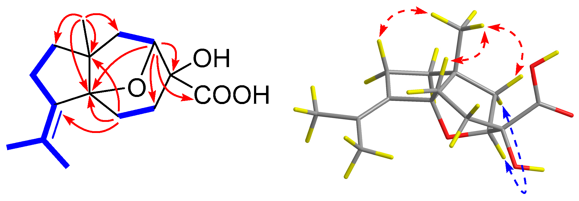
Marinedrugs 17 00507 g002
Figure 3. Experimental and calculated ECD spectra of 1.
Marinedrugs 17 00507 g003
Figure 4. Key COSY ( Marinedrugs 17 00507 i004), HMBC ( Marinedrugs 17 00507 i005), and NOESY ( Marinedrugs 17 00507 i006) correlations of 2.
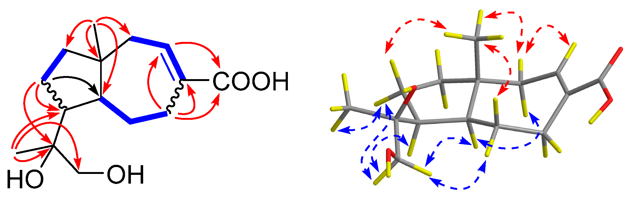
Marinedrugs 17 00507 g004
Figure 5. Experimental and calculated ECD spectra of 2.
Marinedrugs 17 00507 g005
Table 1. 1H (400 MHz) NMR spectroscopic data of 15 (δ in ppm, J in Hz within parentheses).
No.1 a2 a3 b4 a5 a
12.01, dd (13.2, 7.6)2.41, dd (14.9, 9.1)2.00, dd (12.7, 8.5)2.20, dd (13.0, 8.5)2.40, overlap
1.90, d (13.2)2.14, br d (14.2)1.85, d (12.7)2.00, d (13.0)1.48, overlap
24.48, d (7.5)7.07, dt (8.6, 3.1)4.13, d (8.4)4.28, d (8.4)4.31, d (8.4)
42.18, dd (13.5, 6.4)2.99, dd (15.8, 5.8)2.18, dd (13.8, 7.6)2.42, dd (13.7, 7.5)2.28, m
1.69, m1.93, overlap1.20, m1.33, m2.03, overlap
52.74, dt (12.6, 6.2)1.93, overlap1.93, dd (11.0, 8.0)1.77, m2.16, dd (13.1, 8.4)
1.55, overlap1.25, d (13.2)1.43, overlap1.45, dd (13.6, 7.6)2.03, overlap
6-1.59, t (10.5)1.42, overlap1.72, m2.32, m
81.63, m1.41, m1.56, m1.84, m2.40, overlap
1.55, overlap1.26, dd (11.1, 7.6)1.65, m1.67, m
92.38, m1.78, m1.41, overlap2.10, m1.69, m
1.68, m1.61, m1.48, overlap
10-1.93, overlap1.58, m--
121.82, br s1.19, s1.03, s1.76, s1.73, br s
131.63, br s3.45, br s1.05, s5.01, s1.60, br s
4.84, s
151.18, s0.79, s3.64, d (8.0)4.62, d (8.0)3.70, d (8.0)
3.18, d (8.1)3.34, d (7.9)3.35, d (8.7)
a Recorded in CD3OD. b Recorded in DMSO-d6.
Table 2. 13C (100 MHz) NMR spectroscopic data of 15.
No.1 a2 a3 b4 a5 a
140.4 CH242.3 CH235.8 CH238.9 CH235.2 CH2
282.6 CH142.8 CH82.8 CH84.0 CH84.9 CH
374.4 C136.5 C77.6 C79.6 C79.5 C
428.5 CH228.5 CH234.3 CH235.3 CH233.0 CH2
527.7 CH227.0 CH224.8 CH219.8 CH236.8 CH2
693.9 C55.8 CH52.3 CH58.4 CH56.7 CH
754.0 C43.5 C52.7 C52.7 C54.2 C
841.6 CH242.4 CH233.6 CH233.3 CH224.9 CH2
933.3 CH224.6 CH226.8 CH240.0 CH235.1 CH2
10135.6 C50.1 CH51.8 CH86.0 C136.7 C
11131.6 C76.3 C71.5 C150.7 C125.2 C
1221.3 CH322.9 CH326.6 CH319.7 CH321.0 CH3
1323.5 CH369.5 CH229.2 CH3110.3 CH223.4 CH3
14178.0 C172.0 C175.8 C177.9 C177.9 C
1520.1 CH319.2 CH374.5 CH276.2 CH276.5 CH2
a Recorded in CD3OD. b Recorded in DMSO-d6.

