Review

Prostaglandins in Marine Organisms: A Review

Marine Biotechnology Department, Stazione Zoologica Anton Dohrn Napoli, Villa Comunale, 80121 Napoli, Italy
*
Author to whom correspondence should be addressed.
Mar. Drugs 2019, 17(7), 428; https://doi.org/10.3390/md17070428
Submission received: 17 June 2019 / Revised: 15 July 2019 / Accepted: 19 July 2019 / Published: 23 July 2019
(This article belongs to the Special Issue Marine Anti-inflammatory Agents)

Abstract

:
Prostaglandins (PGs) are lipid mediators belonging to the eicosanoid family. PGs were first discovered in mammals where they are key players in a great variety of physiological and pathological processes, for instance muscle and blood vessel tone regulation, inflammation, signaling, hemostasis, reproduction, and sleep-wake regulation. These molecules have successively been discovered in lower organisms, including marine invertebrates in which they play similar roles to those in mammals, being involved in the control of oogenesis and spermatogenesis, ion transport, and defense. Prostaglandins have also been found in some marine macroalgae of the genera Gracilaria and Laminaria and very recently the PGs pathway has been identified for the first time in some species of marine microalgae. In this review we report on the occurrence of prostaglandins in the marine environment and discuss the anti-inflammatory role of these molecules.

1. Introduction

Marine organisms have a great potential to produce a vast variety of bioactive molecules with high antibiotic, anti-proliferative, and anti-inflammatory activity [1]. The biodiversity hosted by the oceans is greater than in terrestrial environments [2] but nonetheless, marine bioresources are still underexplored, and many species await to be discovered.
The high probability to find new interesting bioactive molecules from marine organisms has fostered the effort of the scientific community to adopt new technologies and approaches to increase the success of biodiscovery from marine resources, with a main focus on products with antibiotic, antitumor and anti-inflammatory activities. Of particular interest is the search for new anti-inflammatory compounds since inflammation processes are often related to the onset of chronic pathologies and tumors.
Indeed, inflammation processes represent a fundamental way to restore the original equilibrium of a cell or tissue whose physiology has been impaired by damaging stimuli [3]. At the same time, if inflammation is not blocked it can stimulate a cascade of events that eventually lead to serious diseases such as cancer and autoimmune disorders [4]. The inflammation-resolution process can have different features depending on the type of tissue and injurious stimulus [5], therefore, specific types of pro-resolution stimuli or drugs may be necessary [6]. Both the onset and the resolution of the inflammation are active processes that involve a complex interplay of different molecules [7] like chemokines, cell adhesion molecules, proteolytic enzymes, eicosanoids [8], reactive oxygen species (ROS), and reactive nitrogen species (RNS) [9,10]. Among these, eicosanoids deriving from oxidation of polyunsaturated fatty acids (PUFA) through cyclooxygenase (COX) and lipoxygenase (LOX) pathways play a pivotal role both in the onset and in the resolution of inflammation [9]. The main products of COX enzymes are prostaglandins (PGs), fatty acid derivatives with a molecular structure based on 20 carbon atoms that share a prostanoic acid skeleton.
Prostaglandin E2 was the first PGs to be identified in the early 1930s in human seminal plasma by Von Euler [11] and, independently, by Goldblatt [12] although the chemical structures were determined only 30 years later by Bergström, Samuelsson, and co-workers [13].
PGs are very well studied and described in mammals where they are active in a great variety of physiological and pathological processes such as smooth muscle and vaso-tone regulation, signaling, hemostasis sleep-wake regulation, reproduction, and especially inflammation. In these organisms, they are synthetized and released in response to external stimuli [14] and rapidly inactivated by metabolizing enzymes after they have accomplished their function [15].
PGs represent very important and interesting lipid mediators in all vertebrates and in both terrestrial and marine invertebrates [16,17]. Plants utilize chemically related molecules (jasmonic acid) that have a defensive role, similarly to PGs [16,18,19]. Nevertheless, in some plant species like onions and poplar, a few PGs have also been identified [20].
In the marine environment, prostaglandins have been reported both in vertebrates such as carps, sheatfish and leopard sharks, and invertebrates such as sea squirts, mussels, scallops, crawfish, blue crabs, and sea-anemones and in some macroalgae as the red alga Gracilaria asiatica C. F. Zhang & B. M. Xia, 1985 and the brown alga Laminaria digitata (Hudson) J. V. Lamouroux, 1813.
The earliest report about the presence of prostaglandins in marine invertebrates was by Weinheimer and Spraggins (1969) that discovered 15-epi-prostaglandin A2 and its acetate methyl ester in the gorgonian-type coral Plexaura homomalla Esper, 1794 [21]. The discovery of high PGs levels in gorgonians contributed to the rapid growth of the study and application of PGs in the pharmaceutical and biomedical sector [22].
Very recently, Di Dato et al. (2017) described the presence of all the three series of PGs molecules also in diatoms, an ecologically successful group of marine microalgae [23].
In this review, we present a short background on prostaglandin structure and function and give an updated overview of the presence of PGs in marine organisms, discussing the anti-inflammatory role of PGs from the marine environment.

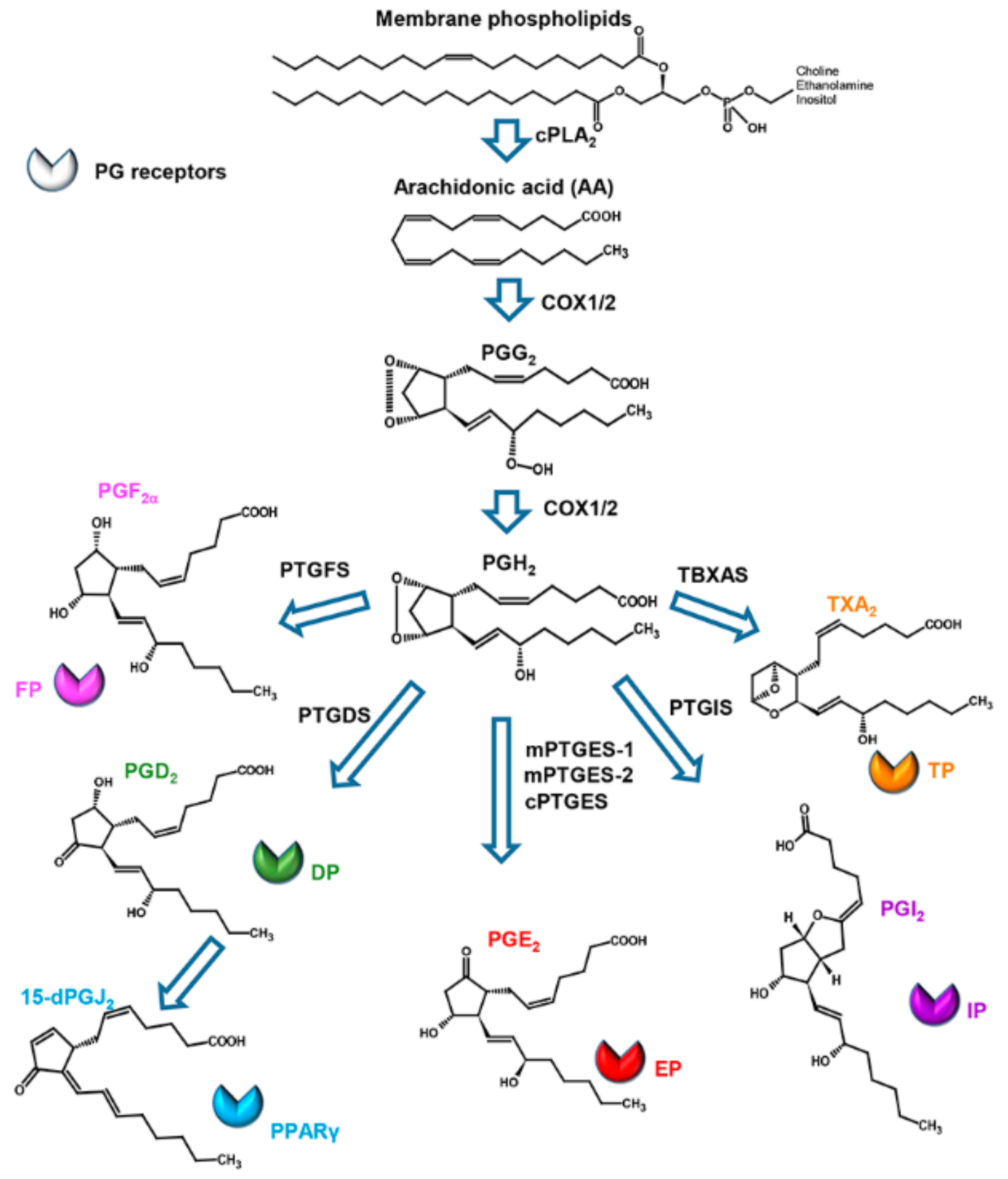
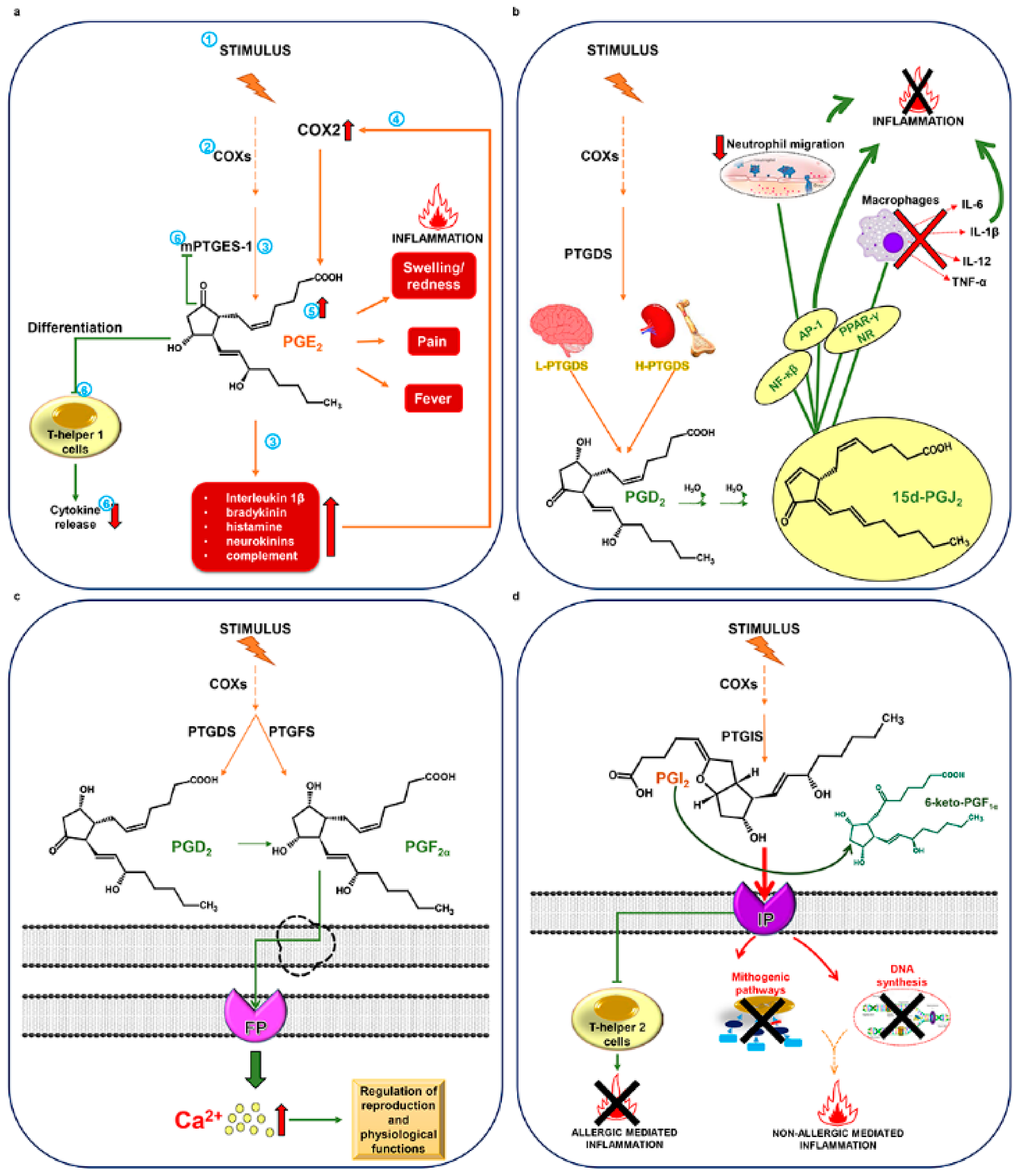
2. Structure, Biosynthesis, and Activity of Prostaglandins in Mammals

Prostaglandins consist of a cyclopentanone nucleus with two side chains. Primary prostaglandins, which include prostaglandin D2 (PGD2), prostaglandin E2 (PGE2), prostaglandin F (PGF), and prostaglandin I2 (PGI2), contain a 15-hydroxyl group with a 13,14-trans double bond (Figure 1).
Currently, three classes of prostaglandins are categorized, based on the number of double bonds present within the molecule and on the fatty acid precursor. Prostaglandins belonging to the series 1 have one double bond and derive from 8,11,14-eicosatrienoic acid (di-homolinolenic acid, ETrA), those of the series 2 have two double bonds and derive from 5,8,11,14-eicosatetraenoic acid (arachidonic acid, ARA), and those of the series 3 have three double bonds and derive from 5,8,11,14,17-eicosapentaenoic acid (EPA).
Their nomenclature comprises 10 specific molecular groups, identified by the letters A through J, that differ by variation in the functional groups attached to positions 9 and 11 of the cyclopentane ring. For PGF, the additional subscript “α” or “β” denotes the spatial configuration of the carbon 9 hydroxyl group. For additional details on the nomenclature of prostaglandins see Lands, 1979 [25].
PGs action is mediated by the interaction with specific receptors present on the plasma membrane. These are transmembrane G-protein coupled receptors (GPCR), named as prostaglandin EP receptor (EP), prostaglandin F receptor (FP), prostaglandin DP receptor (DP), and prostacyclin I2 receptor (IP) receptors, that are highly selective for PGE2 PGF, PGD2, and PGI2, respectively [26]. The EP family comprises four isoforms (EP1-4) that play a relevant role in inflammation processes [27]. The downstream signaling of this receptor family is responsible for the pleiotropic ability of PGE2 to activate different processes, including cell proliferation, apoptosis, angiogenesis, inflammation, and immune surveillance in different cell types [24].
Most prostaglandins display a marked structure-activity specificity mainly determined by substitutions in the cyclopentanone ring and the degree of unsaturation of the side chains. They exert their function once secreted into the extracellular medium, where they are rapidly metabolized by 15-hydroxyprostaglandin dehydrogenase (15-OH-PGDH). This enzyme selectively oxidizes the hydroxyl group at carbon 15 into a 15-keto derivative [28] accompanied by a substantial loss of biological activity.
Prostaglandins derive from the sequential actions of highly specific enzymes (Figure 1). Their synthesis is initiated by phospholipases A2 (PLA2), a family of enzymes that hydrolyze membrane phospholipids at the sn-2 position, liberating free fatty acid precursors, mainly ARA [15]. These enzymes represent a key step in the PG biosynthetic pathway, being regulated by Ca2+ binding and phosphorylation by mitogen-activated protein kinase (MAPK) in response to different stimuli. Membrane-released ARA is then rapidly converted through the cyclization and inclusion of molecular oxygen in the precursor by the action of cyclooxygenase (COXs) enzymes into the unstable metabolite PGG2, which is subsequently reduced to PGH2 by the same enzyme [14]. Cyclooxygenases exist in a substrate-limiting environment; thus, liberation of fatty acids from esterified stores results in the prompt formation of the products. There are two major COX isoforms; COX-1 is constitutively active and present in most cells in the body; expression of the COX-2 isoform is inducible in many tissues by pro-inflammatory and mitogenic stimuli, such as cytokines [29]. The specific transformation of the first product PGH2 to other PGs and thromboxanes (TXs) by downstream enzymes is complexly orchestrated and is cell specific, since each cell tends to form mainly one of these compounds as the major product. For example, in brain and mast cells, PGH2 is converted to PGD2, whereas it is converted in PGF in the uterus; from the same precursor, vascular endothelial cells produce PGI2 (prostacyclin) and platelets release thromboxane A2 (TXA2).
The conversion to PGE2, the most widespread PG, is due to PGE synthase-1, also present in different isoforms in mammals: microsomal PGE synthase-1 (mPTGES-1), mPTGES-2, and cytosolic PGE synthase (cPTGES), the latter of particular interest since frequently associated to the tumorigenic activity of PGE2 [30].
Prostaglandin E2 synthesis is a key event for the development of the three principal signs of inflammation: swelling, redness, pain and fever. Moreover, PGE2 also contributes to the amplification of the inflammatory response by enhancing and prolonging signals produced by pro-inflammatory agents, such as interleukin 1α bradykinin, histamine, neurokinins, and complement [31,32]. These signals, in turn, can increase COX-2 expression thus further increasing PGE2 synthesis. This PG, however, can have a double, inverse role, being also able to act as an immunosuppressant, repressing the differentiation of T helper 1 cells and limiting cytokine release and further prostaglandin synthesis by activating a negative feedback on mPGES-1 (Figure 2a).
PGD2-synthesizing enzymes exist as two distinct genes coding for hematopoietic- and lipocalin-type PGD synthases (H-PTGDS and L-PTGDS, respectively). H-PTGDS is generally localized in the cytosol of immune and inflammatory cells, whereas L-PTGDS has a tissue-based expression [33]. The activity of PGD2 has been mainly associated with inflammatory conditions, being involved in immunologically relevant functions. Its action seems to be mediated by the non-enzymatic production of the PGJ2 family, which occurs through a spontaneous dehydration in aqueous solutions. One of the most studied PGJ2 metabolites, 15-deoxy-Δ12-PGJ2 (15dPGJ2), showed anti-inflammatory properties based on interaction with the intracellular targets Nuclear Factor kappa-light-chain-enhancer of activated B cells (NF-κB), Activator Protein 1 (AP-1), and Peroxisome Proliferator-Activated Receptor gamma (PPAR-γ) [31]. 15dPGJ2 also induces the reduction of neutrophil migration and inhibits the release of Interleukin 6 (IL-6), Interleukin 1β (IL-1β), Interleukin 12 (IL-12), and Tumor Necrosis Factor-α (TNF-α) from macrophages (Figure 2b).
PGD2 can be further metabolized to PGF, although the latter can also be synthesized from PGH2 via PGF synthase. PGF acts via FP receptors, resulting in the elevation of intracellular free calcium concentrations that regulates numerous important physiological functions related to reproduction linking multiple molecular mechanisms that are fine-tuned coordinated events in mammalian physiology [34] (Figure 2c). The emerging role of PGF in acute and chronic inflammation has opened new opportunities for the design of novel anti-inflammatory drugs.
A non-enzymatic dehydration reaction is also responsible for the formation of the PGA series from the corresponding PGE. The PG series A and J contain an α,β-unsaturated carbonyl group within the cyclopentenone ring, which seem to contribute to the anti-inflammatory activity of these PGs [35].
The enzyme prostaglandin I synthase (PGIS), a member of the cytochrome P450 superfamily, specifically converts PGH2 to PGI2 [33]. This PG has anti-mitogenic activity and inhibits DNA synthesis through specific IP receptors. It mediates pro-inflammatory stimuli in non-allergic acute inflammation, while acting as an anti-inflammatory mediator in Th2-mediated allergic inflammatory responses. Soon after having exerted its action, PGI2 is rapidly converted by non-enzymatic processes to the inactive product 6-keto-PGF (Figure 2d).

3. Prostaglandins and Derivative Molecules in Marine Organisms
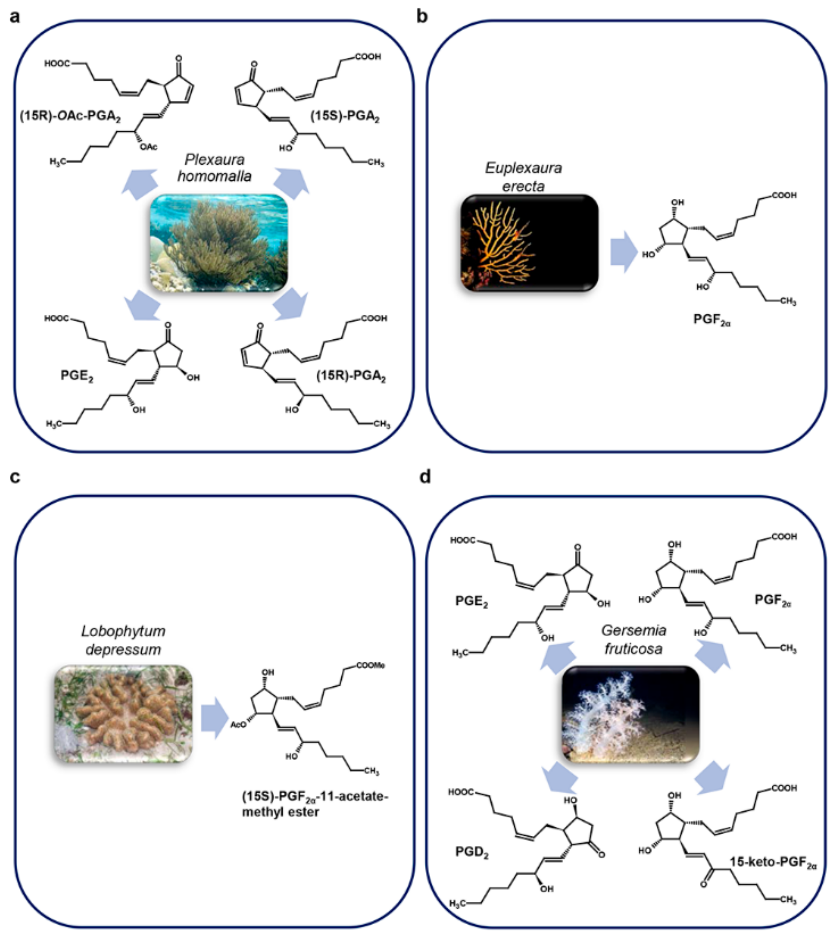
Similar to terrestrial vertebrates, in marine vertebrates prostaglandins are involved in reproduction, osmoregulation, regulation of oxygenation, and cardiovascular system [36]. Marine invertebrates, including sponges, corals, and molluscs, also contain a wide range of prostaglandins, many of which are of the conventional type (PGA2, PGE2, PGD2, PGF), with similar functions as in mammals (reproduction, ion transport) and are also probably used as defense compounds [17]. Interestingly, in some invertebrates, PGs are able to perform different actions based on the tissue or compartment localization [37].
Complex marine photosynthesizing organisms, like some genera in the brown, green, and red algal groups (respectively, Laminaria, Euglena, and Gracilaria species) express the cyclooxygenase gene synthesizing PGE2, PGF, and other PGs whose functions are not yet known, but seem to be associated to a defensive role [17].

3.1. Corals

Among marine invertebrates, corals represent a very interesting group producing specific PGs that are not present in mammals. The Caribbean gorgonian Plexaura homomalla was indeed the species in which PGs were firstly identified in a marine organism, representing also a major source of these compounds in nature [38] (Figure 3a).
Weinheimer A. and Spraggins R. were the first to perform PGs extraction in P. homomalla, identifying in its dry cortex 15-epi-PGA2 and its methyl ester acetate (respectively 0.2% and 1.3%) [15] in an R-configuration on their C-15 asymmetric center, a configuration not present in mammals. In addition to (15R)-epi-PGA2, also (15R)-PGE2, its methyl ester and a complex mixture of other prostaglandins were subsequently found by Light R. and Samuelsson B. [39]. However, these molecules were found also in S configurations [40] and the occurrence of one of the two seems to be related to the geographical distribution of P. homomalla [38].
The high PGs concentration in these invertebrates stimulated the attempt to use them as precursors for the chemical synthesis of the biologically active prostaglandins A2, E2, and F, since their chemical synthesis in large-scale for medical and pharmaceutical purposes was complicated by the necessity of 16 different chemical reactions [41].
The observation that P. homomalla present in coral reefs is not commonly eaten by fishes, led Gerhart [42] to hypothesize that the high amount of PGs they contain could be used as chemical defense against potential predators, since it is known that in mammals they can cause vomiting and nausea when administered orally. Indeed, oral doses of both (15R)-PGA2 and (15S)-PGA2 caused vomiting in a test with fishes, while the PGA2 present in the surrounding water did not cause any effect [42]. However, the hypothesized defensive role seems not to be realistic since PGA2 is stored in coral tissues only as acetoxy methyl esters, whose conversion to (15R)-PGA2 (the compound tested by Gerhart) needs about 24 hours [40]. Moreover, while (15R)-PGA2 inhibits fishes from feeding, the acetoxy methyl ester form, orally delivered, does not show any repellent effect. For this reason, as the process of production of the active (15R)-PGA2 seems too slow to provide the coral with an effective defense mechanism, the function of these molecules in corals is still an open question.
The R-prostaglandins extracted from P. homomalla collected on the Island of San Andreas, in the Caribbean Ocean, were also tested in vivo on mouse ear edema induced by 12-O-tetradecanoylphorbol-13-acetate (TPA) and in vitro, in anti-inflammatory screenings as leucocyte degranulation, myeloperoxidase (MPO), and elastase enzymatic activity inhibition. The results showed that (15R)-PGE2 and, to a lesser extent, (15R)-O-AcPGA2 had an anti-inflammatory activity in vivo and in vitro (Table 1). In particular, in the leucocyte degranulation assay, (15R)-PGE2 greatly inhibited the release of both MPO and elastase, while the other prostaglandins tested were moderately active in the inhibition of elastase release but not of MPO [43].
Euplexaura erecta Kükenthal, 1908 [56] (Figure 3b), Lobophytum depressum Tixier-Durivault, 1966 [57] (Figure 3c), and Gersemia fruticosa Sars, 1860 [58] (Figure 3d) also produce PGs. Interestingly, PGF and its derivatives were principally found in these species: PGF in E. erecta [56]; (15S)-PGF-11-acetate methyl ester, its 18-acetoxy derivative compound and their corresponding free carboxylic acids in L. depressum [57]; PGF and 15-keto-PGF together with PGD2, PGE2 in G. fruticosa [58].
In addition to the identification of PGs molecules in corals, also the enzyme responsible for their synthesis, the cyclooxygenase, was isolated and characterized for the first time from P. homomalla and G. fruticosa [59].

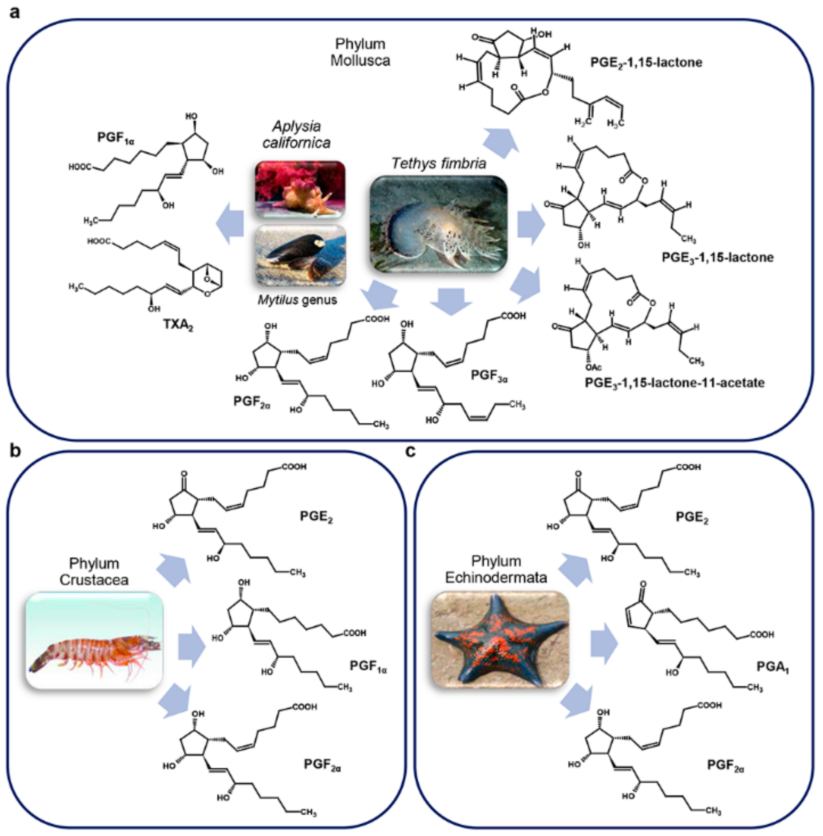
3.2. Other Marine Invertebrates

One of the first studies on marine invertebrates, excluding corals, was a comparative analysis done by Christ E. and Van Dorp D., on representative species in the Mollusca (Mytilus), Crustacea (Homarus), and Cnidaria (Cyanea) phyla versus terrestrial animals [60]. The authors were able to find PGs, particularly PGF but only at very low levels. In some cases, the arachidonic acid precursor was also not detectable making it difficult to assert the existence of PGs and their functional role [60]. These results were confirmed in extended studies including more species in the Chordata, Mollusca, Cnidaria, and Crustacea phyla [16,61,62].
With the improvement of instrument sensitivity, it was possible, more than 10 years later, to conduct functional studies on PGs in molluscs (Figure 4a) and crustaceans (Figure 4b).
Results obtained highlighted different roles for PGs in marine invertebrates, like reproduction, regulation of ion flux, and thermoregulation and fever, mediated respectively by PGF, PGE2, and PGE1 (Table 2) [63].
In the mollusc Modiolus demissus Dillwyn, 1817 the uptake and binding of prostaglandins by gills was investigated, considering that bivalves can both synthetize and accumulate PGs from the surrounding medium [71]. The study revealed the presence of tissue specific, time and pH dependent, PGs binding sites with higher affinity to PGA2 with respect to PGE2 or PGF [71].
Moreover, three prostaglandin-lactones (PGE2-1,15-lactone, PGE3-1,15-lactone, and PGE3-1,15-lactone-11-acetate) of the E and F series were identified in the mantle and body, respectively, of the nudibranch mollusc Tethys fimbria Linnaeus, 1767 (Figure 4a). High quantities of a complex mixture of PGF and PGF 1,15-lactones fatty acid esters (PLFE) were found in its eggs, particularly in mature ovotestis but not in immature ones suggesting a role for PLFE in mollusc reproduction [67]. Altogether, these results led to hypothesize a multiple biological role of prostaglandin-lactones, precursors of PGEs, as defense allormones, and involved in the control of smooth muscle contraction, and egg production and fertilization, depending on their body localization (Table 2) [37].
Crustaceans (Figure 4b) also use PGs for physiological functions. Indeed, PGE2 and PGF were identified in the ovary of the prawn Marsupenaeus japonicus Spence Bate, 1888 where they participate in ovarian maturation (Table 2) [64] whereas in the crab Carcinus maenas Linnaeus, 1758 they are produced in blood cells following a stimulus induced with a calcium ionophore, in the presence of exogenous fatty acids (FA) [72].
Echinoderms (Figure 4c) also represent a source of PGs. Among all the echinoderms analyzed, the starfish Patinia pectinifera Muller and Troschel, 1842 had the highest amount of prostaglandins [73]. PGE2 and PGF have been identified from the sea cucumber Stichopus japonicus Selenka, 1867 using TLC [73]. A study conducted with PGs-3H evidenced that the sea urchin Arbacia punctulata Lamarck, 1816 could accumulate PGs from the surrounding water into its gut or stomach [74], and to accumulate PGA1 both in fertilized and unfertilized eggs, with fertilized eggs accumulating more PGs than unfertilized eggs [74].
Strongylocentrotus nudus A. Agassiz, 1864 and S. intermedius A. Agassiz, 1864 were also reported to have a PGs-like activity in their inner organs [74].

3.3. Marine Vertebrates

As already mentioned, in fish PGs are involved in several processes, such as ovulation, spawning, osmoregulation, regulation of branchial ion fluxes and of the cardiovascular system [36]. Christ and Van Dorp in their comparative studies on PGs considered not only invertebrates, but also fish [60]. They identified moderate yields of PGE1 in freshwater fish and lower yields in homogenates of gills of Salmo sp. [60]. Successively, PGE2 was identified for the first time in the testis of the flounder Paralichthys olivaceus Temminck & Schlegel, 1846, PGF in the semen of the salmon Oncorhynchus keta Walbaum, 1792 and PGE2 and PGF in the testis of the tuna Thunnus thynnus Linnaeus, 1758 [65] (Figure 5a).
In order to explain the reason why prostaglandins are present in these marine animals, although they are oviparous, it was suggested that the identified prostaglandins might be used for the contraction of smooth muscle during ejaculation and for the metabolism of testis in lower animals as in mammals (Table 2) [65]. After this study, PGE2 was isolated and subsequently identified in the gastrointestinal tract and in the skin of the shark Triakis scyllia Müller & Henle, 1839 [16,75] (Figure 5b).
In the brook trout Salvelinus fontinalis PGE2 and PGF (Figure 5a) are synthetized in the follicle wall of mature oocytes, but the highest quantity of prostaglandins was found in the extra-follicular tissue [76]. PGE2 was also found in the skin of Pleuronectes platessa Linnaeus, 1758 (Figure 5a) in response to the fungal extract of Epydermophyton floccosum (Harz) Langeron & Miloch, 1930 known be an inducer of erythema [77].
Leucocytes of the dogfish Scyliorhinus canicula Linnaeus, 1758 secerned high levels of PGE2, PGD2 and PGF (Figure 5b) when exposed to the Ca2+ ionophore A23187 [78]. PGE2 and PGF were found in the red blood cells of the toadfish Opsanus tau Linnaeus, 1766 [79] (Figure 5a).

3.4. Macroalgae

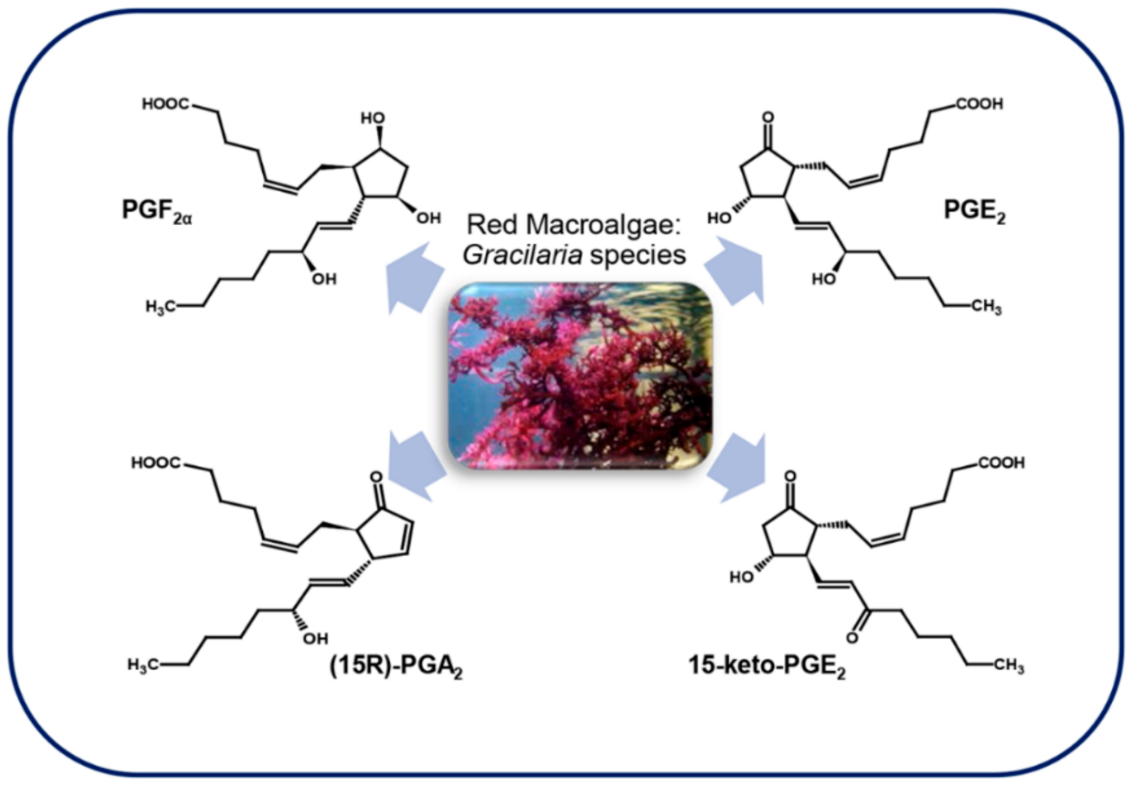
3.4.1. Red Macroalgae

Marine red algae are rich in C20 polyunsaturated fatty acids that, in animals, are precursors of prostaglandins, thromboxane, and other eicosanoids. Although the presence of the prostaglandin-endoperoxide pathway has been demonstrated in non-mammal marine vertebrates and invertebrates, for a long time less was known about these enzymes in non-animal organisms. The first report about the presence of prostaglandins in macroalgae (Figure 6) described the identification of PGE2 and PGF in Gracilaria lichenoides Greville, 1830 [80].
The genus Gracilaria, that comprises algae used in the food and cosmetic industry, is very rich in ARA, varying from 45.9% and 62.0% of the total FA depending on the season. Among these, G. vermiculophylla (Ohmi) Papenfuss, 1967 is one of the algae with the highest content of ARA [81]. This, with other Gracilaria species such as G. asiatica and G. chorda Holmes, 1896, seem to be responsible for a gastrointestinal disorder known as “onogori” poisoning in Japan when it is eaten raw [18]. The possible reason for this poisoning seems to be the fact that the COX contained in raw seaweeds use the highly unsaturated fatty acids to produce great amounts of PGE2 in the stomach of victims in a short lapse of time [81]. G. vermiculophylla is a source of different types of prostaglandins besides PGE2, such as PGA2, PGF2, 15-keto-derivatives of prostaglandins, as well as other eicosanoids [82], whereas the congeneric species G. asiatica produces only 15-keto-PGE2 [83]. G. vermiculophylla use PGA2, PGE2, 15-keto-PGE2 and other eicosanoids as wounding-activated chemical defense molecules (Table 2) [66], and recently a COX gene producing PGF was cloned from this alga and heterologously expressed [84].
Other evidence of prostaglandins as defense molecules in red algae is the fact that gametophytes of the alga Chondrus crispus Stackhouse, 1797, when challenged with pathogens, metabolize C20 and C18 PUFAs not only into the corresponding hydroperoxides and derivatives, but also into molecules with mass fragmentation patterns very similar to prostaglandin B1 and B2 [85]. Furthermore, when the crude extract of this alga was treated with 50–100 µM methyl jasmonate for 6 h, PGA2 and 15-keto-PGE2 were identified [86].

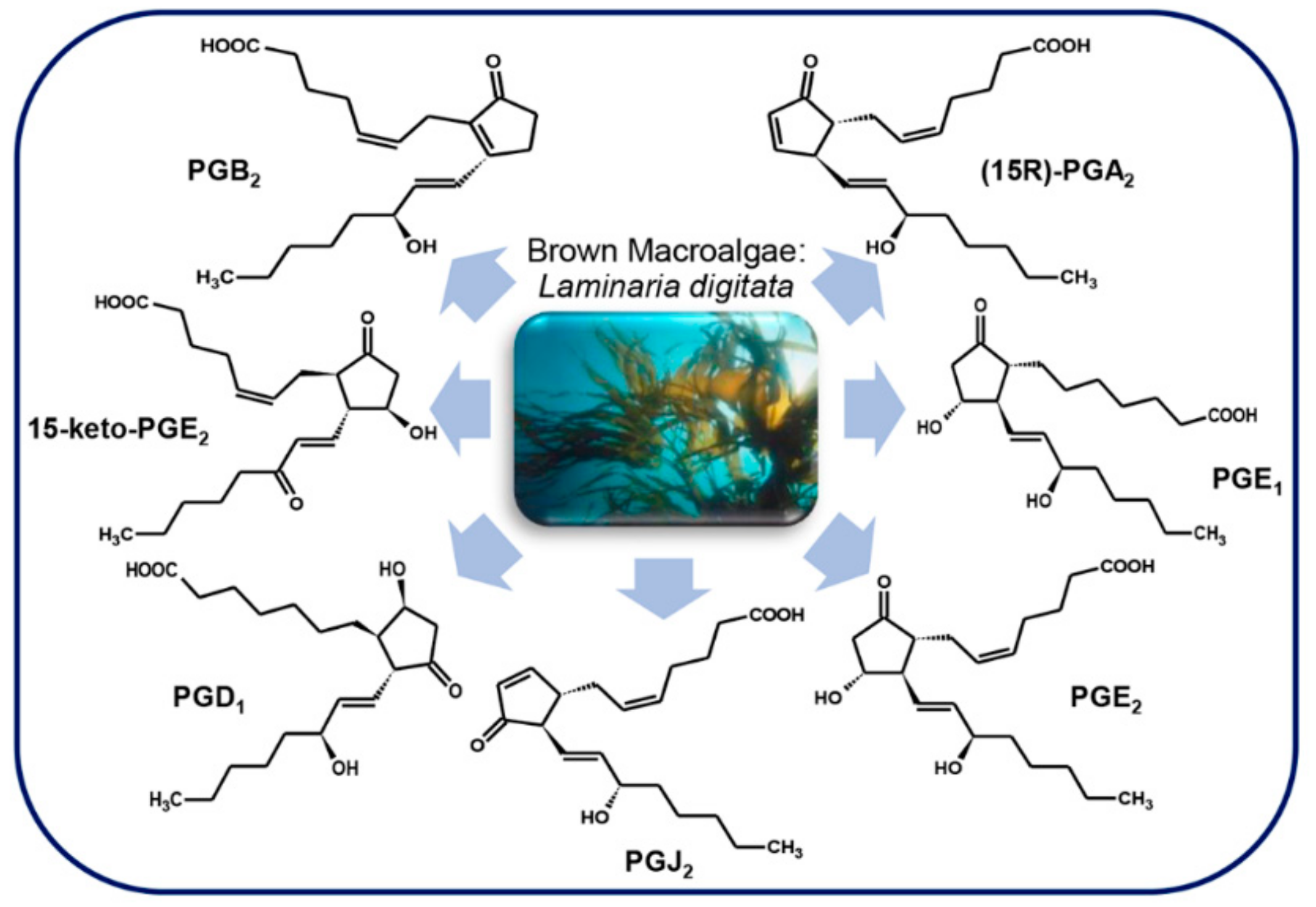
3.4.2. Brown Macroalgae
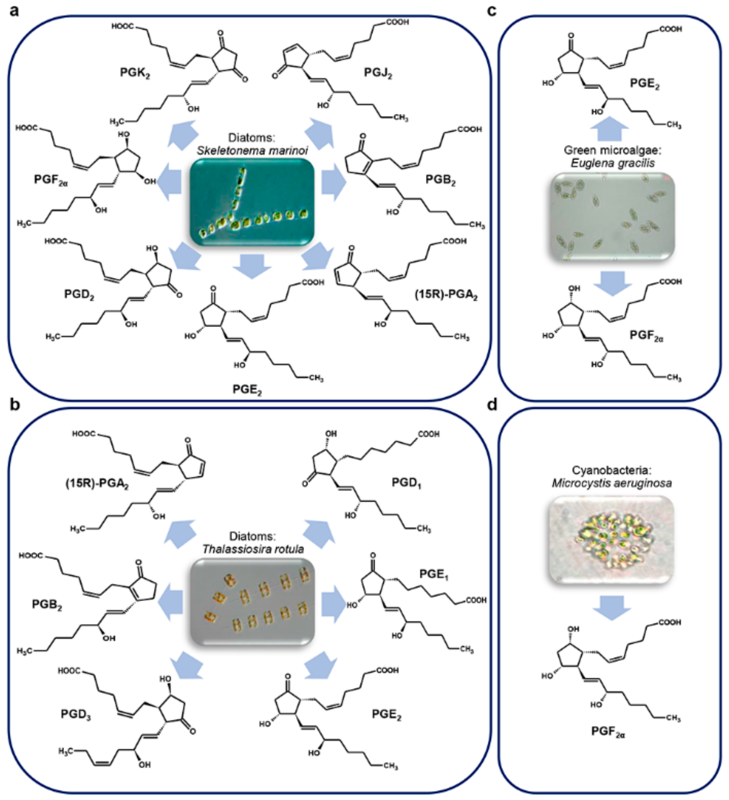
Much less is known about the presence of prostaglandins in brown algae, and most studies have mainly focused on the brown algal kelp Laminaria digitata. Ritter et al. [19] showed that this brown alga uses the generation of oxylipins as a protective mechanism against stress conditions induced by an excess in copper (Table 2). Indeed, high concentrations of copper led to oxygen reactive species (ROS) accumulation in L. digitata cells, and consequently to a cascade of cellular responses. One of these responses was a significant release of PUFAs after 24 h of treatment, followed by the generation of oxylipins. Among complex oxylipins, also PGE1 and PGD1, deriving from ETrA and PGJ2, PGA2, 15-keto-PGE2 and PGB2, deriving from ARA were identified for the first time in brown algae [19]. Successively, the same authors showed that PGA2 does not simply modulate but also triggers an oxidative response in L. digitata that, differently from the response induced by other molecules like methyljasmonate and lipopolysaccharides, occurs in seconds after treatment and in a dose-response-like manner. The authors hypothesized that PGA2 can activate the generation of two different sources of ROS that can be used by the brown alga as defense molecules as in other marine organisms [68] (Figure 7).

3.5. Microalgae

PGs were discovered for the first time in diatoms, phytoplanktonic marine microalgae, only in 2017 [23]. Diatoms are a rich source of polyunsaturated fatty acids (PUFA), that, in some species, are precursors of polyunsaturated aldehydes (PUAs), i.e., oxylipins with pro-apoptotic and anti-proliferative activity, with a defensive role against predator (copepods) grazing [87].
Di Dato et al. [23] explored the presence of PGs in some diatom species, i.e. Skeletonema marinoi Sarno & Zingone, 2005 (Figure 8a) (two different strains, FE7 and FE60) and Thalassiosira rotula Meunier, 1910 (Figure 8b) (Valeria Di Dato, Roberta Barbarinaldi, Alberto Amato, Federica Di Costanzo, Carolina Fontanarosa, Anna Perna, Angela Amoresano, Francesco Esposito, Adele Cutignano, Adrianna Ianora, Giovanna Romano. Variation in prostaglandin metabolism during growth of the diatom Thalassiosira rotula. Sci Rep, under review), identifying in their transcriptome genes involved in PGs biosynthesis, with some differences between the two species.
More specifically, whereas cyclooxygenase-1 (COX-1) and microsomal prostaglandin E synthase 1 (mPTGES) were found in both species; prostaglandin H2 D-isomerase (PTGDS) was found only in S. marinoi, while prostaglandin F synthase (PTGFS) was only detectable in T. rotula. The authors demonstrated the functioning of the pathway by measuring enzyme expression and molecule production by quantitative real time PCR (qPCR) and liquid chromatography/mass spectrometry (LC/MS) analysis, respectively. Interestingly, a wide set of PGs was revealed with representative molecules of each of the three series. In addition, a differential expression and concentration of molecules was reported among different species and strains of the same species, during different phases of growth and nutrient conditions [23]. However, the absence of PTGDS in T. rotula and of PTGFS in S. marinoi could be considered as a “potential absence.” Indeed, the lack of a transcript in a transcriptome annotation can be due to a technical shortcoming and a limitation of the sequencing technology used, as in the case of very low expression levels of a target gene, that may fall under the detection limit.
It is interesting to note that, although the PGs pathway has been experimentally confirmed only in two diatom species, in silico analysis of transcriptomes from different species in different growth conditions, suggests the presence of PGs in many other diatom species [23].
Prostaglandins were identified also in the unicellular green alga Euglena gracilis G. A. Klebs, 1883 (Figure 8c), in which PGE2 and PGF were found at levels three times higher in cells grown in the dark than those grown in the light [88].
The presence of PGA, PGE, and PGF series and their esters have also been documented in the cyanobacteria of the taxa Oscillatoria and Microcystidaceae [89]. Indeed, PGF was identified in Microcystis aeruginosa (Kützing) Kützing, 1846 [90] (Figure 8d).
Currently, to the best of our knowledge, no other marine microalga has been explored for the presence of PGs, and their role in diatoms is still under investigation. Di Dato and co-authors hypothesized that they can be used by diatoms for intercellular signaling and communication [23], in agreement with the role of these molecules in other organisms.
Microorganisms, and in particular microalgae such as diatoms, could represent a potential source of bioactive PGs, as alternative to their chemical synthesis to produce adequate amounts for pharmacological purposes as anti-inflammatory compounds. The biotechnological production of PGs from microalgae may also be convenient due to the natural presence of the full PGs pathway and the possibility to easily grow microalgae on a large scale in bioreactors under controlled culture conditions.

4. Marine Cyclopentenone Prostaglandins

Cyclopentenone prostaglandins are a sub-group of PGs characterized by the presence of a cyclopentenone moiety (CP) with a α,β-unsaturated ketone group in their cyclopentane ring. Their chemical reactivity is responsible for their anti-inflammatory, anti-tumoral and anti-viral activities [91], and the presence of the CP fragment in molecules with anticancer activity, can add more potency to these molecules. For instance, methyl-jasmonate increases its strength of action as an anti-tumor agent when a CP group is inserted in the molecule [92].
The group of cyclopentenone prostaglandins (CPPGs) includes canonical PGs of the A and J series, clavulones, and punaglandins.
Differently from the conventional PGs, that require receptor interaction to trigger their signal, cyclopentenone prostaglandins are actively transported inside the cells [91], where the CP group can interact with a wide variety of target molecules [92] including nuclear factors such as the heat shock protein 70 (HSP70) transcription factor, cyclin-dependent protein kinase (CDK), and NF-ĸB [35] and still not well defined mitochondrial factors [92].

4.1. Clavulones and Related Molecules
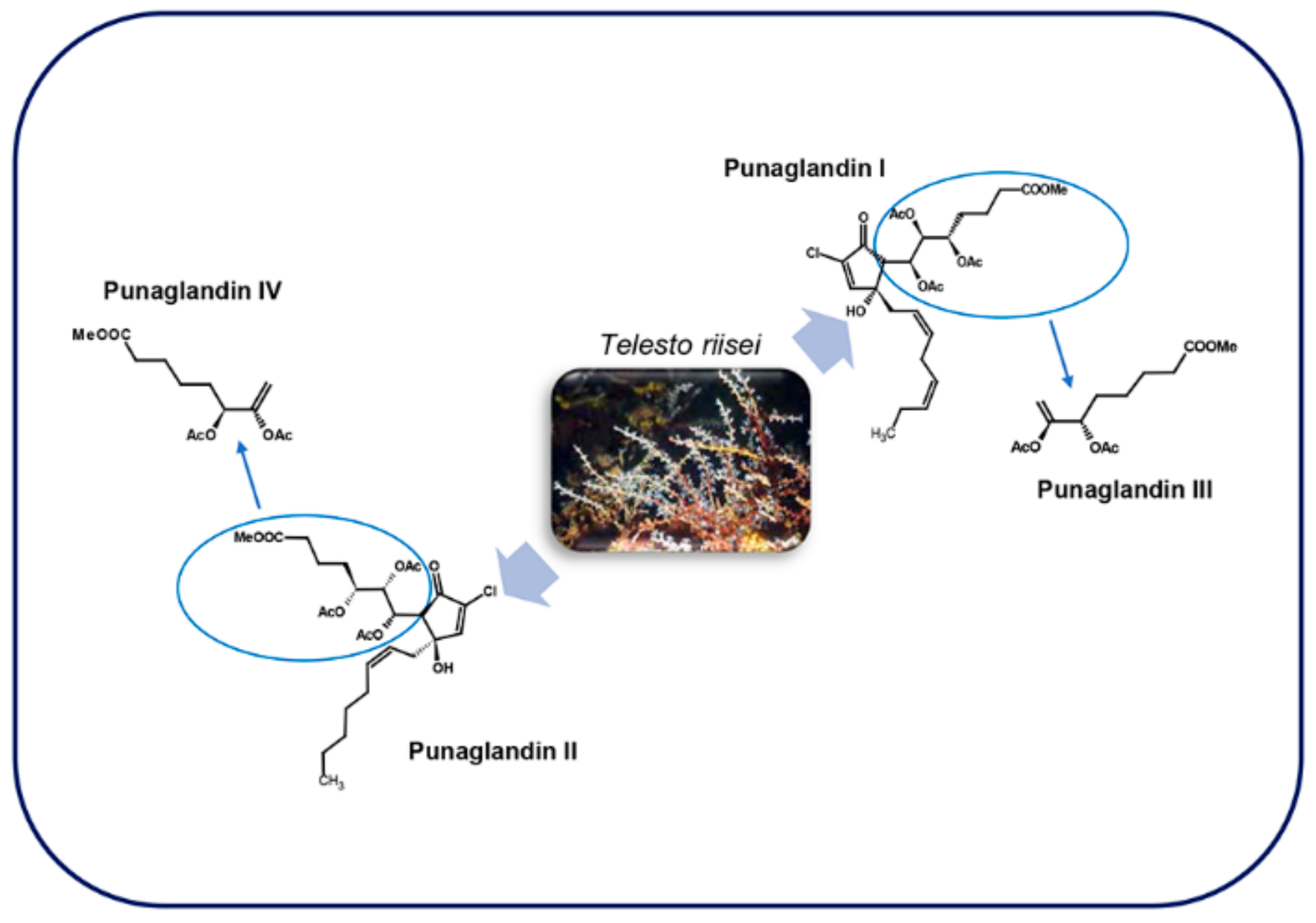
Clavulones (Figure 9) are acetoxy derivatives of PGA [91] characterized by a cyclopentenone fragment, a 12-S acetoxy function and two chains of different length: the α- and the ω-chain with respectively seven and eight carbon atoms [93]. These are anti-tumor marine prostanoids isolated from the soft coral Clavularia viridis Quoy & Gaimard, 1833, a single Clavularia genus that inhabits Okinawa bay in Japan [44,94,95].
Clavulones exists in three isoforms, I, II, and III and two stereoisomers at the C-4 and C-12 positions. These molecules possess a significant anti-inflammatory effect at a concentration of 30 µg/mL in a fertilized chicken egg assay (Table 1) [44,95]. The effects of clavulones on the growth of human cancer cells were first studied using clavulone I, the form that is more abundant in C. viridis. At concentration of 4.0 µM there was an impairment of DNA synthesis in human HL-60 leukemic cells after 1 h incubation, causing irreversible cytotoxic changes after 3 h of exposition [45]. In a more recent study, the anti-cancer activity of clavulone II on HL-60 cells has been deeply studied [46] revealing that low concentrations of clavulone II induce anti-proliferative effects by inducing the down-regulation of cyclin D1 with consequent arrest of the cell cycle in G1. On the contrary, higher concentrations of clavulone II induces apoptosis through the disruption of mitochondrial membrane potential and the activation of caspase-9, -8, and -3 and of B-cell lymphoma 2 (Bcl2)-family proteins (Table 1) [46]. It was suggested that corals might use clavulones as repellent and toxic substances against other marine organisms (Table 2) [45].
Clavulones, similarly to PGAs, also possess antiviral activity. In particular, clavulone II inhibits replication of the vesicular stomatitis virus (VSV) in infected mouse L929 fibroblasts, by blocking the transcription of viral RNA, and consequently the viral protein (Table 1) [47].
Interestingly, both the anti-proliferative and antiviral activities of clavulones are stronger than those of PGAs [47].
Other than clavulone I, II, and III, from the same stolonifer coral, 20-acetoxi-claviridenone b and 20-acetoxi-claviridenone c were discovered [96]. In addition to these, also four halogenated analogues, called chlorovulones I-IV were identified and, among these, chlorovulone I was shown to have anti-proliferative and cytotoxic activities (Table 1) [48]. The structure of an epoxy prostanoid with anti-proliferative activity was also identified in the same soft coral [49] together with bromovulone I and iodovulone I. Both had anti-proliferative and cytotoxic activities even though slightly lower than those of chlorovulone I [50]. All these molecules showed a stronger anti-tumor activity against HL-60 cells in vitro with respect to clavulone I (Table 1) [48,49,50].
More recently, seven new prostanoids were identified from the same soft coral: 4-deacetoxyl-12-O-deacetylclavulone I, 4-deacetoxyl-12-O-deacetylclavulone II, bromovulone II, iodovulone II, 4-deacetoxyl-12-O-deacetylclavulone III, bromovulone III, and iodovulone III. These molecules showed in vitro cytotoxic activity against human prostate carcinoma (PC-3) and colon adenocarcinoma (HT-29) cells and, among these, bromovulone III showed the highest anti-tumor activity, together with chlorovulone II and III, used as standard, that exhibited a slightly lower activity (Table 1) [51].
Through an assay-guided fractionation of a dichloromethane (CH2Cl2) extract of C. viridis, three new cytotoxic molecules were isolated: claviridenone E, claviridenone F, and claviridenone G. In particular, claviridenone F showed a significant cytotoxicity against human lung adenocarcinoma (A549), HT-29, and mouse lymphocytic leukemia cells (P-388), while claviridenone G exhibited a high cytotoxic activity only against A549 (Table 1) [52]. In addition to these molecules, there are clavulone-related oxylipins from C. viridis, named clavirins I and II, for which it was proposed a derivation from clavulone III and I, respectively, having growth-inhibitory activity against human cervix carcinoma cell line (HeLa S3) (Table 1) [53].
Other clavulone-related molecules that have been discovered are tricycloclavulone and clavubicyclone that may be derived from cycloaddition and electrocyclization of clavulone III, respectively. The latter molecule showed a moderate growth-inhibition activity against breast carcinoma (MCF-7) and ovarian carcinoma cells (OVCAR-3) in vitro (Table 1) [54]. Preclavulone lactone I and II and two minor chemical congeners, 17,18-dehydroclavulone I and clavulolactone I, were also isolated and characterized from C. viridis. Among these, 17,18-dehydroclavulone I has a (14Z,17Z)-double bond in the ω side chain, and this suggests that, differently from the other clavulones, it can derive from EPA instead of ARA [97].
Although there are structural similarities between prostaglandins and clavulones, the latter are not generated by a variant of the endoperoxide pathway, but from the lipoxygenase pathway by which arachidonic acid is converted into 8-(R)HPETE and then to preclavulone A [98]. Preclavulone A was identified also in an unrelated Caribbean coral, Pseudoplexaura porosa Houttuyn, 1772, suggesting that this molecule is widespread in corals [99]. In another study, the same authors isolated and defined the structures of 15 new halogenated iodo-, bromo-, and chlorovulones in C. viridis as minor constituents [100], but overall about 50 congeners of clavulones were identified in this soft coral [101].

4.2. Punaglandins

These molecules (Figure 10), structurally related to clavulones, were isolated in the Hawaiian octocoral Telesto riisei Duchassaing & Michelotti, 1860 [102].
The lack of symbiotic algae suggests that T. riisei is the source of these prostaglandins [103]. A total of 19 punaglandins were obtained from this octocoral, which also included acetate and epoxide versions of the four canonical punaglandins [104]. Punaglandins 3 and 4 formally results from elimination of acetic acid from punaglandins 1 and 2 respectively, and possess a cross-conjugated dienone structure [105]. They are halogenated derivatives of PGA [91] characterized by various oxygenations at C -5, -6, -7, and -12 and a 10-chloro-9-cyclopentenone function with a 5S, 6S, 12R stereochemistry [102].
At the moment of their discovery, there was great interest in the study of punaglandins, because of their anti-inflammatory and potent antitumor activities [104]. Indeed, these molecules, and in particular punaglandin 3, was shown to inhibit L1210 leukemia cell proliferation (IC50 = 0.04 µM) (Table 1) [102] with an activity 15-fold higher compared to the corresponding clavulone [55]. This cytotoxic activity is approximately equal to the one of vincristine and doxorubicin that are among the most effective anticancer molecules used today [103].

5. Marine Thromboxane

Thromboxanes (TXs) are closely relate to PGs that bind specific receptors, called thromboxane receptors (TP) and act as strong promoters of platelet aggregation and vaso- and broncho-constriction in mammals [106]. They are labile and biologically active molecules deriving from arachidonic acid in the cyclooxygenase pathway in human platelets. Thromboxane A2 (TXA2) has a short half-life (30 s in human blood) being rapidly converted to a stable B form (TXB2), which is used as a marker for the presence of TXA2 [69].
Other than in mammals, the presence of TXs is documented also in marine organisms.
TXB2, along with prostaglandins, has been reported in aqueous homogenates of the mollusc A. californica J. G. Cooper, 1863 (Figure 4a) [74] using radioimmunoassay. In the marine bivalve M. edulis Linnaeus, 1758 (Figure 4a), arachidonic acid metabolism, in addition to PGs, leads to the production of TXs in the gills, mantle, and adductor tissues [107]. In the polychaetae Arenicola marina Linnaeus, 1758 small amounts of TXB2 are detectable in the digestive and reproductive tracts [107].
Different species of fish also produce TXs [69]. In the plasma of the elasmobranch Dasyatis sabina Lesueur, 1824 (Figure 5b), the presence of an immunoreactive TXB2 (iTXB2) has been detected at concentrations of 0.57 ± 0.03 ng/mL. The level of iTXB2 increased to 3.0 ± 0.27 ng/mL when the plasma clots, but decreased to 1.5 ± 0.17 ng/mL in the presence of indomethacin, a cyclooxygenase inhibitor. These findings suggest that TXA2, the active thromboxane form, may be involved in blood clotting and may be generated by a cyclooxygenase-like enzyme as in mammals (Table 2) [69]. The binding of agonist (radiolabeled [125I]-BOP) and antagonist (L-657925 and L-657926) ligands of mammalian TP receptors to those of D. sabina, also demonstrates the similarity between this elasmobranch and mammalian TP receptors [69]. Three different species of shark (Figure 5b) (Chiloscyllium griseum Müller & Henle, 1838, Carcharhinus plumbeus Nardo, 1827 and Carcharhinus melanopterus Quoy & Gaimard, 1824) were also shown to produce TXB2 as a major product of arachidonic acid metabolism suggesting that TXB2 may be a biologically active prostanoid in these species [70].
Rowley et al. [78] tested the ability of leucocytes extracted from the blood of the dogfish Scyliorhinus canicula (Figure 5b) to produce and release PGs and TXs during their degranulation induced by the calcium ionophore A23187. While PGF, PGE2, and PGD2 levels were always high after the stimulus, the levels of TXB2 were low and detectable in the supernatant only 5 min after the start of the stimulus, with levels increasing after 10 and 15 min [78].
Another direct evidence of the generation of TXB2 in fish was demonstrated by the ability of washed whole blood cells from a variety of fishes of the Arabian Gulf to produce prostanoids from exogenous 14C-arachidonic acid in vitro [70]. In addition, rainbow trout thrombocytes, clotting cells similar to mammalian platelets, were shown to convert arachidonic acid mainly to TXB2 [108,109]. These findings again suggest a role also for TXB2, in rainbow trout as a vasodilator (Table 2), while in mammals TXB2 has no significant biological activity [70]. However, not all fish thrombocytes have the ability to produce thromboxane, possibly suggesting that these molecules are not essential for thrombocyte aggregation in certain species [110].

6. Conclusions

Prostaglandins and their derivatives, although first identified in terrestrial organisms, are also widespread in the marine environment testifying the great importance of these molecules. Nevertheless, the eco-physiological role of marine PGs remains mostly unclear because of contrasting and often old data available in the literature, although it has been suggested that they have a possible role as defensive molecules against predators.
Interestingly, many marine organisms have been shown to produce classical PGs along with new derivatives that are not found in terrestrial animals, e.g., corals that are able to produce punaglandins and clavulones, which are specific of the marine environment and have shown potent pharmacological activity against inflammation, tumors, and viruses. These findings, together with the discovery of PGs in unicellular eukaryotic microalgae, provides new stimuli to pursue the search for new marine PG-derived molecules with anti-inflammatory activity.

Author Contributions

Conceptualization, V.D.D., G.R. and F.D.C.; methodology, V.D.D., G.R. and F.D.C.; resources, V.D.D., G.R. and F.D.C.; writing—original draft preparation, F.D.C., G.R. and V.D.D.; writing—review and editing, F.D.C., V.D.D., G.R. and A.I.; visualization, G.R. and A.I.; supervision, V.D.D., G.R. and A.I.; project administration, G.R.; funding acquisition, G.R.

Funding

This research was funded by Ministero dell’ambiente e della tutela del territorio e del mare, Italia, project name: “Genomics for a Blue Economy,” grant number PGR00765.

Acknowledgments

We thank Adele Cutignano (Institute of Biomolecular Chemistry-National Research Council of Italy) for her help in drawing the molecules.

Conflicts of Interest

The authors declare no conflict of interest. The funders had no role in the design of the study; in the collection, analyses, or interpretation of data; in the writing of the manuscript, or in the decision to publish the results.

References

  1. Cheung, R.C.F.; Ng, T.B.; Wong, J.H.; Chen, Y.; Chan, W.Y. Marine natural products with anti-inflammatory activity. Appl. Microbiol. Biotechnol. 2016, 100, 1645–1666. [Google Scholar] [CrossRef] [PubMed]
  2. Mora, C.; Tittensor, D.P.; Adl, S.; Simpson, A.G.B.; Worm, B. How many species are there on Earth and in the ocean? PLoS Biol. 2011, 9, e1001127. [Google Scholar] [CrossRef] [PubMed]
  3. Buckley, C.D.; Gilroy, D.W.; Serhan, C.N.; Stockinger, B.; Tak, P.P. The resolution of inflammation. Nat. Rev. Immunol. 2013, 13, 59–66. [Google Scholar] [CrossRef] [PubMed]
  4. Fullerton, J.N.; Gilroy, D.W. Resolution of inflammation: A new therapeutic frontier. Nat. Rev. Drug Discov. 2016, 15, 551–567. [Google Scholar] [CrossRef] [PubMed]
  5. Gilroy, D.; De Maeyer, R. New insights into the resolution of inflammation. Semin. Immunol. 2015, 27, 161–168. [Google Scholar] [CrossRef] [PubMed]
  6. Headland, S.E.; Norling, L.V. The resolution of inflammation: Principles and challenges. Semin. Immunol. 2015, 27, 149–160. [Google Scholar] [CrossRef] [PubMed]
  7. Ortega-Gómez, A.; Perretti, M.; Soehnlein, O. Resolution of inflammation: An integrated view. EMBO Mol. Med. 2013, 5, 661–674. [Google Scholar] [CrossRef] [PubMed]
  8. Serhan, C.N.; Chiang, N.; Dalli, J.; Levy, B.D. Lipid mediators in the resolution of inflammation. Cold Spring Harb. Perspect. Biol. 2014, 7, a016311. [Google Scholar] [CrossRef] [PubMed]
  9. Rajakariar, R.; Yaqoob, M.M.; Gilroy, D.W. COX-2 in inflammation and resolution. Mol. Interv. 2006, 6, 199–207. [Google Scholar] [CrossRef] [PubMed]
  10. Murata, T.; Maehara, T. Discovery of anti-inflammatory role of prostaglandin D2. J. Vet. Med. Sci. 2016, 78, 1643–1647. [Google Scholar] [CrossRef] [PubMed] [Green Version]
  11. Euler, U.S.v. Zur Kenntnis der pharmakologischen Wirkungen von Nativsekreten und Extrakten männlicher accessorischer Geschlechtsdrüsen. Arch. Exp. Pathol. Pharmakol. 1934, 175, 78–84. [Google Scholar] [CrossRef]
  12. Goldblatt, M.W. Properties of human seminal plasma. J. Physiol. 1935, 84, 208–218. [Google Scholar] [CrossRef] [PubMed]
  13. Wolfe, L.S. Eicosanoids: Prostaglandins, thromboxanes, leukotrienes, and other derivatives of carbon-20 unsaturated fatty acids. J. Neurochem. 1982, 38, 1–14. [Google Scholar] [CrossRef] [PubMed]
  14. Piper, P.J. Introduction to the biosynthesis and metabolism of prostaglandins. Postgrad. Med. J. 1977, 53, 643–646. [Google Scholar] [CrossRef] [PubMed]
  15. Bhakuni, D.S.; Rawat, D.S. Bioactive Marine Natural Products; Springer: New York, NY, USA; Anamaya: New Delhi, India, 2005; ISBN 978-1-4020-3472-5. [Google Scholar]
  16. Nomura, T.; Ogata, H. Distribution of prostaglandins in the animal kingdom. Biochim. Biophys. Acta 1976, 431, 127–131. [Google Scholar] [PubMed]
  17. Rowley, A.F.; Vogan, C.L.; Taylor, G.W.; Clare, A.S. Prostaglandins in non-insectan invertebrates: Recent insights and unsolved problems. J. Exp. Biol. 2005, 208, 3–14. [Google Scholar] [CrossRef] [PubMed]
  18. Sajiki, J.; Kakimi, H. Identification of eicosanoids in the red algae, Gracilaria asiatica, using high-performance liquid chromatography and electrospray ionization mass spectrometry. J. Chromatogr. A 1998, 795, 227–237. [Google Scholar] [CrossRef]
  19. Ritter, A.; Goulitquer, S.; Salaün, J.-P.; Tonon, T.; Correa, J.A.; Potin, P. Copper stress induces biosynthesis of octadecanoid and eicosanoid oxygenated derivatives in the brown algal kelp Laminaria digitata. New Phytol. 2008, 180, 809–821. [Google Scholar] [CrossRef]
  20. Groenewald, E.G.; van der Westhuizen, A.J. Prostaglandins and related substances in plants. Bot. Rev. 1997, 63, 199–220. [Google Scholar] [CrossRef]
  21. Ruggeri, B.; Thoroughgood, C. Prostaglandins in aquatic fauna: A comprehensive review. Mar. Ecol. Prog. Ser. 1985, 23, 301–306. [Google Scholar] [CrossRef]
  22. Carté, B.K. Biomedical potential of marine natural products. Marine organisms are yielding novel molecules for use in basic research and medical applications. BioScience 1996, 46, 271–286. [Google Scholar]
  23. Di Dato, V.; Orefice, I.; Amato, A.; Fontanarosa, C.; Amoresano, A.; Cutignano, A.; Ianora, A.; Romano, G. Animal-like prostaglandins in marine microalgae. ISME J. 2017, 11, 1722–1726. [Google Scholar] [CrossRef] [PubMed] [Green Version]
  24. Nakanishi, M.; Rosenberg, D.W. Multifaceted roles of PGE2 in inflammation and cancer. Semin. Immunopathol. 2013, 35, 123–137. [Google Scholar] [CrossRef] [PubMed]
  25. Lands, W.E.M. The biosynthesis and metabolism of prostaglandins. Annu. Rev. Physiol. 1979, 41, 633–652. [Google Scholar] [CrossRef] [PubMed]
  26. Bos, C.L.; Richel, D.J.; Ritsema, T.; Peppelenbosch, M.P.; Versteeg, H.H. Prostanoids and prostanoid receptors in signal transduction. Int. J. Biochem. Cell Biol. 2004, 36, 1187–1205. [Google Scholar] [CrossRef] [PubMed]
  27. Funk, C.D. Prostaglandins and leukotrienes: Advances in eicosanoid biology. Science 2001, 294, 1871–1875. [Google Scholar] [CrossRef] [PubMed]
  28. Hansen, H.S. 15-hydroxyprostaglandin dehydrogenase. A review. Prostaglandins 1976, 12, 647–679. [Google Scholar] [CrossRef]
  29. Wang, D.; DuBois, R.N. Prostaglandins and cancer. Gut 2006, 55, 115–122. [Google Scholar] [CrossRef]
  30. Jakobsson, P.J.; Thorén, S.; Morgenstern, R.; Samuelsson, B. Identification of human prostaglandin E synthase: A microsomal, glutathione-dependent, inducible enzyme, constituting a potential novel drug target. Proc. Natl. Acad. Sci. USA 1999, 96, 7220–7225. [Google Scholar] [CrossRef] [Green Version]
  31. Sykes, L.; MacIntyre, D.A.; Teoh, T.G.; Bennett, P.R. Anti-inflammatory prostaglandins for the prevention of preterm labour. Reproduction 2014, 148, R29–R40. [Google Scholar] [CrossRef] [Green Version]
  32. Basu, S. Novel cyclooxygenase-catalyzed bioactive prostaglandin F2α from physiology to new principles in inflammation. Med. Res. Rev. 2007, 27, 435–468. [Google Scholar] [CrossRef] [PubMed]
  33. Ricciotti, E.; FitzGerald, G.A. Prostaglandins and inflammation. Arterioscler. Thromb. Vasc. Biol. 2011, 31, 986–1000. [Google Scholar] [CrossRef] [PubMed]
  34. Watanabe, T.; Nakao, A.; Emerling, D.; Hashimoto, Y.; Tsukamoto, K.; Horie, Y.; Kinoshita, M.; Kurokawa, K. Prostaglandin F2 alpha enhances tyrosine phosphorylation and DNA synthesis through phospholipase C-coupled receptor via Ca(2+)-dependent intracellular pathway in NIH-3T3 cells. J. Biol. Chem. 1994, 269, 17619–17625. [Google Scholar] [PubMed]
  35. Straus, D.S.; Glass, C.K. Cyclopentenone prostaglandins: New insights on biological activities and cellular targets. Med. Res. Rev. 2001, 21, 185–210. [Google Scholar] [CrossRef] [PubMed]
  36. Brown, J.A.; Bucknall, R.M. Antidiuretic and cardiovascular actions of prostaglandin E2 in the rainbow trout Salmo gairdneri. Gen. Comp. Endocrinol. 1986, 61, 330–337. [Google Scholar] [CrossRef]
  37. Di Marzo, V.; Cimino, G.; Crispino, A.; Minardi, C.; Sodano, G.; Spinella, A. A novel multifunctional metabolic pathway in a marine mollusc leads to unprecedented prostaglandin derivatives (prostaglandin 1,15-lactones). Biochem. J. 1991, 273 (Pt 3), 593–600. [Google Scholar] [CrossRef] [Green Version]
  38. Weinheimer, A.J.; Spraggins, R.L. The occurrence of two new prostaglandin derivatives (15-epi-PGA2 and its acetate, methyl ester) in the gorgonian Plexaura homomalla chemistry of coelenterates. XV. Tetrahedron Lett. 1969, 59, 5185–5188. [Google Scholar] [CrossRef]
  39. Light, R.J.; Samuelsson, B. Identification of prostaglandins in the gorgonian, Plexaura homomalla. Eur. J. Biochem. 1972, 28, 232–240. [Google Scholar] [CrossRef]
  40. Schneider, W.P.; Hamilton, R.D.; Rhuland, L.E. Occurrence of esters of (15S)-prostaglandin A 2 and E 2 in coral. J. Am. Chem. Soc. 1972, 94, 2122–2123. [Google Scholar] [CrossRef]
  41. Bayer, F.; Weinheimer, A. Prostaglandins from Plexaura homomalla: Ecology, Utilization and Conservation of a Major Medical Marine Resource. A Symposium; Studies in Tropical Oceanography; University of Miami Press on Behalf of the Upjohn Co.: Coral Gables, FL, USA, 1974. [Google Scholar]
  42. Gerhart, D.J. Prostaglandin A2: An agent of chemical defense in the Caribbean gorgonian Plexaura homomalla. Mar. Ecol. Prog. Ser. 1984, 19, 181–187. [Google Scholar] [CrossRef]
  43. Reina, E.; Ramos, F.A.; Castellanos, L.; Aragón, M.; Ospina, L.F. Anti-inflammatory R-prostaglandins from Caribbean Colombian Soft Coral Plexaura homomalla. J. Pharm. Pharmacol. 2013, 65, 1643–1652. [Google Scholar] [CrossRef] [PubMed]
  44. Kikuchi, H.; Tsukitani, Y.; Iguchi, K.; Yamada, Y. Absolute stereochemistry of new prostanoids clavulone I, II and III, from Clavularia viridis Quoy and Gaimard. Tetrahedron Lett. 1983, 24, 1549–1552. [Google Scholar] [CrossRef]
  45. Honda, A.; Yamamoto, Y.; Mori, Y.; Yamada, Y.; Kikuchi, H. Antileukemic effect of coral-prostanoids clavulones from the stolonifer Clavularia viridis on human myeloid leukemia (HL-60) cells. Biochem. Biophys. Res. Commun. 1985, 130, 515–523. [Google Scholar] [CrossRef]
  46. Huang, Y.-C.; Guh, J.-H.; Shen, Y.-C.; Teng, C.-M. Investigation of anticancer mechanism of clavulone II, a coral cyclopentenone prostaglandin analog, in human acute promyelocytic leukemia. J. Biomed. Sci. 2005, 12, 335–345. [Google Scholar] [CrossRef] [PubMed]
  47. Bader, T.; Yamada, Y.; Ankel, H. Antiviral activity of the prostanoid clavulone II against vesicular stomatitis virus. Antivir. Res. 1991, 16, 341–355. [Google Scholar] [CrossRef]
  48. Iguchi, K.; Kaneta, S.; Mori, K.; Yamada, Y.; Honda, A.; Mori, Y. Chlorovulones, new halogenated marine prostanoids with an antitumor activity from the stolonifer Clavularia viridis Quoy and Gaimard. Tetrahedron Lett. 1985, 26, 5787–5790. [Google Scholar] [CrossRef]
  49. Iguchi, K.; Kaneta, S.; Mori, K.; Yamada, Y. A new marine epoxy prostanoid with an antiproliferative activity from the stolonifer Clavularia viridis Quoy and Gaimard. Chem. Pharm. Bull. 1987, 35, 4375–4376. [Google Scholar] [CrossRef] [PubMed]
  50. Iguchi, K.; Kaneta, S.; Mori, K.; Yamada, Y.; Honda, A.; Mori, Y. Bromovulone I and iodovulone I, unprecedented brominated and iodinated marine prostanoids with antitumour activity isolated from the Japanese stolonifer Clavularia viridis Quoy and Gaimard. J. Chem. Soc. Chem. Commun. 1986, 981–982. [Google Scholar] [CrossRef]
  51. Shen, Y.-C.; Cheng, Y.-B.; Lin, Y.-C.; Guh, J.-H.; Teng, C.-M.; Ko, C.-L. New prostanoids with cytotoxic activity from Taiwanese octocoral Clavularia viridis. J. Nat. Prod. 2004, 67, 542–546. [Google Scholar] [CrossRef]
  52. Duh, C.-Y.; El-Gamal, A.A.H.; Chu, C.-J.; Wang, S.-K.; Dai, C.-F. New cytotoxic constituents from the Formosan Soft Corals Clavularia viridis and Clavularia violacea. J. Nat. Prod. 2002, 65, 1535–1539. [Google Scholar] [CrossRef]
  53. Iwashima, M.; Okamoto, K.; Iguchi, K. Clavirins, a new type of marine oxylipins with growth-inhibitory activity from the Okinawan soft coral, Clavularia viridis. Tetrahedron Lett. 1999, 40, 6455–6459. [Google Scholar] [CrossRef]
  54. Iwashima, M.; Terada, I.; Okamoto, K.; Iguchi, K. Tricycloclavulone and clavubicyclone, novel prostanoid-related marine oxylipins, isolated from the Okinawan Soft Coral Clavularia viridis. J. Org. Chem. 2002, 67, 2977–2981. [Google Scholar] [CrossRef] [PubMed]
  55. Baker, B.J.; Okuda, R.K.; Yu, P.T.K.; Scheuer, P.J. Punaglandins: Halogenated antitumor eicosanoids from the octocoral Telesto riisei. J. Am. Chem. Soc. 1985, 107, 2976–2977. [Google Scholar] [CrossRef]
  56. Komoda, Y.; Kanayasu, T.; Ishikawa, M. Prostaglandin F 2 alpha from the Japanese coastal gorgonian, Euplexaura erecta. Chem. Pharm. Bull. 1979, 27, 2491–2494. [Google Scholar] [CrossRef] [PubMed]
  57. Carmely, S.; Kashman, Y.; Loya, Y.; Benayahu, Y. New prostaglandin (PGF) derivatives from the Soft Coral. Tetrahedron Lett. 1980, 21, 875–878. [Google Scholar] [CrossRef]
  58. Varvas, K.; Järving, I.; Koljak, R.; Vahemets, A.; Pehk, T.; Müürisepp, A.-M.; Lille, Ü.; Samel, N. In vitro biosynthesis of prostaglandins in the White Sea Soft Coral Gersemia fruticosa: Formation of optically active PGD2, PGE2, PGF2α and 15-keto-PGF2α from arachidonic acid. Tetrahedron Lett. 1993, 34, 3643–3646. [Google Scholar] [CrossRef]
  59. Järving, R.; Järving, I.; Kurg, R.; Brash, A.R.; Samel, N. On the evolutionary origin of cyclooxygenase (COX) isozymes: Characterization of marine invertebrate COX genes points to independent duplication events in vertebrate and invertebrate lineages. J. Biol. Chem. 2004, 279, 13624–13633. [Google Scholar] [CrossRef]
  60. Christ, E.J.; Van Dorp, D.A. Comparative aspects of prostaglandin biosynthesis in animal tissues. Biochim. Biophys. Acta (BBA)—Lipids Lipid Metab. 1972, 270, 537–545. [Google Scholar] [CrossRef]
  61. Ogata, H.; Nomura, T.; Hata, M. Prostaglandin biosynthesis in the tissue homogenates of marine animals. NIPPON SUISAN GAKKAISHI 1978, 44, 1367–1370. [Google Scholar] [CrossRef]
  62. Levine, L.; Kobayashi, T. Detection of compounds immunologically related to arachidonic acid transformation products in extracts of invertebrates. Prostaglandins Leukot. Med. 1983, 12, 357–369. [Google Scholar] [CrossRef]
  63. Stanley-Samuelson, D.W. Physiological roles of prostaglandins and other eicosanoids in invertebrates. Biol. Bull. 1987, 173, 92–109. [Google Scholar] [CrossRef] [PubMed]
  64. Tahara, D.; Yano, I. Maturation-related variations in prostaglandin and fatty acid content of ovary in the kuruma prawn (Marsupenaeus japonicus). Comp. Biochem. Physiol. Part A Mol. Integr. Physiol. 2004, 137, 631–637. [Google Scholar] [CrossRef] [PubMed]
  65. Nomura, T.; Ogata, H.; Masao, I.T.O. Occurrence of prostaglandins in fish testis. Tohoku J. Agric. Res. 1973, 24, 138–144. [Google Scholar]
  66. Nylund, G.M.; Weinberger, F.; Rempt, M.; Pohnert, G. Metabolomic Assessment of induced and activated chemical defence in the invasive red alga Gracilaria vermiculophylla. PLoS ONE 2011, 6, e29359. [Google Scholar] [CrossRef] [PubMed]
  67. Cimino, G.; Crispino, A.; Di Marzo, V.; Spinella, A.; Sodano, G. Prostaglandin 1,15-lactones of the F series from the nudibranch mollusk Tethys fimbria. J. Organ. Chem. 1991, 56, 2907–2911. [Google Scholar] [CrossRef]
  68. Zambounis, A.; Gaquerel, E.; Strittmatter, M.; Salaün, J.P.; Potin, P.; Küpper, F.C. Prostaglandin A2 triggers a strong oxidative burst in Laminaria: A novel defense inducer in brown algae? ALGAE 2012, 27, 21–32. [Google Scholar] [CrossRef]
  69. Cabrera, D.M.; Janech, M.G.; Morinelli, T.A.; Miller, D.H. A thromboxane A(2) system in the Atlantic stingray, Dasyatis sabina. Gen. Comp. Endocrinol. 2003, 130, 157–164. [Google Scholar] [CrossRef]
  70. Thomson, M.; al-Hassan, J.M.; al-Saleh, J.; Fayad, S.; Ali, M. Prostanoid synthesis in whole blood cells from fish of the Arabian Gulf. Comp. Biochem. Physiol. B Biochem. Mol. Biol. 1998, 119, 639–646. [Google Scholar] [CrossRef]
  71. Freas, W.; Grollman, S. Uptake and binding of prostaglandins in a marine bivalve, Modiolus demissus. J. Exp. Zool. 1981, 216, 225–233. [Google Scholar] [CrossRef]
  72. Hampson, A.J.; Rowley, A.F.; Barrow, S.E.; Steadman, R. Biosynthesis of eicosanoids by blood cells of the crab, Carcinus maenas. Biochim. Biophys. Acta 1992, 1124, 143–150. [Google Scholar] [CrossRef]
  73. Korotchenko, O.D.; Mishchenko, T.Y.; Isay, S.V. Prostaglandins of Japan Sea invertebrates—I. The quantitation of group B prostaglandins in echinoderms. Comp. Biochem. Physiol. Part C Comp. Pharmacol. 1983, 74, 85–88. [Google Scholar] [CrossRef]
  74. Gerwick, W.H.; Nagle, D.G.; Proteau, P.J. Oxylipins from marine invertebrates. In Marine Natural Products—Diversity and Biosynthesis; Scheuer, P.J., Ed.; Springer: Berlin/Heidelberg, Germany, 1993; Volume 167, pp. 117–180. ISBN 978-3-540-56513-0. [Google Scholar]
  75. Ogata, H.; Nomura, T. Isolation and identification of prostaglandin E2 from gastrointestinal tract of shark Triakis scyllia. Biochim. Biophys. Acta 1975, 388, 84–91. [Google Scholar] [PubMed]
  76. Goetz, F.W.; Duman, P.; Ranjan, M.; Herman, C.A. Prostaglandin F and E synthesis by specific tissue components of the brook trout (Salvelinus fontinalis) ovary. J. Exp. Zool. 1989, 250, 196–205. [Google Scholar] [CrossRef]
  77. Anderson, A.A.; Fletcher, T.C.; Smith, G.M. Prostaglandin biosynthesis in the skin of the plaice Pleuronectes platessa L. Comp. Biochem. Physiol. C Comp. Pharmacol. 1981, 70, 195–199. [Google Scholar] [CrossRef]
  78. Rowley, A.F.; Barrow, S.E.; Hunt, T.C. Preliminary studies on eicosanoid production by fish leucocytes, using GC-mass spectrometry. J. Fish Biol. 1987, 31, 107–111. [Google Scholar] [CrossRef]
  79. Cagen, L.M.; Qureshi, Z.; Nishimura, H. Synthesis of prostaglandin E2 and prostaglandin F2α by toadfish red blood cells. Biochem. Biophys. Res. Commun. 1983, 110, 250–255. [Google Scholar] [CrossRef]
  80. Dang, T.H.; Lee, H.J.; Yoo, E.S.; Hong, J.; Choi, J.S.; Jung, J.H. The occurrence of 15-keto-prostaglandins in the red alga Gracilaria verrucosa. Arch. Pharm. Res. 2010, 33, 1325–1329. [Google Scholar] [CrossRef]
  81. Hsu, B.-Y.; Tsao, C.-Y.; Chiou, T.-K.; Hwang, P.-A.; Hwang, D.-F. HPLC determination for prostaglandins from seaweed. Food Control 2007, 18, 639–645. [Google Scholar] [CrossRef]
  82. Imbs, A.B.; Latyshev, N.A.; Svetashev, V.I.; Skriptsova, A.V.; Le, T.T.; Pham, M.Q.; Nguyen, V.S.; Pham, L.Q. Distribution of polyunsaturated fatty acids in red algae of the genus Gracilaria, a promising source of prostaglandins. Russ. J. Mar. Biol. 2012, 38, 339–345. [Google Scholar] [CrossRef]
  83. De Almeida, C.L.F.; Falcão, H.d.S.; Lima, G.R.d.M.; Montenegro, C.d.A.; Lira, N.S.; de Athayde-Filho, P.F.; Rodrigues, L.C.; de Souza, M.d.F.V.; Barbosa-Filho, J.M.; Batista, L.M. Bioactivities from marine algae of the genus Gracilaria. Int. J. Mol. Sci. 2011, 12, 4550–4573. [Google Scholar] [CrossRef]
  84. Kanamoto, H.; Takemura, M.; Ohyama, K. Identification of a cyclooxygenase gene from the red alga Gracilaria vermiculophylla and bioconversion of arachidonic acid to PGF(2α) in engineered Escherichia coli. Appl. Microbiol. Biotechnol. 2011, 91, 1121–1129. [Google Scholar] [CrossRef] [PubMed]
  85. Bouarab, K.; Adas, F.; Gaquerel, E.; Kloareg, B.; Salaün, J.-P.; Potin, P. The innate immunity of a marine red alga involves oxylipins from both the eicosanoid and octadecanoid pathways. Plant Physiol. 2004, 135, 1838–1848. [Google Scholar] [CrossRef] [PubMed]
  86. Andreou, A.; Brodhun, F.; Feussner, I. Biosynthesis of oxylipins in non-mammals. Prog. Lipid Res. 2009, 48, 148–170. [Google Scholar] [CrossRef] [PubMed]
  87. Ianora, A.; Miralto, A. Toxigenic effects of diatoms on grazers, phytoplankton and other microbes: A review. Ecotoxicology 2010, 19, 493–511. [Google Scholar] [CrossRef] [PubMed]
  88. Levine, L.; Sneiders, A.; Kobayashi, T.; Schiff, J.A. Serologic and immunochromatographic detection of oxygenated polyenoic acids in Euglena gracilis var. bacillaris. Biochem. Biophys. Res. Commun. 1984, 120, 278–285. [Google Scholar] [CrossRef]
  89. Kafanova, T.V.; Busarova, N.G.; Isai, S.V.; Zvyagintseva, T.Y. Fatty acids and prostaglandins of thermal cyanobacteria. Chem. Nat. Compd. 1996, 32, 861–865. [Google Scholar] [CrossRef]
  90. Krüger, G.H.J.; Groenewald, E.G.; de Wet, H.; Botes, P.J. The occurrence of prostaglandin F2α in procaryotic organisms. S. Afr. J. Bot. 1990, 56, 150–153. [Google Scholar] [CrossRef]
  91. Narumiya, S.; Fukushima, M. Cyclopentenone prostaglandins: Anti-proliferative and anti-viral actions and their molecular mechanism. In Eicosanoids and Other Bioactive Lipids in Cancer and Radiation Injury: Proceedings of the 1st International Conference 11–14 October 1989, Detroit, MI, USA; Honn, K.V., Marnett, L.J., Nigam, S., Walden, T.L., Eds.; Developments in Oncology; Springer: Boston, MA, USA, 1991; pp. 439–448. ISBN 978-1-4615-3874-5. [Google Scholar]
  92. Conti, M. Cyclopentenone: A special moiety for anticancer drug design. Anticancer Drugs 2006, 17, 1017–1022. [Google Scholar] [CrossRef]
  93. Lis, L.G.; Zheldakova, T.A. New marine prostanoids, clavulones, halogenovulones, and punaglandins. Chem. Nat. Compd. 1993, 29, 259–274. [Google Scholar] [CrossRef]
  94. Imbs, A.B. Prostaglandins and oxylipins of corals. Russ. J. Mar. Biol. 2011, 37, 325–334. [Google Scholar] [CrossRef]
  95. Kikuchi, H.; Tsukitani, Y.; Iguchi, K.; Yamada, Y. Clavulones, new type of prostanoids from the stolonifer Clavularia viridis Quoy and Gaimard. Tetrahedron Lett. 1982, 23, 5171–5174. [Google Scholar] [CrossRef]
  96. Kitagawa, I.; Kobayashi, M.; Yasuzawa, T.; Son, B.W.; Yoshihara, M.; Kyogoku, Y. New prostanoids from Soft Coral. Tetrahedron 1985, 41, 995–1005. [Google Scholar] [CrossRef]
  97. Iwashima, M.; Okamoto, K.; Konno, F.; Iguchi, K. New marine prostanoids from the Okinawan Soft Coral, Clavularia viridis. J. Nat. Prod. 1999, 62, 352–354. [Google Scholar] [CrossRef] [PubMed]
  98. Corey, E.J.; D’Alarcao, M.; Matsuda, S.P.T.; Lansbury, P.T.; Yamada, Y. Intermediacy of 8-(R)-HPETE in the conversion of arachidonic acid to pre-clavulone a by Clavularia viridis. Implications for the biosynthesis of marine prostanoids. J. Am. Chem. Soc. 1987, 109, 289–290. [Google Scholar] [CrossRef]
  99. Corey, E.J.; Matsuda, S.P.T. Generality of marine prostanoid biosynthesis by the 2-oxidopentadienylcation pathway. Tetrahedron Lett. 1987, 28, 4247–4250. [Google Scholar] [CrossRef]
  100. Watanabe, K.; Sekine, M.; Iguchi, K. Isolation and structures of new halogenated prostanoids from the Okinawan Soft Coral Clavularia viridis. J. Nat. Prod. 2003, 66, 1434–1440. [Google Scholar] [CrossRef] [PubMed]
  101. Watanabe, K.; Sekine, M.; Iguchi, K. Isolation of three marine prostanoids, possible biosynthetic intermediates for clavulones, from the Okinawan Soft Coral Clavularia viridis. Chem. Pharm. Bull. 2003, 51, 909–913. [Google Scholar] [CrossRef]
  102. Gerwick, W.H. Carbocyclic oxylipins of marine origin. Chem. Rev. 1993, 93, 1807–1823. [Google Scholar] [CrossRef]
  103. Munro, M.H.G.; Luibrand, R.T.; Blunt, J.W. The search for antiviral and anticancer compounds from marine organisms. In Bioorganic Marine Chemistry; Scheuer, P.J., Ed.; Springer: Berlin/Heidelberg, Germany, 1987; pp. 93–176. [Google Scholar]
  104. Baker, B.J.; Scheuer, P.J. The punaglandins: 10-chloroprostanoids from the octocoral Telesto riisei. J. Nat. Prod. 1994, 57, 1346–1353. [Google Scholar] [CrossRef]
  105. Suzuki, M.; Morita, Y.; Yanagisawa, A.; Baker, B.J.; Scheuer, P.J.; Noyori, R. Prostaglandin synthesis 15. Synthesis and structural revision of (7E)- and (7Z)-punaglandin 4. J. Org. Chem. 1988, 53, 286–295. [Google Scholar] [CrossRef]
  106. Armstrong, R.A.; Wilson, N.H. Aspects of the thromboxane receptor system. Gen. Pharmacol. 1995, 26, 463–472. [Google Scholar] [CrossRef]
  107. Mustafa, T.; Srivastava, K.C. Prostaglandins (eicosanoids) and their role in ectothermic organisms. In Advances in Comparative and Environmental Physiology; Brouwer, M., Carr, W.E.S., Ellington, W.R., Engel, D.W., Gleeson, R.A., Korsgaard, B., Moerland, T.S., Mustafa, T., Prior, D.J., Sidell, B.D., et al., Eds.; Advances in Comparative and Environmental Physiology; Springer: Berlin/Heidelberg, Germany, 1989; pp. 157–207. ISBN 978-3-642-74510-2. [Google Scholar]
  108. Kayama, M.; Sadõ, T.; Iijima, N. The Prostaglandin synthesis in rainbow trout thrombocyte. NIPPON SUISAN GAKKAISHI 1986, 52, 925. [Google Scholar] [CrossRef]
  109. Pettitt, T.R.; Barrow, S.E.; Rowley, A.F. Thromboxane, prostaglandin and leukotriene generation by rainbow trout blood. Fish Shellfish Immunol. 1991, 1, 71–73. [Google Scholar] [CrossRef]
  110. Hill, D.J.; Hallett, M.B.; Rowley, A.F. Effect of prostanoids and their precursors on the aggregation of rainbow trout thrombocytes. Am. J. Physiol. 1999, 276, R659–R664. [Google Scholar] [CrossRef] [PubMed]
Figure 1. Prostaglandin biosynthetic pathway. Enzymes involved in the pathway are reported next to the arrows. For the abbreviation, refer to the text. Modified from [24].
Marinedrugs 17 00428 g001
Figure 2. Prostaglandins and inflammation. (a) Prostaglandin E2 (PGE2) stimulation of inflammatory response. Numbers indicate the sequence of events from PGE2 synthesis to stimulation of inflammation trough positive feedback on cyclooxygenase (COX)-2 and negative feedback on microsomal PGE synthase-1 (mPTGES-1); (b) The anti-inflammatory role of prostaglandin J2 (PGJ2); (c) prostaglandin F (PGF) signaling; (d) prostaglandin I2 (PGI2) signaling. For details, refer to the text.
Marinedrugs 17 00428 g002
Figure 3. Corals prostaglandins. (a) Plexaura homomalla; (b) Euplexaura erecta; (c) Lobophytum depressum; (d) Gersemia fruticosa.
Marinedrugs 17 00428 g003
Figure 4. Marine invertebrate prostaglandins. (a) Molluscs; (b) Crustaceans; (c) Echinoderms. Except for Tethys fimbria and Aplysia californica, pictures show organisms that are only representatives of each phylum. For details, refer to the text.
Marinedrugs 17 00428 g004
Figure 5. Marine vertebrate prostaglandins. (a) Teleosts (Salmo sp., Paralichthys olivaceus, Oncorhynchus keta, Thunnus thynnus, Salvelinus fontinalis, Pleuronectes platessa, Opsanus tau); (b) Elasmobranchs (Triakis scyllia, Scyliorhinus canicula). Pictures show organisms that are only representatives of each class. For details, refer to the text.
Marinedrugs 17 00428 g005
Figure 6. Red algae prostaglandins. The picture shows an organism representative of the genus. For details, refer to the text.
Marinedrugs 17 00428 g006
Figure 7. Brown algae prostaglandins: only Laminaria digitata has been studied for prostaglandins content. For details, refer to the text.
Marinedrugs 17 00428 g007
Figure 8. Microalgae prostaglandins. (a) Skeletonema marinoi (A. Ianora laboratory clones name FE7 and FE60); (b) Thalassiosira rotula; (c) Euglena gracilis; (d) Microcystis aeruginosa. For details, refer to the text.
Marinedrugs 17 00428 g008
Figure 9. Clavulones and related molecules in Clavularia viridis (Cnidaria, soft corals). For details, refer to the text.
Marinedrugs 17 00428 g009
Figure 10. Punaglandins in Telesto riisei (Cnidaria, octocoral). For details, refer to the text.
Marinedrugs 17 00428 g010
Table 1. List of the tested effects of marine prostaglandins, and their derivatives, on mammalian cells.
ProstaglandinProducer OrganismActivityTarget CellsReference
(15R)-PGE2
(15R)-O-AcPGA2
Plexaura homomallaAnti-inflammatoryLeucocyte/TPA-induced mouse-ear edemaReina et al., 2013 [43]
Clavulones I-IIIClavularia viridisAnti-inflammatoryfertilized chicken eggsKikuchi et al., 1983 [44]
Clavulones I-IIIClavularia viridisAnti-cancerHL-60Honda et al., 1985 [45]; Huang et al., 2005 [46]
Clavulone IIClavularia viridisAnti-viralVSV infected L929Bader et al., 1991 [47]
Chlorovulone IClavularia viridisAnti-proliferative and cytotoxicHL-60Iguchi et al., 1985 [48]
PGs Epoxy-prostanoidClavularia viridisAnti-proliferativeHL-60Iguchi et al., 1987 [49]
Bromovulone I and Iodovulone IClavularia viridisAnti-proliferative and cytotoxicHL-60Iguchi et al., 1986 [50]
Bromovulone IIIClavularia viridisCytotoxicPC-3/HT-29Shen et al., 2004 [51]
Chlorovulones II and IIIClavularia viridisCytotoxicPC-3/HT-29Shen et al., 2004 [51]
Claviridenone FClavularia viridisCytotoxicA549/HT-29/P-388Duh et al., 2002 [52]
Claviridenone GClavularia viridisCytotoxicA549Duh et al., 2002 [52]
Clavirins I-IIClavularia viridisGrowth-inhibitionHeLa S3Iwashima et al., 1999 [53]
ClavubicycloneClavularia viridisGrowth-inhibitionMCF-7/OVCAR-3Iwashima et al., 2002 [54]
Punaglandins I–IVTelesto riiseiCytotoxicL1210Baker et al., 1985 [55]
Table 2. Update of prostaglandins and its derivatives, and functional roles in marine organisms.
CompoundProducer OrganismBiological ActivitiesReference
PGF2αMarsupenaeus japonicusOvarian maturationTahara et al., 2004 [64]
Thunnus thynnusContraction of smooth muscles during ejaculation and metabolism of testisNomura et al., 1973 [65]
PGE1Marine InvertebratesThermoregulation and feverStanley-Samuelson, 1987 [63]
Laminaria digitataProtection against stress conditions induced by copper excessRitter et al., 2008 [19]
Salmo sp.Contraction of smooth muscles during ejaculation and metabolism of testisChirst and Van Dorp, 1972 [60]
PGE2Marsupenaeus japonicusOvarian maturationTahara et al., 2004 [64]
Paralichthys olivaceus and Thunnus thynnusContraction of smooth muscles during ejaculation and metabolism of testisNomura et al., 1973 [65]
Gracilaria vermiculophyllaWounding-activated chemical defense moleculesNylund et al., 2011 [66]
Laminaria digitataProtection against stress conditions induced by copper excessRitter et al., 2008 [19]
PGF2α- and PGF3α-1,15-lactones fatty acid esters (PLFE)Tethys fimbriaReproduction and multiple roles depending on body localizationCimino et al., 1991 [67];
Di Marzo et al., 1991 [37]
PGF1αOncorhynchus ketaContraction of smooth muscles during ejaculation and metabolism of testisNomura et al., 1973 [65]
15-keto-PGE2Gracilaria vermiculophyllaWounding-activated chemical defense moleculesNylund et al., 2011 [66]
Laminaria digitataProtection against stress conditions induced by copper excessRitter et al., 2008 [19]
PGE2-1,15-lactoneTethys fimbriaReproduction and multiple roles depending on body localizationCimino et al., 1991 [67];
Di Marzo et al., 1991 [37]
PGE3-1,15-lactone-11-acetateTethys fimbriaReproduction and multiple roles depending on body localizationCimino et al., 1991 [67];
Di Marzo et al., 1991 [37]
PGE3-1,15-lactoneTethys fimbriaReproduction and multiple roles depending on body localizationCimino et al., 1991 [67];
Di Marzo et al., 1991 [37]
PGD1Laminaria digitataProtection against stress conditions induced by copper excessRitter et al., 2008 [19]
PGA2Gracilaria vermiculophyllaWounding-activated chemical defense moleculesNylund et al., 2011 [66]
Laminaria digitataProtection against copper stress and trigger of oxidative responsesZambounis et al., 2012 [68]
PGB2Laminaria digitataProtection against stress conditions induced by copper excessRitter et al., 2008 [19]
PGJ2Laminaria digitataProtection against stress conditions induced by copper excessRitter et al., 2008 [19]
ClavulonesClavularia viridisSuggested to be hypothetical repellents against other marine organismsHonda et al., 1985 [45]
iTXB2Dayatis sabinaBlood clottingCabrera et al., 2003 [69]
TXB2Oncorhynchus mykissVasodilator agentThomson et al., 1998 [70]

