2. Results and Discussion
The molecular formula of
1 was deduced to be C
20H
20O
8 with 11 degrees of unsaturation by HRFABMS analysis. The
13C NMR data of this compound showed a signal of a ketone carbon at δ
C 197.6 (
Table 1). The signals at δ
C 178.1 and 174.6 could belong to either carbonyl or highly deshielded olefinic carbons. The
13C NMR spectrum, in combination with DEPTs and HSQC spectra (
Supplementary Figure S4), displayed nine nonprotonated sp
2 carbon signals in the δ
C 103.5–162.8 region. The deshielded carbons must be one carbonyl carbon and one olefinic carbon, accounting therefore for seven degrees of unsaturation. The
13C NMR data also showed two oxygen-bearing quaternary sp
3 carbons (δ
C 89.5 and 79.0), one methoxy carbon (δ
C 60.9), one shielded quaternary sp
3 carbon (δ
C 46.9), and four shielded methyl carbons (δ
C 16.4, 16.4, 14.9, and 13.3) (
Table 2). Combining the NMR data and the degrees of unsaturation,
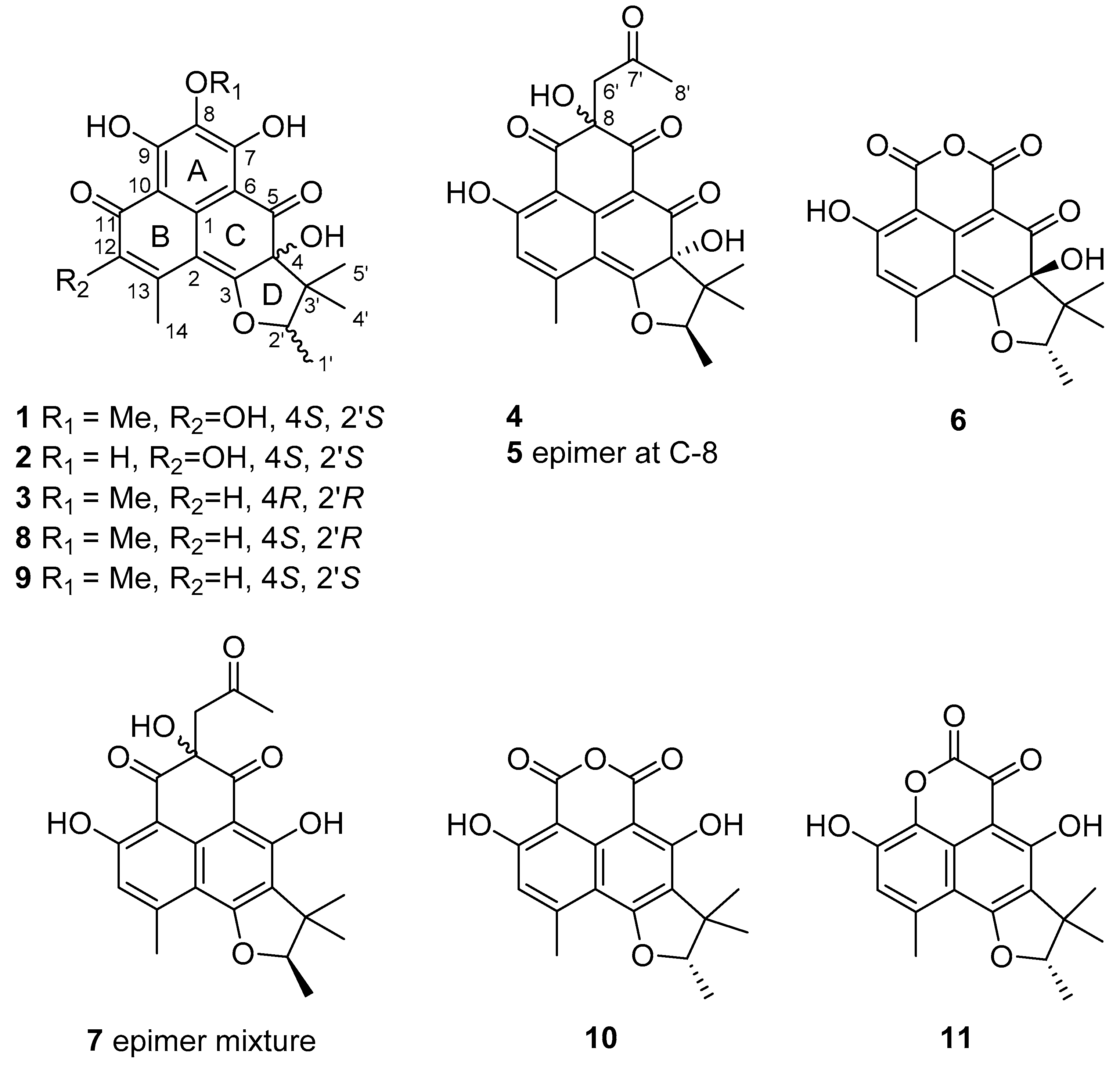
1 must possess four rings featuring the herqueinone class of phenalenones.
Due to the lack of COSY correlations except for that from the methyl doublet at δ
H 1.40 (Me-1′) to the quartet at δ
H 4.99 (H-2′), the structure determination of
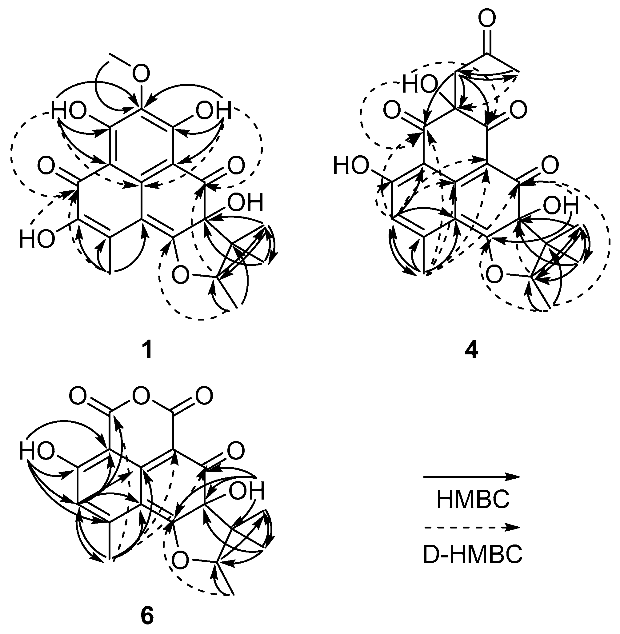
1 had to be carried out through extensive HMBC analyses under diverse measuring conditions (
Figure 1). First, the long-range couplings from OH-7 (δ
H 13.23) to C-6 (δ
C 103.5), C-7 (δ
C 162.8), and C-8 (δ
C 131.6); from OCH
3-8 (δ
H 3.92) to C-8 (δ
C 131.6); and OH-9 (δ
H 13.99) to C-8 (δ
C 131.6), C-9 (δ
C 161.7), and C-10 (δ
C 108.7) lead to a delineation of the C-6 to C-10 fragment. Aided by the four-bond couplings from OH-7 (δ
H 13.23) to C-1 (δ
C 137.3) and OH-9 (δ
H 13.99) to C-1 (δ
C 137.3) by decoupled HMBC (D-HMBC) [
7] experiments, the presence of a hexa-substituted benzene ring (C-1, C-6–C-10; ring A) was confirmed. In addition, the combined HMBC and D-HMBC correlations from OH-12 (δ
H 6.66) and H
3-14 (δ
H 2.47) to neighboring carbons revealed the presence of an
α-hydroxy-
β-methyl-
α,
β-unsaturated ketone group (OH-12 (δ
H 6.66) to C-11 (δ
C 178.1), C-12 (δ
C 143.7), and C-13 (δ
C 124.0); H
3-14 (δ
H 2.47) to C-2 (δ
C 103.7), C-11 (δ
C 178.1), C-12 (δ
C 143.7), and C-13 (δ
C 124.0)), which was directly connected to the benzene ring based on the D-HMBC correlation from OH-7 (δ
H 13.23) to C-11 (δ
C 178.1).
In addition to the correlation from H3-14 (δH 2.47) to C-2 (δC 102.7), a correlation from H3-14 (δH 2.47) to a highly deshielded C-3 (δC 175.4) in the D-HMBC spectrum was crucial evidence for the attachment of an electron-withdrawing oxygen at this position. Subsequently, long-range correlations from OH-4 (δH 7.23) to C-3 (δC 8), C4 (δC 8), and C5 (δC 8) defined not only its connectivity to the C-2 double bond but also placed a carbonyl carbon (δC 198.2) at C-5. These carbon–proton correlations constructed an α,β-dioxycyclohexadienone moiety (C-1–C-6; ring C). The assignment of ring C also secured the formation of the conjugated carbonyl group to another six-membered ring (C-1, C-2, C-10–C-13; ring B).
The remaining C
5 fragment (C-1′–C-5′) of
1 was readily defined as a 2,3-disubstituted 2-methylbutane moiety by a combination of COSY and HMBC data (
Figure 2 and
Supplementary Figures S3–S5). The cyclization of this moiety to the three-ring system was also accomplished by a series of long-range carbon–proton correlations. That is, the connection between C-4 and C-5′ was confirmed by the HMBC correlations from H
3-4′ (δ
H 1.43) and H
3-5′ (δ
H 0.86) to C-4 (δ
C 79.0) as well as a long-range correlation from H-2′ to C-5. The diagnostic chemical shifts of the CH-2′ methine group (δ
C 89.3, δ
H 4.91) suggested its attachment to C-3 via an ethereal bridge. This interpretation was corroborated by the correlation from H
3-1′ (δ
H 1.40) to C-3 (δ
C 174.6), which established a hydrofuran moiety (C-3, C-4, C-2′, and C-4′; ring D). Thus, the structure of
1 was defined as a herqueinone-type tetracyclic phenalenone.
The planar structure of
1 was found to be the same as that of the recently reported peniciherqueinone from the fungus
Penicillium herquei [
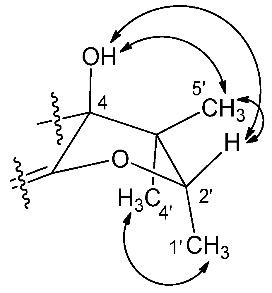
8]. In our study of the configurations of the C-4 and C-2′ stereogenic centers by NOESY analysis (
Figure 3), the OH-4, H-2′, and H
3-5′ protons were oriented toward the same face of the hydrofuran ring based on their mutual cross-peaks. The opposite face was occupied by H
3-1′ and H
3-4′ based on the cross peak between the methyl protons, suggesting that
1 has the same relative configuration (4
S* and 2′
S*) as peniciherqueinone. Interestingly, despite the same signs of optical rotations, there was a remarkable difference in their values of the specific rotations: [α]
(CHCl
3) +203 (
1) and +92 (peniciherqueinone). Since the absolute configurations at C-4 and C-2′ of herqueinones have been the subject of comprehensive investigations [
9,
10], the discrepancy in the specific rotations of
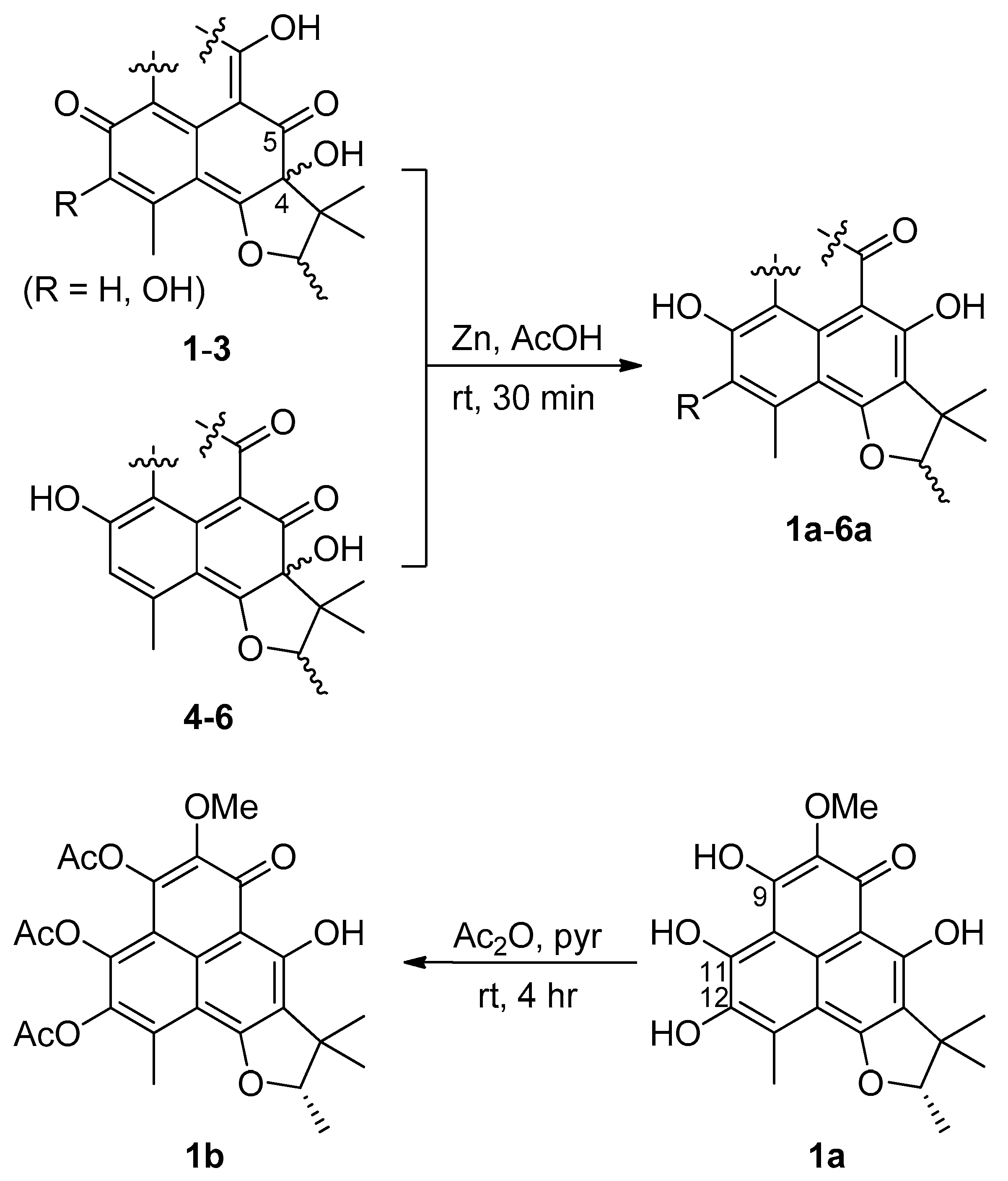
1 and herqueinones needed to be justified. Using a pre-established chemical modification technique [
11,
12,
13],
1 was reduced to
1a, which showed a negative specific rotation ([α]
(CHCl
3) −23); thus, the 2′
S configuration was confirmed. The absolute configuration was further evaluated via the acetylation of
1a to corresponding 9,11,12-triacetyl derivative
1b (
Figure 4). The sign of the specific rotation of
1b ([α]
(MeOH) −42) was opposite to that of herqueinone (
8) but the same as that of isoherqueinone (
9), which proved a 2′
S configuration [
9,
10]. Therefore, the absolute configuration of
1 was assigned as 4
S and 2′
S. Thus,
1, designated as
ent-peniciherqueinone, is a new herqueinone-type phenalenone.
The molecular formula of 2 was deduced to be C19H18O8 based on HRFABMS analysis. The NMR data of this compound were very similar to those of 1, with the absence of a methyl group. A detailed examination of the 13C and 1H NMR data revealed that the OMe-8 of 1 (δC 60.9, δH 3.92) was replaced by a hydroxyl group (δH 8.96) in 2, and this assignment was confirmed by a combination of two-dimensional (2D) NMR analyses. The NOESY data and specific rotation of the reduction product 2a indicated the same 4S and 2′S configuration as in 1. Thus, 2, designated as 12-hydroxynorherqueinone, was determined to be 8-demethyl-ent-peniciherqueinone.
Compound 3 was isolated as an orange amorphous solid with a molecular formula of C20H20O7, based on HRFABMS analysis. The 13C and 1H NMR data of this compound were similar to those obtained for 1. The most noticeable difference was the replacement of a hydroxyl-bearing olefinic carbon with the sp2 methine carbons (δC 122.8, δH 6.36). The structural difference was found to be at C-12 based on the HMBC correlations from H-12 (δH 6.36) to C-2 (δC 103.0), C-10 (δC 109.2), and C-14 (δC 23.8) as well as from H3-14 (δH 2.48) to C-2 (δC 103.0), C-12 (δC 122.8), and C-13 (δC 150.9). However, the sign of the specific rotation of 3 ([α] (MeOH) −69) was opposite to those of 1 and 2, implying a configurational difference. Since the NOESY spectrum showed the same cross-peaks for the hydrofuran moiety as those in the congeners, 3 was proposed to possess the opposite absolute configuration at C-4 and C-2′. As the reduction product of 3 (3a) is dextrorotatory (specific rotation ([α] (MeOH) +39)), the configuration of C-4 and C-2′ are 4R, 2′R, respectively. Thus, 3, designated as ent-isoherqueinone, is a new herqueinone-type phenalenone derivative.
The molecular formula of
4 was also established as C
22H
22O
8 by HRFABMS analysis. Although its spectroscopic data resembled those of
1–
3, several differences were found in both
13C and
1H NMR data. First, aided by the HSQC data, it was found that three additional carbons, i.e., one carbonyl (δ
C 206.9), one methylene sp
2 (δ
C 48.5, δ
H 3.30), and one methyl (δ
C 29.8, δ
H 2.09), were present in this compound (
Table 1 and
Table 2). In the
13C NMR spectrum, resonances of three ketone groups (δ
C 200.6, 193.1, and 189.9) were found for
4, unlike
1–
3. In addition, an aromatic or olefinic carbon had been replaced by an oxygen-bearing nonprotonated sp
3 carbon (δ
C 76.9). A detailed examination of its NMR data revealed that
4 contained the same B and D rings as
1–
3, and the structural differences were located in the remaining portion of the molecule.
The planar structure of
4 was established by extensive HMBC experiments (
Figure 2). Several HMBC correlations were found from an aromatic proton (δ
H 6.75, H-12) and a benzylic methyl proton (δ
H 2.57, H
3-14) to their neighboring carbons (H-12 (δ
H 6.75) to C-2 (δ
C 116.3), C-10 (δ
C 109.4), and C-14 (δ
C 23.5); H
3-14 (δ
H 2.57) to C-2 (δ
C 116.3), C-12 (δ
C 117.5), and C-13 (δ
C 152.3)). Aided by the D-HMBC correlations from H-12 (δ
H 6.75) to C-1 (δ
C 142.7) and C-11 (δ
C 165.6) and from H
3-14 (δ
H 2.57) to C-1 (δ
C 142.7), the long-range carbon–proton correlations led to the establishment of a hydroxyl- and methyl-bearing pentasubstituted benzene as ring B. Additional D-HMBC correlations from these protons to the conspicuous H
3-1′ at δ
H 1.30 (H-12 (δ
H 6.75) to C-6 (δ
C 101.2); H
3-14 (δ
H 2.57) to C-3 (δ
C 175.1), C-5 (δ
C 193.1), and C-6 (δ
C 101.2); H
3-1′ (δ
H 1.30) to C-3 (δ
C 175.1) and C-5 (δ
C 193.1)) defined ring C as a hydroxyl-bearing cyclohexadienone. The ring D was found to be the same as that in other herqueinones by a 2D NMR spectrum.
The remaining portion of
4 consists of three ketone carbonyl (δ
C 206.9, 200.6, and 189.9) and one nonprotonated sp
3 (δ
C 76.9), one methylene sp
3 (δ
C 48.5), and one methyl (δ
C 29.8) carbons. These carbons were initially assembled into a 2-keto-propyl group (C-6′–C-8′) by the HMBC correlations from the methylene and methyl protons to their neighboring carbons (H
2-6′ (δ
H 3.30) to C-7′ (δ
C 206.9) and C-8′ (δ
C 29.8); H
3-8′ (δ
H 2.09) to C-6′ (δ
C 48.5), and C-7′ (δ
C 206.9)) (
Figure 2). Then, this fragment was connected to the core structure by the HMBC correlations from H
2-6′ (H
2-6′ (δ
H 3.30) to C-7 (δ
C 189.9), C-8 (δ
C 76.9), and C-9 (δ
C 200.6)). The confirmation of this assignment as well as the linkage with the B-ring was accomplished by the key D-HMBC correlations from OH-8 (δ
H 6.68) to C-7 (δ
C 189.9) and C-9 (δ
C 200.6); from H-12 (δ
H 6.75) to C-9 (δ
C 200.6); from H
3-14 (δ
H 2.57) to C-9 (δ
C 200.6); and from H
3-8′ (δ
H 2.09) to C-8 (δ
C 76.9). Although it could not be confirmed by 2D-NMR-based carbon–proton correlations, the presence of the four rings, required by the molecular formula and NMR data, directly connected C-6 and C-7 carbonyl carbons to be part of a diketo-bearing six-membered ring as ring A. Thus, the structure of
4 was determined to be a phenalenone related to an acetone adduct of a triketone [
14,
15].
The molecular formula of
5 was the same as that of
4, C
22H
22O
8. Moreover, the
13C and
1H NMR data of these compounds were very similar (
Table 1 and
Table 2). Two-dimensional NMR analyses showed the same carbon–proton correlations throughout the entire molecule, indicating that they have the same planar structure. Therefore,
5 could be an epimer of
4.
In order to clarify the difference in stereochemistry between
4 and
5, NOESY experiments were carried out. The NOESY spectra of both compounds showed the same cross-peaks around the D ring as those observed in other herqueinones, suggesting the 4
R,2′
R or 4
S,2′
S configurations. Then, by chemical conversions to remove the other two stereogenic centers, the absolute configurations at C-4 were determined. That is,
4 and
5 were reduced to
4a and
5a, respectively (
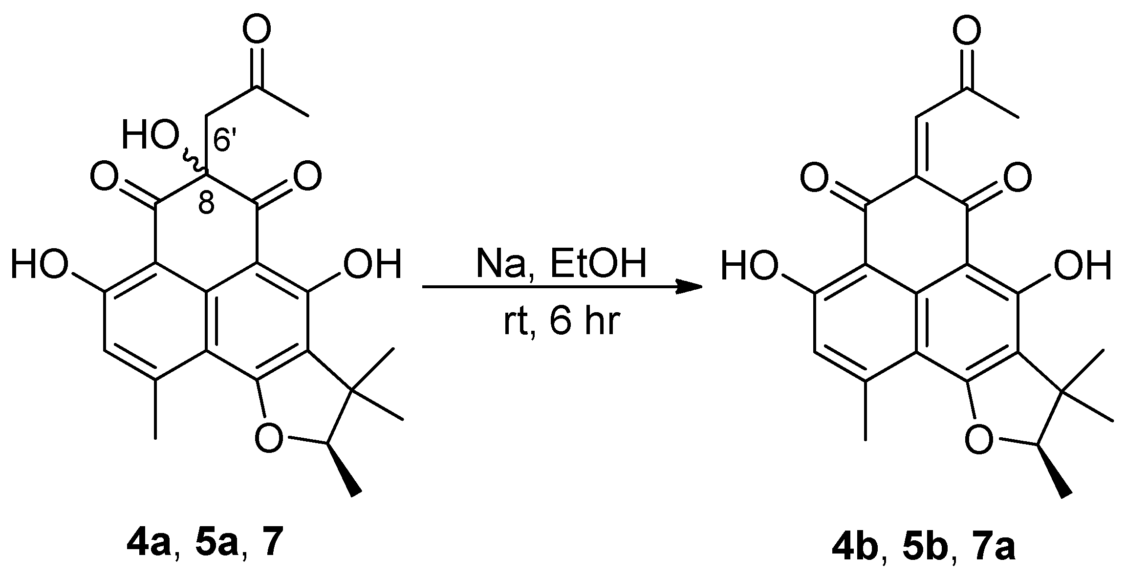
Figure 4), then the compounds were dehydrated to yield 8,15-unsaturated derivatives
4b and
5b, respectively (
Figure 5), and the MS and NMR data of these compounds were identical. Furthermore, their specific rotations were also very similar ([α]
(CHCl
3) −27 and −26 for
4b and
5b, respectively), implying that these were indeed the same compound. The negative specific rotations allow us to confidently assign the 2′
R configuration for both natural products. Thus,
4 and
5, designated as oxopropylisoherqueinones A and B respectively, were elucidated as new phenalenones possessing C
3 side chains. These compounds possessed 4
R, 2′
R configurations. However, the configurations at C-8 remain unassigned despite various chemical and spectroscopic analyses.
In order to determine the absolute configurations at C-8 of
4 and
5, a comparison of the experimental and calculated ECD spectra was carried out. Initially, the experimental CD profiles of these compounds showed opposite signs in the region of 285–340 nm, possibly reflecting the different configuration at C-8 (
Supplementary Figure S51). Despite all the efforts, however, the calculated ECD profiles based on the postulated conformational populations failed to assign the absolute configurations satisfactorily (
Supplementary Figure S52). This could be due to a weak contribution of a single and remote stereogenic center to the ECD in the molecule possessing several UV chromophores and stereogenic centers.
The molecular formula of
6 was deduced to be C
18H
16O
7, which corresponds to 11 degrees of unsaturation, by HRFABMS analysis. The
13C and
1H NMR data of this compound revealed that it is a phenalenone derivative based on the presence of signals for two aromatic rings and a trimethylhydrofuran moiety, which account for eight degrees of unsaturation (
Table 1 and
Table 2). However, only the carbon signals of two nonprotonated quaternary sp
2 carbons (δ
C 164.4 and 155.4) had replaced the NMR signals of the A ring of the other compounds. Therefore, in addition to satisfying the three remaining degrees of unsaturation, the C
2O
3 portion must account for two carbonyls and a cyclic ether or ester group.
The planar structure of
6 was determined with the aid of HMBC experiments (
Figure 2). First, the long-range couplings of key protons, such as the four methyl groups (H
3-14 (δ
H 2.58), H
3-1′ (δ
H 1.36), H
3-4′ (δ
H 0.78), and H
3-5′ (δ
H 1.26)), an aromatic proton (H-12 (δ
H 6.81)), and two hydroxy protons (OH-4 (δ
H 7.41) and OH-11 (δ
H 11.43)), with their neighboring carbons confirmed the presence of the same B-D polycyclic moiety as in
4 and
5. An additional coupling to OH-11 placed a carbonyl carbon (δ
C 164.4) at C-9, which was supported by the key D-HMBC correlation from H
3-14 (δ
H 2.58) to C-9 (δ
C 164.4). The other carbon (δ
C 155.5) must be located at C-7 due to the shielding of C-6 (δ
C 92.3). Although it was not directly proved by NMR spectra, both the MS data and the shielded chemical shifts of the C-7 and C-9 carbonyls were indicative of an oxygen bridge between these positions, leading to a six-membered cyclic acid anhydride moiety as ring A. The NMR data of the ring portion of
6 were similar to those of sclerodin (
10), which was previously reported from the fungus
Gremmeniella abietina thus supporting the structure of
6 [
14,
16].
The NOESY correlations of
6 placed the OH-4, H-2′, and H
3-5′ on one side and H
3-1′ and H
3-4′ on the other side of the hydrofuran moiety, leading to the same relative configuration (4
S* and 2′
S*) as that in
1–
3. Then, the specific rotation of
6 was similar to that of
3 ([α]
−69 and −52 for
3 and
6, respectively), suggesting they have the same absolute configuration (4
R and 2′
R). However, to remove the effect of structural differences in ring A, the reduction of
6 produced the 4-deoxy derivative
6a (=
10), which possessed only the C-2′ stereogenic center (
Figure 4). Interestingly, the specific rotation of
6a showed the same sign as those of
1a and
2a but opposite to those of
3a ([α]
+34 and −18 for
3a and
6a, respectively), confirming the 2′
S configuration. Our results were in good agreement with the specific rotations of natural
6a (
10) and 2′-
epi-
6a, which are levorotatory and dextrorotatory, respectively [
14]. Overall, the configuration of this compound was assigned as 4
S,2′
S. Notably, changing the phenolic A ring to an acid anhydride inverted the sign of the specific rotation of the herqueinone. Thus,
6, designated as 4-hydroxysclerodin, is a new phenalenone derivative and structurally related to sclerodin (
10).
In addition to
1–
6, five previously reported phenalenones (
7–
11) were also isolated. Based on a combination of spectroscopic analyses and a literature survey, these compounds were identified as an acetone adduct of the triketone (
7) [
14], herqueinone (
8) [
3,
17,
18], isoherqueinone (
9) [
19,
20], sclerodin (
10) [
14], and scleroderolide (
11) [
21]. The NMR data of these compounds were in good agreement with the reported values in the literature. Compound
7 was obtained as an unseparated epimeric mixture, which was consistent with the literature [
14,
15]. Compound
7 was dehydrated to
7a by the same method used for
4 and
5, and the 2′
R configuration was thus assigned. In this way, the epimerization of
7 was found to occur not at C-2′ (in the hydrofuran moiety) but at the hydroxy-bearing C-8 stereogenic center.
Compounds
4,
5, and
7 possessed a C
3 oxopropyl moiety (C-6′–C-8′) whose structural resemblance raised the hypothesis that
7 could be the acetone adduct formed during the separation process. This hypothesis has a reliable experimental basis of chemical transformation of a triketone to
7 [
14]. In order to verify if
7 is an acetone adduct or a true natural compound biosynthesized by the fungus, the production of these compounds was monitored by time-scale cultivation and LC-ESI-MS analysis. Weekly mass analysis of the culture media showed that the major metabolite
7 was clearly detected after 6 weeks without using acetone (
Supplementary Figure S53). Thus, these compounds were unambiguously proved to be the natural products produced by the
Penicillum sp. fungus.
Although fungal phenalenones exhibit diverse bioactivities [
1,
2], herqueinone-type compounds have not frequently shown remarkable bioactivities. The mild antioxidant and radical scavenging activities of isoherqueinone (
9) [
9], the antibacterial activity of scleroderolide (
11) [
22], and human leukocyte elastase inhibition of atrovenetinone can be considered exceptions [
23]. Regarding the bioactivities of herqueinones, it is interesting to note that the presence of both OH-5 and OH-11 groups are required for the antibacterial activity [
1]. The cytotoxicity assay revealed that
1–
11 were inactive (IC
50 > 10 μM) against the K562 (human chronic myeloid leukemia) and A549 (adenocarcinomic human alveolar basal epithelial) cancer cell lines. These compounds were also inactive (MIC > 128 μM) against various bacterial and fungal strains, which was consistent with the report on the structure-activity relationships of herqueinones [
1].
Compound
7 moderately inhibited NO production in RAW 264.7 cells with an IC
50 value of 3.2 μM, while the rest of the isolated compounds were inactive (IC
50 > 20 μM). In the angiogenesis assay,
6 inhibited tube formation in HUVECs with an IC
50 of 20.9 μM (
Supplementary Table S1 and Figures S54 and S55), while
1 and
9 induced adipogenesis through PPARγ binding and adiponectin secretion-promoting activity in hBM-MSCs and in a concentration-dependent manner, which was determined by adiponectin secretion-promoting effects with their IC
50 values of 57.5 μM and 39.7 μM, respectively (
Table S1 and Figure S56). All of these bioactivities were found to occur without significant cytotoxicity.
In summary, 11 polyketide-derived phenalenones, including six previously unreported phenalenones, were isolated from the culture broth of a marine-derived Penicillium sp. The absolute configurations of the stereogenic centers in the hydrofuran ring were assigned by chemical modifications and measurements of specific rotations. Compounds 1, 6, 7, and 9 exhibited diverse bioactivities, such as anti-inflammatory, anti-angiogenetic, and adipogenesis-inducing activities.
3. Materials and Methods
3.1. General Experimental Procedures
Optical rotations were measured on a JASCO P-1020 polarimeter (Easton, MD, USA) using a cell with a 1-cm path length. UV spectra were acquired using a Hitachi U-3010 spectrophotometer (Tokyo, Japan). CD spectra were recorded on an Applied Photophysics Ltd. Chirascan plus CD spectrometer (Applied Photophysics Ltd., Leatherhead, Surrey, UK). IR spectra were recorded on a JASCO 4200 FT-IR spectrometer (Easton, MD, USA) using a ZnSe cell. NMR spectra were recorded in DMSO-d6 or CDCl3 solutions on Bruker Avance-400, -500, or -600 instruments (Billerica, MA, USA). High-resolution FABMS data were acquired using a JEOL JMS 700 mass spectrometer (Tokyo, Japan) with 6 keV-energy, emission current 5.0 mA, xenon as inert gas, and meta-nitrobenzyl alcohol (NBA) as the matrix at the Korea Basic Science Institute (Daegu, Korea). Low-resolution ESIMS data were recorded on an Agilent Technologies 6130 quadrupole mass spectrometer (Santa Clara, CA, USA) with an Agilent Technologies 1200 series HPLC (Santa Clara, CA, USA). HPLC separations were performed on a SpectraSYSTEM p2000 equipped with a refractive index detector (SpectraSYSTEM RI-150 (Waltham, MA, USA)) and a UV-Vis detector (Gilson UV-Vis-151 (Middleton, WI, USA)). All solvents used were of spectroscopic grade or were distilled prior to use.
3.2. Fungal Material
The fungal strain Penicillium sp. was isolated from marine sediments collected from Gagudo, Korea, in October 2008. The isolate was identified using standard molecular biological protocols by DNA amplification and sequencing of the ITS region. Genomic DNA extraction was performed using Intron’s i-genomic BYF DNA Extraction Mini Kit according to the manufacturer’s protocol. The nucleotide sequence was deposited in the GenBank database under the accession number JF901804. The 18S rDNA sequence of this strain showed 99% identity with Penicillium herquei GA4 (GenBank accession number EF536027).
3.3. Extraction and Isolation
The fungus was cultivated on YPG medium (5 g of yeast extract, 5 g of peptone, 10 g of glucose in 1 L of artificial seawater) in 2.8 L Fernbach flasks at 30 °C under static conditions in the dark for 6 weeks. The mycelia and culture broth were separated by filtration, and the broth (20 L) was extracted with EtOAc (20 L × 3). The solvent was evaporated under reduced pressure to afford a crude EtOAc extract (6.2 g), which was fractionated by C18 reversed-phase vacuum flash chromatography using mixtures of H2O-MeOH, from 50:50 to 0:100, and acetone and EtOAc as the eluents.
Based on the 1H NMR and LC-MS analyses, the moderately polar fractions (30:70–10:90 H2O-MeOH) were chosen for further separation. The fraction (220 mg) that eluted with H2O-MeOH (30:70) was separated by a semi-preparative reversed-phase HPLC (YMC-ODS-A column, 10 × 250 mm; H2O-MeOH, 45:55; 1.7 mL/min) to yield 4 (tR = 18.4 min, 5.5 mg) and 5 (tR = 18.9 min, 7.7 mg). The fraction (570 mg) that eluted with H2O-MeOH (20:80) was separated by a semi-preparative reversed-phase HPLC (H2O-MeOH, 32:68; 1.7 mL/min) to afford 1 (tR = 37.5 min), 2 (tR = 27.8 min), 3 (tR = 29.1 min), 8 (tR = 25.1 min), and 9 (tR = 21.8 min). Compounds 1 (311.5 mg), 3 (5.6 mg), 8 (16.5 mg), and 9 (4.4 mg) were purified by an analytical HPLC (YMC-ODS-A column, 4.6 × 250 mm; H2O-MeOH, 37:63; 0.7 mL/min; tR = 38.8, 34.5, 30.9, and 27.1 min, respectively). Compound 2 (1.7 mg) was also purified by an analytical HPLC (H2O-MeCN, 48:52; 0.7 mL/min; tR = 35.0 min). The fraction (230 mg) eluted with H2O-MeOH (10:90) was separated by a semi-preparative reversed-phase HPLC (H2O-MeOH, 22:78; 1.7 mL/min) to yield 7 (tR = 19.7 min, 73.4 mg), 10 (tR = 22.8 min), and 11 (tR = 23.5 min). Compounds 10 (3.9 mg) and 11 (3.3 mg) were further purified by an analytical HPLC (H2O-MeOH, 26:74; 0.7 mL/min; tR = 26.8 and 30.1 min, respectively).
ent-Peniciherqueinone (
1): red, amorphous solid; [α]
+203 (
c 1.7, CHCl
3), +254 (
c 1.0, MeOH); UV (MeOH) λ
max (log ε) 217 (4.32), 248 (4.27), 311 (4.20), 427 (3.75) nm; IR (ZnSe) ν
max 3413 (br), 1629, 1590, 1385 cm
−1;
1H and
13C NMR data,
Table 1 and
Table 2; HRFABMS
m/
z 389.1239 [M + H]
+ (calcd for C
20H
21O
8, 389.1239).
12-Hydroxynorherqueinone (
2): red, amorphous solid; [α]
+124 (
c 0.1, MeOH); UV (MeOH) λ
max (log ε) 217 (4.32), 248 (4.31), 311 (4.36), 430 (3.80) nm; IR (ZnSe) ν
max 3445 (br), 1629, 1579, 1461 cm
−1;
1H and
13C NMR data,
Table 1 and
Table 2; HRFABMS
m/
z 375.1079 [M + H]
+ (calcd for C
19H
19O
8, 375.1080).
ent-Isoherqueinone (
3): orange, amorphous solid; [α]
−69 (
c 0.2, MeOH); UV (MeOH) λ
max (log ε) 217 (4.32), 248 (4.29), 311 (4.22), 428 (3.79) nm; IR (ZnSe) ν
max 3422 (br), 1631, 1460 cm
−1;
1H and
13C NMR data,
Table 1 and
Table 2; HRFABMS
m/
z 373.1285 [M + H]
+ (calcd for C
20H
21O
7, 373.1283).
Oxopropylisoherqueinone A (
4): brown, amorphous solid; [α]
+92 (
c 0.2, MeOH); UV (MeOH) λ
max (log ε) 224 (4.36), 274 (4.30), 357 (3.57) nm; IR (ZnSe) ν
max 3382 (br), 1678, 1639, 1297 cm
−1;
1H and
13C NMR data,
Table 1 and
Table 2; HRFABMS
m/
z 415.1396 [M + H]
+ (calcd for C
22H
23O
8, 415.1393).
Oxopropylisoherqueinone B (
5): brown, amorphous solid; [α]
+43 (
c 0.2, MeOH); UV (MeOH) λ
max (log ε) 224 (4.36), 274 (4.30), 357 (3.57) nm; IR (ZnSe) ν
max 3415 (br), 1679, 1640, 1297 cm
−1;
1H and
13C NMR data,
Table 1 and
Table 2; HRFABMS
m/
z 415.1396 [M + H]
+ (calcd for C
22H
23O
8, 415.1393).
4-Hydroxysclerodin (
6): yellow, amorphous solid; [α]
−52 (
c 0.2, MeOH); UV (MeOH) λ
max (log ε) 213 (3.94), 280 (4.21), 312 (3.68) nm; IR (ZnSe) ν
max 3424 (br), 3069, 1729, 1460, 1286 cm
−1;
1H and
13C NMR data,
Table 1 and
Table 2; HRFABMS
m/
z 345.0977 [M + H]
+ (calcd for C
18H
17O
7, 345.0974).
3.4. Reduction of Herqueinones (1–6)
To a solution of 44.3 mg (114 μM) of 1 in 0.5 mL of glacial acetic acid was added 100.0 mg (1.53 mM) of zinc dust under nitrogen atmosphere. The mixture was stirred at room temperature for 30 min and filtered through cotton with 1.0 mL of distilled water. The filtrate was left to stand for 45 min and extracted with 1.5 mL of ethyl acetate. Purification by analytical HPLC (YMC-ODS-A column, 4.6 × 250 mm; H2O-MeCN (50:50); 0.7 mL/min) afforded the 4-deoxy derivative (1a, 6.8 mg) (tR = 15.8 min) as a pure compound. Compounds 2–6 were reduced in a similar manner.
4-Deoxy-ent-peniciherqueinone (1a): [α] −23 (c 0.5, CHCl3); 1H NMR (CDCl3, 400 MHz) δH 13.14 (1H, s), 13.10 (1H, s), 13.07 (1H, s), 4.57 (1H, q, J = 6.5 Hz), 3.99 (3H, s), 2.71 (3H, s), 1.50 (3H, s), 1.44 (3H, d, J = 6.5 Hz), 1.25 (3H, s); ESIMS m/z 373.1 [M + H]+ (calcd for C20H21O7, 373.1).
4-Deoxy-12-hydroxynorherqueinone (2a): [α] −20 (c 0.3, CHCl3); 1H NMR (DMSO-d6, 400 MHz) δH 14.32 (1H, s), 13.52 (1H, s), 9.28 (1H, s), 8.72 (1H, s), 4.64 (1H, q, J = 6.5 Hz), 2.66 (3H, s), 1.49 (3H, s), 1.38 (3H, d, J = 6.5 Hz), 1.26 (3H, s); ESIMS m/z 359.1 [M + H]+ (calcd for C19H19O7, 359.1).
4-Deoxy-ent-isoherqueinone (3a): [α] +34 (c 0.5, CHCl3); 1H NMR (DMSO-d6, 400 MHz) δH 13.52 (1H, s), 8.13 (1H, s), 7.48 (1H, br s), 7.14 (1H, br s), 4.69 (1H, q, J = 6.5 Hz), 3.13 (3H, s), 2.66 (3H, s), 1.47 (3H, s), 1.42 (3H, d, J = 6.5 Hz), 1.22 (3H, s); ESIMS m/z 357.3 [M + H]+ (calcd for C20H21O6, 357.3).
4-Deoxy-oxopropylisoherqueinone A (4a): [α] +8 (c 0.5, CHCl3), +10 (c 0.5, MeOH); 1H NMR (DMSO-d6, 400 MHz) δH 13.27 (1H, s), 8.48 (1H, s), 6.18 (1H, s), 5.73 (1H, s), 4.13 (1H, q, J = 6.5 Hz), 3.16 (2H, s), 2.80 (3H, s), 2.05 (3H, s), 1.35 (3H, s), 1.25 (3H, d, J = 6.5 Hz), 1.05 (3H, s); ESIMS m/z 399.1 [M + H]+ (calcd for C22H23O7, 399.1).
4-Deoxy-oxopropylisoherqueinone B (5a): [α] +3 (c 0.5, CHCl3), +5 (c 0.5, MeOH); 1H NMR (DMSO-d6, 400 MHz) δH 13.27 (1H, s), 8.47 (1H, s), 6.18 (1H, s), 5.71 (1H, s) 4.10 (1H, q, J = 6.5 Hz), 3.16 (2H, s), 2.80 (3H, s), 2.05 (3H, s), 1.34 (3H, s), 1.26 (3H, d, J = 6.5 Hz), 1.05 (3H, s); ESIMS m/z 399.1 [M + H]+ (calcd for C22H23O7, 399.1).
Sclerodin (6a = 10): [α] −18 (c 0.5, CHCl3); 1H NMR (CDCl3, 400 MHz) δH 11.5 (1H, s), 6.75 (1H, s), 5.06 (1H, q, J = 6.5 Hz), 2.69 (3H, s), 1.48 (3H, d, J = 6.5 Hz), 1.41 (3H, s), 0.92 (3H, s); ESIMS m/z 329.1 [M + H]+ (calcd for C18H17O6, 329.1).
3.5. Acetylation of 4-Deoxy-ent-peniciherqueinone (1a)
To a solution of 3.0 mg (2.7 mM) of 1a in 3.0 mL of pyridine was added 0.4 mL of Ac2O. After stirring the mixture for 4 h at room temperature, the pyridine and excess Ac2O were removed under vacuum. Purification by analytical HPLC (YMC-ODS column, 4.6 × 250 mm; 0.7 mL/min; H2O-MeCN (40:60)) yielded 4-deoxy-9,11,12-triacetyl-ent-peniciherqueinone (1b) (tR = 35.8 min): [α] −35 (c 0.5, CHCl3), −42 (c 0.5, MeOH); 1H NMR (CDCl3, 400 MHz) δH 4.72 (1H, q, J = 6.5 Hz), 4.05 (3H, s), 2.71 (3H, s), 2.404 (3H, s), 2.401 (3H, s), 2.39 (3H, s), 1.59 (3H, s), 1.50 (3H, d, J = 6.5 Hz), 1.35 (3H, s); ESIMS m/z 499.5 [M + H]+ (calcd for C26H27O10, 499.5).
3.6. Dehydration of Herqueinones (4a, 5a, and 7)
To a solution of 0.5 mg (44 mM) of Na in 500 μL of anhydrous ethanol was added 1.5 mg (7.5 mM) of 4a under nitrogen atmosphere. After stirring the mixture for 6 h at room temperature, the solvent was removed under vacuum. Purification by analytical HPLC (YMC-ODS column, 4.6 × 250 mm; 0.7 mL/min; H2O-MeCN (40:60)) afforded the 8(6′)-dehydroxy derivative (4b) (tR = 12.2 min) as a pure compound. Compounds 5a and 7 were dehydrated to 5b and 7a, respectively, in the same manner.
4-Deoxy-8(6′)-dehydroxyoxopropylisoherqueinone A (4b): [α] −27 (c 0.5, CHCl3); 1H NMR (CDCl3, 400 MHz) δH 13.31 (1H, s), 12.79 (1H, s), 6.33 (1H, s), 5.61 (1H, s) 4.42 (1H, q, J = 6.5 Hz), 2.56 (3H, s), 2.38 (3H, s), 1.46 (3H, s), 1.38 (3H, d, J = 6.5 Hz), 1.20 (3H, s); ESIMS m/z 381.1 [M + H]+ (calcd for C22H21O6, 381.1).
4-Deoxy-8(6′)-dehydroxyoxopropylisoherqueinone B (5b): [α] −26 (c 0.5, CHCl3); 1H NMR (CDCl3, 400 MHz) δH 13.31 (1H, s), 12.79 (1H, s), 6.33 (1H, s), 5.61 (1H, s) 4.42 (1H, q, J = 6.5 Hz), 2.56 (3H, s), 2.38 (3H, s), 1.46 (3H, s), 1.38 (3H, d, J = 6.5 Hz), 1.20 (3H, s); ESIMS m/z 381.1 [M + H]+ (calcd for C22H21O6, 381.1).
8(6′)-Dehydroxy derivative of 7 (7a): [α] −26 (c 0.5, CHCl3); 1H NMR (CDCl3, 400 MHz) δH 13.31 (1H, s), 12.79 (1H, s), 6.33 (1H, s), 5.61 (1H, s) 4.42 (1H, q, J = 6.5 Hz), 2.56 (3H, s), 2.38 (3H, s), 1.46 (3H, s), 1.38 (3H, d, J = 6.5 Hz), 1.20 (3H, s); ESIMS m/z 381.1 [M + H]+ (calcd for C22H21O6, 381.1).
3.7. ECD Calcualtions
The conformational searches for the C-8 position of
4 and
5 were performed using Macromodel (Version 9.9, Schrodinger LLC.) software with “Mixed torsional/Low Mode sampling” in the GAFF force field. The experiments were conducted in the gas phase with a 50 kJ/mol energy window limit and a maximum of 10,000 steps to thoroughly examine all low-energy conformers. The Polak–Ribiere conjugate gradient (PRCG) method was utilized for minimization processes with 10,000 maximum iterations and a 0.001 kJ (mol Å)
−1 convergence threshold on the RMS gradient. Conformers within 10 kJ/mol of each global minimum for
R and
S form of
4 and
5 were used for gauge-independent atomic orbital (GIAO) shielding constant calculations without geometry optimization employing TmoleX Version 4.2.1 (COSMOlogic GmbH & Co. KG, Leverkusen, Germany) at the B3LYP/6-31G(d,p) level in the gas phase. The ECD spectra were simulated by overlapping each transition, where
σ is the width of the band at 1/
e height. Δ
Ei and
Ri are the excitation energies and rotatory strengths, respectively, for transition
i. In the current work, the value was 0.10 eV.
3.8. Cytotoxicity and Antibacterial Assays
The cytotoxicity assay was performed in accordance with the published protocols [
24]. The antimicrobial assay was performed according to the method described previously [
25].
3.8.1. iNOS Assay
Mouse macrophage RAW 264.7 cells obtained from the American Type Culture Collection (ATCC, Rockville, MD, USA) were cultured in Dulbecco’s modified Eagle’s medium (DMEM) supplemented with 10% heat-inactivated fetal bovine serum (FBS) with antibiotics-antimycotics (PSF; 100 units/mL penicillin G sodium, 100 ng/mL streptomycin, and 250 ng/mL amphotericin B) [
26,
27]. The cells were seeded in 24-well plates (2 × 10
5 cells/mL). The next day, the culture media was changed to 1% FBS-DMEM, and the samples were treated with the test compounds. After pretreatment with the drug for 1 h, 1 μg/mL lipopolysaccharides (LPS) was added to stimulate NO production. The cells were incubated for an additional 18 h, and the amount of NO produced in the supernatant was determined by Griess reaction. Then, the absorbance was measured at 540 nm, and the nitrite concentration was determined by comparison with a sodium nitrite standard curve. The percent inhibition was calculated using the following formula: [1 − (NO level of test samples/NO levels of vehicle-treated control)] × 100. The IC
50 values were calculated through nonlinear regression analysis using TableCurve 2-D v5.01 (Systat Software Inc., San Jose, CA, USA). At the same time, MTT assays were also performed to test cell viability. MTT solution (final concentration of 500 μg/mL) was added to the cells, and they were incubated for 4 h at 37 °C. The culture media was removed, and the remaining dyes were dissolved in DMSO. The absorbance of each well was measured at 570 nm using a VersaMax ELISA microplate reader (Molecular Devices, Sunnyvale, CA, USA). The percent survival was determined by comparison with a control group (LPS+).
3.8.2. Tube Formation Assay
Human umbilical vascular endothelial cells (HUVECs) were purchased from the American Type Culture Collection (ATCC, Rockville, MD, USA), and cultured in EGM-2 (Lonza, Walkerswille, MD, USA) supplemented with 10% FBS and antibiotics-antimycotics (PSF) [
28,
29]. The cells were maintained at 37 °C under a humidified atmosphere containing 5% CO
2. A 96-well plate was coated with Matrigel (Corning) for 30 min at 37 °C under a humidified atmosphere containing 5% CO
2. HUVECs (1.8 × 10
4 cells/well) were mixed with the test compounds in 0.5% FBS EBM-2 medium with VEGF (50 ng/mL) or 0.5% FBS EBM-2 medium only for the VEGF negative control. The cells were incubated for 6 h and photographed using an inverted microscope (Olympus Optical Co. Ltd., Tokyo, Japan). Images were quantified with an angiogenesis analyzer using ImageJ software. Tube formation activity was calculated using the following formula: [(Total segment # (tested compound) − Total segment # (VEGF−)]/[Total segment # (VEGF+) − Total segment # (VEGF−)] × 100 (# stands for tubule segment number). The IC
50 values were calculated through nonlinear regression analysis using TableCurve 2-D v5.01 (Systat Software Inc., San Jose, CA, USA). Cell viabilities were evaluated with the MTT assay. HUVECs (0.8 × 10
4 cells/well) were seeded into a 96-well plate and indicated for 1 day. The culture medium was replaced with serum-free medium, and the cells were incubated overnight. After starvation, the cells were treated with the test compounds and VEGF (50 ng/mL) in 2% FBS EBM-2 medium. Cells were incubated for a further 24 h, and MTT solution (final concentration of 500 μg/mL) was added to the cells to measure the cell viability. The formazan products were dissolved in DMSO. The absorbance of each well was measured at 570 nm using a VersaMax ELISA microplate reader (Molecular Devices, Sunnyvale, CA, USA).
3.8.3. Adiponectin Production Assay
Human bone marrow-mesenchymal stem cells (hBM-MSCs) were purchased from Lonza, Inc. (Walkersville, MD, USA) and cultured in low-glucose (1 g/L) DMEM supplemented with 10% FBS, penicillin-streptomycin, and Glutamax
TM (Invitrogen, Carlsbad, CA, USA). To induce adipogenesis, the cell growth medium was replaced with high-glucose (4.5 g/L) DMEM supplemented with 10% FBS, penicillin-streptomycin, 10 μg/mL insulin, 0.5 μM dexamethasone, and 0.5 mM 3-isobutyl-1-methylxanthine (IBMX) (IDX conditions) [
30]. IBMX, pioglitazone, and aspirin were purchased from Sigma-Aldrich (St. Louis, MO, USA). hBM-MSCs were stained with 0.2% oil red O (ORO) reagent for 10 min at 24 °C, and then washed with H
2O four times. Following a 10-min elution with isopropanol, the absorbance was measured at 500 nm using a spectrophotometer. To visualize the nucleus, the hBM-MSCs were counterstained with hematoxylin reagent for 2 min and then washed twice with H
2O. The level of adipocyte differentiation was observed and counted using an inverted phase microscope. A Quantikine immunoassay kit (R&D Systems, Minneapolis, MN, USA) was used for quantitative determination of adiponectin in the cell culture supernatants.
3.8.4. Receptor Binding Assay
The time-resolved fluorescence resonance energy transfer (TR-FRET)-based nuclear receptor binding assay to evaluate binding of the ligand to PPARγ was performed using Lanthanscreen
TM competitive binding assay kits (Invitrogen) [
30]. All assay measurements were performed using a CLARIOstar instrument (BMG LABTECH, Ortenberg, Germany) with the settings described in the TR-FRET manufacturer’s instructions.
