Article

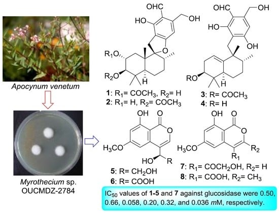
Meroterpenoids and Isocoumarinoids from a Myrothecium Fungus Associated with Apocynum venetum

1
State Key Laboratory of Functions and Applications of Medicinal Plants, Guizhou Medical University, Guiyang 550014, China
2
Key Laboratory of Marine Drugs, Ministry of Education of China, School of Medicine and Pharmacy, Ocean University of China, Qingdao 266003, China
3
Laboratory for Marine Drugs and Bioproducts, Qingdao National Laboratory for Marine Science and Technology, Qingdao 266003, China
4
Guangxi Key Laboratory of Chemistry and Engineering of Forest Products, School of Chemistry and Chemical Engineering, Guangxi University for Nationalities, Nanning 530006, China
*
Authors to whom correspondence should be addressed.
These authors contributed equally to this paper.
Mar. Drugs 2018, 16(10), 363; https://doi.org/10.3390/md16100363
Submission received: 31 August 2018 / Revised: 25 September 2018 / Accepted: 27 September 2018 / Published: 1 October 2018
(This article belongs to the Special Issue Natural Products from Marine Fungi)

Abstract

:
Four new meroterpenoids 14 and four new isocoumarinoids 58, along with five known isocoumarinoids (913), were isolated from the fungus Myrothecium sp. OUCMDZ-2784 associated with the salt-resistant medicinal plant, Apocynum venetum (Apocynaceae). Their structures were elucidated by means of spectroscopic analysis, X-ray crystallography, ECD spectra and quantum chemical calculations. Compounds 15, 7, 9 and 10 showed weak α-glucosidase inhibition with the IC50 values of 0.50, 0.66, 0.058, 0.20, 0.32, 0.036, 0.026 and 0.37 mM, respectively.

Graphical Abstract

1. Introduction

Since the discovery of penicillin, fungi have been an important source of lead compounds for drug development, which have provided a lot of attractive natural products (NPs) with different biological activities [1,2,3]. With the increase of study on the terrestrial fungal NPs, more and more known compounds were isolated repeatedly. Therefore, many researchers turned their attention to the fungi isolated from specific habitats, such as the marine-derived fungi [4,5,6,7] and the fungi associated with the plants or animals [8,9,10,11].
As part of our ongoing studies to search for bioactive NPs from fungi derived from special niche [12,13,14,15,16], we screened the fungus Myrothecium sp. OUCMDZ-2784 which is associated with the salt-resistant plant A. venetum (Apocynaceae) growing in the Yellow River Delta, a traditional Chinese medicine used for treatment of hypertension [17] and heart failure [18]. Myrothecium sp. has been reported to produce trichothecenes [19], sesquiterpenes [20,21], diterpenes [22] and cyclopeptides [23] with cytotoxic and antibacterial activities. The ethyl acetate (EtOAc) extract of the fermentation of Myrothecium sp. OUCMDZ-2784 showed 75% inhibition of α-glucosidase at 286 μg/mL. Chemical study resulted in the isolation and identification of four new meroterpenoids, myrothecisins A–D (14) and four new isocoumarinoids, myrothelactones A–D (58), together with five known isocoumarinoids that were identified as tubakialactone B (9) [24], acremonone G (10) [25], 6,8-dihydroxy-3-methylisocoumarin (11) [26], 3,4-dimethyl-6,8-dihydroxyisocoumarin (12) [27] and sescandelin B (13) [28], respectively by comparing 1H and 13C NMR spectra (Figure S57, Table S1) as well as ESIMS spectra (Figure S59) with those reported.

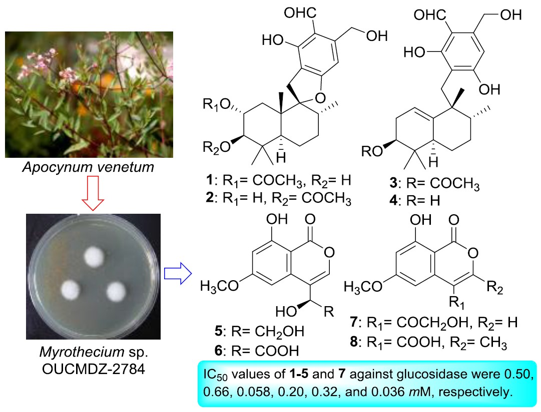
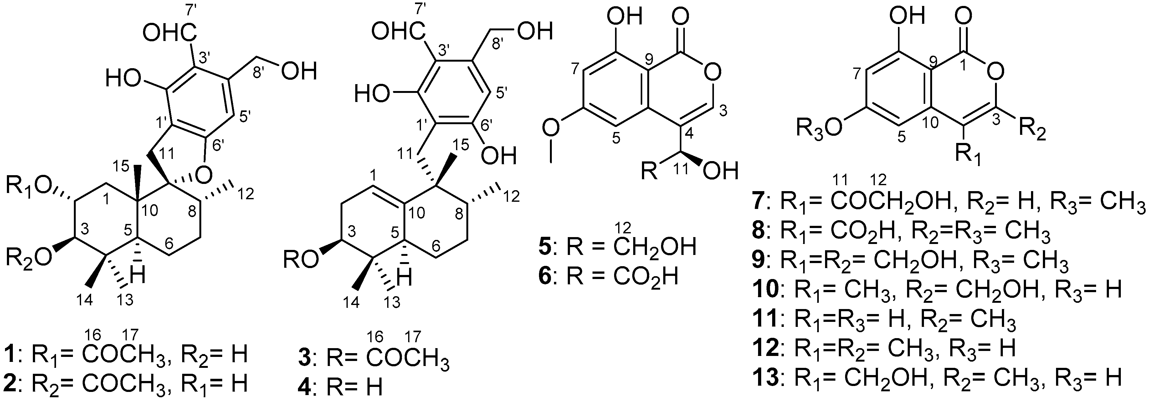
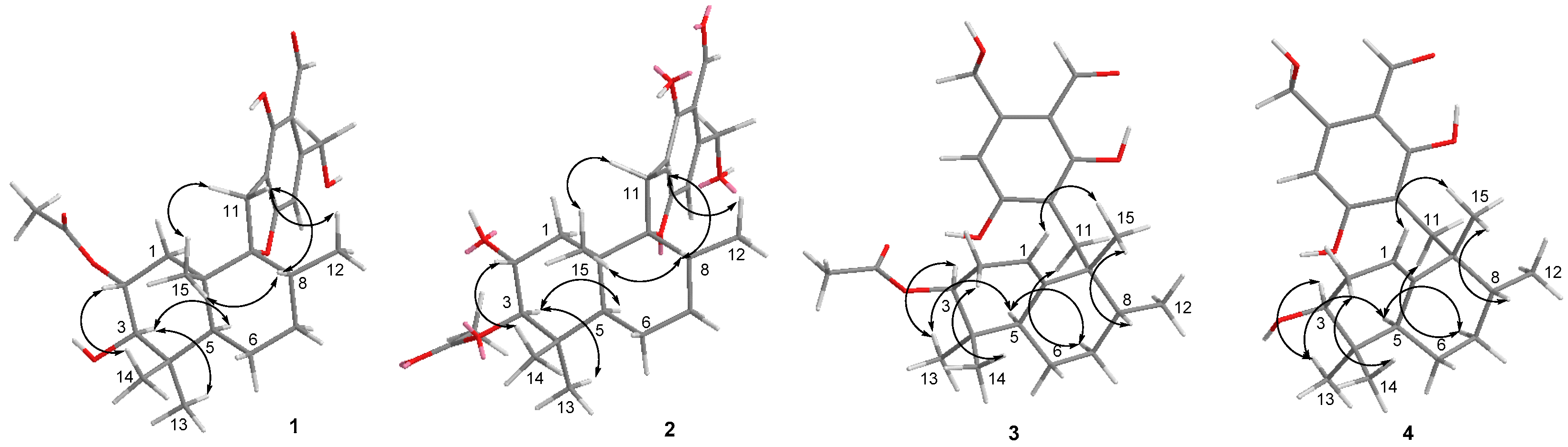
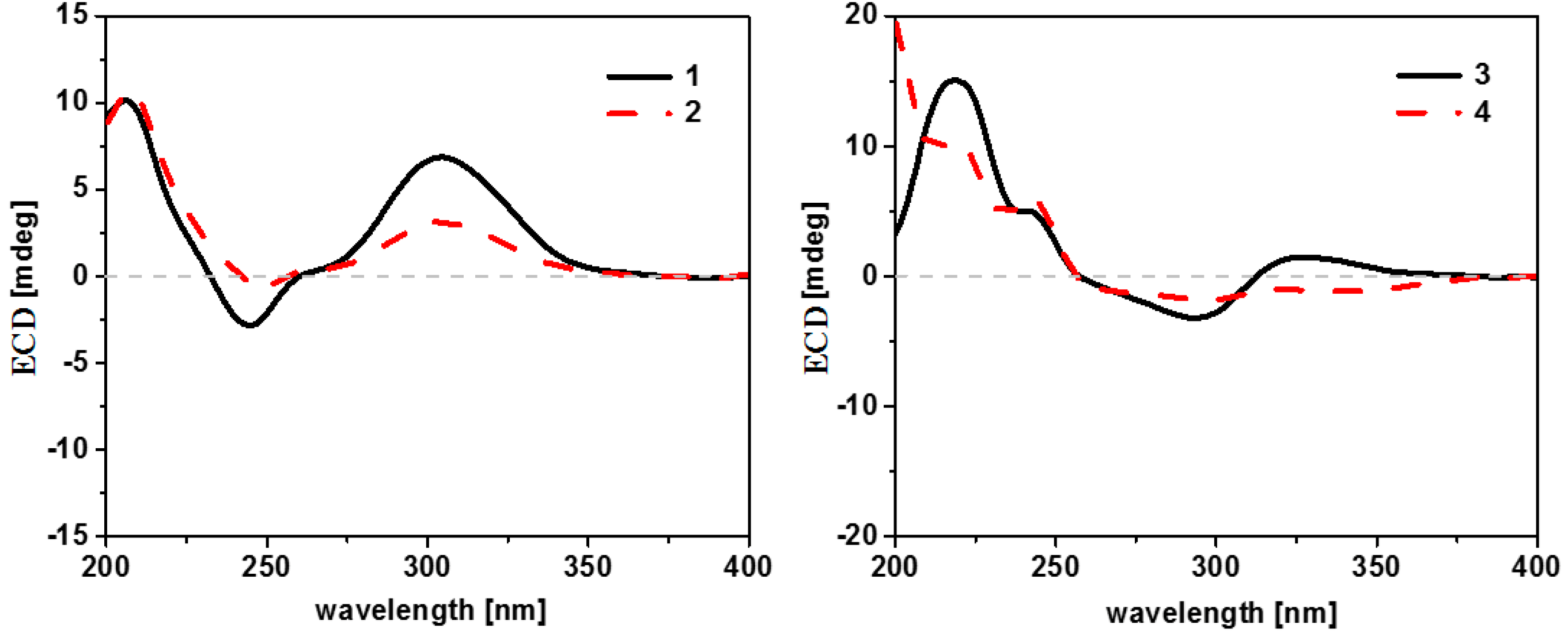
2. Results and Discussion

Myrothecisin A (1) was isolated as a pale-yellow oil. Its molecular formula was assigned as C25H34O7 by the HRESIMS peak at m/z 469.2188 [M + Na]+ (Figure S58A), indicating nine degrees of unsaturation. The 13C NMR (Figure S6) spectrum of 1 showed 25 signals that were classified by DEPT (Figure S7) and HSQC (Figure S8) as an aldehyde carbonyl carbon (δC 193.8), one acyl carbonyl carbon (δC 170.3), five sp2 non-protonated carbons (δC 167.3, 159.8, 149.5, 112.3, 111.2) and three sp3 non-protonated carbons (δC 98.8, 42.8, 39.2), one sp2 methine (δC 101.5) and four sp3 methines (δC 78.2, 71.4, 45.6, 36.0), five sp3 methylenes (δC 60.5, 35.1, 30.6, 30.4, 20.7) and five methyl carbons (δC 28.7, 21.3, 17.0, 16.6, 15.2) (Table 1). The 1H (Figure S5) and HSQC NMR showed the singlet signals at δH 10.06 and 6.56 for an aldehyde proton and an aromatic proton, respectively. The key HMBC (Figure S10) correlations from H-7′ (δH 10.06) to C-2′/C-3′/C-4′, 2′-OH (δH 12.16) to C-1′/C-2′/C-3′, H-5′ (δH 6.56) to C-1′/C-4′/C-6′/C-8′, H-8′ (δH 4.74) to C-3′/C-4′/C-5′ and 8′-OH (δH 5.44) to C-4′/C-8′ suggested a penta-substituted benzene ring (Figure 2). The COSY (homonuclear correlation spectroscopy) correlations from H-1 through H-2 to H-3 and H-5 through H-6, H-7 and H-8 to H-12 (Figure 1 and Figure S9), along with the key HMBC correlations from H-2 to C-4/C-10/C-16, H-3 to C-5/C-13/C-14, 3-OH to C-2/C-3/C-4, H-1 to C-2/C-3/C-10/C-15, H-15 to C-1/C-5/C-9/C-10, H-5 to C-3/C-4/C-6/C-13, H-6 to C-5, H-7 to C-8, H-13 to C-3/C-5/C-14, H-14 to C-3/C-5/C-13, H-12 to C-7/C-8/C-9 and H-17 to C-16 revealed a sesquiterpene fragment (Figure 2). The connection of the above-mentioned two fragments were confirmed by the key HMBC correlations from H-11 to C-8/C-9/C-10/C-1′/C-2′/C-6′ (Figure 2) [29]. The relative configuration of 1 was determined by the NOESY correlations from H-8 to H-11 and H-15, H-3 to H-5 and H-13, H-2 to H-14 and H-15 and H-11 to H-15 (Figure 3 and Figure S11). The absolute configuration of 1 was determined by calculation of electronic circular dichroism (ECD) using time-dependent density functional theory (TDDFT) (Figure S1) [30,31] and the measured ECD spectrum of 1 matched well with the calculated ECD spectrum for (2R,3R,5S,8R,9R,10S)-1 (Figure 4).
The molecular formula of 2 was also determined as C25H34O7 by the HRESIMS peak at m/z 469.2200 [M + Na]+ (Figure S58B), implying that 2 is an isomer of 1. Comparison of its 1H and 13C NMR spectra (Figures S12–S16) with those of 1 revealed that the acetyloxy group in 2 was on C-3. This was confirmed by the HMBC (Figure S17) correlation from H-3 (δH 4.29) to C-16 (δC 170.3) (Figure 2). The similar NOESY correlations (Figure 3 and Figure S18) suggested that 2 has the same relative configuration as 1. The similarity of ECD curves between 2 and 1 (Figure 5) indicated the same absolute configurations of its stereogenic carbons. Therefore 2 was named myrothecisin B.
Compound 3 was also obtained as a pale-yellow oil. Its molecular formula was determined as C25H34O6 according to the HRESIMS peak at m/z 453.2238 [M + Na]+ (Figure S58C). The 13C NMR (Figure S20) spectrum of 3 showed one aldehyde carbonyl carbon (δC 193.3), one acyl carbonyl carbon (δC 169.8), six sp2 non-protonated carbons (δC 164.6, 164.5, 145.4, 143.9, 111.4, 110.2) and two sp3 non-protonated carbons (δC 43.7, 35.6), two sp2 methines (δC 114.7, 107.5) and three sp3 methines (δC 75.6, 43.3, 43.1), five sp3 methylenes (δC 59.9, 30.2, 28.0, 26.7, 23.5) and five methyl carbons (δC 25.0, 23.0, 21.0, 17.1, 17.1) (Table 1). Analysis of its 1D and 2D NMR (Figures S19–S24) data revealed the presence of a substituted benzene ring and a sesquiterpene unit, indicating 3 was an analogue of 1 and 2. Comparison of the 1H and 13C NMR spectra with those of 1 and 2 suggested a same pentasubstituted benzene ring. The structure of the sesquiterpene unit was slightly modified and was determined by the COSY (Figure S23) correlations from H-1 through H-2 to H-3 and H-5 through H-6, H-7 and H-8 to H-12 and the key HMBC (Figure S24) correlations from H-3 to C-2/C-4/C-10/C-13/C-14, H-17 to C-16, H-1 to C-3/C-9, H-15 to C-10, H-8 to C-9, H-5 to C-9/C-13, H-12 to C-7 and H-2 to C-4/C-10 (Figure 2). The HMBC correlations from H2-11 (δH 2.68/2.53) to C-8/C-10/C-15/C-2′/C-6′ (Figure 2) confirmed the connection between the sesquiterpene fragment and the benzene ring. The relative configuration of 3 was determined by the NOESY (Figure S25) correlations from H-13 to H-3 and H-5, H-8 to H-15, as well as H-5 to H-11 (Figure 3). The absolute configuration was determined as (3S,5R,8R,9R)- by comparison of the calculated and experimental ECD spectra (Figure 4 and Figure S2). Therefore 3 was named myrothecisin C.
The molecular formula of 4 was assigned as C23H32O5 by the HRESIMS peak at m/z 411.2139 [M + Na]+ (Figure S58D), which was C2H2O less than that of 3. The similarity of the UV and NMR data between 3 and 4 (Table 1) suggested that 4 possesses the same skeleton as 3. Careful comparison of their 1H and 13C NMR spectra (Figures S26–S31) showed that the acetyloxy group (δC 21.0/δH 1.98 and δC 169.8) in 3 was replaced by a hydroxy group (δH 4.13) in 4 (Table 1). The NOESY data (Figure 3 and Figure S32) suggested that 4 has the same relative configuration as 3. The ECD Cotton effects of 4 were nearly identical to those of 3 (Figure 5), indicating the same absolute configurations of the corresponding stereogenic carbons. Thus, 4 was named myrothecisin D.
Compound 5 was obtained as a colorless crystal with the molecular formula C12H12O6 from the HRESIMS peak at m/z 251.0563 [M − H] (Figure S58E). The 1H NMR spectrum showed two meta-coupled aromatic protons at δH 6.78 (d, J = 2.2 Hz) and δH 6.63 (d, J = 2.2 Hz) (Table 2, Figure S33), indicating the presence of a tetra-substituted benzene ring. The 13C (Figure S34) NMR spectrum showed 12 carbon signals that were classified by DEPT (Figure S35) and HSQC (Figure S36) spectra as six sp2 non-protonated carbons (δC 166.4, 165.4, 163.4, 137.6, 118.6, 99.9), three sp2 methines (δC 143.1, 100.5, 100.4) and one sp3 methine (δC 68.8), one sp3 methylene (δC 64.8) and one methoxy group (δC 56.0) (Table 2). The key HMBC correlations (Figure 2 and Figure S38) from CH3O-6 to C-6, HO-11 to C-4/C-12, H-11 to C-3/C-10, H-3 to C-1/C-10, H-5 to C-4/C-7/C-9 and H-7 to C-9 along with the continuous COSY correlations of HO-11 (δH 5.50)/H-11 (δH 4.66)/H-12 (δH 3.51, 3.62)/HO-12 (δH 4.81) (Figure S37) revealed that 5 possesses a 4,6,8-trisubstituted isocoumarin skeleton with a hydroxy, a methoxy and a 1,2-dihydroxy ethyl at C-8, C-6 and C-4, respectively. The structure of 5 was further confirmed by X-ray crystallography (Figure 6). Because the value of the Flack parameter [−0.2(2)] was large, the absolute configuration determined by X-ray crystallography was not reliable. Thus, the ECD calculation method was used to further confirm the absolute configuration of C-11 of 5 as 11R- (Figure 4 and Figure S3). Consequently, 5 was named myrothelactone A.
Compound 6 was obtained as a white powder. Its molecular formula was determined as C12H10O7 based on the HRESIMS peak at m/z 265.0355 [M − H] (Figure S58F). The UV and 13C NMR data of 6 (Table 2) were similar to those of 5, indicating that they have the same isocoumarin scaffold. Comparison of their 1H and 13C data (Figures S39–S43) indicated that the hydroxymethyl group (δC/H 64.8/3.62&3.51, δH 4.81) in 5 was replaced by the carboxyl group (δC 173.4). This change was verified by the key HMBC (Figure S44) correlations from H-11 to C-3/C-10/C-12. The absolute configuration of C-11 of 6 was determined as 11R- by comparison of the calculated and experimental ECD spectra (Figure 4 and Figure S4). Therefore, 6 was name myrothelactone B.
Compound 7 was obtained as a white powder. Its molecular formula was determined as C12H10O6 according to its HRESIMS peak at m/z 249.0408 [M – H] (Figure S58G), which was only two hydrogen atoms less than that of 5. The difference observed in the NMR spectra of 7 and 5 was that the signals for hydroxymethine (δC/H 68.8/4.66) in 5 were replaced by the signal of a carbonyl group (δC-11 198.3) in 7 (Table 2, Figures S45–S49). The HMBC (Figure S50) correlations from H-3 (δH 8.47) and H-12 (δH 4.57) to C-11 further confirmed the structure of 7 which was name myrothelactone C (Figure 2).
The molecular formula of compound 8 was determined as C12H10O6 on the basis of its HRESIMS peak at m/z 249.0407 [M − H] (Figure S58H), which is an isomer of 7. Analysis of its 1H and 13C NMR spectra showed that 8 also had the same isocoumarin scaffold, whose difference is the replacement of carbonyl (δC 198.3), hydroxymethyl (δC 65.9, δH 4.57/5.28) and sp2 methine (δC 153.1, δH 8.47) signals in 7 by two sp2 non-protonated carbons (δC 173.9, 151.0) and methyl (δC 17.9, δH 2.29) signals in 8 (Table 2, Figures S51–S55). The HMBC correlations from H-12 (δH 2.29) to C-3 (δC 151.0) and C-4 (δC 118.7) suggested the methyl substitution at C-3 (Figure 2 and Figure S56). The chemical shift of the carboxyl signal (δC 173.9) together with 2D NMR data indicated the carboxyl substitution at C-4. The structure of myrothelactone D (8) was therefore determined (Figure 2).
The α-glucosidase inhibitory activity of 113 was preliminarily investigated. Compounds 15, 7, 9 and 10 exhibited inhibitory activity against the human-sourced α-glucosidase recombinant expressed in Saccharomyces cerevisiae [31,32,33] with IC50 values of 0.50, 0.66, 0.058, 0.20, 0.32, 0.036, 0.026 and 0.37 mM, while the IC50 value of positive control acarbose was 0.47 mM. Due to the low activity, the deeper investigation of the mechanism and type of enzymatic inhibition as well as the binding mode were not done.

3. Experimental Section

3.1. General Experimental Procedures

Optical rotations were measured using a JASCO P-1020 digital polarimeter (JASCO Corporation, Tokyo, Japan). UV spectra were obtained on a Beckman DU 640 spectrophotometer (Beckman Coulter, Inc., Brea, CA, USA). CD data were performed using a JASCO J-815 spectropolarimeter (JASCO Corporation, Tokyo, Japan). IR spectra were obtained on a Nicolet Nexus 470 spectrophotometer (Thermo Nicolet Corporation, Madison, WI, USA) as KBr discs. NMR spectra were recorded on a Varian System 500 spectrometer (Varian, Palo Alto, CA, USA) or a Bruker Avance 600 spectrometer (Bruker, Fallanden, Switzerland) using residual solvent signals for referencing and chemical shifts were recorded as δ values. HRESIMS spectra were measured using the Q-TOF ULTIMA GLOBAL GAA076 LC mass spectrometer (Waters Asia, Ltd., Singapore). Semi-preparative HPLC was performed using an ODS column (YMC-pack ODS-A, 10 mm × 250 mm, 5 μm, 4.0 mL/min, Kyoto, Japan). TLC and column chromatography (CC) were performed on plates pre-coated with silica gel GF254 (10–40 µm, Qingdao Marine Chemical Factory, Qingdao, China) and Sephadex LH-20 (Amersham Biosciences, Uppsala, Sweden), respectively. Vacuum-liquid chromatography (VLC) utilized silica gel H (Qingdao Marine Chemical Factory).

3.2. Collection and Phylogenetic Analysis

The fungus OUCMDZ-2784 was isolated from Apocynum venetum (Apocynaceae) collected from the estuary of Yellow River, Dongying, China. The leaves of the plant were washed with tap water and sterile distilled water in sequence. Then, it was cut into small pieces, which were then put into a centrifuge tubes filled with different concentrations of sucrose solution. These tubes were centrifuged at 1200 rpm for 20 min. Four zones were separated by improved discontinuous sucrose gradient centrifugation. The interface between the third and the fourth bands was deposited on a PDA (200 g potato, 20 g glucose, 20 g agar per liter of sea water) plate containing chloramphenicol (100 μg/mL) as a bacterial inhibitor and was then cultured at 28 °C for 3 days. A single colony was transferred to PDA agar media and was identified as Myrothecium sp. according to its morphological characteristics and 18S rRNA gene sequences (GenBank accession No. KF977010).

3.3. Cultivation and Extraction

Fungus OUCMDZ-2784 was prepared on PDA agar medium. Spores were incubated at 28 °C for 48 h on a rotary shaker with shaking at 120 rpm in a 500 mL cylindrical flask containing 150 mL liquid medium (20 g maltose, 20 g mannitol, 10 g glucose, 3 g yeast extract, 10 g monosodium glutamate per liter of sea water). The cultures were transferred to 350 × 1000 mL Erlenmeyer flasks and each containing 300 mL liquid fermentation media (1 g peptone, 10g soluble starch per liter of sea water, pH 7.0). The flasks were incubated at room temperature under static conditions for 30 days. The cultures were extracted three times by EtOAc and the combined EtOAc extracts were dried in vacuo to yield 20.1 g of extract.

3.4. Purification

The extract (20.1 g) was fractionated by VLC, eluting with a step gradient of CH2Cl2-petroleum ether (50–100%) and MeOH-CH2Cl2 (0–50%) and five fractions (Fr.1–Fr.5) were collected. Fraction 2 (3.2 g) was subjected to Sephadex LH-20 chromatography eluting with CH2Cl2/MeOH (1:1) to afford three subfractions (Fr.2.1–Fr.2.3). Fr.2.1 (1.0 g) was further purified by HPLC on an ODS column (80% MeOH/H2O) to give compounds 1 (25.2 mg, tR 6.3 min) and 3 (30.1 mg, tR 10.2 min). Fr.2.2 (50.2 mg) was purified by HPLC on an ODS column (60% MeOH/H2O) to yield compounds 11 (3.5 mg, tR 10.4 min) and 8 (3.3 mg, tR 15.2 min). Fr.2.3 (46.3 mg) was purified by HPLC on an ODS column (60% MeOH/H2O) to yield compounds 12 (5.1 mg, tR 12.6 min) and 13 (12.0 mg, tR 16.4 min). Fraction 3 (3.8 g) was separated into three subfractions (Fr.3.1–Fr.3.3) by Sephadex LH-20 eluting with MeOH-CH2Cl2 (1:1). Fr.3.1 (0.5 g) was purified by semi preparative HPLC on an ODS column (85% MeOH/H2O) to yield compound 2 (36.2 mg, tR 10.2 min). Fr.3.2 (1.1 g) was separated by silica gel VLC column eluting with CH2Cl2-petroleum (2:1) to yield compounds 10 (100.3 mg) and 5 (200.8 mg). Fr.3.3 (0.5 g) was further purified by Sephadex LH-20 eluting with MeOH to yield compound 9 (15.3 mg). Fraction 4 (1.8 g) was separated into three subfractions (Fr.4.1–Fr.4.3) by Sephadex LH-20 eluting with MeOH. Fr.4.1 (0.6 g) was further purified by semi preparative HPLC on an ODS column (85% MeOH/H2O) to yield compound 4 (34.3 mg, tR 12.2 min). Fr.4.2 (0.2 g) was further purified by semi preparative HPLC on an ODS column (50% MeOH/H2O) to yield compound 7 (6.0 mg, tR 12.7 min). Fr.4.3 (0.3 g) was purified by HPLC on an ODS column (40% MeOH/H2O) to yield compound 6 (5.8 mg, tR 10.6 min).
Myrothecisin A (1): pale yellow oil; [ α ] D 20 + 9.0 (c 0.1, CHCl3); UV (MeOH) λmax (log ε) 212 (4.13), 237 (3.60), 305 (3.93) nm; ECD (0.002 M, MeOH) λmaxε) 207 (+1.69), 244 (−0.51), 304 (+1.06) nm; IR (KBr) νmax 3443, 2926, 1719, 1635, 1372, 1267 cm−1; 1H and 13C NMR data, see Table 1; HRESIMS m/z 469.2188 [M + Na]+ (calcd for C25H34O7Na, 469.2197).
Myrothecisin B (2): pale yellow oil; [ α ] D 20 + 70.1 (c 0.1, CHCl3); UV (MeOH) λmax (log ε) 211 (4.10), 238 (3.62), 305 (3.88) nm; ECD (0.002 M, MeOH) λmaxε) 207 (+1.62), 249 (−0.09), 305 (+0.49) nm; IR (KBr) νmax 3424, 2940, 1733, 1638, 1460, 1371, 1263 cm−1; 1H and 13C NMR data, see Table 1; HRESIMS m/z 469.2200 [M + Na]+ (calcd for C25H34O7Na, 469.2197).
Myrothecisin C (3): pale yellow oil; [ α ] D 20 + 63.5 (c 0.1, CHCl3); UV (MeOH) λmax (log ε) 212 (4.09), 239 (3.65), 307 (3.81) nm; ECD (0.002 M, MeOH) λmaxε) 219 (+2.29), 293 (−0.49), 327 (+0.22) nm; IR (KBr) νmax 3480, 2936, 1732, 1612, 1373, 1254 cm−1; 1H and 13C NMR data, see Table 1; HRESIMS m/z 453.2238 [M + Na]+ (calcd for C25H34O6Na, 453.2248).
Myrothecisin D (4): pale yellow oil; [ α ] D 20 + 20.0 (c 0.1, CHCl3); UV (MeOH) λmax (log ε) 203 (4.16), 224 (3.56), 300 (3.86) nm; ECD (0.002 M, MeOH) λmaxε) 220 (+1.57), 242 (+0.92), 295 (−0.27) nm; IR (KBr) νmax 2928, 1718, 1621, 1370, 1264, 1027 cm−1; 1H and 13C NMR data, see Table 1; HRESIMS m/z 411.2139 [M + Na]+ (calcd for C23H32O5Na, 411.2142).
Myrothelactone A (5): colorless crystal; melting point (mp) 174–175 °C; [ α ] D 20 − 36.0 (c 0.1, MeOH); UV (MeOH) λmax (log ε) 246 (3.82), 328 (3.19) nm; ECD (0.004 M, MeOH) λmaxε) 206.5 (−0.22), 241 (+0.24), 259 (−0.27) nm; IR (KBr) νmax 3747, 3630, 3159, 2959, 1668, 1558, 1398, 1237 cm−1; 1H and 13C NMR data, see Table 2; HRESIMS m/z 251.0563 [M − H] (calcd for C12H11O6, 251.0561).
Myrothelactone B (6): white powder; mp 169–171 °C; [ α ] D 20 − 30.0 (c 0.1, MeOH); UV (MeOH) λmax (log ε) 245 (3.81), 328 (3.17) nm; ECD (0.004 M, MeOH) λmaxε) 206.5 (−0.78), 241 (+0.24), 259 (−0.13) nm; IR (KBr) νmax 3749, 2922, 1681, 1651, 1619, 1459, 1399 cm−1; 1H and 13C NMR data, see Table 2; HRESIMS m/z 265.0355 [M − H] (calcd for C12H9O7, 265.0354).
Myrothelactone C (7): white powder; mp 160–161 °C; UV (MeOH) λmax (log ε) 228 (3.91), 263 (3.43), 325 (3.16) nm; IR (KBr) νmax 3750, 3675, 3615, 1736, 1651, 1558, 1540, 1399 cm−1; 1H and 13C NMR data, see Table 2; HRESIMS m/z 249.0408 [M − H] (calcd for C12H9O6, 249.0405).
Myrothelactone D (8): white powder; mp 219–221 °C; UV (MeOH) λmax (log ε) 231 (3.92), 263 (3.45), 325 (3.16) nm; IR (KBr) νmax 3749, 3673, 3445, 3197, 1716, 1682, 1539, 1457, 1399 cm−1; 1H and 13C NMR data, see Table 2; HRESIMS m/z 249.0407 [M − H] (calcd for C12H9O6, 249.0405).

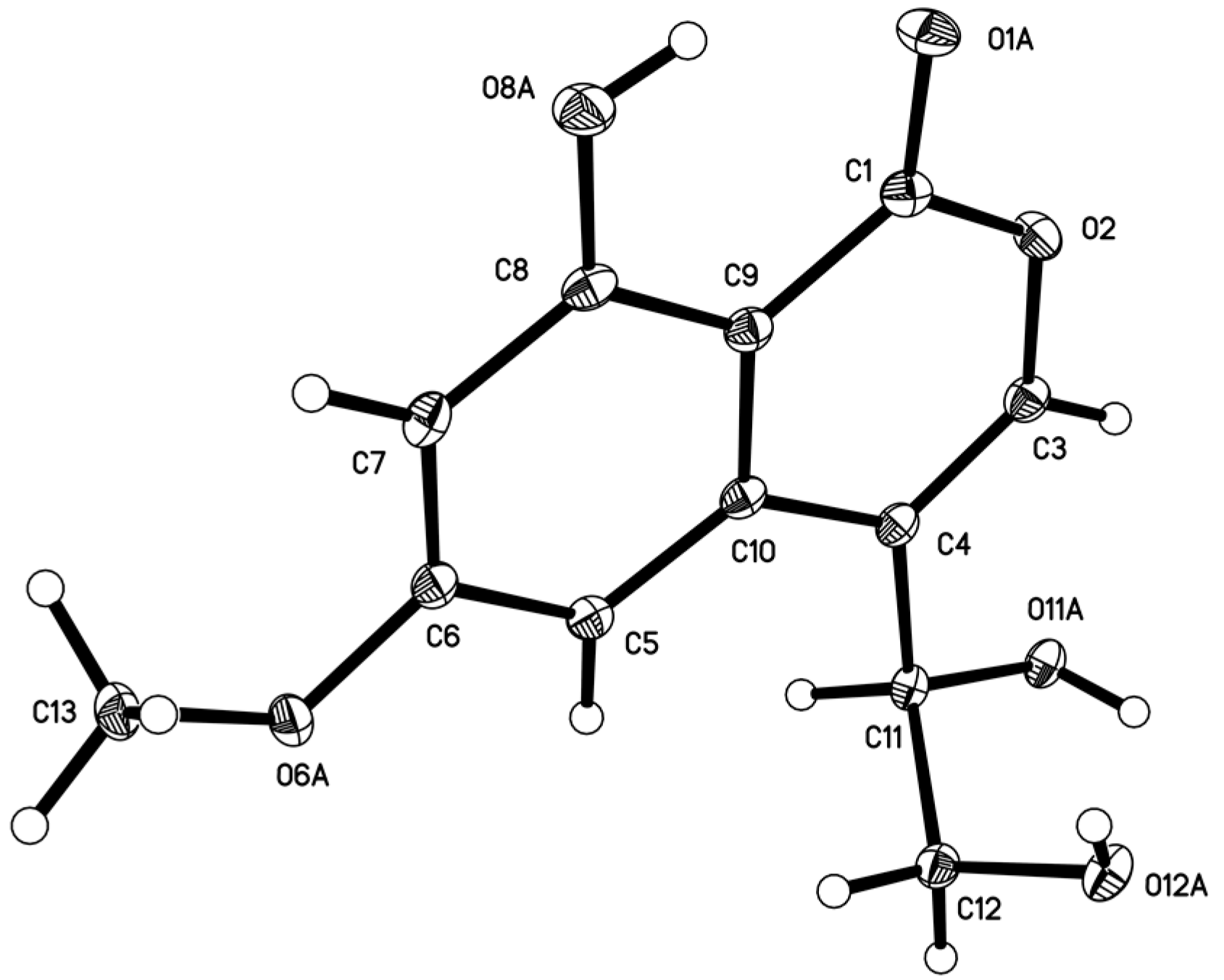
3.5. X-ray Structure Determination of Compound 5

Compound 5 was obtained as a colorless needles crystal with molecular formula C12H12O6. Orthorhombic, space group P212121, a = 4.9041(2) Å, b = 13.8470(5) Å, c = 15.7443(6) Å, α = 90.00°, β = 90.00°, γ = 90.00°, V = 1069.15(7) Å3, Z = 4, Dcalcd = 1.567 Mg/m3, μ = 1.089 mm−1, F(000) = 528, crystal size 0.30 mm × 0.18 mm × 0.15 mm, T = 293(2) K. A total of 1478 unique reflections (2θ < 50°) were collected on a CCD area detector diffractometer with graphite monochromated Cu Kα radiation (λ = 1.54178 Å). The structure was solved by direct methods (SHELXS-97) and expanded using Fourier techniques (SHELXL-97). The final cycle of full-matrix least squares refinement was based on 1478 unique reflections (2θ < 50°) and 165 variable parameters and converged with unweighted and weighted agreement factors of R1 = 0.0326, wR2 = 0.0885 and R = 0.0880 for I > 2sigma(I) data. Absolute structure parameter: −0.2(2). The deposited number of compound 5 in the Cambridge Crystallographic Data Centre is 980155.

3.6. α-Glucosidase Inhibitory Assays

The human-sourced α-glucosidase was recombinant expressed in the yeast Saccharomyces cerevisiae and the inhibitory effects of compounds 113 were tested using p-nitrophenyl-α-d-glucopyranoside (pNPG) as substrate [31,32,33]. The sample was dissolved in sodium phosphate buffer (PBS, pH 6.8) at three concentrations. 10 μL of the sample solution, 20 μL of 2.5 mM pNPG solution (in phosphate buffer) and 20 μL of PBS were mixed in a 96-well microplate and incubated at 37 °C for 5 min. A volume of 10 μL of α-glucosidase diluted to 0.2 U/mL by 0.01 M PBS was then added to each well. After incubating at 37 °C for 15 min, the absorbance at 405 nm was recorded by a Spectra max 190 micro plate reader (Molecular Devices Inc., San Jose, CA, USA). The blank was prepared by adding phosphate buffer instead of the α-glucosidase and the positive control was acarbose. Blank readings (no enzyme) were subtracted from each well and results were compared to the control. The inhibition (%) was calculated as [1 − (ODdrug/ODblank)] × 100%. The IC50 value was calculated as the compound concentration that is required for 50% inhibition and the IC50 value of acarbose was 0.47 mM.

4. Conclusions

This study revealed eight new fungal NPs, meroterpenoids 14 and isocoumarinoids 58, from the culture of the salt-tolerant plant-associated fungus Myrothecium sp. OUCMDZ-2784. The new compounds 15 and 7 exhibited α-glucosidase inhibitory activity. Combined with bioactive NPs from mangrove-derived fungi [34,35,36,37,38], the results indicated that fungi living in the salt-tolerant plants are an important biological resources for new and bioactive NPs.

Supplementary Materials

The following are available online at https://www.mdpi.com/1660-3397/16/10/363/s1, Figures S1–S4: DFT-optimized structures for low-energy conformers of compounds 1, 3, 5 and 6, Figures S5–S11: NMR spectra of compound 1 in DMSO-d6, Figures S12–S18: NMR spectra of compound 2 in DMSO-d6, Figures S19–S25: NMR spectra of compound 3 in DMSO-d6, Figures S26–S32: NMR spectra of compound 4 in DMSO-d6, Figures S33–S38: NMR spectra of compound 5 in DMSO-d6, Figures S39–S44: NMR spectra of compound 6 in DMSO-d6, Figures S45–S50: NMR spectra of compound 7 in DMSO-d6, Figures S51–S56: NMR spectra of compound 8 in DMSO-d6, Figure S57: 1H- and 13C-NMR spectra of compounds 913 in DMSO-d6, Figure S58: HRESI-MS spectra of compounds 18, Figure S59: ESI-MS spectra of compounds 913, Table S1: 1H and 13C NMR data for compounds 913 in DMSO-d6.

Author Contributions

Y.X. performed the most experiments; C.W. prepared the draft of the manuscript; H.L. tested the α-glucosidase inhibitory activity; G.Z. performed the ECD calculations; P.F. checked the data; L.W. revised the manuscript; W.Z. designed and supervised the research and revised the final version.

Funding

This research was funded by the NSFC (Nos. 81561148012, U1501221, 81741150, U1606403), the 100 Leading Talents of Guizhou Province (fund for W. Zhu), the science and technology project of Guizhou (Grant No. QKHT Z-2014-4007) and the academician workstation of Guizhou (Grant No. QKH YSZ-2015-4009).

Conflicts of Interest

The authors declare no conflict of interest.

References

  1. Aly, A.H.; Debbab, A.; Proksch, P. Fifty years of drug discovery from fungi. Fungal Divers. 2011, 50, 3–19. [Google Scholar] [CrossRef]
  2. Bladt, T.T.; Frisvad, J.C.; Knudsen, P.B.; Larsen, T.O. Anticancer and antifungal compounds from Aspergillus, Penicillium and other filamentous fungi. Molecules 2013, 18, 11338–11376. [Google Scholar] [CrossRef] [PubMed] [Green Version]
  3. Khan, A.A.; Bacha, N.; Ahmad, B.; Lutfullah, G.; Farooq, U.; Cox, R.J. Fungi as chemical industries and genetic engineering for the production of biologically active secondary metabolites. Asian Pac. J. Trop. Biomed. 2014, 4, 859–870. [Google Scholar] [CrossRef]
  4. Rateb, M.E.; Ebel, R. Secondary metabolites of fungi from marine habitats. Nat. Prod. Rep. 2011, 28, 290–344. [Google Scholar] [CrossRef] [PubMed]
  5. Bugnia, T.S.; Ireland, C.M. Marine-derived fungi: A chemically and biologically diverse group of microorganisms. Nat. Prod. Rep. 2004, 21, 143–163. [Google Scholar] [CrossRef] [PubMed]
  6. Xu, L.; Meng, W.; Cao, C.; Wang, J.; Shan, W.; Wang, Q. Antibacterial and antifungal compounds from marine fungi. Mar. Drugs 2015, 13, 3479–3513. [Google Scholar] [CrossRef] [PubMed]
  7. Moghadamtousi, S.Z.; Nikzad, S.; Kadir, H.A.; Abubakar, S.; Zandi, K. Potential antiviral agents from marine fungi: An overview. Mar. Drugs 2015, 13, 4520–4538. [Google Scholar] [CrossRef] [PubMed]
  8. Strobel, G.; Daisy, B.; Castillo, U.; Harper, J. Natural products from endophytic microorganisms. J. Nat. Prod. 2004, 67, 257–268. [Google Scholar] [CrossRef] [PubMed]
  9. Borges, W.S.; Borges, K.B.; Bonato, P.S.; Said, S.; Pupo, M.T. Endophytic fungi: natural products, enzymes and biotransformation reactions. Curr. Org. Chem. 2009, 13, 1137–1163. [Google Scholar] [CrossRef]
  10. Nisa, H.; Kamili, A.N.; Nawchoo, I.A.; Shafi, S.; Shameem, N.; Bandh, S.A. Fungal endophytes as prolific source of phytochemicals and other bioactive natural products: A review. Microb. Pathog. 2015, 82, 50–59. [Google Scholar] [CrossRef] [PubMed]
  11. Tan, R.X.; Zou, W.X. Endophytes: A rich source of functional metabolites. Nat. Prod. Rep. 2001, 18, 448–459. [Google Scholar] [CrossRef] [PubMed]
  12. Sun, K.; Li, Y.; Guo, L.; Wang, Y.; Liu, P.; Zhu, W. Indole diterpenoids and isocoumarin from the fungus, Aspergillus flavus, isolated from the prawn, Penaeus vannamei. Mar. Drugs 2014, 12, 3970–3981. [Google Scholar] [CrossRef] [PubMed]
  13. Zhu, G.; Kong, F.; Wang, Y.; Fu, P.; Zhu, W. Cladodionen, a cytotoxic hybrid polyketide from the marine-derived Cladosporium sp. OUCMDZ-1635. Mar. Drugs 2018, 16, 71. [Google Scholar] [CrossRef] [PubMed]
  14. Wang, Y.; Zheng, J.; Liu, P.; Wang, W.; Zhu, W. Three new compounds from Aspergillus terreus PT06-2 grown in a high salt medium. Mar. Drugs 2011, 9, 1368–1378. [Google Scholar] [CrossRef] [PubMed]
  15. Wang, Y.; Lu, Z.; Sun, K.; Zhu, W. Effects of high salt stress on secondary metabolites from marine-derived fungus Spicaria elegans. Mar. Drugs 2011, 9, 535–542. [Google Scholar] [CrossRef] [PubMed]
  16. Wang, Y.; Wang, L.; Zhuang, Y.; Kong, F.; Zhang, C.; Zhu, W. Phenolic polyketides from the co-cultivation of marine-derived Penicillium sp. WC-29-5 and Streptomyces fradiae 007. Mar. Drugs 2014, 12, 2079–2088. [Google Scholar] [CrossRef] [PubMed]
  17. Chinese Pharmacopeia Committee of Ministry of Public Health of the People’s Republic of China. Chinese Pharmacopeia 2000; Chemical and Technical Press: Beijing, China, 2000; p. 170. [Google Scholar]
  18. Shaanxi Provincial and Municipal Collaborative Group for Prevention and Treatment of Coronary Heart Diseases. Clinical observation and pharmacological experiment of Apocynum venetum root in the treatment of heart failure. Shaanxi Med. J. 1974, 5, 10–14. [Google Scholar]
  19. Isaka, M.; Punya, J.; Lertwerawat, Y.; Tanticharoen, M.; Thebtaranonth, Y. Antimalarial activity of macrocyclic trichothecenes isolated from the fungus Myrothecium verrucaria. J. Nat. Prod. 1999, 62, 329–331. [Google Scholar] [CrossRef] [PubMed]
  20. Fu, Y.; Wu, P.; Xue, J.; Wei, X. Cytotoxic and antibacterial quinone sesquiterpenes from a Myrothecium fungus. J. Nat. Prod. 2014, 77, 1791–1799. [Google Scholar] [CrossRef] [PubMed]
  21. Fu, Y.; Wu, P.; Xue, J.; Li, H.; Wei, X. Myrothecols G and H, two new analogues of the marine-derived quinone sesquiterpene Penicilliumin A. Mar. Drugs 2015, 13, 3360–3367. [Google Scholar] [CrossRef] [PubMed]
  22. Hsu, Y.H.; Hirota, A.; Shima, S.; Nakagawa, M.; Nozaki, H.; Tada, T.; Nakayama, M. Structure of myrocin C, a new diterpene antibiotic produced by a strain of Myrothecium sp. Agric. Biol. Chem. 1987, 51, 3455–3457. [Google Scholar] [CrossRef]
  23. Zou, X.; Niu, S.; Ren, J.; Li, E.; Liu, X.; Che, Y. Verrucamides A–D, antibacterial cyclopeptides from Myrothecium verrucaria. J. Nat. Prod. 2011, 74, 1111–1116. [Google Scholar] [CrossRef] [PubMed]
  24. Nakashima, K.; Tomida, J.; Hirai, T.; Morita, Y.; Kawamura, Y.; Inoue, M. Tubakialactones A–E, new polyketides from the endophytic fungus Tubakia sp. ECN-111. Tetrahedron Lett. 2017, 58, 2248–2251. [Google Scholar] [CrossRef]
  25. Rukachaisirikul, V.; Rodglin, A.; Sukpondma, Y.; Phongpaichit, S.; Buatong, J.; Sakayaroj, J. Phthalide and isocoumarin derivatives produced by an Acremonium sp. isolated from a mangrove Rhizophora apiculata. J. Nat. Prod. 2012, 75, 853–858. [Google Scholar] [CrossRef] [PubMed]
  26. Kornsakulkarn, J.; Thongpanchang, C.; Lapanun, S.; Srichomthong, K. Isocoumarin glucosides from the scale insect fungus Torrubiella tenuis BCC 12732. J. Nat. Prod. 2009, 72, 1341–1343. [Google Scholar] [CrossRef] [PubMed]
  27. Tanaka, A.K.; Sato, C.; Shibata, Y.; Kobayashi, A.; Yamashita, K. Growth promoting activities of sclerotinin A and its analogs. Agric. Biol. Chem. 1974, 38, 1311–1315. [Google Scholar] [CrossRef]
  28. Kimura, Y.; Nakadoi, M.; Nakajima, H.; Hamasaki, T.; Nagai, T.; Kohmoto, K.; Shimada, A. Structure of sescandelin-B, a new metabolite produced by the fungus Sesquicillium candelabrum. Agric. Biol. Chem. 1991, 55, 1887–1888. [Google Scholar]
  29. Zhao, J.; Feng, J.; Tan, Z.; Liu, J.; Zhao, J.; Chen, R.; Xie, K.; Zhang, D.; Li, Y.; Yu, L.; et al. Stachybotrysins A−G, phenylspirodrimane derivatives from the fungus Stachybotrys chartarum. J. Nat. Prod. 2017, 80, 1819–1826. [Google Scholar] [CrossRef] [PubMed]
  30. Berova, N.; Bari, L.D.; Pescitelli, G. Application of electronic circular dichroism in configurational and conformational analysis of organic compounds. Chem. Soc. Rev. 2007, 36, 914–931. [Google Scholar] [CrossRef] [PubMed]
  31. Kong, F.; Zhao, C.; Hao, J.; Wang, C.; Wang, W.; Huang, X.; Zhu, W. New α-glucosidase inhibitors from a marine sponge-derived fungus, Aspergillus sp. OUCMDZ-1583. RSC Adv. 2015, 5, 68852–68863. [Google Scholar] [CrossRef]
  32. Wang, C.; Guo, L.; Hao, J.; Wang, L.; Zhu, W. α-Glucosidase inhibitors from the marine-derived fungus Aspergillus flavipes HN4-13. J. Nat. Prod. 2016, 79, 2977–2981. [Google Scholar] [CrossRef] [PubMed]
  33. Shim, Y.-J.; Doo, H.-K.; Ahn, S.-Y.; Kim, Y.-S.; Seong, J.-K.; Park, I.-S.; Min, B.-H. Inhibitory effect of aqueous extract from the gall of Rhus chinensis on alpha-glucosidase activity and postprandial blood glucose. J. Ethnopharmacol. 2003, 85, 283–287. [Google Scholar] [CrossRef]
  34. Lin, Z.; Zhu, T.; Fang, Y.; Gu, Q.; Zhu, W. Polyketides from Penicillium sp. JP-1, an endophytic fungus associated with the mangrove plant Aegiceras corniculatum. Phytochemistry 2008, 69, 1273–1278. [Google Scholar] [CrossRef] [PubMed]
  35. Lu, Z.; Zhu, H.; Fu, P.; Wang, Y.; Zhang, Z.; Lin, H.; Liu, P.; Zhuang, Y.; Hong, K.; Zhu, W. Cytotoxic polyphenols from the marine-derived fungus Penicillium expansum. J. Nat. Prod. 2010, 73, 911–914. [Google Scholar] [CrossRef] [PubMed]
  36. Wang, J.; Lu, Z.; Liu, P.; Wang, Y.; Li, J.; Hong, K.; Zhu, W. Cytotoxic polyphenols from the fungus Penicillium expansum 091006 endogenous with the mangrove plant Excoecaria agallocha. Planta Med. 2012, 78, 1861–1866. [Google Scholar] [PubMed]
  37. Kong, F.; Wang, Y.; Liu, P.; Dong, T.; Zhu, W. Thiodiketopiperazines from the marine-derived fungus Phoma sp. OUCMDZ-1847. J. Nat. Prod. 2014, 77, 132–137. [Google Scholar] [CrossRef] [PubMed]
  38. Wang, L.; Han, X.; Zhu, G.; Wang, Y.; Chairoungdua, A.; Piyachaturawat, P.; Zhu, W. Polyketides from the endophytic fungus Cladosporium sp. isolated from the mangrove plant Excoecaria agallocha. Front. Chem. 2018, 6, 344. [Google Scholar] [CrossRef] [PubMed]
Figure 1. Structures 113 isolated from Myrothecium sp. OUCMDZ-2784.
Marinedrugs 16 00363 g001
Figure 2. Key homonuclear correlation spectroscopy (COSY) and key HMBC correlations for 18.
Marinedrugs 16 00363 g002
Figure 3. NOESY correlations for 14.
Marinedrugs 16 00363 g003
Figure 4. Measured and calculated ECD spectra for 1, 3, 5 and 6.
Marinedrugs 16 00363 g004
Figure 5. ECD spectra for 14.
Marinedrugs 16 00363 g005
Figure 6. ORTEP diagram of 5.
Marinedrugs 16 00363 g006
Table 1. 1H (600 MHz) and 13C (150 MHz) NMR data for 14 in DMSO-d6.
No.1234
δC, TypeδH, Mult. (J in Hz)δC, TypeδH, Mult. (J in Hz)δC, TypeδH, Mult. (J in Hz)δC, TypeδH, Mult. (J in Hz)
135.1, CH21.16, m; 1.55 a38.4, CH21.17, m; 1.57 a114.7, CH4.94, brs116.3, CH4.88, s
271.4, CH4.83, ddd (11.5, 10.1, 4.1)64.4, CH3.65, m28.0, CH21.89, m; 1.23, m31.6, CH21.72, m
378.2, CH2.96, dd (10.1, 4.8)83.3, CH4.29, d (9.9)75.6, CH4.62, dd (8.6, 6.5)71.5, CH3.30 a
439.2, C 39.0, C 35.6, C 36.9, C
545.6, CH1.56 a45.5, CH1.63 a43.1, CH2.54, m43.7, CH2.45, m
620.7, CH21.54 a; 1.47, m20.4, CH21.56 a; 1.48, m26.7, CH21.79, m26.7, CH21.77, m
730.6, CH21.55 a; 1.35, m30.5, CH21.57 a; 1.38, m30.2, CH21.54, m30.3, CH21.52, m
836.0, CH1.83, m35.6, CH1.85, m43.3, CH1.30, m42.9, CH1.29, m
998.8, C 98.8, C 43.7, C 43.7, C
1042.8, C 42.5, C 143.9, C 143.0, C
1130.4, CH23.03, d (16.1);
2.79, d (16.0)
30.2, CH23.11, d (16.5);
2.81, d (16.5)
23.5, CH22.68, d (12.9);
2.53, d (12.9)
23.3, CH22.63, d (12.7);
2.50, d (12.7)
1215.2, CH30.64, d (6.3)15.1, CH30.68, d (6.3)17.1, CH31.03, d (6.6)17.2, CH31.02, d (6.7)
1328.7, CH30.97, s28.4, CH30.79, s25.0, CH30.88, s25.2, CH30.95, s
1417.0, CH30.78, s17.5, CH30.81, s17.1, CH30.73, s14.8, CH30.59, s
1516.6, CH31.04, s16.6, CH31.03, s23.0, CH30.96, s22.9, CH30.91, s
16170.3, C 170.3, C 169.8, C
1721.3, CH31.91, s21.0, CH32.02, s21.0, CH31.98, s
1′111.2, C 111.2, C 111.4, C 111.3, C
2′159.8, C 159.8, C 164.5, C 164.8, C
3′112.3, C 112.2, C 110.2, C 110.2, C
4′149.5, C 149.4, C 145.4, C 145.4, C
5′101.5, CH6.56, s101.6, CH6.61, s107.5, CH6.51, s107.3, CH6.50, s
6′167.3, C 167.3, C 164.6, C 164.6, C
7′193.8, CH10.06, s193.8, CH10.07, s193.3, CH9.96, s193.4, CH9.96, s
8′60.5, CH24.74, s60.4, CH24.74, s59.9, CH24.70, s59.8, CH24.70, s
2/3-OH 4.94, d (4.7) 4.62, d (4.9) 4.13, s
2′-OH 12.16, s 12.24, s 12.84, s 12.78, s
6′-OH 10.55, brs
8′-OH 5.44, s 5.39, s 5.35, s 5.36, s
a Overlapped.
Table 2. 1H and 13C NMR data for 58 in DMSO-d6.
No.5 a6 b7 b8 b
δC, TypeδH, mult. (J in Hz)δC, TypeδH, mult. (J in Hz)δC, TypeδH, mult. (J in Hz)δC, TypeδH, mult. (J in Hz)
1165.4, C 164.9, C 163.4, C 165.2, C
3143.1, CH7.45, s144.4, CH7.62, s153.1, CH8.47, s151.0, C
4118.6, C 117.7, C 114.7, C 118.7, C
5100.5, CH6.78, d (2.2)101.6, CH6.84, s102.3, CH7.46, s101.6, CH6.86, s
6166.4, C 166.4, C 166.8, C 166.3, C
7100.4, CH6.63 (d, 2.2)100.6, CH6.63, s101.2, CH6.68, s99.8, CH6.48, s
8163.4, C 163.3, C 163.2, C 162.6, C
999.9, C 99.9, C 99.9, C 99.2, C
10137.6, C 136.8, C 134.8, C 138.6, C
1168.8, CH4.66, td (5.2, 5.2)68.5, CH5.01, s198.3, C 173.9, C
1264.8, CH23.62, ddd (11.3, 5.3, 5.3);
3.51, ddd (11.3, 5.3, 5.3)
173.4, C 65.9, CH24.57, d (4.7)17.9, CH32.29, s
6-OCH356.0, CH33.88, s56.0, CH33.84, s56.0, CH33.87, s55.8, CH33.80, s
8-OH 11.37, s 11.22, s 11.03, s 11.21, s
11-OH 5.50, d (4.7)
12-OH 4.81, t (5.2) 5.28, t (5.0)
a Data were measured at 600 MHz (1H) and 150 MHz (13C). b Data were measured at 500 MHz (1H) and 125 MHz (13C).

