Abstract
Sulphated polysaccharides (SP) extracted from seaweeds have antiviral properties and are much less cytotoxic than conventional drugs, but little is known about their mode of action. Combination antiviral chemotherapy may offer advantages over single agent therapy, increasing efficiency, potency and delaying the emergence of resistant virus. The paramyxoviridae family includes pathogens causing morbidity and mortality worldwide in humans and animals, such as the Newcastle Disease Virus (NDV) in poultry. This study aims at determining the antiviral activity and mechanism of action in vitro of an ulvan (SP from the green seaweed Ulva clathrata), and of its mixture with a fucoidan (SP from Cladosiphon okamuranus), against La Sota NDV strain. The ulvan antiviral activity was tested using syncytia formation, exhibiting an IC50 of 0.1 μg/mL; ulvan had a better anti cell-cell spread effect than that previously shown for fucoidan, and inhibited cell-cell fusion via a direct effect on the F0 protein, but did not show any virucidal effect. The mixture of ulvan and fucoidan showed a greater anti-spread effect than SPs alone, but ulvan antagonizes the effect of fucoidan on the viral attachment/entry. Both SPs may be promising antivirals against paramyxovirus infection but their mixture has no clear synergistic advantage.
1. Introduction
Since the first report of the antiviral activity of sulphated polysaccharides in 1958 [1], a large number of SP have been found to possess a broad spectrum of antiviral activity. Nowadays a number of these SP have been demonstrated to be potent inhibitors of paramyxoviruses, including parainfluenza virus, respiratory syncytial virus, mumps virus, measles virus, NDV, and distemper canine virus [2,3,4]. Ulvales (chlorophyta) are seaweeds distributed worldwide. The genera Ulva, before Enteromorpha, is grown and collected for food consumption [5,6]. Among the polymers synthetized by these Ulva sp., cell wall polysaccharides represents 38%–54% of all dry algal matter [7], and can be classified in four polysaccharide families: two major ones, ulvan and cellulose and two minor ones, xyloglucan and glucurunan. Ulvan, the most important one, has been characterized with a variety of biological features [8,9,10,11,12,13,14]; among these, the immunomodulatory effect and the antiviral activity have focused the attention of scientists worldwide and marked a trend in drugs development. The main constituents reported in ulvans from several Ulvales species [15,16,17,18,19] are: Rhamnose (16.8%–45.0% dw), xylose (2.1%–12.0%), glucose (0.5%–6.4%), uronic acid (6.5%–19.0%), sulphate (16.0%–23.2%) and iduronic acid (1.1%–9.1%), rhamnose occurring mainly in the form of the aldobiouronic acid, 4-O-β-d-glucuronosyl-l-rhamnose.
Newcastle disease virus (NDV) is the causative agent of one of the most serious threats to the poultry industry and causes enormous economic losses because of its worldwide distribution and high flock mortality [20]. It belongs to the Paramyxoviridae family, which groups enveloped, negative-sense single-stranded RNA viruses [21], and utilizes its genome to code for six proteins from the 3′ terminus to the 5′ terminus [22]. The viral lipid envelope contains the viral glycoproteins HN (attachment protein) and F (fusion protein), both being needed for efficient infection of cells; the F glycoprotein mediates the penetration of the cellular membrane during viral entry [22]. Currently there are no antiviral agents available against NDV in poultry, hence the importance of developing new alternative control measures.
In our study, we evaluated the anti-NDV activity and mechanism of action of the SP ulvan (isolated from the green algae Ulva clathrata) and of a mixture with Cladosiphon okamuranus fucoidan, which mode of action has previously been characterized [3,4], for the purpose of developing new candidates anti- paramyxovirus drugs.
2. Results and Discussion
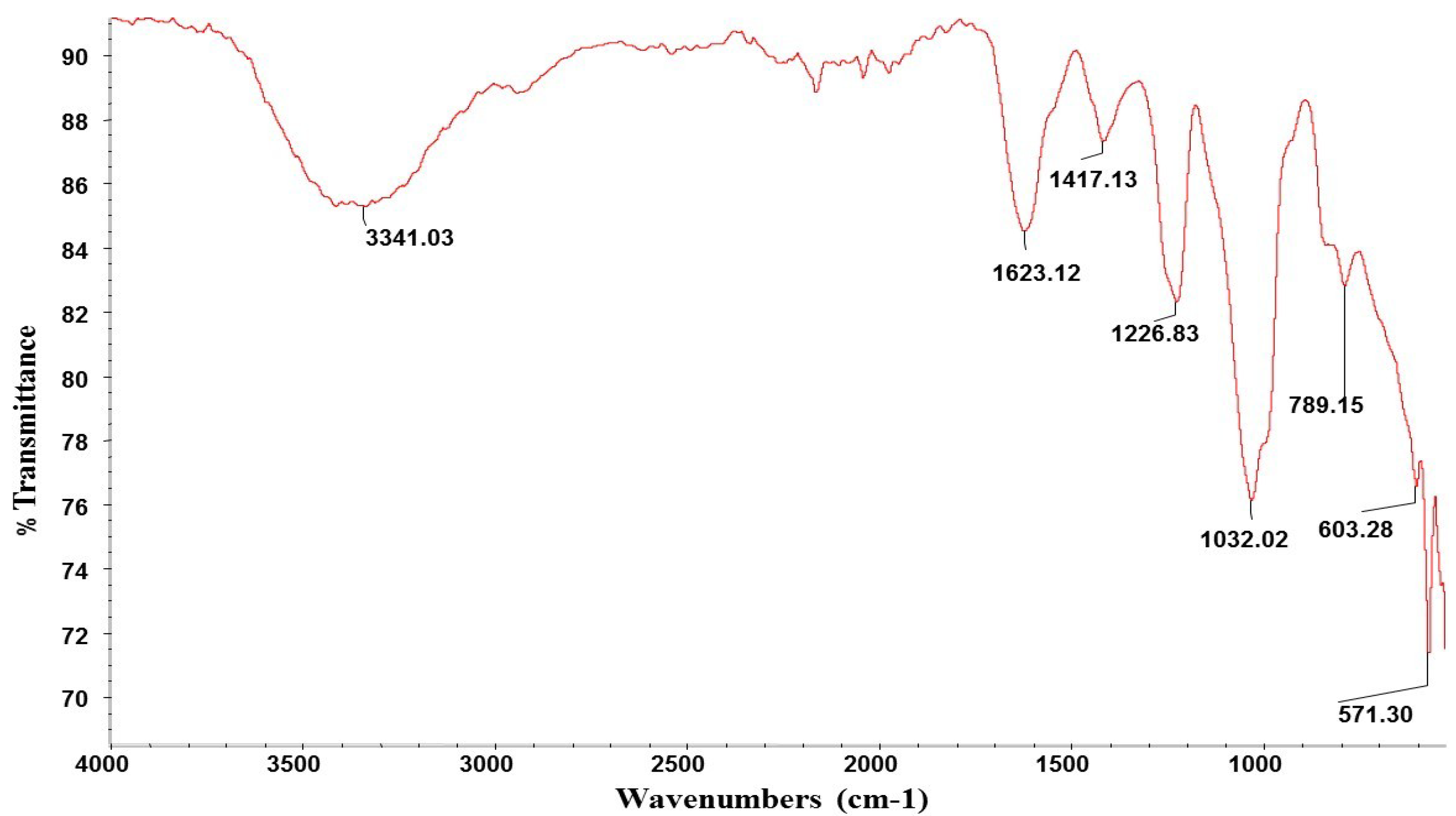
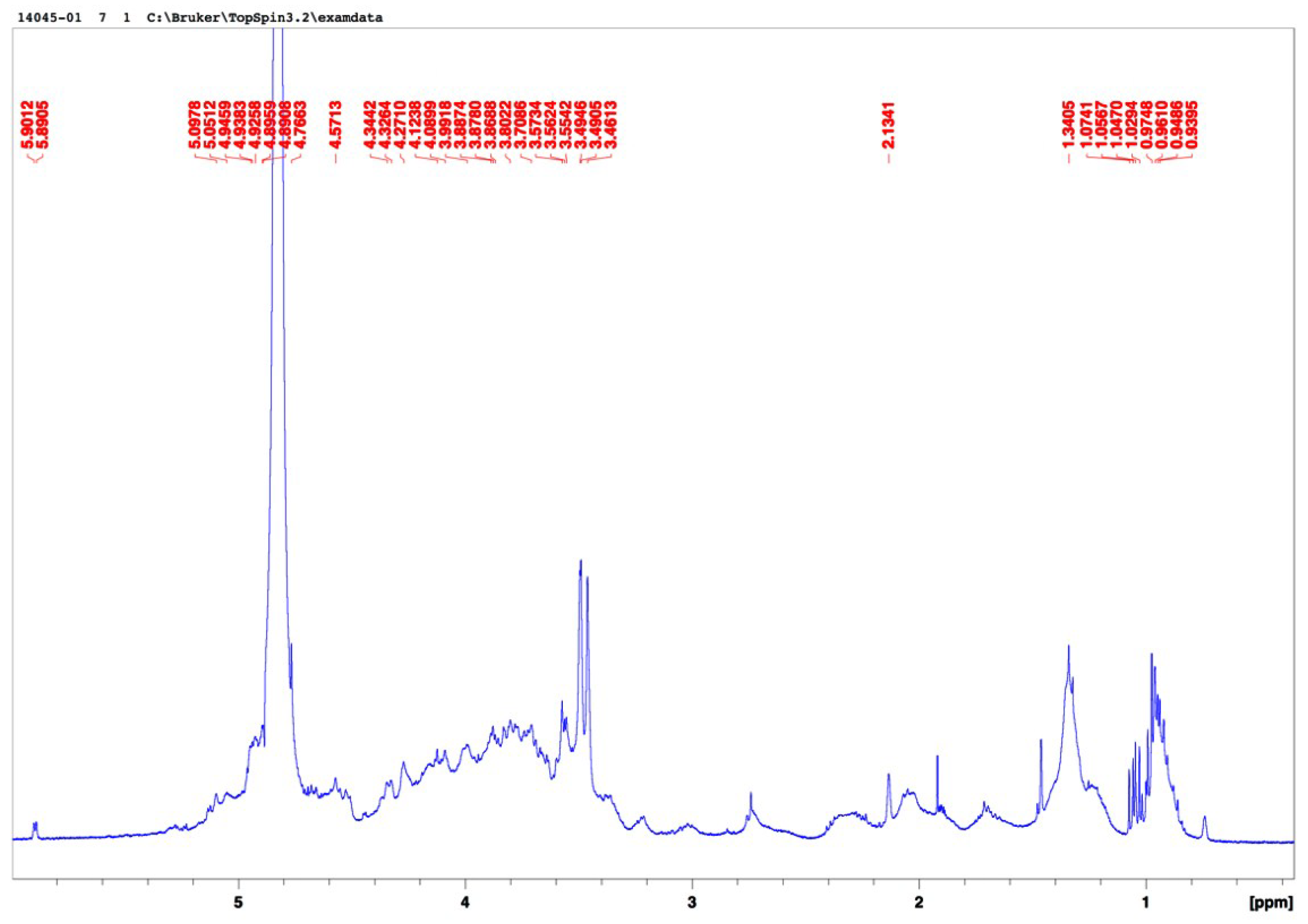
2.1. Composition of Ulva Clathrata Powder and Ulvan Extract
The composition of the starting Ulva clathrata powder was 22.4% ash, 15.3% protein, 9.5% sulphate, 11.4% rhamnose, 7.3% glucuronic acid, 3.0% xylose, 2.5% glucose and 1.4% galactose. The final composition of the ulvan extract was 19% ash, 10.0% protein, 11.0% sulphate, 21.7% rhamnose, 13.9% glucuronic acid, 6.1% glucose, 5.0% xylose, 3.6% iduronic acid, and 2.2% galactose. The molecular weight was 359,800 g·mol−1 equivalent pullulan. Ulva sp. powder is known to contain high amounts of good-quality protein, carbohydrate, vitamins and minerals [23]. Ulva clathrata chemical composition found in present study is on the line with that reported by Peña-Rodriguez et al. [15] for the same Ulva species. Ulvan composition has been widely studied [7,19] and showed to vary according to several factors including the period of collection, the ecophysiological growth conditions, the taxonomic origins and the post-collection treatment of the algal sources [7]. Extraction is conventionally achieved by using warm water (80–90 °C) containing ammonium oxalate as a divalent cation chelator, and the recovery of ulvan is generally obtained by precipitation in ethanol. As ulvan precipitation with ethanol is not specific, the extraction and purification are not completely effective in the removal of cellulose, hemicellulose, proteins and ash [24]. The final step for removing contaminants may vary considerably. Ulvan is mainly built on disaccharides repeating sequences composed of sulphated rhamnose and glucuronic acid, iduronic acid or xylose. Low proportions of galactose, glucose and protein are also generally found in ulvan [7,19,25]. The sugar composition of the purified U. clathrata ulvan obtained in present study is very similar to that given by Quemener et al. [25] for an U. lactuca ulvan that contained rhamnose 20.8%, glucuronic acid 16.0%, iduronic acid 3.7%, xylose 3.5%, glucose 2.8%, and galactose 0.7%; this Ulva lactuca ulvan also contained protein 11.6% and sulphate 21.2%. These values are congruent with those reported for ulvan from other origins [26,27,28]. The polysaccharide extracted from U. clathrata in present study was more sulphated than those reported for Ulva armoricana (10.3%–13.8%) and Ulva rotundata (9.2%–12.5%) [16,29] but similar to those obtained from Ulva conglobata (23.04%–35.2%) [14], and less sulphated than the Ulva clathrata ulvan reported by Hernandez-Garibay et al. [30]. Ulvan PS characterization was completed by FT-IR and H-NMR spectra: the FT-IR spectrum (Figure 1) is comparable with the previously published data for Ulva rigida and Ulva pertusa polysaccharides [28,31]. A characteristic band at 3341.03 cm-1 corresponding to -OH groups, which is consistent with the structure of disaccharides is observed. Two bands at 1226.83 cm−1 and 789 cm−1 are indicatives to the bounds C-O-S of sulfated groups. The band 1623 cm−1 is allocated to the vibration of C=O of the uronic acid; H-NMR spectrum (Figure 2) is comparable with data for ulvans [32]. All aromatic protons bound to -OH groups are in the region of 4.5 to 5.0. Protons attached to ether groups are allocated in the region of 3.4 to 3.6. Methyl protons of rhamnose are assigned in the region of 0.8 to 1.1.
Figure 1.
Infrared absorption spectrum of the native ulvan from Ulva clathrata.
Figure 2.
1H-NMR spectrum in D2O of the native ulvan from Ulva clathrata.
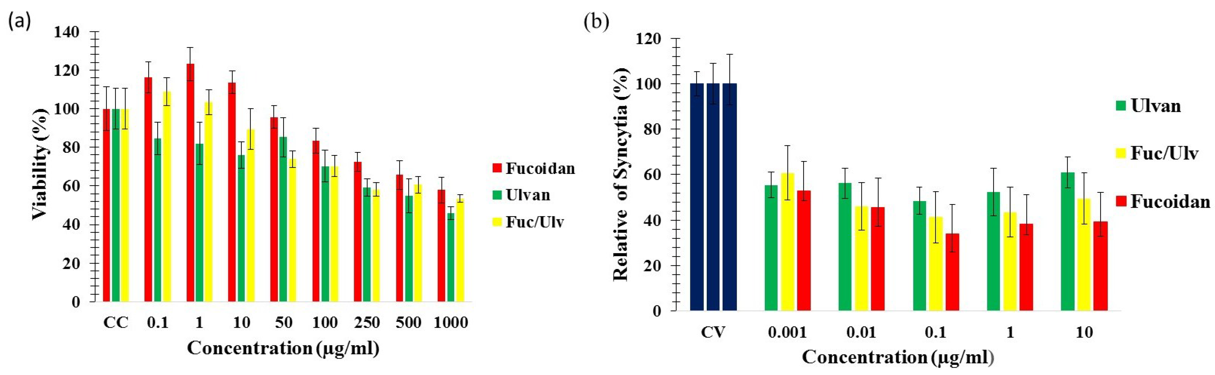
2.2. Cytotoxicity of the SP
To determine the cytotoxicity of SP and their mixture, MTT assays were performed. No significant cytotoxicity was detected for the SP at concentrations up to 100 μg/mL in Vero cells; the 50% cytotoxic concentrations (CC50) for fucoidan, ulvan and the mixture were of 1336 μg/mL, 810 μg/mL and 823 μg/mL, respectively (Figure 3a, Table 1). This is consistent with studies that have assessed the cytotoxicity of other SP in cell culture, showing lower toxicity than commercial, FDA-approved, antiviral drugs [3,8,33].
Figure 3.
Cytotoxicity and antiviral activity of sulphated polysaccharides (SP) against Newcastle Disease Virus (NDV). (a) Vero cells (6.5 × 10³ cells), seeded in 96-well plates, were treated with different concentrations of SP for 48 h. Cell viability was then determined in triplicates by the MTT assay and compared with that in untreated cells (100% viability); (b) Vero cells (6.7 × 10⁴ cells), seeded in 24-well plates, were infected with NDV and treated with different concentrations of SP during infection for 1 h, and the antiviral activity was determined in a syncytia reduction assay. The data shown are the mean ± standard deviation (SD) of triplicate cultures, and the experiment was repeated three times. CC, cellular control; CV, viral control.
Table 1.
Antiviral activity and cytotoxicity of sulphated polysaccharides.
| Polysaccharide | Vero Cell Line | ||
|---|---|---|---|
| CC50 (μg/mL) a | IC50 (μg/mL) b | SI c | |
| Fucoidan | 1136 | 0.01 | 113,633 |
| Ulvan | 810 | 0.1 | 8102.5 |
| Fuc/Ulv | 823 | 0.01 | 82,325 |
a Concentration of test compound (μg/mL) that reduced Vero cell viability by 50%. b Concentration of a test compound that reduced the number of NDV syncytia in Vero cells by 50%. c Values of selectivity index.
2.4. Virucidal Activity of Ulvan
Mendes et al. [10] demonstrated that algae extracts from Ulva fasciata have the possibility to inactivate virions. To analyse the possibility that U. clathrata ulvan acts directly on the virus particle, leading to infectivity inactivation, a virucidal assay against NDV virions was conducted. Ulvan did not exert virucidal effect against NDV virions at the tested concentrations, indicating that the inhibitory effect detected by the syncytia reduction assay was actually due to an interference with some step of the NDV replication cycle (Table 2). The lack of virucidal activity is in agreement with previous studies with the C. okamuranus fucoidan [3,33] and other pure algae SP that cannot induce significant virions inactivation [45,46,47].
Table 2.
Virucidal assay based in the presence or absence of the cytopathic effect of NDV.
| Time | Viral Titer | |||
|---|---|---|---|---|
| 1 × 10⁴ TCID50 | 1 × 10⁵ TCID50 | |||
| CC | ― | ― | ||
| CV | +++ | +++ | ||
| Ulvan concentrations | 10 μg/mL | 0 h | +++ | +++ |
| 10 μg/mL | 3 h | +++ | +++ | |
| 10 μg/mL | 6 h | +++ | +++ | |
| 100 μg/mL | 0 h | +++ | +++ | |
| 100 μg/mL | 3 h | +++ | +++ | |
| 100 μg/mL | 6 h | +++ | +++ | |
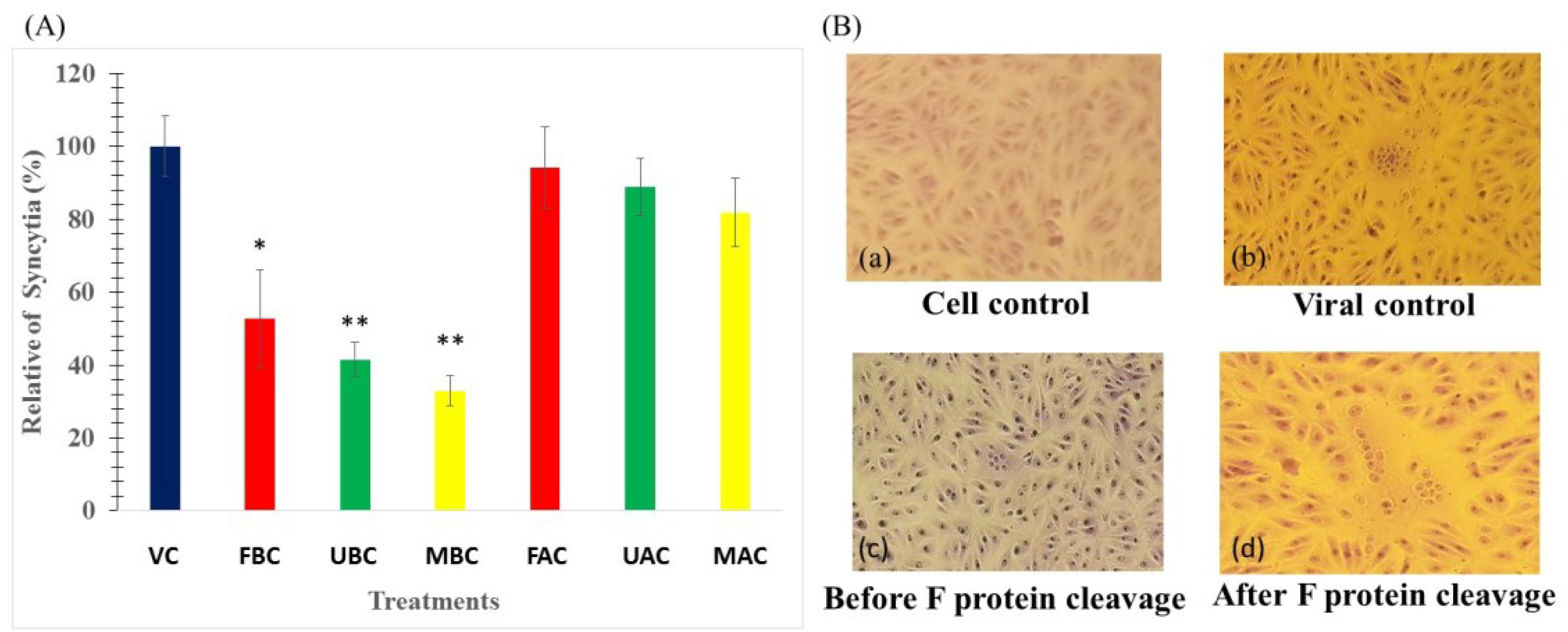
2.5. Ability of the SP to Block NDV-Induced Cell-Cell Fusion
To determine whether the compound inhibits the cell-cell spread of NDV, we performed cell fusion inhibition assays as described in the experimental section. The NDV envelope contains two proteins related to entry: the attachment protein HN and the fusion protein F. After one replication cycle, both glycoproteins are displayed in the cell envelope. Fusion of an infected cell with a non-infected occurs due to the recognition of sialic acid by HN and a conformational change of F protein, driving to syncytia formation, the characteristic cytopathic effect of NDV. Avirulent strains of NDV are characterized by their inability to form syncytia because the F0 protein cannot be cleaved in F1/F2. However, after trypsin digestion, syncytia formation was observed. Our results show that the SP and their mixture inhibited syncytia formation only if they were added before F protein cleavage (before trypsin digestion; Figure 5B). Before F protein cleavage, the mixture inhibited syncytia formation by 67% and reduced the syncytia size, compared with the findings in untreated viral-infected control cells, while fucoidan and ulvan inhibited syncytia formation by 47% and 59% respectively; therefore ulvan and the mixture apparently have a better anti-NDV cell-cell fusion spread than fucoidan alone. However, after F protein cleavage via trypsin digestion, the SP and their mixture lost the ability to inhibit syncytia formation (Figure 5A). This suggests that ulvan inhibits viral fusion by interacting with the intact F0 protein but not with the mature protein, as previously found with fucoidan [3], and that ulvan has the same mechanism of action as fucoidan.
Figure 5.
Inhibition of an avirulent NDV (La Sota) strain-mediated cell fusion. (A) Fusion inhibition assay with SP and their mixture treatment before, before cleavage [BC] and after F protein cleavage, after cleavage [AC]. Data are expressed as the percent of the number of syncytia compared with the findings in virus-infected control cells. VC, viral control; F, fucoidan; U, ulvan; M, mixture. The data shown are the mean ± SD of triplicate experiments. The asterisks indicate a significant difference between the treatment and viral control (* p < 0.05 and ** p < 0.001); (B) Vero cells infected with NDV and treated by trypsin digestion: (a) Uninfected control cells (100×); (b) Vero cells infected with NDV and treated with trypsin (100×); (c) Infected Vero cells were treated with 0.1 μg/mL of the SP and their mixture before F protein cleavage, at which point syncytia formation could be inhibited if SP were added (100×); (d) Infected Vero cells were treated with SP and their mixture after F protein cleavage, and the SP could not inhibit syncytia formation in this condition (100×).
Our results agree with other reports about algae SP being able to exert the ability to inhibit the cell-cell fusion [48]. Nyberg et al. [49] demonstrated that the yeast-derived SP PI-88 could inhibit the cell-cell spread fusion and consequent syncytia formation by HSV in GMK AH1 cells. Cagno et al. [50] reported the anti-cell-cell spread fusion of SP derived from Escherichia coli of RSV; they observed a considerable reduction of syncytia inHep-2 and A549 cells. Thus SP from different sources could be powerful inhibitors of viral cell-cell fusion.
3. Experimental Section
3.2. Cell Line and VIRUS
Green African monkey kidney (Vero) cells were grown in DMEM/FL-12 supplemented with 5% (v/v) fetal bovine serum and 1% (v/v) antibiotics. The flasks were maintained in a humidified atmosphere with 5% CO2 at 37 °C. The La Sota NDV strain was obtained from FORT DODGE® (Pfizer, New York, NY, USA) and tittered by fifty percent tissue culture infectious dose (TCID50) assay, according to their CPE effect as described by Reed and Muench [54].
3.3. Cytotoxicity Assay
To evaluate the cytotoxicity of the compounds MTT’s assays were performed. Vero cells were seeded in 96-well plates at an initial density of 7 × 10³ cells per well. The cells were incubated with increasing concentrations of the SP diluted in DMEM, for 48 h at 37 °C and 5% CO2. MTT (3-(4,5-dimethylthiazol-2-yl)-2,5-diphenyltetrazolium bromide; 0.5 mg/mL; 40 μL/well) was added to the cells, which were further incubated for 1 h 15 min. MTT was removed, and dimethylsulfoxide (DMSO; 100 μL/well) was added to dissolve the formazan crystals. The optical density of the cells was measured at 570 nm (Synergy™ HT multi-mode microplatereader; BIOTEK instruments Inc., Winooski, VT, USA). Each experiment was performed in triplicate, and experiments were repeated at least three times. The cytotoxicity was expressed as the CC50, which was the concentration of the test substances that inhibited the growth Vero cells by 50% compared with the growth of the untreated cells.
3.4. Virucidal Assay
The virucidal activity of ulvan against La Sota NDV was assessed using syncytia reduction assays with monolayers of Vero cells grown in 24-well plates. The assays where performed by adding the SP (0, 10, or 100 μg/mL) to an equal volume to NDV (1 × 10⁴ and 1 × 10⁵ TCID50). After 0, 3 and 6 h, the mixtures were added to Vero cells and further incubated 1 h at 37 °C. Thereafter, the mixtures were removed and DMEM with no serum and antibiotics was added. Because La Sota NDV is an avirulent strain, trypsin (0.001%; GIBCO) was added after 24 h to mediate syncytia formation after proteolytic cleavage of F0 protein and incubated for 30 min at 37 °C, afterwards trypsin was removed and DMEM was added. Monolayers were fixed with methanol:acetone (3:1) then incubated for 24 h at 37 °C and 5% CO2 and stained with 1% crystal violet in an incubator, after which syncytia presence or absence was evaluated.
3.6. Fusion Inhibition Assay
To determine if the SP has the possibility to inhibit cell-cell spread of NDV, Fusion inhibition assays were performed according to the method reported by Zhu et al. [55]. Vero cell monolayers in 24-well plates were infected with 1 × 105.5 TCID50 of La Sota NDV. After 12 h of infection, the monolayers were washed two times with PBS, digested by trypsin (0.001%; GIBCO) in DMEM at room temperature for 20 min, and then washed two times with PBS, the monolayers where incubated in DMEM without serum/antibiotics for 12 h. To detect SP ability to inhibit fusion and further cell-cell viral spread, they were added to the medium before or after trypsin digestion. Monolayers were fixed with 3:1 methanol:acetone and stained with 1% crystal violet.
3.7. Statistical Analysis
Data analysis was performed by PASW 18 software (PASW 18.0 (2009), Chicago, IL, USA). All variables were tested in triplicate for each experiment and experiments were repeated at least three times. The CC50 values were determined by Probit regression analysis. One- way ANOVA with Dunnet’s post hoc test was used for comparisons vs. viral control. Values were expressed as the mean ± standard deviation (SD). The results of the comparisons vs. viral control were considered significantly different if p < 0.05 or highly significantly different if p < 0.001.
4. Conclusions
Characterization by chemical analysis indicates that the structure of the obtained sulphated polysaccharide is similar to that of ulvan-rich extracts already reported in the literature.
The SP and their mixture inhibit NDV in vitro and they did not exhibit significant toxicity at effective concentrations. Ulvan exerted no direct inactivating effect on virions in virucidal assay. Based on our findings from syncytia reduction and cell-cell fusion inhibition assays, we support the concept that ulvan acts at the same stages of the infection as fucoidan. The polygonogram and the median-effect plot show that ulvan acts as an antagonist of fucoidan in their mixture at the syncytia reduction assay, probably because both act on the same target.
Our results indicate that ulvan inhibits syncytia formation only if it is added before F protein cleavage (before trypsin digestion). This suggests that ulvan inhibits viral fusion by interacting with the intact F0 protein but not with the mature F protein. Ulvan alone showed better anti cell-cell spread activity than fucoidan, and their mixture apparently exhibited more potent effect.
Understanding the mechanism of antiviral activity of brown and green algae SPs is important for their future development as antiviral drugs for clinical or veterinary use. Nevertheless, it would be very useful to search for natural compounds that act on different targets along the viral infection process (perhaps other compounds from seaweed) and in a synergy with ulvan or fucoidan, in such a way to obtain a more effective combination therapy, which could exhibit a viral suppression and lower resistance than a single compound.
Acknowledgments
This work was supported by Consejo Nacional de Ciencia y Tecnología (CONACYT) México (Project No. 99862).
Author Contributions
J.A.A., and L.M.T. designed the research. J.A.A., performed antiviral experimental work. J.A.A., and P.Z.B. performed cytotoxicity assays. L.E.C., J-F.S. and D.R. performed extraction and characterization of the SP. J.A.A., L.E.C., E.M., C.R., D.R. and L.M.T. participated in writing the paper.
Conflicts of Interest
The authors declare no conflict of interest.
References
- Gerber, P.; Dutcher, J.D.; Adams, E.V.; Sherman, J.H. Protective effect of seaweed extracts for chicken embryos infected with influenza B or mumps virus. Proc. Soc. Exp. Biol. Med. 1958, 99, 590–593. [Google Scholar] [CrossRef] [PubMed]
- Damonte, E.B.; Matulewicz, M.C.; Cerezo, A.S. Sulfated seaweed polysaccharides as antiviral agents. Curr. Med. Chem. 2004, 11, 2399–2419. [Google Scholar] [CrossRef] [PubMed]
- Elizondo-Gonzalez, R.; Cruz-Suarez, L.E.; Ricque-Marie, D.; Mendoza-Gamboa, E.; Rodriguez-padilla, C.; Trejo-Avila, L.M. In vitro characterization of the antiviral activity of fucoidan from Cladosiphon okamuranus against Newcastle Disease Virus. Virol. J. 2012, 9, 307. [Google Scholar] [CrossRef] [PubMed]
- Trejo-Avila, L.M.; Morales-Martínez, M.L.; Ricque-Marie, D.; Cruz-Suarez, L.E.; Zapata-Benavides, P.; Morán-Santibañez, K.; Rodríguez-Padilla, C. In vitro anti-canine distemper virus activity of fucoidan extracted from the brown alga Cladosiphon okamuranus. Virus Dis. 2014, 25, 474–480. [Google Scholar] [CrossRef]
- Patarra, R.F.; Paiva, L.; Neto, A.I.; Lima, E.; Baptista, J. Nutritional value of selected macroalgae. J. Appl. Phycol. 2011, 23, 205–208. [Google Scholar] [CrossRef]
- Lahaye, M.; Kaeffer, B. Seaweed dietary fibres: Structure, physico-chemical and biological properties relevant to intestinal physiology. Sci. Aliments 1997, 17, 563–584. [Google Scholar]
- Lahaye, M.; Robic, A. Structure and functional properties of ulvan, a polysaccharide from green seaweeds. Biomacromolecules 2007, 8, 1765–1774. [Google Scholar] [CrossRef] [PubMed]
- Alves, A.; Sousa, R.A.; Reis, R.L. In vitro cytotoxicity assessment of ulvan, a polysaccharide extracted from green algae. Phytother. Res. 2013, 27, 1143–1148. [Google Scholar]
- Qi, H.; Liu, X.; Zhang, J.; Duan, Y.; Wang, X.; Zhang, Q. Synthesis and antihyperlipidemic activity of acetylated derivative of ulvan from Ulva pertusa. Int. J. Biol. Macromol. 2012, 50, 270–272. [Google Scholar] [CrossRef] [PubMed]
- Mendes, G.S.; Soares, A.R.; Martins, F.O.; Albuquerque, M.C.; Costa, S.S.; Yoneshigue-Valentin, Y.; Gestinari, L.M.; Santos, N.; Romanos, M.T. Antiviral activity of green marine alga Ulva fasciata on the replication of human metapneumovirus. Rev. Inst. Med. Trop. Sao Paulo 2010, 52, 3–10. [Google Scholar] [CrossRef] [PubMed]
- Jaulneau, V.; Laffite, C.; Jacquet, C.; Fournier, S.; Salamagne, S.; Briand, X.; Esquerré- Tugayé, M.T.; Dumas, B. Ulvan, a sulfated polysaccharide from green algae, activates plant immunity through the jasmonic acid signaling pathway. J. Biomed. Biotechnol. 2010. [Google Scholar] [CrossRef]
- Tabarsa, M.; Han, J.H.; Kim, C.Y.; You, S.G. Molecular characteristics and immunomodulatory activities of water soluble sulfated polysaccharides from Ulva pertusa. J. Med. Food 2012, 15, 135–144. [Google Scholar] [CrossRef] [PubMed]
- Leiro, J.M.; Castro, C.; Arranz, J.A.; Lamas, J. Immunomodulating activities of acidic sulphated polysaccharides obtained from the seaweed Ulva rigida C. Agardh. Int. Immunopharmacol. 2007, 7, 879–888. [Google Scholar] [CrossRef] [PubMed]
- Mao, W.; Zang, X.; Li, Y.; Zhang, H. Sulfated polysaccharides from marine green algae Ulva conglobata and their anticoagulant activity. J. Appl. Phycol. 2006, 18, 9–14. [Google Scholar] [CrossRef]
- Peña-Rodríguez, A.; Mawhinney, T.P.; Ricque-Marie, D.; Cruz-Suárez, L.E. Chemical composition of cultivated seaweed Ulva clathrata (Roth) C. Agardh. Food Chem. 2011, 129, 491–498. [Google Scholar] [CrossRef]
- Robic, A.; Sassi, J-F.; Lahaye, M. Impact of stabilization treatments of the green seaweed Ulva rotundata (Chlorophyta) on the extraction yield, the physico-chemical and rheological properties of ulvan. Carbohydr. Polym. 2008, 74, 344–352. [Google Scholar] [CrossRef]
- Tabatabai, M.A. Determination of sulfate in water samples. Sulphur. Inst. 1974, 10, 11–13. [Google Scholar]
- Quemener, B.; Marot, C.; Mouillet, L.; da Riz, V.; Diris, J. Quantitative analysis of hydrocolloids in food systems by methanolysis coupled to reverse HPLC. Part 1. Gelling carrageenans. Food Hydrocoll. 2000, 14, 9–17. [Google Scholar] [CrossRef]
- Robic, A.; Rondeau-Mouro, C.; Sassi, J.-F.; Lerat, Y.; Lahaye, M. Structure and interactions of ulvan in the cell wall of the marine green algae Ulva rotundata (Ulvales, Chlorophyceae). Carbohydr. Polym. 2009, 77, 206–216. [Google Scholar] [CrossRef]
- Miller, P.J.; Decanini, E.L.; Afonso, C.L. Newcastle disease: Evolution of genotypes and the related diagnostic challenges. Infect. Genet. Evol. 2010, 10, 26–35. [Google Scholar] [CrossRef] [PubMed]
- Mayo, M.A. Virus taxonomy-Houston 2002. Arch. Virol. 2002, 147, 1071–1076. [Google Scholar] [CrossRef] [PubMed]
- Lamb, R.A.; Parks, G.D. Paramyxoviridae: The viruses and their replication. In Fields Virology, 5th ed.; Knipe, D.M., Howley, P.M., Eds.; Lippincott WIlliams & Wilkins, Wolters Kluwer: Philadelphia, PA, USA, 2007; Volume 1, pp. 1449–1496. [Google Scholar]
- Taboada, C.; Millán, R.; Miguez, I. Composition, nutritional aspects and effect on serum parameters of marine algae Ulva rigida. J. Sci. Food Agric. 2010, 90, 445–449. [Google Scholar] [CrossRef] [PubMed]
- Yaich, H.; Garna, H.; Besbes, S.; Paquot, M.; Blecker, C.; Attia, H. Effect of extraction conditions on the yield and purity of ulvan extracted from Ulva lactuca. Food Hydrocoll. 2013, 31, 375–382. [Google Scholar] [CrossRef]
- Quemener, B.; Lahaye, M. Sugar determination in ulvans by a chemical-enzymatic method coupled to high performance anion exchange chromatography. J. Appl. Phycol. 1997, 9, 179–188. [Google Scholar] [CrossRef]
- DeReviers, B.; Leproux, A. Characterization of polysaccharides from Enteromorpha intestinalis (L.) Link. Chlorophyta. Carbohydr. Polym. 1993, 22, 253–259. [Google Scholar] [CrossRef]
- Percival, E.; McDowell, R.H. Chemistry and Enzymology of Marine Algal Polysaccharides; Academic Press: London, UK, 1967; p. 219. [Google Scholar]
- Ray, B.; Lahaye, M. Cell-wall polysaccharides from the marine green alga Ulva “rigida” (Ulvales Chlorophyta)-Chemical structure of ulvan. Carbohydr. Res. 1995, 274, 313–318. [Google Scholar] [CrossRef]
- Robic, A.; Bertrand, D.; Sassi, J.F.; Lerat, Y.; Lahaye, M. Determination of the chemical composition of ulvan, a cell wall polysaccharide from Ulva spp. (Ulvales, Chlorophyta) by FT-IR and chemometrics. J. Appl. Phycol. 2009, 21, 451–456. [Google Scholar] [CrossRef]
- Hernández-Garibay, E.; Zertuche-González, J.A.; Pacheco-Ruíz, I. Isolation and chemical characterization of algal polysaccharides from the green seaweed Ulva clathrata (Roth) C. Agardh. J. Appl. Phycol. 2010, 23, 537–542. [Google Scholar] [CrossRef]
- Pengzhan, Y.; Quanbin, Z.; Ning, L.; Zuhong, X.; Yanmei, W.; Zhi’en, L. Polysaccharides from Ulva pertusa (Chlorophyta) and preliminary studies on their antihyperlipidemia activity. J. Appl. Phycol. 2003, 15, 21–27. [Google Scholar] [CrossRef]
- Chiellini, F.; Morelli, A. Ulvan: A Versatile Platform of Biomaterials from Renewable Resources. In Biomaterials—Physics and Chemistry; Pignatello, R., Ed.; InTech: Rijeka, Croatia, 2011; pp. 75–98. [Google Scholar]
- Song, X.; Yin, Z.; Zhao, X.; Cheng, A.; Jia, R.; Yuan, G.; Xu, J.; Fan, Q.; Dai, S.; Lu, H.; et al. Antiviral activity of sulfated Chuanmingshen violaceum polysacchare against Newcastle disease virus. J. Gen. Virol. 2013, 94, 2164–2174. [Google Scholar] [CrossRef] [PubMed]
- Ivanova, V.; Rouseva, R.; Kolarova, M.; Serkedjieva, J.; Rachev, R.; Manolova, N. Isolation of a polysaccharide with antiviral effect from Ulva lactuca. Prep. Biochem. 1994, 24, 83–97. [Google Scholar] [PubMed]
- Chiu, Y.H.; Chan, Y.L.; Li, T.L.; Wu, C.J. Inhibition of Japanese encephalitis virus infection by the sulfated polysaccharide extracts from Ulva lactuca. Mar. Biotechnol. (N.Y.) 2012, 14, 468–478. [Google Scholar] [CrossRef]
- Lee, E.; Pavy, M.; Young, N.; Freeman, C.; Lobigs, M. Antiviral effect of the heparan sulfate mimetic, PI-88, against dengue and encephalitic flaviviruses. Antivir. Res. 2006, 69, 31–38. [Google Scholar] [CrossRef] [PubMed]
- Hayashi, T.; Hayashi, K.; Kanekiyo, K.; Ohta, Y.; Lee, J.B. Promising antiviral Glyco-molecules from an edible alga. In Combating the Threat of Pandemic Influenza: Drug Discovery Approaches; Torrence, P.F., Ed.; John Wiley & Sons: Hoboken, NJ, USA, 2007; pp. 166–182. [Google Scholar]
- Vlieghe, P.; Clerc, T.; Pannecouque, C.; Witvrouw, M.; de Clercq, E.; Salles, J.P.; Kraus, J.L. Synthesis of new covalently bound kappa-carrageenan-AZT conjugates with improved anti-HIV activities. J. Med. Chem. 2002, 45, 1275–1283. [Google Scholar] [CrossRef] [PubMed]
- Takebe, Y.; Saucedo, C.J.; Lund, G.; Uenishi, R.; Hase, S.; Tsuchiura, T.; Kneteman, N.; Ramessar, K.; Tyrrell, D.L.; Shirakura, M.; et al. Antiviral lectins from red and blue-green algae show potent in vitro and in vivo activity against hepatitis C virus. PLoS One 2013, 8, e64449. [Google Scholar] [CrossRef] [PubMed]
- Chou, T.C. Theoretical basis, experimental design, and computerized simulation of synergism and antagonism in drug combination studies. Pharmacol. Rev. 2006, 58, 621–681. [Google Scholar] [CrossRef] [PubMed]
- Chou, T.C.; Talalay, P. Quantitative analysis of dose-effect relationships: The combined effects of multiple drugs or enzyme inhibitors. Adv. Enzyme Regul. 1984, 22, 27–55. [Google Scholar] [CrossRef] [PubMed]
- Feng, J.Y.; Ly, J.K.; Myrick, F.; Goodman, D.; White, K.L.; Svarovskaia, E.S.; Borroto-Esoda, K.; Miller, M.D. The triple combination of tenofovir, emtricitabine and efavirenz shows synergistic anti-HIV-1 activity in vitro: A mechanism of action study. Retrovirology 2009, 6, 44. [Google Scholar] [CrossRef] [PubMed]
- Klein, M.B.; Campeol, N.; Lalonde, R.G.; Brenner, B.; Wainberg, M.A. Didanosine, interferon-alfa and ribavirin: A highly synergistic combination with potential activity against HIV-1 and hepatitis C virus. AIDS 2003, 17, 1001–1008. [Google Scholar] [CrossRef] [PubMed]
- Yang, Z.H.; Crouch, J.Y.; Chou, T.C.; Hsiung, G.D. Combined antiviral effects of paired nucleosides against guinea pig cytomegalovirus replication in vitro. J. Antivir. Res. 1990, 14, 249–266. [Google Scholar] [CrossRef]
- Damonte, E.B.; Matulewicz, M.C.; Cerezo, A.S.; Coto, C. Herpes simplex virus inhibitory sulfated xylogalactans from the red seaweed Nothogenia fastigiata. Chemotherapy 1996, 42, 57–64. [Google Scholar] [CrossRef] [PubMed]
- Mandal, P.; Pujol, C.A.; Carlucci, M.J.; Chattopadhyay, K.; Damonte, E.B.; Ray, B. Anti-herpetic activity of a sulfated xylomannan from Scinaia hatei. Phytochemistry 2008, 69, 2193–2199. [Google Scholar] [CrossRef] [PubMed]
- Bouhlal, R.; Haslin, C.; Chermann, J.-C.; Colliec-Jouault, S.; Sinquin, C.; Simon, G.; Cerantola, S.; Riadi, H.; Bourgougnon, N. Antiviral activities of sulfated polysaccharides isolated from Sphaerococcus coronopifolius (Rhodophytha, Gigartinales) and Boergeseniella thuyoides (Rhodophyta, Ceramiales). Mar. Drugs 2011, 9, 1187–1209. [Google Scholar] [CrossRef] [PubMed]
- Paskaleva, E.E.; Lin, X.; Li, W.; Cotter, R.; Klein, M.T.; Roberge, E.; Yu, E.K.; Clark, B.; Veille, J.C.; Liu, Y.; et al. Inhibition of highly productive HIV-1 infection in T cells, primary human macrophages, microglia, and astrocytes by Sargassum fusiforme. AIDS Res. Ther. 2006, 3, 15. [Google Scholar] [CrossRef] [PubMed]
- Nyberg, K.; Ekblad, M.; Bergström, T.; Freeman, C.; Parish, C.R.; Ferro, V.; Trybala, E. The low molecular weight heparan sulfate-mimetic, PI-88, inhibits cell-to-cell spread of herpes simplex virus. Antivir. Res. 2004, 63, 15–24. [Google Scholar] [CrossRef] [PubMed]
- Cagno, V.; Donalisio, M.; Civra, A.; Volante, M.; Veccelli, E.; Oreste, P.; Rusnati, M.; Lembo, D. Highly sulfated K5 Escherichia coli polysaccharide derivatives inhibit respiratory syncytial virus infectivity in cell lines and human tracheal-bronchial histocultures. Antimicrob. Agents Chemother. 2014, 58, 4782–4794. [Google Scholar] [CrossRef] [PubMed]
- Tako, M.; Yoza, E.; Thoma, S. Chemical characterization of acetyl fucoidan and alginate from commercially cultured Cladosiphon okamuranus. Bot. Mar. 2000, 43, 393–398. [Google Scholar] [CrossRef]
- Moll, B. Aquatic Surface Barriers and Methods for Culturing Seaweed; World Intelectual Property Organization: Davis, CA, USA, 2004. [Google Scholar]
- AOAC. Method 984.13. In Official Methods of Analysis, 16th ed., 3rd revision; Cunniff, P., Ed.; AOAC International (formerly The Association of Official Analytical Chemists): Gaithersburg, MD, USA, 1997; p. 1033. [Google Scholar]
- Reed, L.J.; Muench, H. A simple method of estimating fifty percent endpoints. Am. J. Hyg. 1938, 27, 493–497. [Google Scholar]
- Zhu, J.; Jiang, X.; Liu, Y.; Tien, P.; Gao, G.F. Design and characterization of viral polypeptide inhibitors targeting Newcastle disease virus fusion. J. Mol. Biol. 2005, 354, 601–613. [Google Scholar] [CrossRef] [PubMed]
© 2015 by the authors; licensee MDPI, Basel, Switzerland. This article is an open access article distributed under the terms and conditions of the Creative Commons Attribution license (http://creativecommons.org/licenses/by/4.0/).