New Marker of Brain–Heart Interaction: Tpeak–Tend Interval

 , , , , , and
Abstract
1. Introduction
2. Materials and Methods
2.1. Study Population
2.2. Study Protocol
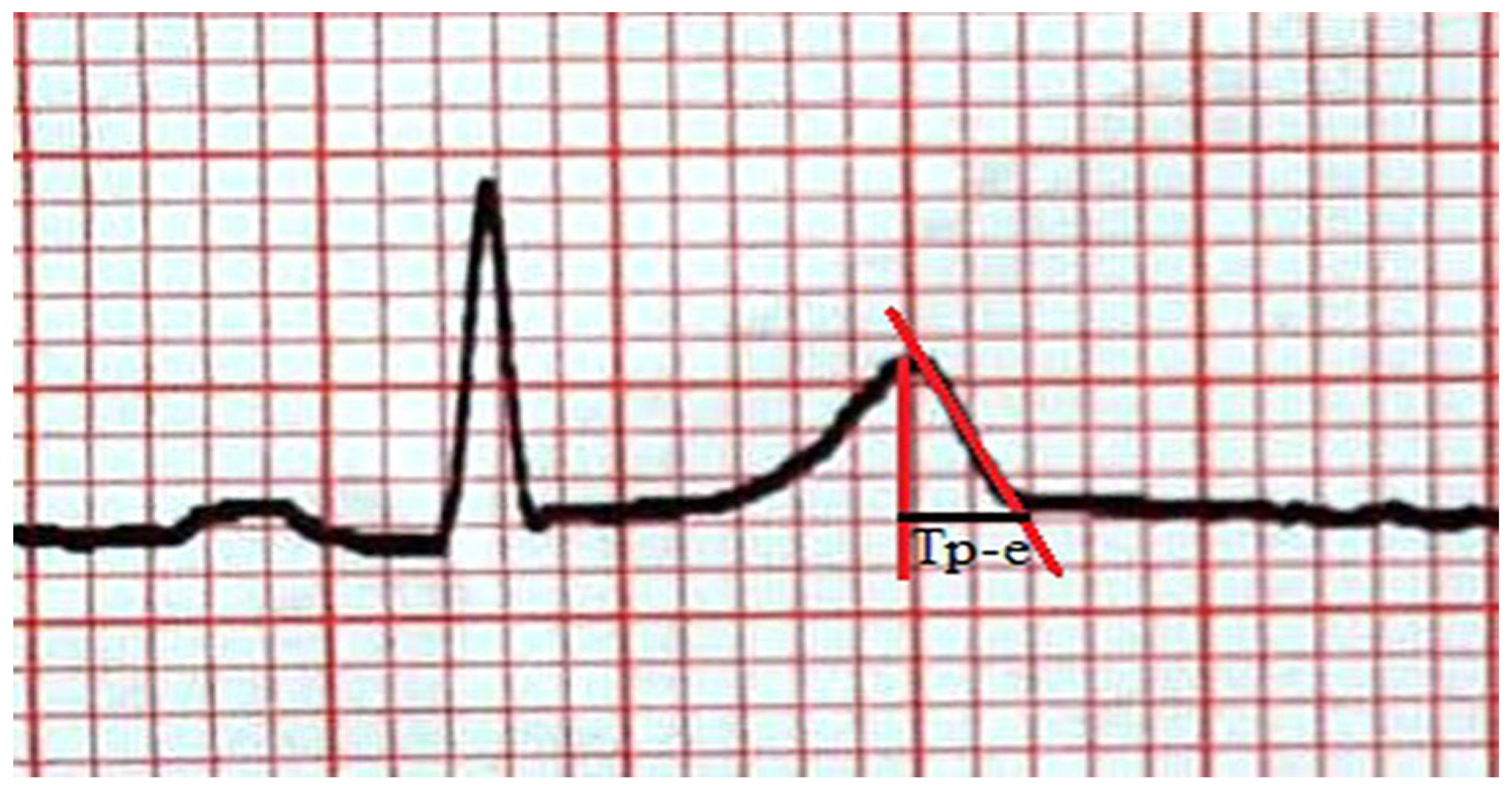
2.3. ECG Measurements
2.4. Statistical Analysis
3. Results
4. Discussion
Limitations
5. Conclusions
Author Contributions
Funding
Institutional Review Board Statement
Informed Consent Statement
Data Availability Statement
Conflicts of Interest
References
- Kim, M.S.; Kim, J.J. Heartand Brain Interconnection–Clinical Implications of Changes in Brain Function During Heart Failure. Circ. J. 2015, 79, 942–947. [Google Scholar] [CrossRef]
- Kenmogne-Domning, G.H.; Kamtchum-Tatuene, J. Cardiac complications after stroke: Protocol for a systematic review and meta-analysis. BMJ Open 2018, 8, e021416. [Google Scholar] [CrossRef]
- Prosser, J.; MacGregor, L. Predictors of early cardiac morbidity and mortality after ischemic stroke. Stroke 2007, 38, 2295–2302. [Google Scholar] [CrossRef]
- Battaglini, D.; Robba, C. Brain–heart interaction after acute ischemic Stroke. Crit. Care 2020, 24, 163. [Google Scholar] [CrossRef]
- Yan, G.X.; Wu, Y. Phase 2 early after depolarization as a trigger of polymorphic ventricular tachycardia in acquired long-QT syndrome: Direct evidence from intracellular recordings in the intact left ventricular wall. Circulation 2001, 103, 2851–2856. [Google Scholar] [CrossRef]
- Tse, G.; Gong, M. The Tpeak–Tend interval as an electrocardiographic risk marker of arrhythmic and mortality outcomes: A systematic review and meta-analysis. Heart Rhythm 2017, 14, 1131–1137. [Google Scholar] [CrossRef] [PubMed]
- Odacı, E.; Bahadır, A. Cavalieri prensibi kullanılarak bilgisayarlı tomografi ve manyetik rezonans görüntüleri üzerinden hacim hesaplanması ve klinik kullanımı. Turk. Klin. J. Med. Sci. 2005, 25, 421–428. [Google Scholar]
- Satran, D.; Henry, C.R. Cardiovascular manifestations of moderate to severe carbonmonoxide poisoning. J. Am. Coll. Cardiol. 2005, 45, 1513–1516. [Google Scholar] [CrossRef] [PubMed]
- Hlaing, T.; Guo, D. The QT and Tp-e intervals in left and right chest leads: Comparison between patients with systemic and pulmonary hypertension. J. Electrocardiol. 2005, 38, 154–158. [Google Scholar] [CrossRef]
- Gunnoo, T.; Hasan, N. Quantifying the risk of heart disease following acute ischaemic stroke: A meta-analysis of over 50,000 participants. BMJ Open 2016, 6, e009535. [Google Scholar] [CrossRef]
- Xu, C.; Zheng, A. Brain–Heart Axis and Biomarkers of Cardiac Damage and Dysfunction after Stroke: A Systematic Review and Meta-Analysis. Int. J. Mol. Sci. 2020, 21, 2347. [Google Scholar] [CrossRef]
- Sörös, P.; Hachinsk, V. Cardiovascular and neurological causes of sudden death after ischaemic stroke. Lancet Neurol. 2012, 11, 179–188. [Google Scholar] [CrossRef]
- Li, Y.; Fitzgibbons, T.P. Left ventricular ejection fraction and clinically defined heart failure to predict 90-day functional outcome after ischemic stroke. J. Stroke Cerebrovasc. Dis. 2019, 28, 371–380. [Google Scholar] [CrossRef]
- Danese, A.; Cappellari, M. The dispersion of myocardial repolarization in ischemic stroke and intracranial hemorrhage. J. Electrocardiol. 2018, 51, 691–695. [Google Scholar] [CrossRef]
- Micieli, G.; Cavallini, A. The autonomic nervous system and ischemic stroke: A reciprocal interdependence. Clin. Auton. Res. 2008, 18, 308–317. [Google Scholar] [CrossRef]
- Christensen, H.; FoghChristensen, A. Abnormalities on ECG and telemetry predict stroke outcome at 3 months. J. Neurol. Sci. 2005, 234, 99–103. [Google Scholar] [CrossRef]
- Sugimoto, K.; Inamasu, J. The role of norepinephrine and estradiol in the pathogenesis of cardiac wall motion abnormality associated with subarachnoid hemorrhage. Stroke 2012, 43, 1897–1903. [Google Scholar] [CrossRef]
- Sugimoto, K.; Inamasu, J. Association between elevated plasma norepinephrine levels and cardiac wall motion abnormality in poor-grade subarachnoid hemorrhage patients. Neurosurg. Rev. 2013, 36, 259–266. [Google Scholar] [CrossRef] [PubMed]
- Samuels, M.A. The brain–heart connection. Circulation 2007, 116, 77–84. [Google Scholar] [CrossRef] [PubMed]
- Oppenheimer, S.M.; Kedem, G. Left-insular cortex lesions perturbcardiac autonomic tone in humans. Clin. Auton. Res. 1996, 6, 131–140. [Google Scholar] [CrossRef] [PubMed]
- Koppikar, S.; Baranchuk, A. Stroke and ventricular arrhythmias. Int. J. Cardiol. 2013, 168, 653–659. [Google Scholar] [CrossRef] [PubMed]
- Taggart, P. Brain-heart interactions and cardiac ventricular arrhythmias. Neth. Heart J. 2013, 21, 78–81. [Google Scholar] [CrossRef] [PubMed]
- Stead, L.G.; Gilmore, R.M. Prolonged QTc as a predictor of mortality in acute ischemic stroke. J. Stroke Cerebrovasc. Dis. 2019, 18, 469–474. [Google Scholar] [CrossRef] [PubMed]


| Age, years, median (IQR) | 72.3 (10.5) |
| Sex, n (%) | |
| Male | 49 (45.4) |
| GCS, median (IQR) | 14 (6) |
| Stroke type, n (%) | |
| Infarct | 89 (82.4) |
| Hemorrhage | 19 (17.6) |
| CAD, n (%) | 22 (20.4) |
| DM, n (%) | 46 (42.6) |
| Hypertension, n (%) | 79 (73.1) |
| CRF, n (%) | 7 (6.5) |
| Lesion location, n (%) | |
| Right hemisphere, n (%) | 60 (55.6) |
| Insular cortex involvement, n (%) | 50 (46.3) |
| Thalamus involvement, n (%) | 19 (17.6) |
| Supratentorial location, n (%) | 81 (75) |
| Volume, cm3, median (IQR) | 12.4 (46.4) |
| Systolic blood pressure, mmHg, median (IQR) | 155.7 (32.1) |
| Diastolic blood pressure, mmHg, median (IQR) | 85.9 (15.3) |
| Pulse/min median (IQR) | 81.9 (22) |
| Troponin, median (IQR) | 0.04 (0.06) |
| Ejection fraction, median (IQR) | 54.6 (11.05) |
| ST elevation, n (%) | 39 (36.1) |
| ST depression, n (%) | 20 (18.5) |
| T inversion, n (%) | 34 (31.5) |
| QTV2, ms, median (IQR) | 343 (41) |
| QTc V2, ms, median (IQR) | 382 (44) |
| Tp-eV2, ms median (IQR) | 92 (16) |
| QTV5, ms, median (IQR) | 354 (45) |
| QTcV5, ms, median (IQR) | 393 (43) |
| Tp-eV5, ms, median (IQR) | 92 (17) |
| Pulse-24.h/min, median (IQR) | 84 (21) |
| ST elevation-24 h, median (IQR) | 18 (16.7) |
| ST depression-24 h, median (IQR) | 10 (9.3) |
| T inversion-24 h, median (IQR) | 20 (18.5) |
| QTV2-24 h, ms, median (IQR) | 345 (44) |
| QTc V2-24 h, ms, median (IQR) | 388 (37) |
| Tp-eV2-24 h, ms, median (IQR) | 84 (16) |
| QTV5-24 h, ms, median (IQR) | 349 (46) |
| QTcV5-24 h, ms, median (IQR) | 390 (32) |
| Tp-eV5-24 h, ms, median (IQR) | 81 (18) |
| In-hospital mortality, n (%) | 37 (34.3) |
| Unadjusted OR (95% CI) | p Value | Adjusted OR (95% CI) | p Value | |
|---|---|---|---|---|
| SBP | 0.99 (0.98–1.000) | 0.34 | ||
| DBP | 0.96 (0.93–0.99) | 0.02 | 0.96 (0.93–1.000) | 0.05 |
| Pulse/min | 1.00 (0.98–1.02) | 0.47 | ||
| CAD | 1.3 (0.51–3.5) | 0.53 | ||
| Troponin | 0.73 (0.46–1.14) | 0.17 | ||
| Ejection fraction | 1.03 (0.98–1.38) | 0.23 | ||
| ST elevation | 0.68 (0.29–1.61) | 0.38 | ||
| ST depression | 1.4 (0.51–3.9) | 0.49 | ||
| T inversion | 1.07 (0.45–2.5) | 0.87 | ||
| QTV2 | 0.99 (0.97–1.002) | 0.09 | ||
| QTcV2 | 0.99 (0.97–1.004) | 0.24 | ||
| Tp-eV2 | 0.96 (0.93–0.99) | 0.008 | 0.96 (0.93–0.99) | 0.008 |
| QTV5 | 0.99 (0.98–1.004) | 0.25 | ||
| QTcV | 0.99 (0.97–1.005) | 0.41 | ||
| Tp-eV5 | 0.98 (0.96–1.005) | 0.12 |
Disclaimer/Publisher’s Note: The statements, opinions and data contained in all publications are solely those of the individual author(s) and contributor(s) and not of MDPI and/or the editor(s). MDPI and/or the editor(s) disclaim responsibility for any injury to people or property resulting from any ideas, methods, instructions or products referred to in the content. |
© 2026 by the authors. Published by MDPI on behalf of the Lithuanian University of Health Sciences. Licensee MDPI, Basel, Switzerland. This article is an open access article distributed under the terms and conditions of the Creative Commons Attribution (CC BY) license.
Share and Cite
Akilli, N.B.; Mutlu, H.; Dundar, Z.D.; Ozberk, O.; Koylu, R.; Gunaydın, Y.K.; Cander, B. New Marker of Brain–Heart Interaction: Tpeak–Tend Interval. Medicina 2026, 62, 695. https://doi.org/10.3390/medicina62040695
Akilli NB, Mutlu H, Dundar ZD, Ozberk O, Koylu R, Gunaydın YK, Cander B. New Marker of Brain–Heart Interaction: Tpeak–Tend Interval. Medicina. 2026; 62(4):695. https://doi.org/10.3390/medicina62040695
Chicago/Turabian StyleAkilli, Nazire Belgin, Huseyin Mutlu, Zerrin Defne Dundar, Omer Ozberk, Ramazan Koylu, Yahya Kemal Gunaydın, and Basar Cander. 2026. "New Marker of Brain–Heart Interaction: Tpeak–Tend Interval" Medicina 62, no. 4: 695. https://doi.org/10.3390/medicina62040695
APA StyleAkilli, N. B., Mutlu, H., Dundar, Z. D., Ozberk, O., Koylu, R., Gunaydın, Y. K., & Cander, B. (2026). New Marker of Brain–Heart Interaction: Tpeak–Tend Interval. Medicina, 62(4), 695. https://doi.org/10.3390/medicina62040695