Quantitative and Qualitative MRI Assessment of Perivascular Spaces in Parkinson’s Disease Patients †
 , ,
, ,  , , , ,
Abstract
1. Introduction
1.1. Perivascular Spaces in Neurodegeneration
1.2. Perivascular Spaces as Cognitive Biomarkers in Parkinson’s Disease
2. Materials and Methods
2.1. Participants
2.2. MRI Acquisition
2.3. Quantification of Perivascular Spaces
- grade 0 indicates no visible PVS,
- grade 1 corresponds to less than 10,
- grade 2 to 11–20,
- grade 3 to 21–40, and
- grade 4 to more than 40 PVS.
- grade 1 (<2 mm),
- grade 2 (2–3 mm),
- grade 3 (>3 mm).
2.4. Cognitive Assessment
2.5. Statistical Analysis
3. Results
3.1. Demographic Characteristics
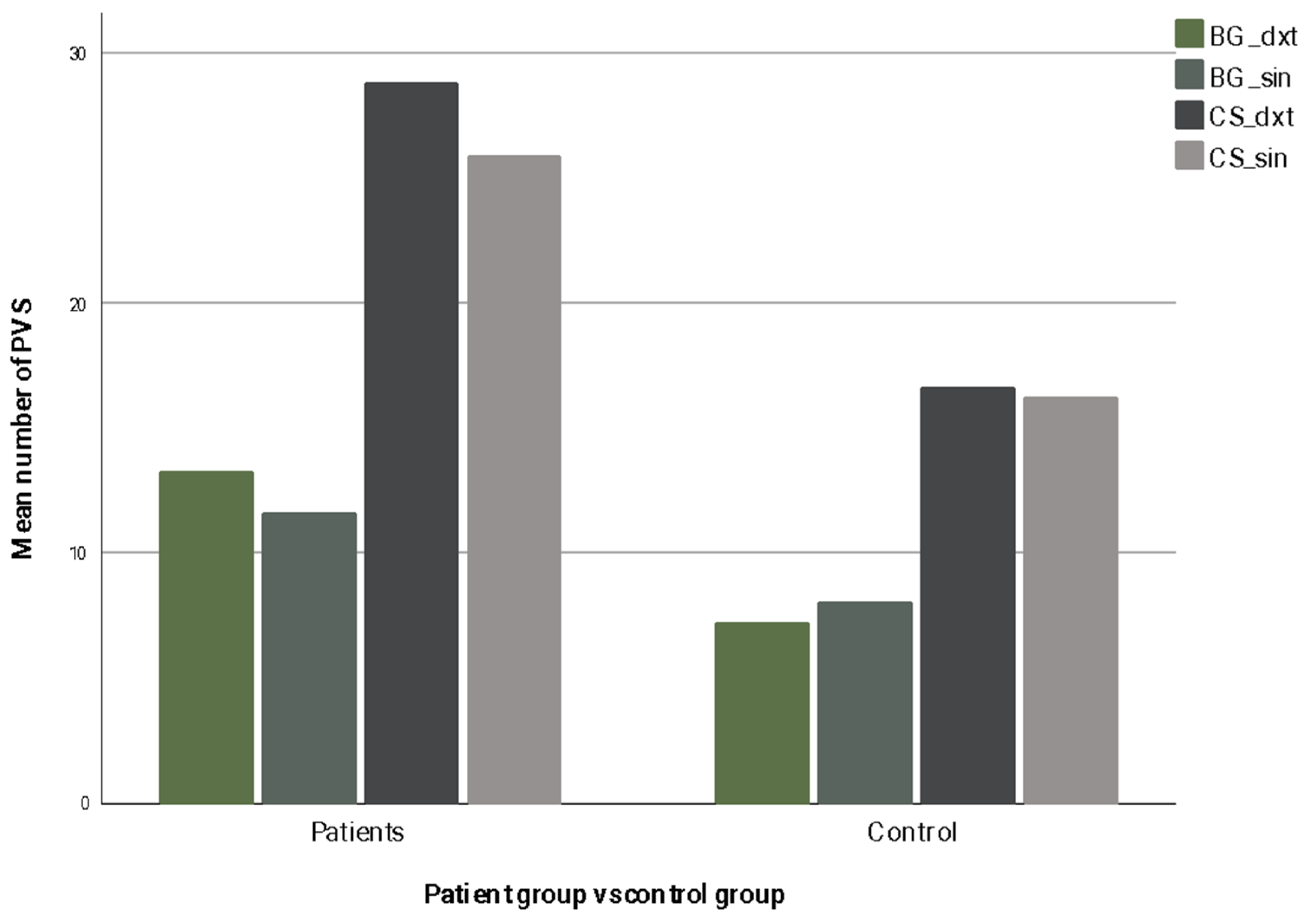
3.2. Quantitative PVS Measures
3.3. Visual Rating Scales
3.4. Largest-per-Slice PVS Counts
3.5. Perivascular Spaces Diameters
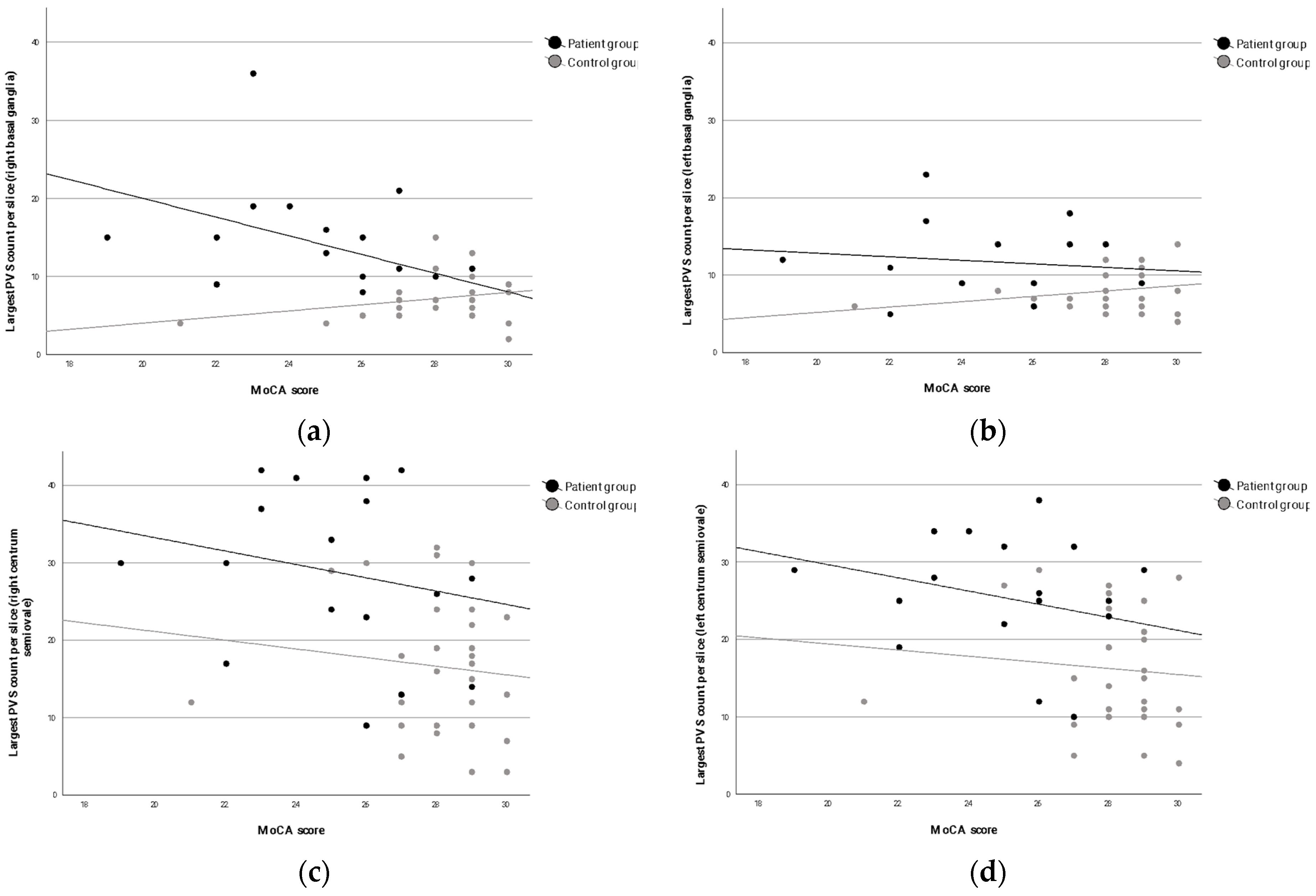
3.6. Associations with Cognitive Performance
4. Discussion
4.1. Main Findings
4.2. Interpretation and Comparison with the Literature
4.3. Strengths and Limitations
4.4. Clinical Implications
4.5. Future Directions
5. Conclusions
Supplementary Materials
Author Contributions
Funding
Institutional Review Board Statement
Informed Consent Statement
Data Availability Statement
Acknowledgments
Conflicts of Interest
References
- Brown, R.; Benveniste, H.; Black, S.E.; Charpak, S.; Dichgans, M.; Joutel, A.; Nedergaard, M.; Smith, K.J.; Zlokovic, B.V.; Wardlaw, J.M. Understanding the Role of the Perivascular Space in Cerebral Small Vessel Disease. Cardiovasc. Res. 2018, 114, 1462–1473. [Google Scholar] [CrossRef]
- Liu, W.; Bu, W.; Meng, L.; Luo, W.; Liu, C.; Li, X.; Li, W.; Ren, H. Spatial Distribution of Enlarged Perivascular Spaces as a Potential Biomarker for Distinguishing Vascular Dementia from Alzheimer’s Disease in Older Adults. Clin. Neurol. Neurosurg. 2025, 257, 109098. [Google Scholar] [CrossRef]
- Riba-Llena, I.; Jiménez-Balado, J.; Castañé, X.; Girona, A.; López-Rueda, A.; Mundet, X.; Jarca, C.I.; Álvarez-Sabin, J.; Montaner, J.; Delgado, P. Arterial Stiffness Is Associated with Basal Ganglia Enlarged Perivascular Spaces and Cerebral Small Vessel Disease Load. Stroke 2018, 49, 1279–1281. [Google Scholar] [CrossRef]
- Bown, C.W.; Carare, R.O.; Schrag, M.S.; Jefferson, A.L. Physiology and Clinical Relevance of Enlarged Perivascular Spaces in the Aging Brain. Neurology 2022, 98, 107–117. [Google Scholar] [CrossRef] [PubMed]
- Yang, Y.; Wang, M.; Luan, M.; Song, X.; Wang, Y.; Xu, L.; Zhong, M.; Zheng, X. Enlarged Perivascular Spaces and Age-Related Clinical Diseases. Clin. Interv. Aging 2023, 18, 855–867. [Google Scholar] [CrossRef] [PubMed]
- Gouveia-Freitas, K.; Bastos-Leite, A.J. Perivascular Spaces and Brain Waste Clearance Systems: Relevance for Neurodegenerative and Cerebrovascular Pathology. Neuroradiology 2021, 63, 1581–1597. [Google Scholar] [CrossRef]
- Kim, S.; Na, H.K.; Sun, Y.; Yoon, Y.J.; Chung, S.J.; Sohn, Y.H.; Lyoo, C.H.; Lee, P.H. Regional Burden of Enlarged Perivascular Spaces and Cognition and Neuropsychiatric Symptoms in Drug-Naive Patients with Parkinson Disease. Neurology 2024, 102, e209483. [Google Scholar] [CrossRef] [PubMed]
- Yu, L.; Hu, X.; Li, H.; Zhao, Y. Perivascular Spaces, Glymphatic System and MR. Front. Neurol. 2022, 13, 844938. [Google Scholar] [CrossRef]
- Wardlaw, J.M.; Smith, E.E.; Biessels, G.J.; Cordonnier, C.; Fazekas, F.; Frayne, R.; Lindley, R.I.; O’Brien, J.T.; Barkhof, F.; Benavente, O.R.; et al. Neuroimaging Standards for Research into Small Vessel Disease and Its Contribution to Ageing and Neurodegeneration. Lancet Neurol. 2013, 12, 822–838. [Google Scholar] [CrossRef]
- Rudie, J.D.; Rauschecker, A.M.; Nabavizadeh, S.A.; Mohan, S. Neuroimaging of Dilated Perivascular Spaces: From Benign and Pathologic Causes to Mimics. J. Neuroimaging Off. J. Am. Soc. Neuroimaging 2018, 28, 139–149. [Google Scholar] [CrossRef]
- Duperron, M.-G.; Tzourio, C.; Sargurupremraj, M.; Mazoyer, B.; Soumaré, A.; Schilling, S.; Amouyel, P.; Chauhan, G.; Zhu, Y.-C.; Debette, S. Burden of Dilated Perivascular Spaces, an Emerging Marker of Cerebral Small Vessel Disease, Is Highly Heritable. Stroke 2018, 49, 282–287. [Google Scholar] [CrossRef]
- Dubost, F.; Yilmaz, P.; Adams, H.; Bortsova, G.; Ikram, M.A.; Niessen, W.; Vernooij, M.; de Bruijne, M. Enlarged Perivascular Spaces in Brain MRI: Automated Quantification in Four Regions. NeuroImage 2019, 185, 534–544. [Google Scholar] [CrossRef]
- Bloem, B.R.; Okun, M.S.; Klein, C. Parkinson’s Disease. Lancet 2021, 397, 2284–2303. [Google Scholar] [CrossRef]
- Shen, T.; Hu, J.; Jiang, Y.; Zhao, S.; Lin, C.; Yin, X.; Yan, Y.; Pu, J.; Lai, H.-Y.; Zhang, B. Early-Onset Parkinson’s Disease Caused by PLA2G6 Compound Heterozygous Mutation, a Case Report and Literature Review. Front. Neurol. 2019, 10, 915. [Google Scholar] [CrossRef] [PubMed]
- Gökçal, E.; Gür, V.E.; Selvitop, R.; Babacan Yildiz, G.; Asil, T. Motor and Non-Motor Symptoms in Parkinson’s Disease: Effects on Quality of Life. Arch. Neuropsychiatry 2017, 54, 143–148. [Google Scholar] [CrossRef]
- Dong-Chen, X.; Yong, C.; Yang, X.; Chen-Yu, S.; Li-Hua, P. Signaling Pathways in Parkinson’s Disease: Molecular Mechanisms and Therapeutic Interventions. Signal Transduct. Target. Ther. 2023, 8, 73. [Google Scholar] [CrossRef]
- Lee, H.-J.; Baek, S.M.; Ho, D.-H.; Suk, J.-E.; Cho, E.-D.; Lee, S.-J. Dopamine Promotes Formation and Secretion of Non-Fibrillar Alpha-Synuclein Oligomers. Exp. Mol. Med. 2011, 43, 216–222. [Google Scholar] [CrossRef] [PubMed]
- Chen, Z.; Li, G.; Liu, J. Autonomic Dysfunction in Parkinson’s Disease: Implications for Pathophysiology, Diagnosis, and Treatment. Neurobiol. Dis. 2020, 134, 104700. [Google Scholar] [CrossRef] [PubMed]
- MacMahon Copas, A.N.; McComish, S.F.; Fletcher, J.M.; Caldwell, M.A. The Pathogenesis of Parkinson’s Disease: A Complex Interplay Between Astrocytes, Microglia, and T Lymphocytes? Front. Neurol. 2021, 12, 666737. [Google Scholar] [CrossRef] [PubMed]
- Kouli, A.; Torsney, K.M.; Kuan, W.-L. Parkinson’s Disease: Etiology, Neuropathology, and Pathogenesis. In Parkinson’s Disease: Pathogenesis and Clinical Aspects; Stoker, T.B., Greenland, J.C., Eds.; Codon Publications: Brisbane, Australia, 2018; ISBN 978-0-9944381-6-4. [Google Scholar]
- Fang, Y.; Gu, L.-Y.; Tian, J.; Dai, S.-B.; Chen, Y.; Zheng, R.; Si, X.-L.; Jin, C.-Y.; Song, Z.; Yan, Y.-P.; et al. MRI-Visible Perivascular Spaces Are Associated with Cerebrospinal Fluid Biomarkers in Parkinson’s Disease. Aging 2020, 12, 25805–25818. [Google Scholar] [CrossRef]
- Cao, X.; Gan, C.; Zhang, H.; Yuan, Y.; Sun, H.; Zhang, L.; Wang, L.; Zhang, L.; Zhang, K. Altered Perivascular Spaces in Subcortical White Matter in Parkinson’s Disease Patients with Levodopa-Induced Dyskinesia. npj Park. Dis. 2024, 10, 71. [Google Scholar] [CrossRef]
- Weil, R.S.; Costantini, A.A.; Schrag, A.E. Mild Cognitive Impairment in Parkinson’s Disease—What Is It? Curr. Neurol. Neurosci. Rep. 2018, 18, 17. [Google Scholar] [CrossRef]
- Kletzel, S.L.; Hernandez, J.M.; Miskiel, E.F.; Mallinson, T.; Pape, T.L.-B. Evaluating the Performance of the Montreal Cognitive Assessment in Early Stage Parkinson’s Disease. Park. Relat. Disord. 2017, 37, 58–64. [Google Scholar] [CrossRef] [PubMed]
- Donahue, E.K.; Foreman, R.P.; Duran, J.J.; Jakowec, M.W.; O’Neill, J.; Petkus, A.J.; Holschneider, D.P.; Choupan, J.; Van Horn, J.D.; Venkadesh, S.; et al. Increased Perivascular Space Volume in White Matter and Basal Ganglia is Associated with Cognition in Parkinson’s Disease. Brain Imaging Behav. 2024, 18, 57–65. [Google Scholar] [CrossRef]
- Shen, T.; Yue, Y.; Zhao, S.; Xie, J.; Chen, Y.; Tian, J.; Lv, W.; Lo, C.-Y.Z.; Hsu, Y.-C.; Kober, T.; et al. The Role of Brain Perivascular Space Burden in Early-Stage Parkinson’s Disease. npj Park. Dis. 2021, 7, 12. [Google Scholar] [CrossRef]
- Postuma, R.B.; Berg, D.; Stern, M.; Poewe, W.; Olanow, C.W.; Oertel, W.; Obeso, J.; Marek, K.; Litvan, I.; Lang, A.E.; et al. MDS Clinical Diagnostic Criteria for Parkinson’s Disease. Mov. Disord. 2015, 30, 1591–1601. [Google Scholar] [CrossRef]
- Potter, G.M.; Chappell, F.M.; Morris, Z.; Wardlaw, J.M. Cerebral Perivascular Spaces Visible on Magnetic Resonance Imaging: Development of a Qualitative Rating Scale and Its Observer Reliability. Cerebrovasc. Dis. 2015, 39, 224–231. [Google Scholar] [CrossRef] [PubMed]
- Heier, L.A.; Bauer, C.J.; Schwartz, L.; Zimmerman, R.D.; Morgello, S.; Deck, M.D. Large Virchow-Robin Spaces: MR-Clinical Correlation. AJNR Am. J. Neuroradiol. 1989, 10, 929–936. [Google Scholar] [PubMed] [PubMed Central]
- Adams, H.H.H.; Cavalieri, M.; Verhaaren, B.F.J.; Bos, D.; van der Lugt, A.; Enzinger, C.; Vernooij, M.W.; Schmidt, R.; Ikram, M.A. Rating Method for Dilated Virchow–Robin Spaces on Magnetic Resonance Imaging. Stroke 2013, 44, 1732–1735. [Google Scholar] [CrossRef] [PubMed]
- Zhu, Y.-C.; Dufouil, C.; Mazoyer, B.; Soumaré, A.; Ricolfi, F.; Tzourio, C.; Chabriat, H. Frequency and Location of Dilated Virchow-Robin Spaces in Elderly People: A Population-Based 3D MR Imaging Study. AJNR Am. J. Neuroradiol. 2011, 32, 709–713. [Google Scholar] [CrossRef]
- Patankar, T.F.; Mitra, D.; Varma, A.; Snowden, J.; Neary, D.; Jackson, A. Dilatation of the Virchow-Robin Space Is a Sensitive Indicator of Cerebral Microvascular Disease: Study in Elderly Patients with Dementia. AJNR Am. J. Neuroradiol. 2005, 26, 1512–1520. [Google Scholar] [PubMed] [PubMed Central]
- Nasreddine, Z.S.; Phillips, N.A.; Bédirian, V.; Charbonneau, S.; Whitehead, V.; Collin, I.; Cummings, J.L.; Chertkow, H. The Montreal Cognitive Assessment, MoCA: A Brief Screening Tool for Mild Cognitive Impairment. J. Am. Geriatr. Soc. 2005, 53, 695–699. [Google Scholar] [CrossRef]
- Heinrichs-Graham, E.; Santamaria, P.M.; Gendelman, H.E.; Wilson, T.W. The Cortical Signature of Symptom Laterality in Parkinson’s Disease. NeuroImage Clin. 2017, 14, 433–440. [Google Scholar] [CrossRef] [PubMed]
- Li, P.; Ensink, E.; Lang, S.; Marshall, L.; Schilthuis, M.; Lamp, J.; Vega, I.; Labrie, V. Hemispheric Asymmetry in the Human Brain and in Parkinson’s Disease is Linked to Divergent Epigenetic Patterns in Neurons. Genome Biol. 2020, 21, 61. [Google Scholar] [CrossRef]
- Su, W.; Li, K.; Li, C.-M.; Ma, X.-X.; Zhao, H.; Chen, M.; Li, S.-H.; Wang, R.; Lou, B.-H.; Chen, H.-B.; et al. Motor Symptom Lateralization Influences Cortico-Striatal Functional Connectivity in Parkinson’s Disease. Front. Neurol. 2021, 12, 619631. [Google Scholar] [CrossRef]
- Stagisa, E.; Silovs, A.; Skuburs, G.K.; Zdanovskis, N.; Sevcenko, A.; Mednieks, J.; Naudins, E.; Bartusevica, S.; Umbrasko, S.; Zarina, L.; et al. Quantitative and Qualitative MRI Assessment of Perivascular Spaces in Parkinson’s Disease Patients. In Proceedings of the 2026 American Society of Neuroradiology (ASNR) Annual Meeting, Austin, TX, USA, 17–20 May 2026. [Google Scholar]



| Scoring System | Basis of Rating | Brain Regions Evaluated | Grading Criteria |
|---|---|---|---|
| Potter scale [28] | Number of PVS | Basal ganglia, centrum semiovale, midbrain | 0 = none; 1 = <10; 2 = 11–20; 3 = 21–40; 4 = >40; Midbrain 0–1 scale |
| Heier scale [29] | Diameter of PVS | Basal ganglia | 1 = <2 mm; 2 = 2–3 mm; 3 = >3 mm |
| Adams scale [30] | Number and size of PVS | Basal ganglia, centrum semiovale, hippocampus, midbrain | 0–4 scale based on number and size (>1 mm) |
| Zhu scale [31] | Number of PVS | Basal ganglia, cerebral white matter | For BG: 1 = ≤5, 2 = 6–10, 3 = >10 but countable, 4 = innumerable. For WM: 1 = ≤10 total, 2 = >10 total but <10 in highest section, 3 = 10–20 in highest section, 4 = >20 in highest section |
| Patankar scale [32] | Number of PVS | Basal ganglia, centrum semiovale, midbrain, subinsular region | For BG: 0 = only one side and <5, 1 => 5 on one side or any in nucleus lentiformis, 2 = any in nucleus caudatus. For CS: 0 = none, 1 = <5 per side, 2 = >5 on one or both sides. Subinsular region: 0 = none, 1= <5 on one side, 2 = >5 on one or both sides |
| Variable | Patients (n = 21) | Controls (n = 21) |
|---|---|---|
| Age (years) | 66.0 ± 12.2 Median: 64 | 62.2 ± 7.7 Median: 64 |
| MoCA score | 25.60 ± 2.74 Median: 26 | 28.10 ± 1.82 Median: 29 |
| Gender, (%) | 11 male (52.4%) 10 female (47.6%) | 10 male (47.6%) 11 female (52.4%) |
| Region | PD Patients (Median) | Controls (Median) | p-Value |
|---|---|---|---|
| Right basal ganglia | 2.0 | 1.0 | <0.001 |
| Left basal ganglia | 2.0 | 1.0 | 0.025 |
| Right centrum semiovale | 3.0 | 2.0 | <0.001 |
| Left centrum semiovale | 3.0 | 2.0 | 0.001 |
Disclaimer/Publisher’s Note: The statements, opinions and data contained in all publications are solely those of the individual author(s) and contributor(s) and not of MDPI and/or the editor(s). MDPI and/or the editor(s) disclaim responsibility for any injury to people or property resulting from any ideas, methods, instructions or products referred to in the content. |
© 2026 by the authors. Published by MDPI on behalf of the Lithuanian University of Health Sciences. Licensee MDPI, Basel, Switzerland. This article is an open access article distributed under the terms and conditions of the Creative Commons Attribution (CC BY) license.
Share and Cite
Stagisa, E.; Silovs, A.; Skuburs, G.K.; Zdanovskis, N.; Sevcenko, A.; Mednieks, J.; Naudins, E.; Bartusevica, S.; Umbrasko, S.; Zarina, L.; et al. Quantitative and Qualitative MRI Assessment of Perivascular Spaces in Parkinson’s Disease Patients. Medicina 2026, 62, 613. https://doi.org/10.3390/medicina62040613
Stagisa E, Silovs A, Skuburs GK, Zdanovskis N, Sevcenko A, Mednieks J, Naudins E, Bartusevica S, Umbrasko S, Zarina L, et al. Quantitative and Qualitative MRI Assessment of Perivascular Spaces in Parkinson’s Disease Patients. Medicina. 2026; 62(4):613. https://doi.org/10.3390/medicina62040613
Chicago/Turabian StyleStagisa, Evelina, Arturs Silovs, Gvido Karlis Skuburs, Nauris Zdanovskis, Aleksejs Sevcenko, Janis Mednieks, Edgars Naudins, Santa Bartusevica, Solvita Umbrasko, Liga Zarina, and et al. 2026. "Quantitative and Qualitative MRI Assessment of Perivascular Spaces in Parkinson’s Disease Patients" Medicina 62, no. 4: 613. https://doi.org/10.3390/medicina62040613
APA StyleStagisa, E., Silovs, A., Skuburs, G. K., Zdanovskis, N., Sevcenko, A., Mednieks, J., Naudins, E., Bartusevica, S., Umbrasko, S., Zarina, L., Zelge, L., Pastare, A. A., Smilga, J., Skilters, J., Pinna, B., & Platkajis, A. (2026). Quantitative and Qualitative MRI Assessment of Perivascular Spaces in Parkinson’s Disease Patients. Medicina, 62(4), 613. https://doi.org/10.3390/medicina62040613