Immunological Biomarkers to Assess Activity and Treatment Response in IgG4-Related Disease
 , , , , , , and
Abstract
1. Introduction
2. Materials and Methods
2.1. Study Design and Population
2.2. Clinical Data
2.3. Laboratory Variables
2.4. Cytokine Profiling
2.5. B Cell Subpopulations
2.6. Outcomes
2.7. Statistical Analysis
3. Results
3.1. Patient Characteristics and Phenotypic Distribution
3.2. Baseline Disease Activity and Imaging Findings
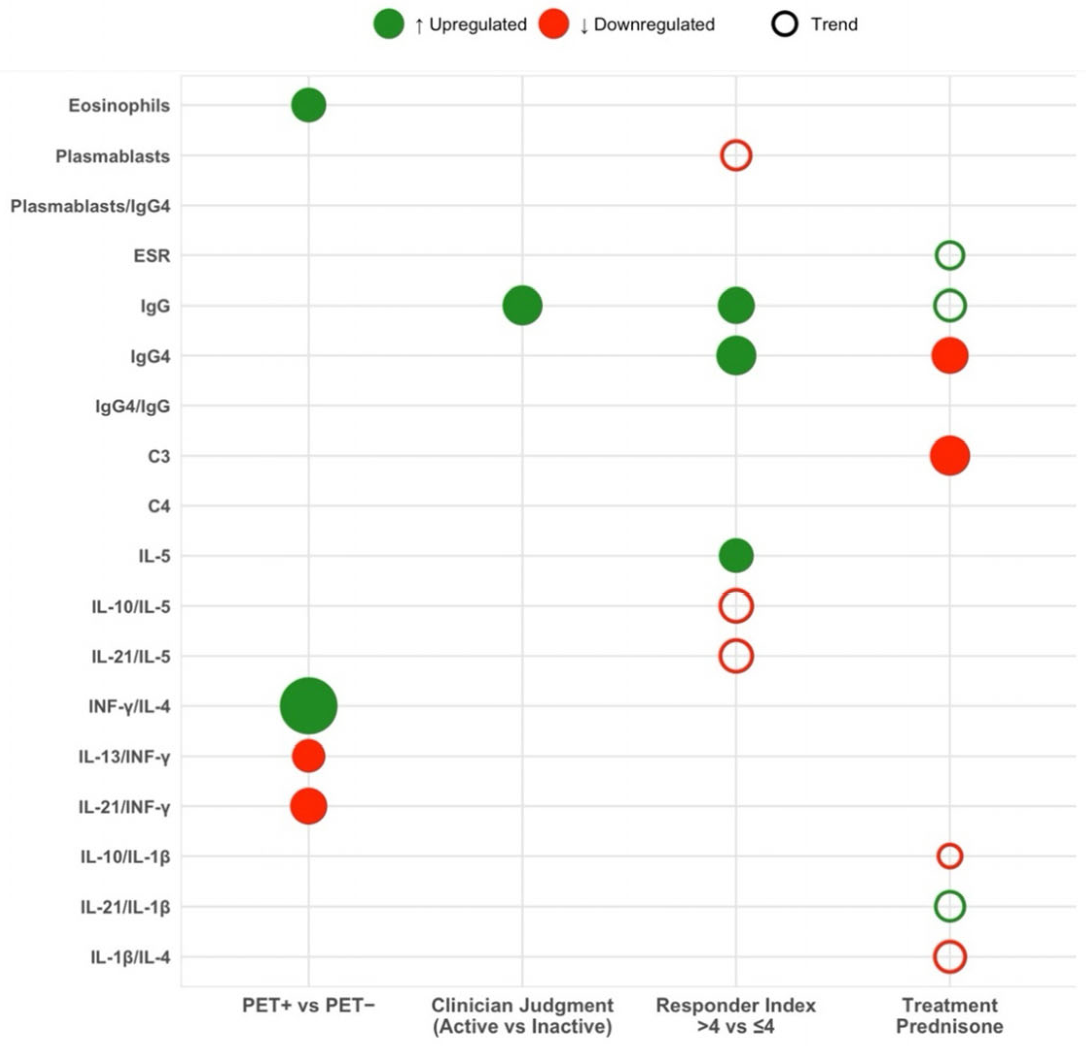
3.3. Biomarker Profiles According to [18F]-FDG-PET/CT Activity
3.4. Biomarker Profiles According to Clinician-Assessed Activity
3.5. Biomarker Profiles According to IgG4-RD Responder Index
3.6. Biomarker Correlation with the IgG4-RD Damage Index
3.7. Impact of Immunosuppressive Treatment on Biomarker Levels
4. Discussion
5. Conclusions
Author Contributions
Funding
Institutional Review Board Statement
Informed Consent Statement
Data Availability Statement
Conflicts of Interest
Abbreviations
| ACR/EULAR | American College of Rheumatology/European League Against Rheumatism |
| C3 | Complement component 3 |
| C4 | Complement component 4 |
| CRP | C-reactive protein |
| CT | Computed tomography |
| ESR | Erythrocyte sedimentation rate |
| IFN-γ | Interferon gamma |
| IgG | Immunoglobulin G |
| IgG4 | Immunoglobulin G4 |
| IL | Interleukin |
| P25–P75 | 25th to 75th percentile (interquartile range) |
| PET | 18F-fluorodeoxyglucose positron emission tomography |
| PET/CT | 18F-fluorodeoxyglucose positron emission tomography/computed tomography |
| RI | Responder Index |
| SD | Standard deviation |
References
- Umehara, H.; Okazaki, K.; Masaki, Y.; Kawano, M.; Yamamoto, M.; Saeki, T.; Matsui, S.; Sumida, T.; Mimori, T.; Tanaka, Y.; et al. A novel clinical entity, IgG4-related disease (IgG4RD): General concept and details. Mod. Rheumatol. 2012, 22, 1–14. [Google Scholar] [CrossRef]
- Wallace, Z.S.; Katz, G.; Hernandez-Barco, Y.G.; Baker, M.C. Current and future advances in practice: IgG4-related disease. Rheumatol. Adv. Pract. 2024, 8, rkae020. [Google Scholar] [CrossRef] [Scilit]
- Parums, D.V. IgG4-Related Disease: A Review of Persistent Challenges in the Pathogenesis, Diagnosis, and Approaches to Treatment. Med. Sci. Monit. 2025, 31, e950212. [Google Scholar] [CrossRef] [Scilit]
- Wallace, Z.S.; Zhang, Y.; Perugino, C.A.; Naden, R.; Choi, H.K.; Stone, J.H. Clinical phenotypes of IgG4-related disease: An analysis of two international cross-sectional cohorts. Ann. Rheum. Dis. 2019, 78, 406–412. [Google Scholar] [CrossRef] [Scilit] [PubMed]
- Lanzillotta, M.; Campochiaro, C.; Mancuso, G.; Ramirez, G.A.; Capurso, G.; Falconi, M.; Dagna, L.; Della-Torre, E. Clinical phenotypes of IgG4-related disease reflect different prognostic outcomes. Rheumatology 2020, 59, 2435–2442. [Google Scholar] [CrossRef] [Scilit] [PubMed]
- Lanzillotta, M.; Mancuso, G.; Della-Torre, E. Advances in the diagnosis and management of IgG4 related disease. BMJ 2020, 369, m1067. [Google Scholar] [CrossRef] [Scilit]
- Maehara, T.; Moriyama, M.; Nakamura, S. Review of a novel disease entity, immunoglobulin G4-related disease. J. Korean Assoc. Oral. Maxillofac. Surg. 2020, 46, 3–11. [Google Scholar] [CrossRef] [Scilit]
- Tsuboi, H.; Honda, F.; Takahashi, H.; Ono, Y.; Abe, S.; Kondo, Y.; Matsumoto, I.; Sumida, T. Pathogenesis of IgG4-related disease. Comparison with Sjögren’s syndrome. Mod. Rheumatol. 2020, 30, 7–16. [Google Scholar] [CrossRef] [Scilit]
- Della-Torre, E.; Feeney, E.; Deshpande, V.; Mattoo, H.; Mahajan, V.; Kulikova, M.; Wallace, Z.S.; Carruthers, M.; Chung, R.T.; Pillai, S.; et al. B-cell depletion attenuates serological biomarkers of fibrosis and myofibroblast activation in IgG4-related disease. Ann. Rheum. Dis. 2015, 74, 2236–2243. [Google Scholar] [CrossRef] [Scilit] [PubMed]
- Maehara, T.; Koga, R.; Nakamura, S. Immune dysregulation in immunoglobulin G4-related disease. Jpn. Dent. Sci. Rev. 2023, 59, 1–7. [Google Scholar] [CrossRef] [Scilit]
- Ito, F.; Kamekura, R.; Yamamoto, M.; Takano, K.; Takaki, H.; Yabe, H.; Ikegami, I.; Shigehara, K.; Himi, T.; Takahashi, H.; et al. IL-10(+) T follicular regulatory cells are associated with the pathogenesis of IgG4-related disease. Immunol. Lett. 2019, 207, 56–63. [Google Scholar] [CrossRef] [Scilit]
- Tang, J.; Cai, S.; Ye, C.; Dong, L. Biomarkers in IgG4-related disease: A systematic review. Semin. Arthritis Rheum. 2020, 50, 354–359. [Google Scholar] [CrossRef] [Scilit]
- Wallace, Z.S.; Mattoo, H.; Carruthers, M.; Mahajan, V.S.; Della Torre, E.; Lee, H.; Kulikova, M.; Deshpande, V.; Pillai, S.; Stone, J.H. Plasmablasts as a biomarker for IgG4-related disease, independent of serum IgG4 concentrations. Ann. Rheum. Dis. 2015, 74, 190–195. [Google Scholar] [CrossRef] [Scilit]
- Culver, E.L.; Sadler, R.; Bateman, A.C.; Makuch, M.; Cargill, T.; Ferry, B.; Aalberse, R.; Barnes, E.; Rispens, T. Increases in IgE, Eosinophils, and Mast Cells Can be Used in Diagnosis and to Predict Relapse of IgG4-Related Disease. Clin. Gastroenterol. Hepatol. 2017, 15, 1444–1452.e6. [Google Scholar] [CrossRef] [Scilit]
- Lopez-Gomez, M.; Moya-Alvarado, P.; Park, H.S.; Martín, M.C.; Calleja, S.; Codes-Mendez, H.; Magallares, B.; Castellví, I.; Barros-Membrilla, A.J.; Laiz, A.; et al. Comparative Analysis of Classification Criteria in IgG4-Related Disease and Evaluating Diagnostic Accuracy from a Retrospective Cohort in Clinical Practice. Diagnostics 2024, 14, 2583. [Google Scholar] [CrossRef] [Scilit]
- Wallace, Z.S.; Naden, R.P.; Chari, S.; Choi, H.K.; Della-Torre, E.; Dicaire, J.-F.; A Hart, P.; Inoue, D.; Kawano, M.; Khosroshahi, A.; et al. The 2019 American College of Rheumatology/European League Against Rheumatism Classification Criteria for IgG4-Related Disease. Arthritis Rheumatol. 2020, 72, 7–19. [Google Scholar] [CrossRef] [Scilit] [PubMed]
- Khosroshahi, A.; Wallace, Z.S.; Crowe, J.L.; Akamizu, T.; Azumi, A.; Carruthers, M.N.; Chari, S.T.; Della-Torre, E.; Frulloni, L.; Goto, H.; et al. International Consensus Guidance Statement on the Management and Treatment of IgG4-Related Disease. Arthritis Rheumatol. 2015, 67, 1688–1699. [Google Scholar] [CrossRef] [Scilit]
- Khosroshahi, A.; Bloch, D.B.; Deshpande, V.; Stone, J.H. Rituximab therapy leads to rapid decline of serum IgG4 levels and prompt clinical improvement in IgG4-related systemic disease. Arthritis Rheum. 2010, 62, 1755–1762. [Google Scholar] [CrossRef] [Scilit]
- Carruthers, M.N.; Topazian, M.D.; Khosroshahi, A.; E Witzig, T.; Wallace, Z.S.; A Hart, P.; Deshpande, V.; Smyrk, T.C.; Chari, S.; Stone, J.H. Rituximab for IgG4-related disease: A prospective, open-label trial. Ann. Rheum. Dis. 2015, 74, 1171–1177. [Google Scholar] [CrossRef] [Scilit] [PubMed]
- Carruthers, M.N.; Stone, J.H.; Deshpande, V.; Khosroshahi, A. Development of an IgG4-RD Responder Index. Int. J. Rheumatol. 2012, 2012, 259408. [Google Scholar] [CrossRef] [Scilit] [PubMed]
- Wallace, Z.S.; Khosroshahi, A.; Carruthers, M.D.; Perugino, C.A.; Choi, H.; Campochiaro, C.; Culver, E.L.; Cortazar, F.; Della-Torre, E.; Ebbo, M.; et al. An International Multispecialty Validation Study of the IgG4-Related Disease Responder Index. Arthritis Care Res. 2018, 70, 1671–1678. [Google Scholar] [CrossRef] [Scilit] [PubMed]
- Peng, L.; Li, J.; Zhou, J.; Fei, Y.; Dong, L.; Liu, Y.-Y.; Zhang, D.; Wang, Y.; Chi, S.; Wang, F.; et al. The development and initial validation of IgG4-related disease damage index: A consensus report from Chinese IgG4-RD Consortium. RMD Open 2024, 10, e003938. [Google Scholar] [CrossRef] [Scilit]
- Tang, C.Y.L.; Chua, W.M.; Cheng, L.T.J.; Fong, W.; Zaheer, S.; Lam, W.W. (18)F-FDG PET/CT Manifestations of IgG4-related Disease. Br. J. Radiol. 2021, 94, 20210105. [Google Scholar] [CrossRef] [Scilit]
- StataCorp. Stata Statistical Software, Release 17; StataCorp LLC: College Station, TX, USA, 2021. [Google Scholar]
- R Core Team. A Language and Environment for Statistical Computing; R Foundation for Statistical Computing: Vienna, Austria, 2021; Available online: https://www.r-project.org/ (accessed on 10 October 2021).
- Kamisawa, T.; Zen, Y.; Pillai, S.; Stone, J.H. IgG4-related disease. Lancet 2015, 385, 1460–1471. [Google Scholar] [CrossRef] [Scilit]
- Wallace, Z.S.; Deshpande, V.; Mattoo, H.; Mahajan, V.S.; Kulikova, M.; Pillai, S.; Stone, J.H. IgG4-Related Disease: Clinical and Laboratory Features in One Hundred Twenty-Five Patients. Arthritis Rheumatol. 2015, 67, 2466–2475. [Google Scholar] [CrossRef] [Scilit]
- Ming, B.; Zhong, J.; Dong, L. Role of eosinophilia in IgG4-related disease. Clin. Exp. Rheumatol. 2022, 40, 1038–1044. [Google Scholar] [CrossRef] [Scilit]
- Peng, Y.; Li, J.Q.; Zhang, P.P.; Zhang, X.; Peng, L.Y.; Chen, H.; Zhou, J.X.; Zhang, S.Z.; Yang, H.X.; Liu, J.J.; et al. Clinical outcomes and predictive relapse factors of IgG4-related disease following treatment: A long-term cohort study. J. Intern. Med. 2019, 286, 542–552. [Google Scholar] [CrossRef] [Scilit]
- Zhang, X.; Zhang, P.; Li, J.; He, Y.; Fei, Y.; Peng, L.; Shi, Q.; Zhang, W.; Zhao, Y. Different clinical patterns of IgG4-RD patients with and without eosinophilia. Sci. Rep. 2019, 9, 16483. [Google Scholar] [CrossRef] [Scilit] [PubMed]
- Zhang, N.; Peng, Y.; Li, R.; Nie, Y.; Li, J.; Yang, X.; Zhang, J.; Wang, Y.; Luo, Q.; Zhou, J.; et al. Elevated eosinophil-to-lymphocyte ratio (ELR) as a predictor of relapse for IgG4-related disease: A retrospective study across a decade. Clin. Exp. Med. 2025, 25, 193. [Google Scholar] [CrossRef] [Scilit] [PubMed]
- Liu, C.; Zhang, P.; Zhang, W. Immunological mechanism of IgG4-related disease. J. Transl. Autoimmun. 2020, 3, 100047. [Google Scholar] [CrossRef] [Scilit]
- Gieseck, R.L.; Wilson, M.S.; Wynn, T.A. Type 2 immunity in tissue repair and fibrosis. Nat. Rev. Immunol. 2018, 18, 62–76. [Google Scholar] [CrossRef] [Scilit]
- Della-Torre, E.; Rigamonti, E.; Perugino, C.; Baghai-Sain, S.; Sun, N.; Kaneko, N.; Maehara, T.; Rovati, L.; Ponzoni, M.; Milani, R.; et al. B lymphocytes directly contribute to tissue fibrosis in patients with IgG(4)-related disease. J. Allergy Clin. Immunol. 2020, 145, 968–981.e14. [Google Scholar] [CrossRef] [Scilit]
- Akiyama, M.; Alshehri, W.; Ishigaki, S.; Saito, K.; Kaneko, Y. Human T follicular helper cells and their impact on IgE and IgG4 production across allergy, malignancy, and IgG4-related disease. Allergol. Int. 2025, 74, 25–32. [Google Scholar] [CrossRef] [Scilit] [PubMed]
- Akiyama, M.; Suzuki, K.; Yamaoka, K.; Yasuoka, H.; Takeshita, M.; Kaneko, Y.; Kondo, H.; Kassai, Y.; Miyazaki, T.; Morita, R.; et al. Number of circulating follicular helper 2 T cells correlates with IgG4 and interleukin-4 levels and plasmablast numbers in IgG4-related disease. Arthritis Rheumatol. 2015, 67, 2476–2481. [Google Scholar] [CrossRef] [Scilit]
- Kwon, O.C.; Park, M.C.; Kim, Y.G. Correlation between serologic parameters and disease activity of IgG4-related disease: Differences between patients with normal and elevated serum IgG4 concentrations. Front. Immunol. 2022, 13, 1020459. [Google Scholar] [CrossRef] [Scilit]
- Chen, L.Y.C.; Mattman, A.; Seidman, M.A.; Carruthers, M.N. IgG4-related disease: What a hematologist needs to know. Haematologica 2019, 104, 444–455. [Google Scholar] [CrossRef] [Scilit]
- Zen, Y.; Fujii, T.; Harada, K.; Kawano, M.; Yamada, K.; Takahira, M.; Nakanuma, Y. Th2 and regulatory immune reactions are increased in immunoglobin G4-related sclerosing pancreatitis and cholangitis. Hepatology 2007, 45, 1538–1546. [Google Scholar] [CrossRef] [Scilit]
- Tanaka, A.; Moriyama, M.; Nakashima, H.; Miyake, K.; Hayashida, J.; Maehara, T.; Shinozaki, S.; Kubo, Y.; Nakamura, S. Th2 and regulatory immune reactions contribute to IgG4 production and the initiation of Mikulicz disease. Arthritis Rheum. 2012, 64, 254–263. [Google Scholar] [CrossRef] [Scilit] [PubMed]
- Yamamoto, M.; Takano, K.I.; Kamekura, R.; Aochi, S.; Suzuki, C.; Ichimiya, S.; Nakase, H.; Himi, T.; Takahashi, H. Interleukin 5-producing ST2(+) memory Th2 cells in IgG4-related dacryoadenitis and sialadenitis. Mod. Rheumatol. 2019, 29, 856–860. [Google Scholar] [CrossRef] [Scilit] [PubMed]
- Yamamoto, M.; Takano, K.; Kamekura, R.; Suzuki, C.; Ichimiya, S.; Himi, T.; Nakase, H.; Takahashi, H. Stage classification of IgG4-related dacryoadenitis and sialadenitis by the serum cytokine environment. Mod. Rheumatol. 2018, 28, 1004–1008. [Google Scholar] [CrossRef] [Scilit]
- Mattoo, H.; Mahajan, V.S.; Della-Torre, E.; Sekigami, Y.; Carruthers, M.; Wallace, Z.S.; Deshpande, V.; Stone, J.H.; Pillai, S. De novo oligoclonal expansions of circulating plasmablasts in active and relapsing IgG4-related disease. J. Allergy Clin. Immunol. 2014, 134, 679–687. [Google Scholar] [CrossRef] [Scilit] [PubMed]
- Yoshifuji, H.; Umehara, H. Glucocorticoids in the treatment of IgG4-related disease-Prospects for new international treatment guidelines. Mod. Rheumatol. 2023, 33, 252–257. [Google Scholar] [CrossRef] [Scilit] [PubMed]
- Peng, L.; Lu, H.; Zhou, J.; Zhang, P.; Li, J.; Liu, Z.; Wu, D.; Zhang, S.; Yang, Y.; Bai, W.; et al. Clinical characteristics and outcome of IgG4-related disease with hypocomplementemia: A prospective cohort study. Arthritis Res. Ther. 2021, 23, 102. [Google Scholar] [CrossRef] [Scilit] [PubMed]


| Category | Variable | IgG4-RD Patients n = 35 |
|---|---|---|
| Demographics, n (%) | Age, median (P25–P75) | 63.0 (52–72) |
| Sex (men) | 27 (77.1) | |
| Diagnostic Criteria, n (%) | Okazaki | 23 (65.7) |
| Umehara | ||
| Possible | 20 (57.1) | |
| Probable | 2 (5.7) | |
| Definite | 10 (28.6) | |
| ACR/EULAR | 23 (65.7) | |
| Clinical Scores and Indices, n (%) | Responder Index, mean (SD) | 4.83 (4.0) |
| >4 | 19 (54.3) | |
| Clinically assessed active disease | 12 (34.3) | |
| Damage Control Index, mean (SD) | 3.09 (1.5) | |
| Organ Involvement, n (%) | Increased organ size | 33 (94.3) |
| Glandular | ||
| >1 | 3 (8.8) | |
| >2 | 3 (8.8) | |
| Pancreas | ||
| Pancreas and biliary tract | 7 (20.0) | |
| Diffuse pancreatic enlargement with capsule-like rim of low density | 2 (5.7) | |
| Diffuse pancreatic enlargement (loss of lobulation) | 1 (2.9) | |
| Renal | ||
| Bilateral low-density areas in the renal cortex | 10 (28.6) | |
| Hydronephrosis | 5 (14.3) | |
| Hypocomplementemia | 4 (11.4) | |
| Thickening of the renal pelvis/soft tissue | 3 (8.6) | |
| Retroperitoneal | ||
| Peripheral or anterolateral soft tissue around the infrarenal aorta or iliac arteries | 12 (34.3) | |
| Diffuse thickening of the abdominal aortic wall | 8 (22.9) | |
| Thorax | ||
| Peribronchovascular and septal thickening | 5 (14.3) | |
| Soft tissue resembling a paravertebral band | 2 (5.7) | |
| Aneurism | 13 (37.1) | |
| Wallace Subtype Classification, n (%) | Group 1. Pancreato-biliary | 8 (22.9) |
| Group 2. Retroperitoneum and aorta | 21 (60.0) | |
| Group 3. Systemic Mikulicz | 5 (14.3) | |
| Group 4. Head and neck | 1 (2.9) | |
| Systemic Symptoms, n (%) | Low back pain | 8 (22.9) |
| Toxic syndrome | 7 (20.0) | |
| Fever | 5 (14.3) | |
| Neurological and Head Involvement, n (%) | Hypophysitis | 1 (2.9) |
| Pachymeningitis | 1 (2.9) | |
| Orbital pseudotumor | 0 (0.0) | |
| Uveal involvement | 0 (0.0) | |
| Imaging Findings, n (%) | Activity based on positive PET | 8 (28.6) |
| CT | 14 (40.0) | |
| Aorta involvement | 3 (21.4) | |
| Retroperitoneum involvement | 3 (21.4) | |
| Histological Findings, n (%) | 11 (31.4) | |
| Plasmatic cells | 10 (90.9) | |
| Lymphocytic infiltration | 10 (90.9) | |
| Fibrosis | 4 (36.4) | |
| Phlebitis | 3 (27.3) | |
| Serologic biomarkers | IgG (P25–P75) | 1168 (954–1808.5) (mg/dL) |
| IgG4 (P25–P75) | 91 (33–138) (mg/dL) | |
| Eosinophil count (P25–P75) | 0.16 (0.08–0.26) × 103 cells/μL) | |
| CRP (P25–P75) | 2.5 (1–5.05) (mg/L) | |
| ERS (P25–P75) | 9 (0–24) (mm/h) | |
| Category | Variable | PET + (n = 8) | PET − (n = 20) | p-Value | Clinician Assessment: Active (n = 12) | Clinician Assessment: Not Active (n = 23) | p-Value | RI ≥ 4 (n = 19) | RI < 4 (n = 16) | p-Value |
|---|---|---|---|---|---|---|---|---|---|---|
| Cellular markers, median (IQR) | Plasmablasts, cells/μL | 1226 (141.75–2024.75) | 202.50 (0–1121) | 0.22 | 401 (0–1219) | 722 (0–1606) | 0.71 | 223 (0–1054) | 1096 (0–2955) | 0.07 |
| Plasmablasts/IgG4 | 0.77 (0–10.06) | 8.04 (3.48–16.85) | 0.31 | 4.4 (0–32.52) | 2.61 (0–10.5) | 0.63 | 0.97 (0–7.26) | 19.30 (0–43.09) | 0.08 | |
| Hematologic markers, median (IQR) | Eosinophils. ×103 cells/μL | 0.24 (0.17–0.39) | 0.15 (0.08–0.21) | 0.03 | 0.15 (0.09–0.23) | 0.16 (0.08–0.34) | 0.4 | 0.15 (0.08–0.23) | 0.19 (0.10–0.37) | 0.18 |
| Hemoglobin, g/dL | 13 (11.88–14.55) | 14.4 (13.55–15) | 0.175 | 14.1 (12.65–15.45) | 14.3 (13.45–14.8) | 0.7 | 14.2 (13.20–14.95) | 14.35 (13.30–14.95) | 0.65 | |
| Acute-phase markers, median (IQR) | CRP, mg/L | 2.55 (1.62–5.22) | 3.15 (1–6.35) | 0.78 | 1.75 (1–5.15) | 2.5 (1–5) | 0.96 | 2.50 (1–5.3) | 2.40 (1–4.28) | 0.80 |
| ESR, mm/h | 9 (0–22) | 11.5 (4–24.75) | 0.6 | 11 (4–13) | 6.5 (0–37) | 0.64 | 10 (0–18.50) | 6 (1–31.00) | 0.84 | |
| Complement system, mean (SD) | C3, mg/dL | 130.25 (25.8) | 125.27 (32.2) | 0.71 | 135.17 (26.6) | 118.07 (28.3) | 0.12 | 129.93 (27.3) | 120.55 (30.1) | 0.41 |
| C4, mg/dL | 26.10 (11.3) | 26.61 (6.3) | 0.88 | 28.12 (8.7) | 25.36 (6.9) | 0.37 | 27.36 (8.2) | 25.65 (7.4) | 0.58 | |
| Humoral markers, median (IQR) | IgG, mg/dL | 1759.5 (920.5–2046.5) | 1003.5 (931–1348.25) | 0.33 | 1751.5 (1131.25–2046.5) | 1009 (933–1313) | 0.01 | 1327 (1051–1919.5) | 975 (878–1366.25) | 0.02 |
| IgG4, mg/dL | 103.5 (47.25–132.50) | 82 (37.25–118.75) | 0.74 | 114 (81.5–176.25) | 75 (37–114) | 0.2 | 117 (72.5–226) | 60.5 (32–97) | 0.01 | |
| IgG4/IgG | 0.06 (0.05–0.07) | 0.07 (0.03–0.10) | 0.57 | 0.06 (0.04–0.1) | 0.07 (0.04–0.1) | 0.74 | 0.10 (0.08) | 0.06 (0.03) | 0.21 | |
| Cytokines, median (IQR) | INF-γ, pg/mL | 5.95 (3.91–9.71) | 4.29 (2.17–7.38) | 0.19 | 4.32 (2.79–8.13) | 5.21 (2.25–7.72) | 0.64 | 4.68 (2.75–8.64) | 4.58 (2.14–7.47) | 0.46 |
| IL-10, pg/mL | 1.88 (0.56–12.64) | 5.64 (1.65–9.66) | 0.27 | 1.28 (0.56–8.15) | 3.99 (1.58–6.04) | 0.43 | 2.32 (0.67–7.51) | 3.59 (1.37–5.79) | 0.99 | |
| IL-13, pg/mL | 0.23 (0.17–1.29) | 1.94 (0.23–9.34) | 0.16 | 0.23 (0.23–9.34) | 1.75 (0.23–2.94) | 0.87 | 1.85 (0.23–4.96) | 0.23 (0.23–2.32) | 0.21 | |
| IL-1β, pg/mL | 0.57 (0.34–1.09) | 0.42 (0.14–0.83) | 0.52 | 0.49 (0.33–1.08) | 0.49 (0.14–0.84) | 0.52 | 0.55 (0.32–1.21) | 0.44 (0.14–0.67) | 0.33 | |
| IL-21, pg/mL | 0.21 (0.15–0.44) | 0.26 (0.14–0.44) | 0.98 | 0.21 (0.14–0.46) | 0.25 (0.14–0.4) | 1 | 0.3 (0.14–0.47) | 0.21 (0.14–0.39) | 0.58 | |
| IL-4, pg/mL | 2.67 (1.12–5.41) | 6.83 (2.62–25.91) | 0.17 | 3.69 (1.12–18.40) | 4.59 (1.25–9.42) | 0.89 | 4.24 (1.79–20.2) | 4.31 (1.12–9.02) | 0.55 | |
| IL-5, pg/mL | 1.35 (0.69–2.21) | 1.58 (0.60–2.63) | 1 | 1.85 (0.7–2.85) | 1.01 (0.41–2.00) | 0.15 | 1.63 (0.97–2.66) | 0.8 (0.27–2) | 0.03 | |
| Cytokine ratios, median (IQR) | INF-γ/IL-10 | 3.76 (0.99–7.39) | 1.3 (0.36–2.14) | 0.14 | 2.93 (1.22–6.29) | 1.5 (0.51–2.84) | 0.26 | 1.81 (0.99–4.8) | 1.48 (0.43–2.67) | 0.46 |
| IL-13/INF-γ | 0.04 (0.02; 0.34) | 0.31 (0.11–1.01) | 0.04 | 0.1 (0.06–1.01) | 0.2 (0.08–0.77) | 0.82 | 0.29 (0.07–0.91) | 0.15 (0.04–0.53) | 0.45 | |
| INF-γ/IL-1β | 9.14 (6.28–20.78) | 8.84 (6.88–14.34) | 0.82 | 7.34 (6.44–11.57) | 9.24 (6.2–14.82) | 0.52 | 8.37 (6.33–15.21) | 9.05 (6.28–14.34) | 0.72 | |
| IL-21/INF-γ | 0.03 (0.02–0.06) | 0.07 (0.04–0.10) | 0.03 | 0.06 (0.03–0.07) | 0.07 (0.03–0.09) | 0.48 | 0.06 (0.03–0.07) | 0.07 (0.03–0.09) | 0.46 | |
| INF-γ/IL-4 | 3.46 (1.06–4.57) | 0.98 (0.37–1.32) | <0.001 | 1.22 (0.5–2.79) | 1.11 (0.48–1.63) | 0.67 | 1.11 (0.51–1.69) | 1.18 (0.44–2.22) | 0.78 | |
| INF-γ/IL-5 | 3.44 (2.69–8.45) | 2.98 (1.94–5.48) | 0.38 | 3.44 (2.75–4.45) | 3.45 (2.38–12.2) | 0.67 | 3.03 (2.65–3.83) | 5.76 (2.35–14.35) | 0.16 | |
| IL-13/IL-10 | 0.41 (0.04–0.46) | 0.41 (0.14–1.15) | 0.4 | 0.41 (0.37–0.81) | 0.41 (0.12–1.04) | 0.79 | 0.49 (0.33–1.19) | 0.41 (0.09–0.81) | 0.22 | |
| IL-10/IL-1β | 4.7 (1.23–18.87) | 7.44 (4.09–38.25) | 0.24 | 4.06 (1.37–6.46) | 6.6 (3.84–25.5) | 0.22 | 4.69 (2.47–11.59) | 6.84 (3.74–18.87) | 0.45 | |
| IL-21/IL-10 | 0.1 (0.01–0.32) | 0.07 (0.02–0.16) | 0.94 | 0.16 (0.05–0.27) | 0.09 (0.03–0.18) | 0.48 | 0.08 (0.04–0.24) | 0.1 (0.03–0.2) | 0.93 | |
| IL-10/IL-4 | 0.68 (0.37–1.79) | 0.48 (0.22–1.25) | 0.51 | 0.48 (0.24–0.81) | 0.5 (0.25–1.32) | 0.59 | 0.47 (0.23–0.82) | 0.5 (0.26–1.25) | 0.45 | |
| IL-10/IL-5 | 1.1 (0.50–12.00) | 3.91 (2.04–9.94) | 0.38 | 1.6 (0.7–4.79) | 4.1 (1.19–10.06) | 0.9 | 2.39 (0.67–6.69) | 4.17 (1.21–14.27) | 0.056 | |
| IL-13/IL-1β | 1.11 (0.19–3.13) | 2.29 (1.57–10.77) | 0.14 | 1.64 (0.56–6.36) | 1.64 (1.50–5.82) | 0.65 | 2.50 (1.07–7.32) | 1.64 (0.97–3.81) | 0.55 | |
| IL-13/IL-21 | 1.53 (0.72–4.89) | 3.40 (1.64–18.47) | 0.17 | 1.64 (1.49–13.64) | 3.3 (1.64–9.54) | 1 | 4.32 (1.64–16.6) | 1.64 (1.18–5.47) | 0.18 | |
| IL-13/IL-4 | 0.15 (0.05–0.29) | 0.21 (0.14–0.29) | 0.43 | 0.21 (0.1–0.36) | 0.21 (0.15–0.29) | 0.79 | 0.21 (0.14–0.43) | 0.21 (0.12–0.24) | 0.32 | |
| IL-13/IL-5 | 0.24 (0.08–1.09) | 1.6 (0.32–5.29) | 0.07 | 0.89 (0.18–3.45) | 1.5 (0.46–2.18) | 0.63 | 1.50 (0.26–2.94) | 1.04 (0.32–2.05) | 0.79 | |
| IL-21/IL-1β | 0.55 (0.3–0.95) | 0.73 (0.48–1) | 0.26 | 0.47 (0.4–0.70) | 0.73 (0.48–1) | 0.21 | 0.49 (0.41–0.78) | 0.83 (0.49–1) | 0.14 | |
| IL-1β/IL-4 | 0.13 (0.11–0.45) | 0.07 (0.04–0.13) | 0.06 | 0.13 (0.06–0.29) | 0.12 (0.05–0.14) | 0.38 | 0.1 (0.04–0.23) | 0.12 (0.07–0.13) | 1 | |
| IL-1β/IL-5 | 0.46 (0.22–0.65) | 0.29 (0.15–1.02) | 0.74 | 0.5 (0.14–0.67) | 0.35 (0.23–1.13) | 0.66 | 0.36 (0.15–0.72) | 0.53 (0.28–1.17) | 0.3 | |
| IL-21/IL-4 | 0.08 (0.03–0.13) | 0.05 (0.02–0.11) | 0.56 | 0.07 (0.02–0.13) | 0.07 (0.03–0.12) | 0.78 | 0.04 (0.02–0.13) | 0.08 (0.03–0.12) | 0.67 | |
| IL-21/IL-5 | 0.17 (0.08–0.27) | 0.21 (0.11–0.52) | 0.44 | 0.17 (0.08–0.42) | 0.23 (0.15–0.52) | 0.25 | 0.19 (0.09–0.36) | 0.37 (0.17–0.67) | 0.056 | |
| IL-4/IL-5 | 3.67 (0.93–4.60) | 6 (2.62–28.48) | 0.07 | 4.68 (1.46–7.44) | 4.54 (2.74–19.49) | 0.38 | 3.50 (1.95–9.18) | 4.71 (3.83–18.75) | 0.24 |
| Category | Variable | PREDNISONE | ||
|---|---|---|---|---|
| Yes (n = 21) | No (n = 7) | p-Value | ||
| Cellular markers, median (IQR) | Plasmablasts, cells/μL | 416 (0–1139) | 579 (571–1638) | 0.12 |
| Plasmablasts/IgG4 | 4.4 (0–10.95) | 2.67 (0.12–35.2) | 0.87 | |
| Hematologic markers, median (IQR) | Eosinophils, ×103 cells/μL | 0.15 (0.08–0.2) | 0.08 (0.08–0.16) | 0.166 |
| Hemoglobin. g/dL | 14.3 (13.6–14.9) | 14.4 ( | 0.72 | |
| Acute-phase markers, median (IQR) | CRP, mg/L | 8.58 (16.2) | 1 (1–1.4) | 0.04 |
| ESR, mm/h | 20.80 (22.5) | 11 ( | 0.80 | |
| Complement system, mean (SD) | C3, mg/dL | 118.19 (26.2) | 144.72 (17.1) | 0.007 |
| C4, mg/dL | 27.02 (6.4) | 31.1 (4.5) | 0.106 | |
| Humoral markers, median (IQR) | IgG, mg/dL | 998 (887–1275) | 1189 (998.5–1831) | 0.06 |
| IgG4, mg/dL | 70 (26–104) | 128 (32–186) | 0.04 | |
| IgG4/IgG | 0.06 (0.02–0.09) | 0.03 (0.02–0.182) | 0.69 | |
| Cytokines, median (IQR) | INF-γ, pg/mL | 5.21 (2.28–8.22) | 5.4 (3.24–8.88) | 0.63 |
| IL-10, pg/mL | 4.82 (1.64–6.51) | 2.07 (0.73–6.18) | 0.8 | |
| IL-13, pg/mL | 1.75 (0.23–4.63) | 3.13 (1.32–3.40) | 0.12 | |
| IL-1β, pg/mL | 0.44 (0.14–0.72) | 0.66 (0.38–0.99) | 0.31 | |
| IL-21, pg/mL | 0.3 (0.14–0.48) | 0.26 (0.20–0.32) | 0.91 | |
| IL-4, pg/mL | 4.48 (1.37–16.58) | 9.65 (6.17–13.34) | 0.22 | |
| IL-5, pg/mL | 1.54 (0.42–2.36) | 2.02 (0.81–2.65) | 0.59 | |
| Cytokine ratios, median (IQR) | INF-γ/IL-10 | 1.49 (0.58–2.30) | 1.81 (1.32–3.31) | 0.88 |
| IL-13/INF-γ | 0.20 (0.06–0.98) | 0.34 (0.21–1.03) | 0.17 | |
| INF-γ/IL-1β | 9.90 (6.36–15.79) | 8.5 (7.34–9.57) | 0.6 | |
| IL-21/INF-γ | 0.07 (0.04–0.11) | 0.05 (0.045–0.069) | 0.9 | |
| INF-γ/IL-4 | 1.11 (0.48–1.42) | 1.16 (0.64–1.22) | 0.5 | |
| INF-γ/IL-5 | 0.03 (2.15–6.00) | 3.29 (2.79–9.9) | 0.46 | |
| IL-13/IL-10 | 0.41 (0.14–1.12) | 0.06 | ||
| IL-10/IL-1β | 0.6 (3.85–34.43) | 3.67 (3.05–5.63) | 0.39 | |
| IL-21/IL-10 | 0.09 (0.03–0.19) | 0.2 (0.12–0.31) | 0.41 | |
| IL-10/IL-4 | 0.47 (0.27–1.18) | 0.47(0.23–0.57) | 0.34 | |
| IL-10/IL-5 | 3.62 (1.24–9.82) | 3.62 (1.72–5.05) | 0.88 | |
| IL-13/IL-1β | 1.64 (1.64–7.33) | ) | 0.3 | |
| IL-13/IL-21 | 3.3 (1.64–13.90) | ) | 0.91 | |
| IL-13/IL-4 | 0.21 (0.17–0.29) | 0.3 (0.23–0.2) | 0.9 | |
| IL-13/IL-5 | 1.5 (0.35–2.61) | 2.01 (0.43–2.30) | 0.09 | |
| IL-21/IL-1β | 0.84 (0.52–1) | 0.67 (1.01–4.8) | 0.08 | |
| IL-1β/IL-4 | 0.1 (0.04–0.12) | 0.12 (0.07–0.16) | 0.7 | |
| IL-1β/IL-5 | 0.35 (0.21–0.61) | 0.6 (0.33–1.14) | 0.39 | |
| IL-21/IL-4 | 0.07 (0.03–0.12) | ) | 0.59 | |
| IL-21/IL-5 | 0.23 (0.15–0.50) | 0.34 (0.13–0.4) | 0.81 | |
| IL-4/IL-5 | 4.59 (2.80–20.98) | 5.87 (2.91–9.3) | 0.78 | |
Disclaimer/Publisher’s Note: The statements, opinions and data contained in all publications are solely those of the individual author(s) and contributor(s) and not of MDPI and/or the editor(s). MDPI and/or the editor(s) disclaim responsibility for any injury to people or property resulting from any ideas, methods, instructions or products referred to in the content. |
© 2026 by the authors. Published by MDPI on behalf of the Lithuanian University of Health Sciences. Licensee MDPI, Basel, Switzerland. This article is an open access article distributed under the terms and conditions of the Creative Commons Attribution (CC BY) license.
Share and Cite
Moya-Alvarado, P.; Lopez-Gomez, M.; Martínez-Martinez, L.; Park, H.S.; Leyva, T.F.; Martín, M.C.; Codes-Mendez, H.; Lacruz, A.C.; Calleja, S.; Magallares, B.; et al. Immunological Biomarkers to Assess Activity and Treatment Response in IgG4-Related Disease. Medicina 2026, 62, 323. https://doi.org/10.3390/medicina62020323
Moya-Alvarado P, Lopez-Gomez M, Martínez-Martinez L, Park HS, Leyva TF, Martín MC, Codes-Mendez H, Lacruz AC, Calleja S, Magallares B, et al. Immunological Biomarkers to Assess Activity and Treatment Response in IgG4-Related Disease. Medicina. 2026; 62(2):323. https://doi.org/10.3390/medicina62020323
Chicago/Turabian StyleMoya-Alvarado, Patricia, Marta Lopez-Gomez, Laura Martínez-Martinez, Hye Sang Park, Teresa Franco Leyva, Mar Concepción Martín, Helena Codes-Mendez, Anna Calvet Lacruz, Sara Calleja, Berta Magallares, and et al. 2026. "Immunological Biomarkers to Assess Activity and Treatment Response in IgG4-Related Disease" Medicina 62, no. 2: 323. https://doi.org/10.3390/medicina62020323
APA StyleMoya-Alvarado, P., Lopez-Gomez, M., Martínez-Martinez, L., Park, H. S., Leyva, T. F., Martín, M. C., Codes-Mendez, H., Lacruz, A. C., Calleja, S., Magallares, B., Castellví, I., Barros-Membrilla, A. J., Bernárdez, J., & Corominas, H. (2026). Immunological Biomarkers to Assess Activity and Treatment Response in IgG4-Related Disease. Medicina, 62(2), 323. https://doi.org/10.3390/medicina62020323