Article

Correlation Between Core Stability and Plantar Pressure Distribution During Double-Leg Stance, Single-Leg Stance, and Squat Positions in Healthy Male Athletes

Department of Physical Therapy, College of Applied Medical Sciences, Imam Abdulrahman Bin Faisal University, P.O. Box 2435, Dammam 31451, Saudi Arabia
*
Author to whom correspondence should be addressed.
Medicina 2025, 61(7), 1188; https://doi.org/10.3390/medicina61071188
Submission received: 20 May 2025 / Revised: 22 June 2025 / Accepted: 27 June 2025 / Published: 30 June 2025
(This article belongs to the Special Issue Clinical Recent Research in Rehabilitation and Preventive Medicine)

Abstract

Background: Core stability is a cornerstone of optimum athletic performance, and its reduction is a risk factor for athletic injuries. Evidence has shown that core impairments can alter lower-limb mechanics through the kinetic chains. Additionally, plantar pressure can be influenced by proximal conditions, such as core muscle fatigue. Therefore, this study aimed to investigate the correlation between core endurance and plantar pressure distribution (PPD) during double-leg stance, single-leg stance, and single-leg squat positions in healthy male athletes. Methods: A total of 21 healthy male recreational athletes between 19 and 26 years of age volunteered to participate in this correlational study. The McGill core endurance test was used to measure the endurance of their core flexors, extensors, and lateral flexors. The participants’ PPD was evaluated using the Tekscan Mobile Mat pressure measurement system in three positions (double-leg stance, single-leg stance, and single-leg squat) for both the dominant and non-dominant feet. Results: There was a poor and insignificant correlation (p > 0.05) between the core flexors’, extensors’, and side flexors’ endurance and the peak and total PPD in all the tested positions for both the dominant and non-dominant feet. Conclusions: Core muscle endurance is neither a component that affects nor is affected by the PPD in this study population. Thus, the endurance of core flexors, extensors, and side flexors may not be considered in screening, examination, or intervention for the total and peak pressure during double-leg stance, single-leg stance, and single-leg squat positions for both the dominant and non-dominant feet in the study population. Further similar studies are warranted in various sports and during dynamic tasks to better understand the different dimensions of the studied relationship in athletes.

1. Introduction

The ‘core,’ or ‘lumbopelvic-hip complex,’ includes the pelvis, lumbar vertebrae, hip joints, and the muscles and tendons that influence their movement. Both active and passive components contribute to core stability, along with neural elements within the central nervous system. Core stability maintains structural integrity and limits displacements during static and dynamic conditions, providing proximal stability for optimal distal mobility [1]. Studies show that extremity movement is precipitated by core muscle activation, as observed through electromyography [2]. This is believed to occur via the kinetic chain concept, a fascial link that connects the core to the upper and lower limbs, forming a network across body parts. This connection is evident in studies linking core stability exercises with performance tests for the upper and lower extremities [3].
Core stability requires various components, including muscle strength, endurance, proprioception, and neuromuscular control, with a power element added during athletic activities. The integration of these constructs ensures optimal core stability and function at the extremities. In sports, the focus on a specific core stability component varies by the sport’s needs. Among those components, core endurance is commonly tested, defined as the ability to maintain core position or perform low-load tasks repeatedly without fatigue [4,5,6]. It is vital for muscle coordination, spinal stabilisation, and sustaining athletic performance [7].
Many studies on sports like running, hockey, tennis, soccer, and basketball discussed the link between core endurance, athletic performance, and injuries [6,8]. Core endurance is related to athletic performance metrics and exercises, including single-leg squats, vertical jumps, push-ups [9], the 20-metre sprint [10], Yo-Yo tests [9], Y-balance tests [11], and medicine ball throws [12]. Additionally, core endurance is positively correlated with athletic injuries such as running-related injuries in rugby players, low back pain in equestrian athletes, and knee and ankle injuries in soccer players [13,14,15].
Plantar pressure distribution (PPD), or pedobarography, examines pressure on the foot’s plantar surface. Measuring PPD aids in analysing balance and foot function, designing custom shoes, and preventing injuries. Increased PPD is a key risk factor for foot and lower-limb injuries like stress fractures and ankle sprains, while even distribution contributes to better athletic performance. It is worth noting that PPD can be affected by leg muscle fatigue [16,17,18] and, more proximally, by core muscle fatigue [19].
Considering that core stability (endurance component) can have an influence, distally, on the mechanics of the lower extremities via the fascial link and that PPD is affected by proximal structures such as core fatigue, to the best of the author’s knowledge, the relationship between core endurance and PPD has not yet been established. Therefore, our study aimed to investigate the relationship between PPD and core endurance in static positions, including double-leg stance, single-leg stance, and single-leg squat, in healthy male athletes. The study findings may provide valuable information for use in screening methods, injury prevention programs, treatment protocols, and other applications to save costs and time lost due to sports injuries.

2. Materials and Methods

Ethical approval for the study was obtained from the Institutional Review Board Committee at Imam Abdulrahman bin Faisal University in Dammam, Kingdom of Saudi Arabia (approval number: IRB-PGS-2022-03-086), on 20 February 2022.

2.1. Participants

This cross-sectional study was conducted at the Physical Therapy Laboratory, Physical Therapy Department, College of Applied Medical Sciences, Imam Abdulrahman bin Faisal University, Dammam. A sample size of 21 participants was determined using https://www.ai-therapy.com/psychology-statistics/sample-size-calculator, (accessed on 10 January 2022) powered to detect the relationship between core endurance and PPD. Based on previous research linking arch height and PPD in a normal population [20], the current study assumed a correlation coefficient of 0.7, a significance level (p) of 0.05, and an expected power (β) of 0.8. Twenty-one healthy male recreational athletes aged 21.3 ± 1.65 years, weighing 71.65 ± 7.84 kg, with an average height of 171.33 ± 6.05 cm and a BMI of 24.41 ± 2.65, participated in the study. Recreational athletes were defined as individuals who participate regularly in sporting events one to three times per week [21]. Exclusion criteria included recent limb/trunk injuries, back pain requiring medical attention, fractures, surgeries, lower-limb deep vein thrombosis, neurological deficits, systemic diseases, chronic illnesses, concussions, or biomechanical deformities. Additionally, athletes on medications that could influence testing, those reporting pain, and smokers were also excluded. The information mentioned earlier was collected verbally, as reported by participants, and through inspection and palpation performed by the primary researcher (RB). Eligible athletes refrained from strenuous activities for 24 h before testing. The study procedure was explained, and informed consent was obtained from all the participants. Participants had the right to withdraw from the study at any time without consequences.

2.2. Procedure

The study protocol began with participant screening and consent, followed by testing procedures. The timeline of the study procedure is illustrated in Figure 1. The dominant lower limb was identified based on participants’ preferred limb for three tasks: kicking an imaginary ball, drawing the numeral ‘8’ on the floor, and extinguishing an imaginary fire [22]. Participants were not informed about the purposes of the tasks or which limb to use beforehand. The limb used instinctively in two or more tasks was deemed the dominant limb [22]. The testing environment, lap temperature, and testing timing were standardised among the study participants. It was reported that an improvement in endurance training performance was observed during daytime [23]. Therefore, collecting data was standardised among all participants during the duty hours from 10:00 am to 3:00 pm, assuming that the current study participants are (morning chronotype) since they are all either university students or daytime employees. Moreover, the lab temperature was centrally preset to 24 °C to ensure comfort for all participants. Racinais et al. (2005) concluded that muscle contractility is improved when daytime and warm climate factors are combined [24].

2.3. Overview of Outcome Measures

The current study utilised clinical tests for core endurance and a device for PPD assessment. Core endurance was evaluated using the McGill Core Endurance Test (MCET), a static isometric endurance measure that includes four tests for core flexors, extensors, and side flexors. PPD was measured with the Tekscan MobileMat pressure measurement system (Tekscan, Inc., South Boston, MA, USA) in three positions: double-leg stance, single-leg stance, and single-leg squat. To minimise variability, all tests of core endurance and PPD were administered by the primary researcher (R.B). Tests were performed randomly to avoid learning and order effects. For recovery, a 5 min rest was allowed between core endurance tests [4]. A coin toss determined which limb to test first (right or left) before PPD testing during the single-leg stance and squat. Each testing procedure lasted about 45 min per participant. The timeline of the outcome measures is shown in Table 1.

2.4. Core Endurance Testing

MCET assessed core endurance through four tests: trunk flexor, trunk extensor, and right and left lateral plank. All tests were performed on a floor mat, except the trunk extensor, which was on a plinth. Participants were instructed to maintain specific positions for as long as possible. They received no encouragement or timing information [27]. Testing time was recorded in seconds.

2.4.1. Core Flexor Endurance Test

Participants were seated on a sports mat with their backs flat against a wooden wedge positioned at 60°, hands across their chest, and knees at 90° flexion. The researcher measured knee joint angles using a universal goniometer. The axis of the goniometer was aligned with the knee joint, with the top arm placed on the lateral thigh towards the greater trochanter and the lower arm on the lateral leg towards the lateral malleolus [28]. The same method was used for the other knee. When both knee angles were at 90° flexion, the assistant moved the wedge 10 cm away from the participant, guided by a 10 cm line pre-drawn on the floor. The investigator then said ‘Start’ to record the time. The time was stopped when the participant’s trunk moved from the starting position [27,29]. This test has been reported to have an excellent test–retest reliability, with ICC scores ranging from 0.95 to 0.98 [4].

2.4.2. Core Extensor Endurance Test

Participants took the test in a prone position, with all body parts above the anterior superior iliac spine positioned off the plinth and their ankles placed on a small pillow. Three Velcro straps stabilised their lower bodies: one above the ankles, one above the knees, and one above the gluteal fold. They rested their palms on a stand placed on the floor at shoulder level vertically while an assistant stabilised their legs. When ready, participants folded their arms across their chests. When their trunk was parallel to the floor, the researcher said ‘start’ and began recording time. The recording stopped upon visual detection of trunk deviation. This test exhibits good reliability, with ICC scores ≥ 0.77 [30].

2.4.3. Left and Right Lateral Plank Tests

In a side-lying position on the mat, participants placed their top foot over the lower and propped their body up using their elbows and feet. The elbow was at 90° flexion, aligned with the shoulder, and the top arm rested on the torso [31]. Participants were instructed to keep their trunk, pelvis, and legs straight, with both knees extended. When the correct side plank position was achieved, the researcher said ‘start’ and began recording time. If the researcher visually noticed any trunk deviation from alignment, they said ‘stop’ and ceased recording [27]. These tests have shown excellent intra-rater reliability, with ICC scores of ≥0.97 [4].

2.5. Plantar Pressure Distribution Testing

The Tekscan Mobile Mat pressure measurement system (Tekscan, Inc., Boston, MA, USA) with FootMat Clinical Software version 7.00-65 measured PPD. This reliable [32], user-friendly device, used in previous studies [33,34], features a 2.5 × 2.5-foot mat that senses PPD through paper-thin embedded sensors and connects to a computer via USB.
The step calibration method was employed since the activities to be performed (i.e., double-leg stance, single-leg stance, and single-leg squat) require participants to maintain static positions. To set up (step) calibration, participants started with both feet off the FootMat. The researcher entered the participants’ weights and clicked the start button on the computer screen, after which a timer appeared in the calibration window. After a few seconds, the system required participants to step with one foot (left or right) on the FootMat and keep the assumed position for five to ten seconds. Once the calibration was successful, real data collection started. A “Step calibration” was performed for each participant individually before starting real data collection as recommended by the Tekscan user manual.

Collecting Plantar Pressure Data

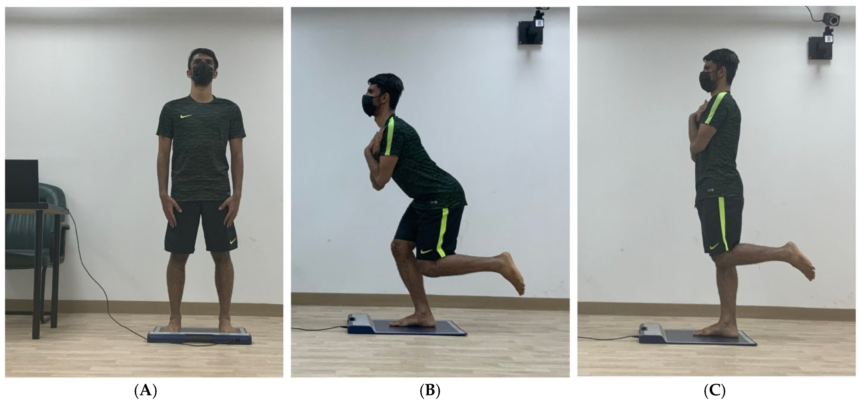
All PPD tests were conducted barefoot to ensure sensor contact and standardisation. Each participant completed two official trials for PPD measurement in double-leg and single-leg stances (left and right) and single-leg squats (left and right) to reduce errors. The official two trials were preceded by two practice trials in each tested position [26]. The researcher averaged participants’ scores from the official trials for analysis. Each trial lasted 6 s [33]. To gather PPD data in the double-leg stance, participants simply stood on the testing mat with feet shoulder-width apart and arms at their sides, looking forward (Figure 2).
To collect PPD data in a single-leg stance, each participant stepped onto the FootMat with one foot. The non-supporting limb was at 90° knee flexion, aligned but not touching the supporting limb. The researcher measured the knee joint angle using a universal goniometer, placing the axis on the knee joint. The top arm was positioned on the lateral thigh, pointing to the greater trochanter, while the lower arm pointed to the lateral malleolus on the lateral leg [28]. Participants folded their arms against their chest and looked forward (Figure 2). They maintained this position for 6 s as PPD data were recorded [28].
PPD data were collected during a single-leg squat, as described by Gwynne [35], but the degree of squatting on a single limb was chosen after [36]. Participants stepped onto the FootMat with the tested leg while the non-supporting leg was at 90° knee flexion, aligned but not touching. A goniometer measured the angle of the non-supporting knee, similar to the PPD measurement in single-leg stance. Participants folded their arms to eliminate the swing and looked forward. They squatted on the tested leg to about 30° knee flexion, maintaining balance (Figure 2). The researcher also measured the supporting leg’s knee angle to confirm the squatting degree. Participants held the position for 6 s while PPD data were recorded. Participants were asked to maintain a neutral foot position during the squat.

2.6. Data Analysis

Statistical analysis was carried out using the SPSS version 27 (IBM Corp., Armonk, NY, USA) software. The Shapiro–Wilk test was used to check the normality of the data. Pearson’s correlation test was used to correlate between normally distributed data, while with non-normally distributed data, Spearman’s correlation test was used. The significance level was set at <0.05. The correlation strength was interpreted as follows: +/− (>0.8), +/− (0.7–0.8), +/− (0.5–0.6), +/− (0.3–0.4), and +/− (0.0–0.2) represent almost perfect, strong, moderate, fair, and poor correlations, respectively [37].

3. Results

The means and SDs of the normally distributed data are provided in Table 2, and the medians and 25–75 percentiles of the non-normally distributed data are provided in Table 3. The correlations between the study variables were made according to the normality of the data. A total of 48 correlations between the participants’ MCET test and PPD data were measured. All the correlations were non-significant, with strengths ranging from poor to fair (Table 4).

Additional Findings

Table 5 shows weak and insignificant correlations between the PPD values of the dominant foot in the tested static positions, except for the significant, moderate, and positive relationship of the total and peak PPD of the dominant foot in the double-leg stance with the peak PPD in the single-leg stance (r = 0.527, p = 0.014; r = 0.570, p = 0.007). As for the non-dominant foot, Table 6 also shows weak and insignificant correlations between its PPD values in the tested static positions.

4. Discussion

This study investigates the link between core muscle endurance and PPD in three static positions among healthy male athletes. To the authors’ knowledge, it is the first to correlate these factors in both athletic and non-athletic populations. The chosen static positions are key to establishing basic knowledge in sports performance, and injury prevention, paving the way for advanced sport-specific research. The chosen static positions are sport-specific [38,39,40] and used in practice and performance testing [41,42]. Single-leg stances and squats are linked to various athletic injuries [43,44,45]. Recreational athletes aged 18 to late 30s were selected for this study, as they are typically in peak health, physically and mentally mature, and self-directed [46]. They excel physiologically in muscle strength, cardiac function, sensory abilities, and reaction time, placing them at the peak of athletic performance and profession [46].
This study finds no correlation between MCET test scores and PPD values (total and peak pressure) in static positions (double-leg stance, single-leg stance, and single-leg squat) among healthy male athletes. Previous studies explored core and leg muscle fatigue’s effect on PPD during dynamic activities [16,17,18,19]. For instance, one study with 46 novice runners found a positive relationship between core muscle fatigue and PPD [19]. Muscular fatigue leads to tiredness and reduced performance. Askari and Esmaeili [19] findings contradict this study’s lack of correlation between core muscle endurance and PPD, likely due to different testing positions: cyclic dynamic in their research versus static in the present study. Thus, core endurance seems more related to PPD in dynamic than static activities [19].
Weist, Eils, and Rosenbaum [16] studied the effect of leg muscle fatigue on PPD during running in 30 experienced runners and found that leg muscle fatigue increased PPD under the medial aspect of the midfoot and the second and third metatarsal heads. Lung, Liau, Peters, He, Townsend, and Jan [17] observed changes in PPD induced by tibialis anterior muscle fatigue during walking and running, and found an increase in peak PPD under the big toe and first metatarsal head. Zhao [18] investigated the effect of leg muscle fatigue on PPD during walking in college students. After fatigue, plantar loading was reduced under the medial arch, distal metatarsals, rear heel, and cuboid bone. Despite the conflicting results of these studies, it is evident that there is a relationship between leg muscle fatigue and PPD during dynamic activities, which negates our findings. Based on the idea that fatigue depletes endurance, it may be concluded that leg muscle endurance also has a correlation with PPD during dynamic activities.
On the other hand, Vie, Gomez, Brerro-Saby, Weber, and Jammes [47] investigated the effect of leg muscle fatigue on the measurement of plantar surface area/cm2 during the double-leg standing position (static). The researchers found that leg muscle (tibialis anterior) fatigue increased the mean pressure and the centre of the pressure surface area/cm2 in 12 healthy male subjects [47]. Again, if fatigue is considered the opposite of endurance, leg muscle endurance may be related to PPD measured in static positions, while in dynamic activities, both core and leg endurance may have a correlation with PPD. Accordingly, a question may be raised as to whether a positive correlation might have been found in the current study if leg muscle endurance were measured and correlated with PPD. Seeking to answer this question may pave the way for future research.
Finally, Lee, Wang, Lee, Yu, Kim, Kim, and heon Hong [48] conducted a study on 21 healthy male and female adults to compare the influences of progressive ankle- and core-muscle-strengthening exercises on static and dynamic balance. The exercises were performed for four weeks. Static balance was measured with stability and weight distribution indices using a Tetrax® device. The dynamic balance of both legs was evaluated using the Y-balance test. It was concluded that both static and dynamic balance abilities were positively affected by the progressive theraband exercises. Thus, core-muscle-strengthening exercises were reported to be more effective with dynamic balance, while ankle-muscle-strengthening exercises were more effective with static balance. Considering that the weight distribution in PPD measurement is directly related to balance, PPD can be used as a measurement of balance and postural sway [49]. The Findings of Lee, Wang, Lee, Yu, Kim, Kim, and heon Hong [48] can perhaps support the proposition that PPD is more related to leg endurance in static positions and to core endurance in dynamic activities.

4.1. Additional Findings

The findings in Table 5 and Table 6 reveal weak, insignificant correlations between PPD measurements across almost all tested positions in both dominant and non-dominant feet. This indicates that PPD measurements in these positions are independent. This novel discovery fills a significant gap in current knowledge. While some studies have examined PPD values during dynamic sports activities [50] or in specific static positions like the single-leg squat [51], none have compared PPD differences across various static positions. The minimal correlation may stem from differing muscle activation in each position, as lower-limb activation varies between double-leg stance [52], single-leg stance [53], and single-leg squat [51]. Thus, the muscle activity variance in these static positions could influence PPD parameters, making the lack of correlation understandable. Nevertheless, further research is necessary to clarify the reasons behind the lack of correlation between PPD in the tested positions.
The previous finding has one exception. Table 5 shows a significant, moderate, positive correlation between the total and peak PPD of the dominant foot in a double-leg stance and peak PPD in a single-leg stance. This novel finding fills a knowledge gap, appearing only in the dominant foot during single-leg stance (not in the non-dominant foot). This may result from the lack of comparability of PPD between feet [54]. Static peak PPD significantly differs between dominant and non-dominant feet, with higher static forces linked to body weight on the dominant side [54]. Further research is needed to identify the reasons for this relationship.

4.2. Clinical Implications

The study findings suggest no correlation between most PPD values for the dominant and non-dominant feet, except for the total and peak PPD in the double-leg stance, which correlated with the peak PPD of the dominant foot in the single-leg stance. Thus, PPD values are largely independent. When PPD measurement is needed to customise insoles to meet sports demands, it may be important to conduct it in sport-specific positions. Further investigation into these findings is recommended.

4.3. Study Limitations

Recruiting only male recreational athletes limited the generalisability of findings to other populations. However, it improved internal validity within the tested group. Another limitation was including individuals from various sports, which reduced sample homogeneity and internal validity but enhanced external validity. Additionally, the MCET test assessed only part of the lower core musculature, boosting internal validity while lowering external validity. Moreover, prolonged sitting weakens back extensor muscles and endurance. This variable was not addressed. Participants’ internal motivation during data collection, which may have affected core muscle endurance values, was not assessed. However, the effect may be minimised since no external motivational talk was given before testing. Also, foot type is a risk factor for changes in PPD values [55]. Research shows that core lateral flexor endurance is lower in university students with flat feet than healthy peers, yet this was only visually inspected here.
The quadriceps angle influences PPD values but was not measured in this study. The lack of control over participants’ fat profiles, like waist circumference and calf girth, is another limitation. While BMI classifies weight status, it does not measure body fat or muscle mass. Despite these limitations, the study offers valuable insights into core endurance and PPD in healthy male recreational athletes, providing guidance for athletes, therapists, and sports organisations to save time and costs.

4.4. Recommendations for Future Studies

Future research may confirm the results of the current study and consider different populations, such as females, the elderly, and individuals with special needs. It may also consider the psychological, nutritional, and lifestyle factors of participants, along with different core stability constructs (strength, power, proprioception). Additionally, researching the correlation of core and leg muscle endurance with PPD in both static and dynamic settings will clarify how endurance relates to PPD in sports, carrying important clinical implications.

5. Conclusions

This study concludes that trunk flexor, extensor, and side flexor endurance are not relevant for screening foot pressure during double-leg stance, single-leg stance, and single-leg squat. Therefore, core endurance neither influences nor is influenced by PPD measurements in this population.

Author Contributions

Conceptualization, R.A.B. and S.N.; methodology, R.A.B. and S.N.; formal analysis, S.N. and Q.M., data curation, R.A.B. and S.N. writing—original draft preparation, R.A.B.; writing—review and editing, Q.M. and T.A.; supervision, S.N. and Q.M.; project administration, T.A. and Q.M. All authors have read and agreed to the published version of the manuscript.

Funding

This study did not receive any specific grant from funding agencies in public, commercial, or not-for-profit sectors.

Institutional Review Board Statement

Ethical approval for the study was obtained from the Institutional Review Board Committee at Imam Abdulrahman bin Faisal University in Dammam, Kingdom of Saudi Arabia (approval number: IRB-PGS-2022-03-086).The study was carried out following the Declaration of Helsinki’s principles.

Informed Consent Statement

All participants in the study provided their informed consent.

Data Availability Statement

The data that support the findings of this study are available from the corresponding author upon request.

Acknowledgments

Special Thanks are due to Zainb AlShami, Alaa Ibrahim, Rayan Alim, Moath Al-Musallam, Hifa Almansoof, Abdulaziz Al-Kwiekbi, Ahlam Alamri, Abdullah Al-Sultan, and Mohammed Abu-Saleh for their valuable support and volunteering during the research process.

Conflicts of Interest

The authors declare no conflicts of interest.

References

  1. Dong, K.; Yu, T.; Chun, B. Effects of core training on sport-specific performance of athletes: A meta-analysis of randomized controlled trials. Behav. Sci. 2023, 13, 148. [Google Scholar] [CrossRef] [PubMed]
  2. Hodges, P.W.; Richardson, C.A. Contraction of the abdominal muscles associated with movement of the lower limb. Phys. Ther. 1997, 77, 132–142. [Google Scholar] [CrossRef] [PubMed]
  3. Butcher, S.J.; Craven, B.R.; Chilibeck, P.D.; Spink, K.S.; Grona, S.L.; Sprigings, E.J. The effect of trunk stability training on vertical takeoff velocity. J. Orthop. Sports Phys. Ther. 2007, 37, 223–231. [Google Scholar] [CrossRef] [PubMed]
  4. McGill, S.M.; Childs, A.; Liebenson, C. Endurance times for low back stabilization exercises: Clinical targets for testing and training from a normal database. Arch. Phys. Med. Rehabil. 1999, 80, 941–944. [Google Scholar] [CrossRef]
  5. Saeterbakken, A.H.; Fimland, M.; Navarsete, J.; Kroken, T.; van der Tillaar, R. Muscle activity, and the association between core strength, core endurance and core stability. J. Nov. Physiother. Phys. Rehabil. 2015, 2, 28–34. [Google Scholar] [CrossRef]
  6. de Bruin, M.; Coetzee, D.; Schall, R. The relationship between core stability and athletic performance in female university athletes. S. Afr. J. Sports Med. 2021, 33, v33i31a10825. [Google Scholar] [CrossRef]
  7. Sato, K.; Mokha, M. Does core strength training influence kinetic efficiency, lower extremity stability, and 5000m performance in runners? J. Strength Cond. Res. 2009, 23, 133–140. [Google Scholar] [CrossRef]
  8. Cobanoglu, G.; Keklik, S.S.; Zorlular, A.; Polat, E.A.; Akaras, E. The relationship between scapular and core muscle endurance in professional athletes. Ann. Med. Res. 2019, 26, 1295–1300. [Google Scholar]
  9. Santos, M.S.; Behm, D.G.; Barbado, D.; DeSantana, J.M.; Da Silva-Grigoletto, M.E. Core endurance relationships with athletic and functional performance in inactive people. Front. Physiol. 2019, 10, 1490. [Google Scholar] [CrossRef]
  10. Nesser, T.W.; Lee, W.L. The relationship between core strength and performance in Division I female soccer players. J. Exerc. Physiol. Online 2009, 12, 21–26. [Google Scholar]
  11. Mohammadi, H.; Fathi, J. The Relationship Between Core Endurance and Performance in National Female Badmin-ton Athletes. Phys. Treat. 2018, 8, 123–131. [Google Scholar]
  12. I Shaikh, A.; Nuhmani, S.; Kachanathu, S.J.; I Muaidi, Q. Relationship of Core Power and Endurance with Performance in Random Intermittent Dynamic Type Sports. Asian J. Sports Med. 2019, 10, e62843. [Google Scholar] [CrossRef]
  13. Wilkerson, G.B.; Giles, J.L.; Seibel, D.K. Prediction of core and lower extremity strains and sprains in collegiate football players: A preliminary study. J. Athl. Train. 2012, 47, 264–272. [Google Scholar] [CrossRef] [PubMed]
  14. Abdallah, A.A.; Mohamed, N.A.; Hegazy, M.A. A comparative study of core musculature endurance and strength between soccer players with and without lower extremity sprain and strain injury. Int. J. Sports Phys. Ther. 2019, 14, 525–536. [Google Scholar] [CrossRef]
  15. Cejudo, A.; Ginés-Díaz, A.; Rodríguez-Ferrán, O.; Santonja-Medina, F.; Sainz de Baranda, P. Trunk Lateral Flexor Endurance and Body Fat: Predictive Risk Factors for Low Back Pain in Child Equestrian Athletes. Children 2020, 7, 172. [Google Scholar] [CrossRef]
  16. Weist, R.; Eils, E.; Rosenbaum, D. The influence of muscle fatigue on electromyogram and plantar pressure patterns as an explanation for the incidence of metatarsal stress fractures. Am. J. Sports Med. 2004, 32, 1893–1898. [Google Scholar] [CrossRef]
  17. Lung, C.-W.; Liau, B.-Y.; Peters, J.A.; He, L.; Townsend, R.; Jan, Y.-K. Effects of various walking intensities on leg muscle fatigue and plantar pressure distributions. BMC Musculoskelet. Disord. 2021, 22, 831. [Google Scholar] [CrossRef]
  18. Zhao, C. Effect of sports fatigue on plantar pressure distribution of healthy male college students. Rev. Bras. Med. Esporte 2022, 28, 398–401. [Google Scholar] [CrossRef]
  19. Askari, Z.; Esmaeili, H. Effect of trunk muscles fatigue on plantar pressure distribution in novice runners. J. Biomech. 2021, 122, 110487. [Google Scholar] [CrossRef]
  20. Teyhen, D.S.; Stoltenberg, B.E.; Collinsworth, K.M.; Giesel, C.L.; Williams, D.G.; Kardouni, C.H.; Molloy, J.M.; Goffar, S.L.; Christie, D.S.; McPoil, T. Dynamic plantar pressure parameters associated with static arch height index during gait. Clin. Biomech. 2009, 24, 391–396. [Google Scholar] [CrossRef]
  21. Chappell, J.D.; Yu, B.; Kirkendall, D.T.; Garrett, W.E. A comparison of knee kinetics between male and female recreational athletes in stop-jump tasks. Am. J. Sports Med. 2002, 30, 261–267. [Google Scholar] [CrossRef] [PubMed]
  22. Vaisman, A.; Guiloff, R.; Rojas, J.; Delgado, I.; Figueroa, D.; Calvo, R. Lower Limb Symmetry: Comparison of Muscular Power Between Dominant and Nondominant Legs in Healthy Young Adults Associated With Single-Leg-Dominant Sports. Orthop. J. Sports Med. 2017, 5, 2325967117744240. [Google Scholar] [CrossRef] [PubMed]
  23. Hammouda, O.; Chtourou, H.; Chaouachi, A.; Chahed, H.; Bellimem, H.; Chamari, K.; Souissi, N. Time-of-day effects on biochemical responses to soccer-specific endurance in elite Tunisian football players. J. Sports Sci. 2013, 31, 963–971. [Google Scholar] [CrossRef] [PubMed]
  24. Racinais, S.; Blonc, S.; Jonville, S.; Hue, O. Time of day influences the environmental effects on muscle force and contractility. Med. Sci. Sports Exerc. 2005, 37, 256–261. [Google Scholar] [CrossRef]
  25. Okada, T.; Huxel, K.C.; Nesser, T.W. Relationship Between Core Stability, Functional Movement, and Performance. J. Strength Cond. Res. 2011, 25, 252–261. [Google Scholar] [CrossRef]
  26. Al-Magsoosi, S.K.; Chong, A.K. Foot Loading Pattern Variations between Normal Weight, Overweight, and Obese Adults Aged 24 to 50 Years. J. Biosci. Med. 2019, 7, 34–49. [Google Scholar] [CrossRef]
  27. Ambegaonkar, J.P.; Mettinger, L.M.; Caswell, S.V.; Burtt, A.; Cortes, N. Relationships between core endurance, hip strength, and balance in collegiate female athletes. Int. J. Sports Phys. Ther. 2014, 9, 604–616. [Google Scholar]
  28. Shamsi, M.; Mirzaei, M.; Khabiri, S.S. Universal goniometer and electro-goniometer intra-examiner reliability in measuring the knee range of motion during active knee extension test in patients with chronic low back pain with short hamstring muscle. BMC Sports Sci. Med. Rehabil. 2019, 11, 4. [Google Scholar] [CrossRef]
  29. Abhilash, P.; Sudeep, S.; Anjana, K. Relationship between core endurance and dynamic balance in professional basketball players: A pilot study. Int. J. Phys. Educ. Sports Health 2021, 8, 1–5. [Google Scholar]
  30. Evans, K.; Refshauge, K.M.; Adams, R. Trunk muscle endurance tests: Reliability, and gender differences in athletes. J. Sci. Med. Sport 2007, 10, 447–455. [Google Scholar] [CrossRef]
  31. McGill, S. McGill’s torso muscular endurance test battery. Am. Counc. Exercise. Pristup. 2015, 3, 2022. [Google Scholar]
  32. Zammit, G.V.; Menz, H.B.; Munteanu, S.E. Reliability of the TekScan MatScan® system for the measurement of plantar forces and pressures during barefoot level walking in healthy adults. J. Foot Ankle Res. 2010, 3, 1–9. [Google Scholar] [CrossRef] [PubMed]
  33. Da, H.K.; Lee, J.D.; Kim, K. Plantar pressures in individuals with normal and pronated feet according to static squat depths. J. Phys. Ther. Sci. 2015, 27, 2833–2835. [Google Scholar]
  34. Bodkin, S.G.; Simpson, A.S.; Kirsch, A.N.; Hart, J.M. Postural Control in Patients with ACL Reconstruction Using Automated Error Detection from Instrumented Balance Measures. Athl. Train. Sports Health Care 2021, 13, e323–e328. [Google Scholar] [CrossRef]
  35. Gwynne, C.R. Alterations in center of pressure during single-limb loading in individuals with patellofemoral pain. J. Am. Podiatr. Med. Assoc. 2020, 110, 5. [Google Scholar] [CrossRef]
  36. Ugalde, V.; Brockman, C.; Bailowitz, Z.; Pollard, C.D. Single leg squat test and its relationship to dynamic knee valgus and injury risk screening. PMR 2015, 7, 229–235, quiz 235. [Google Scholar] [CrossRef]
  37. Portney, L.G.; Watkins, M.P. Foundations of Clinical Research: Applications to Practice; Pearson Prentice Hall: Upper Saddle River, NJ, USA, 2009; Volume 892. [Google Scholar]
  38. Riemann, B.L.; Schmitz, R. The relationship between various modes of single leg postural control assessment. Int. J. Sports Phys. Ther. 2012, 7, 257–266. [Google Scholar]
  39. Vecchio, L.; Daewoud, H.; Green, S. The health and performance benefits of the squat, deadlift, and bench press. MOJ Yoga Phys. Ther. 2018, 3, 40–47. [Google Scholar] [CrossRef]
  40. Ramirez-Campillo, R.; Andrade, D.C.; Nikolaidis, P.T.; Moran, J.; Clemente, F.M.; Chaabene, H.; Comfort, P. Effects of plyometric jump training on vertical jump height of volleyball players: A systematic review with meta-analysis of randomized-controlled trial. J. Sports Sci. Med. 2020, 19, 489–499. [Google Scholar]
  41. Onofrei, R.R.; Amaricai, E.; Petroman, R.; Suciu, O. Relative and absolute within-session reliability of the modified Star Excursion Balance Test in healthy elite athletes. PeerJ 2019, 7, e6999. [Google Scholar] [CrossRef]
  42. Scinicarelli, G.; Trofenik, M.; Froböse, I.; Wilke, C. The reliability of common functional performance tests within an experimental test battery for the lower extremities. Sports 2021, 9, 100. [Google Scholar] [CrossRef] [PubMed]
  43. Nevitt, M.C.; Cummings, S.R.; Hudes, E.S. Risk factors for injurious falls: A prospective study. J. Gerontol. 1991, 46, M164–M170. [Google Scholar] [CrossRef] [PubMed]
  44. Trojian, T.H.; McKeag, D.B. Single leg balance test to identify risk of ankle sprains. Br. J. Sports Med. 2006, 40, 610–613, discussion 613. [Google Scholar] [CrossRef] [PubMed]
  45. Stickler, L.; Finley, M.; Gulgin, H. Relationship between hip and core strength and frontal plane alignment during a single leg squat. Phys. Ther. Sport 2015, 16, 66–71. [Google Scholar] [CrossRef]
  46. Tyler, S. Physical Development in Early Adulthood. In Human Behavior and the Social Environment I; University of Arkansas: Fayetteville, AR, USA, 2020. [Google Scholar]
  47. Vie, B.; Gomez, N.; Brerro-Saby, C.; Weber, J.P.; Jammes, Y. Changes in stationary upright standing and proprioceptive reflex control of foot muscles after fatiguing static foot inversion. J. Biomech. 2013, 46, 1676–1682. [Google Scholar] [CrossRef]
  48. Lee, K.-S.; Wang, J.-W.; Lee, D.Y.; Yu, J.H.; Kim, J.S.; Kim, S.G.; heon Hong, J. Effects of progressive core and ankle muscle strengthening exercises using thera-band on body balance. J. Korean Phys. Ther. 2022, 34, 121–127. [Google Scholar] [CrossRef]
  49. Kim, S.M.; Hyun, G.J.; Jung, T.-W.; Son, Y.D.; Cho, I.-H.; Kee, B.S.; Han, D.H. Balance deficit and brain connectivity in children with attention-deficit/hyperactivity disorder. Psychiatry Investig. 2017, 14, 452. [Google Scholar] [CrossRef]
  50. Eils, E.; Streyl, M.; Linnenbecker, S.; Thorwesten, L.; Völker, K.; Rosenbaum, D. Characteristic plantar pressure distribution patterns during soccer-specific movements. Am. J. Sports Med. 2004, 32, 140–145. [Google Scholar] [CrossRef]
  51. Knoll, M.G.; Davidge, M.; Wraspir, C.; Korak, J.A. Comparisons of single leg squat variations on lower limb muscle activation and center of pressure alterations. Int. J. Exerc. Sci. 2019, 12, 950–959. [Google Scholar] [CrossRef]
  52. Jeon, W.; Whitall, J.; Griffin, L.; Westlake, K.P. Trunk kinematics and muscle activation patterns during stand-to-sit movement and the relationship with postural stability in aging. Gait Posture 2021, 86, 292–298. [Google Scholar] [CrossRef]
  53. Chakravarty, K.; Chatterjee, D.; Das, R.K.; Tripathy, S.R.; Sinha, A. Analysis of muscle activation in lower extremity for static balance. In Proceedings of the 2017 39th Annual International Conference of the IEEE Engineering in Medicine and Biology Society (EMBC), Jeju Island, Republic of Korea, 11–15 July 2017; pp. 4118–4122. [Google Scholar] [CrossRef]
  54. Imamura, M.; Imamura, S.T.; Salomão, O.; Pereira, C.A.M.; De Carvalho Jr, A.E.; Neto, R.B. Pedobarometric evaluation of the normal adult male foot. Foot Ankle Int. 2002, 23, 804–810. [Google Scholar] [CrossRef]
  55. Woźniacka, R.; Oleksy, Ł.; Jankowicz-Szymańska, A.; Mika, A.; Kielnar, R.; Stolarczyk, A. The association between high-arched feet, plantar pressure distribution and body posture in young women. Sci. Rep. 2019, 9, 17187. [Google Scholar] [CrossRef]
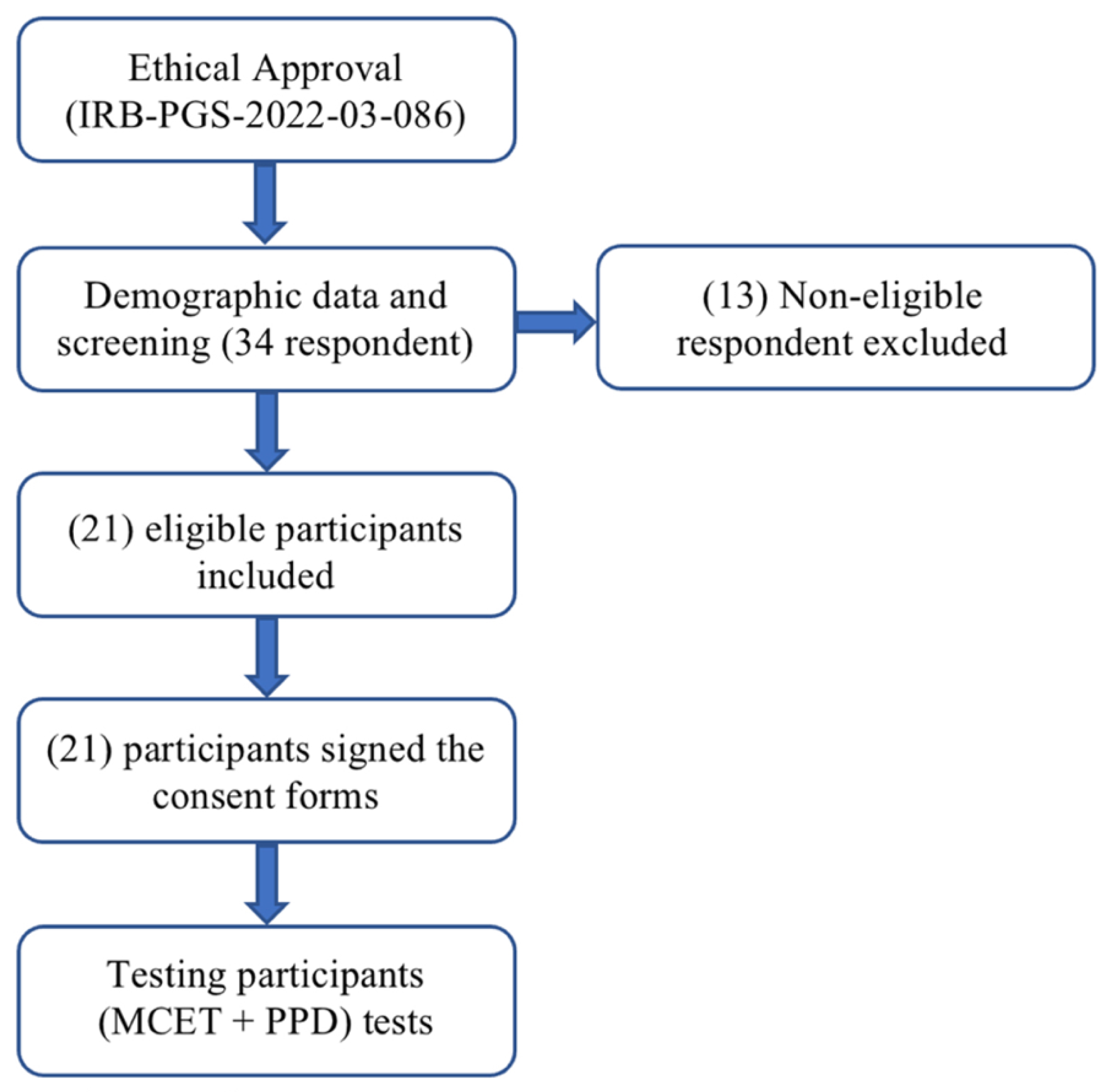
Figure 1. The flow chart of the study procedure.
Medicina 61 01188 g001
Figure 2. Measurement of plantar pressure distribution, (A). in double-leg stance position, (B). single-leg stance position, and (C). single-leg squat position.
Medicina 61 01188 g002
Table 1. The timeline of the outcome measure testing.
Outcome MeasuresTesting Sequence
The sequence of the testing procedure was undertaken randomly for both McGill core endurance and PPD tests (participants chose from folded papers blindly) to rule out that one test might affect the result of the other.
McGill core endurance tests:

- Flexors endurance test.
- Extensors endurance test.
- Left lateral plank test.
- Right lateral plank test.
Explanation and demonstration of the testing position and procedure was provided to each participant.
- Each participant was allowed to practice the testing position for no longer than 5 s to avoid fatigue.
- One official trial was completed only to avoid fatigue [25].
- Once participants assumed the desired position, the official trial started with the word “Start” and ended with “Stop” upon the trunk deviation from the midline noticed.
- The holding time was recorded in seconds.
- Participants were allowed to rest for five minutes between the testing trials to eliminate the effect of fatigue on the next test’s performance [4].
Plantar pressure distribution measurement:
Data were recorded during the following static positions:

- Double-leg stance.
- Single-leg stance.
- single-leg squat.
For all the testing positions:
- Explanation and demonstration of the testing position and procedure was provided to each participant.
- Two practicing trials before the formal ones were allowed.
- Two formal trials were conducted [26].
- Formal trial recording started (after assuming the testing position) with the word “Start” and ended with “Stop”.
- Each formal trial lasted six seconds.
- The average of the two formal trials was taken in data analysis.
For PPD testing in single-leg stance and squat positions:
- The selection of which foot to test first (dominant or non-dominant) was made randomly after a coin flip.
- Before recording, the researcher measured (using a Goniometer) the non-supporting leg’s 90° knee flexion in the single-leg stance and single-leg squat positions. Besides measuring the supporting leg’s 30° knee flexion in the single-leg squat position.
PPD: Plantar pressure distribution.
Table 2. Normally distributed variables’ mean and standard deviation.
Normally Distributed VariablesMean ± Standard Deviation
McGill core endurance test
Extension test (s)34.32 ± 13.88
Left lateral plank test (s)30.76 ± 14.50
Bilateral stance average values of plantar pressure distribution
DPPA22.81 ± 10.32
NDPP25.50 ± 9.97
DPA16.61 ± 6.59
NDPA18.40 ± 6.00
Single-leg stance average values of plantar pressure distribution
DPPA47.09 ± 7.15
NDPPA50.42 ± 10.34
DPA32.90 ± 6.34
NDPA38.04 ± 7.51
Single-leg squat average values of plantar pressure distribution
DPPA53.85 ± 9.58
NDPPA52.61 ± 8.52
DPA35.52 ± 7.19
NDPA34.57 ± 5.32
DPPA, dominant leg peak plantar pressure average; NDPPA, non-dominant leg peak plantar pressure average; DPA, dominant leg total plantar pressure average; NDPA, non-dominant leg total plantar pressure average.
Table 3. Non-normally distributed variables’ median and 25–75 percentiles.
Non-Normally Distributed VariablesMedian (25–75 Percentiles)
McGill core endurance test
Flexion test (s)74.38 (47.78–109.13)
Right lateral plank test (s)24.63 (15.76–35.02)
Table 4. Correlations of the tested variables.
Plantar Pressure DataMcGill Core Endurance Tests
Flexion Endurance Test Extension Endurance Test Right Lateral Plank TestLeft Lateral Plank Test
Double-leg stance position
DPPArho = −0.117
p = 0.614
r = 0.148
p = 0.523
rho = 0.068
p = 0.769
r = 0.030
p = 0.896
NDPPArho = 0.114
p = 0.622
r = 0.159
p = 0.490
rho = 0.112
p = 0.630
r = 0.034
p = 0.883
DPArho = −0.149
p = 0.520
r = 0.125
p = 0.590
rho = 0.109
p = 0.640
r = 0.061
p = 0.794
NDPArho = 0.129
p = 0.578
r = 0.215
p = 0.348
rho = 0.050
p = 0.829
r = 0.060
p = 0.796
Single-leg stance position
DPPArho = −0.079
p = 0.732
r = −0.025
p = 0.915
rho = 0.120
p = 0.605
r = 0.175
p = 0.449
NDPPArho = −0.040
p = 0.865
r = −0.299
p = 0.188
rho = 0.136
p = 0.555
r = −0.104
p = 0.653
DPArho = −0.024
p = 0.918
r = −0.331
p = 0.142
rho = 0.112
p = 0.630
r = 0.011
p = 0.963
NDPArho = 0.025
p = 0.913
r = −0.297
p = 0.191
rho = 0.279
p = 0.220
r = 0.078
p = 0.737
Single-leg squat position
DPPArho = 0.364
p = 0.105
r = 0.341
p = 0.131
rho = 0.068
p = 0.769
r = −0.051
p = 0.826
NDPPArho = 0.182
p = 0.430
r = 0.147
p = 0.526
rho = −0.178
p = 0.440
r = −0.146
p = 0.527
DPArho = 0.331
p = 0.143
r = 0.307
p = 0.176
rho = 0.003
p = 0.991
r = 0.111
p = 0.631
NDPArho = 0.337
p = 0.135
r = 0.257
p = 0.261
rho = −0.071
p = 0.760
r = 0.164
p = 0.476
DPPA, dominant leg peak plantar pressure average; NDPPA, non-dominant leg peak plantar pressure average; DPA, dominant leg total plantar pressure average; NDPA, non-dominant leg total plantar pressure average; r, Pearson’s correlation coefficient; rho, Spearman’s correlation coefficient; p, p-value =< 0.05.
Table 5. Correlations between plantar pressure measurements of the dominant leg.
Double-Leg Stance DPPA Double-Leg Stance DPA Single-Leg Stance DPPASingle-Leg Stance DPA Single-Leg Squat DPPASingle-Leg Squat DPA
Double-leg stance DPPA
Double-leg stance DPA
Single-leg stance DPPAr = 0.570
p = 0.007 *
r = 0.527
p = 0.014 *
Single-leg stance DPAr = 0.335
p = 0.137
r = 0.320
p = 0.157
Single-leg squat DPPAr = 0.088
p = 0.706
r = 0.031
p = 0.894
r = 0.086
p = 0.711
r = −0.052
p = 0.823
Single-leg squat DPAr = 0.283
p = 0.213
r = 0.208
p = 0.367
r = 0.310
p = 0.172
r = 0.236
p = 0.302
DPPA, dominant leg peak plantar pressure average; DPA, dominant leg total plantar pressure average; r, Pearson’s correlation coefficient; p, p-value =< 0.05; * Statistically significant at p < 0.05
Table 6. Correlations between plantar pressure measurements of the non-dominant leg.
Double-Leg Stance NDPPA Double-Leg Stance NDPA Single-Leg Stance NDPPASingle-Leg Stance NDPA Single-Leg Squat NDPPASingle-Leg Squat NDPA
Double-leg stance NDPPA
Double-leg stance NDPA
Single-leg stance NDPPAr = 0.265
p = 0.245
r = 0.210
p = 0.362
Single-leg stance NDPAr = 0.110
p = 0.634
r = 0.108
p = 0.640
Single-leg squat NDPPAr = 0.091
p = 0.695
r = 0.051
p = 0.827
r = 0.247
p = 0.280
r = 0.210
p = 0.362
Single-leg squat NDPAr = 0.284
p = 0.212
r = 0.247
p = 0.281
r= 0.006
p = 0.978
r = −0.137
p = 0.554
NDPPA, non-dominant leg peak plantar pressure average; NDPA, non-dominant leg total plantar pressure average; r, Pearson’s correlation coefficient; p, p-value =< 0.05.
Disclaimer/Publisher’s Note: The statements, opinions and data contained in all publications are solely those of the individual author(s) and contributor(s) and not of MDPI and/or the editor(s). MDPI and/or the editor(s) disclaim responsibility for any injury to people or property resulting from any ideas, methods, instructions or products referred to in the content.