Article

Comparison of Asymptomatic Brain Lesions Between Thalassemia Major and Sickle Cell Anemia Patients

by
Derya Yavuz Demiray
1,*,
Özge Eriş Davut
2 and
Gönül Oktay
3
1
Department of Neurology, School of Medicine, Mustafa Kemal University, Hatay 31100, Turkey
2
Department of Psychiatry, Ankara Etlik City Hospital, Ankara 06710, Turkey
3
Department of Thalassemia Unit, Hatay Education and Research Hospital, Hatay 31027, Turkey
*
Author to whom correspondence should be addressed.
Medicina 2025, 61(1), 159; https://doi.org/10.3390/medicina61010159

Submission received: 15 December 2024 / Revised: 31 December 2024 / Accepted: 15 January 2025 / Published: 19 January 2025
(This article belongs to the Section Hematology and Immunology)

Abstract

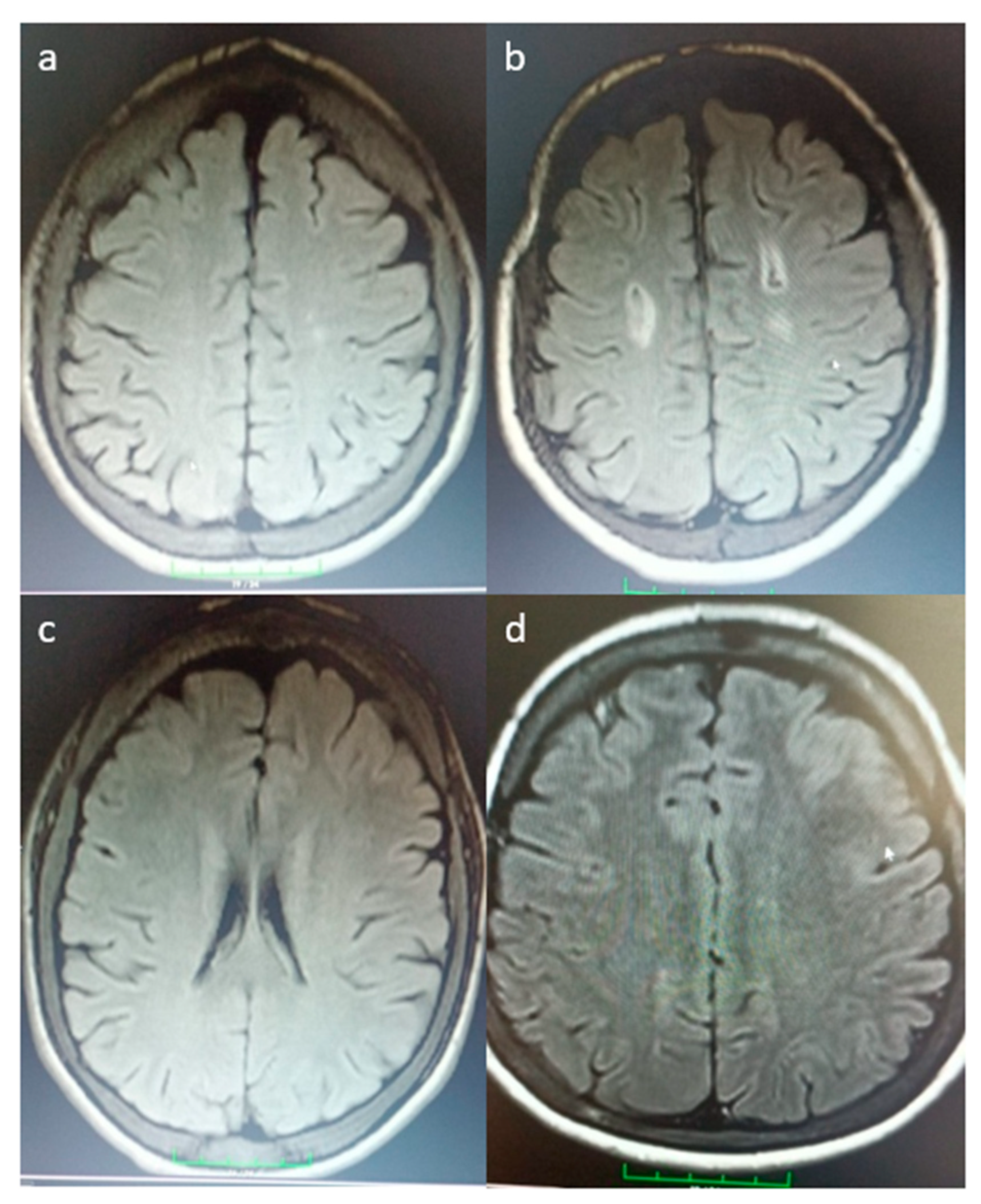
Background and Objectives: This study aimed to identify asymptomatic brain lesions in patients with β-thalassemia major (TM) and sickle cell anemia (SCA) and evaluate the correlation of these lesions with factors such as splenectomy, thrombocytosis, and blood transfusions. Materials and Methods: A total of 26 patients with thalassemia major and 23 patients with sickle cell anemia were included. Ischemic lesions were categorized as lacunar, small vessel, or multifocal. Variables including age, years of education, presence and type of MRI-detected ischemia, smoking status, hemoglobin, hematocrit, platelet count, ferritin levels, vitamin B12 levels, fasting blood sugar, splenectomy status, chelation therapy, and hydroxyurea treatment were compared between the two groups. Results: The mean age was 27.33 years in the thalassemia major group and 32.65 years in the sickle cell anemia group (p = 0.010). No statistically significant difference was observed in the distribution of ischemia types between the groups (p = 0.303). The thalassemia major group had a lower mean hemoglobin level (8.37 g/dL) compared to the sickle cell anemia group (9.57 g/dL) (p = 0.003). Ferritin levels were significantly higher in the thalassemia major group (2018.92 ng/mL) than in the sickle cell anemia group (660.39 ng/mL) (p < 0.001). Conclusions: Although ischemic lesions were more frequently observed in patients with sickle cell anemia, the difference was not statistically significant. These findings emphasize the importance of ongoing surveillance and individualized management to mitigate cerebrovascular risks in both patient populations.
Keywords: thalassemia major; sickle cell anemia; silent infarct; vascular pathology; blood levels; complications; management; neuropathic pain; depression thalassemia major; sickle cell anemia; silent infarct; vascular pathology; blood levels; complications; management; neuropathic pain; depression