Case Report

Cutaneous Calcified Mass of Foot in Pseudohypoparathyoidism: Case Report

Department of Orthopaedic Surgery, Soonchunhyang University Hospital Bucheon, 170, Jomaru-ro, Wonmi-gu, Gyeonggi-do, Bucheon-si 14584, Republic of Korea
*
Author to whom correspondence should be addressed.
These authors contributed equally to this work and are co-first authors.
Medicina 2024, 60(4), 595; https://doi.org/10.3390/medicina60040595

Submission received: 1 March 2024 / Revised: 18 March 2024 / Accepted: 2 April 2024 / Published: 4 April 2024

Abstract

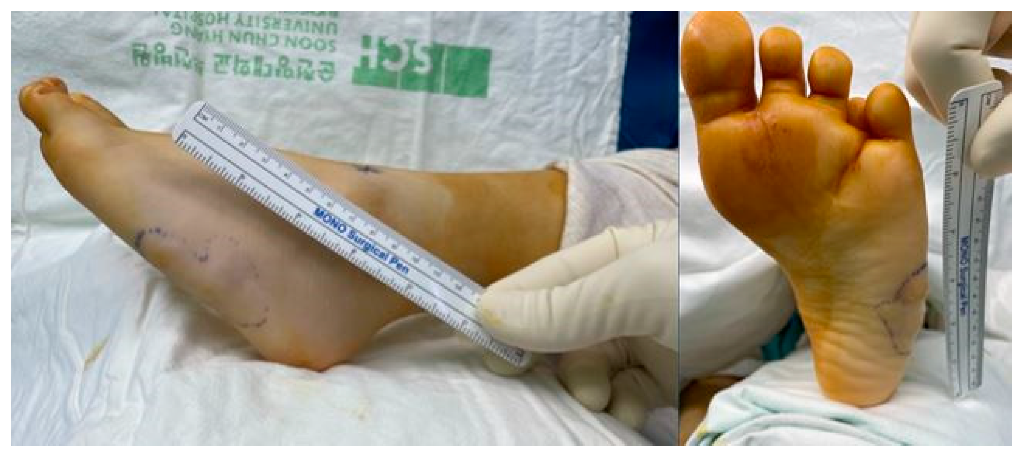
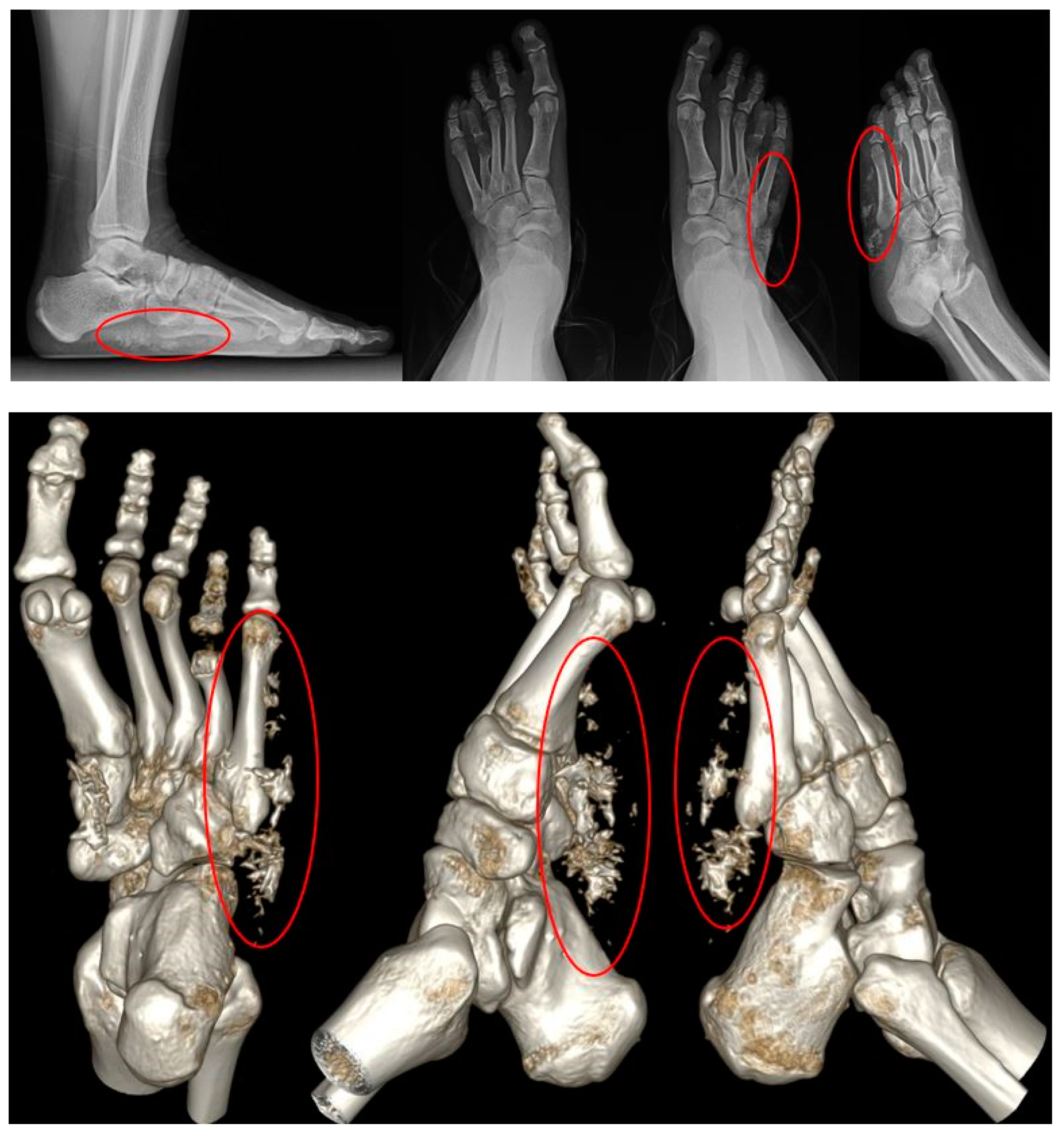
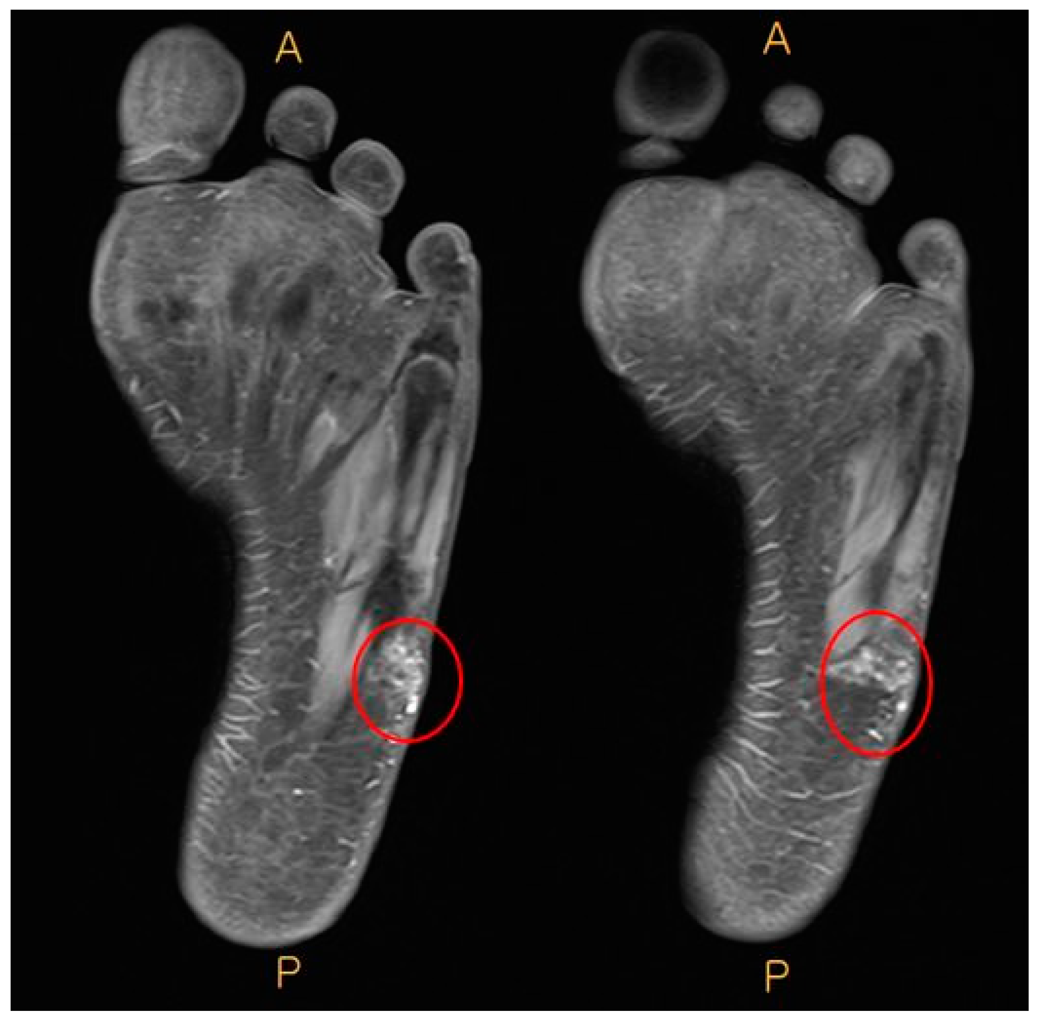
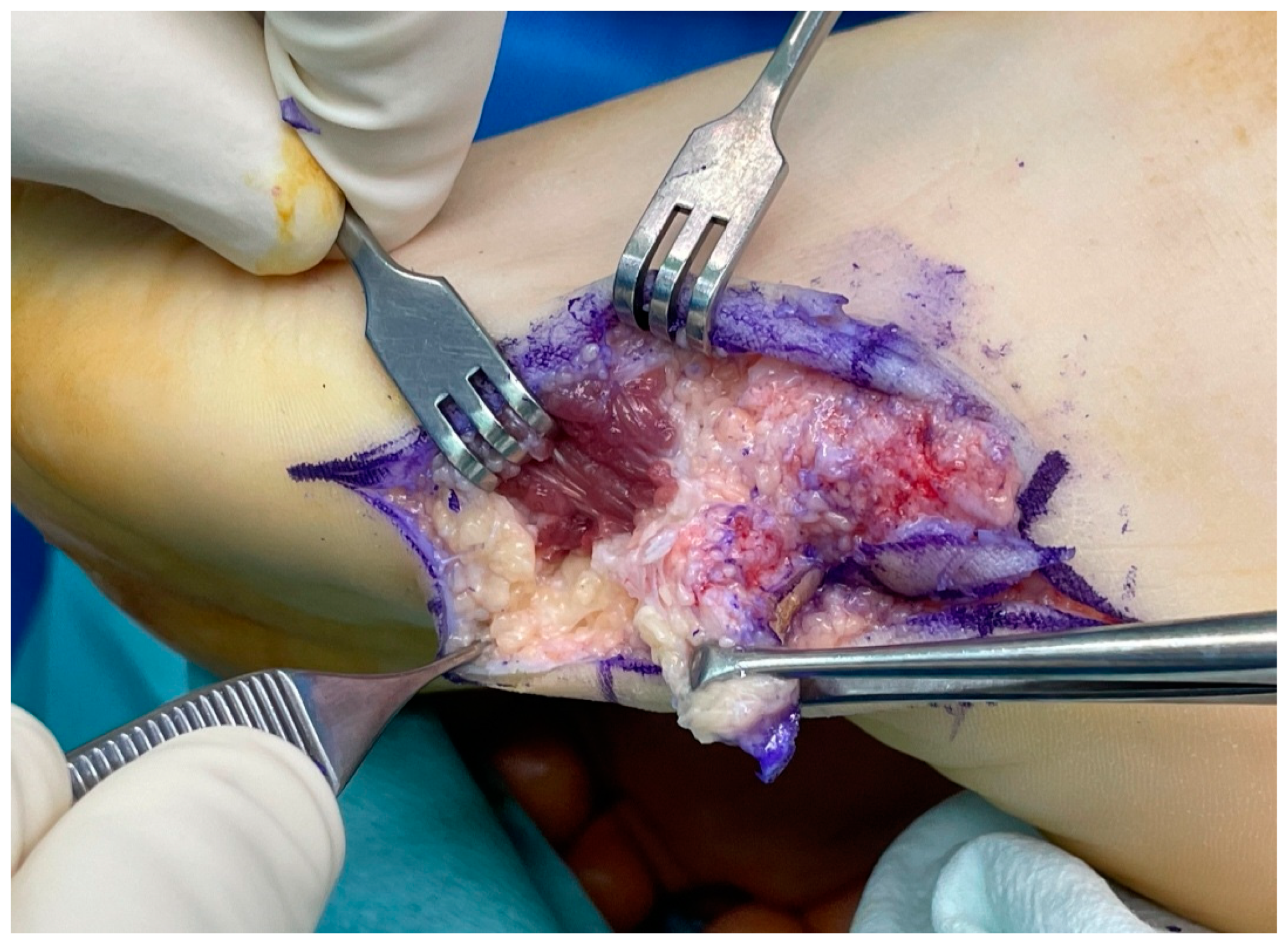
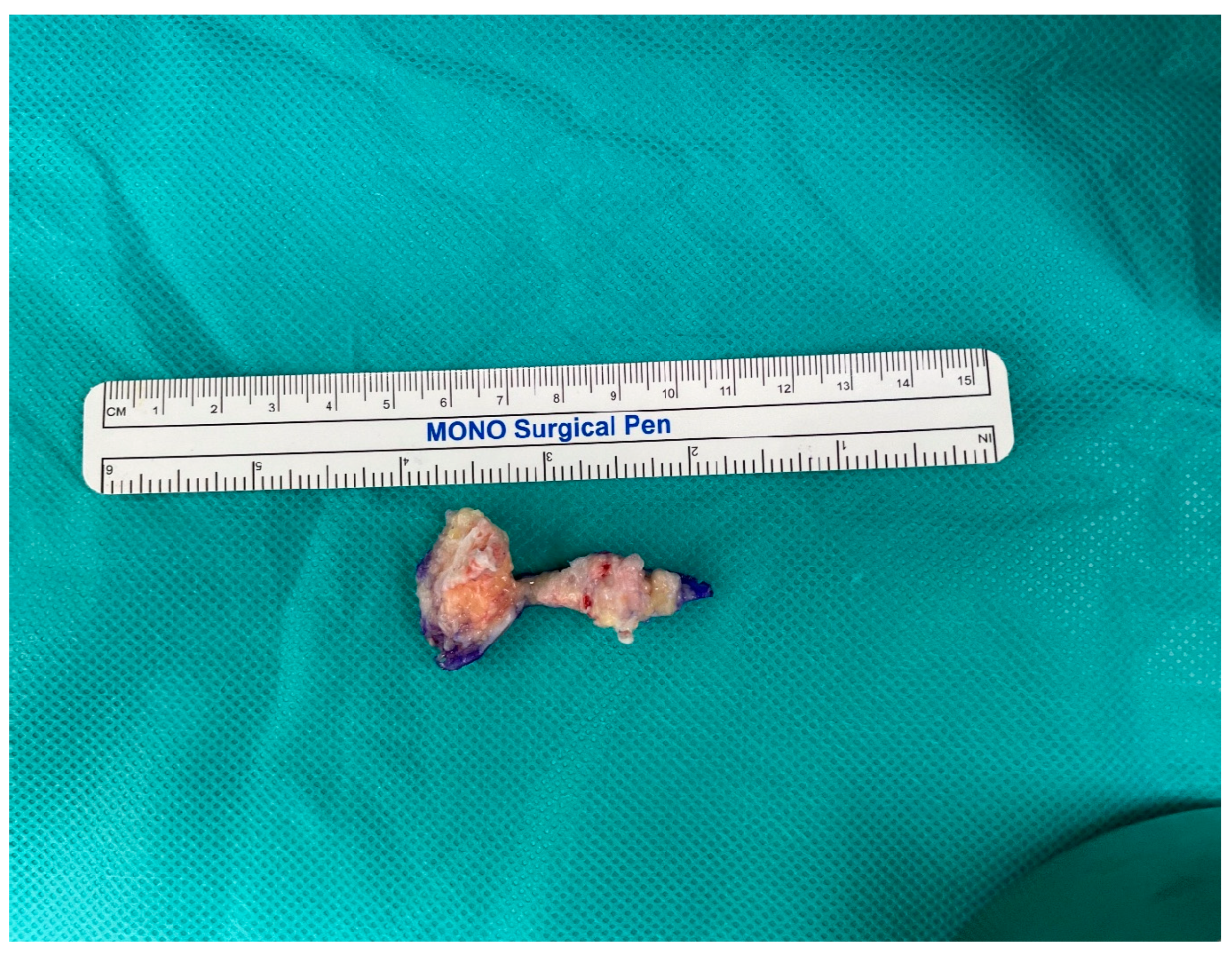
Soft tissue calcifications frequently appear on imaging studies, representing a prevalent but non-specific discovery, varying from a local reaction without clear cause to suggesting an underlying systemic condition. Because calcifications like these can arise from various causes, an accurate differential diagnosis is crucial. Differential diagnosis entails a methodical assessment of the patient, encompassing clinical presentation, medical history, radiological and pathological findings, and other pertinent factors. Through scrutiny of the patient’s medical and trauma history, we can refine potential causes of calcification to vascular, metabolic, autoimmune, neoplastic, or traumatic origins. Furthermore, routine laboratory assessments, including serum levels of calcium, phosphorus, ionized calcium, vitamin D metabolites, and parathyroid hormone (PTH), aid in identifying metabolic etiologies. We describe a rare occurrence of osteoma cutis in a 15-year-old female patient with a history of pseudohypoparathyroidism (PHP) and Albright’s hereditary osteodystrophy (AHO). The patient presented with a painful mass on the lateral side of her left foot. The diagnosis was based on medical history, laboratory tests, and imaging, leading to an excisional biopsy and complete pain relief post-surgery. Understanding such rare occurrences and related conditions is crucial for accurate diagnosis and management.
Keywords: calcification; parathyroid hormone; pseudohypoparathyroidism calcification; parathyroid hormone; pseudohypoparathyroidism