Article

Evaluation of the Risk Factors for Cellulitis among Patients with Peripheral Artery Disease

1
Institute of Medicine, Chung Shan Medical University, Taichung 402, Taiwan
2
School of Medicine, Chung Shan Medical University, Taichung 402, Taiwan
3
Department of Emergency Medicine, Chung Shan Medical University Hospital, Taichung 402, Taiwan
4
Department of Medical Research, Chung Shan Medical University Hospital, Taichung 402, Taiwan
5
Division of Cardiology, Department of Internal Medicine, Changhua Christian Hospital, Yunlin Branch, Changhua 648, Taiwan
6
Department of Post-Baccalaureate Medicine, College of Medicine, National Chung Hsing University, Taichung 402, Taiwan
7
Department of Surgery, Chung Shan Medical University Hospital, Taichung 402, Taiwan
*
Author to whom correspondence should be addressed.
Medicina 2023, 59(5), 933; https://doi.org/10.3390/medicina59050933
Submission received: 6 April 2023 / Revised: 26 April 2023 / Accepted: 11 May 2023 / Published: 12 May 2023
(This article belongs to the Topic Public Health and Healthcare in the Context of Big Data)

Abstract

:
Background and objectives: The objective of this study is to elucidate peripheral occlusion artery disease (PAOD) as a risk factor for cellulitis. Materials and Methods: This is a retrospective population-based cohort study. The database is the Longitudinal Health Insurance Database, which covers two million beneficiaries from the entire population of the 2010 registry for beneficiaries in Taiwan. The PAOD group is composed of patients who were newly diagnosed with PAOD from 2001 to 2014. The non-PAOD group is composed of patients who were never diagnosed with PAOD from 2001 to 2015. All patients were followed until the onset of cellulitis, death, or until the end of 2015. Results: Finally, 29,830 patients who were newly diagnosed with PAOD were included in the PAOD group, and 29,830 patients who were never diagnosed with PAOD were included in the non-PAOD group. The incidence densities (ID) of cellulitis were 26.05 (95% CI = 25.31–26.80) patients per 1000 person-years in the PAOD group and 49.10 (95% CI = 48.04–50.19) in the non-PAOD group. The PAOD group had an increased risk of cellulitis (adjusted HR = 1.94, 95% CI = 1.87–2.01) compared to the non-PAOD group. Conclusions: Patients with PAOD were associated with a higher risk of subsequent cellulitis compared to patients without PAOD.

1. Introduction

Peripheral artery occlusion disease (PAOD), also known as peripheral artery disease (PAD), is a manifestation of atherosclerosis that causes the narrowing or occlusion of the arterial lumen. Blood flow and perfusion are impaired in the area of the distal part of the affected arteries [1]. Figures on the prevalence of PAOD vary with diagnostic criteria and the study population. The prevalence of PAOD was about 4.67–14% and up to 24.7% in individuals over the age of 70 [2,3,4]. It is estimated that over 200 million people have PAOD globally [5]. Cigarette smoking, diabetes, hypertension, hyperlipidemia, and age are the presently known risk factors [6,7,8]. Intermittent claudication (IC) is the most common symptom of PAOD, which manifests as leg pain that is induced by walking and relieved by rest. The severity of symptoms is determined by the extent of arterial stenosis, the number of affected arteries, and the intensity of exercise. Symptoms of PAOD in the male population were reported to be more severe than in the female population [6]. The diagnosis of PAD was also presented in the previous study [1].
Cellulitis is an infection of the subcutaneous tissue and deep dermis when indigenous flora enters the dermis and colonises the dermis by breaking the skin barrier; it manifests as an area of skin erythema, swelling, sensation of heat, and tenderness in the affected area [9]. The incidence of cellulitis varies by country. In the USA, approximately 25/1000 person-years was reported in 2006 [10]. In the Netherlands, the annual incidence was estimated to be 22/1000 person-years [11]. A study estimated that the incidence of cellulitis in the UK was 16.4/1000 person-years [12]. Cellulitis became a health burden, causing more than 650,000 hospitalizations and 3.7 billion USD in ambulatory care costs annually in the USA. Infectious organisms are identified in about 15% of cases; most of them are β-hemolytic Streptococcus and Staphylococcus aureus [9]. The infection can occur anywhere on the body, and the lower extremities are affected in most cases [13]. Cellulitis is usually thought to be caused by β-hemolytic Streptococcus and Staphylococcus aureus, and the presence of β-hemolytic Streptococcus and/or Staphylococcus aureus in the toe webs was strongly considered as a risk factor for cellulitis. Other risk factors associated with cellulitis include trauma, diabetes, smoking, chronic venous insufficiency, edema, destroyed skin barrier, previous history of bacterial cellulitis, existence of erosions or ulcers of the lower extremities, and a history of saphenectomy [13,14,15].
Although it remains controversial, there is increasing evidence linking skin lesions to infectious diseases [16,17,18]. A previous review article showed that patients with eosinophilic skin diseases, in which tissue layers or skin structures were affected by eosinophil infiltration and/or degranulation, had limited defense against certain parasitic infections [19,20]. Another meta-analysis showed that the pooled adjusted odds ratio of non-purulent cellulitis was more than 13 times higher in patients with current leg ulcers and 6 times higher in patients with excoriating skin diseases compared to patients in the control group [21]. In addition, maintaining the heath of skin tissue and protecting against microbial invasion depend on whether the functions of the blood vessels are normal or not. Impaired function of the skin microcirculation leads to being unable to maintain skin defense mechanisms. A comparative study described the relationship among skin lesions, vascular disease, and infection, and found that patients with chronic skin lesions and with peripheral vascular lesions simultaneously had differences in cutaneous bacterial flora compared to patients only with chronic skin lesions [17]. PAOD can cause arterial dysfunction and impaired blood perfusion; a progressive reduction of nutrition and oxygen supplement may change the physiological integrity of the skin tissue. According to the PAOD classification system, ischemia, tissue loss, ulcers, and even necrosis can occur as the disease progresses [22]. Therefore, PAOD patients are more susceptible to develop consequent cellulitis is a reasonable assumption, but research on the association between PAOD and cellulitis is still limited with only a few studies indicating this possibility without examining the time sequence between PAOD and cellulitis. Consequently, this study aimed to investigate the association of PAOD with the development of subsequent cellulitis.

2. Materials and Methods

2.1. Data Source

This retrospective, population-based cohort study was conducted using the dataset from the Longitudinal Health Insurance Database (LHID) 2010. LHID 2010 is an electronic database built based on the National Health Program that contains more than 99% of the population and is administered and regulated by the Health and Welfare Data Science Center in Taiwan. The database is composed of two million Taiwanese residents who were randomly enrolled and sampled by stratification according to age, sex, and region from the nearly entire Taiwan population of the 2010 registry in the mandatory National Health Insurance program. Its contents are comprised of patients’ information, such as the registry for beneficiaries, administrative claims in ambulatory care visits and admissions, diagnosis, medication, and interventions. All insurance claims were monitored by reimbursement specialists and peer reviewers, which made the diagnosis coding highly reliable. We can trace the medical record of patients from 2000 to 2015 by an analysis of the LHID 2010. This study protocol was reviewed by the Chung Shan Medical University Hospital ethics review board (CS1-20056).

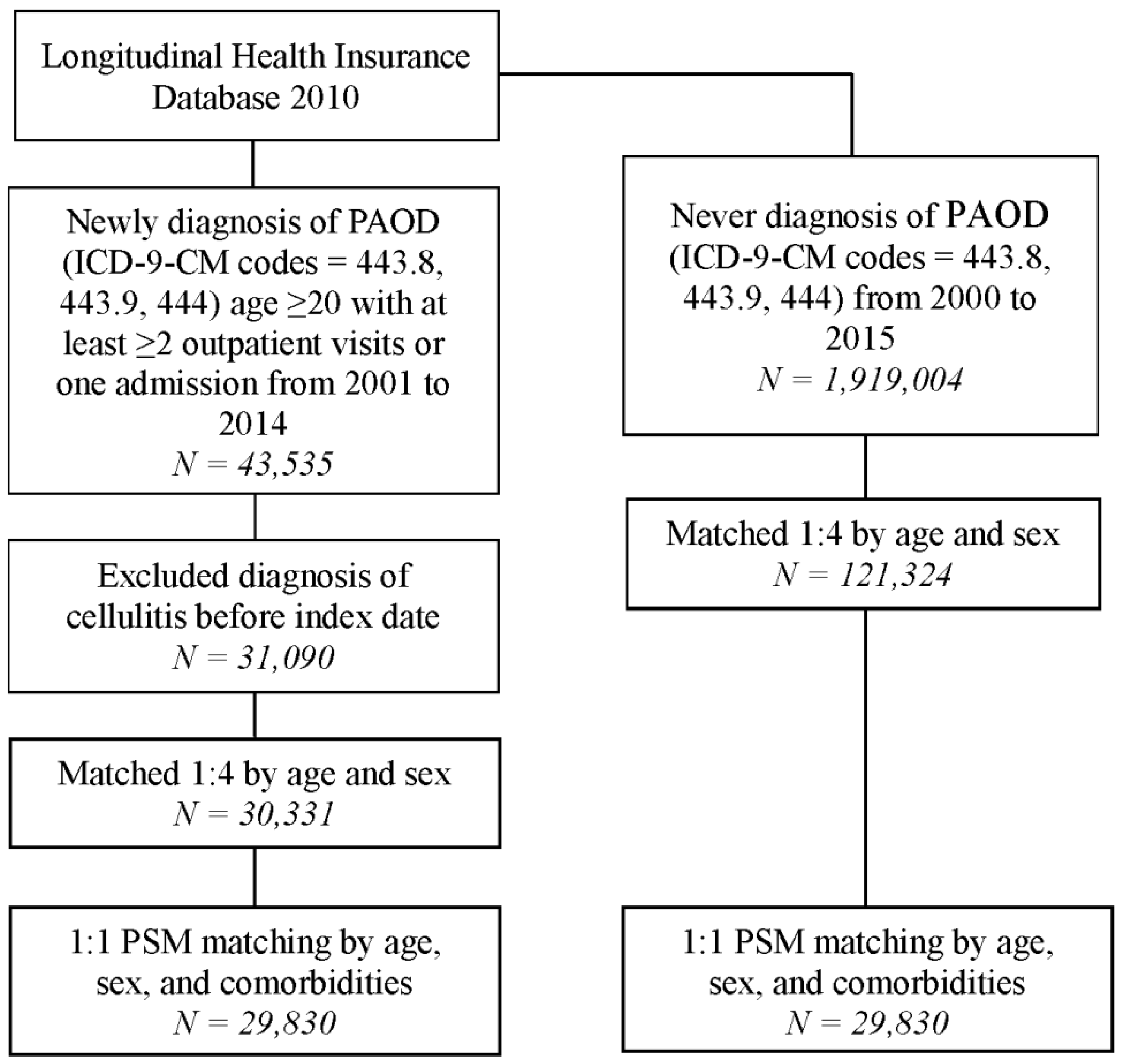
2.2. Patient Selection and Outcome

The study sample was divided into two stratified cohorts based on the presence or absence of PAOD disease. Patients had newly diagnosed PAOD if they had received PAOD-related diagnostic codes (ICD-9-CM codes: 443.8, 443.9, and 444) in more than two outpatient visits or one admission, according to their medical records from 2001 to 2014, and comprised the PAOD group. The index date was the date of the first diagnosis of PAOD. To accurately explicate the association between PAOD and cellulitis, patients with a diagnosis of cellulitis before the index date were excluded. Patients without a PAOD diagnosis from 2000 to 2015 comprised the non-PAOD group. The outcome variable was the diagnosis of cellulitis, according to the diagnostic codes (ICD-9-CM codes: 376.01, 478.21, 478.71, 528.3, 614.3, 614.4, 681.0, 681, and 682). Both groups were followed-up until the onset of cellulitis, death, or 31 December 2015, whichever happened first.

2.3. Covariates and Matching

The demographic characteristics included in the analysis were age and sex and the comorbidities of hypertension, hyperlipidemia, diabetes, hyperthyroidism, hypothyroidism, rheumatoid arthritis, systemic lupus erythematosus, Sjogren’s syndrome, ankylosing spondylitis, chronic pulmonary disease, renal disease, and liver disease. An individual was defined if they received a diagnosed within 1 year before the index date for at least 2 outpatient visits or one admission.
First, a 1:4 age and sex matching was used to ensure an identical index date for any given PAOD participant with their non-PAOD counterparts. Subsequently, propensity score matching (PSM) was conducted by age, sex, and all comorbidities. The propensity score was calculated using a binary logistic regression. The binary variable was a PAOD diagnosis. PSM ensured the balanced heterogeneity of the two groups. For the subgroup analysis, the samples of the study group were divided into different subgroups according to age (20–40, 40–60, and those older than 60 years old), sex, and comorbidities (Supplementary Table S1).

2.4. Sensitivity Analysis

In order to verify the stability of the results, we conducted a sensitivity analysis with hypoglycemic drugs (ATC codes: A10A, A10BA, A10BB, A10BF, A10BG02, A10BG03, A10BH, A10BX02, and A10BX03) and statins (ATC code: C10AA) that were included in this study variable. Moreover, chronic limb threatening ischaemia (CLTI, ICD-9-CM = 707.10–707.15, 707.19, 730.05, 730.06, 730.07, 730.15, 730.16, 730.17, 785.4), and end-stage renal disease (ESRD, ICD-9-CM = 585 with use of erythropoietin, ATC codes: B03XA01, B03XA02, and B03XA03) were also added to control the interference of disease severity.

2.5. Statistical Analysis

The characteristics of the PAOD and the non-PAOD groups were compared using the absolute standardized difference (ASD). An ASD < 0.1 indicates similarity in the characteristics of both groups. The incidence density of cellulitis in the PAOD and non-PAOD groups was calculated by dividing the number of events by 1000 person-years at risk. The Kaplan–Meier curves were used to calculate the cumulative incidence of PAOD development between the study group and the control group. The relative risk (RR) of cellulitis and the confidence intervals (CI) were calculated using Poisson regression. The hazard ratio (HR) of cellulitis between the two groups was estimated by using the multivariate Cox proportional hazard model. Whether there was a significant difference between the two groups is determined by using the log-rank test. All the statistical analyzes in this study were performed in SAS version 9.4 (SAS Institute, Cary, NC, USA). Statistical significance was set at a p-value smaller than 0.05.

3. Results

3.1. Characteristics of the Participants

After selection, a total of 29,830 patients were included in the PAOD group (study group), while another 29,830 patients were enrolled in the non-PAOD group (control group). The study framework is shown in Figure 1. The baseline characteristics and comorbidities between the PAOD and the non-PAOD groups are shown in Table 1. The mean age of patients with and without PAOD was 63.70 ± 13.98 and 62.96 ± 14.48, respectively. The male/female ratio was similar in both groups (47.4%:52.6% in the PAOD group; 47.5%:52.5% in the non-PAOD group). There was no difference in demographic data and comorbidities between the study or control groups after the propensity score-matching process. The mean track time was 5.45 ± 4.00 and 6.07 ± 3.85 years in the PAOD and non-PAOD groups, while the time to event in the PAOD and non-PAOD groups was 3.46 ± 3.21 and 4.39 ± 3.25 years.

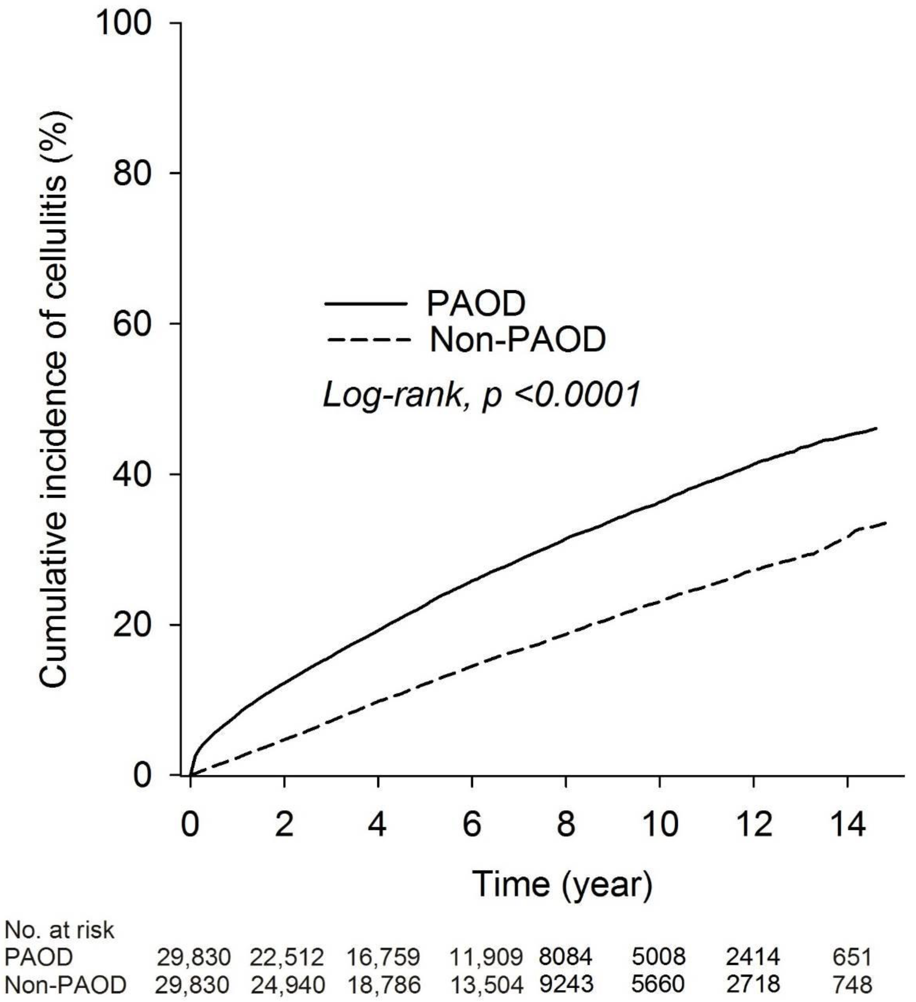
3.2. Risk of Cellulitis between PAOD and Non-PAOD Groups

The incidence densities of cellulitis were 26.05 (95% CI = 25.31–26.80) and 49.10 (95% CI = 48.04–50.19) patients per 1000 person years in the non-PAOD and PAOD groups, respectively. The RR of cellulitis was 1.89-fold (95% CI = 1.82–1.95) higher in the PAOD group compared to the non-PAOD group (Table 2). The cumulative incidence of cellulitis in both groups revealed that cellulitis was higher in the PAOD group compared to the non-PAOD group (log-rank test, p < 0001; Figure 2).

3.3. Comparison of the Risk of Cellulitis between PAOD and Non-PAOD Groups

After the statistical adjustment of age, sex, and associated comorbidities, the risk of cellulitis in the PAOD group was 1.94-fold (95% CI = 1.87–2.01) higher compared to the non-PAOD group. Compared to the subgroup aged 20–40 years old, the risk of cellulitis increased markedly with age. Compared to the female population, the risk of cellulitis was higher in the male population. The analyses of comorbidities, hypertension, diabetes, rheumatoid arthritis, SLE, ankylosing spondylitis, chronic pulmonary disease, renal disease, and liver disease as risk factors for cellulitis are shown in Table 3. In the sensitivity analysis, there were no differences between the study and control groups after the propensity score-matching process (Supplementary Table S2). After adjusting demographic data, comorbidities, and medications, the risk of cellulitis in the PAOD group was 1.85-fold (95% CI = 1.78–1.92) higher than the non-PAOD group (Supplementary Table S3).

3.4. Subgroup Analysis of Cellulitis Risk in the PAOD Group Relative to the Non-PAOD Group after Propensity Score Matching

In a subgroup analysis, the HR of cellulitis is higher in the PAOD group than in the non-PAOD group in all age subgroups. Compared to patients without PAOD, patients in the PAOD group aged 20 to 40 years (HR = 1.75, 95% CI = 1.48–2.08), aged 40 to 60 years (HR = 1.79; 95% CI = 1.70–1.90), and aged ≥60 years (HR = 2.12; 95% CI = 2.02–2.23) had a higher risk of cellulitis. In addition, in the PAOD group, the risk of cellulitis was higher in both male (HR = 2.12; 95% CI = 2.02–2.24) and female (HR = 1.84; 95% CI = 1.75–1.94) populations. Patients with PAOD were shown to have a higher risk of cellulitis compared to patients without PAOD, whether the following diseases were present or not: hypertension, hyperlipidemia, diabetes, hyperthyroidism, hypothyroidism, rheumatoid arthritis, Sjogren’s syndrome, chronic pulmonary disease, renal disease, and liver disease. Furthermore, the risk of cellulitis was significantly higher in the PAOD group compared to the non-PAOD group for patients without the comorbidity of ankylosing spondylitis. The details of the subgroup analyses are shown in Table 4.

4. Discussion

Patients with or without a new diagnosis of PAOD were enrolled to analyze their association with cellulitis. In this study, we found that the incidence of cellulitis increased in patients with PAOD compared to those without PAOD. PAOD is associated with atherosclerosis, which results in the subintimal accumulation of lipid and fibrous materials and causes the vessel lumen to narrow. PAOD is often asymptomatic, and symptoms of PAOD such as claudication, rest pain, ulceration, and gangrene are mainly based on the extent of vascular narrowing and the locations of affected vessels. In patients with PAOD, the adhesion and recruitment of leukocyte and platelet cells, increased activity of platelet activation and thrombosis formation, and dysregulation of vessel tone may cause endothelial dysfunction [23,24]. The selective barrier function of regulating the transport of fluid and intercellular components, vascular reactivity via NO synthase to control blood flow, and proinflammatory activation are considered vital functions of endothelial cells [24,25,26,27,28]. Dysfunction of endothelial cells affects the regulation of coagulation, platelet, and complement activation, as well as leukocyte recruitment in the microvasculature [29]. The progressive reduction of blood flow also gives rise to free radical production and lower oxygen supplementation [30]. Oxygen is important for wound healing, cellular function maintenance, and prophylaxis against infection [31]. Inadequate perfusion of skin tissue below the level adequate to maintain skin structure may change the physiological integrity of skin tissue and, finally, lead to poor wound healing, an ischemic ulcer, or gangrene. The alteration of the immune defenses and destruction of skin integrity result in an increasing risk of developing infection.
Several studies have documented the association between PAOD and infectious diseases. Wang et al. revealed that patients with PAOD were at an increased risk of future sepsis events [32]. The other retrospective cohort study in 2022 showed a similar finding; it reported patients with PAOD were prone to suffer from impaired immune responses due to endothelial damage and dysfunction. Those patients also had a higher risk of sepsis after infection [33]. Kim et al. found that when admitted with an infection of SARS-CoV-2, patients with PAOD have over 40% mortality [34]. Moreover, an observational cohort study in the United States during the period of 2011–2013, which enrolled patients aged 71 to 90 years, reported that the contribution of PAOD to infection risk was similar to that of coronary heart disease and stroke, and PAOD was firmly associated with infection-related hospitalization in older individuals [35]. By contrast, we found that patients with PAOD were associated with a higher HR of cellulitis in all age subgroups.
A retrospective study in Sweden from 2005 to 2010 indicated the rate of infection was significantly associated with the degree of lower extremity ischemia [36]. This study has clinical implications. Patients with PAOD have a high risk of cellulitis. The length of time from the index date to the diagnosis of cellulitis is shorter in the PAOD group than in the non-PAOD group (3.46 ± 3.21 vs 4.39 ± 3.25 years). Skin necrosis, ulcers, gangrene, or chronic limb-threatening ischemia may be represented as the progression of PAOD and the susceptibility to infection. In addition, coexistence with complex wounds and superimposed infections also increases the difficulty of treatments. On the other hand, severe PAOD may develop chronic limb-threatening ischemia (CLTI), which has a high rate of mortality and limb loss. The risk of limb loss in patients without treatment is estimated to be about 20% by 1 year, and the mortality rate is typically over 50% by 5 years [37,38]. It is predictable that PAOD will become increasingly universal in the future with aging of the global population. Despite the fact that it is extremely common, the early stage of PAOD is often asymptomatic, and the majority of PAOD patients are undiagnosed and undertreated [39]. Identifying the risk factors and paying attention to the early detection of PAOD are imperative. Lifestyle modifications, such as smoking cessation, weight reduction, and treatment of other metabolic or cardiovascular diseases, can reduce rates of adverse events and complications [40,41]. To prevent further infections and failures in wound healing, angioplasty, bypass surgery, and hyperbaric oxygen therapy should also be considered [42,43,44].
There are several limitations to this study. First, the physiological and laboratory data of the studied patients were not contained in the database. We cannot evaluate the severity and process of infectious diseases in the absence of C-reactive protein (CRP), erythrocyte sedimentation rate (ESR) level, the results of blood or pus culture, or the ankle-brachial index (ABI), which is a non-invasive assessment tool used to measure the severity of atherosclerotic burden. CRP and ESR are two frequently ordered laboratory tests that may help physicians diagnose and access disease states. However, in some cases, CRP and ESR were not recommended as tools used to alter medical decision-making and the course of treatment in a patients with an indeterminate clinical suspicion of disease because of low sensitivity or specificity [45]. On the other hand, ABI is considered a useful, simple, and inexpensive tool for the diagnosis and assessment of the severity of PAOD; however, it has a wide range of sensitivity, especially in elderly individuals and patients with diabetes [46,47]. Second, the laboratory markers of disease severity of risk factors for cellulitis, such as the glycated hemoglobin (HBA1c) level in diabetes and renal function in chronic kidney disease, cannot be collected from the claims database. The site of ulcer or gangrene and the region of cellulitis were also not fully obtained. It was hard to determine the time sequence and analyze the existing association between soft tissue lesions such as venous ulcers or lymphedema and cellulitis. In addition, we did not know the mechanism of cellulitis related to open wounds, the distribution of cellulitis at each stage of PAOD, or the proportion of undiagnosed PAOD patients in the control group in this study, which cannot answer whether patients with each stage of PAOD were associated a higher risk of subsequent cellulitis compared to patients without PAOD. Third, the database did not provide the timing of the intervention, such as debridement, and the differences in therapeutic strategies between PAOD and non-PAOD groups were not compared. In order to conquer these limitations, we used a propensity score by matching age, sex, and comorbidities to reduce the heterogeneity between the two groups. Fourth, this is an observational and retrospectively designed study, and the accuracy and precision of the results may be restricted. The exact mechanism linking the correlations between PAOD and cellulitis is required to be clarified by further investigation.

5. Conclusions

In conclusion, the results of this study revealed that patients with PAOD were associated with a higher risk of subsequent cellulitis compared to patients without PAOD. PAOD had a significant risk of cellulitis among patients who were ≥60 years old, male, had diabetes, had ankylosing spondylitis, and were in the renal disease subgroup.

Supplementary Materials

The following supporting information can be downloaded at: https://www.mdpi.com/article/10.3390/medicina59050933/s1, Table S1: Disease code of comorbidities; Table S2: Sensitivity analysis of demographic characteristics of PAOD and Non-PAOD; Table S3: Sensitivity analysis of Cox proportional hazard model analysis for risk of cellulitis.

Author Contributions

B.-Y.W., S.-F.Y. and C.-B.Y. conceptualized the study; Y.-H.W. conducted formal analysis; B.-Y.W., K.-H.T., M.-C.C. and C.-B.Y. prepared the original draft; B.-Y.W., S.-F.Y. and C.-B.Y. reviewed and edited the manuscript. All authors have read and agreed to the published version of the manuscript.

Funding

This study was funded by Chung Shan Medical University Hospital (CSH-2022-C-010 and CSH-2022-A-005).

Institutional Review Board Statement

The study was conducted in accordance with the Declaration of Helsinki guidelines and was approved by the Ethical Review Board of the Chung Shan Medical University Hospital (CS1-20056) on 25 May 2020.

Informed Consent Statement

Because this study involved the processing of a database, patient consent was waived by both the National Health Insurance Administration and the Institutional Review Board at Chung Shan Medical University Hospital.

Data Availability Statement

The data used in this study were obtained from the National Health Insurance database and are available from the authors with permission of the National Health Insurance Administration of Taiwan, subject to certain restrictions.

Conflicts of Interest

The authors declare no conflict of interest.

References

  1. Yeh, Y.T.; Tseng, Y.S.; Wu, Y.L.; Yang, S.F.; Wang, B.Y.; Wang, Y.H.; Yeh, L.T.; Yeh, Y.T.; Chan, C.H. Risk of Peripheral Arterial Occlusive Disease with Periodontitis and Dental Scaling: A Nationwide Population-Based Cohort Study. Int. J. Environ. Res. Public Health 2022, 19, 10057. [Google Scholar] [CrossRef] [PubMed]
  2. Binaghi, F.; Fronteddu, P.F.; Cannas, F.; Caredda, E.; Uras, A.; Garau, P.; Pitzus, F. Prevalence of peripheral arterial occlusive disease and associated risk factors in a sample of southern Sardinian population. Int. Angiol. 1994, 13, 233–245. [Google Scholar]
  3. Hiatt, W.R.; Hoag, S.; Hamman, R.F. Effect of diagnostic criteria on the prevalence of peripheral arterial disease. The San Luis Valley Diabetes Study. Circulation 1995, 91, 1472–1479. [Google Scholar] [CrossRef] [PubMed]
  4. Hooi, J.D.; Stoffers, H.E.; Kester, A.D.; Rinkens, P.E.; Kaiser, V.; van Ree, J.W.; Knottnerus, J.A. Risk factors and cardiovascular diseases associated with asymptomatic peripheral arterial occlusive disease: The Limburg PAOD Study. Scand. J. Prim. Health Care 1998, 16, 177–182. [Google Scholar] [CrossRef]
  5. Fowkes, F.G.; Rudan, D.; Rudan, I.; Aboyans, V.; Denenberg, J.O.; McDermott, M.M.; Norman, P.E.; Sampson, U.K.; Williams, L.J.; Mensah, G.A.; et al. Comparison of global estimates of prevalence and risk factors for peripheral artery disease in 2000 and 2010: A systematic review and analysis. Lancet 2013, 382, 1329–1340. [Google Scholar] [CrossRef] [PubMed]
  6. Criqui, M.H.; Aboyans, V. Epidemiology of peripheral artery disease. Circ. Res. 2015, 116, 1509–1526. [Google Scholar] [CrossRef]
  7. Joosten, M.M.; Pai, J.K.; Bertoia, M.L.; Rimm, E.B.; Spiegelman, D.; Mittleman, M.A.; Mukamal, K.J. Associations between conventional cardiovascular risk factors and risk of peripheral artery disease in men. JAMA 2012, 308, 1660–1667. [Google Scholar] [CrossRef]
  8. Song, P.; Rudan, D.; Zhu, Y.; Fowkes, F.J.I.; Rahimi, K.; Fowkes, F.G.R.; Rudan, I. Global, regional, and national prevalence and risk factors for peripheral artery disease in 2015: An updated systematic review and analysis. Lancet Glob. Health 2019, 7, e1020–e1030. [Google Scholar] [CrossRef]
  9. Raff, A.B.; Kroshinsky, D. Cellulitis: A Review. JAMA 2016, 316, 325–337. [Google Scholar] [CrossRef]
  10. Ellis Simonsen, S.M.; van Orman, E.R.; Hatch, B.E.; Jones, S.S.; Gren, L.H.; Hegmann, K.T.; Lyon, J.L. Cellulitis incidence in a defined population. Epidemiol. Infect. 2006, 134, 293–299. [Google Scholar] [CrossRef]
  11. Cranendonk, D.R.; Lavrijsen, A.P.M.; Prins, J.M.; Wiersinga, W.J. Cellulitis: Current insights into pathophysiology and clinical management. Neth. J. Med. 2017, 75, 366–378. [Google Scholar] [PubMed]
  12. McCormick, A. Morbidity statistics from general practice. In Fourth National Study 1991–1992; Office of Population Censuses and Surveys: London, UK, 1995. [Google Scholar]
  13. Bhagat, T.S.; Kumar, L.; Garg, P.; Goel, A.; Aggarwal, A.; Gupta, S. To Study the Clinical Profile and Management of Cellulitis of Lower Limb in Northern India. Int. J. Low. Extrem. Wounds 2021, 22, 1534734620986679. [Google Scholar] [CrossRef] [PubMed]
  14. Roujeau, J.C.; Sigurgeirsson, B.; Korting, H.C.; Kerl, H.; Paul, C. Chronic dermatomycoses of the foot as risk factors for acute bacterial cellulitis of the leg: A case-control study. Dermatology 2004, 209, 301–307. [Google Scholar] [CrossRef] [PubMed]
  15. Björnsdóttir, S.; Gottfredsson, M.; Thórisdóttir, A.S.; Gunnarsson, G.B.; Ríkardsdóttir, H.; Kristjánsson, M.; Hilmarsdóttir, I. Risk factors for acute cellulitis of the lower limb: A prospective case-control study. Clin. Infect. Dis. 2005, 41, 1416–1422. [Google Scholar] [CrossRef] [PubMed]
  16. Chambers, E.S.; Vukmanovic-Stejic, M. Skin barrier immunity and ageing. Immunology 2020, 160, 116–125. [Google Scholar] [CrossRef] [PubMed]
  17. Chisari, G.; Chisari, E.M.; Borzì, A.M.; Grasso, A.; Chisari, C.G. Etiology of chronic skin lesions in subjects with peripheral arterial disease. Clin. Ter. 2018, 169, e51–e57. [Google Scholar] [CrossRef] [PubMed]
  18. Kujath, P.; Kujath, C. Complicated skin, skin structure and soft tissue infections—Are we threatened by multi-resistant pathogens? Eur. J. Med. Res. 2010, 15, 544–553. [Google Scholar] [CrossRef] [PubMed]
  19. Jackson, D.J.; Akuthota, P.; Roufosse, F. Eosinophils and eosinophilic immune dysfunction in health and disease. Eur. Respir. Rev. 2022, 31, 210150. [Google Scholar] [CrossRef]
  20. Long, H.; Zhang, G.; Wang, L.; Lu, Q. Eosinophilic Skin Diseases: A Comprehensive Review. Clin. Rev. Allergy Immunol. 2016, 50, 189–213. [Google Scholar] [CrossRef]
  21. Quirke, M.; Ayoub, F.; McCabe, A.; Boland, F.; Smith, B.; O’Sullivan, R.; Wakai, A. Risk factors for nonpurulent leg cellulitis: A systematic review and meta-analysis. Br. J. Dermatol. 2017, 177, 382–394. [Google Scholar] [CrossRef]
  22. Hardman, R.L.; Jazaeri, O.; Yi, J.; Smith, M.; Gupta, R. Overview of classification systems in peripheral artery disease. Semin. Intervent. Radiol. 2014, 31, 378–388. [Google Scholar] [CrossRef] [PubMed]
  23. Kavurma, M.M.; Bursill, C.; Stanley, C.P.; Passam, F.; Cartland, S.P.; Patel, S.; Loa, J.; Figtree, G.A.; Golledge, J.; Aitken, S.; et al. Endothelial cell dysfunction: Implications for the pathogenesis of peripheral artery disease. Front. Cardiovasc. Med. 2022, 9, 1054576. [Google Scholar] [CrossRef] [PubMed]
  24. Gimbrone, M.A. Vascular Endothelium in Hemostasis and Thrombosis; Churchill Livingstone: London, UK, 1986; Volume 2. [Google Scholar]
  25. Majno, G. The Healing Hand: Man and Wound in the Ancient World; Harvard University Press: Cambridge, MA, USA, 1991. [Google Scholar]
  26. Lin, Z.; Natesan, V.; Shi, H.; Dong, F.; Kawanami, D.; Mahabeleshwar, G.H.; Atkins, G.B.; Nayak, L.; Cui, Y.; Finigan, J.H. Kruppel-like factor 2 regulates endothelial barrier function. Arterioscler. Thromb. Vasc. Biol. 2010, 30, 1952–1959. [Google Scholar] [CrossRef] [PubMed]
  27. Hobbs, A.J.; Higgs, A.; Moncada, S. Inhibition of nitric oxide synthase as a potential therapeutic target. Annu. Rev. Pharmacol. Toxicol. 1999, 39, 191–220. [Google Scholar] [CrossRef]
  28. Pober, J.S.; Cotran, R.S. The role of endothelial cells in inflammation. Transplantation 1990, 50, 537–544. [Google Scholar] [CrossRef] [PubMed]
  29. Jackson, S.P.; Darbousset, R.; Schoenwaelder, S.M. Thromboinflammation: Challenges of therapeutically targeting coagulation and other host defense mechanisms. Blood 2019, 133, 906–918. [Google Scholar] [CrossRef]
  30. Lin, S.; Lin, R.; Zhang, H.; Xu, Q.; He, Y. Peripheral vascular remodeling during ischemia. Front. Pharmacol. 2022, 13, 1078047. [Google Scholar] [CrossRef]
  31. Eisenbud, D.E. Oxygen in wound healing: Nutrient, antibiotic, signaling molecule, and therapeutic agent. Clin. Plast. Surg. 2012, 39, 293–310. [Google Scholar] [CrossRef]
  32. Wang, H.E.; Shapiro, N.I.; Griffin, R.; Safford, M.M.; Judd, S.; Howard, G. Chronic medical conditions and risk of sepsis. PLoS ONE 2012, 7, e48307. [Google Scholar] [CrossRef]
  33. Wang, B.Y.; Chou, Y.H.; Chung, C.T.; Yang, S.F.; Tzeng, S.L.; Wang, Y.H.; Chou, M.C.; Yeh, C.B.; Chan, C.H. Association of Peripheral Arterial Occlusive Disease and Deep Venous Thrombosis with Risk of Consequent Sepsis Event: A Retrospective Population-Based Cohort Study. Int. J. Environ. Res. Public Health 2022, 19, 6710. [Google Scholar] [CrossRef]
  34. Smolderen, K.G.; Lee, M.; Arora, T.; Simonov, M.; Mena-Hurtado, C. Peripheral Artery Disease and COVID-19 Outcomes: Insights from the Yale DOM-CovX Registry. Curr. Probl. Cardiol. 2022, 47, 101007. [Google Scholar] [CrossRef]
  35. Mok, Y.; Ishigami, J.; Lutsey, P.L.; Tanaka, H.; Meyer, M.L.; Heiss, G.; Matsushita, K. Peripheral Artery Disease and Subsequent Risk of Infectious Disease in Older Individuals: The ARIC Study. Mayo Clin. Proc. 2022, 97, 2065–2075. [Google Scholar] [CrossRef]
  36. Daryapeyma, A.; Östlund, O.; Wahlgren, C.M. Healthcare-associated infections after lower extremity revascularization. Eur. J. Vasc. Endovasc. Surg. 2014, 48, 72–77. [Google Scholar] [CrossRef]
  37. Abu Dabrh, A.M.; Steffen, M.W.; Undavalli, C.; Asi, N.; Wang, Z.; Elamin, M.B.; Conte, M.S.; Murad, M.H. The natural history of untreated severe or critical limb ischemia. J. Vasc. Surg. 2015, 62, 1642–1651.e1643. [Google Scholar] [CrossRef] [PubMed]
  38. Duff, S.; Mafilios, M.S.; Bhounsule, P.; Hasegawa, J.T. The burden of critical limb ischemia: A review of recent literature. Vasc. Health Risk Manag. 2019, 15, 187–208. [Google Scholar] [CrossRef] [PubMed]
  39. Norgren, L.; Hiatt, W.R.; Dormandy, J.A.; Nehler, M.R.; Harris, K.A.; Fowkes, F.G.; Rutherford, R.B. Inter-society consensus for the management of peripheral arterial disease. Int. Angiol. 2007, 26, 81–157. [Google Scholar]
  40. Polonsky, T.S.; McDermott, M.M. Lower Extremity Peripheral Artery Disease without Chronic Limb-Threatening Ischemia: A Review. JAMA 2021, 325, 2188–2198. [Google Scholar] [CrossRef] [PubMed]
  41. Soyoye, D.O.; Abiodun, O.O.; Ikem, R.T.; Kolawole, B.A.; Akintomide, A.O. Diabetes and peripheral artery disease: A review. World J. Diabetes 2021, 12, 827–838. [Google Scholar] [CrossRef]
  42. Dick, F.; Diehm, N.; Galimanis, A.; Husmann, M.; Schmidli, J.; Baumgartner, I. Surgical or endovascular revascularization in patients with critical limb ischemia: Influence of diabetes mellitus on clinical outcome. J. Vasc. Surg. 2007, 45, 751–761. [Google Scholar] [CrossRef]
  43. Federman, D.G.; Ladiiznski, B.; Dardik, A.; Kelly, M.; Shapshak, D.; Ueno, C.M.; Mostow, E.N.; Richmond, N.A.; Hopf, H.W. Wound Healing Society 2014 update on guidelines for arterial ulcers. Wound Repair Regen. 2016, 24, 127–135. [Google Scholar] [CrossRef]
  44. Korhonen, M.; Biancari, F.; Söderström, M.; Arvela, E.; Halmesmäki, K.; Albäck, A.; Lepäntalo, M.; Venermo, M. Femoropopliteal balloon angioplasty vs. bypass surgery for CLI: A propensity score analysis. Eur. J. Vasc. Endovasc. Surg. 2011, 41, 378–384. [Google Scholar] [CrossRef]
  45. Bray, C.; Bell, L.N.; Liang, H.; Haykal, R.; Kaiksow, F.; Mazza, J.J.; Yale, S.H. Erythrocyte Sedimentation Rate and C-reactive Protein Measurements and Their Relevance in Clinical Medicine. Wmj 2016, 115, 317–321. [Google Scholar] [PubMed]
  46. Dachun, X.; Jue, L.; Liling, Z.; Yawei, X.; Dayi, H.; Pagoto, S.L.; Yunsheng, M. Sensitivity and specificity of the ankle--brachial index to diagnose peripheral artery disease: A structured review. Vasc. Med. 2010, 15, 361–369. [Google Scholar] [CrossRef] [PubMed]
  47. Watson, E.L.; Patel, B.; Katsogridakis, E.; Pepper, C.J.; Messeder, S.J.; Saratzis, A.; Zubair, M.; Nicholls, J.K.; Chung, E.; Bown, M.J. Selecting Portable Ankle/Toe Brachial Pressure Index Systems for a Peripheral Arterial Disease Population Screening Programme: A Systematic Review, Clinical Evaluation Exercise, and Consensus Process. Eur. J. Vasc. Endovasc. Surg. 2022, 64, 693–702. [Google Scholar] [CrossRef] [PubMed]
Figure 1. Flowchart of patient selection.
Medicina 59 00933 g001
Figure 2. Kaplan–Meier curves of the cumulative proportions of cellulitis in PAOD patients.
Medicina 59 00933 g002
Table 1. Demographic characteristics of PAOD and non-PAOD.
Before PSM Matching After PSM Matching
Non-PAOD
(N = 121,324)
PAOD
(N = 30,331)
PAOD
(N = 29,830)
Non-PAOD
(N = 29,830)
n%n%ASDn%n%ASD
Age <0.001 <0.001
20–4083126.920786.9 19446.520506.9
40–6054,33244.813,58344.8 13,14644.113,25544.4
≥6058,68048.414,67048.4 14,74049.414,52548.7
Mean ± SD62.95 ± 14.4862.95 ± 14.48<0.00163.70 ± 13.9862.96 ± 14.480.063
Sex <0.001 0.003
Female63,46852.315,86752.3 15,70052.615,65352.5
Male57,85647.714,46447.7 14,13047.414,17747.5
Hypertension36,46030.114,70048.50.38414,39448.314,21647.70.012
Hyperlipidemia13,63311.2586319.30.226587319.7574219.20.011
Diabetes14,82312.2838227.60.393790826.5790426.5<0.001
Hyperthyroidism7200.62380.80.0232470.82340.80.005
Hypothyroidism5110.41720.60.0211700.61650.60.002
Rheumatoid arthritis6680.63561.20.0673501.23441.20.002
Systemic lupus erythematosus880.1750.20.044560.2600.20.003
Sjogren’s syndrome4650.42120.70.0432220.72060.70.006
Ankylosing spondylitis1330.1740.20.032620.2730.20.008
Chronic pulmonary disease98988.2382312.60.146386313.0374612.60.012
Renal disease26252.225768.50.28519736.620807.00.014
Liver disease49464.120126.60.11420426.819726.60.009
ASD: absolute standardized differences; PAOD: peripheral arterial occlusion disease.
Table 2. Poisson regression of the relative risk of PAOD and non-PAOD.
Non-PAODPAOD
N29,83029,830
Person-years180,947162,677
No. of cellulitis47137988
ID (95% CI)26.05 (25.31–26.80)49.10 (48.04–50.19)
Relative risk (95% CI)Reference1.89 (1.82–1.95)
ID: incidence density (per 1000 person-years); CI: confidence interval; PAOD: peripheral arterial occlusion disease.
Table 3. Cox proportional hazard model analysis for the risk of cellulitis.
Univariate Multivariate †
HR (95% CI)p ValueHR (95% CI)p Value
Group
Non-PAODReference Reference
PAOD1.87 (1.81–1.94)<0.0011.94 (1.87–2.01)<0.001
Age
20–40Reference Reference
40–601.48 (1.36–1.61)<0.0011.30 (1.19–1.41)<0.001
≥602.31 (2.12–2.51)<0.0011.84 (1.69–2.01)<0.001
Sex
FemaleReference Reference
Male1.16 (1.12–1.21)<0.0011.14 (1.10–1.18)<0.001
Hypertension1.42 (1.37–1.47)<0.0011.13 (1.09–1.17)<0.001
Hyperlipidemia1.12 (1.07–1.17)<0.0010.91 (0.87–0.95)<0.001
Diabetes1.74 (1.68–1.81)<0.0011.62 (1.56–1.69)<0.001
Hyperthyroidism0.90 (0.74–1.11)0.3240.97 (0.79–1.18)0.734
Hypothyroidism0.99 (0.77–1.28)0.9490.99 (0.77–1.27)0.923
Rheumatoid arthritis1.43 (1.24–1.65)<0.0011.40 (1.21–1.62)<0.001
Systemic lupus erythematosus1.57 (1.13–2.18)0.0071.78 (1.28–2.47)<0.001
Sjogren’s syndrome1.14 (0.92–1.40)0.2271.05 (0.86–1.30)0.621
Ankylosing spondylitis1.68 (1.24–2.26)<0.0011.70 (1.26–2.30)<0.001
Chronic pulmonary disease1.44 (1.37–1.51)<0.0011.26 (1.20–1.32)<0.001
Renal disease1.87 (1.75–1.99)<0.0011.59 (1.50–1.70)<0.001
Liver disease1.18 (1.10–1.26)<0.0011.10 (1.03–1.18)0.003
HR: hazard ratio; CI: confidence interval; PAOD: peripheral arterial occlusion disease; † adjusted for age, sex, hypertension, hyperlipidemia, diabetes, hyperthyroidism, hypothyroidism, rheumatoid arthritis, systemic lupus erythematosus, Sjogren’s syndrome, ankylosing spondylitis, chronic pulmonary disease, renal disease, and liver disease.
Table 4. Subgroup analysis of the risk of cellulitis.
Non-PAODPAOD
NNo. of Cellulitis CasesNNo. of Cellulitis CasesHR (95% CI)p Value
Age 1
20–40194421020503791.75 (1.48–2.08)<0.001
40–6013,146193513,25531721.79 (1.70–1.90)<0.001
≥6014,740256814,52544372.12 (2.02–2.23)<0.001
p for interaction < 0.001
Sex 1
Female15,700249915,65339971.84 (1.75–1.94)<0.001
Male14,130221414,17739912.12 (2.02–2.24)<0.001
p for interaction < 0.001
Hypertension 1
No15,436223515,61437911.91 (1.82–2.02)<0.001
Yes14,394247814,21641972.03 (1.93–2.13)<0.001
p for interaction = 0.0640
Hyperlipidemia 1
No23,957380424,08864281.97 (1.89–2.05)<0.001
Yes5873909574215602.01 (1.85–2.18)<0.001
p for interaction = 0.5347
Diabetes 1
No21,922330821,92651871.75 (1.68–1.83)<0.001
Yes79081405790428012.49 (2.34–2.66)<0.001
p for interaction < 0.001
Hyperthyroidism 2
No29,583467329,59679351.98 (1.91–2.05)<0.001
Yes24740234531.69 (1.11–2.56)0.015
p for interaction = 0.4887
Hypothyroidism 3
No29,660468929,66579511.97 (1.90–2.05)<0.001
Yes17024165372.04 (1.19–3.49)0.010
p for interaction = 0.8925
Rheumatoid arthritis 1
No29,480464029,48678691.97 (1.90–2.04)<0.001
Yes350733441192.11 (1.57–2.84)<0.001
p for interaction = 0.4890
Systemic lupus erythematosus 4
No29,774469829,77079671.98 (1.91–2.05)<0.001
Yes561560211.56 (0.68–3.56)0.292
p for interaction = 0.7374
Sjogren’s syndrome 5
No29,608467229,62479401.98 (1.91–2.05)<0.001
Yes22241206481.61 (1.04–2.50)0.034
p for interaction = 0.2626
Ankylosing spondylitis 6
No29,768469129,75779671.98 (1.91–2.05)<0.001
Yes622273211.15 (0.60–2.20)0.680
p for interaction = 0.0161
Chronic pulmonary disease 1
No25,967397326,08467871.96 (1.88–2.04)<0.001
Yes3863740374612012.05 (1.87–2.25)<0.001
p for interaction = 0.2229
Renal disease 1
No27,857436227,75072781.92 (1.85–2.00)<0.001
Yes197335120807102.65 (2.33–3.02)<0.001
p for interaction <0.001
Liver disease 1
No27,788433927,85873871.98 (1.90–2.05)<0.001
Yes204237419726011.94 (1.71–2.21)<0.001
p for interaction = 0.8008
1 Adjusted for age, sex, and comorbidities. 2 Adjusted for age, sex, and comorbidities, excluding ankylosing spondylitis. 3 Adjusted for age, sex, and comorbidities, excluding systemic lupus erythematosus, Sjogren’s syndrome, and ankylosing spondylitis. 4 Adjusted for age, sex, and comorbidities, excluding hypothyroidism, and ankylosing spondylitis. 5 Adjusted for age, sex, and comorbidities, excluding hypothyroidism. 6 Adjusted for age, sex, and comorbidities, excluding hyperthyroidism, and systemic lupus erythematosus.
Disclaimer/Publisher’s Note: The statements, opinions and data contained in all publications are solely those of the individual author(s) and contributor(s) and not of MDPI and/or the editor(s). MDPI and/or the editor(s) disclaim responsibility for any injury to people or property resulting from any ideas, methods, instructions or products referred to in the content.

