Correction: Lu et al. Periodontal Pathogen Adhesion, Cytotoxicity, and Surface Free Energy of Different Materials for an Implant Prosthesis Screw Access Hole. Medicina 2022, 58, 329

 ,
, 
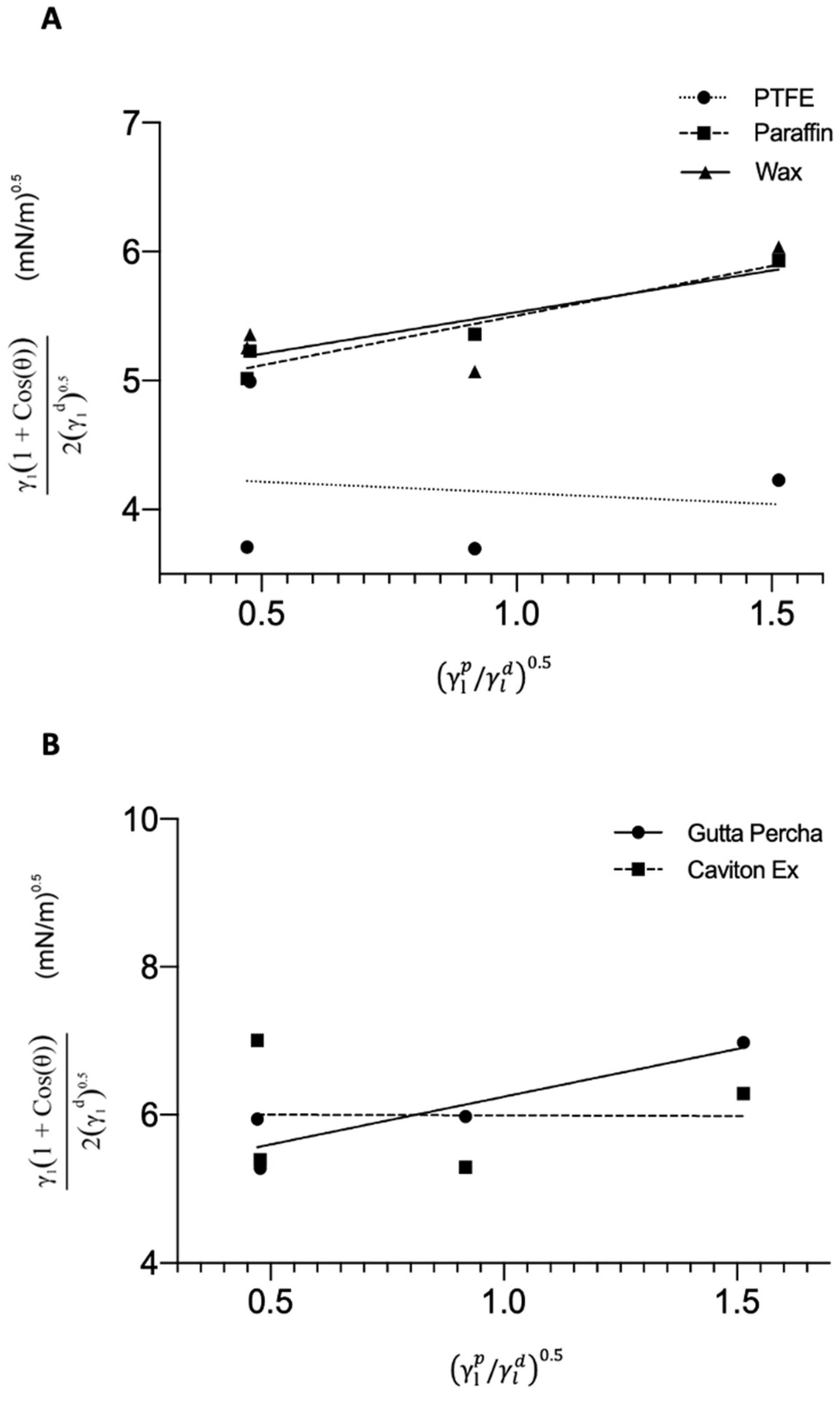
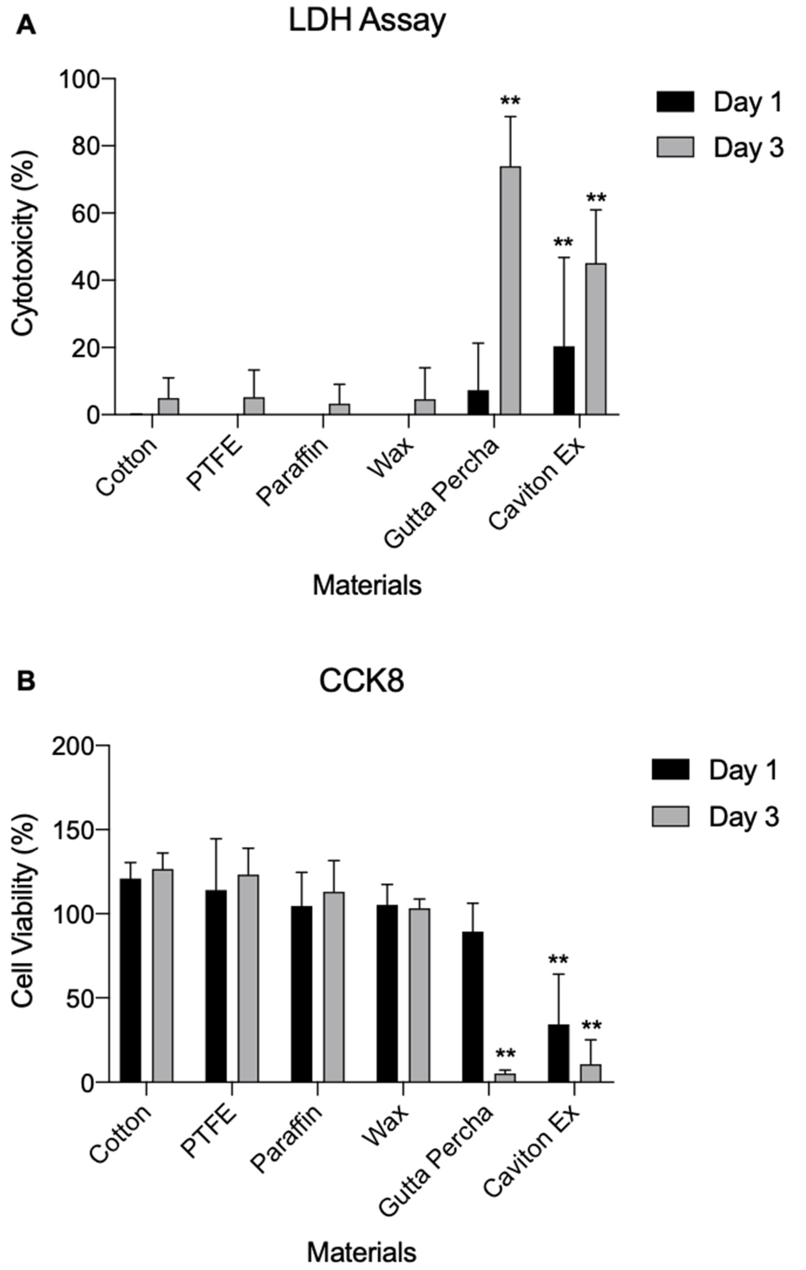
Error in Figure
Text Correction
Reference
- Lu, H.-Y.; Hou, J.; Takahashi, Y.; Tamura, Y.; Kasugai, S.; Kuroda, S.; Nakata, H. Periodontal Pathogen Adhesion, Cytotoxicity, and Surface Free Energy of Different Materials for an Implant Prosthesis Screw Access Hole. Medicina 2022, 58, 329. [Google Scholar] [CrossRef] [PubMed]
Publisher’s Note: MDPI stays neutral with regard to jurisdictional claims in published maps and institutional affiliations. |
© 2022 by the authors. Licensee MDPI, Basel, Switzerland. This article is an open access article distributed under the terms and conditions of the Creative Commons Attribution (CC BY) license (https://creativecommons.org/licenses/by/4.0/).
Share and Cite
Lu, H.-Y.; Hou, J.; Takahashi, Y.; Tamura, Y.; Kasugai, S.; Kuroda, S.; Nakata, H. Correction: Lu et al. Periodontal Pathogen Adhesion, Cytotoxicity, and Surface Free Energy of Different Materials for an Implant Prosthesis Screw Access Hole. Medicina 2022, 58, 329. Medicina 2022, 58, 1413. https://doi.org/10.3390/medicina58101413
Lu H-Y, Hou J, Takahashi Y, Tamura Y, Kasugai S, Kuroda S, Nakata H. Correction: Lu et al. Periodontal Pathogen Adhesion, Cytotoxicity, and Surface Free Energy of Different Materials for an Implant Prosthesis Screw Access Hole. Medicina 2022, 58, 329. Medicina. 2022; 58(10):1413. https://doi.org/10.3390/medicina58101413
Chicago/Turabian StyleLu, Hsin-Ying, Jason Hou, Yuta Takahashi, Yukihiko Tamura, Shohei Kasugai, Shinji Kuroda, and Hidemi Nakata. 2022. "Correction: Lu et al. Periodontal Pathogen Adhesion, Cytotoxicity, and Surface Free Energy of Different Materials for an Implant Prosthesis Screw Access Hole. Medicina 2022, 58, 329" Medicina 58, no. 10: 1413. https://doi.org/10.3390/medicina58101413
APA StyleLu, H.-Y., Hou, J., Takahashi, Y., Tamura, Y., Kasugai, S., Kuroda, S., & Nakata, H. (2022). Correction: Lu et al. Periodontal Pathogen Adhesion, Cytotoxicity, and Surface Free Energy of Different Materials for an Implant Prosthesis Screw Access Hole. Medicina 2022, 58, 329. Medicina, 58(10), 1413. https://doi.org/10.3390/medicina58101413