Review

Handheld Ultrasound and Focused Cardiovascular Echography: Use and Information

Cardiology University of Perugia, 06156 Perugia, Italy
*
Author to whom correspondence should be addressed.
Medicina 2019, 55(8), 423; https://doi.org/10.3390/medicina55080423
Submission received: 22 May 2019 / Revised: 23 July 2019 / Accepted: 25 July 2019 / Published: 31 July 2019

Abstract

:
The availability of miniaturized ultrasound machines has changed our approach to many cardiovascular diseases. Handheld ultrasound imaging can be performed at the bedside, it is easy to use, and the information provided, although limited, is of unquestionable importance for a quick diagnosis that leads to early treatment. They have unique characteristics: Low cost, wide availability, safety, accuracy, and can be used in different clinical scenarios and by operators with different backgrounds. Image acquisition and interpretation is rapid and provides, in each situation, useful information for diagnosis, prognosis, and clinical and therapeutic management. This review focuses on the use of handheld ultrasound devices, describes differences with other equipment, their limitations, and the numerous advantages derived from their use.

1. Introduction

Cardiovascular ultrasound is an imaging investigation with unique characteristics: It is safe, low cost, widely available, repeatable, and accurate. In fact, it is the most used imaging technique in clinical practice. In cardiology, the use of various echocardiographic modalities allows a comprehensive study of cardiac structure, function, and hemodynamics. Mono-dimensional (M-mode), two-dimensional (2D), and three-dimensional (3D) echo define cardiac chamber dimensions and volumes, systolic function, and valvular morphology: Color and Doppler (pulsed and continuous) techniques allow accurate investigation of cardiac hemodynamics and diastolic function; tissue Doppler imaging (TDI) and two-dimensional strain (2DS) are able to detect clinical and subclinical systolic and diastolic dysfunction [1]. All derived echocardiographic information has a well-documented diagnostic and prognostic utility [2].
For many decades, echocardiography equipment was immobile, ultrasound scans were performed in specific echo-laboratories, and cardiologists were the only competent operators. With technological development, echocardiography equipment has become movable, portable, and miniaturized, and ultrasound use has become more extensive, at the bedside, in wider clinical scenarios, in critically ill patients, and in emergencies/urgencies, and its use has expanded to physicians with different backgrounds [3].
Nowadays, various types of equipment are available, with different sizes, different echo modalities, and different diagnostic capabilities. This review focuses on the use of handheld ultrasound devices (HUDs), and describes differences with other equipment, their limitations, and the numerous advantages that come from their use.
HUDs can be performed easily, rapidly, and allow basic information to be obtained for diagnosis and clinical management of various diseases; an HUD can be used by physicians from different disciplines, in many situations and clinical scenarios, especially in emergencies/urgencies and its use has transformed almost all aspects of our daily practice.

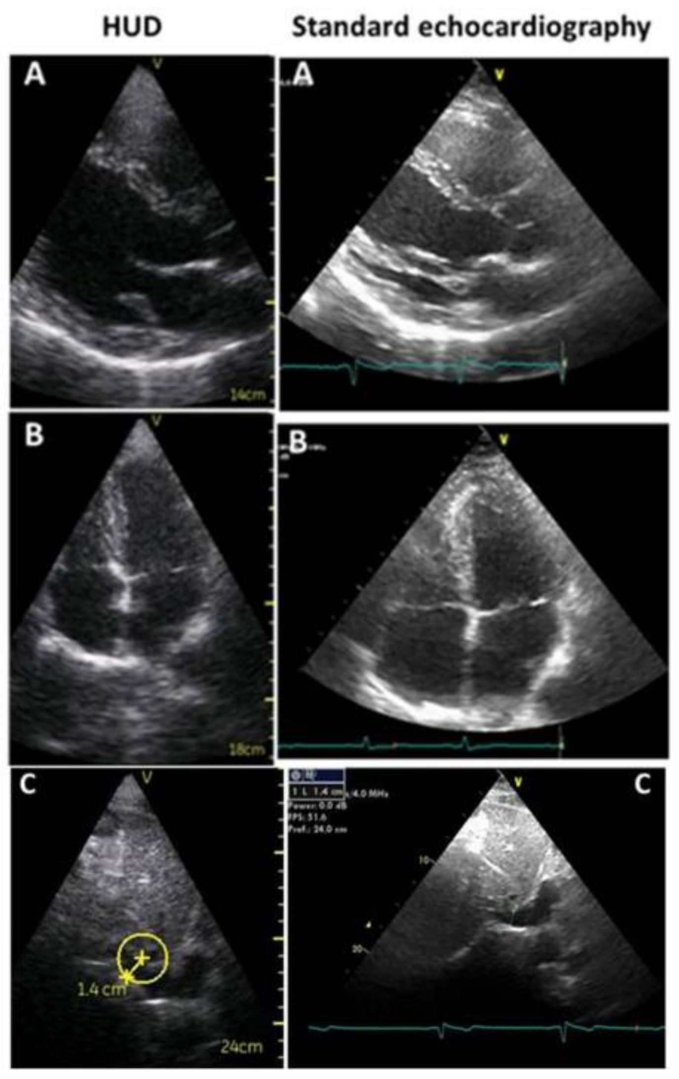
2. Differentiation of the Various Echocardiographic Equipment: From Standard, Comprehensive Transthoracic Echocardiography to Focused Cardiac Ultrasound

Standard echocardiography performed in echo-labs by cardiologists or sonographers/cardiologists provides information about cardiac size, structure, function, and hemodynamics. All these data are obtained using stationary systems that are equipped with various modality system and transducers: 2D, M-mode, Doppler (pulsed and continuous), color, and TDI, transesophageal approach (TEE), and, in many cases with advanced modalities, such as 2DS and three-dimensional echo (3D). During the exam, all modalities can be utilized (if necessary, also the advanced modalities), all standardized echo sections are performed, simultaneously with ECG-guided comprehensive measurements of cardiac structure, heart function, and hemodynamics. Thus, the final report is complete and accurate [1,2,4].
Portable machines are smaller, and allow a basic comprehensive exam of 2D, M-mode, pulse, and continuous Doppler and color. Generally, they do not have advanced modalities, the quality of images is good, and the exam is clinically complete. The final report describes all basic cardiac morphological and functional information.
Handheld ultrasound devices are the smallest machines, very simple to use, with a limited number of basic controls for adjusting the depth and gain, to freeze and store images (in JPEG) and little loops (in MPEG-4), and the available measurements are few and limited to a simple distance and area assessment. The devices only have the 2D modality with grey-scale images and color-Doppler, simultaneous ECG, M-mode, TDI, and advanced technologies are lacking. Images have lower spatial (640 × 480 pixels) and temporal resolution than other equipment. However, 2D and color Doppler are in real-time; the field-of-view (2D and color flow), maximum depth (25 cm), automatic frame rate (28 frames per second), and transducers (phased array—1.7 to 3.8 MHz) are similar; images have good technical quality; and a correct final diagnosis can be made in most cases (Figure 1) [5,6,7,8]. The exam is performed with a limited number of echo sections; often the evaluation is qualitative, with a bimodal (yes/no) or semi quantitative (normal/reduced) response. It represents an extension of the physical examination, is focused to recognize specific signs that lead to an answer to a clinical diagnostic suspicion in a specific clinical setting, and, because it is focused on getting a few findings, it is also named FoCUS [9,10] (Table 1).

3. Focused-Echo in Various Clinical Scenarios: When to Use It

FoCUS/HUDs can be used in many situations and different scenarios, with various acquisition protocols, in stable and unstable patients. It can be used in in- and out-patients and by operators with different specialties. Few measurements are possible, such as left and right atrium and ventricle dimensions, wall thickness, ascending aorta, and inferior vena cava (IVC), but HUDs can qualitatively provide useful information about atrial dilatation, left and right ventricle global systolic function, ventricular dilatation or hypertrophy, significant valvular stenosis and regurgitation, pericardial effusion, and tamponade [8,9,10]. In the literature, many studies have demonstrated that HUDs provide a more accurate diagnosis than physical examination for the majority of common cardiovascular diseases and FoCUS results correlate well with standard echocardiography [8,10,11,12,13,14,15,16,17].
In stable patients, FoCUS is appropriate for the screening of structural heart disease. It allows early diagnosis, defines prognostic stratification, and directs to the appropriate therapy. The exam can rapidly define/exclude qualitative ejection fraction, wall motion abnormalities, ventricular systolic dysfunction, left ventricular hypertrophy, cardiomyopathies with hypertrophic pattern, dilated cardiomyopathies, pulmonary congestion, significant valvulopathies, ascending aorta and aortic root dilatation, pericardial and pleural effusion, and IVC size and respiratory collapsibility [3,7,8,11,12,14,16] (Table 2).
Below are some examples of the effective use of HUD:
In patients with clinical suspicion of congestive heart failure, FoCUS helps to define atrial dilatation, wall motion abnormalities and thicknesses, cardiac dimension and function, significant mitral regurgitation, lung congestion (B-lines at lung ultrasound, also called “comets”), IVC collapse, and it can differentiate between systolic or diastolic heart failure (Figure 2) [18,19,20].
In atrial fibrillation, FoCUS defines left atrial dimensions (dilatation) and left ventricular function, which are strong predictors of sinus rhythm restoration and lead to proper treatment [21,22].
In patients with valvular stenosis, FoCUS can qualitatively show valvular morphology, such as thickened and calcified leaflets with reduced mobility and turbulent transvalvular flow by color Doppler. All these indirect signs lead to the suspicion of significant valvulopathy. Quantitative assessment is not possible, nor are pulmonary pressures, but in patients with signs/symptoms of heart failure, atrial fibrillation, or syncope, these data define the underlying pathophysiology [9,10,23].
In valvular regurgitation, color-HUDs can differentiate significant mild/trivial valvulopathy.
In patients with pericarditis, FoCUS can be used for basic follow-up of pericardial effusion (Figure 3) [9,20].
This approach is a screening tool that provides all information for diagnosis and immediate start for proper therapy, but it cannot be considered as a definitive investigation and standard echocardiography must follow later for a comprehensive heart disease evaluation [3,4,24].
In unstable patients, HUDs provide crucial information in critically ill patients or impending critical situations, is an essential technology for improving early diagnosis, help to rule-in/rule-out different pathological conditions, evaluates the pathophysiology of clinical status, and allows proper clinical management [15,25]. When an HUD is used at the point of care, the acronym FoCUS is changed to POCUS (point of care ultrasound).
For 20 years, many POCUS protocols have been proposed in the literature to standardize the procedure. They preferentially evaluate cardiac, pulmonary, and abdomen diseases, or are elective for searching for trauma lesions. Actually, because there are many clinical scenarios, data acquisition depends on the specific imaging target rather than specific protocol.
In ill patients, POCUS is extremely useful in terms of a differential diagnosis of clinical conditions that manifest with the same symptoms (Table 3).
In acute respiratory failure, lung ultrasound can differentiate pneumothorax from pleural effusion or cardiogenic and non-cardiogenic pulmonary edema [26].
In shock patients, POCUS is useful for diagnosis, management, and monitoring of treatment efficacy and clinical progression. Chamber size, left ventricular systolic function, IVC dimensions and collapsibility, pericardial effusion, and pulmonary congestion (lung B lines—comets) in lung ultrasounds identifies cardiogenic shock [27].
In cardiac tamponade, POCUS is useful to identify pericardial effusion size and distribution and directs to the best approach for pericardiocentesis [28].
In the emergency department, POCUS helps to differentiate the nature of chest pain. In addition to history, clinical examination, electrocardiogram, and biomarkers, echocardiography can visualize wall-motion abnormalities and left ventricular function, dilated right ventricle with free wall hypokinesia, ascending aorta dimension and morphology, significant aortic regurgitation, and pericardial effusion. In this way, it is a first step to a differential diagnosis among acute coronary syndromes, acute aortic syndromes, pulmonary embolism, congestive heart failure, and pericarditis [29].
In patients with cardiac arrest due to complex arrhythmias, HUDs can exclude/diagnose all cardiac arrhythmogenic diseases, such as hypertrophic, dilated, or arrhythmogenic right ventricular cardiomyopathies [30].
In cardiopulmonary resuscitation and during BLS (basic life support) and ALS (advanced life support) protocols, POCUS can help to diagnose potentially treatable causes of cardiac arrest, such as cardiac tamponade, massive pulmonary embolism, severe ventricular dysfunction, and hypovolemia [31,32].

4. Specialties That Use HUDs and Training of Operators

Since FoCUS and POCUS are simplified exams with restricted protocols, operators with different backgrounds can utilize them. Actually, cardiologists, internists, anesthesiologists, and specialists in emergency/urgency frequently utilize this tool. The approach is simplified; the findings to obtain are few and are guided by symptoms, so a short training period is required. Some studies have shown that brief training is sufficient to perform and interpret a FoCUS scan, the agreement between FoCUS and standard echocardiography is satisfactory, the trainee’s performance improves over a short period of time, and the inter-observer variability in FoCUS is low [33,34,35,36]. Nowadays, both European and American guidelines recommend a theoretical background on cardiovascular diseases and practical experience to obtain competence in image acquisition and in their interpretation [9,10]. The educational path is carried out with a tutor, who verifies correct image acquisition. At the beginning, data interpretation is discussed together and, subsequently, each operator individually acquires and interprets the data. Specific education and training in the use of HUDs is obtained by individually interpreting 25 to 50 exams [8,10,37].

5. Conclusions

The use of HUDs has completely changed the daily approach to cardiac diseases. Handheld devices have allowed fundamental information about morphology and cardiac function to be obtained in stable and, especially, unstable patients. They extend the physical examination with a short and simplified ultrasound examination aimed at achieving rapid diagnosis, early treatment, and basic monitoring of some cardiac diseases. Its use serves to guide diagnosis in specific clinical situations, mainly defined by patient symptoms; it is a limited and non-exhaustive technique, but it is fast, repeatable, and easy to perform. The training of operators and the learning curve is short (albeit rigorous) and can be used from physicians of different background and specialties. FoCUS should not be considered as a replacement of echocardiographic examination but as a clinical tool similar to a stethoscope to aid early diagnosis (at the bedside), define the event pathophysiology and prognostic stratification, and direct to the appropriate therapy. In this setting and in specific clinical situations, it is extremely useful.

Author Contributions

K.S. designed and wrote the article. G.A. reviewed and commented the review.

Funding

This research received no external funding.

Conflicts of Interest

The authors declare no conflict of interest.

References

  1. Mitchell, C.; Rahko, P.S.; Blauwet, L.A.; Canaday, B.; Finstuen, J.A.; Foster, M.C.; Horton, K.; Ogunyankin, K.O.; Palma, R.A.; Velazquez, E.J. Guidelines for Performing a Comprehensive Transthoracic Echocardiographic Examination in Adults: Recommendations from the American Society of Echocardiography. J. Am. Soc. Echocardiogr. 2019, 32, 1–64. [Google Scholar] [CrossRef]
  2. Douglas, P.S.; Khandheria, B.J.; Stainback, R.F. ACCF/ASE/ACEP/ASNC/SCAI/SCCT/SCMR 2007 appropriateness criteria for transthoracic and transesophageal echocardiography: A report of the American College of Cardiology Foundation Quality strategic directions committee appropriateness criteria working group, American Society of Echocardiography, American College of Emergency Physicians, American Society of Nuclear Cardiology, Society for Cardiovascular Angiography and Interventions, Society of Cardiovascular Computed Tomography, and the Society for Cardiovascular Magnetic Resonance. Endorsed by the American College of Chest Physicians and the Society of Critical Care Medicine. J. Am. Soc. Echocardiogr. 2007, 20, 787–805. [Google Scholar] [CrossRef]
  3. Chamsi-Pasha, M.A.; Sengupta, P.P.; Zoghbi, W.A. Handheld Echocardiography. Circulation 2017, 136, 2178–2188. [Google Scholar] [CrossRef]
  4. Picard, M.H.; Adams, D.; Bierig, S.M.; Dent, J.M.; Douglas, P.S.; Gillam, L.D.; Keller, A.M.; Malenka, D.J.; Masoudi, F.A.; McCulloch, M.; et al. American Society of Echocardiography Recommendations for Quality Echocardiography Laboratory Operations. J. Am. Soc. Echocardiogr. 2011, 24, 1–10. [Google Scholar] [CrossRef]
  5. Razaak, M.; Martini, M.G.; Savino, K. A Study on Quality Assessment for Medical Ultrasound Video Compressed via HEVC. IEEE J. Biomed. Health Inform. 2014, 18, 1552–1559. [Google Scholar] [CrossRef] [Green Version]
  6. Martini, M.G.; Iacobelli, L.; Bergeron, C.; Hewage, C.T.; Panza, G.; Piri, E.; Vehkaperä, J.; Amon, P.; Mazzotti, M.; Savino, K.; et al. Real-time multimedia communications in medical emergency—The CONCERTO project solution. In Proceedings of the 2015 37th Annual International Conference of the IEEE Engineering in Medicine and Biology Society (EMBC), Milan, Italy, 25–29 August 2015; Volume 2015, pp. 7324–7327. [Google Scholar]
  7. Di Bello, V.; La Carruba, S.; Conte, L.; Fabiani, I.; Posteraro, A.; Antonini-Canterin, F.; Barletta, V.; Nicastro, I.; Mariotti, E.; Severino, S.; et al. Incremental value of pocket-sized echocardiography in addition to physical examination during inpatient cardiology evaluation: A multicenter Italian study (SIEC). Echocardiography 2015, 32, 1463–1470. [Google Scholar] [CrossRef]
  8. Cardim, N.; Dalen, H.; Voigt, J.U.; Ionescu, A.; Price, S.; Neskovic, A.N.; Edvardsen, T.; Galderisi, M.; Sicari, R.; Donal, E.; et al. The use of handheld ultrasound devices: A position statement of the European Association of Cardiovascular Imaging (2018 update). Eur. Heart J. Cardiovasc. Imaging 2019, 20, 245–252. [Google Scholar] [CrossRef]
  9. Spencer, K.T.; Kimura, B.J.; Korcarz, C.E.; Pellikka, P.A.; Rahko, P.S.; Siegel, R.J. Focused Cardiac Ultrasound: Recommendations from the American Society of Echocardiography. J. Am. Soc. Echocardiogr. 2013, 26, 567–581. [Google Scholar] [CrossRef]
  10. Nesković, A.N.; Skinner, H.; Price, S.; Via, G.; De Hert, S.; Stankovic, I.; Galderisi, M.; Donal, E.; Muraru, D.; Sloth, E.; et al. Focus cardiac ultrasound core curriculum and core syllabus of the European Association of Cardiovascular Imaging. Eur. Hear. J. Cardiovasc. Imaging 2018, 19, 475–481. [Google Scholar] [CrossRef]
  11. Fukuda, S.; Shimada, K.; Kawasaki, T.; Fujimoto, H.; Maeda, K.; Inanami, H.; Yoshida, K.; Jissho, S.; Taguchi, H.; Yoshiyama, M.; et al. Pocket-Sized Transthoracic Echocardiography Device for the Measurement of Cardiac Chamber Size and Function. Circ. J. 2009, 73, 1092–1096. [Google Scholar] [CrossRef] [Green Version]
  12. Culp, B.C.; Mock, J.D.; Chiles, C.D.; Culp, W.C. The Pocket Echocardiograph: Validation and Feasibility. Echocardiography 2010, 27, 759–764. [Google Scholar] [CrossRef]
  13. Culp, B.C.; Mock, J.D.; Ball, T.R.; Chiles, C.D.; Culp, W.C., Jr. The Pocket Echocardiograph: A Pilot Study of Its Validation and Feasibility in Intubated Patients. Echocardiography 2011, 28, 371–377. [Google Scholar] [CrossRef]
  14. Kitada, R.; Fukuda, S.; Watanabe, H.; Oe, H.; Abe, Y.; Yoshiyama, M.; Song, J.M.; Sitges, M.; Shiota, T.; Ito, H.; et al. Diagnostic Accuracy and Cost-Effectiveness of a Pocket-Sized Transthoracic Echocardiographic Imaging Device. Clin. Cardiol. 2013, 36, 603–610. [Google Scholar] [CrossRef]
  15. Andersen, C.A.; Holden, S.; Vela, J.; Rathleff, M.S.; Jensen, M.B. Point-of-Care Ultrasound in General Practice: A Systematic Review. Ann. Fam. Med. 2019, 17, 61–69. [Google Scholar] [CrossRef] [Green Version]
  16. Giusca, S.; Jurcut, R.; Ticulescu, R.; Dumitru, D.; Vladaia, A.; Savu, O.; Voican, A.; Popescu, B.A.; Ginghina, C. Accuracy of handheld echocardiography for bedside diagnostic evaluation in a tertiary cardiology center: Comparison with standard echocardiography. Echocardiography 2011, 28, 136–141. [Google Scholar] [CrossRef]
  17. Mantella, L.E.; Colledanchise, K.; Bullen, M.; Hétu, M.F.; Day, A.G.; McLellan, C.S.; Johri, A.M. Handheld versus conventional vascular ultrasound for assessing carotid artery plaque. Int. J. Cardiol. 2019, 278, 295–299. [Google Scholar] [CrossRef]
  18. Mirabel, M.; Celermajer, D.; Beraud, A.-S.; Jouven, X.; Marijon, E.; Hagège, A.A. Pocket-sized focused cardiac ultrasound: Strengths and limitations. Arch. Cardiovasc. Dis. 2015, 108, 197–205. [Google Scholar] [CrossRef] [Green Version]
  19. Razi, R.; Estrada, J.R.; Doll, J.; Spencer, K.T. Bedside Hand-Carried Ultrasound by Internal Medicine Residents Versus Traditional Clinical Assessment for the Identification of Systolic Dysfunction in Patients Admitted with Decompensated Heart Failure. J. Am. Soc. Echocardiogr. 2011, 24, 1319–1324. [Google Scholar] [CrossRef]
  20. Kono, Y.; Fukuda, S.; Shimada, K.; Oe, H.; Maeda, K.; Kawasaki, T.; Fujimoto, H.; Otsuka, K.; Kubo, T.; Jissho, S.; et al. Pocket-sized echo for evaluation of mitral and tricuspid regurgitation. JACC Cardiovasc. Imaging 2011, 4, 921. [Google Scholar] [CrossRef]
  21. Cameli, M.; Mandoli, G.E.; Ambrosio, A.; Cerbai, E.; Coiro, S.; Emdin, M.; Marcucci, R.; Morrone, D.; Palazzuoli, A.; Savino, K.; et al. Arterial hypertension and atrial fibrillation: Standard and advanced echocardiography from diagnosis to prognostication. J. Cardiovasc. Med. 2018, 19, 51–61. [Google Scholar] [CrossRef]
  22. Beltrami, M.; Palazzuoli, A.; Padeletti, L.; Cerbai, E.; Coiro, S.; Emdin, M.; Marcucci, R.; Morrone, D.; Cameli, M.; Savino, K.; et al. The importance of integrated left atrial evalutation: From hypertension to heart failure with preserved ejection fraction. Int. J. Clin. Pract. 2018, 72, 130–150. [Google Scholar] [CrossRef]
  23. Abe, Y.; Ito, M.; Tanaka, C.; Ito, K.; Naruko, T.; Itoh, A.; Haze, K.; Muro, T.; Yoshiyama, M.; Yoshikawa, J. A Novel and Simple Method Using Pocket-Sized Echocardiography to Screen for Aortic Stenosis. J. Am. Soc. Echocardiogr. 2013, 26, 589–596. [Google Scholar] [CrossRef]
  24. Wiley, B.; Bibhu Mohanty, B. Handheld ultrasound and diagnosis of cardiovascular disease at the bedside. J. Am. Coll. Cardiol. 2014, 64, 229–230. [Google Scholar] [CrossRef]
  25. Moore, C.L.; Copel, J.A. Point-of-care ultrasonography. N. Engl. J. Med. 2011, 364, 749–757. [Google Scholar] [CrossRef]
  26. Lichtenstein, D.A. Lung ultrasound in the critically ill. Ann. Intensive Care 2014, 4, 1–12. [Google Scholar] [CrossRef]
  27. Peterson, D.; Arntfield, R.T. Critical Care Ultrasonography. Emerg. Med. Clin. N. Am. 2014, 32, 907–926. [Google Scholar] [CrossRef]
  28. Byhahn, C.; Bingold, T.M.; Zwissler, B.; Maier, M.; Walcher, F. Prehospital ultrasound detects pericardial tamponade in a pregnant victim of stabbing assault. Resuscitation 2008, 76, 146–148. [Google Scholar] [CrossRef]
  29. Douglas, P.S.; Garcia, M.J.; Haines, D.E.; Lai, W.W.; Manning, W.J.; Patel, A.R.; Picard, M.H.; Polk, D.M.; Ragosta, M.; Ward, R.P.; et al. ACCF/ASE/AHA/ASNC/HFSA/HRS/SCAI/ SCCM/SCCT/SCMR 2011 Appropriate use criteria for echocardiography. J. Am. Soc. Echocardiogr. 2011, 24, 229–267. [Google Scholar] [CrossRef]
  30. Savino, K.; Bagliani, G.; Crusco, F.; Padeletti, M.; Lombardi, M. Electrocardiogram and Imaging: An Integrated Approach to Arrhythmogenic Cardiomyopathies. Card. Electrophysiol. Clin. 2018, 10, 413–429. [Google Scholar] [CrossRef]
  31. Olasveengen, T.M.; De Caen, A.R.; Mancini, M.E.; Maconochie, I.K.; Aickin, R.; Atkins, D.L.; Berg, R.A.; Bingham, R.M.; Brooks, S.C.; Castrén, M.; et al. 2017 International Consensus on Cardiopulmonary Resuscitation and Emergency Cardiovascular Care Science With Treatment Recommendations Summary. Circulation 2017, 136, e424–e440. [Google Scholar] [CrossRef] [Green Version]
  32. Gundersen, G.H.; Norekval, T.M.; Haug, H.H.; Skjetne, K.; Kleinau, J.O.; Graven, T.; Dalen, H. Adding point of care ultrasound to assess volume status in heart failure patients in a nurse-led outpatient clinic. A randomised study. Heart 2016, 102, 29–34. [Google Scholar] [CrossRef]
  33. Panoulas, V.F.; Daigeler, A.L.; Malaweera, A.S.; Lota, A.S.; Baskaran, D.; Rahman, S.; Nihoyannopoulos, P. Pocket-size hand-held cardiac ultrasound as an adjunct to clinical examination in the hands of medical students and junior doctors. Eur. Heart J. Cardiovasc. Imaging 2013, 14, 323–330. [Google Scholar] [CrossRef]
  34. Alexander, J.H.; Peterson, E.D.; Chen, A.Y.; Harding, T.M.; Adams, D.B.; Kisslo, J.A. Feasibility of point-of-care echocardiography by internal medicine house staff. Am. Hear. J. 2004, 147, 476–481. [Google Scholar] [CrossRef]
  35. Kobal, S.L.; Trento, L.; Baharami, S.; Tolstrup, K.; Naqvi, T.Z.; Cercek, B.; Neuman, Y.; Mirocha, J.; Kar, S.; Forrester, J.S.; et al. Comparison of Effectiveness of Hand-Carried Ultrasound to Bedside Cardiovascular Physical Examination. Am. J. Cardiol. 2005, 96, 1002–1006. [Google Scholar] [CrossRef]
  36. Evangelista, A.; Galuppo, V.; Mendez, J.; Evangelista, L.; Arpal, L.; Rubio, C.; Vergara, M.; Liceran, M.; López, F.; Sales, C.; et al. Handheld cardiac ultrasound screening performed by family doctors with remote expert support interpretation. Heart 2016, 102, 376–382. [Google Scholar] [CrossRef]
  37. Ryan, T.; Berlacher, K.; Lindner, J.R.; Mankad, S.V.; Rose, G.A.; Wang, A. COCATS 4 task force 5: Training in echocardiography: Endorsed by the American Society of Echocardiography. J. Am. Soc. Echocardiogr. 2015, 28, 615–627. [Google Scholar] [CrossRef]
Figure 1. Echocardiographic images acquired in the same patient, both with HUD (on the left) and standard echocardiography (on the right). (A): parasternal long axis view, (B): apical four chambers view, (C): subcostal view for inferior vena cava. HUD defines qualitatively normal ventricular diameters and wall thicknesses, and mild left and right atrium dilatation. HUD measurement is accurate (see C). In standard echocardiography endocardium, mitral and aortic valves are better visualized.
Medicina 55 00423 g001
Figure 2. FoCUS examples in clinical suspicion of congestive heart failure. Parasternal long axis view (A) and apical four chambers view (B) in a patient with dilated cardiomyopathy and systolic heart failure: left ventricle is markedly dilated, wall thicknesses are normal, and ejection fraction is reduced. Parasternal long axis view (C) and apical four chambers view (D) in a patient with hypertrophic cardiomyopathy and diastolic heart failure: left ventricle is normal, wall thicknesses have increased, both left and right atrium are dilated, and mild pericardial effusion is present (arrows).
Medicina 55 00423 g002
Figure 3. Parasternal long axis view (A) and left ventricle short axis view (B) in a patient with pericarditis. Arrows show a moderate pericardial effusion.
Medicina 55 00423 g003
Table 1. Differences between standard and FoCUS echocardiography.
Standard Echocardiography
Medicina 55 00423 i001
Focused Echo
Medicina 55 00423 i002
EquipmentStationary, top level technologyHandheld ultrasound devices
Echo purposeAll heart diseases studyFew clinical questions
Echo goalAccurate studyDifferential diagnosis
US sectionsAll recommendedFew
Execution time>30 minFew minutes
DiagnosisIn electionRapid
Operator trainingAccurate/longRapid/25–50 exams
Table 2. FoCUS diagnostic capabilities in stable patients.
Clinical SuspectFoCUS Goals
Congestive heart failureLeft atrium and ventricle dimension, LV EF, significant valvular regurgitation, IVC dimension and collapse, pulmonary congestion
CardiomyopathiesLeft ventricular hypertrophy, dilated cardiomyopathies
Atrial fibrillationLeft atrial dimensions, left ventricular function
Stable coronary artery diseaseWall motion abnormalities, ejection fraction
Valvular stenosisThickness and calcification leaflet, reduced leaflets mobility, turbulent transvalvular flow at color-Doppler
Valvular regurgitationExtent regurgitation jet at color-Doppler
PericarditisPericardial effusion presence, size and distribution
LV: left ventricle, EF: ejection fraction, IVC: inferior vena cava.
Table 3. POCUS diagnostic capabilities in unstable patients.
Clinical ScenariosPOCUS GoalRule-In/Rule-Out Diseases
Acute respiratory failureNo lung sliding sign (lung movement absence), Anechoic space between visceral and parietal pleura, Lines B—cometsPneumotorax
Pleural effusion Pulmonary congestion
ShockChambers size and shape, LV EF, IVC size and collapsibility, pericardial effusion, pulmonary congestionCardiogenic shock
Cardiac tamponadePericardial fluid size and distribution Guide to pericardiocentesisMassive pericardial effusion
Acute chest pain LV wall motion abnormalities, LV EF, RV size and function, Ascending aorta dimensions and morphology, aortic regurgitation, pericardial effusion Acute coronary syndrome Pulmonary embolism Aortic dissection
Pericarditis
Arrhythmic cardiac arrestLeft ventricular hypertrophy, LV dilatation, LV dysfunction, RV dilatation and dysfunction Hypertrophic cardiomyopathy Dilated cardiomyopathy Arrhythmogenic right ventricular cardiomyopathy
Cardiopulmonary resuscitationMassive pericardial effusion, RV dilatation, RV severe dysfunction, LV severe dysfunction, LV very small dimension (empty ventricle)Cardiac tamponade Massive pulmonary embolism
Severe congestive heart failure
Hypovolemia
LV: left ventricle, EF: ejection fraction, IVC: inferior vena cava, RV: right ventricle.

