Article

Development of Wedelia (Sphagneticola trilobata) and Sembung Rambat (Mikania micrantha) Extracts as Herbal Medicine for Chronic Obstructive Pulmonary Disease (COPD)


1
Department of Chemistry, Faculty of Mathematics and Natural Sciences, IPB University, Bogor 16680, West Java, Indonesia
2
Tropical Biopharmaca Research Center, IPB University, Bogor 16680, West Java, Indonesia
3
School of Veterinary Medicine and Biomedical Sciences, IPB University, Bogor 16680, West Java, Indonesia
4
Faculty of Medicine, Padjadjaran University, Bandung 45363, West Java, Indonesia
5
Faculty of Medicine, Universitas Indonesia, Jakarta 10430, Central Jakarta, Indonesia
*
Author to whom correspondence should be addressed.
Curr. Issues Mol. Biol. 2026, 48(7), 720; https://doi.org/10.3390/cimb48070720
Submission received: 31 May 2026 / Revised: 12 July 2026 / Accepted: 13 July 2026 / Published: 15 July 2026
(This article belongs to the Special Issue The Role of Bioactives in Inflammation, 2nd Edition)

Abstract

Chronic Obstructive Pulmonary Disease (COPD) is a chronic respiratory condition characterized by progressive deterioration in lung function and persistent symptoms, particularly dyspnea, which can lead to death. Inflammation is important in COPD pathogenesis because it causes persistent inflammatory responses in the respiratory tract. Therefore, this study aimed to investigate H2O extracts derived from Wedelia (Sphagneticola trilobata) and Sembung Rambat (Mikania micrantha), respectively, as anti-inflammatory agents and to analyze the metabolite profiles contained in the extracts. The results showed that Sembung Rambat H2O extract demonstrated significant inhibition of IL-2 (100 ± 0.00%), while Wedelia extract inhibited 74.68 ± 7.13% release of pro-inflammatory cytokine IL-6. Liquid Chromatography–Tandem Mass Spectrometry (LC-MS/MS) analysis mainly identified the presence of amino acids, phenolic acids, organic acids, and terpenoids in both extracts. These results suggest the promising potential of Wedelia and Sembung Rambat extracts as herbal therapy for COPD.

1. Introduction

The coronavirus SARS-CoV-2 is the root cause of COVID-19. The 2019–2020 coronavirus pandemic was caused by this illness. The pathogenesis is possibly related to the body’s hyperinflammatory response, which is identified by the presence of cytokine storm. COVID-19 patients may develop fever, dry cough, and more severe symptoms, including trouble breathing and chest tightness. The virus predominantly affects the respiratory system, which is already weakened in patients with Chronic Obstructive Pulmonary Disorder (COPD) [1,2].
Airflow from the lungs is blocked by COPD, which is a long-lasting inflammatory condition of the respiratory system. The primary physiological function of the lungs, the main organs of the respiratory system, is to enable the exchange of gases. WHO documented that COPD accounted for the third-highest mortality burden worldwide in 2019 [3]. COPD-related lung disease is caused by airway abnormalities (chronic bronchitis or bronchiolitis) and/or alveoli (emphysema). The primary sign of chronic bronchitis is a persistent cough, which is indicative of airway inflammation. Additionally, it prevents airflow by causing goblet cells to produce excessive amounts of mucus. On the other hand, emphysema causes alveolar damage and produces symptoms such as dyspnea. Alveolar septa disintegration, an increase in airspace volume, and a reduction in elasticity brought on by redox imbalance, as well as excessive inflammatory response, are the hallmarks of the pathophysiology. An inflammatory immune response or long-term inflammation of the lungs’ airways is brought on by exposure to dangerous particles and chemicals, especially those found in cigarette smoke. COPD’s pathophysiology is caused by this inflammatory reaction [4]. An increase in cells, including neutrophils and lymphocytes, as well as macrophages, causes the release of pro-inflammatory cytokines, including IL-6, TNF-α, IL-8, and IL-1β, as well as IL-2, which are mostly found in the lung parenchyma, peripheral airways, and pulmonary blood vessels, and typically indicate chronic inflammation [5,6]. Cytokines, in particular, are polypeptides that function as intercellular mediators. In respiratory tract illnesses, the generation of these inflammation-promoting cytokines adds to lung tissue damage and affects respiratory tract inflammation. Patients with COPD can be managed and treated using a variety of strategies. Oral medicines, like inhaled inflammation-inhibiting agents (corticosteroids) and bronchodilators, are prominent therapeutic options. Nevertheless, sustained corticosteroid therapy may predispose patients to multiple adverse clinical effects, including diabetes mellitus, skeletal demineralization, lens opacification, and an elevated susceptibility to infections [7,8,9,10].
Herbal plants contribute significantly to culture and traditional medicine practices in many countries. Indonesia is a megabiodiverse country with over 30,000 plant taxa that are suitable to be utilized as raw materials for herbal medicines. Flavonoids, terpenoids, alkaloids, and saponins are among the bioefficacious phytochemicals identified in plants of medicinal value that exhibit diverse pharmacodynamic properties. These compounds exhibit beneficial effects against different pathological conditions. Indonesians have locally used herbal plants as traditional medicines [11,12,13].
Globally, there is currently a growing trend toward a return to a traditional lifestyle, which includes the utilization of natural or herbal therapeutic compounds, with a prevalence of 88% [14]. The WHO advises utilizing herbs to preserve the health of the population. Globally, especially in Indonesia, the herbal product market is currently quite popular and has a lot of promise. With a market size estimated at USD 165.66 billion in 2021, the herbal products sector is forecast to attain a value of USD 5 trillion by 2050 [15,16].
A comparative study on three methods of treatment and management for COPD patients in China showed that the combination of treatment methods and Traditional Chinese Medicine (TCM) was the most successful method [17]. Several medicinal plants have been employed in China for COPD treatment, including milkvetch root (Astragalus monholicus). This plant reportedly improved immune function in patients with COPD who experienced acute aggravation [18].
Five Indonesian medicinal herbs have been demonstrated to decrease inflammatory regulation related to the respiratory system, according to a comprehensive review based on earlier publications. The plants are Sambiloto (Andrographis paniculata), Babadotan (Ageratum conyzoides), Kersen (Muntingia calabura), and Legetan Warak (Adenostemma lavenia), as well as Sembung Rambat (Mikania micrantha Kunth) [19]. An A. paniculata 98% ethanol extract inhibited the cytokines that promote inflammation release, like IL-1β and IL-6, as well as TNF-α, in LPS-treated RAW 264.7 cells in vitro, according to Lukito et al. [20]. A. lavenia aqueous extract attenuated IL-6 levels according to another in vitro work by Astuti et al. [21]. Additionally, an M. calabura ethanol-based extract reduced the activity of cyclooxygen-ase-2 (COX-2) [22]. Specifically, COX-2 is an enzyme that produces prostaglandins, which cause inflammation [19].
In vivo models have demonstrated several plant-derived extracts’ inflammation-modulating activity. In a hamster model, an A. paniculata extract intervention markedly decreased IL-6 secretion and expression in pulmonary tissue, as reported by Kongsomros et al. [6]. Likewise, Widyaningrum et al. [23] observed that an M. calabura ethanolic extract exhibited significant inflammation-reducing activity by reducing carrageenan-induced edema in mice within 4 h after induction with 1% carrageenan. Furthermore, Xu et al. [24] demonstrated that A. conyzoides extract suppressed the activation of the NLRP3 inflammasome pathway in a murine model of peritonitis. Additionally, according to Deori et al. [25], an M. micrantha ethanolic extract exhibited inflammation-reducing activity potential by lowering rat paw edema in acute and sub-acute inflammation caused by carrageenan. The compounds 1a,9b-dihydro-1H-cyclopropa[a]anthracene, found in A. lavenia extract, and 7-hydroxyflavone, found in M. calabura extract, have a strong affinity for binding COX-2 as an inflammatory receptor in molecular dynamics studies, according to an in silico study by Tuwalaid et al. [22].
Inflammation is closely associated with COPD because the disease is related to inflammation of the respiratory system. Therefore, further investigation of anti-COPD in vitro is essential to produce a candidate herbal medicine supported by strong scientific evidence, along with the working mechanism, characteristic compounds, and active compounds. Based on the available literature, studies to develop H2O extracts derived from Wedelia (Sphagneticola trilobata) and Sembung Rambat (Mikania micrantha) have not been carried out. Ethanol extract of M. micrantha was examined by Deori et al. [25] in vivo, with the results showing significant inflammation-inhibiting properties. However, in vitro studies on S. trilobata and M. micrantha plants have not previously been conducted. Accordingly, the goal of this study is to employ in vitro methods to determine the key inflammatory mediator inhibitory activity, like IL-1β, IL-6, and IL-2, released from the aqueous extract of S. trilobata and M. micrantha on LPS-treated RAW 264.7 macrophages. The secondary metabolite profile was examined in each extract to determine which compounds act as inhibitors of inflammation related to the respiratory system. The results of this study can help the pharmaceutical industry achieve the mission to provide trusted health products to improve public health. It also supports the vision of becoming a leading health product manufacturer that dominates the market in Indonesia and Asia, with minimal side effects and by using natural raw materials from local sources.

2. Materials and Methods

2.1. Preparation of Raw Materials

Wedelia (S. trilobata) and Sembung Rambat (M. micrantha) were obtained from the Biopharmaceutical Cultivation Conservation Unit (UKBB) Garden at the Tropical Biopharmaceutical Study Center, IPB University. The plants were harvested at the age of 3–4 months by pruning the stems, and then the fresh leaves were made into simple drugs through a process of washing, drying in a 45 °C oven for 36 h, and grinding into powder using a grinder with an 80 mesh sieve. The extraction was carried out using the maceration method. The ratio of the plant powder to the solvent was 1:10, with distilled water used as the solvent at room temperature. The maceration process was carried out for 3 × 24 h. All macerate was collected and evaporated using a rotary evaporator to obtain a thick extract, which was then freeze-dried to obtain a dry extract.

2.2. Determination of Standardization of Raw Materials for Simplicia and Extracts

Post-harvest evaluation at the supplier/farmer level was conducted to evaluate the standardized post-harvest process for raw materials. The standardization was conducted in accordance with Indonesian Food and Drug Authority (BPOM/Badan Pengawas Obat dan Makanan) requirements. The analyses consist of moisture, ash, microbiological, and heavy metal content to prepare a Certificate of Analysis (CoA) for the raw materials.
Standardization of raw materials was carried out in accordance with BPOM requirements. Analysis carried out for the standardization of raw materials includes moisture content, ash content, acid-insoluble ash content, water-soluble extract content, ethanol-soluble extract content, microbiological content, and heavy metal content for lead (Pb) and cadmium (Cd). The procedures followed standard methods, including AOAC [26] and Indonesian National Standard (SNI) by BSN [27,28,29] as follows:
  • Moisture content—A covered porcelain dish was heated in an oven at 105 °C for 30 min, cooled in a desiccator for 15 min, and weighed. Approximately 10 g of sample was accurately weighed into the pre-weighed dish and dried at 105 °C for 5 h. The dish containing the sample was then cooled in a desiccator for 30 min and weighed. Drying was continued at 1 h intervals until the difference between two consecutive weighings was not more than 0.25%. The moisture content was calculated using Equation (1).
    M o i s t u r e   c o n t e n t = W ( W 1 W 2 ) W × 100 %
    W = s a m p l e   w e i g h t   b e f o r e   d r y i n g   g W 1 = s a m p l e + c o v e r e d   p o r c e l a i n   d i s h   w e i g h t   a f t e r   d r y i n g   g W 2 = h e a t e d   e m p t y   c o v e r e d   p o r c e l a i n   d i s h   w e i g h t   ( g )
  • Ash content—2–3 g of sample was accurately weighed into a porcelain crucible of known weight. The sample was charred over a burner flame, then ashed in an electric furnace at a maximum temperature of 550 °C until complete ashing (occasionally, the furnace door is opened slightly to allow oxygen to enter). The crucible and sample were cooled in a desiccator, then weighed until the weight remained constant. The ash content was calculated using Equation (2).
    A s h   c o n t e n t = W 1 W 2 W × 100 %
    W = s a m p l e   w e i g h t   b e f o r e   a s h i n g   g W 1 = s a m p l e + c o v e r e d   p o r c e l a i n   d i s h   w e i g h t   a f t e r   a s h i n g   g W 2 = h e a t e d   e m p t y   c o v e r e d   p o r c e l a i n   d i s h   w e i g h t   g
  • Acid-insoluble ash content—The ash obtained from the previous ash content determination was dissolved in 25 mL of 10% HCl and boiled for 5 min. The solution was then filtered through ashless filter paper and washed with distilled water until chloride-free. The filter paper was placed in a porcelain cup of known weight and dried in an oven, then ashed. The porcelain cup was cooled in a desiccator to room temperature, then weighed. The weighing was repeated until a constant weight was achieved. The acid-insoluble ash content was calculated using Equation (2).
  • Water- or ethanol-soluble extract content—Five grams of sample were accurately weighed using a stoppered flask. The sample was macerated with 100 mL of water or ethanol (95%) while shaking for the first 6 h and then left for 18 h. The mixture was quickly filtered to avoid solvent evaporation. The filtrate was transferred to a 100 mL volumetric flask, calibrated, and shaken until homogeneous. Twenty milliliters of the solution were accurately taken and dried in an oven at 105 °C in a shallow dish of known weight. Next, it was cooled in a desiccator and weighed. The drying, cooling, and weighing procedures were repeated until a constant weight was obtained. The water or ethanol extract content was calculated using Equation (3).
    W a t e r   o r   e t h a n o l   s o l u b l e   e x t r a c t   c o n t e n t = W 1 W 2 W × 100 % × 5
    W = s a m p l e   w e i g h t   b e f o r e   d r y i n g   g W 1 = s a m p l e + d i s h   w e i g h t   a f t e r   d r y i n g   g W 2 = e m p t y   d i s h   w e i g h t   ( g )
  • Microbial content—
    (i)
    Total Plate Count (TPC): Initially, 1 g of the sample was weighed and thoroughly mixed into 10 mL of sterile water to create the stock suspension. A serial dilution was then performed to achieve the required concentration levels. From the prepared dilution tubes, aliquots of 1 mL and 0.1 mL were drawn and transferred into sterile Petri dishes. Subsequently, molten Plate Count Agar (PCA) or Nutrient Agar (NA) (Oxoid) was poured into the dishes to mix with the sample. Finally, the Petri dishes were incubated at 37 °C for 24 to 48 h to allow the microbial colonies to develop.
    (ii)
    Coliform Test: Initially, 1 g of the sample was weighed and introduced into 10 mL of sterile water. A serial dilution was then performed to obtain the required dilution series. From these dilutions, aliquots of 1 mL and 0.1 mL were drawn and transferred into sterile Petri dishes. Subsequently, Eosin Methylene Blue Agar (EMBA) (Oxoid) was poured into the dishes to mix with the sample. Finally, the Petri dishes were incubated at 25 °C for 24 h to allow for microbial observation.
    (iii)
    Mold/Yeast Test: Initially, 1 g of the sample was weighed and mixed into 10 mL of sterile water to create the stock suspension. A serial dilution was then performed to achieve the required dilution levels. From the prepared dilution tubes, aliquots of 1 mL and 0.1 mL were drawn and transferred into sterile Petri dishes. Subsequently, Potato Dextrose Agar (PDA) (Oxoid) was poured into the dishes to mix thoroughly with the sample. Finally, the Petri dishes were incubated at 30 °C for 2 to 4 days to allow for the growth and observation of mold and yeast colonies.
  • Heavy metal content for Pb and Cd—Initially, 5 g of the sample was weighed and transferred into a porcelain dish or a 100 mL Pyrex beaker. The container was then moved into a muffle furnace at 200 °C, where the temperature was gradually increased to 500 °C over 2 h, and the sample was ashed overnight at 450–500 °C. Following this, the beaker was removed from the furnace and allowed to cool on an asbestos mat; if carbon residue remained, 1 mL of water and 2 mL of pro analyst HNO3 were added after cooling, dried over a water bath, and reheated at 500 °C for 1 h until a white ash was obtained. Next, 5 mL of HNO3 was added along the wall of the beaker, and the mixture was heated over a water bath until the ash was fully dissolved. The solution was quantitatively transferred into a 50 mL volumetric flask, diluted to volume with distilled water, and filtered using Whatman 540 filter paper. A blank was prepared concurrently using the same reagents. Subsequently, the absorption of the standard solutions, blank, and sample was measured using an atomic absorption spectrophotometer at specific wavelengths for each metal (217.0 nm for Pb and 228.8 nm for Cd). Finally, a calibration curve was constructed with absorbance plotted on the Y-axis and concentration (in μ g mL   o r   ppm ) on the X-axis, from which the metal content in the sample was calculated using Equation (4).
    H e a v y   m e t a l   c o n t e n t   ( μ g / g ) = μ g   l o g a m / m L f r o m   c a l i b r a t i o n   c u r v e × V m
    V = d i s s o l u t i o n   v o l u m e   m L m = s a m p l e   w e i g h t   ( g )

2.3. LC-MS/MS Analysis

The H2O extracts’ chemical profiling from Wedelia and Sembung Rambat was conducted using LC-MS/MS on a UHPLC Vanquish system combined with a Thermo Scientific Q Exactive Plus Orbitrap HRMS. Potential constituents were assigned by searching the acquired spectral data against a laboratory-curated database using a maximum mass deviation of ±5 ppm. An Accucore C18 column (100 mm × 2.1 mm, 1.5 μm particle size) was employed for analytes’ chromatographic separation. The sample injection volume, flow rate, and column temperature were set at 5.0 μL, 0.2 mL/min, and 30 °C, respectively. The elution solvents comprised 0.1% formic acid in water (phase A) and 0.1% formic acid in acetonitrile (phase B). The gradient elution program consisted of 5%, 5–95%, 95%, and 5% solvent B over 0–1, 1–25, 25–28, and 28–33 min, respectively. The MS electrospray source was configured in negative-ion mode, with the spray voltage, capillary temperature, auxiliary and sheath gas flow rate set at 3.8 kV, 320 °C, 3 mL/min, and 15 mL/min, respectively. Fragmentation analysis employed collision energies of 18, 35, and 53 eV, and ions were acquired over an m/z range of 100–1500. Furthermore, LC-MS/MS data were analyzed through an in-house database to identify potential compounds and used a mass error of ±5 ppm. The identification of compounds was conducted by comparing fragmentation patterns and retention duration to data from an in-house database, and also various scientific journals [30].

2.4. RAW 264.7 Macrophage Cell Culture

Murine RAW 264.7 macrophage supplied by the Primate Research Center of IPB University (Bogor, Indonesia) served as the experimental cell line. The cells were regularly cultivated in DMEM (Dulbecco’s Modified Eagle’s Medium) supplemented with 1% penicillin-streptomycin and 10% FBS. Until sufficient confluence was reached, cell cultures were kept at 37 °C in a moist atmosphere with 5% CO2, after which cell detachment was performed using trypsin–EDTA (ethylenediaminetetraacetic acid) [20].

2.5. Cell Viability Assay

The MTT (3-(4,5-dimethylthiazol-2-yl)-2,5-diphenyltetrazolium bromide) technique was utilized for the cell viability test. For the assay, RAW 264.7 macrophages were plated in 96-well plates at 1 × 10⁴ cells/well initial density and they were then incubated for a full day. After replacing the rejected medium with a fresh one and adding it to the sample at 15.625, 31.25, and 62.50 ppm, the sample was incubated for a full day. After removing the medium, PBS was used to wash the cells. Each well received around 10 μL of a 5 mg/mL MTT solution, and then placed in an incubator for another four hours. One hundred microliters of DMSO were utilized to dissolve the formazan crystals. A microplate reader set to 570 nm was utilized to estimate uptake [20]. Cell viability percentage was defined utilizing Equation (5).
%   C e l l   v i a b i l i t y = A b s o r b a n c e s a m p l e A b s o r b a n c e b l a n k A b s o r b a n c e c o n t r o l A b s o r b a n c e b l a n k × 100 %

2.6. Pro-Inflammatory Cytokine Inhibitory Activity Assay

Using the ELISA technique, increased IL-6, IL-2, and IL-1β levels in LPS-treated RAW 264.7 macrophages were measured to assess cytokine-suppressive activity. Moreover, 96-well plates were filled with 1 × 10³ RAW 264.7 macrophages per well, which were then cultured for a full day. After discarding the medium, a fresh one was applied to the specimen at the 31.25 ppm concentration. After two more hours of incubation, the specimens were activated with 1 μg/mL LPS followed by culturing for a full day. The medium was collected and agitated for 10 min at 2000 rpm.
The amounts of IL-6, IL-2, and IL-1β present in the cell culture supernatants were quantified utilizing Elabscience mouse ELISA kits by adhering to the supplier’s prescribed protocol [20]. The percentages of cytokines that promote inflammation inhibition were computed using Equation (6).
%   i n h i b i t i o n = C o n c e n t r a t i o n n e g a t i v e c o n t r o l C o n c e n t r a t i o n s a m p l e C o n c e n t r a t i o n n e g a t i v e c o n t r o l × 100 %

2.7. Statistical Analysis

One-way ANOVA and Tukey’s post hoc test at a 95% confidence level were used to analyze the data employing statistical methods, which were depicted as the three replicates ± the SD average. A p-value ≤ 0.05 indicates a significant difference between groups in the analyzed populations.

3. Results and Discussion

3.1. Extraction Results

Extraction is the technique of utilizing a solvent to separate secondary metabolites or bioactive components from plant materials. The yield is dependent on the pH, temperature, solvent polarity, sample composition, and extraction duration [30,31]. In this study, the H2O extract of Sembung Rambat had a higher yield (26.65%) than Wedelia (20.04%). Wedelia and Sembung Rambat were derived employing H2O solvent, as indicated in Table 1; hence, there is no discernible difference in the yields of the two extracts, and both belong to the same group, Asteraceae [32,33].

3.2. Standardization of Raw Materials for Simplicia and Extracts

To guarantee the medicinal plant extract’s quality and safety criteria, standardization is required. Standardization of raw materials is carried out in accordance with the Herbal Pharmacopoeia and BPOM Regulation Number 32 of 2019 concerning the Safety and Quality Requirements for Traditional Medicines. The results of standardization assays on the raw materials and extracts of Wedelia and Sembung Rambat (Tables S1 and S2) for moisture and ash content, as well as microbial contamination parameters, met the standards in accordance with the Herbal Pharmacopoeia and BPOM Regulation Number 32 of 2019 [34] concerning the Safety and Quality Requirements for Traditional Medicines. There is no standard reference pertaining to the acid-insoluble ash, water, and ethanol solubility of the Wedelia and Sembung Rambat plants and their extracts, but these data give information on the solvents used for further preparation of plant-based products.
Meanwhile, the analysis showed that the levels of Cd metal in the Wedelia and Sembung Rambat plants and water extracts exceeded the specified threshold. Naturally, Cd is found in sedimentary rocks, and the weathering of these rocks slowly releases Cd into the soil, which can be absorbed by plants. In addition, sources of heavy metal contamination can also come from added fertilizer base materials or from the environment. Heavy metals are often found in the use of organic and inorganic fertilizers and pesticides during cultivation [35]. Moreover, both plants have been reported to have the capacity as heavy metal bioremediators, which explains their correlation with high levels of cadmium [36,37]. The sample standardization analysis results of Wedelia and Sembung Rambat are illustrated in the Supplementary Materials (Tables S1 and S2). The data in this study indicate that selecting the right growing location and appropriate cultivation practices are important factors in reducing Cd content in plants. Furthermore, the extraction solvent’s choice is also important to minimize the transfer of Cd into the plant extract. These factors require greater attention in the development of herbal products to meet the safety standards. It is also recommended for future cultivation using soilless cultivation techniques to reduce heavy metal contamination in these two plants.

3.3. Putative Compounds in Wedelia and Sembung Rambat H2O Extracts

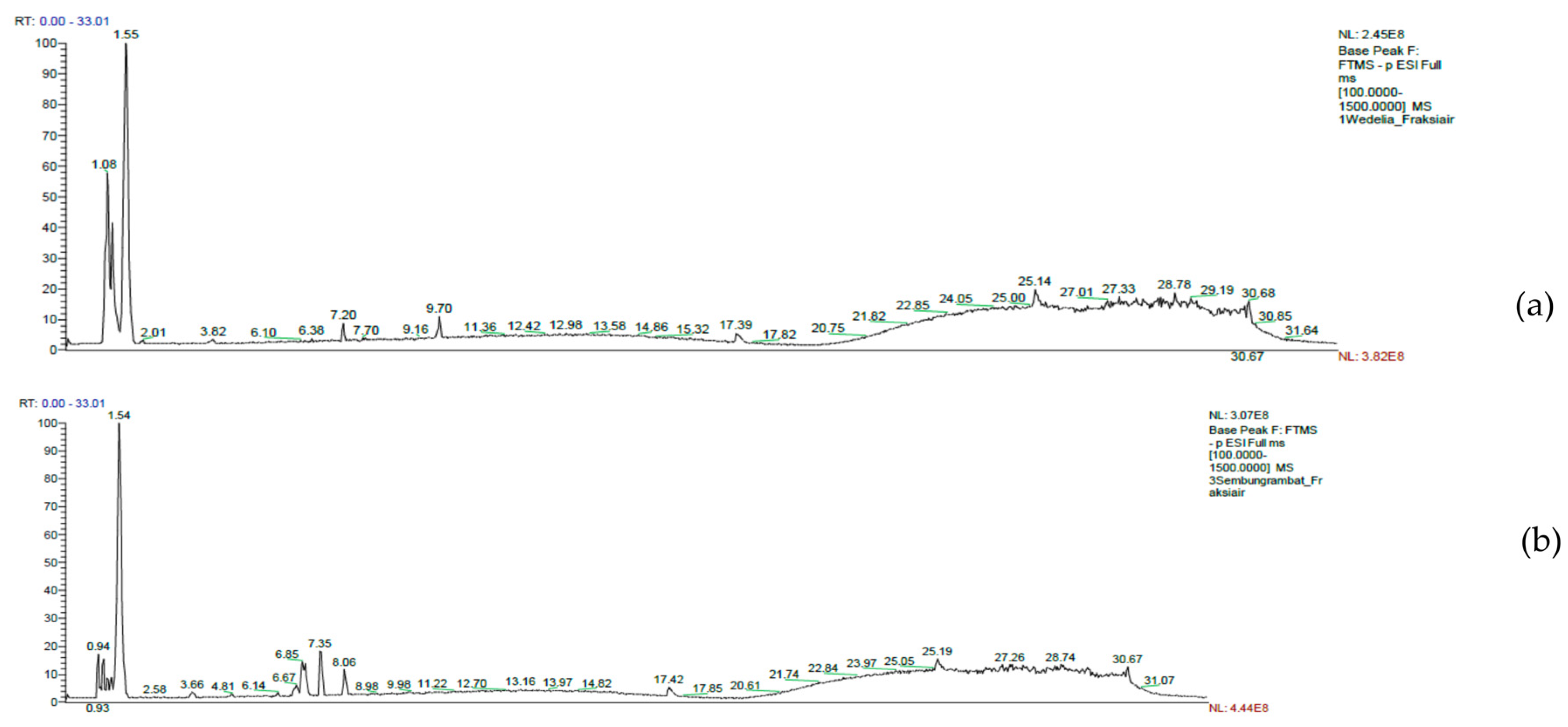
The LC-MS/MS chromatogram for the H2O extracts of Wedelia and Sembung Rambat can be seen in Figure 1. This research found that amino acids, phenolic acids, and organic acids are 4 groups of compounds that are found in large quantities in both extracts. In addition, terpenoids were only found in the Wedelia H2O extract. The entire group of compounds (4 groups of compounds) consists of 17 compounds. It has been reported that all these groups of compounds function as anti-inflammatories, particularly by inhibiting the release of pro-inflammatory cytokines IL-6, TNF-α, and IL-1β (Table 2) [38,39,40,41]. Three compounds were also found together in both extracts, including L-(+)-Valine, 4-Aminobenzoic acid, and Protocatechuic acid.

3.4. Cell Viability

In order to analyze the anti-inflammatory properties of Wedelia and Sembung Rambat extracts, we initially measured their cytotoxicity in RAW 264.7 macrophages. This method is based on determining cell viability in cell culture to investigate the effects of pharmaceuticals in vitro on cell proliferation by cell-mediated cytotoxicity analysis. In this study, we used the MTT (3-(4,5-dimethylthiazol-2-yl)-2,5-diphenyltetrazolium bromide) assay. The principle of this method is to measure the mitochondrial dehydrogenase activity of living cells, which has the ability to convert MTT into formazan. The intensity of the purple color of formazan is determined using an absorbance value that is related to the number of living cells. The more intense the purple color, the higher the absorption value. This demonstrates that the higher the number of living cells reacting with tetrazolium salt, the more formazan is produced [19,50].
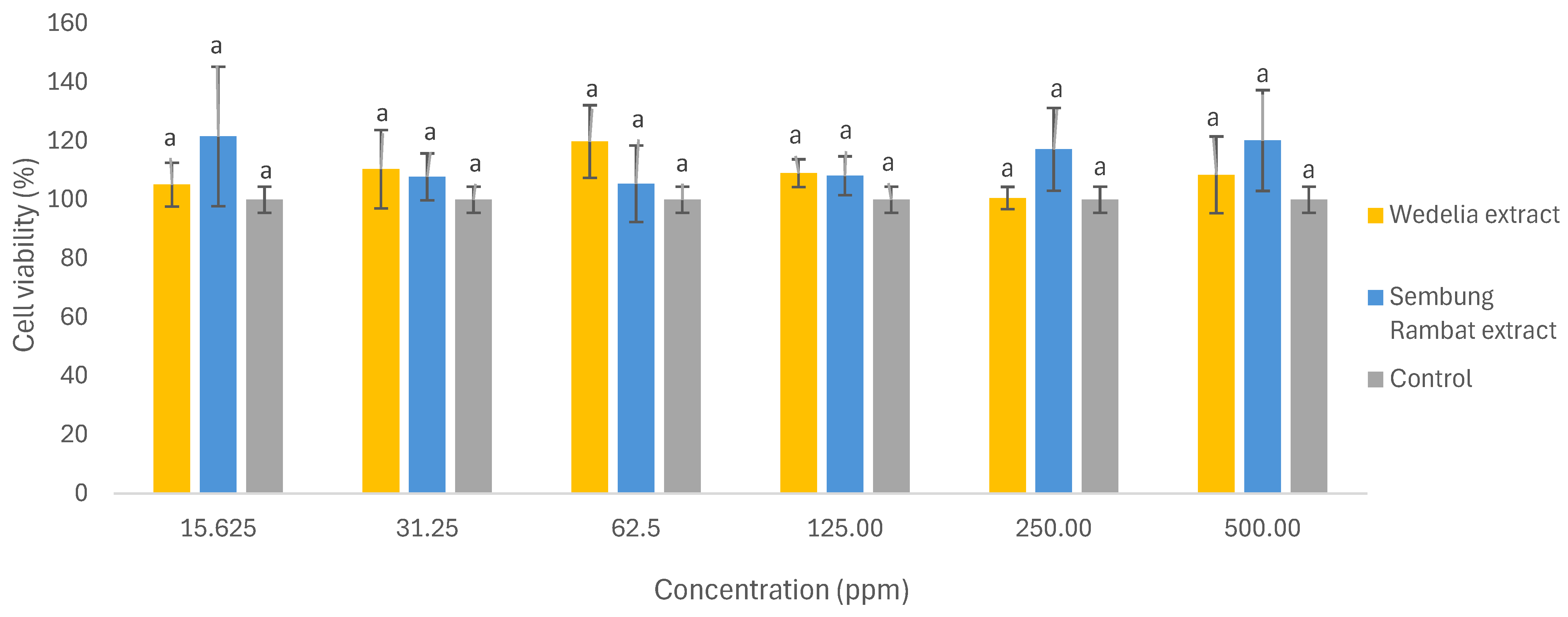
The viability of RAW 264.7 macrophage cells when treated with H2O extracts of Wedelia and Sembung Rambat can be observed in Figure 2 and Table S3. This study used sample concentrations of 15.625, 31.25, 62.50, 125.00, 250.00, and 500.00 ppm. The cell viability assay is used to identify the concentration that does not cause macrophage cell death when treated with the optimum formula. A macrophage cell viability value of 264.7 is considered non-toxic if the cell viability reaches above 80% [51,52]. The results showed that the RAW 264.7 macrophage’s viability reached above 80% for all assay concentrations following treatment with aqueous extracts of Wedelia and Sembung Rambat. This result serves as the basis for determining safe concentration levels [53] in extract inhibition analysis against LPS-treated IL-6, IL-2, and IL-1β.

3.5. Inhibitory Activity of Single Extracts Against Pro-Inflammatory Cytokines

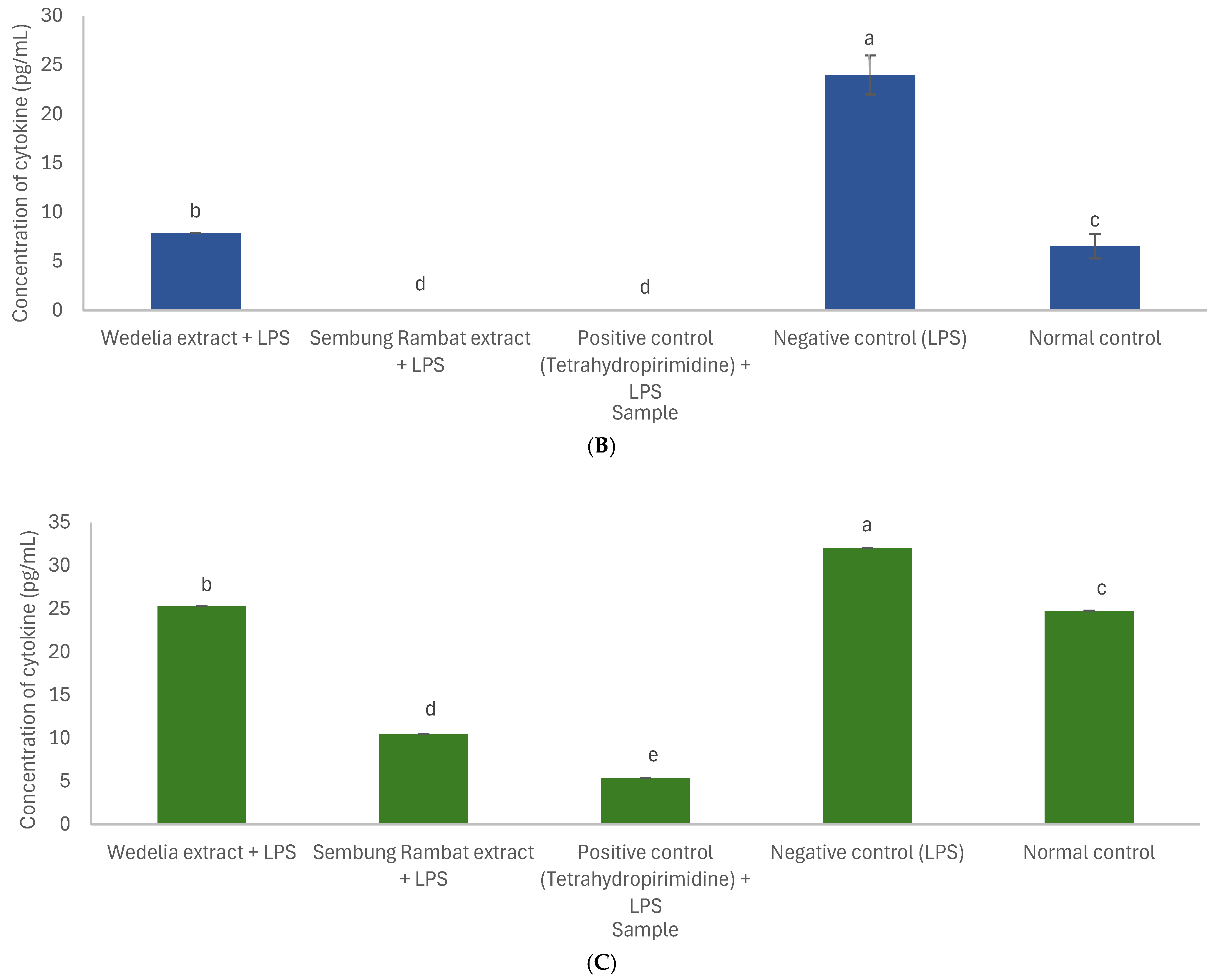
An extract concentration of 31.25 ppm was used in the inhibition assay of pro-inflammatory cytokine release from Wedelia and Sembung Rambat extracts. This concentration was selected because it is considered safe when converted to a human drug dose. Furthermore, the moderate concentration is expected to demonstrate effectiveness as a drug or inhibit the in vitro release of pro-inflammatory cytokines [54,55].
Tetrahydropyrimidine was used as the positive control because it is used as an anti-inflammatory drug (non-steroidal anti-inflammatory drugs/NSAIDs) that suppresses the production of pro-inflammatory cytokines and works by inhibiting inflammatory pathway enzymes such as COX-2 (cyclooxygenase-2) and iNOS (inducible nitric oxide synthase) [56,57].
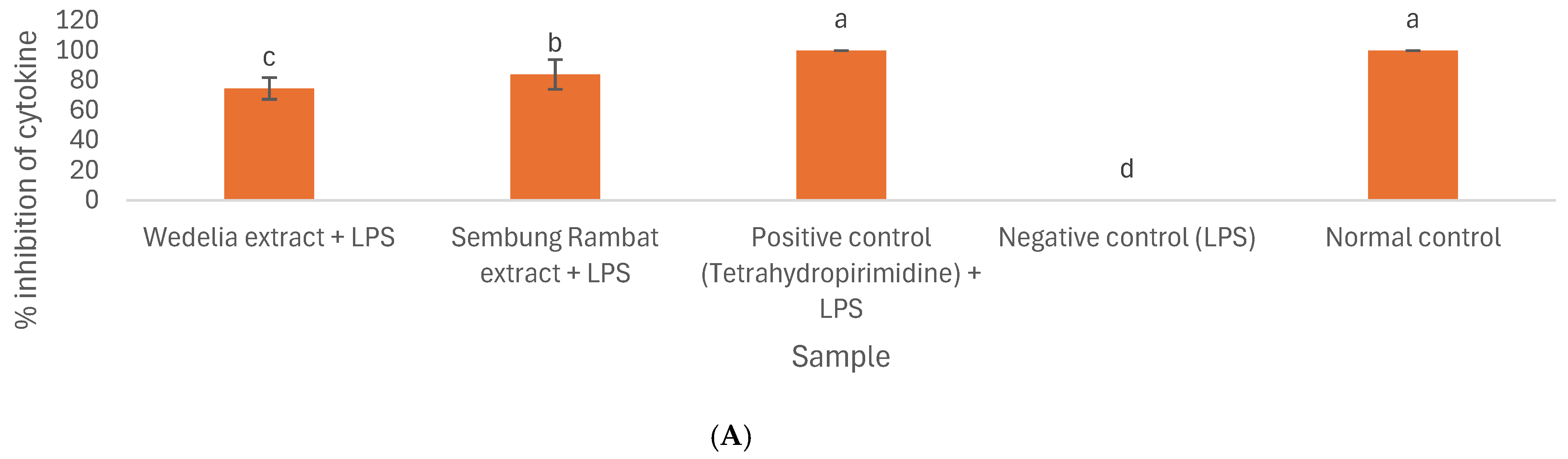
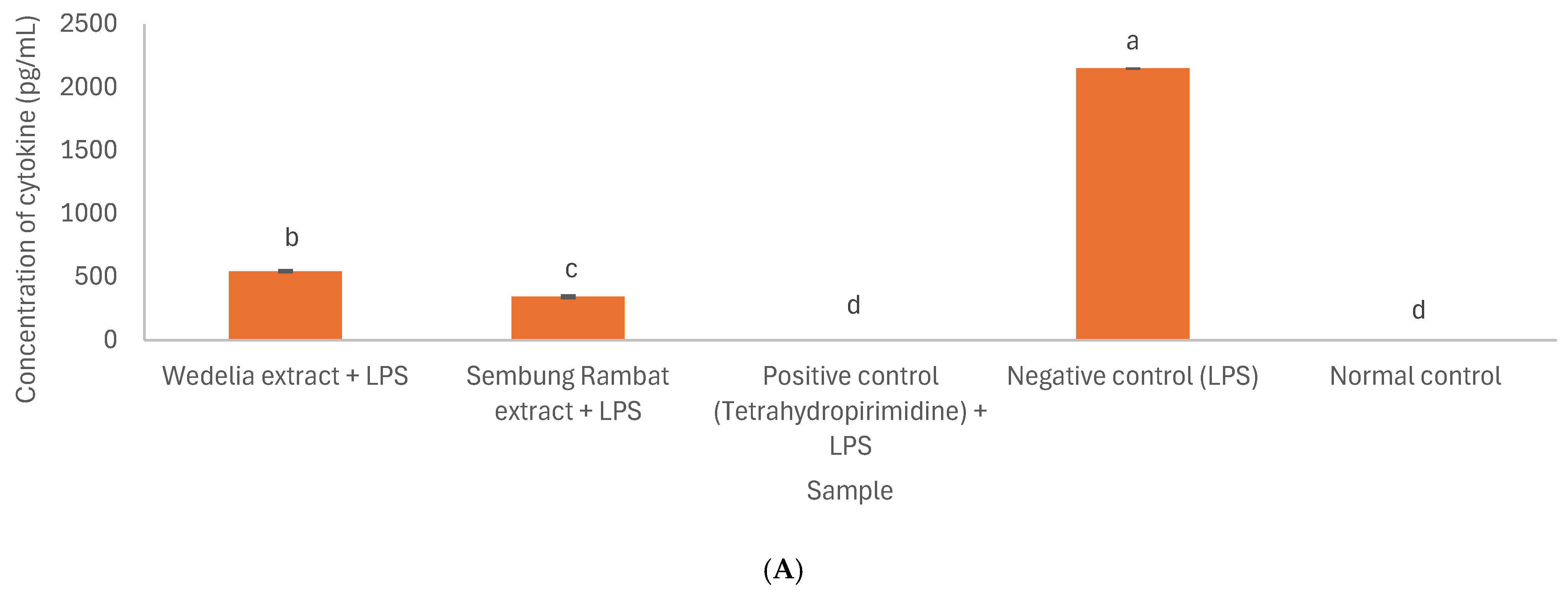
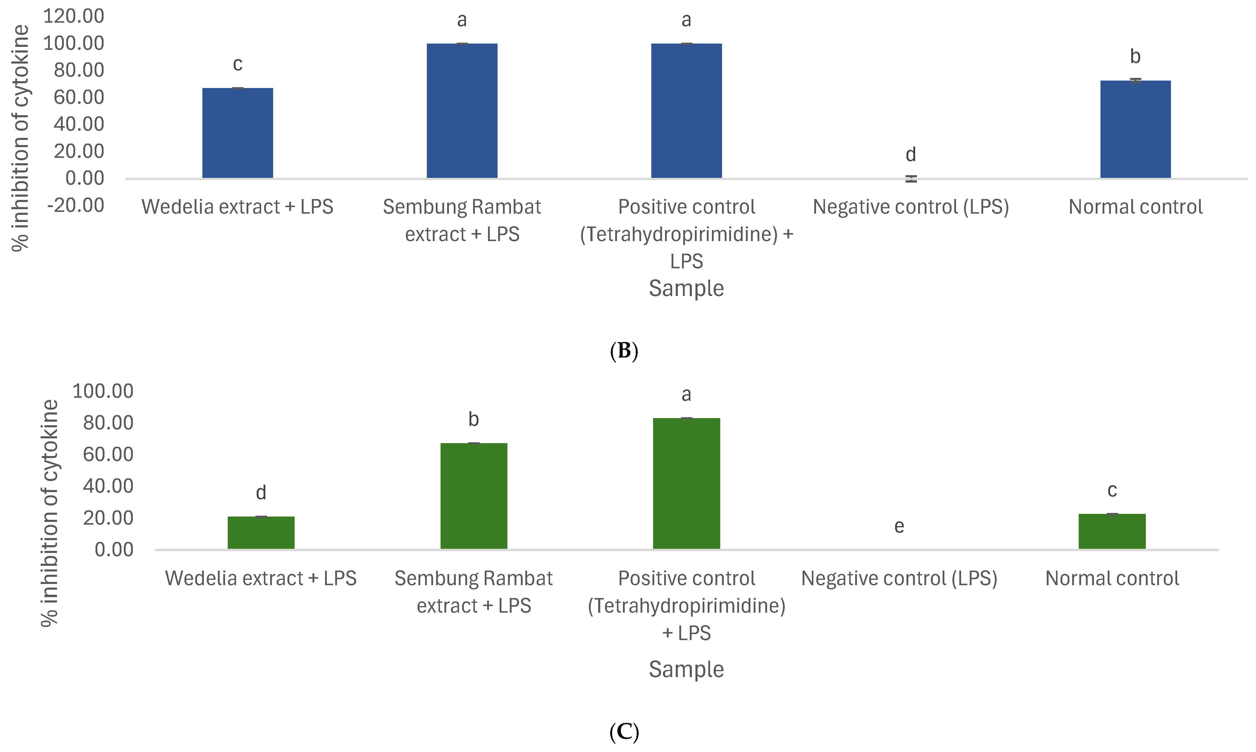
The findings noted that in the IL-6 assay, the Sembung Rambat extract had the highest inhibition (84.03 ± 9.91%) and the lowest concentration (343.25 ± 9.91 pg/mL). In comparison, the Wedelia extract showed an IL-6 concentration of 544.222 ± 7.13 pg/mL with an inhibition of 74.68 ± 7.13%. Compared with both the negative and positive control groups, the two extracts demonstrated significantly different inhibitory effects. The positive ones explained an inhibition of 100.00 ± 0.00%, while the negative control demonstrated no inhibition of IL-6 cytokine release (0.00 ± 0.00%). The normal control did not show any IL-6 cytokine concentration or have an inhibition of 100.00 ± 0.00%.
In the IL-2 assay, the Sembung Rambat extract demonstrated the lowest concentration of 0.00 ± 0.00 pg/mL and the maximum inhibition of 100.00 ± 0.00%. This inhibition was similar to the reference treatment (100.00 ± 0.00%), with no significant difference. Meanwhile, the Wedelia extract had an IL-2 concentration of 7.88 ± 0.0 pg/mL and an inhibition of 67.15 ± 0.0. The negative control showed no inhibition of IL-2 cytokine release (0.00 ± 0.00%), and the normal control had a concentration of 6.55 ± 1.26 pg/mL or an inhibition of 72.72 ± 1.26%. However, the Sembung Rambat extract had greater inhibition of IL-2 cytokine release than the normal control.
In the IL-1β assay, the Sembung Rambat extract had the lowest concentration of 10.46 ± 0.00 pg/mL and the highest inhibition of 67.34 ± 0.00%. The Wedelia extract yielded an IL-1β concentration of 25.32 ± 0.00 pg/mL with an inhibition of 20.95 ± 0.00%. Meanwhile, the positive control had a higher inhibition than the Rambat extract, namely 83.16 ± 0.00%. The untreated control did not exhibit inhibition of IL-β cytokine release. The normal control showed an IL-1β concentration of 24.78 ± 0.00 pg/mL or 22.63 ± 0.00% inhibition. Nevertheless, the extract’s inhibition was still higher than the normal control. Based on the test, both extracts have the potential to inhibit pro-inflammatory cytokine production. Inhibition data from each extract, the positive control, the negative control, and the normal control are presented in Figure 3 and Figure 4, as well as Tables S4 and S5.
A major transcription factor in macrophages, the Nuclear Factor-kappa B (NF-κB) signaling pathway is necessary for the induction of many pro-inflammatory cytokine genes, including TNF-α, IL-1β, IL-6, IL-12, and COX-2. NF-κB activation occurs via two primary mechanisms: the classical (canonical) pathway and the alternative (non-canonical) pathway. The classical pathway is triggered by pro-inflammatory stimuli such as TNF-α, IL-1, and LPS. This pathway induces protein degradation, resulting in the release and translocation of NF-κB dimers to the cell nucleus, where they activate the expression of genes associated with inflammation and cellular defense. As this pathway operates rapidly and plays a dominant role in the acute inflammatory response, NF-κB is closely linked to the pathogenesis of inflammatory diseases such as COPD. Therefore, extracts of Wedelia and Sembung Rambat, which can inhibit the release of the pro-inflammatory cytokines IL-6, IL-2, and IL-1β, will have implications for the NF-κB signaling pathway [58,59].
The assay results are consistent with the metabolite profile data, showing that amino and phenolic acids dominate in reducing pro-inflammatory cytokine production. Previous studies have also confirmed these results. For example, Ellergezen et al. [43] reported that pregabalin, an amino acid, could inhibit the release of IL-6 and IL-2 in individuals with fibromyalgia syndrome (FMS) at a dose of 150 mg/day for at least three months. Furthermore, Li et al. [39] stated that Protocatechuic acid from the phenolic acids group at a dose of 0.25–1.0 µM can reduce the levels of IL-6, IL-1β, and TNF-α in LPS-induced macrophages.
Research related to the anti-inflammatory activity of Sembung Rambat extract has also been conducted by Deori et al. [25] in vivo, which stated that Sembung Rambat extract can reduce rat paw edema in carrageenan-induced acute and sub-acute inflammation at a dose of 200–400 mg/kgBW of Sembung Rambat ethanol extract. Furthermore, Samsuar et al. [60] also revealed that the ethanol fraction of Sembung Rambat at a dose of 450 mg/kgBW in vivo has anti-inflammatory activity on rat paw edema induced by carrageenan for acute inflammation.
An in vivo inflammation-inhibiting study related to inflammation in the respiratory tract was conducted by Castro et al. [61], who stated that IL-1β inhibition reduced rat lung inflammation induced by exposure to cigarette smoke. Furthermore, Widowati et al. [62] stated that Camellia sinensis L. extract reduced IL-6 expression in an ARDS experimental rat model. Lee et al. [63] also indicated that glycyrrhizin inhibited IL-6 lipopolysaccharide-induced chronic pulmonary damage in a rat model. Algheshairy et al. [64] also stated that Citrus limon juice can reduce IL-6 levels in a respiratory-irritated rat model. Several in vivo studies have shown that inhibition of pro-inflammatory cytokines can reduce inflammation in the respiratory tract, thus aligning with the in vitro studies we have currently conducted.

4. Conclusions

Despite the significant results, certain limitations must be acknowledged. Standardization of raw materials for simplicia and extracts was carried out in accordance with the requirements of BPOM Number 32 of 2019 concerning the Safety and Quality Requirements for Traditional Medicines. The Cd ion content is of particular concern because it exceeds the BPOM standard limit for simplicia and both extracts. This may be attributed to contamination originating from fertilizer inputs or environmental sources. Heavy metals are frequently detected in both organic and inorganic fertilizers as well as pesticides during cultivation [35]. The results indicate that determining the ideal growth site and using suitable cultivation techniques are crucial for lowering the amount of Cd in plants.
Metabolite profiling of the two extracts was conducted by LC-MS/MS using a Thermo Scientific Vanquish UHPLC coupled with a Q Exactive Plus Orbitrap high-resolution mass spectrometer. Although this instrument is putative, the compound determination has a mass error of ±5 ppm and compound fragmentation, which increases the reliability of the identified compounds. However, further studies should be conducted using standard compounds to validate the compounds obtained.
The standardization assay and in vitro studies of pro-inflammatory cytokine release inhibition (IL6, IL-2, and IL-1β) showed that Wedelia and Sembung Rambat extracts possess inflammation-inhibiting activity related to COPD. The Sembung Rambat water extract inhibited 100.00 ± 0.00% of IL-2 and 67.34 ± 0.00 of IL-1β cytokine release in LPS-treated macrophages. Meanwhile, the Wedelia water extract inhibited 84.03 ± 9.91% of IL-6 cytokine production. The extract concentration used in this in vitro assay for the inhibition of pro-inflammatory cytokine release was 31.25 ppm. This specific concentration was selected because the subsequent research aims to develop an herbal medicine. Therefore, a moderate concentration is required—one that can be converted into a therapeutic dosage and demonstrate efficacy as an anti-inflammatory inhibitor. Nevertheless, assays across the extract concentration range of 15.625–500 ppm are still required to provide a complete set of assay results.
The results are limited to in vitro conditions, and the observed inflammation-inhibiting effects cannot be directly extrapolated to therapeutic efficacy in patients with COPD respiratory tract inflammation. Therefore, further studies are needed to develop in vitro formulations of these two extracts, followed by in vivo evaluation using appropriate animal models and clinical investigations to determine the effectiveness in treating respiratory inflammation associated with COPD patients.
In conclusion, the extracts of Wedelia and Sembung Rambat fulfilled the standards for moisture and ash content, as well as microbial contamination parameters, which is in line with the standardization assays conducted in accordance with the Herbal Pharmacopoeia and BPOM Regulation Number 32 of 2019. The findings indicated that both aqueous extracts demonstrated substantial anti-inflammation activity through inhibition of pro-inflammatory cytokine release in LPS-treated RAW 264.7 macrophages. Amino compounds together with phenolic acids detected in the aqueous extract are considered potential contributors to its inflammation-inhibiting properties, particularly through suppression of pro-inflammatory cytokine release, including IL-6, IL-2, and IL-1β. Both the Wedelia and Sembung Rambat extracts have considerable potential for further development, either individually or in combination, as inflammation-inhibiting agents for COPD management, particularly through the reduction in inflammation in the respiratory system.

Supplementary Materials

The following supporting information can be downloaded at: https://www.mdpi.com/article/10.3390/cimb48070720/s1.

Author Contributions

Conceptualization, D.I. and M.R.; methodology, D.I., M.R., T.T., S.S. and N.H.; validation, D.I. and M.R.; investigation, R.N.R.S., R.A.C., R.M.S., R.Z. and M.K.; writing—original draft preparation, R.N.R.S. and R.M.S.; writing—review and editing, R.N.R.S., R.A.C., R.M.S., N.H. and D.I.; supervision, D.I., M.R. and N.H.; project administration, S.I. All authors have read and agreed to the published version of the manuscript.

Funding

This research was supported by the BRIN RIIM LPDP Grant for the second year, number 41781/IT3.D10/PT.01.03/P/B/2023.

Institutional Review Board Statement

Not applicable.

Informed Consent Statement

Not applicable.

Data Availability Statement

The original contributions in this study are included in the article. Further inquiries can be directed to the corresponding author.

Acknowledgments

The authors express their gratitude to BRIN for providing financial support through the RIIM LPDP Grant for the second year.

Conflicts of Interest

The authors declare no conflicts of interest.

References

  1. Ghaffarpour, S.; Ghazanfari, T.; Ardestani, S.K.; Naghizadeh, M.M.; Mahdavi, M.R.V.; Salehi, M.; Majd, A.M.M.; Rashidi, A.; Chenary, M.R.; Mostafazadeh, A.; et al. Cytokine profiles dynamics in COVID-19 patients: A longitudinal analysis of disease severity and outcomes. Sci. Rep. 2025, 15, 14209. [Google Scholar] [CrossRef] [Scilit] [PubMed]
  2. Zanza, C.; Romenskaya, T.; Manetti, A.C.; Franceschi, F.; La Russa, R.; Bertozzi, G.; Maiese, A.; Savioli, G.; Volonnino, G.; Longhitano, Y. Cytokine Storm in COVID-19: Immunopathogenesis and Therapy. Medicina 2022, 58, 144. [Google Scholar] [CrossRef] [Scilit]
  3. Mara, G.; Nini, G.; Cotoraci, C. Chronic Obstructive Pulmonary Disease and COVID-19: The Impact of Hematological Biomarkers on Disease Severity and Outcomes. J. Clin. Med. 2025, 14, 2765. [Google Scholar] [CrossRef] [Scilit]
  4. Lin, H.; Wang, C.; Yu, H.; Liu, Y.; Tan, L.; He, S.; Li, Z.; Wang, C.; Wang, F.; Li, P.; et al. Protective effect of total Saponins from American ginseng against cigarette smoke-induced COPD in mice based on integrated metabolomics and network pharmacology. Biomed. Pharmacother. 2022, 149, 112823. [Google Scholar] [CrossRef] [Scilit] [PubMed]
  5. Xu, J.; Zeng, Q.; Li, S.; Su, Q.; Fan, H. Inflammation mechanism and research progress of COPD. Front. Immunol. 2024, 15, 1404615. [Google Scholar]
  6. Kongsomros, S.; Boonyarattanasoonthorn, T.; Phongphaew, W.; Kasorndorkbua, C.; Sunyakumthorn, P.; Im-Erbsin, R.; Lugo-Roman, L.A.; Kongratanapasert, T.; Paha, J.; Manopwisedjaroen, S.; et al. In vivo evaluation of Andrographis paniculata and Boesenbergia rotunda extract activity against SARS-CoV-2 Delta variant in Golden Syrian hamsters: Potential herbal alternative for COVID-19 treatment. J. Tradit. Complement. Med. 2024, 14, 598–610. [Google Scholar] [CrossRef] [Scilit] [PubMed]
  7. Clarke, R.; Lundy, F.T.; McGarvey, L. Herbal treatment in asthma and COPD—Current evidence. Clin. Phytosci. 2015, 1, 4. [Google Scholar] [CrossRef] [Scilit]
  8. Patel, R.; Naqvi, S.A.; Griffiths, C.; Bloom, C.I. Systemic adverse effects from inhaled corticosteroid use in asthma: A systematic review. BMJ Open Respir. Res. 2020, 7, e000756. [Google Scholar] [CrossRef] [Scilit] [PubMed]
  9. Rusmini, H.; Ma’rifah, S. Gambaran Penggunaan Kortikosteroid Sistemik Jangka Panjang Terhadap Kejadian Katarak Di Poli Mata Rumah Sakit Pertamina Bintang Amin Bandar Lampung. J. Ilmu Kedokt. Kesehat. 2017, 4, 91–97. [Google Scholar]
  10. Budianti, T. Chronic Obstructive Pulmonary Disease: An Overview. J. Biol. Trop. 2025, 25, 5075–5084. [Google Scholar] [CrossRef] [Scilit]
  11. Hapid, A.; Ariyanti, A.; Erniwati, E.; Suena, N.M.D.S.; Adrianta, K.A.; Yuniarti, K.; Muthmainnah, M. Diversity of Types of Medicinal Plants and Local Wisdom of the Kaili Tribe in Processing Medicinal Plants Around the Forest Areas of Central Sulawesi, Indonesia. Pharmacogn. J. 2023, 15, 535–540. [Google Scholar] [CrossRef] [Scilit]
  12. Davis, C.C.; Choisy, P. Medicinal plants meet modern biodiversity science. Curr. Biol. 2024, 34, R158–R173. [Google Scholar] [CrossRef] [Scilit] [PubMed]
  13. Hasyim, D.M.; Anurogo, D.; Bansaleng, Y.F.; Hiola, S.F.; Anrip, N. Potential of Medicinal Plants in Indonesian Forest Biodiversity Conservation in Synergy with Pharmaceutical Technology for Modern Medicine. Int. J. Eng. Sci. Inf. Technol. 2025, 5, 200–207. [Google Scholar] [CrossRef] [Scilit]
  14. World Health Organization. WHO Global Report on Traditional and Complementary Medicine 2019; World Health Organization: Geneva, Switzerland, 2019. [Google Scholar]
  15. Fortune Business Insights. Herbal Medicine Market Size, Share & COVID-19 Impact Analysis, by Application (Pharmaceutical & Nutraceutical, Food & Beverages, and Personal Care & Beauty Products), By Form (Powder, Liquid & Gel, and Tablets & Capsules), and Regional Forecast, 2024–2032; Fortune Business Insights: Pune, India, 2024. [Google Scholar]
  16. Tahir, A.H.; Hussain, Z.; Yousuf, H.; Fazal, F.; Tahir, M.A.; Kashif, M. Traditional Herbal Medicine and Its Clinical Relevance: A Need to Preserve the Past for the Future. J. Biosci. Med. 2022, 10, 64–75. [Google Scholar] [CrossRef]
  17. Li, J.S.; Xie, Y.; Li, S.Y.; Yu, X.Q. Comparison of conventional medicine, TCM treatment, and combination of both conventional medicine and TCM treatment for patients with chronic obstructive pulmonary disease: Study protocol of a randomized comparative effectiveness research trial. Trials 2014, 15, 153. [Google Scholar] [CrossRef] [Scilit] [PubMed]
  18. Jiang, D.; Wang, X.; Su, Q.; Jiang, S.; Yuan, F.; Zhang, C.; Gong, F.; Dong, Q.; Shi, J.; Chen, B. Milkvetch root improves immune function in patients with acute exacerbation of COPD. Biomed. Mater. Eng. 2015, 26, S2113–S2121. [Google Scholar] [CrossRef] [Scilit] [PubMed]
  19. Iswantini, D.; Rahminiwati, M.; Trivadila, T.; Sadiah, S.; Hanif, N.; Sianipar, R.N.R.; Indariani, S. Inhibition of Inflammatory Regulators for Chronic Obstructive Pulmonary Disease (COPD) Treatment from Indonesian Medicinal Plants: A Systematic Review. Curr. Issues Mol. Biol. 2025, 47, 262. [Google Scholar] [CrossRef] [Scilit]
  20. Lukito, E.W.; Iswantini, D.; Antariksa, B.; Rafi, M.; Wahyudi, S.T. Screening and Identification of Metabolites from Sambiloto (Andrographis paniculata) Ethanol Extract for Pro-Inflammatory Cytokines Inhibitory through In Silico and In Vitro Approaches. Pharmacogn. J. 2024, 16, 131–140. [Google Scholar] [CrossRef] [Scilit]
  21. Astuti, F.W. Analisis Potensi Ekstrak Adenostemma lavenia dan Ageratum conyzoides sebagai Inhibitor Interleukin-6; Skripsi Sarjana, Institut Pertanian Bogor: Bogor, Indonesia, 2021. [Google Scholar]
  22. Tuwalaid, B.; Iswantini, D.; Wahyudi, S.T. Potential Adenostemma lavenia and Muntingia calabura Extracts to Inhibit Cyclooxygenase-2 Activity as a Therapeutic Strategy for Anti-inflammation: Experimental and Theoretical Studies. Indones. J. Chem. 2022, 22, 754–769. [Google Scholar] [CrossRef] [Scilit]
  23. Widyaningrum, N.R.; Wahyuningsih, S.S.; Priyono, C. Antiinflammation activity of Muntingia calabura L. leaves ethanol, ethylacetate and chloroform extracts. Nat. Sci. J. Sci. Technol. 2022, 11, 7–13. [Google Scholar] [CrossRef] [Scilit]
  24. Xu, Z.; Ji, R.; Zha, X.; Zhao, H.; Zhou, S. The aqueous extracts of Ageratum conyzoides inhibit inflammation by suppressing NLRP3 inflammasome activation. J. Ethnopharmacol. 2023, 309, 116353. [Google Scholar] [CrossRef] [Scilit] [PubMed]
  25. Deori, C.; Dutta, G.; Das, S.; Phukan, D.; Gogoi, G. To Evaluate the Anti-Inflammatory Activity of Ethanolic Extract of Leaves of Mikania micrantha on Experimental Animal Models. J. Evol. Med. Dent. Sci. 2017, 6, 3818–3821. [Google Scholar] [CrossRef] [Scilit]
  26. Association of Official Analytical Collaboration (AOAC). AOAC Guideline for Single Laboratory Validation of Chemical Methods for Dietary Supplements and Botanicals; AOAC International: Rockville, MD, USA, 2013. [Google Scholar]
  27. SNI 01-2891-1992; Cara Uji Makanan dan Minuman. Badan Standardisasi Nasional (BSN): Jakarta, Indonesia, 1992.
  28. SNI 01–2896–1998; Cara Uji Cemaran Logam dalam Makanan. Badan Standardisasi Nasional (BSN): Jakarta, Indonesia, 1998.
  29. SNI 2897:2008; Metode Pengujian Cemaran Mikroba Pada Pangan. Badan Standardisasi Nasional (BSN): Jakarta, Indonesia, 2008.
  30. Sianipar, R.N.R.; Iswantini, D.; Charlena, C.; Wahyudi, S.T.; Prasetyo, J. Simultaneous Optimization of Extraction Yield, Flavonoid, and Phenolic Compounds from Spatholobus littoralis Hassk. and Sauropus androgynus L. Using Response Surface Methodology. Iran. J. Chem. Chem. Eng. 2025, 44, 3007–3029. [Google Scholar]
  31. Rafi, M.; Meitary, N.; Septaningsih, D.A.; Bintang, M. Phytochemical profile and antioxidant activity of Guazuma ulmifolia leaves extracts using different solvent extraction. Indones. J. Pharm. 2020, 31, 171–180. [Google Scholar] [CrossRef] [Scilit]
  32. Borghi, S.M.; Mizokami, S.S.; Carvalho, T.T.; Rasquel-Oliveira, F.S.; Ferraz, C.R.; Fattori, V.; Hayashida, T.H.; Peron, J.P.S.; Camilios-Neto, D.; Ambrosio, S.R.; et al. The diterpene from Sphagneticola trilobata (L.) Pruski, kaurenoic acid, reduces lipopolysaccharide-induced peritonitis and pain in mice. J. Ethnopharmacol. 2021, 273, 113980. [Google Scholar] [CrossRef] [Scilit] [PubMed]
  33. Datta, S.; Bhattacharjee, S.; Seal, T. Anti-diabetic, anti-inflammatory and anti-oxidant properties of four underutilized ethnomedicinal plants of West Bengal, India: An in vitro approach. S. Afr. J. Bot. 2022, 149, 768–780. [Google Scholar] [CrossRef] [Scilit]
  34. Utami, Y.P.; Umar, A.H.; Syahruni, R.; Kadullah, I. Standardisasi Simplisia dan Ekstrak Etanol Daun Leilem (Clerodendrum minahassae Teisjm. & Binn.). J. Pharm. Med. Sci. 2017, 2, 32–39. [Google Scholar] [CrossRef] [Scilit]
  35. Hindarwati, Y.; Soeprobowati, T.R.; Izzati, M.; Hadiyanto, H. Kontaminan Logam Berat (Pb, Cd, dan Cu) pada Tanah dari Pemupukan Berbasis Jerami Padi. J. Ilmu Lingk. 2023, 21, 8–14. [Google Scholar]
  36. Leung, H.M.; Yue, P.Y.K.; Sze, S.C.W.; Au, C.K.; Cheung, K.C.; Chan, K.L.; Yung, K.L.K.; Li, W.C. The potential of Mikania micrantha (Chinese creeper) to hyperaccumulate heavy metals in soil contaminated by electronic waste. Environ. Sci. Pollut. Res. 2019, 26, 35275–35280. [Google Scholar] [CrossRef] [Scilit]
  37. Liu, Y.; Chen, S.; Wu, Y.; Zhu, H.; Lin, W.; Zheng, Y.; Yu, Z. Research on the potential of Sphagneticola trilobata (L.) Pruski for historical tungsten mining area soils remediation based on physiological integration analysis. SSRN Electron. J. 2025, 5495677. [Google Scholar] [CrossRef] [Scilit]
  38. Lin, Y.; Caesar, C.; Sutopo, Y.; Wang, S.; Chen, J.; Kao, Y.; Chen, Y. Free amino-acid and imidazole-ring dipeptide profiles of chicken-liver-hydrolysate supplement and its modulatory effects on lipid metabolism, oxidative status, and inflammation in livers, as well as gut microbiota in a high-fat diet. Poult. Sci. 2026, 105, 106533. [Google Scholar] [PubMed]
  39. Li, S.; Du, Y.; Chen, G.; Mao, Y.; Zhang, W.; Kang, M.; Zhu, S.; Wang, D. Protocatechuic Acid Attenuates Inflammation in Macrophage-like Vascular Smooth Muscle Cells in ApoE-/- Mice. Nutrients 2025, 17, 1090. [Google Scholar] [CrossRef] [Scilit] [PubMed]
  40. Kui, L.; Wang, G.; Huang, J.; Yang, X.; Yu, J.; Li, X.; Peng, H.; Leng, B.; Jiao, Y.; Zhang, Z.; et al. Therapeutic efficacy and mechanisms of gentiopicroside in various diseases. Front. Pharmacol. 2025, 16, 1634722. [Google Scholar] [CrossRef] [Scilit] [PubMed]
  41. Zhang, J.; Bissih, F.; Tan, B.; Zhang, S.; Yang, Q.; Dong, X. Citric acid could strengthen the intestinal tight junction barrier and improve intestinal immune function in vivo and in vitro experiments of hybrid grouper (Epinephelus fuscoguttatus ♀ × E. lanceolatus ♂). Aquac. Rep. 2026, 46Trace, 103282. [Google Scholar] [CrossRef] [Scilit]
  42. Ahil, S.B.; Hira, K.; Shaik, A.B.; Pal, P.P.; Kulkarni, O.P.; Araya, H.; Fujimoto, Y. L-Proline-based-cyclic dipeptides from Pseudomonas sp. (ABS-36) inhibit pro-inflammatory cytokines and alleviate crystal-induced renal injury in mice. Int. Immunopharmacol. 2019, 73, 395–404. [Google Scholar] [CrossRef] [Scilit]
  43. Ellergezen, P.; Alp, A.; Çavun, S.; Çelebi, M.; Macunluoğlu, A.C. Pregabalin inhibits proinflammatory cytokine release in patients with fibromyalgia syndrome. Arch. Rheumatol. 2023, 38, 307–314. [Google Scholar] [CrossRef] [Scilit] [PubMed]
  44. Tekin, S.; Bolat, M.; Atasever, A.; Bolat, İ.; Çinar, B.; Shadidizaji, A.; Dağ, Y.; Şengül, E.; Yildirim, S.; Hacimuftuoglu, A. Mechanistic insights into the P-coumaric acid protection against bisphenol A-induced hepatotoxicity in in vivo and in silico models. Sci. Rep. 2025, 15, 8709. [Google Scholar] [CrossRef] [Scilit]
  45. Ye, Z.; Cheng, L.; Xuan, Y.; Yu, K.; Li, J.; Gu, H. Chlorogenic acid alleviates the development of severe acute pancreatitis by inhibiting NLPR3 inflammasome activation via Nrf2/HO-1 signaling. Int. Immunopharmacol. 2025, 151, 114335. [Google Scholar] [CrossRef] [Scilit] [PubMed]
  46. Klebeko, J.; Ossowicz-Rupniewska, P.; Świątek, E.; Szachnowska, J.; Janus, E.; Krachmarova, E.; Guncheva, M. Salicylic Acid as Ionic Liquid Formulation May Have Enhanced Potency to Treat Some Chronic Skin Diseases. Molecules 2021, 27, 216. [Google Scholar] [CrossRef] [Scilit] [PubMed]
  47. Peng, L.; Zuo, M.; Qiu, T.; Lan, W.; Wen, Y. Sweroside: Unveiling broad therapeutic potential—From mechanistic insights to clinical potential. Front. Pharmacol. 2025, 16, 1594278. [Google Scholar] [CrossRef] [Scilit]
  48. Ahmed, A.A.; Elmujtba, M.; Essa, A.; Mollica, A.; Stefanucci, A.; Zengin, G.; Ahmed, H. Gum Arabic modifies anti-inflammatory cytokine in mice fed with high fat diet induced obesity. Bioact. Carbohydr. Diet. Fibre 2021, 25, 100258. [Google Scholar] [CrossRef] [Scilit]
  49. Zainul, R.; Afif, I.S.; Goh, K.W.; Wulandari, A.P.; Wandira, A.; Etika, S.B.; Oktavia, B.; Nelza, N. Molecular Docking and Pharmacokinetic Evaluation of Terpenoid Compounds from Ramie (Boehmeria nivea (L.) Gaudich.) Leaves as Potential Natural Agents for Inflammatory Bowel Disease. Chem. Methodol. 2026, 10, 489–503. [Google Scholar]
  50. Ghasemi, M.; Turnbull, T.; Sebastian, S.; Kempson, I. The MTT Assay: Utility, Limitations, Pitfalls, and Interpretation in Bulk and Single-Cell Analysis. Int. J. Mol. Sci. 2021, 22, 12827. [Google Scholar] [CrossRef] [Scilit] [PubMed]
  51. Wu, Y.; Tang, K.; Wang, C.; Song, H.; Zhou, F.; Guo, Y. Establishment of interpretable cytotoxicity prediction models using machine learning analysis of transcriptome features. Acta Pharm. Sin. B 2025, 15, 1344–1358. [Google Scholar] [CrossRef] [Scilit] [PubMed]
  52. Madorran, E.; Ambrož, M.; Knez, J.; Sobočan, M. An Overview of the Current State of Cell Viability Assessment Methods Using OECD Classification. Int. J. Mol. Sci. 2025, 26, 220. [Google Scholar]
  53. Uddin, S.; Han, S.B.; Cho, S. Anti-inflammatory and antioxidant effects of Pogostemon stellatus (Lour.) Kuntze via MAPK, NF-κB, and Nrf2 signaling pathways in LPS-activated RAW 264.7 macrophages. Front. Pharmacol. 2025, 16, 1679919. [Google Scholar] [CrossRef] [Scilit]
  54. Hamed, A.M.; Enaili, S.S.; Mohammed, W.I.; Abouelella, A.M.A.; Elsayed, Z.; Mohana, E.; Monir, D.M.; Soliman, S.S.; Eldeeb, E.; Hamouda, M.; et al. Novel Benzenesulfonamide Derivatives of 5′-Aminospirotriazolotriazine Exhibit Anti-Inflammatory Activity by Suppressing Pro-Inflammatory Mediators: In Vitro and In Vivo Evaluation Using a Rat Model of Carrageenan-Induced Paw Edema. Biomedicines 2025, 13, 1732. [Google Scholar] [CrossRef] [Scilit]
  55. Jacob, S.; Nair, A.B.; Morsy, M.A. Dose Conversion Between Animals and Humans: A Practical Solution. Indian J. Pharm. Educ. Res. 2022, 56, 600–607. [Google Scholar] [CrossRef] [Scilit]
  56. Tylińska, B.; Janicka-Kłos, A.; Gębarowski, T.; Nowotarska, P.; Plińska, S.; Wiatrak, B. Pyrimidine Derivatives as Selective COX-2 Inhibitors with Anti-Inflammatory and Antioxidant Properties. Int. J. Mol. Sci. 2024, 25, 11011. [Google Scholar] [CrossRef] [Scilit] [PubMed]
  57. Ali, A.R.; Al-Kubeisi, A.K.; Elwakil, B.H.; Darwish, A.A.E.; Almutairi, T.M.; Elshatanofy, M.M.; Alshargabi, T.Q.; El Sawy, M.A. Design, synthesis, and biological evaluation of new tetrahydropyrimidine derivatives as multitarget anti-inflammatory agents. Eur. J. Med. Chem. Rep. 2025, 13, 100259. [Google Scholar] [CrossRef] [Scilit]
  58. Liu, T.; Zhang, L.; Joo, D.; Sun, S. NF-κB signaling in inflammation. Signal Transduct. Target. Ther. 2017, 2, 17023. [Google Scholar] [CrossRef] [Scilit] [PubMed]
  59. Bharti, K.; Pandey, R.; Srivastava, N.; Kashyap, S.; Kumar, D.; Kumar, L.; Suman, S.K.; Patel, S.K.S. Role of Inflammatory Mediators in Chronic Obstructive Pulmonary Disease Pathogenesis: Updates and Perspectives. Curr. Issues Mol. Biol. 2025, 47, 1856–1882. [Google Scholar]
  60. Samsuar, S.; Rokiban, A.; Nur, R. Fraksi Etanol Daun Sembung Rambat (Mikania micrantha Kunth) sebagai Antiinflamasi terhadap Tikus Putih Jantan Galur Wistar. JFL J. Farm. Lampung 2018, 7, 51–56. [Google Scholar]
  61. Castro, P.; Legora-Machado, A.; Cardilo-Reis, L.; Valença, S.; Cristóvão Porto, L.; Walker, C.; Zuany-Amorim, C.; Gonçalves Koatz, V.L. Inhibition of interleukin-1β reduces mouse lung inflammation induced by exposure to cigarette smoke. Eur. J. Pharmacol. 2004, 498, 279–286. [Google Scholar] [CrossRef] [Scilit] [PubMed]
  62. Widowati, W.; Priyandoko, D.; Sari, H.; Kusuma, W. The Potency of Camellia sinensis L. to Reduce Proinflammatory Cytokine Levels in the Acute Respiratory Distress Syndrome Rat Model. Nama J. Teridentifikasi 2023, 55, 92–108. [Google Scholar] [CrossRef] [Scilit]
  63. Lee, S.A.; Lee, S.H.; Kim, J.Y.; Lee, W.S. Effects of glycyrrhizin on lipopolysaccharide-induced acute lung injury in a mouse model. J. Thorac. Dis. 2019, 11, 1287–1302. [Google Scholar] [CrossRef] [Scilit] [PubMed]
  64. Algheshairy, R.M.; Ali, H.A. Antioxidant, anti-inflammatory, and immunomodulatory pathways of Citrus limon juice in a respiratory-irritated rat model. Front. Nutr. 2026, 13, 1743575. [Google Scholar] [CrossRef] [Scilit] [PubMed]
Figure 1. LC-MS/MS chromatogram for Wedelia H2O extract (a) and Sembung Rambat H2O extract (b).
Cimb 48 00720 g001
Figure 2. Viability of RAW 264.7 macrophage cells when treated with the extract. For each sample, values with different letters in the same column indicate significant differences at p-value ≤ 0.05 based on one-way ANOVA followed by Tukey’s test (n = 3). The results are sorted in ascending order: a > b > c > d > e.
Cimb 48 00720 g002
Figure 3. Concentrations of IL-6 (A), IL-2 (B), and IL-1β (C) produced by Wedelia and Sembung Rambat extracts. For each sample, values with different letters in the same column indicate significant differences at p-value ≤ 0.05 based on one-way ANOVA followed by Tukey’s test (n = 3). The results are sorted in ascending order: a > b > c > d > e.
Cimb 48 00720 g003aCimb 48 00720 g003b
Figure 4. Inhibition of Wedelia and Sembung Rambat extracts against IL-6 (A), IL-2 (B), and IL-1ß (C). For each sample, values with different letters in the same column indicate significant differences at p-value ≤ 0.05 based on one-way ANOVA followed by Tukey’s test (n = 3). The results are sorted in ascending order: a > b > c > d > e.
Cimb 48 00720 g004aCimb 48 00720 g004b
Table 1. Yields of Wedelia and Sembung Rambat extracts.
SampleYield (%)
Wedelia20.04
Sembung Rambat26.65
Table 2. Data on compounds that have the potential to be inhibitors of pro-inflammatory cytokines in the H2O extracts of Wedelia and Sembung Rambat.
No.Groups of CompoundsNo.CompoundsWedelia H2O ExtractSembung Rambat H2O ExtractAnti-Inflammatory ActivityReference
1.Amino acids1.L-(+)-ValineThe free amino acid composition contained in chicken liver hydrolysate-based supplements can reduce IL-6, TNF-α, and IL-1β inflammation in mice fed a long-term high-fat diet (HFD).[38]
2.L-(+)-Leucine-The free amino acid composition contained in chicken liver hydrolysate-based supplements can reduce IL-6, TNF-α, and IL-1β inflammation in mice fed a long-term high-fat diet (HFD).[38]
3.DL-Phenylalanine-The free amino acid composition contained in chicken liver hydrolysate-based supplements can reduce IL-6, TNF-α, and IL-1β inflammation in mice fed a long-term high-fat diet (HFD).[38]
4.DL-Glutamic acid-The free amino acid composition contained in chicken liver hydrolysate-based supplements can reduce IL-6, TNF-α, and IL-1β inflammation in mice fed a long-term high-fat diet (HFD).[38]
5.4-Aminobenzoic acidThe free amino acid composition contained in chicken liver hydrolysate-based supplements can reduce IL-6, TNF-α, and IL-1β inflammation in mice fed a long-term high-fat diet (HFD).[38]
6.DL-Tyrosine-The free amino acid composition contained in chicken liver hydrolysate-based supplements can reduce IL-6, TNF-α, and IL-1β inflammation in mice fed a long-term high-fat diet (HFD).[38]
7.L-Proline-L-Proline-based cyclic dipeptides derived from Pseudomonas sp. decrease pro-inflammatory cytokines in vitro.[42]
8.Pregabalin-Pregabalin could inhibit the release of IL-6 and IL-2 in individuals with fibromyalgia syndrome (FMS) at a dose of 150 mg/day for at least three months.[43]
2.Phenolic acids9.Protocatechuic acidProtocatechuic acid can reduce the levels of IL-6, IL-1β, and TNF-α in LPS-induced macrophages.[39]
10.(E)-p-coumaric acid-(E)-p-coumaric acid can lower the levels of IL-6, TNF-α, and IL-1β in liver tissue of bisphenol A-induced rats. [44]
11.Chlorogenic acid-Chlorogenic acid can reduce levels of IL-6, TNF-α, and IL-1β in severe acute pancreatitis rats.[45]
12. Salicylic acid-Salicylic acid can inhibit the production of IL-6 in LPS-stimulated keratinocytes.[46]
3.Organic acids13.Gentiopicrin-Gentiopicrin reduces the levels of IL-6, TNF-α, and IL-1β in LPS-induced inflammation.[40]
14.Sweroside-Sweroside can reduce the production of IL-6, TNF-α, and IL-1β in vitro.[47]
15.Citric acid-Citric acid suppressed the secretion of IL-6, TNF-α, and IL-1β in an LPS-stimulated inflammation model of primary intestinal epithelial cells.[41]
16.Arabic acid-Arabic acid lowered the levels of IL-6, TNF-α, and IL-1β in mice treated with high-fat diet-induced obesity.[48]
4.Terpenoids17.Navenone A-Navenone A can reduce the levels of IL-6, TNF-α, and IL-1β in vitro.[49]
Disclaimer/Publisher’s Note: The statements, opinions and data contained in all publications are solely those of the individual author(s) and contributor(s) and not of MDPI and/or the editor(s). MDPI and/or the editor(s) disclaim responsibility for any injury to people or property resulting from any ideas, methods, instructions or products referred to in the content.