Article

Functional Characterization and Inhibition Analysis of a Glutathione Transferase from Cryptosporidium parvum: A Potential Target for Antiparasitic Drug Development

by
Panagiota D. Pantiora
1,
Nikolaos D. Georgakis
1,
Dimitris Matiadis
2,
Marina Sagnou
3,4 and
Nikolaos E. Labrou
1,5,*
1
Laboratory of Enzyme Technology, Department of Biotechnology, School of Applied Biology and Biotechnology, Agricultural University of Athens, 75 Iera Odos Street, 11855 Athens, Greece
2
Laboratory of Chemistry, Department of Food Science & Human Nutrition, School of Food and Nutritional Sciences, Agricultural University of Athens, Iera Odos 75, 11855 Athens, Greece
3
Institute of Biosciences & Applications, National Centre for Scientific Research “Demokritos”, 15310 Athens, Greece
4
Chemical Biology Laboratory, RTIN and Department of Science and Mathematics—Deree, The American College of Greece, 15342 Athens, Greece
5
Institute of Nanobiotechnology, Sustainable Development and Artificial Intelligence, Research and Innovation Center, Agricultural University of Athens, Iera Odos 75, 11855 Athens, Greece
*
Author to whom correspondence should be addressed.
Pharmaceuticals 2026, 19(7), 1106; https://doi.org/10.3390/ph19071106
Submission received: 13 June 2026 / Revised: 9 July 2026 / Accepted: 9 July 2026 / Published: 17 July 2026

Abstract

Background/Objectives: Cryptosporidiosis, caused by Cryptosporidium parvum, is a significant cause of diarrheal disease, particularly affecting young children and immunocompromised individuals. With current treatments offering limited efficacy, there is an urgent need for novel therapeutic targets. Methods: In this study, we report the cloning, expression, and functional characterization of a glutathione transferase (GST) from C. parvum (CpGST). Results: Biocomputing analysis revealed a single gene encoding a cytosolic enzyme with distinct structural features, compared to human cytosolic homologs. Structural modeling indicated a non-canonical thioredoxin fold and a truncated C-terminal domain, suggesting functional divergence. CpGST was expressed in Escherichia coli, and its enzymatic properties were characterized. Although the enzyme displayed a narrow substrate spectrum, it showed a distinct substrate preference, retaining catalytic activity toward the standard GST substrates 1-chloro-2,4-dinitrobenzene (CDNB) and cumene hydroperoxide (CuOOH). Steady-state kinetic analysis revealed limited affinity for both reduced glutathione (GSH) and CDNB. Inhibition analysis identified several polyphenols and synthetic curcumin analogues as potent inhibitors, with IC50 values in the low micromolar range. Kinetic analysis with the most potent inhibitor revealed a mixed-type inhibition mechanism. Conclusions: These findings support the classification of CpGST as a structurally and functionally distinct member of the GST family, likely adapted to the parasite’s physiology and metabolism. The enzyme’s divergence from human GSTs, along with its favorable druggability profile, underscores its potential as a target for anti-cryptosporidial drug development, particularly in strategies aimed at disrupting stress response and detoxification pathways.

1. Introduction

Cryptosporidiosis is an intestinal disease caused by intracellular protozoan parasites of the genus Cryptosporidium [1]. These parasites are a major cause of waterborne infections and outbreaks [2,3], targeting the intestinal epithelium of various hosts, including humans, livestock, birds, fish, and reptiles [4]. In humans, infection occurs primarily through ingestion of oocysts present in contaminated water or food or via direct contact with infected individuals or animals [5,6,7]. The oral–fecal route remains the predominant mode of transmission [8,9].
Cryptosporidiosis has been recognized by the World Health Organization (WHO) as one of the leading causes of parasitic gastroenteritis and diarrheal outbreaks [6]. Although it is often self-limiting in healthy individuals, the infection can become life-threatening in vulnerable populations, including young children, the elderly, and immunocompromised patients, due to prolonged and severe diarrheal episodes [10,11,12]. Cryptosporidium parvum and Cryptosporidium hominis account for up to 90% of human cases, representing a significant public health challenge [4,13,14].
Effective treatment options for cryptosporidiosis remain limited [12,15]. To date, nitazoxanide is the only drug approved by the U.S. Food and Drug Administration (FDA) for the treatment of cryptosporidiosis [16,17]. However, its efficacy is significantly reduced in immunocompromised individuals and malnourished children, populations at the highest risk of severe disease [16,17]. Other compounds, including azithromycin, paromomycin, roxithromycin, spiramycin, and bovine-derived anticryptosporidial immunoglobulins, have been explored for their therapeutic potential, but their clinical benefits remain modest [18,19,20].
Given the widespread prevalence of cryptosporidiosis in both humans and animals, the identification of novel drug targets is a critical research priority. Cryptosporidium species possess unique biological features that make them amenable to targeted drug development. For example, they have a compact genome and relatively simple cellular architecture, with fewer organelles compared to related protozoan parasites such as Cyclospora, Plasmodium, and Toxoplasma [21,22,23]. Recent studies have highlighted several promising molecular targets, particularly those involved in essential metabolic pathways and macromolecular processes. These include parasite-specific protein kinases, such as calcium-dependent protein kinase 1 (CDPK1) and phosphatidylinositol 4-kinase (PI4K) [24,25,26], as well as enzymes involved in nucleic acid synthesis [27], proteolysis (e.g., cysteine and subtilisin-like serine proteases) [28], and lipid metabolism [29].
In addition to established molecular targets, enzymes involved in the metabolism of xenobiotics and toxic compounds represent a promising yet underexplored opportunity for cryptosporidiosis drug development. Among these, glutathione transferases (GSTs) are a diverse and functionally significant superfamily of enzymes involved in detoxification, redox regulation, and xenobiotic metabolism across all domains of life [30]. They catalyze the conjugation of reduced glutathione (GSH) to a wide range of endogenous and xenobiotic electrophiles, including hydroperoxides, thereby enhancing their solubility and facilitating excretion while simultaneously protecting cells from oxidative stress [31,32]. GSTs in humans have been implicated in the development of resistance to chemotherapeutic agents and in pathogen adaptation to hostile host environments. These functional properties make GSTs attractive targets for drug development [33,34,35].
In protozoan parasites, GSTs are increasingly recognized not only for their role in cellular defense mechanisms but also as attractive drug targets, owing to their essential functions in survival and adaptation within host environments [36,37]. Among protozoa, the GSTs from Plasmodium falciparum and Toxoplasma gondii have been the most extensively studied [37,38,39]. For example, P. falciparum expresses a multifunctional GST involved in heme detoxification during hemoglobin digestion, a critical survival mechanism for the parasite in erythrocytes [37]. The inhibition of P. falciparum GST has been proposed as a viable antimalarial strategy due to its crucial involvement in oxidative stress management. Despite their well-established roles in other organisms, GSTs remain largely uncharacterized in Cryptosporidium species. Genomic analysis of Cryptosporidium parvum Iowa II by Abrahamsen et al. (2004) identified several proteins involved in the parasite’s defense mechanisms, including a single cytosolic GST [40]. This enzyme plays key roles in redox homeostasis, signal transduction, and apoptosis regulation. Importantly, it is expressed throughout all developmental stages of the Cryptosporidium life cycle, clearly suggesting its fundamental importance to parasite viability [41]. Recent genomic analyses have identified three genes encoding distinct GST isoforms in Cryptosporidium species [42]. Based on amino acid sequence length, these isoforms can be divided into two groups: shorter variants (157–268 residues) and longer ones (373–466 residues), with size differences attributed to the presence or absence of N- or C-terminal extensions. These GSTs are cytosolic and belong to three novel classes, Vega (θ), Psi (ψ), and Gamma (γ), which appear to be unique to Cryptosporidium spp. [42].
In the present study, we report for the first time the cloning and molecular characterization of a GST from the parasitic pathogen Cryptosporidium parvum Iowa II (CpGST). Sequence analysis revealed that, although the protein shares some features with cytosolic GSTs, it exhibits substantial divergence from human GST orthologs, suggesting promising potential for selective targeting. A library of natural products, including curcumin and some of its monocarbonyl derivatives, was evaluated for their potential to inhibit the activity of CpGST. This study provides the first comprehensive characterization of CpGST, including its structural and functional analysis and the identification of some potent inhibitors that may act as lead therapeutic compounds.

2. Results and Discussion

2.1. Phylogenetic Analysis

Gene mining was conducted to identify putative GST genes within the Cryptosporidium parvum Iowa II genome (GenBank: AAEE00000000.1). The analysis led to the identification of a single open reading frame (ORF) encoding a cytosolic-type GST, which was annotated under accession number XP_628625.1 and selected for further structural and biochemical characterization. This finding should be distinguished from recent comparative genomic studies reporting three GST isoform classes across Cryptosporidium species, which likely reflect broader interspecies diversity within the genus. The enzyme is composed of 161 amino acids, with a theoretical pI of 4.87 and a molecular weight of 18,759.51 Da. InterPro analysis revealed that the gene encodes a protein with conserved GST structural domains, including the typical N-terminal thioredoxin-like fold (IPR004045) and a C-terminal α-helical domain (IPR010987). The obtained results indicate that the sequence contains the characteristic structural features of GSTs, thus confirming its classification within the GST family. Unlike many other protozoa that possess multiple GST isoforms, C. parvum appears to encode a single cytosolic GST, suggesting a potentially non-redundant and essential function. This uniqueness is in contrast to the finding in Giardia lamblia, where GST-like enzymes have also been identified as key antioxidant defenses, despite the organism’s limited oxidative stress response pathways [43].
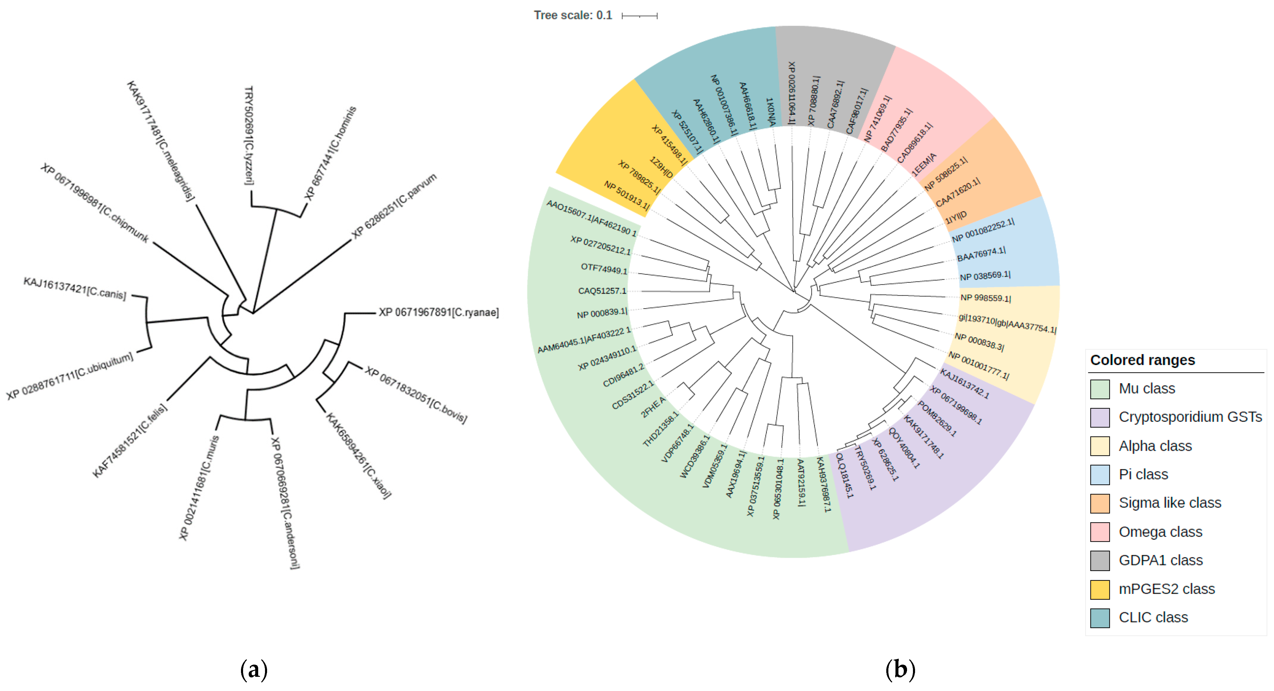
A BLASTp search using the CpGST sequence identified homologous GST-like proteins across various Cryptosporidium species. The retrieved sequences exhibited pairwise identity values ranging from 55.97% for C. muris to 98.76% for C. hominis, while the corresponding query coverage values ranged from 94% to 100%. The high query coverage observed for most homologues indicates that the similarity extends across a substantial portion of the CpGST sequence, supporting moderate to high conservation of GST-like proteins within the genus. These sequences were subsequently used to construct a phylogenetic tree, enabling the analysis of the evolutionary positioning of CpGST (XP_628625.1) in relation to its homologs from other Cryptosporidium species (Figure 1a).
CpGST clusters most closely with the enzyme from C. hominis. This supports the well-established genetic similarity between C. parvum and C. hominis, both of which infect humans and share similar biological and pathogenic features. The evolutionary analysis of GSTs from various Cryptosporidium species, using representative sequences from all known animal GST classes, revealed that CpGST clusters closely with the mammalian mu GST class, yet it forms a distinct and separate clade. This phylogenetic positioning suggests that CpGSTs probably represent a novel class of animal GSTs, diverging early from the canonical mu-type lineage (Figure 1b).

2.2. Structure Prediction and Analysis of CpGST

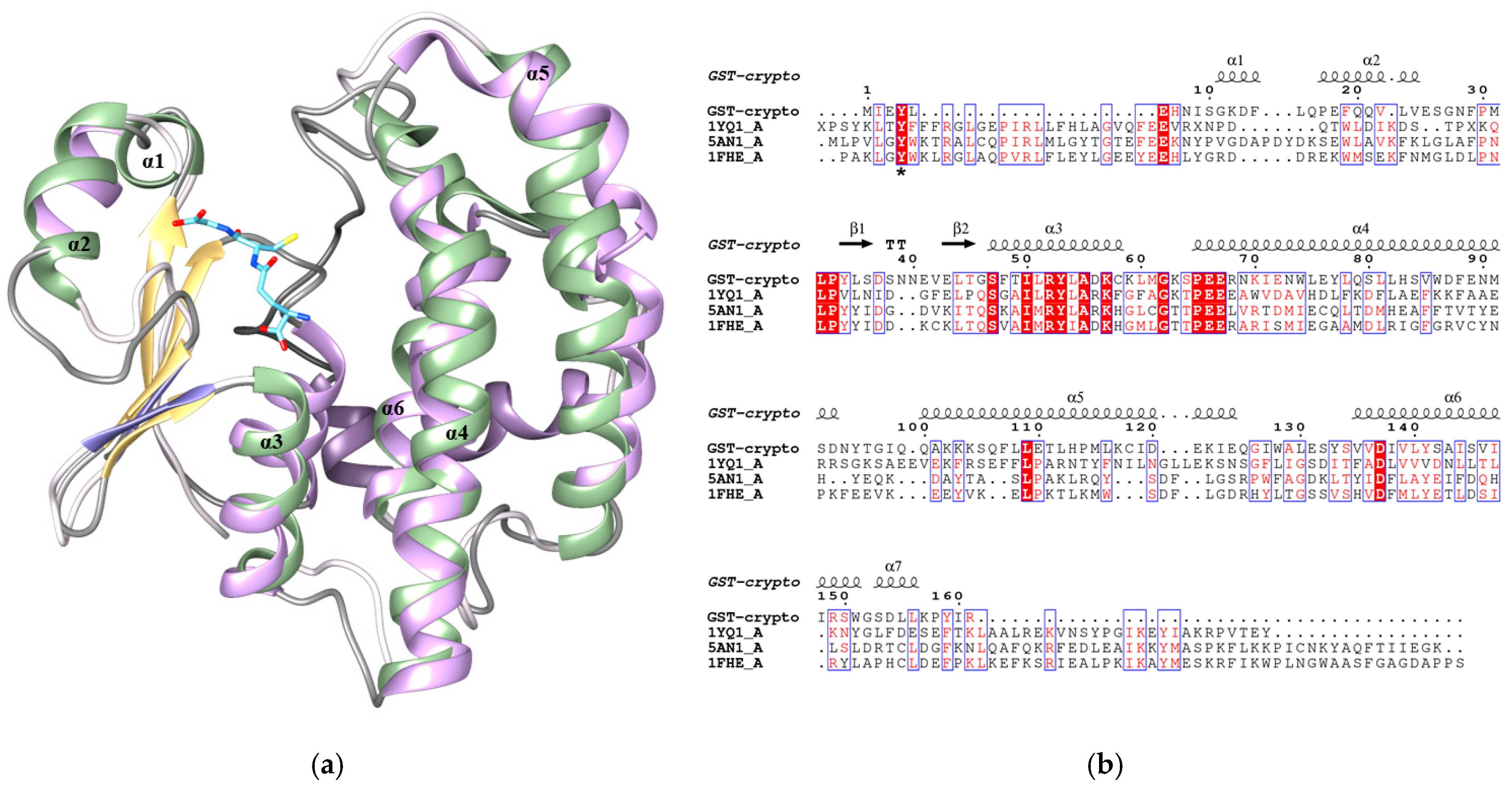
Structure prediction of CpGST was performed using the I-TASSER server. Among the five models generated, the one with the highest confidence score was selected for further analysis. Overall, the predicted structure (Figure 2a) is characterized by the typical GST architecture, consisting of two distinct domains. The N-terminal thioredoxin-like domain, spanning residues 1 to 62, harbors the glutathione-binding site (G-site), and a C-terminal domain is responsible for binding hydrophobic xenobiotic substrates (H-site). Interestingly, although cytosolic GSTs generally exhibit the canonical βαβαββα thioredoxin-like fold, CpGST displays a non-canonical thioredoxin fold with an ααββα motif, suggesting potential structural and functional divergence. The C-terminal all-alpha domain is located between residues 64 and 161. This domain is significantly shorter compared to that of other parasite and human orthologs. The C-terminal helix of GSTs plays a pivotal role in maintaining the structural integrity of the active site and shaping the hydrophobic substrate-binding pocket (H-site). It contributes to substrate orientation, active-site closure, and catalytic efficiency, often participating in induced-fit conformational changes upon ligand binding [44,45].
To increase confidence in the predicted architecture, the CpGST model was further compared with AlphaFold2-derived predictions and evaluated using structural quality and confidence metrics (see Supplementary Information Figure S1a). AlphaFold2 confidence analysis showed that the predicted CpGST structure has generally good to very high local confidence across most of the sequence. The five ranked models displayed highly similar pLDDT profiles, indicating good consistency among the independent predictions (Figure S1b). The best-ranked CpGST model showed a mean pLDDT value of 88.87, with an uncertainty of 2.57, supporting very good overall confidence in the predicted structure. The overall agreement between independent modeling approaches supports the proposed fold.
All these features together—the structural divergences, the non-canonical thioredoxin fold and the missing C-terminal helix compared to cytosolic GSTs—make the parasite-specific CpGST a compelling candidate drug target, offering opportunities for the design of selective inhibitors that minimize off-target effects on host (human) GSTs.
Structure-based alignments (Figure 2b) with mu-class GST homologs from Fasciola hepatica (45.45% identity), Penaeus vannamei (37.18% identity), and Caenorhabditis elegans (26.36% identity) revealed the presence of a conserved catalytic tyrosine residue at position 4 (Tyr4) [46,47]. Interestingly, in F. hepatica, Gln66 and Lys44 are key contributors to GSH binding [46]. In contrast, the corresponding residues in CpGST is occupied by Gly46, and no Lys residue is present. These substitutions highlight the low degree of homology between the two enzymes at the G-site, which may in turn contribute to a reduced affinity for GSH.

2.3. Cloning, Heterologous Expression and Purification

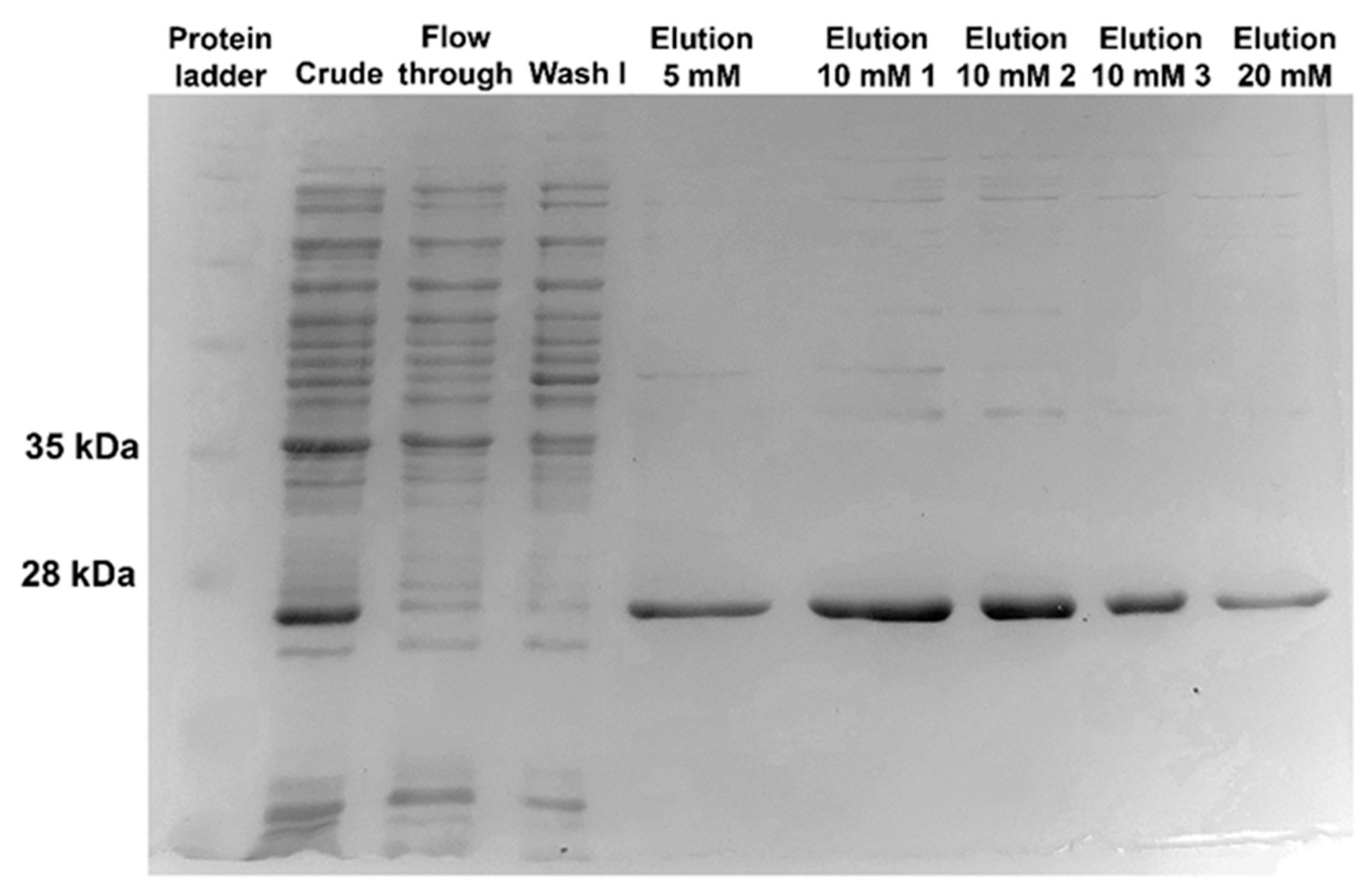
The synthetic gene encoding CpGST was cloned into the pET-28b(+) plasmid vector under the control of a T7 promoter, allowing for strong IPTG-inducible expression. The expression construct was transformed into E. coli Rosetta™ 2 (DE3) pLysS cells. Following induction with IPTG, the recombinant CpGST was efficiently expressed and subsequently purified using Ni2+-IDA-Sepharose affinity chromatography (Figure 3). The SDS–PAGE analysis of the purification process showed a distinct band consistent with the expected molecular weight of the His-tagged CpGST protein. The protein was eluted using a stepwise imidazole gradient (5 to 20 mM). This elution profile suggests a low affinity of the His-tagged CpGST for the Ni2+ resin. Following purification, the enzyme preparation exhibited a specific activity of 0.431 U/mg, corresponding to an enzyme recovery of 81.8%. The purification protocol resulted in a 45.4-fold increase in specific activity compared with the crude extract.

2.4. Substrate Specificity and Kinetic Analysis

The substrate specificity of CpGST was assessed by measuring its specific activity against a panel of common GST substrates (Table 1). Using CDNB, a standard model substrate for GSTs, the enzyme exhibited the highest specific activity, confirming its suitability for baseline enzymatic characterization.
Notably, CpGST also showed activity toward CuOOH, a known marker of GSH peroxidase-like activity, suggesting a potential role in peroxide detoxification and redox homeostasis. In contrast, the enzyme exhibited minimal activity toward tert-butyl hydroperoxide, indicating a narrow substrate range for hydroperoxides and a possible structural preference for aromatic over aliphatic hydroperoxides. Fluorodifen, a substituted diphenyl ether herbicide, was accepted as a substrate with moderate activity, which is consistent with GSTs involved in xenobiotic metabolism. Interestingly, no detectable activity was observed toward several other known GST substrates, including dehydroascorbate, ethacrynic acid, and isothiocyanates (allyl isothiocyanate and phenethyl isothiocyanate). The inability to metabolize ethacrynic acid and isothiocyanates, substrates commonly conjugated by human GSTs [48], further supports the structural divergence of CpGST from host homologs and emphasizes its unique substrate recognition profile.
Collectively, these findings suggest that CpGST is catalytically active and has a restricted but distinct substrate range. Its preference for CDNB and CuOOH highlights its dual potential in both xenobiotic conjugation and peroxidase-like activity, while its inactivity toward several common GST substrates underscores its structural and functional specificity.
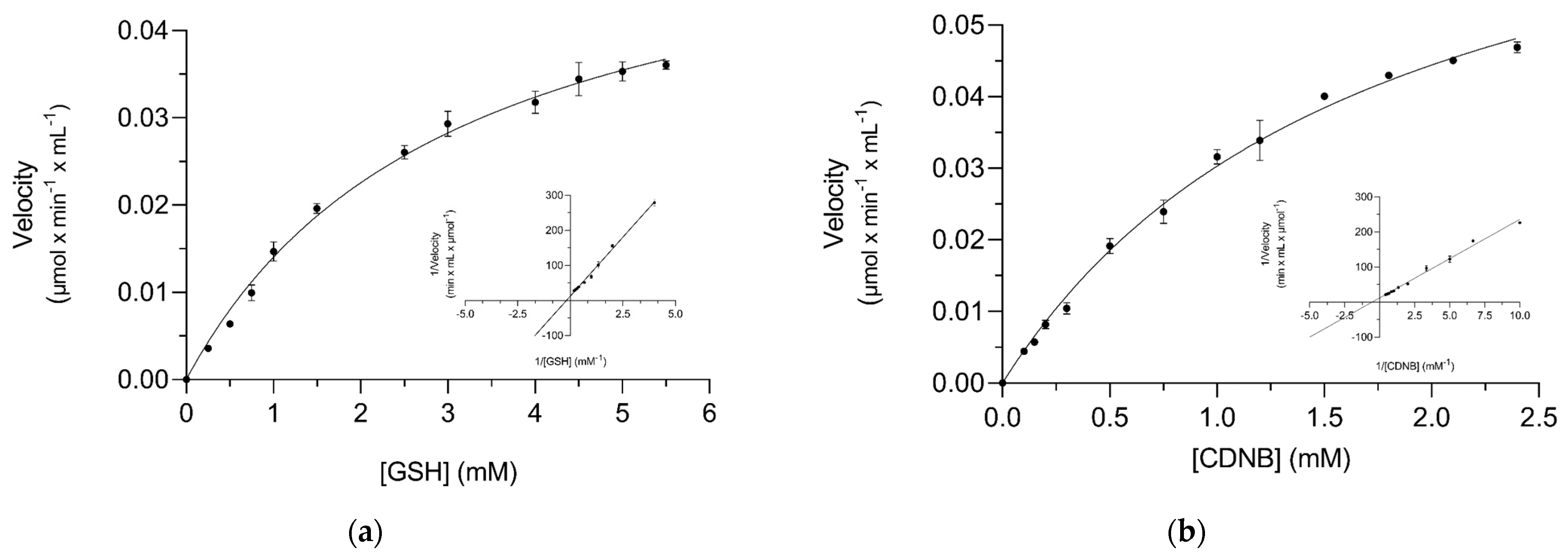
The kinetic parameters of the recombinant CpGST enzyme were determined using GSH and CDNB. The results for the kinetic analysis are shown in Figure 4 and the related kinetic parameters are summarized in Table 2. The enzyme displayed Michaelis–Menten-type kinetics for both substrates. The high Km value suggests a relatively low affinity of CpGST for GSH compared to many human GSTs, which often have Km values for GSH below 1 mM [49]. This observation may reflect structural divergence in the GSH-binding site, as supported by the sequence alignment and structural modeling data (Figure 2). The lower Km for CDNB, compared to GSH, indicates a somewhat higher affinity for this electrophilic substrate relative to GSH, though both affinities remain in the low millimolar range. This kinetic profile aligns with the enzyme’s modest specific activity values and further supports its classification as a catalytically active but relatively low-efficiency GST compared to canonical human GSTs [49,50,51].
The low catalytic activity and substrate affinity are likely attributable to the absence of the C-terminal helix, which plays a key role in active-site stabilization and substrate positioning. In several GST classes, residues within this region directly influence substrate specificity and enzyme stability. Multiple studies have demonstrated that removal or alteration of the C-terminal helix profoundly impairs GST function. For example, studies on alpha- and theta-class GSTs have shown that removal of this region alters substrate specificity, reduces activity and disrupts proper active-site architecture [44,45]. The relatively low catalytic activity of CpGST may be partly associated with its predicted structural divergence from canonical cytosolic GSTs, including the shortened C-terminal region. The purified recombinant enzyme was soluble, migrated at the expected molecular weight, and showed reproducible activity toward CDNB and CuOOH, following Michaelis–Menten kinetics. These observations support the conclusion that CpGST is catalytically competent and unlikely to be misfolded. Nevertheless, other factors, including subtle conformational effects of heterologous expression, parasite-specific molecular partners, or preference for physiological substrates not tested here, may also contribute to its low catalytic efficiency. Further structural and functional studies will be required to clarify the molecular basis of CpGST activity.
These results suggest that CpGST may not serve a broad detoxification role under basal conditions but could instead be adapted for specific or inducible metabolic tasks within the parasite’s metabolic framework.

2.5. Inhibition Analysis of Polyphenols and Synthetic Curcumin Analogues Against CpGST

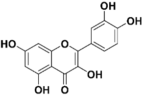
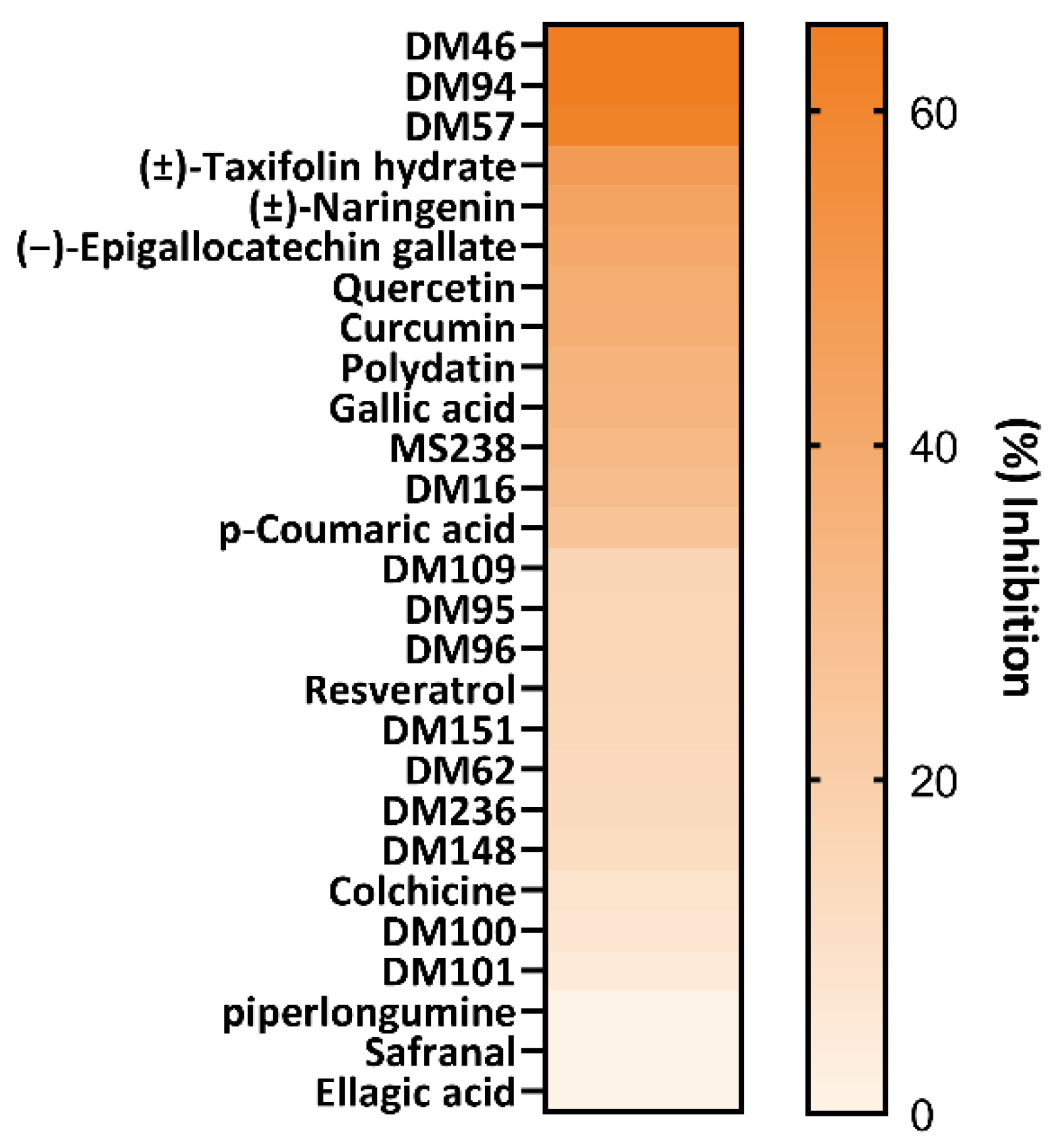
Inhibition analysis was conducted to assess the inhibitory potency of a range of natural products, mainly polyphenols and synthetic curcumin analogues, against CpGST. Each compound was tested at a final concentration of 25 μM. The results were visualized as a heatmap (Figure 5) and listed in Table 3. The screening revealed distinct trends in inhibitor structural preferences, suggesting that certain chemical scaffolds and substituents may be critical for enzyme binding and inhibition. Among the tested natural compounds, polyphenolic flavonoids and catechins demonstrated the highest inhibitory activity. Notably, taxifolin hydrate (47.6%), naringenin (43.5%), and epigallocatechin gallate (40.7%) showed strong inhibition (>40%), suggesting that the flavonoid scaffold, particularly with multiple hydroxyl groups, enhances binding affinity to the enzyme.
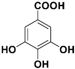
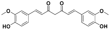
Quercetin (38.1%) and curcumin (37.6%) followed closely, reinforcing the importance of planar, aromatic structures with conjugated systems and phenolic hydroxyls that can potentially engage in hydrogen bonding or π–π stacking within the active site. Gallic acid (34.3%) and polydatin (34.7%) also exhibited moderate inhibition, which may be related to their trihydroxybenzene cores, albeit with a structurally simpler architecture compared to that of flavonoids. In contrast, p-coumaric acid (25.8%) and resveratrol (15.4%) were less potent and were suboptimal for effective binding. Interestingly, colchicine (8.7%), safranal, piperlongumine, and ellagic acid showed negligible or no inhibition.
Among the most effective inhibitors identified was curcumin (37.6%). The effects of curcumin on the infectivity and development of C. parvum have been studied in an in vitro system combining infection of human ileocecal adenocarcinoma cell cultures with quantification of intracellular parasites by quantitative polymerase chain reaction. Curcumin exhibited >95% inhibition of parasite growth at 50 μM for 24 h when infected cultures were exposed for more than 12 h [52]. The anti-cryptosporidial effect of curcumin was also demonstrated in a study by Asadpour et al., which reported a significant reduction in oocyst shedding and intestinal lesions in both the jejunum and ileum of mice treated with either curcumin or paromomycin [53]. Interestingly, the strong antioxidant effect of curcumin was postulated to be at least partially responsible for its therapeutic mechanism in a number of studies [53,54,55]. The inhibition of phospholipase A2 and histone deacetylase family enzymes reported in C. parvum is an additional postulated mode of the anti-cryptosporidial action of curcumin [56,57]. To the best of our knowledge, this is the first time that this activity has been related to the inhibitory potency of curcumin against CpGST.
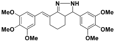
Consequently, these results prompted us to assess the inhibitory potency of several monocarbonyl curcumin analogues against the enzyme. More specifically, the synthetic analogues displayed a broad range of inhibitory effects, highlighting the impact of substituent type, position, and hydrophobicity. DM46 (65.2%), DM94 (64.9%), and DM57 (62.3%) were the most potent inhibitors, surpassing the parent curcumin compound. Moderate inhibitors included DM16 (29.5%), MS238 (31.6%), and DM109 (16.3%). The majority of the analogues, including DM62, DM95, DM96, DM151 and DM236, exhibited only low inhibition (≤15%), and DM101 and DM100 were among the least active (≤7%), suggesting that even slight structural changes strongly affect enzyme binding.
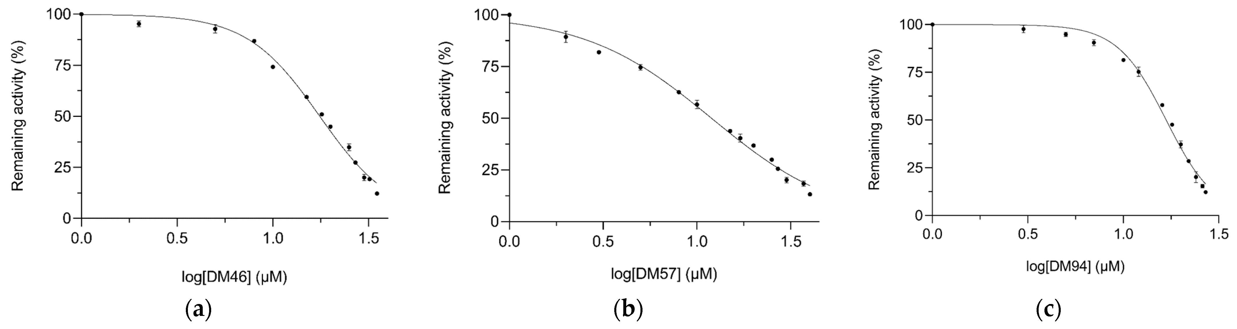
Although more compounds should have been screened to establish a clearer structure–activity relationship profile, some features may still be identified as crucial for activity. The current inhibitor panel provides a useful starting point for CpGST inhibitor discovery, while a larger and more structurally diverse compound library will be required in future studies to establish a robust SAR and to identify more selective and potent inhibitors. In particular, the presence of methoxy groups at various substitution positions and numbers may be identified as a highly favourable structural characteristic. This is supported not only by the fact that all three highest-activity inhibitors—DM46, DM57 and DM94—possess one, two or three methoxy groups, respectively, on each of the symmetrical aromatic rings but also by the activity of the remaining methoxylated compounds. The moderate activity for DM16 and MS238 may similarly be connected to the presence of methoxy substitution, regardless of the monocarbonyl or dicarbonyl nature of the curcuminoid skeleton. In contrast, most of the phenolic compounds exhibit rather low inhibitory potential. Additionally, the low activity of DM236 could be interpreted as an indication of the importance of the monocarbonyl moiety for effective inhibition. It is noteworthy that in our ongoing investigation for the inhibitory potential of this type of molecule against various human GSTs, like P1-1 and A4-4, the phenolic motif appeared to be more active [58]. This may provide further support for the structural divergence in the GSH-binding site. Based on these results, DM46, DM57, and DM94 were selected for further dose–response analysis to determine their IC50 values (Figure 6). As summarized in Table 4, DM57 emerged as the most potent inhibitor, with an IC50 value of 11.8 ± 0.5 μM, followed by DM94 (16.8 ± 0.5 μM) and DM46 (17.6 ± 0.6 μM).
Although the applied workflow follows a standard inhibitor-screening approach, it provides a necessary first step toward the identification of novel CpGST-targeting compounds and establishes a basis for future structure-guided optimization and more advanced functional studies.
Previous work with human GSTP1-1 [31], the most widely distributed and therapeutically relevant GST isoenzyme, showed that the two most active CpGST inhibitors, DM57 and DM46, exhibited only modest inhibitory activity against GSTP1-1, reducing enzyme activity by 16.75% and 8.5%, respectively [31]. Conversely, DM96 and DM100, which were weak inhibitors of CpGST, showing 7.2% and 15.53% inhibition, respectively (Table 3), displayed strong inhibitory activity against human GSTP1-1, with inhibition values of 86.99% and 40%, respectively. These findings suggest a degree of selectivity of DM57 and DM46 toward CpGST over human GSTP1-1, although this conclusion should be interpreted with caution, as the compounds have not yet been evaluated against the full panel of human GST isoenzymes.
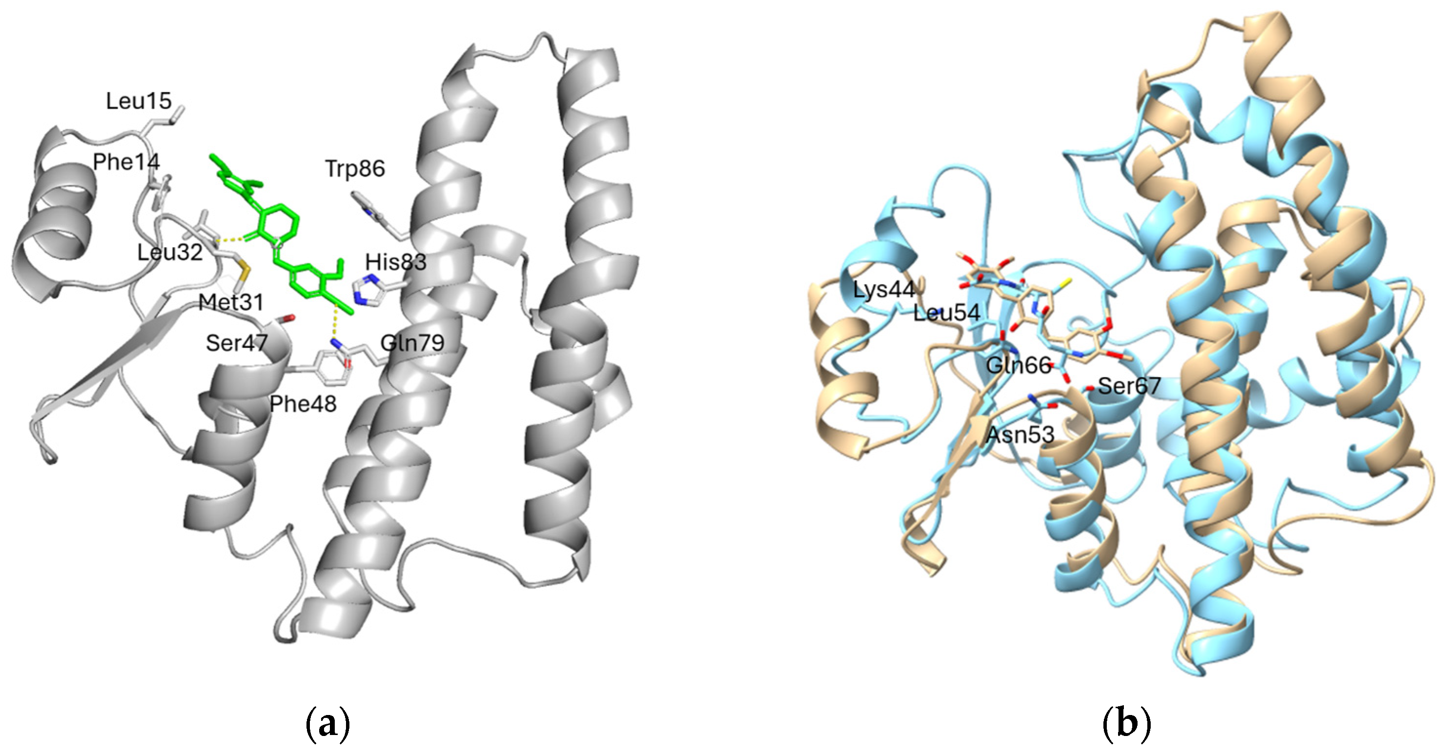
Molecular docking was performed to predict the best ligand binding site and explore the putative binding mode of DM57 within the predicted CpGST structure (Figure 7). The docking model indicated that DM57 is accommodated in a binding cavity formed mainly by hydrophobic residues, including Phe14, Leu15, Met31, Leu32, Phe48, and Trp86, together with polar or potentially hydrogen-bonding residues such as Ser47, Gln79, and His83. The aromatic and methoxy-substituted scaffold of DM57 appears to be stabilized predominantly by hydrophobic contacts and π-related interactions with aromatic residues, particularly Phe14, Phe48, and Trp86. In addition, possible polar interactions involving Ser47, Gln79, and/or His83 may further contribute to ligand stabilization. Overall, the predicted pocket displays a mainly hydrophobic character, with localized polar residues capable of supporting specific ligand orientation.
Structural superposition of the CpGST–DM57 docking model with the mu-class GST from Fasciola hepatica in complex with GSH (Figure 7) showed that the predicted DM57-binding region is located close to the GSH-binding region. This supports the hypothesis that DM57 may inhibit CpGST through binding to a partially overlapping pocket near the substrate-binding region.

2.6. Kinetic Inhibition Analysis of CpGST by DM94 and Molecular Docking Assessment of Its Binding Mode

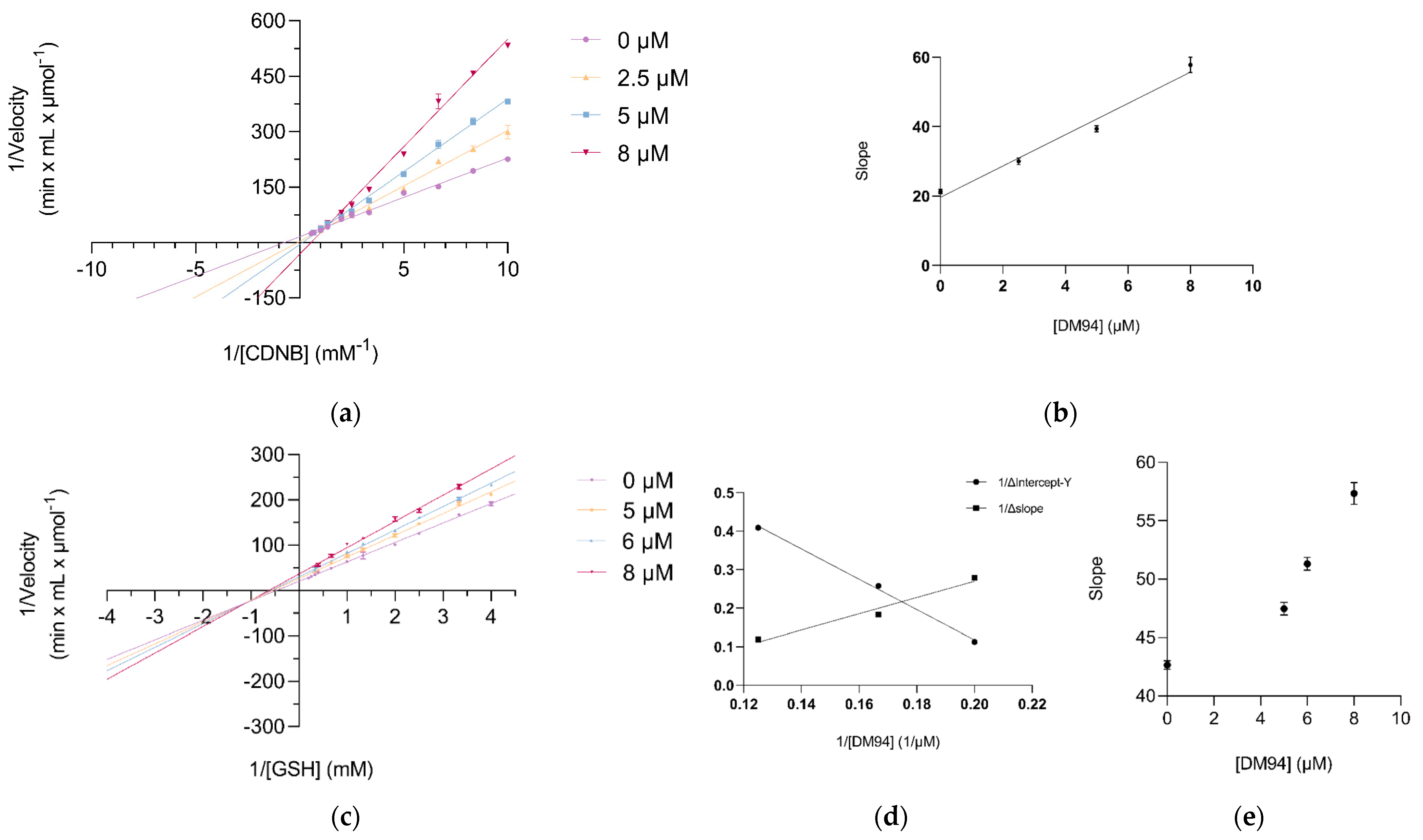
Given its strong inhibitory activity and the absence of prior reports on its interaction with GSTs, the compound DM94 was selected for detailed kinetic inhibition studies to determine its inhibition constant (Ki) and the mode of inhibition. The Lineweaver–Burk plots using CDNB as the variable substrate are shown in Figure 8a. The kinetic lines in the absence and presence of three different concentrations of DM94 intersected in the first quadrant, which is indicative of a mixed-type inhibition pattern. This suggests that DM94 is capable of binding to both the free enzyme and the enzyme–substrate (Enzyme-CDNB) complex, with respective inhibition constants Ki and Ki′. A secondary plot of the slopes from the double reciprocal plots versus DM94 concentration (Figure 8b) displayed a linear relationship (R2 = 0.9838), confirming a purely mixed-type inhibition mechanism. The calculated inhibition constants were Ki = 4.35 ± 1.47 μM and Ki′ = 3.20 ± 0.61 μM.
When GSH was used as the variable substrate, the Lineweaver–Burk plots again demonstrated mixed-type inhibition (Figure 8c). The corresponding secondary plot exhibited a nonlinear (parabolic) relationship (Figure 8d), indicating a partial mixed-type inhibition mechanism. The estimated inhibition constants were Ki = 5.04 ± 0.36 μM for the free enzyme and Ki′ = 4.13 ± 0.68 μM for the enzyme–GSH complex (Figure 8e).

2.7. Therapeutic Implications

From a therapeutic standpoint, GSTs in protozoa offer several advantages as drug targets. First, their role in mitigating oxidative stress and metabolizing xenobiotics renders them essential under host-induced stress conditions. Second, significant structural divergence from mammalian GSTs allows for the development of parasite-selective inhibitors. Third, as shown in other protozoa, GSTs are implicated in resistance mechanisms, making them suitable for combination therapies aimed at sensitizing parasites to conventional drugs [59]. In light of these considerations and given the structural and functional divergence of CpGST from human GST orthologs, which underscores its potential as a selective target with minimal off-target effects on host enzymes, CpGST can be considered as a promising candidate for anti-cryptosporidial drug development. Its structural uniqueness, essential functional role, and druggability provide a solid foundation for future structure-based drug discovery efforts.
Prior biochemical studies have shown that C. parvum oocysts exhibit extremely limited antioxidant enzyme activity, with only low levels of superoxide dismutase (SOD) detected and no measurable activity of catalase, peroxidase, or classical GSTs [55]. These findings suggest that the GSTs encoded in the C. parvum genome may perform noncanonical or stress-induced roles, possibly activated only under specific environmental conditions. The absence of detectable GST activity in earlier studies may be attributed to the enzyme’s expression pattern or to its low specific activity, as demonstrated in the present study. Moreover, the role of GSTs in redox homeostasis must be considered in the broader context of Cryptosporidium’s redox biology. Recent work by Gabriele et al. (2025) has shown that C. parvum lacks authentic glutathione reductase and instead relies on a thioredoxin reductase (CpTrxR)-based system to maintain glutathione in its reduced form [60]. CpTrxR, a type II thioredoxin reductase with unique structural features, serves as a central redox regulator and is essential for parasite viability [60]. This suggests a functional integration between the thioredoxin and glutathione systems, with GSTs potentially participating downstream in detoxification and signaling pathways. Given that CpTrxR is irreversibly inhibited by the gold-based drug auranofin, which also impairs GST-related pathways, this finding raises the possibility of redox-targeted combination therapies in cryptosporidiosis.
Although the present biochemical and inhibition data support CpGST as a promising candidate for further investigation, its essentiality for C. parvum survival, infectivity, or life-cycle progression has not yet been experimentally demonstrated. Future studies should include genetic or functional validation approaches and parasite infectivity assays in relevant host cell models. Such studies will be necessary to determine the biological importance of CpGST and to establish whether its inhibition can compromise parasite viability or infectivity.

3. Materials and Methods

3.1. Materials

Natural products (ellagic acid, (±)-taxifolin hydrate, gallic acid, (−)-epigallocatechin gallate, p-coumaric acid, (±)-naringenin, quercetin, curcumin, resveratrol, colchicine, polydatin, safranal, piperlongumine) were obtained from Sigma-Aldrich (St. Louis, MO, USA). The curcumin analogues were synthesized as previously reported [31]. Dimethyl sulfoxide (DMSO), acetone and ethanol were purchased from Scharlau (Barcelona, Spain). The antibiotics kanamycin and chloramphenicol were bought from AppliChem (Darmstadt, Germany). CDNB, reduced glutathione, cumene hydroperoxide and all other substrates were purchased from Sigma-Aldrich (St. Louis, MO, USA). The plasmid vector pET-28b bearing the 6-His tag from Novagen was employed for the subcloning of the CpGST gene. PCR experiments were performed using the KAPA Taq polymerase from KAPA Biosystems (Wilmington, MA, USA) and Platinum™ Pfx from Invitrogen™ (Carlsbad, CA, USA). Moreover, CloneAmp™ HiFi PCR Premix polymerase from Clontech Laboratories, Inc. (Takara Bio, San Jose, CA, USA) was used in the subcloning reactions.

3.2. Methods

3.2.1. Biocomputing-Based Structural Characterization of CpGST

Structural prediction of the CpGST enzyme was performed using the I-TASSER server [61,62]. Among the generated models, the one with the highest confidence score (C-score = −0.13) was selected for further analysis. The predicted 3D structure was visualized and examined using UCSF Chimera 1.16. Functional domain analysis was carried out via the InterPro database [63], while homologous sequences were identified through BLASTp searches against the Protein Data Bank (PDB) and non-redundant protein sequences [64]. The UniProt database and the resulting hits were used to construct a phylogenetic tree using the iTOL web tool (version 7.6) [65]. Multiple sequence alignment of the retrieved homologs was conducted using Clustal Omega [66,67], and the results were graphically represented with ESPript [68]. The three-dimensional structure of the enzyme was also predicted using the AlphaFold2 server [69]. AlphaFold2 model quality was evaluated from the per-residue pLDDT traces, using the standard interpretation that pLDDT ≥ 90 indicates high accuracy, 70–90 indicates confident backbone placement, and <70 indicates flexible/low-confidence regions [70].
Molecular docking analysis was performed to investigate the putative binding mode of the best ligand DM57 within the predicted three-dimensional structure of CpGST. The AlphaFold2-predicted structure of CpGST was used as the receptor model. Molecular docking calculations were carried out using DiffDock-L [71] (run on the Neurosnap web server https://neurosnap.ai/, accessed on 9 July 2026). Docking evaluation was based on binding affinity, minimized affinity, minimized RMSD, and intramolecular energy. The best-ranked docking pose was selected based on the docking confidence score. Prediction of the ligand binding site and analysis of the enzyme–ligand complex were carried out using the ProteinsPlus web server (https://proteins.plus) [72].

3.2.2. Molecular Cloning of the CpGST

The gene encoding a putative GST from Cryptosporidium parvum Iowa II (Accession No.: XP_628625.1) was identified through an extensive search of the NCBI database. The CpGST gene was synthesized (GeneScript, Piscataway, NJ, USA) and codon-optimized for enhanced expression in Escherichia coli. PCR amplification was performed using the In-Fusion® HD Cloning Kit (Takara Bio USA, Inc.) with the following primers:
Forward: 5′-AGAAGGAGATATACCATGATTGAATATCTAGAGCACAATATATC-3′.
Reverse: 5′-GTGGTGGTGGTGGTGACGGATGTACGGCTTTAAC-3′.
The PCR reaction (25 μL) contained 12.5 μL of CloneAmp™ HiFi PCR Premix (1X), 0.3 μM of each primer, 16 ng of DNA template and 9.5 μL of ddH2O. The amplification protocol included an initial denaturation at 98 °C for 4 min, followed by 35 cycles of denaturation at 98 °C for 10 s, annealing at 62 °C for 15 s, and extension at 72 °C for 10 s. A final extension step was carried out at 72 °C for 10 min. The resulting amplicon was cloned into the pET-28b expression vector (Lucigen, Middleton, WI, USA), which contains a C-terminal hexahistidine (6×His) tag. The resulting construct, pET-28b_CpGST_6×His, was initially used to transform E. coli Stellar™ competent cells. Plasmid DNA was then isolated using the NucleoSpin® Plasmid kit (Macherey-Nagel, Düren, Germany), sequenced to confirm the correct insertion and the presence of the C-terminal 6×His tag, and subsequently used to transform a range of E. coli expression strains, including BL21 (DE3) pLysS, BL21 (DE3), BL21 OverExpress™ C41 (DE3), BL21 Rosetta™ 2 (DE3) pLysS, BL21 Shuffle® T7 Express, and BL21 Tuner™ (DE3).

3.2.3. Heterologous Expression and Purification of CpGST

High levels of heterologous expression of the pET-28b_CpGST_6×His construct were achieved in E. coli Rosetta™ 2 (DE3) pLysS cells. The expression protocol was adapted with minor modifications from a previously established method [69]. Briefly, transformed cells were initially grown in LB medium at 37 °C for 16 h. A suitable volume of this pre-culture was then used to inoculate flasks containing 2×YT medium (1.6% w/v tryptone, 1% w/v yeast extract, and 0.5% w/v NaCl) supplemented with kanamycin (100 µg/mL) and chloramphenicol (34 µg/mL). When the optical density at 600 nm reached approximately 0.5–0.6, protein expression was induced by the addition of 1 mM IPTG, followed by incubation at 37 °C for 18–20 h. After expression, cells were harvested by centrifugation at 8000× g for 8 min, and the resulting pellet was resuspended in wash buffer (300 mM NaCl, 50 mM NaH2PO4, pH 8.0). Cell lysis was performed by sonication (6 cycles of 15 s at 60 Hz, 50 W), and the lysate was clarified by centrifugation at 12,000× g for 10 min for purification.
Recombinant CpGST was purified by immobilized metal affinity chromatography (IMAC) using Ni2+-IDA Sepharose resin pre-equilibrated with wash buffer [70]. The column was washed, and the bound protein was sequentially eluted with buffers containing increasing concentrations of imidazole: 10 mL with 5 mM, 15 mL with 10 mM, and a final 5 mL fraction with 20 mM imidazole, all in 50 mM NaH2PO4 buffer containing 300 mM NaCl (pH 8.0). All purification steps were carried out at 4 °C. Eluted fractions were diluted with glycerol to a final concentration of 50% (v/v) and stored at −20 °C for further analysis.

3.2.4. Kinetic Analysis

The kinetic characterization of the recombinant CpGST enzyme was performed to determine its Michaelis–Menten parameters (Km, Vmax, kcat) using GSH and 1-chloro-2,4-dinitrobenzene (CDNB) as substrates. Enzyme activity assays were conducted based on established protocols with slight modifications [58,71]. Reactions were carried out in a final volume of 1 mL at 25 °C. For assays in which GSH served as the variable substrate, the reaction mixture contained 0.1 M KH2PO4 buffer (pH 6.5), a fixed concentration of CDNB, and varying concentrations of GSH (0–5.5 mM). Conversely, when CDNB was the variable substrate, the mixture contained the same buffer and CDNB concentrations ranging from 0 to 2.4 mM. In all assays, the enzyme was pre-incubated with GSH in the buffer for 5 min at 37 °C prior to the addition of CDNB to initiate the reaction. The kinetic parameters Km, Vmax and kcat were calculated by fitting the data to the Michaelis–Menten equation using GraphPad Prism software (version 9.3.1).

3.2.5. Inhibitor Screening and Kinetic Inhibition Analysis

Selected natural products [ellagic acid, (±)-taxifolin hydrate, gallic acid, (−)-epigallocatechin gallate, p-coumaric acid, (±)-naringenin, quercetin, curcumin, resveratrol, colchicine, polydatin, safranal, piperlongumine] and the synthetic curcumin analogues were evaluated as potential inhibitors of CpGST. Compounds were dissolved in DMSO to a final concentration of 100 μM. Inhibitory activity was assessed by measuring CpGST catalysis using GSH and CDNB as substrates at final concentrations of 2.5 mM and 1 mM, respectively. The formation of the conjugated product was monitored at 340 nm for 120 s at 25 °C. Test compounds were added to the reaction mixture at a final concentration of 25 μM. Prior to initiating the reaction with CDNB, the enzyme was pre-incubated with GSH and the respective compound in buffer at 37 °C for 5 min. Compounds showing the greatest inhibitory effect were selected for IC50 determination. For this, enzymatic activity was measured in the presence of increasing concentrations of each inhibitor under the same assay conditions. All measurements were performed in triplicate, and appropriate controls and corrections were applied as needed. IC50 values were calculated by nonlinear regression using GraphPad Prism (version 9.3.1). Kinetic inhibition analysis was conducted to elucidate the mode of inhibition, following the methodology used for the initial kinetic characterization. The substrate concentration (GSH or CDNB) was varied while keeping the other constant, in the absence and presence of fixed concentrations of the inhibitor. All assays were conducted in triplicate. Kinetic data were analyzed using Lineweaver–Burk plots, and the inhibition type was determined based on the intersection patterns of the plotted lines. All enzymatic assays were performed in triplicate, and data are presented as mean ± standard deviation. Kinetic parameters and IC50 values were calculated by nonlinear regression analysis using GraphPad Prism. For inhibitor screening performed at a single compound concentration, the data were used for comparative ranking, and no pairwise statistical significance was inferred unless specifically indicated.

4. Conclusions

This study offers the first in-depth molecular and functional characterization of a GST from Cryptosporidium parvum, unveiling a structurally distinct enzyme with conserved catalytic residues and unique features that distinguish it from host homologs. The enzyme’s low substrate affinity, atypical structural elements, and evolutionary classification suggest a specialized role in the parasite’s biology. The identification of high affinity curcumin inhibitors opens new scaffolds for rational drug design. Furthermore, the parasite’s reliance on a thioredoxin-based redox system, as recently elucidated, may enhance the therapeutic relevance of GST inhibitors when used in combination with redox-disrupting agents. Future studies should assess the effects of DM46, DM57, and DM94 in C. parvum-infected cell culture models, including parasite growth/infectivity assays and host cell cytotoxicity evaluation, to help in advance CpGST as a viable target in the fight against cryptosporidiosis.

Supplementary Materials

The following supporting information can be downloaded at: https://www.mdpi.com/article/10.3390/ph19071106/s1, Figure S1: Structural comparison and AlphaFold2 confidence analysis of CpGST.

Author Contributions

Conceptualization, N.E.L.; methodology, P.D.P., N.D.G., N.E.L.; formal analysis, P.D.P., N.D.G.; investigation, P.D.P., N.D.G., D.M., M.S.; resources, D.M., M.S.; writing—original draft preparation, P.D.P., N.D.G., N.E.L.; writing—review and editing, P.D.P., N.D.G., M.S., N.E.L. All authors have read and agreed to the published version of the manuscript.

Funding

This research received no external funding.

Institutional Review Board Statement

Not applicable.

Informed Consent Statement

Not applicable.

Data Availability Statement

The original contributions presented in this study are included in the article/Supplementary Material. Further inquiries can be directed to the corresponding author.

Conflicts of Interest

The authors declare no conflicts of interest.

Abbreviations

The following abbreviations are used in this manuscript.
CDNB1-Chloro-2,4-dinitrobenzene
CpTrxRCryptosporidium parvum Thioredoxin Reductase
GSHGlutathione (reduced form)
GSTGlutathione transferase
CpGSTCryptosporidium parvum GST
H-siteHydrophobic substrate-binding site of GST
IC50Half-maximal Inhibitory Concentration
IPTGIsopropyl β-D-1-thiogalactopyranoside
LBLuria–Bertani medium
Ni2+-IDANickel–iminodiacetic acid (used in affinity chromatography)
ORFOpen Reading Frame
SODSuperoxide Dismutase
TrxThioredoxin
TrxRThioredoxin Reductase

References

  1. Wu, M.; Yang, B.; Wang, D.; Zhang, Y.; Li, X.; Zhi, Y.; Zhao, X.; Yin, J.; Zhu, G. Zoonotic Cryptosporidium parasites possess a unique carbohydrate-binding protein (malectin) that is absent in other apicomplexan lineages. Zoonoses 2022, 2, 988. [Google Scholar] [CrossRef] [Scilit]
  2. Helmy, Y.A.; El-Adawy, H.; Abdelwhab, E.M. A comprehensive review of common bacterial, parasitic and viral zoonoses at the human–animal interface in Egypt. Pathogens 2017, 6, 33. [Google Scholar] [CrossRef] [Scilit] [PubMed]
  3. Gharpure, R.; Perez, A.; Miller, A.D.; Wikswo, M.E.; Silver, R.; Hlavsa, M.C. Cryptosporidiosis outbreaks—United States, 2009–2017. Morb. Mortal. Wkly. Rep. 2019, 68, 568–572. [Google Scholar] [CrossRef] [Scilit]
  4. Hassan, E.M.; Örmeci, B.; DeRosa, M.C.; Dixon, B.R.; Sattar, S.A.; Iqbal, A. A review of Cryptosporidium spp. and their detection in water. Water Sci. Technol. 2021, 83, 1–25. [Google Scholar] [PubMed]
  5. dos Santos Toledo, R.; Martins, F.D.C.; Freire, R.L. Waterborne Giardia and Cryptosporidium: Contamination of human drinking water by sewage and cattle feces. Semin. Ciênc. Agrár. 2017, 38, 3395–3415. [Google Scholar] [CrossRef] [Scilit]
  6. Khan, S.M.; Witola, W.H. Past, current, and potential treatments for cryptosporidiosis in humans and farm animals: A comprehensive review. Front. Cell. Infect. Microbiol. 2023, 13, 1115522. [Google Scholar] [CrossRef] [Scilit] [PubMed]
  7. Martinez, M.P.; Carmena, D.; Herrador, B.R.G.; Miguel, M.P.; Campelli, G.S.; Álvarez, R.M.G.; Guerrero-Vadillo, M.; Dashti, A.; Köster, P.C.; Alemany, E.G.; et al. Marked increase in cryptosporidiosis cases, Spain, 2023. Eurosurveillance 2024, 29, 2300733. [Google Scholar] [CrossRef] [Scilit]
  8. Pinto, D.J.; Vinayak, S. Cryptosporidium: Host–parasite interactions and pathogenesis. Curr. Clin. Microbiol. Rep. 2021, 8, 62–67. [Google Scholar] [CrossRef] [Scilit] [PubMed]
  9. European Centre for Disease Prevention and Control. Cryptosporidiosis—Annual Epidemiological Report for 2021; ECDC: Stockholm, Sweden, 2021. [Google Scholar]
  10. O’Connor, R.M.; Shaffie, R.; Kang, G.; Ward, H.D. Cryptosporidiosis in patients with HIV/AIDS. AIDS 2011, 25, 549–560. [Google Scholar] [CrossRef] [Scilit] [PubMed]
  11. Bhadauria, D.; Goel, A.; Kaul, A.; Sharma, R.K.; Gupta, A.; Ruhela, V.; Gupta, A.; Vardhan, H.; Prasad, N. Cryptosporidium infection after renal transplantation in an endemic area. Transpl. Infect. Dis. 2015, 17, 48–55. [Google Scholar] [CrossRef] [Scilit] [PubMed]
  12. Helmy, Y.A.; Hafez, H.M. Cryptosporidiosis: From prevention to treatment, a narrative review. Microorganisms 2022, 10, 2456. [Google Scholar] [CrossRef] [Scilit] [PubMed]
  13. Xiao, L.; Fayer, R.; Ryan, U.; Upton, S.J. Cryptosporidium taxonomy: Recent advances and implications for public health. Clin. Microbiol. Rev. 2004, 17, 72–97. [Google Scholar] [CrossRef] [Scilit] [PubMed]
  14. Ryan, U.; Fayer, R.; Xiao, L. Cryptosporidium species in humans and animals: Current understanding and research needs. Parasitology 2014, 141, 1667–1685. [Google Scholar] [CrossRef] [Scilit] [PubMed]
  15. Love, M.S.; Choy, R.K.M. Emerging treatment options for cryptosporidiosis. Curr. Opin. Infect. Dis. 2021, 34, 455–462. [Google Scholar] [CrossRef] [Scilit] [PubMed]
  16. Amadi, B.; Mwiya, M.; Musuku, J.; Watuka, A.; Sianongo, S.; Ayoub, A.; Kelly, P. Effect of nitazoxanide on morbidity and mortality in Zambian children with cryptosporidiosis: A randomised controlled trial. Lancet 2002, 360, 1375–1380. [Google Scholar] [CrossRef] [Scilit] [PubMed]
  17. Rossignol, J.F. Cryptosporidium and Giardia: Treatment options and prospects for new drugs. Exp. Parasitol. 2010, 124, 45–53. [Google Scholar] [CrossRef] [Scilit] [PubMed]
  18. Blagburn, B.L.; Soave, R. Prophylaxis and chemotherapy: Human and animal. In Cryptosporidium and Cryptosporidiosis; Fayer, R., Ed.; CRC Press: Boca Raton, FL, USA, 1997; pp. 111–128. [Google Scholar]
  19. Maggi, P.; Larocca, A.M.; Quarto, M.; Serio, G.; Brandonisio, O.; Angarano, G.; Pastore, G. Effect of antiretroviral therapy on cryptosporidiosis and microsporidiosis in patients infected with human immunodeficiency virus type 1. Eur. J. Clin. Microbiol. Infect. Dis. 2000, 19, 213–217. [Google Scholar] [CrossRef] [Scilit] [PubMed]
  20. Zardi, E.M.; Picardi, A.; Afeltra, A. Treatment of cryptosporidiosis in immunocompromised hosts. Chemotherapy 2005, 51, 193–196. [Google Scholar] [CrossRef] [Scilit] [PubMed]
  21. Giacometti, A.; Cirioni, O.; Barchiesi, F.; Ancarani, F.; Scalise, G. Activity of nitazoxanide alone and in combination with azithromycin and rifabutin against Cryptosporidium parvum in cell culture. J. Antimicrob. Chemother. 2000, 45, 453–456. [Google Scholar] [CrossRef] [Scilit] [PubMed]
  22. Checkley, W.; White, A.C., Jr.; Jaganath, D.; Arrowood, M.J.; Chalmers, R.M.; Chen, X.-M.; Fayer, R.; Griffiths, J.K.; Guerrant, R.L.; Hedstrom, L.; et al. A review of the global burden, novel diagnostics, therapeutics, and vaccine targets for Cryptosporidium. Lancet Infect. Dis. 2015, 15, 85–94. [Google Scholar] [CrossRef] [Scilit] [PubMed]
  23. Wang, B.; Castellanos-Gonzalez, A.; White, A.C., Jr. Novel drug targets for treatment of cryptosporidiosis. Expert Opin. Ther. Targets 2020, 24, 915–922. [Google Scholar] [CrossRef] [Scilit] [PubMed]
  24. Murphy, R.C.; Ojo, K.K.; Larson, E.T.; Castellanos-Gonzalez, A.; Perera, B.G.K.; Keyloun, K.R.; Kim, J.E.; Bhandari, J.G.; Muller, N.R.; Verlinde, C.L.M.J.; et al. Discovery of potent and selective inhibitors of CDPK1 from C. parvum and T. gondii. ACS Med. Chem. Lett. 2010, 1, 331–335. [Google Scholar] [CrossRef] [Scilit] [PubMed]
  25. Ojo, K.K.; Larson, E.T.; Keyloun, K.R.; Castaneda, L.J.; DeRocher, A.E.; Inampudi, K.K.; Kim, J.E.; Arakaki, T.L.; Murphy, R.C.; Zhang, L.; et al. Toxoplasma gondii calcium-dependent protein kinase 1 is a target for selective kinase inhibitors. Nat. Struct. Mol. Biol. 2010, 17, 602–607. [Google Scholar] [CrossRef] [Scilit] [PubMed]
  26. Manjunatha, U.H.; Vinayak, S.; Zambriski, J.A.; Chao, A.T.; Sy, T.; Noble, C.G.; Bonamy, G.M.C.; Kondreddi, R.R.; Zou, B.; Gedeck, P.; et al. A Cryptosporidium PI(4)K inhibitor is a drug candidate for cryptosporidiosis. Nature 2017, 546, 376–380. [Google Scholar] [CrossRef] [Scilit] [PubMed]
  27. Mead, J.R.; Arrowood, M.J. (Eds.) Cryptosporidium: Methods and Protocols; Methods in Molecular Biology; Humana Press: New York, NY, USA, 2020; Volume 2052. [Google Scholar]
  28. Forney, J.R.; Yang, S.; Du, C.; Healey, M.C. Efficacy of serine protease inhibitors against Cryptosporidium parvum infection in a bovine fallopian tube epithelial cell culture system. J. Parasitol. 1996, 82, 638–640. [Google Scholar] [CrossRef] [Scilit] [PubMed]
  29. Guo, F.; Zhang, H.; Payne, H.R.; Zhu, G. Differential gene expression and protein localization of Cryptosporidium parvum fatty acyl-CoA synthetase isoforms. J. Eukaryot. Microbiol. 2016, 63, 233–246. [Google Scholar] [PubMed]
  30. Labrou, N.E.; Papageorgiou, A.C.; Pavli, O.; Flemetakis, E. Plant GSTome: Structure and functional role in xenome network and plant stress response. Curr. Opin. Biotechnol. 2015, 32, 186–194. [Google Scholar] [CrossRef] [Scilit] [PubMed]
  31. Pantiora, P.; Furlan, V.; Matiadis, D.; Mavroidi, B.; Perperopoulou, F.; Papageorgiou, A.C.; Sagnou, M.; Bren, U.; Pelecanou, M.; Labrou, N.E. Monocarbonyl curcumin analogues as potent inhibitors against human glutathione transferase P1-1. Antioxidants 2023, 12, 63. [Google Scholar]
  32. Sabt, A.; Kitsos, S.; Ebaid, M.S.; Furlan, V.; Pantiora, P.D.; Tsolka, M.; Elkaeed, E.B.; Hamissa, M.F.; Angelis, N.; Tsitsilonis, O.E.; et al. Novel coumarin-6-sulfonamide-chalcone hybrids as glutathione transferase P1-1 inhibitors. PLoS ONE 2024, 19, e0306124. [Google Scholar] [CrossRef] [Scilit] [PubMed]
  33. Laborde, E. Glutathione transferases as mediators of signaling pathways involved in cell proliferation and cell death. Cell Death Differ. 2010, 17, 1373–1380. [Google Scholar] [CrossRef] [Scilit] [PubMed]
  34. Karpusas, M.; Axarli, I.; Chiniadis, L.; Papakyriakou, A.; Bethanis, K.; Scopelitou, K.; Clonis, Y.D.; Labrou, N.E. The interaction of the chemotherapeutic drug chlorambucil with human glutathione transferase A1-1: Kinetic and structural analysis. PLoS ONE 2013, 8, e56337. [Google Scholar] [CrossRef] [Scilit] [PubMed]
  35. Perperopoulou, F.; Pouliou, F.; Labrou, N.E. Recent advances in protein engineering and biotechnological applications of glutathione transferases. Crit. Rev. Biotechnol. 2018, 38, 511–528. [Google Scholar] [PubMed]
  36. Atamna, H.; Ginsburg, H. Heme degradation in the presence of glutathione: A proposed mechanism to account for the high levels of non-heme iron found in the membranes of hemoglobinopathic red blood cells. J. Biol. Chem. 1995, 270, 24876–24883. [Google Scholar] [CrossRef] [Scilit] [PubMed]
  37. Perbandt, M.; Eberle, R.; Fischer-Riepe, L.; Cang, H.; Liebau, E.; Betzel, C. High resolution structures of Plasmodium falciparum GST complexes provide novel insights into the dimer–tetramer transition and a novel ligand-binding site. J. Struct. Biol. 2015, 191, 365–375. [Google Scholar] [CrossRef] [Scilit] [PubMed]
  38. Li, S.; Liu, J.; Zhang, H.; Sun, Z.; Ying, Z.; Wu, Y.; Xu, J.; Liu, Q. Toxoplasma gondii glutathione S-transferase 2 plays an important role in partial secretory protein transport. FASEB J. 2021, 35, e21352. [Google Scholar] [CrossRef] [Scilit] [PubMed]
  39. Al-Malki, E.S. Exploring the infectious drug target glutathione S-transferase in Plasmodium falciparum with the inhibitory potential of Azadirachta indica phytocompounds. Adv. Public Health 2024, 2024, 8486021. [Google Scholar] [CrossRef] [Scilit]
  40. Abrahamsen, M.S.; Templeton, T.J.; Enomoto, S.; Abrahante, J.E.; Zhu, G.; Lancto, C.A.; Deng, M.; Liu, C.; Widmer, G.; Tzipori, S.; et al. Complete genome sequence of the apicomplexan, Cryptosporidium parvum. Science 2004, 304, 441–445. [Google Scholar] [CrossRef] [Scilit] [PubMed]
  41. Mauzy, M.J.; Enomoto, S.; Lancto, C.A.; Abrahamsen, M.S.; Rutherford, M.S. The Cryptosporidium parvum transcriptome during in vitro development. PLoS ONE 2012, 7, e31715. [Google Scholar] [CrossRef] [Scilit] [PubMed]
  42. Mfeka, M.S.; Martínez-Oyanedel, J.; Chen, W.; Achilonu, I.; Syed, K.; Khoza, T. Comparative analyses and structural insights of new class glutathione transferases in Cryptosporidium species. Sci. Rep. 2020, 10, 20370. [Google Scholar] [CrossRef] [Scilit] [PubMed]
  43. Leitsch, D.; Müller, J.; Müller, N. Evaluation of Giardia lamblia thioredoxin reductase as drug activating enzyme and as drug target. Int. J. Parasitol. Drugs Drug Resist. 2016, 6, 148–153. [Google Scholar] [CrossRef] [Scilit] [PubMed]
  44. Dirr, H.W.; Wallace, L.A. Role of the C-terminal helix 9 in the stability and ligandin function of class α glutathione transferase A1-1. Biochemistry 1999, 38, 15631–15640. [Google Scholar] [CrossRef] [Scilit] [PubMed]
  45. Shokeer, A.; Mannervik, B. Minor modifications of the C-terminal helix reschedule the favored chemical reactions catalyzed by theta class glutathione transferase T1-1. J. Biol. Chem. 2010, 285, 5639–5645. [Google Scholar] [CrossRef] [Scilit] [PubMed]
  46. Rossjohn, J.; Feil, S.C.; Wilce, M.C.J.; Sexton, J.L.; Spithill, T.W.; Parker, M.W. Crystallization, structural determination and analysis of a novel parasite vaccine candidate: Fasciola hepatica glutathione S-transferase. J. Mol. Biol. 1997, 273, 857–872. [Google Scholar] [CrossRef] [Scilit] [PubMed]
  47. Juárez-Martínez, A.B.; Sotelo-Mundo, R.R.; Rudiño-Piñera, E. Crystal structure of a class-mu glutathione S-transferase from whiteleg shrimp Litopenaeus vannamei: Structural changes in the xenobiotic binding H-site may alter the spectra of molecules bound. J. Biochem. Mol. Toxicol. 2017, 31, e21838. [Google Scholar]
  48. Kolm, R.H.; Danielson, U.H.; Zhang, Y.; Talalay, P.; Mannervik, B. Isothiocyanates as substrates for human glutathione transferases: Structure–activity studies. Biochem. J. 1995, 311, 453–459. [Google Scholar] [CrossRef] [Scilit] [PubMed]
  49. Morgenstern, R. Kinetic behavior of glutathione transferases: Understanding cellular protection from reactive intermediates. Biomolecules 2024, 14, 641. [Google Scholar] [CrossRef] [Scilit] [PubMed]
  50. Sheehan, D.; Meade, G.; Foley, V.M.; Dowd, C.A. Structure, function and evolution of glutathione transferases: Implications for classification of non-mammalian members of an ancient enzyme superfamily. Biochem. J. 2001, 360, 1–16. [Google Scholar] [CrossRef] [PubMed]
  51. Dourado, D.F.A.R.; Fernandes, P.A.; Ramos, M.J. Mammalian cytosolic glutathione transferases. Curr. Protein Pept. Sci. 2008, 9, 325–337. [Google Scholar] [CrossRef] [Scilit] [PubMed]
  52. Shahiduzzaman, M.; Dyachenko, V.; Khalafalla, R.E.; Desouky, A.Y.; Daugschies, A. Effects of curcumin on Cryptosporidium parvum in vitro. Parasitol. Res. 2009, 105, 1155–1161. [Google Scholar] [CrossRef] [Scilit] [PubMed]
  53. Asadpour, M.; Namazi, F.; Razavi, S.M.; Nazifi, S. Comparative efficacy of curcumin and paromomycin against Cryptosporidium parvum infection in a BALB/c model. Vet. Parasitol. 2018, 250, 7–14. [Google Scholar] [CrossRef] [Scilit] [PubMed]
  54. Leitch, G.J.; He, Q. Reactive nitrogen and oxygen species ameliorate experimental cryptosporidiosis in the neonatal BALB/c mouse model. Infect. Immun. 1999, 67, 5885–5891. [Google Scholar] [CrossRef] [Scilit] [PubMed]
  55. Entrala, E.; Mascaró, C.; Barrett, J. Anti-oxidant enzymes in Cryptosporidium parvum oocysts. Parasitology 1997, 114, 13–17. [Google Scholar] [CrossRef] [Scilit] [PubMed]
  56. Rider, S.D., Jr.; Zhu, G. An apicomplexan ankyrin-repeat histone deacetylase with relatives in photosynthetic eukaryotes. Int. J. Parasitol. 2009, 39, 747–754. [Google Scholar] [CrossRef] [Scilit] [PubMed]
  57. Pollok, R.C.; McDonald, V.; Kelly, P.; Farthing, M.J. The role of Cryptosporidium parvum-derived phospholipase in intestinal epithelial cell invasion. Parasitol. Res. 2003, 90, 181–186. [Google Scholar] [CrossRef] [Scilit] [PubMed]
  58. Tsouri, S.; Tselo, E.; Premetis, G.E.; Furlan, V.; Pantiora, P.D.; Mavroidi, B.; Matiadis, D.; Pelecanou, M.; Papageorgiou, A.C.; Bren, U.; et al. A monocarbonyl curcuminoid derivative inhibits the activity of human glutathione transferase A4-4 and chemosensitizes glioblastoma cells to temozolomide. Pharmaceuticals 2024, 17, 365. [Google Scholar] [CrossRef] [Scilit] [PubMed]
  59. Pramanik, P.K.; Alam, M.N.; Roy Chowdhury, D.; Chakraborti, T. Drug resistance in protozoan parasites: An incessant wrestle for survival. J. Glob. Antimicrob. Resist. 2019, 18, 1–11. [Google Scholar] [CrossRef] [Scilit] [PubMed]
  60. Gabriele, F.; Bogard, J.A.; Palerma, M.; Ardini, M.; Byrne, M.E.; Chen, X.-M.; Petukhov, P.A.; Ippoliti, R.; Angelucci, F.; Williams, D.L. Targeting apicomplexan parasites: Structural and functional characterization of Cryptosporidium thioredoxin reductase as a novel drug target. Biochemistry 2025, 64, 2212–2225. [Google Scholar] [CrossRef] [Scilit] [PubMed]
  61. Yang, J.; Zhang, Y. I-TASSER server: New development for protein structure and function predictions. Nucleic Acids Res. 2015, 43, W174–W181. [Google Scholar] [CrossRef] [Scilit] [PubMed]
  62. Zhang, C.; Freddolino, P.L.; Zhang, Y. COFACTOR: Improved protein function prediction by combining structure, sequence and protein–protein interaction information. Nucleic Acids Res. 2017, 45, W291–W299. [Google Scholar] [CrossRef] [Scilit] [PubMed]
  63. Blum, M.; Chang, H.-Y.; Chuguransky, S.; Grego, T.; Kandasaamy, S.; Mitchell, A.; Nuka, G.; Paysan-Lafosse, T.; Qureshi, M.; Raj, S.; et al. The InterPro protein families and domains database: 20 years on. Nucleic Acids Res. 2021, 49, D344–D354. [Google Scholar] [PubMed]
  64. Priyam, A.; Woodcroft, B.J.; Rai, V.; Moghul, I.; Munagala, A.; Ter, F.; Chowdhary, H.; Pieniak, I.; Maynard, L.J.; Gibbins, M.A.; et al. Sequenceserver: A modern graphical user interface for custom BLAST databases. Mol. Biol. Evol. 2019, 36, 2922–2924. [Google Scholar] [CrossRef] [Scilit] [PubMed]
  65. Letunic, I.; Bork, P. Interactive Tree Of Life (iTOL) v5: An online tool for phylogenetic tree display and annotation. Nucleic Acids Res. 2021, 49, W293–W296. [Google Scholar] [CrossRef] [Scilit] [PubMed]
  66. Sievers, F.; Wilm, A.; Dineen, D.; Gibson, T.J.; Karplus, K.; Li, W.; Lopez, R.; McWilliam, H.; Remmert, M.; Söding, J.; et al. Fast, scalable generation of high-quality protein multiple sequence alignments using Clustal Omega. Mol. Syst. Biol. 2011, 7, 539. [Google Scholar] [CrossRef] [Scilit] [PubMed]
  67. Madeira, F.; Park, Y.M.; Lee, J.; Buso, N.; Gur, T.; Madhusoodanan, N.; Basutkar, P.; Tivey, A.R.N.; Potter, S.C.; Finn, R.D.; et al. The EMBL-EBI search and sequence analysis tools APIs in 2019. Nucleic Acids Res. 2019, 47, W636–W641. [Google Scholar] [CrossRef] [Scilit] [PubMed]
  68. Robert, X.; Gouet, P. Deciphering key features in protein structures with the new ENDscript server. Nucleic Acids Res. 2014, 42, W320–W324. [Google Scholar] [CrossRef] [Scilit] [PubMed]
  69. Jumper, J.; Evans, R.; Pritzel, A.; Green, T.; Figurnov, M.; Ronneberger, O.; Tunyasuvunakool, K.; Bates, R.; Žídek, A.; Potapenko, A.; et al. Highly accurate protein structure prediction with AlphaFold. Nature 2021, 596, 583–589. [Google Scholar] [CrossRef] [Scilit] [PubMed]
  70. Tunyasuvunakool, K.; Adler, J.; Wu, Z.; Green, T.; Zielinski, M.; Žídek, A.; Bridgland, A.; Cowie, A.; Meyer, C.; Laydon, A.; et al. Highly accurate protein structure prediction for the human proteome. Nature 2021, 596, 590–596. [Google Scholar] [CrossRef] [Scilit] [PubMed]
  71. Corso, G.; Deng, A.; Fry, B.; Polizzi, N.; Barzilay, R.; Jaakkola, T. Deep Confident Steps to New Pockets: Strategies for Docking Generalization. arXiv 2024, arXiv:2402.18396. [Google Scholar]
  72. Stierand, K.; Rarey, M. From Modeling to Medicinal Chemistry: Automatic Generation of Two-Dimensional Complex Diagrams. ChemMedChem 2007, 2, 853–860. [Google Scholar] [CrossRef] [Scilit] [PubMed]
Figure 1. Phylogenetic analysis of CpGST. (a) Phylogenetic tree of CpGST homologs from Cryptosporidium species. The tree was constructed based on protein sequence alignments produced by ClustalO to illustrate evolutionary relationships among homologs from multiple Cryptosporidium species. Labels include GenBank accession numbers followed by species names. (b) Phylogenetic analysis of CpGST. The CpGST sequence with representative homologues from all known animal classes of GSTs. ClustalO was used for tree construction and illustrated using iTOL v5.
Pharmaceuticals 19 01106 g001
Figure 2. (a) Structural superposition of CpGST with the mu-class GST from Fasciola hepatica in complex with GSH (PDB ID: 1FHE). CpGST is shown in light green, F. hepatica GST is depicted in purple, and the bound GSH molecule is represented in light blue. (b) Multiple sequence alignment of CpGST with structurally and functionally characterized GSTs. The aligned sequences include 1FHE_A (Fasciola hepatica, 45.45% identity), 5AN1_A (Penaeus vannamei, 37.18% identity), and 1YQ1_A (Caenorhabditis elegans, 26.36% identity). The predicted catalytic residue (Tyr4) is marked with a star (★). The alignment highlights conserved structural features and residues relevant to enzymatic function.
Pharmaceuticals 19 01106 g002
Figure 3. SDS–PAGE (12% w/v) analysis of CpGST purification using Ni2+-IDA-Sepharose affinity chromatography. Lane annotations are as follows: Protein ladder (molecular weight markers), crude lysate, flow-through, wash I, and elution fractions collected with increasing imidazole concentrations (5 mM, 10 mM, and 20 mM).
Pharmaceuticals 19 01106 g003
Figure 4. Kinetic analysis of CpGST. (a) GSH was used as the variable substrate at a fixed CDNB concentration (1 mM), and (b) CDNB was used as the variable substrate at a fixed GSH concentration (2.5 mM). Enzyme activity was monitored at 340 nm, and initial velocities were calculated and fitted to the Michaelis–Menten equation. Insets show the corresponding Lineweaver–Burk double-reciprocal plots.
Pharmaceuticals 19 01106 g004
Figure 5. Heatmap illustrating the percentage inhibition of CpGST enzyme activity by selected natural products and curcumin analogues at a concentration of 25 μM. Inhibitory potency is color-coded from low (light) to high (dark orange), as indicated by the scale bar on the right.
Pharmaceuticals 19 01106 g005
Figure 6. Concentration–response curves for the determination of IC50 values of curcumin analogues DM46 (a), DM57 (b), and DM94 (c) against CpGST enzyme activity. Remaining enzymatic activity (%) was measured after incubation with increasing concentrations of each inhibitor. Data represent the mean ± standard deviation (SD) of three independent experiments.
Pharmaceuticals 19 01106 g006
Figure 7. Molecular docking analysis of DM57 with CpGST. (a) Predicted binding mode of DM57 within the AlphaFold-modeled structure of CpGST. DM57 is shown in green, and amino acid residues predicted to participate in ligand binding are labelled. The binding cavity is mainly formed by hydrophobic residues, including Phe14, Leu15, Met31, Leu32, Phe48, and Trp86, together with polar residues such as Ser47, Gln79, and His83 that may contribute to ligand stabilization. Yellow dashed lines indicate predicted polar interactions. (b) Structural superposition of the CpGST–DM57 docking model with the mu-class GST from Fasciola hepatica in complex with GSH (PDB ID: 1FHE). CpGST and DM57 are shown in light brown, while F. hepatica GST and bound GSH are shown in light blue. Residues involved in GSH binding in F. hepatica GST are labelled. The comparison indicates that DM57 binds close to, but not identically within, the canonical GSH-binding region, supporting a possible non-canonical binding mode in CpGST.
Pharmaceuticals 19 01106 g007
Figure 8. Kinetic inhibition analysis of CpGST by compound DM94. (a) Lineweaver–Burk plots using CDNB as the variable substrate at fixed GSH concentration, with increasing concentrations of DM94 (0, 2.5, 5, and 8 μM). (b) Secondary plot of slopes from panel (a) as a function of DM94 concentration, used to calculate the inhibition constant (Ki). (c) Lineweaver–Burk plots using GSH as the variable substrate at fixed CDNB concentration, with increasing concentrations of DM94 (0, 5, 6, and 8 μM). (d) Secondary plot of slopes from panel (c) as a function of DM94 concentration. (e) Tertiary plot showing 1/ΔIntercept-Y and 1/ΔSlope versus 1/[DM94], confirming mixed-type inhibition and enabling the determination of Ki and Ki′ values.
Pharmaceuticals 19 01106 g008
Table 1. Substrate specificity of purified recombinant CpGST. Results represent the means of triplicate determinations with a variation of less than 5% in all cases.
SubstrateSpecific Activity
(U/mg)
CDNB
(1-chloro-2.4 dinitrobenzene)
0.43
CuOOH
(Cumene hydroperoxide)
0.25
tert-BuOOH
Tert-butyl-hydroperoxide
0.01
Fluorodifen0.18
DHA
(Dehydroascorbate)
Undetectable
2-[2,3-Dichloro-4-(2-methylidenebutanoyl)phenoxy]acetic acid
(Ethacrynic acid)
Undetectable
AITC
(Allyl isothiocyanate)
Undetectable
PEITC
(Phenethyl isothiocyanate)
Undetectable
Table 2. Kinetic parameters of the CpGST enzyme determined using GSH and CDNB as variable substrates. Values represent the mean ± standard deviation (SD) from three independent experiments.
Kinetic ParametersSubstrateCpGST
Km (mM)GSH3.10 ± 0.40
Km (mM)CDNB1.75 ± 0.22
kcat (s−1) 4.46 ± 0.06
Table 3. Screening of the inhibitory potential of selected natural products and curcumin analogues against CpGST enzyme activity. Compounds were tested at a final concentration of 25 μM. Results are expressed as the percentage inhibition relative to the control (no inhibitor) and represent the mean ± SD of three independent experiments. ND: Not determined due to the absence of inhibition.
Natural Products
Molecular StructureEnzyme Inhibition (%)Molecular StructureEnzyme Inhibition (%)
(±)-Taxifolin hydrate
Pharmaceuticals 19 01106 i001
47.6 ± 0.1Polydatin
Pharmaceuticals 19 01106 i002
34.7 ± 1.1
(±)-Naringenin
Pharmaceuticals 19 01106 i003
43.5 ± 1.0Gallic acid
Pharmaceuticals 19 01106 i004
34.3 ± 2.6
(−)Epigallocatechin gallate
Pharmaceuticals 19 01106 i005
40.7 ± 1.7p-Coumaric acid
Pharmaceuticals 19 01106 i006
25.8 ± 1.4
Quercetin
Pharmaceuticals 19 01106 i007
38.1 ± 0.1Resveratrol
Pharmaceuticals 19 01106 i008
15.4 ± 1.0
Curcumin
Pharmaceuticals 19 01106 i009
37.6 ± 2.0Colchicine
Pharmaceuticals 19 01106 i010
8.7 ± 1.1
Ellagic acid
Pharmaceuticals 19 01106 i011
NDSafranal
Pharmaceuticals 19 01106 i012
ND
Piperlongumine
Pharmaceuticals 19 01106 i013
ND
Curcumin analogues
DM94
Pharmaceuticals 19 01106 i014
64.9 ± 2.4DM109
Pharmaceuticals 19 01106 i015
16.3 ± 0.9
DM46
Pharmaceuticals 19 01106 i016
65.2 ± 1.2DM95
Pharmaceuticals 19 01106 i017
15.9 ± 0.7
DM57
Pharmaceuticals 19 01106 i018
62.3 ± 1.5DM96
Pharmaceuticals 19 01106 i019
15.5 ± 0.9
MS238
Pharmaceuticals 19 01106 i020
31.6 ± 1.3DM151
Pharmaceuticals 19 01106 i021
14.2 ± 2.9
DM16
Pharmaceuticals 19 01106 i022
29.5 ± 2.3DM62
Pharmaceuticals 19 01106 i023
13.4 ± 0.8
DM236
Pharmaceuticals 19 01106 i024
13.0 ± 1.4DM101
Pharmaceuticals 19 01106 i025
4.5 ± 1.9
DM148
Pharmaceuticals 19 01106 i026
11.8 ± 2.0DM15
Pharmaceuticals 19 01106 i027
ND
DM100
Pharmaceuticals 19 01106 i028
7.2 ± 2.5
ND: Not determined due to the absence of inhibition.
Table 4. IC50 values of the tested compounds DM46, DM57, and DM94 determined by dose–response kinetic inhibition assays against CpGST enzyme activity.
Tested CompoundIC50 Values (μM)
DM4617.6 ± 0.6
DM5711.8 ± 0.5
DM9416.8 ± 0.5
Disclaimer/Publisher’s Note: The statements, opinions and data contained in all publications are solely those of the individual author(s) and contributor(s) and not of MDPI and/or the editor(s). MDPI and/or the editor(s) disclaim responsibility for any injury to people or property resulting from any ideas, methods, instructions or products referred to in the content.