Article

Evaluation of Antileishmanial and Antibacterial Activity of Bioconjugate Guanidine–Temporin

by
Gabriel Antunes Santoro
1,†,
Natalia Caroline Souza Costa
2,†,
Sarah Tolentino Rocha Brandão
1,
Jhonatan Santos de Lima
2,
Angela Maria Arenas Velasquez
2,
Luana Ribeiro dos Anjos
3,
Cauã Dias Abrão
3,
João Victor Marcelino de Souza
1,
Marcela Nunes Argentin
4,
Ilana Lopes Baratella da Cunha Camargo
4,
Marcia A. S. Graminha
2,
Eduardo Rene P. Gonzalez
3,* and
Eduardo Maffud Cilli
1,*
1
Department of Biochemistry and Organic Chemistry, Institute of Chemistry, São Paulo State University (UNESP), Araraquara 14800-060, SP, Brazil
2
School of Pharmaceutical Sciences, São Paulo State University (UNESP), Araraquara 14800-903, SP, Brazil
3
School of Sciences and Technology, São Paulo State University (UNESP), Presidente Prudente 19060-080, SP, Brazil
4
Department of Physics and Interdisciplinary Science, São Carlos Institute of Physics, University of São Paulo (USP), São Carlos 13563-120, SP, Brazil
*
Authors to whom correspondence should be addressed.
These authors contributed equally to this work.
Pharmaceuticals 2026, 19(6), 835; https://doi.org/10.3390/ph19060835
Submission received: 13 April 2026 / Revised: 22 May 2026 / Accepted: 23 May 2026 / Published: 27 May 2026

Abstract

Background: Peptides are promising tools in medicine for viral, bacterial, and parasitic infections. MAP1 and MAP2 are peptides with antimicrobial activity. Aiming to increase selectivity, enhance antimicrobial activity, and reduce its cytotoxicity, two bioconjugates (GVL1-MAP2 and GVL1-MAP1) with the guanidine derivative group in the N-terminus position were synthesized. Methods: The peptides and bioconjugates were synthesized by SPPS. The biological activity and inhibition of the CPB enzyme were evaluated. Results: Antileishmanial activity was evaluated, and the bioconjugates exhibited higher activity in both the promastigote and amastigote forms than isolated GVL1 and peptides alone. Discussion: GVL1-MAP2 bioconjugate demonstrated not only the greatest activity against L. amazonensis in the promastigote (IC50 = 3.2 μM) and amastigote (IC50 = 0.6 μM) forms but also prevented the parasite from infecting new host cells, reducing the infection rate by 3-fold compared to the untreated control. Similar results were obtained in L. infantum, with IC50 = 2.5 and 1.0 μM for promastigote and amastigote forms, respectively. In active serum, GVL1-MAP2 continued to show high activity. GVL1-MAP2 also showed bactericidal activity against most strains tested. The bioconjugate GVL1-MAP2 showed lower cytotoxicity than GVL1-MAP1 and amphotericin B in macrophages. Permeabilization studies and enzyme inhibition revealed that the peptide acts via at least two distinct mechanisms, with the primary mechanism targeting the membrane and inner targets as an inhibitor of the CPB enzyme. Conclusions: These data demonstrated that the synthesis of bioconjugates can be a tool for the development of bactericidal and antileishmanial compounds with improved potency and selectivity.

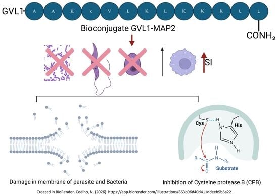
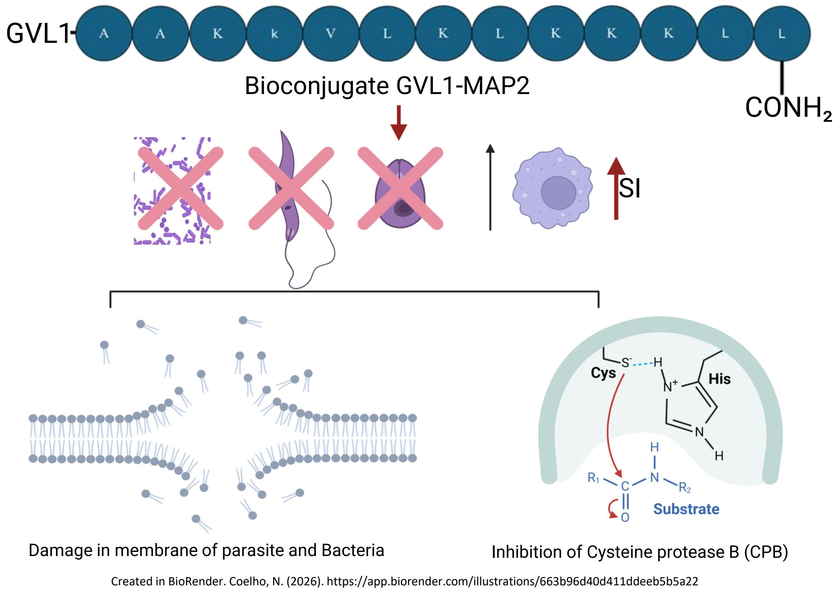
Graphical Abstract

1. Introduction

Infectious diseases are caused by microorganisms such as bacteria, parasites, and fungi and represent a global challenge, especially in regions with precarious socioeconomic conditions, where access to adequate treatments is limited. These diseases represent a major public health concern, especially considering the increasing number of immunocompromised patients and those living in poverty [1].
Leishmaniasis is a tropical, parasitic infection caused by the protozoan Leishmania spp. and transmitted through the bite of an infected female Phlebotomine [2]. These are considered neglected diseases and occur in underdeveloped regions, such as the Global South. Despite leishmaniasis being an endemic disease, little to no effort is employed in treating and preventing it. Leishmaniasis remains a major global health threat, affecting millions of people primarily in developing countries. The clinical management of this neglected tropical disease is severely hampered by a limited therapeutic arsenal and the emergence of drug-resistant Leishmania strains. Resistance to pentavalent antimonials is now widespread in several endemic regions, and decreased susceptibility to second-line drugs, such as amphotericin B and miltefosine, has been increasingly reported. The use of extremely cytotoxic drugs such as amphotericin B (Amp B), which must be injected in the patient in several, painful shots, leads to severe side effects [3]. This alarming trend of drug resistance, coupled with the high toxicity and cost of current treatments, underscores an urgent need for the development of novel, safe, and effective antileishmanial agents, such as antimicrobial peptides and their bioconjugates [3].
Other infectious diseases, particularly bacterial ones, are also on the rise, with cases emerging worldwide and medical treatments that were once remarkably effective now becoming flawed and outdated.
The World Health Organization (WHO) has categorized the bacteria most urgently needed for new therapies as ESKAPE (Enterococcus faecium, Staphylococcus aureus, Klebsiella pneumoniae, Acinetobacter baumannii, Pseudomonas aeruginosa, and Enterobacter species). This classification was based on criteria such as mortality rate, treatability, healthcare burden, and resistance prevalence, among others (WHO, 2019) [4]. Antibiotic resistance makes treatment of these and other bacterial infections challenging, increasing associated morbidity and mortality. Prolonged and uncontrolled use of drugs against bacteria and parasites promotes the prevalence of resistant pathogens [5]. Furthermore, there are a few therapeutic options for treating bacteria. Therefore, the search for new therapeutic alternatives is particularly important.
Antimicrobial peptides (AMPs) are short amino acid chains that occur from mammals to fungi that serve as a defense mechanism against predators [6]. They have had a plethora of applications in modern medicine and research fields when fighting the recent downfall of antibiotic efficacy [5]. The use of AMPs has shown effects on bacterial and protozoan cell membranes, thus leading to the death of the pathogen through a vast number of mechanisms, such as cell penetration and pore-inducing, disturbing the lipid bilayer structure of the cell, thus inducing its death [7].
Our group has evaluated bioconjugates containing AMPs and organic active compounds and has obtained promising results against parasites, bacteria, and viruses [7,8]. Recently, naturally occurring molecules that contain a guanidine center (or moiety) have been studied. Guanidines are widely used due to their oxidative phosphorylation properties [9], which can target intracellular parts of the pathogen. The data supports the actions of guanidine-derivative compounds at the cellular level, inhibiting enzymes such as cysteine protease B (CPB) in the Leishmania spp. protozoan, an enzyme responsible for its thriving when phagocytosed by a macrophage and, therefore, infection of the host. Its inhibiting action favors the treatment of the disease, thus not allowing the parasite to infect the cell [7,10].
Bioconjugates containing guanidine derivatives and peptides have already been explored by our group’s research against antileishmanial activities [7,11] using a cell penetration peptide (CPP) and the TSHa peptide against Leishmania. Given the potential of AMPs and guanidine derivatives, we synthesized two bioconjugates containing guanidine derivatives with the peptide analogues HP-MAP1 (MAP1) and HP-MAP2 (MAP2) (amide form), derived from temporin-PTa. Previously, these peptides have shown antimicrobial activity [12], while temporin-PTa is antiparasitic. A guanidine derivative called GVL1 ((Z)-4-((2-benzoyl3-(4-bromophenyl)guanidino)methyl) benzoic acid) (Figure S1) was used for this conjugation. Thus, this work explores the use of new bioconjugates, GVL1-MAP1 and GVL1-MAP2, as promising molecules against some ESKAPE bacterial strains and for antileishmanial activity in the treatment of Leishmania spp.

2. Results

2.1. Synthesis of GVL1-MAP1 and GVL1-MAP2 Bioconjugates

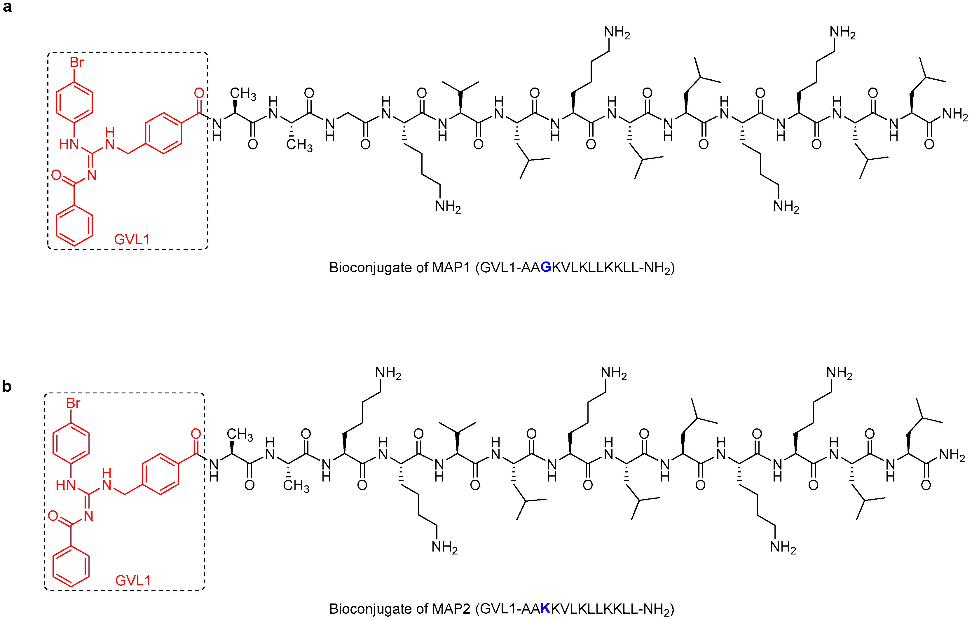
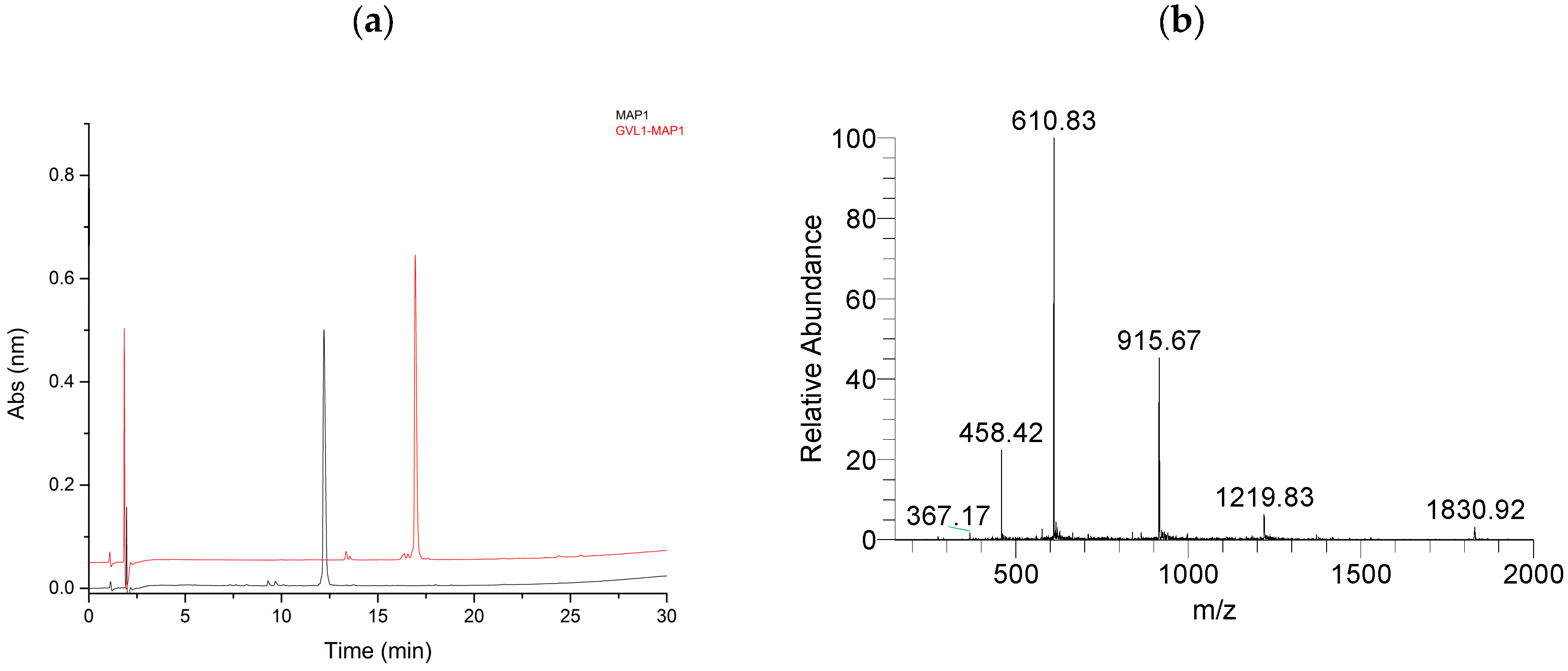
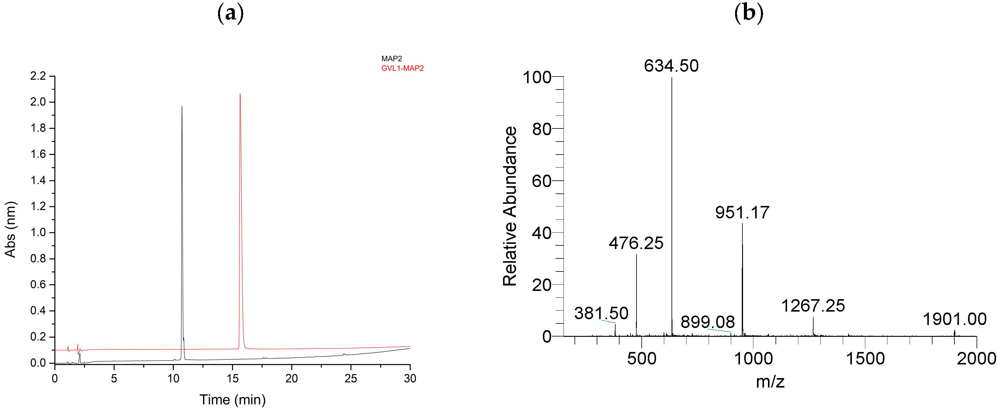
The MAP1 (AAGKVLKLLKKLL-NH2) (Figure S2) and MAP2 (AAKKVLKLLKKLL-NH2) (Figure S3) peptides were synthesized via SPPS (Solid Phase Peptide Synthesis) [13] using the Fmoc protocol. GVL1 was coupled to the N-terminal group of MAP1 (Figure 1a) and MAP2 (Figure 1b) using HATU/DIEA via its carboxyl group [7]. The peptides and bioconjugates were purified by reverse-phase HPLC in semi-preparative mode (Figure 2a and Figure 3a). The material obtained was analyzed by mass spectrometry, confirming that the desired material had been obtained (Figure 2b and Figure 3b). The mass spectra of both MAP1 and MAP2 peptides are in the Supplementary Data (Figures S4 and S5).

2.2. Biological Assays

2.2.1. Antileishmanial and Cytotoxic Activities Evaluation

The antileishmanial activity against L. amazonensis showed that GVL1 had IC50 values of 51.8 and 7.5 μM for the promastigote and amastigote forms, respectively, and the MAP1 peptide showed IC50 values of 43 μM in promastigote and 10 μM in amastigote forms (Table 1). The GVL1-MAP1 bioconjugate exhibited higher activity than GVL1 or MAP1 alone in both the promastigote and amastigote forms, with IC50 values of 7.3 and 0.9 μM, respectively (Table 1). In amastigotes, the increase in activity was approximately 8.5 times greater than that of GVL1 alone and 11 times greater than that of the MAP1 peptide. The GVL1-MAP1 bioconjugate showed CC50 = 21 μM in murine peritoneal macrophages, a lower value when compared to the MAP1 peptide alone and, similarly, Amp B. The selectivity index (SI) for this compound was smaller than that of Amp B, indicating worse biological properties. The SI indicates the compound’s toxicity towards the parasite relative to host mammalian cells (SI = CC50/IC50). Regarding the GVL1-MAP2 bioconjugate, it showed the best antileishmanial activity for the promastigote (IC50 = 3.2 µM) and amastigote (IC50 = 0.6 µM) forms (Table 1), with an increase in potency of approximately 2-fold when compared to GVL1-MAP1. In addition, this bioconjugate has the highest CC50 value (125 µM) and selectivity index (SI = 201). This last value was approximately six times higher than that of Amp B (SI = 32) and seven times higher than that of the MAP2 peptide, demonstrating that the coupling of GVL1 increases the potency and selectivity of GVL1 and MAP2 alone.
The bioconjugates with better antileishmanial activity against L. amazonensis, GVL1-MAP2, and the MAP2 peptide were also evaluated against L. infantum. The results were similar to those obtained for L. amazonensis. GVL1-MAP2 showed better potency and SI, with an IC50 value of 1.0 µM for the amastigote form, similar to that of the control drug Amp B (1.25 µM) (Table 2). In addition, this compound was more selective for the parasite, with SI = 125, higher than that obtained for Amp B (SI = 19), indicating that the bioconjugate was 5.2 times more selective for the parasite than Amp B.
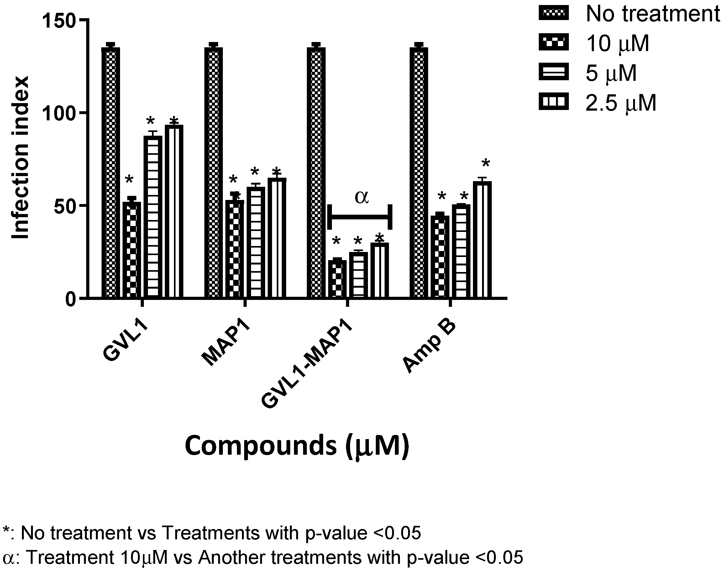
The ability to infect new macrophages was evaluated at concentration values of 2.5, 5.0 and 10 μM. The lower the infection rate of Leishmania, the greater its ability to prevent the infection of new macrophages. GVL1-MAP1 in L. amazonensis, after 24 h, decreased the infection rate almost 6.7-fold in the three tested concentrations in comparison to infected cells that were not treated (Figure 4). In comparison to the control drug Amp B, the bioconjugate was 3.5 times more effective at 10 μM, while the MAP1 peptide and GVL1 alone had smaller infection index values than GVL1-MAP1.
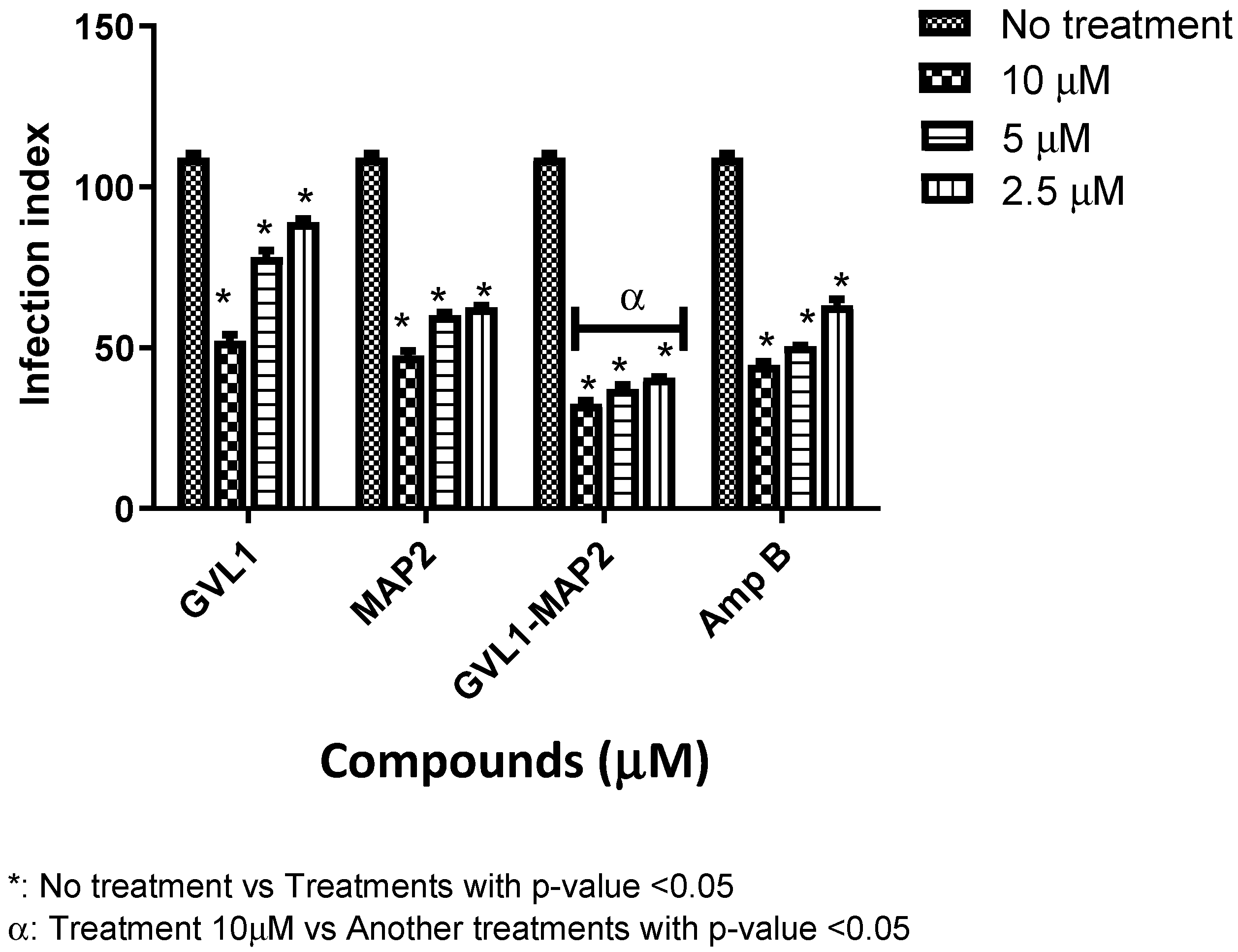
The GVL1-MAP2 bioconjugate also showed greater selectivity for the parasite than for the mammalian host cell. Moreover, it was also able to prevent the parasite from infecting new host cells. There was a 3-fold decrease in the infection rate when compared to the untreated control for L. amazonensis (Figure 5), and the infection index value was also lower than that found in relation to Amp B. The lower infection rate found for GVL1-MAP1 could be related to its higher toxicity in macrophages.
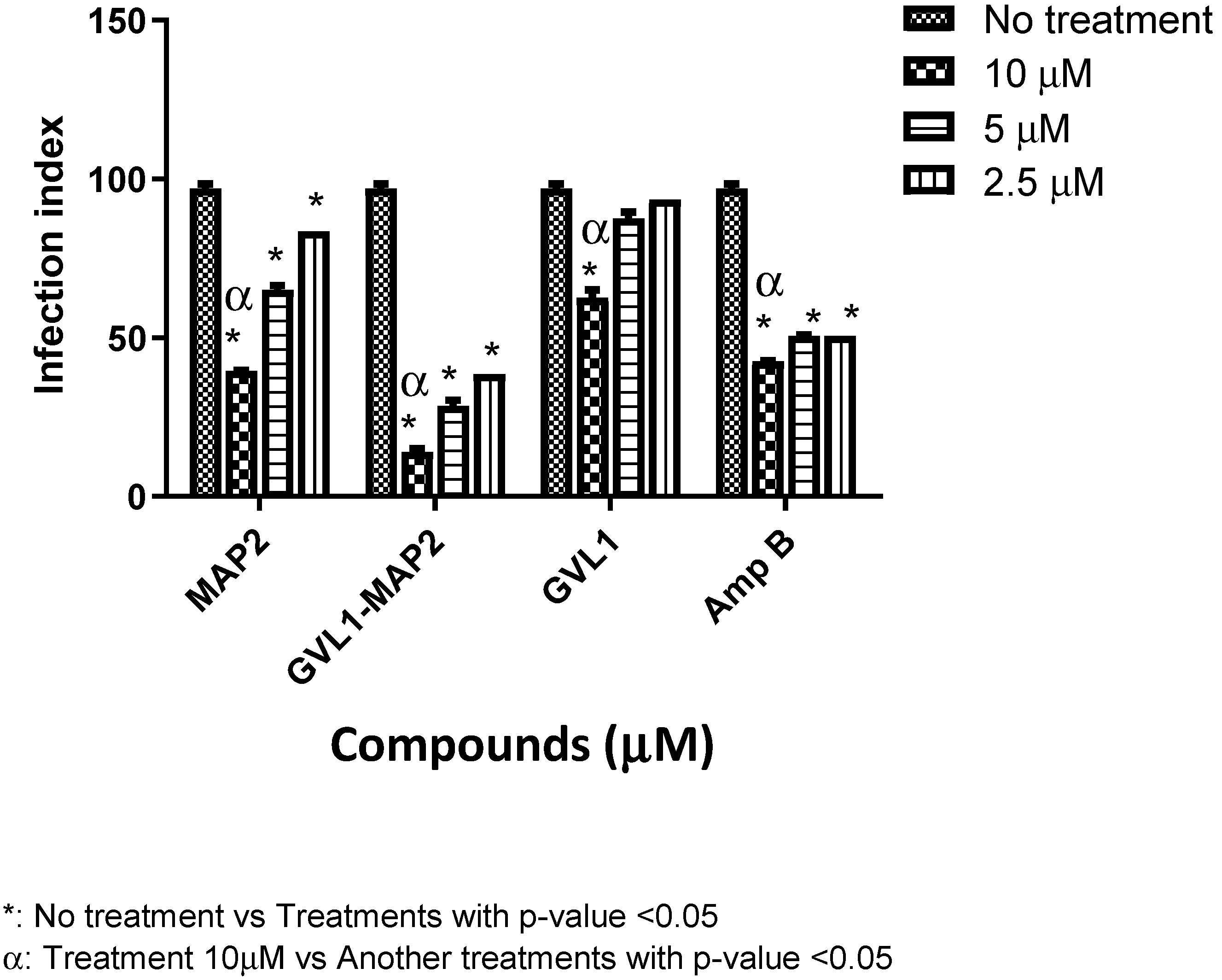
The bioconjugate GVL1-MAP2 was also able to reduce the infection rate for in vitro treatment with L. infantum; compared to macrophages infected without treatment, there was a 7.5-fold decrease in the infection rate (Figure 6) at 10 μM. In comparison to the control drug Amp B, there was a 3-fold decrease (Figure 6), demonstrating that, in addition to improving the selectivity of this compound, it is also able to interfere with the infection process, preventing new macrophages from being infected.

2.2.2. Stability Assay with Active Fetal Bovine Serum

In inactivated serum, the MAP2 peptide alone had not shown activity against Leishmania in the promastigote form, but in the presence of active serum, the IC50 values were 81.5 and 17.4 μM for L. amazonensis and L. infantum, respectively (Table 3).
The bioconjugate GVL1-MAP2 exhibited IC50 = 9.9 μM against L. amazonensis promastigote and 4.4 μM against L. infantum (Table 3). Despite undergoing partial degradation, the bioconjugate maintained significant biological potency. The decrease in activity (IC50) was 3- and 2-fold compared with those found for L. amazonensis and for L. infantum, respectively, in inactive serum.

2.2.3. Antibacterial Assays

The antibacterial activity of the bioconjugates was tested against Gram-positive and Gram-negative bacteria (Enterococcus faecium (Gram-positive); Staphylococcus aureus (Gram-positive); Klebsiella pneumoniae (Gram-negative); Acinetobacter baumannii (Gram-negative); Pseudomonas aeruginosa (Gram-negative); Enterobacter spp. (Gram-negative)), including ESKAPE bacteria, and expressed as minimum inhibitory concentration (MIC) and minimum bactericidal concentration (MBC) (Table 4 and Table 5). The Staphylococcus epidermidis is not classified within the ESKAPE group but was included in this study due to its status as a major opportunistic pathogen in hospital settings. The S. epidermidis strain is a well-characterized biofilm producer, representing a significant challenge in the treatment of device-related infections and chronic wounds, exhibiting multidrug resistance profiles similar to those found in ESKAPE species [5]. Except for S. epidermidis ATCC 35984, the GVL1-MAP1 bioconjugate was more active against Gram-positive bacteria than MAP1 (Table 4), particularly against the vancomycin-resistant E. faecium ATCC 700221 (MIC = 8 mg/mL). For Gram-negative bacteria, the GVL1-MAP1 bioconjugate showed activity only against A. baumannii ATCC 19606 and P. aeruginosa ATCC 27853, and the MIC values were higher than those of the MAP1 peptide. GVL1-MAP2 demonstrated activity against all tested Gram-positive bacteria, with higher activity than the isolated peptide MAP2, except for the strain S. epidermidis ATCC 35984. For Gram-negative bacteria, GVL1-MAP2 showed activity against all strains tested, but it was more active than MAP2 alone only in P. aeruginosa ATCC 27853 (Table 5).

2.3. Mechanism of Action

2.3.1. Vesicle Permeabilization

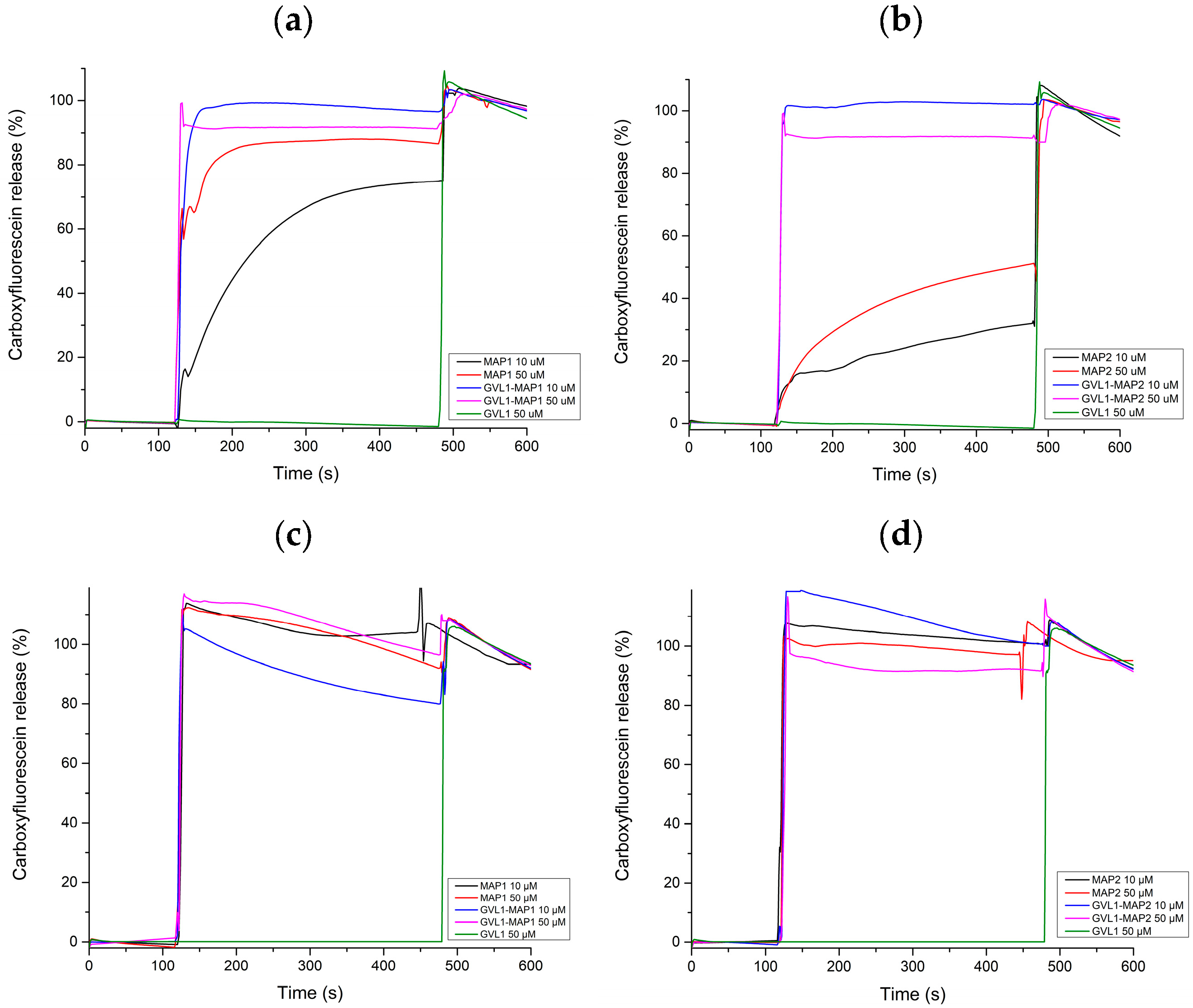
To evaluate a possible mechanism of action of the GVL1-MAP2 bioconjugate and the MAP2 peptide on the membranes, a permeabilization assay based on increasing the fluorescence of carboxyfluorescein (CF) leakage promoted by pore formation in unilamellar vesicles [15] was used. Thus, two membrane mimetic systems were evaluated, one containing 1-palmitoyl-2-oleoyl-sn-glycero-3-phosphocholine (POPC), 1-palmitoyl-2-oleoyl-sn-glycero-3-phospho-L-serine (POPS), and ergosterol, as a Leishmania membrane mimetic, and the other containing 1-palmitoyl-2-oleoyl-sn-glycero-3-phosphocholine (POPC) and cholesterol, as a eukaryotic membrane mimetic [16,17,18]. GVL1 alone did not permeabilize the vesicle mimetics of either mimetic vesicle (Figure 7), indicating, as expected, that the mechanism of action of GLV1 is not related to the cell membrane. The bioconjugates were able to cause faster membrane damage than peptides in eukaryotic mimetic membrane-containing POPC/cholesterol (Figure 7a,b), indicating that the conjugation strategy increases the capacity for membrane damage. These data are in accordance with the CC50 found in macrophages. The CC50 values of the bioconjugates were smaller than those found for the peptides. The peptides alone or conjugated with GVL1 in the Leishmania membrane mimetic (Figure 7c,d) caused rapid and more intense membrane damage than in POPC/cholesterol vesicles.

2.3.2. Cysteine Protease Enzyme Inhibition

We evaluated the action of MAP2 and GVL1-MAP2 on recombinant cysteine protease from Leishmania mexicana (CPB) (rCPB2.8ΔCTE), an important enzyme for the cell viability of the parasite. The peptide and bioconjugate inhibited enzymatic activity by approximately 95% and 89%, respectively, at 20 μM (Table 6). The IC50 for CPB inhibitory activity was also determined, demonstrating the peptide’s superior inhibitory activity against CPB.

3. Discussion

The retention times of the bioconjugates in the HPLC profiles were higher than those of peptides, indicating an increase in the hydrophobicity of the compound. These data are in agreement with the low solubility of the guanidine derivatives. The peptide increases the solubility of the guanidine derivative and its activity and decreases the toxicity.
One focus of this work is the development of molecules with anti-Leishmania activity. L. amazonensis is one of the species that causes cutaneous leishmaniasis (CL) and affects 12 million people, with 2 million new cases occurring annually [19]. Therefore, the development of less toxic and less painful therapies is needed [20]. However, the financial resources are limited for developing new molecules, vaccines, and diagnostic methods for leishmaniosis, a disease that mainly affects underprivileged people [21].
Peptides from the temporin class have the membrane as the main mechanism of action [22,23] and could facilitate the entry of active compounds, such as guanidine derivatives, into cells and promote inhibitory activity in internal proteins [12]. Even though temporin-class peptides are toxic to mammalian cells, this problem could be resolved by modifying the peptide sequence [12,24]. A study involving the peptide TSHa demonstrated that modifications of the peptide sequence at positions 4 and 7 (2 Gly for 2 Ala) reduced the toxicity of these analogues to red blood cells and other cell lines. This occurred due to an increase in hydrophobicity [25]. MAP1 and MAP2 were derived from temporin-PTa (FFGSVLKLIPKIL). MAP1 has the amino acids Ser4 and Pro10 replaced by Lys. In MAP2, Gly3 was replaced by a Lys. In both peptides, alanine residues replaced Phe at the N-terminus to assist the extension of the α-helical segment, and isoleucine residues were replaced by leucine residues aiming at reduced hydrophobicity. Both analogues displayed in vitro activity against bacteria without presenting hemolytic effects [12].
In this work, MAP2 showed a higher CC50 than MAP1 in macrophages. These results were attributed to the higher hydrophobicity of MAP1 and the different α-helical amphipathic structure content [12]. The α-helical amphipathic structure content was also related to a reduction in cytotoxicity activity in mammalian cells [26]. The antileishmanial activity of MAP1 was higher than that of MAP2 in promastigote form, but it was lower in amastigote form. The Leishmania membrane undergoes radical remodeling to adapt to its host environments. Promastigotes possess a glycocalyx dominated by lipophosphoglycan (LPG), which imparts a strong negative surface charge necessary for sandfly midgut attachment [27]. In terms of lipid composition, they are rich in ergosterol-type sterols and unsaturated fatty acids, maintaining high membrane fluidity. Conversely, amastigotes drastically downregulate LPG, replacing it with smaller glycoinositolphospholipids (GIPLs) and sphingolipids (such as inositol phosphorylceramide) and cholesterol scavenged from the host. These modifications result in a neutralized or less negative charge and a more rigid membrane in amastigote form [28,29]. The difference in the membrane composition could be related to different anti-Leishmania activities in the amastigote and promastigote forms.
The use of bioconjugates containing membrane-bound peptides can facilitate the entry of the guanidine derivative into the cell and promote its inhibitory activity on intracellular proteins via transient pore formation [30]. In the amastigote form, bioconjugation increased the IC50 by approximately 10 times compared to GVL1, and the values were similar to those of Amp B. Moreover, the SI of GVL1-MAP2 was 6.3 and 6.6 times higher than that of Amp B for L. amazonensis and L. infantum, respectively. The addition of the guanidine derivative to the N-terminus of the peptides decreased the CC50. This could be due to the charge of the N-terminus group. The bioconjugation of GVL1 in the amino-terminal region decreased the overall charge of the peptide, which could make the initial interaction with mammalian cell membranes difficult, causing a decrease in the CC50 value. The N-terminus group is important to the biological activity of the peptides and could change the selectivity of the bioconjugates [31]. This data substantiates the exceptional biological activity of the bioconjugate.
The synthesis of the bioconjugate may also have other effects on the peptide structure. The use of AMPs in clinical applications remains a challenge due to stability and bioavailability [32]. Several strategies have been employed to increase the stability of peptides, including the incorporation of non-natural amino acids, modification to the N- and/or C-terminal, cyclization, the use of non-peptide structures (peptidomimetics), and multimerization [33]. To evaluate the influence of the GVL1 compound on the stability of MAP2 (GVL1-MAP2), the antileishmanial activity was performed in the presence of active fetal bovine serum added to the culture medium of the L. amazonensis and L. infantum parasites in their promastigote form. The active fetal bovine serum contains proteases that can degrade these molecules [34]. The bioconjugates showed degradation but sustained relevant biological activity. The minor activity could be explained by forming a less active metabolite obtained from degradation or by reducing the guanidine derivative concentration inside the parasite and its activity. The formation of an active metabolite is confirmed for MAP2: in this case, the product of degradation was more active than the original compound in promastigote form. Similar results were found for GA-metabolite 5 obtained from the GA-Hecate peptide. Metabolite 5 (the degradation product) was the most efficient in inhibiting ZIKV strains of the African and Asian lineages during virus entry and replication [8].
One of the biggest global health problems is antimicrobial resistance, which causes around 9% of all global deaths. In 2019, approximately 13.7 million deaths were related to infectious diseases, of which those directly related to antimicrobial resistance accounted for around 1.27 million. The effects of morbidity or mortality related to antimicrobial resistance (AMR) are particularly severe in low- and middle-income countries, aggravating extreme poverty [35,36], causing a global economic problem. It is estimated that by 2050, low- and middle-income countries will have suffered an estimated loss of more than 5% of their gross domestic product because of these diseases [36,37]. In this manner, the current investment in developing new molecules for the treatment of infectious diseases contrasts sharply with an ever-increasing demand for new antimicrobials to treat infections caused by resistant microorganisms. In addition, the severity of leishmaniasis can be amplified by secondary infections of bacterial origin, whether opportunistic or pathogenic [38]. Studies using classic rodent models have advanced the understanding of how these bacteria interact with the host and disrupt its microbiota during the disease [39,40,41]. The development of new antileishmanial and bacterial compounds could solve this problem.
The peptides MAP1 and MAP2 showed activity against most bacterial strains. This behavior may be associated with the presence of lysine residues, which provided this peptide with a higher charge. Amphipathic peptides rich in lysine are multitargets for several pathogenic cells [24,42]. The addition of guanidine derivatives to peptides maintained the antimicrobial activity of the peptide. The GVL1-MAP2 bioconjugate generally exhibited similar or higher MIC values compared to GVL1-MAP1 against Gram-positive bacteria, indicating comparable or lower antibacterial potency. However, GVL1-MAP2 showed activity against all Gram-negative strains, whereas GVL1-MAP1 was active against fewer strains. Antimicrobial peptides typically have MICs in the 1–32 mg/mL range, as compared to the <1 mg/L used for traditional antibiotics [43]. In comparison to antimicrobial peptides that are already being used in clinical studies, such as the peptide nisin [44], the bioconjugate showed 8-fold less activity against the Gram-positive species S. aureus, but it showed the same activity against the Gram-negative species P. aeruginosa (different ATCC strain). These data showed the good activity of bioconjugates against bacteria with bactericidal effects in most cases.
The mechanism of action of peptides and bioconjugates was evaluated. Antimicrobial peptides (AMPs) can act on membranes, causing damage to them through mechanisms that lead to the formation of pores, such as barrel–stave, toroidal–pore, and transient pore, or causing the destabilization of the lipid bilayer, such as carpet-like mechanism [21,30]. The vesicle permeabilization studies showed that peptides and bioconjugates act in vesicles mimicking a Leishmania membrane, probably through carpet-like mechanism, with a rapid burst and instantaneous release of the fluorophore (Figure 7c,d). In this mechanism, the partially positively charged peptides adhere parallel to the membrane surface; the main binding force in this mechanism is assumed to be the electrostatic attraction between the negative charge of the vesicle bilayer surface that mimics the parasite membrane, from which micelles are formed [45]. For peptides in eukaryotic membrane–mimetic vesicles (Figure 7a,b), the leakage percentage increased with peptide concentration, characterized by a sustained release of a fluorophore. This kind of profile is attributed to the pore formation mechanism [46]. The difference in mechanisms in both vesicles is due to different lipid compositions. In addition, the conjugation strategy increases the capacity for membrane damage in eukaryotic mimetic vesicle (POPC/cholesterol). These data are in accordance with the CC50 found in macrophages, where the bioconjugates showed smaller values than those found for the peptides.
In addition to the membrane, the peptides may have other therapeutic targets. In concentrations lower than MIC, the peptide and bioconjugate may induce small and transient pores in the target membrane [47]. When the transient pores are broken down, the peptide sequence could cross the membrane and act on intracellular targets, as enzymes. Enzymes are essential for functional integrity and virulence. Cysteine proteases B (CPB) are important in the infectious process; their inhibition has shown high therapeutic potential, enabling the design of new drugs [48,49,50]. In L. mexicana, three cysteine protease genes were identified as virulence factors: lmcpa, lmcpb and lmcpc. The last encodes a cathepsin L-like protein and is the most abundant in the amastigote form of the parasite, making this enzyme an important therapeutic target. Guanidine derivatives target the cysteine protease, such as the compound E64 (L-trans-epoxysuccinyl-leucylamido-(4-guanidino)butane), which is a potent irreversible inhibitor of Leishmania CPB [51]. The guanidine derivative LQOF-G6 showed selective inhibitory activity on Leishmania major cysteine protease LmCPB2.8DCTE (CPB) and was not toxic [52]. Compounds belonging to the azapeptide class have also been reported to be commercial inhibitors of cysteine proteases, such as papain, cathepsin B, and cathepsin K. The peptide Z-Arg-Leu-Val-Gly-Ile-Val-Ome is also a potent inhibitor of cathepsin B, demonstrating the high potential of peptides as cysteine protease inhibitors [53].
The MAP2 peptide and the GVL1-MAP2 bioconjugate inhibited CPB enzyme activity. The enzymatic inhibition of the peptide was greater than that of the bioconjugate and can be explained by the increased size and volume of the molecule, which may cause steric hindrance in contact with the CPB enzyme site. GVL1 alone was also tested and showed the worst inhibitory action, indicating the peptide contribution in enzyme-bound areas. Cysteine proteases may also interfere with the parasite infection process. Leishmania expresses high levels of several classes of CP belonging to the papain family, which are crucial for the parasite’s metabolism, reproduction, and intracellular survival [54,55,56]. During the process of Leishmania infection in macrophages, CPB modulates the host’s responses by negatively regulating protective Th1 immune responses, particularly IFN-γ production, through degradation of the transcription factor NF-κB and subsequent inhibition of IL-12 production by infected macrophages. Therefore, the protozoan cysteine protease CPB can alter host macrophage signaling by increasing IL-12 transcription, which hampers the establishment of infection [49,57]. The literature demonstrates the fundamental role of parasite CPB in the infectious process and suggests the high therapeutic potential of inhibiting these enzymes [48,49,58,59]. Nonetheless, the bioconjugate showed a lower infection index than the MAP2 peptide. The bioconjugate could act on targets other than CPB, such as trypanothione reductase [7]. This could explain the higher activity of the bioconjugate.
These data show that GVL1-MAP2 is a multifunctional therapeutic tool. This bioconjugate not only overcomes the limitations of traditional AMPs regarding toxicity and stability [12,23] but also provides a protective immunomodulatory response essential for controlling Leishmania infection [51]. Together, the data obtained showed that the GVL1-MAP2 bioconjugate fulfills key pharmacological criteria for anti-Leishmania candidates, showing high potency and a selectivity index (SI > 10) that exceeds the requirements for visceral leishmaniasis treatment [16,18].

4. Materials and Methods

4.1. Peptides and Bioconjugates Synthesis

The synthesis of the peptides and their bioconjugates was made by the SPPS [13] using the Fmoc chemistry protocol [13]. The resin employed was the Rink amide-MBHAR resin (AAPPTEC, Louisville, KY, USA), which makes peptides with an amide at the C-terminal position.
The amino acid sequences that correspond to the MAP1 and MAP2 peptides are AAGKVLKLLKKLL-NH2 and AAKKVLKLLKKLL-NH2, respectively.
Their bioconjugates have GVL1 at the N-terminus. Coupling agents such as N,N’-diisopropylcarbodiimide (DIC)/1-hydroxybenzotriazole (HOBt) (Oakwood Chemical, Estill, SC, USA) or 2-(1-H-7-azabenzotriazol1-yl)-1,1,3,3-tetramethyluroniumhexafluorophosphate (HATU) (AAPPTEC, Louisville, KY, USA)/N,N-diisopropylethylamine (DIPEA) (Merck, Darmstadt, Germany), in N,N-dimethylformamide (DMF) (Êxodo Científica, Sumaré, SP, Brazil), are used for peptide bonds between Fmoc-amino acids (AAPPTEC, Louisville, KY, USA) and for the bioconjugation of the guanidine derivative moiety, respectively.
For the cleavage of the peptides and bioconjugates, a cocktail containing 2.5% H2O, 2.5% triisopropylsilane (TIS), and 95% trifluoroacetic acid (Oakwood Chemical, Estill, SC, USA) was used under constant agitation. After two hours, the peptides were precipitated with anhydrous ethyl ether (Êxodo Científica, Sumaré, SP, Brazil) and separated from the soluble non-peptide material by centrifugation. The peptide obtained was dissolved in an aqueous solution containing 0.045% TFA in water and lyophilized for subsequent characterization.

4.2. Molecule Purification and Characterization

The molecules were purified by reversed-phase high-performance liquid chromatography (HPLC), in semi-preparative mode using a C18 Sepax column (25 cm × 1 mm and 5 μm particle size) [7]. The molecules were then analyzed by mass spectrometry and HPLC to confirm their molecular weights and purity [7].

4.3. Antileishmanial Activity

L. amazonensis promastigotes (MPRO/BR/1972/M1841-LV-79) were cultivated in LIT medium, while L. infantum promastigotes (MNYC/BZ/62/M379) were raised in 199 culture medium (Sigma-Aldrich, St. Louis, MO, USA) supplemented with 10% heat-inactivated fetal bovine serum (FBS; Gibco/Invitrogen, Thermo Fisher Scientific, Waltham, MA, USA) [7] and 10% urine obtained from healthy male human donors among the members of the research group, at 28 °C, until the mid-log phase of growth was achieved. For macrophage recruitment, 2–3 mL of 3% thioglycolate was applied to the peritoneal cavity of the animals, three days before the start of the experiment. After this period, the animals were euthanized with CO2, and the macrophages were collected by washing the peritoneal cavity with 5 mL of sterile PBS. Viable cell counting was performed using trypan blue, and the concentration was adjusted to 1 to 5 × 10⁶ cells/mL. In a 96-well plate, 100 µL of the macrophage suspension was distributed per well and incubated for 4 to 6 h at 37 °C with 5% CO2 to allow for cell adhesion. After adhesion, the medium was removed, and 97 µL of complete RPMI medium and 3 µL of the compound to be tested were added, followed by incubation for 24 h, according to the methodology described by Almeida et al. [60]. For antileishmanial assays performed on the promastigote form to obtain IC50 measurements, cultures of the protozoans L. infantum and L. amazonensis were grown until the exponential phase and then transferred to 96-well plates at a final concentration of 1 × 10⁷ promastigotes × mL−1. Thereafter, the compounds of interest were added to those wells at concentrations ranging from 1.28 to 100 µmol L−1 to determine their anti-promastigote activity. After 72 h, viable protozoans were determined using the MTT method [61]. For antileishmanial assay performance on the amastigote form, macrophages were infected with Leishmania infantum and Leishmania amazonensis stationary-growth-phase promastigotes, using a ratio of five parasites per macrophage for Leishmania amazonensis and ten to one for Leishmania infantum at 37 °C in five-percent carbon dioxide for 4 h. Infected macrophages were treated with different concentrations of the peptides and bioconjugates and incubated for 24 h. The infection index was determined by the product of the percentage of infected macrophages and the average number of amastigotes per infected cell. All experiments were done in triplicate. The concentration responsible for inhibiting fifty percent of the growth of amastigote forms compared to the untreated control was determined by non-linear regression analysis using GraphPad Prism software. Normality was assessed using Shapiro–Wilk tests and statistical analysis (including ANOVA and the t-test), comparing the results with control groups and standard drugs, such as amphotericin B. Twelve healthy male Swiss mice obtained from the Central Vivarium of the School of Pharmaceutical Sciences—UNESP Araraquara (six to eight weeks of age, twenty-five to thirty grams) were used, eleven for the amastigote assay and one for the control. The infection indices were conducted as previously described [62].

4.4. Active Serum Stability

Peptide stability was investigated using anti-promastigote assays with L. amazonensis and L. infantum using LIT or 199 culture media with active fetal bovine serum, as previously detailed by de Souza et al. [7], in which the peptide is submitted to the same protocol as previously described for anti-promastigote assays, varying only the serum utilized: in this assay, the serum contains various proteases that can degrade the peptide, so it allows for a measurement of its stability when facing such enzymes.

4.5. CPB Enzyme Inhibition

CPB inhibition experiments were performed as described previously by Moreira et al., 2022 [52], utilizing pre-established methods consisting of the measurement of fluorescence, obtained from the cleavage of the substrate caused by the enzyme treated with the compounds.

4.6. Antibacterial Activity

The minimum inhibitory concentrations (MICs) of GVL, MAP1, GVL1-MAP1, MAP2 and GVL1-MAP2 were determined against clinically important species strains, including ESKAPE species: S. epidermidis ATCC35984, E. faecalis ATCC29212, S. aureus ATCC25923, S. aureus ATCC8095, E. faecium ATCC 700221, K. pneumoniae ATCC 700603, E. coli ATCC25922, A. baumannii ATCC19606, and P. aeruginosa ATCC27853. Although S. epidermidis is not part of the ESKAPE group, it was included due to its clinical relevance as an opportunistic pathogen associated with nosocomial infections. S. epidermidis is the most common cause of implant-associated infections and is a canonical opportunistic biofilm former. Each compound was diluted in water to prepare a 10× concentrated stock solution. Subsequently, the stock solution was diluted 1:10 in Cation-Adjusted Mueller Hinton broth (MHCA) (BD), according to CLSI (2013) [63]. Each compound was tested at 512 mg/mL or at the highest concentration at which it could be dissolved without precipitation. Serial dilutions (1:2) were made from the 512 mg/mL well to the 0.06 mg/mL well. Incubation was performed at 37 °C, and the results were visually read after 24 h, at which point the concentration at which the compound inhibited the microorganism’s growth was observed. For the negative and positive controls, MHCA broth was added. In the positive control, bacteria were added without the compound to observe their growth in MHCA broth. In the negative control, only the culture medium, MHCA broth, was present, without bacteria, to show that it was not contaminated. The tests were performed in triplicate.
To determine the minimum bactericidal concentration (MBC), 100 μL from each well corresponding to all tested concentrations in the MIC assay was subcultured onto MHCA agar plates and incubated at 35 °C for 24 h. The MBC was then determined as the lowest peptide concentration at which no visible growth was observed on the plate.

4.7. LUV Permeabilization Model Assay

Vesicles of POPC/cholesterol (4:1, m:m), for simulating human membranes, and POPG/POPS/ergosterol (8:1:1; m/m/m), for protozoan membrane simulation, containing fluorescein in their insides, were made according to the protocol described by Lorenzón et al. [64]. These vesicles had a carboxyfluorescein (CF) inner structure to detect extravasation via an increase in fluorescence due to pore formation. The wavelength of excitation used was 490 nm, and the wavelength of emission was 512 nm.
For a blank passage, the vesicles were introduced to the spectrofluorometer in a quartz cuvette, diluted in PBS, for a time of 480 s (0% of release). Then, a 10% solution of Triton (polyethylene glycol p-(1,1,3,3-tetramethylbutyl)-phenyl ether) was added to the cuvette, therefore releasing all the fluorescent material (100% of release). Evaluation of the GVL1, peptide, and bioconjugate in concentrations of 50 µM and 10 µM was done by the addition of these compounds at 120 s. After 480 s, Triton was added to reach the maximum read of fluorescence (100% of release).

4.8. Cytotoxicity Assay on Murine Peritoneal Macrophages

Macrophages were sourced from the peritoneal cavity of Swiss mice (details in Section 4.3), using the methodology outlined by Almeida et al., 2017 [65]. The MTT colorimetric assay was carried out. The drug concentration corresponding to 50% cell growth inhibition was expressed as the cytotoxic concentration (CC50). Seven male Swiss mice (six to eight weeks of age; twenty-five to thirty grams) were used, six for the cytotoxicity assay and one for the control.

4.9. Statistical Analysis

Data were expressed as the mean ± standard deviation (SD) of at least three independent experiments performed in triplicate. The normality of the data distribution was assessed using the Shapiro–Wilk test. For comparisons between multiple groups, such as the antileishmanial and antibacterial assays, one-way analysis of variance (ANOVA), followed by Tukey’s post hoc test, was employed. For enzyme inhibition assays, comparisons were made using Student’s t-test.
The IC50, CC50, and EC50 values were determined by non-linear regression analysis (log[inhibitor] vs. response—variable slope). All statistical analyses and graphical representations were performed using GraphPad Prism software version 8.0 (GraphPad Software, San Diego, CA, USA). A p-value of less than 0.05 (p < 0.0) was considered “statistically significant.”
All assays were performed in triplicate.

5. Conclusions

This study describes the successful development of bioconjugate containing guanidine derivatives and antimicrobial peptides as an effective strategy for creating new compounds to treat leishmaniasis and bacterial infections. The key achievement is obtaining a compound with high antileishmanial activity and elevated selectivity, surpassing the lack of selectivity observed with amphotericin B, the current standard treatment. In addition, the development of antibacterial and antileishmanial compounds can avoid secondary infections, whether opportunistic or pathogenic. The bioconjugation strategy proved effective in enhancing the compound’s potency and selectivity, offering a superior alternative for developing new drugs. Although degradation assays indicated that the bioconjugate suffers degradation in active serum, it nonetheless maintained its biological activity, suggesting sufficient functional stability. Permeabilization studies and enzyme inhibition analyses revealed that the peptide acts via at least two distinct mechanisms, with the primary one focusing on the membrane and inner targets as an inhibitor of CPB enzyme. These results provide valuable information for designing new antileishmanial and antibacterial peptides. The use of the guanidine derivative–peptide conjugation shows excellent potential in new drug discovery, offering crucial insights for the development of therapeutics against leishmaniasis and bacteria, significant global public health concerns.

Supplementary Materials

The following supporting information can be downloaded at https://www.mdpi.com/article/10.3390/ph19060835/s1: Figure S1. Structure of the guanidine derivative GVL1. Figure S2. Structure of the peptide MAP1 (AAGKVLKLLKKLL-NH2). Figure S3. Structure of the peptide MAP2 (AAKKVLKLLKKLL-NH2). Figure S4. Mass spectrum of the MAP1 peptide. Figure S5. Mass spectrum of the MAP2 peptide.

Author Contributions

Conceptualization, E.M.C., M.A.S.G., I.L.B.d.C.C. and E.R.P.G.; methodology, all authors; validation, N.C.S.C., A.M.A.V., I.L.B.d.C.C., E.M.C., M.A.S.G., and E.R.P.G.; formal analysis, all authors; investigation, G.A.S., N.C.S.C., J.V.M.d.S., S.T.R.B., J.S.d.L., M.N.A., A.M.A.V., L.R.d.A., and C.D.A.; resources, N.C.S.C., E.M.C., M.A.S.G., I.L.B.d.C.C. and E.R.P.G.; data curation, all authors; writing—original draft preparation, G.A.S., N.C.S.C., E.M.C., M.A.S.G. and E.R.P.G.; writing—review and editing, G.A.S., N.C.S.C., E.M.C., M.A.S.G., I.L.B.d.C.C. and E.R.P.G.; supervision, project administration and funding acquisition, E.M.C., M.A.S.G., I.L.B.d.C.C. and E.R.P.G. All authors have read and agreed to the published version of the manuscript.

Funding

This study was financed, in part, by the São Paulo Research Foundation (FAPESP), Brazil—Process Numbers 22/05411-8, 20/04415-4, 2013/07600-3, 2021/02595-8 and PhD scholarship 2025/01056-7—and the National Council of Scientific and Technological Development, grant numbers 304739/2021-9 and FINEP 01.23.0034.00 (0419/22).

Institutional Review Board Statement

This research did not involve human subjects. Animal experiments were conducted in accordance with the protocol approved by the Institutional Ethics Committee on the Use of Animals (CEUA/FCF/Car, #03/2019), approved on 28 August 2019.

Informed Consent Statement

Not applicable.

Data Availability Statement

The original contributions presented in this study are included in the article/supplementary material. Further inquiries can be directed to the corresponding authors.

Acknowledgments

We acknowledge Coordenação de Aperfeiçoamento de Pessoal de Nível Superior—Brasil (CAPES)—Finance Code 001. The authors would like to thank BioRender.com for assistance in creating the graphical abstract (Created in Biorender. Coelho, N. (2026) https://BioRender.com/23yx4pz (accessed on 21 May 2026)). We would like to thank the editor, Christophe Dardonville, for his suggestions throughout the process of expanding this paper.

Conflicts of Interest

The authors declare no conflicts of interest.

Abbreviations

The following abbreviations are used in this manuscript:
AMPsAntimicrobial peptides
Amp BAmphotericin B
AMRAntimicrobial resistance
CFCarboxyfluorescein
CLCutaneous leishmaniasis
CPBCysteine proteases B
CPPCell penetration peptide
DIEAN,N-Diisopropylethylamine
GIPLGlycoinositolphospholipids
GVL1(Z)-4-((2-benzoyl-3-(4-bromophenyl)guanidino)methyl) benzoic acid
HATU2-(1-H-7-azabenzotriazol1-yl)-1,1,3,3-tetramethyluroniumhexafluorophophate
LPGLipophosphoglycan
MAP1AAGKVLKLLKKLL-NH2
MAP2AAKKVLKLLKKLL-NH2
MBCMinimum bactericidal concentration
MICMinimum inhibitory concentration
POPC1-palmitoyl-2-oleoyl-sn-glycero-3-phosphocholine
POPS 1-palmitoyl-2-oleoyl-sn-glycero-3-phospho-L-serine
SISelectivity index (MBC/MIC ratio)

References

  1. Baker, R.E.; Mahmud, A.S.; Miller, I.F.; Rajeev, M.; Rasambainarivo, F.; Rice, B.L.; Takahashi, S.; Tatem, A.J.; Wagner, C.E.; Wang, L.F.; et al. Infectious Disease in an Era of Global Change. Nat. Rev. Microbiol. 2022, 20, 193–205. [Google Scholar] [CrossRef] [PubMed]
  2. Taslimi, Y.; Zahedifard, F.; Rafati, S. Leishmaniasis and Various Immunotherapeutic Approaches. Parasitology 2018, 145, 497–507. [Google Scholar] [CrossRef]
  3. Ponte-Sucre, A.; Gamarro, F.; Dujardin, J.C.; Barrett, M.P.; López-Vélez, R.; García-Hernández, R.; Pountain, A.W.; Mwenechanya, R.; Papadopoulou, B. Drug Resistance and Treatment Failure in Leishmaniasis: A 21st Century Challenge. PLoS Negl. Trop. Dis. 2017, 11, e0006052. [Google Scholar] [CrossRef]
  4. World Health Organization. 2019 Antibacterial Agents in Clinical Development: An Analysis of the Antibacterial Clinical Development Pipeline; World Health Organization: Geneva, Switzerland, 2019; Available online: https://iris.who.int/server/api/core/bitstreams/945de890-a22e-47ee-95ae-610c9dc0250a/content (accessed on 12 April 2026).
  5. Huemer, M.; Mairpady Shambat, S.; Brugger, S.D. Antibiotic Resistance and Persistence—Implications for Human Health and Treatment Perspectives. EMBO Rep. 2020, 21, e51034. [Google Scholar] [CrossRef]
  6. Alzain, M.; Daghistani, H.; Shamrani, T.; Almoghrabi, Y.; Daghistani, Y.; Alharbi, O.S.; Sait, A.M.; Mufrrih, M.; Alhazmi, W.; Alqarni, M.A.; et al. Antimicrobial Peptides: Mechanisms, Applications, and Therapeutic Potential. Infect. Drug Resist. 2025, 18, 4385–4426. [Google Scholar] [CrossRef] [PubMed]
  7. de Souza, J.V.M.; Costa, N.C.S.; Brasil, M.C.O.A.; dos Anjos, L.R.; de Menezes, R.P.B.; Zampieri, E.H.; de Lima, J.S.; Velasquez, A.M.A.; Scotti, L.; Scotti, M.T.; et al. Guanidines Conjugated with Cell-Penetrating Peptides: A New Approach for the Development of Antileishmanial Molecules. Molecules 2025, 30, 264. [Google Scholar] [CrossRef] [PubMed]
  8. da Silva Sanches, P.R.; Sanchez-Velazquez, R.; Batista, M.N.; Carneiro, B.M.; Bittar, C.; De Lorenzo, G.; Rahal, P.; Patel, A.H.; Cilli, E.M. Antiviral Evaluation of New Synthetic Bioconjugates Based on GA-Hecate: A New Class of Antivirals Targeting Different Steps of Zika Virus Replication. Molecules 2023, 28, 4884. [Google Scholar] [CrossRef]
  9. Hollunger, G. Guanidines and Oxidative Phosphorylations. Acta Pharmacol. Toxicol. 1955, 11, 1–84. [Google Scholar] [CrossRef]
  10. Alexander, J.; Coombs, G.H.; Mottram, J.C. Leishmania Mexicana Cysteine Proteinase-Deficient Mutants Have Attenuated Virulence for Mice and Potentiate a Th1 Response1. J. Immunol. 1998, 161, 6794–6801. [Google Scholar] [CrossRef]
  11. Costa, N.C.S.; dos Anjos, L.R.; de Souza, J.V.M.; Brasil, M.C.O.d.A.; Moreira, V.P.; Graminha, M.A.S.; Lubec, G.; Gonzalez, E.R.P.; Cilli, E.M. Development of New Leishmanicidal Compounds via Bioconjugation of Antimicrobial Peptides and Antileishmanial Guanidines. ACS Omega 2023, 8, 34008–34016. [Google Scholar] [CrossRef]
  12. Souza e Silva, P.; Ferreira, M.A.; de Moraes, L.F.R.; de Barros, E.; Preza, S.L.E.; Cardoso, M.H.; Franco, O.L.; Migliolo, L. Synthetic Peptides Bioinspired in Temporin-PTa with Antibacterial and Antibiofilm Activity. Chem. Biol. Drug Des. 2022, 100, 51–63. [Google Scholar] [CrossRef] [PubMed]
  13. Merrifield, R.B. Solid Phase Peptide Synthesis. III. An Improved Synthesis of Bradykinin. Biochemistry 1964, 3, 1385–1390. [Google Scholar] [CrossRef]
  14. Pankey, G.A.; Sabath, L.D. Clinical Relevance of Bacteriostatic versus Bactericidal Mechanisms of Action in the Treatment of Gram-Positive Bacterial Infections. Clin. Infect. Dis. 2004, 38, 864–870. [Google Scholar] [CrossRef]
  15. Krauson, A.J.; He, J.; Wimley, W.C. Determining the Mechanism of Membrane Permeabilizing Peptides: Identification of Potent, Equilibrium Pore-Formers. Biochim. Et Biophys. Acta (BBA) Biomembr. 2012, 1818, 1625–1632. [Google Scholar] [CrossRef] [PubMed]
  16. Cabezas, Y.; Legentil, L.; Robert-Gangneux, F.; Daligault, F.; Belaz, S.; Nugier-Chauvin, C.; Tranchimand, S.; Tellier, C.; Gangneux, J.-P.; Ferrières, V. Leishmania Cell Wall as a Potent Target for Antiparasitic Drugs. A Focus on the Glycoconjugates. Org. Biomol. Chem. 2015, 13, 8393–8404. [Google Scholar] [CrossRef]
  17. dos Santos, M.G.; Muxel, S.M.; Zampieri, R.A.; Pomorski, T.G.; Floeter-Winter, L.M. Transbilayer Dynamics of Phospholipids in the Plasma Membrane of the Leishmania Genus. PLoS ONE 2013, 8, e55604. [Google Scholar] [CrossRef]
  18. Dueñas-Romero, A.M.; Loiseau, P.M.; Saint-Pierre-Chazalet, M. Interaction of Sitamaquine with Membrane Lipids of Leishmania Donovani Promastigotes. Biochim. Biophys. Acta Biomembr. 2007, 1768, 246–252. [Google Scholar] [CrossRef] [PubMed]
  19. Baileyid, F.; Mondragon-Shem, K.; Haines, L.R.; Olabi, A.; Alorfi, A.; Ruiz-Postigo, J.A.; Alvar, J.; Hotez, P.; Adams, E.R.; Vélez, I.D.; et al. Cutaneous Leishmaniasis and Co-Morbid Major Depressive Disorder: A Systematic Review with Burden Estimates. PLoS Negl. Trop. Dis. 2019, 13, e0007092. [Google Scholar] [CrossRef]
  20. Katsuno, K.; Burrows, J.N.; Duncan, K.; Van Huijsduijnen, R.H.; Kaneko, T.; Kita, K.; Mowbray, C.E.; Schmatz, D.; Warner, P.; Slingsby, B.T. Hit and Lead Criteria in Drug Discovery for Infectious Diseases of the Developing World. Nat. Rev. Drug Discov. 2015, 14, 751–758. [Google Scholar] [CrossRef]
  21. Schroeder, J.; Aebischer, T. Vaccines for Leishmaniasis: From Proteome to Vaccine Candidates. Hum. Vaccin. 2011, 7, 10–15. [Google Scholar] [CrossRef]
  22. Maharjan, R.; Khan, A.I.; Nadeem-ul-Haque, M.; Maresca, M.; Choudhary, M.I.; Shaheen, F.; Simjee, S.U. Serum Stable and Low Hemolytic Temporin-SHa Peptide Analogs Disrupt Cell Membrane of Methicillin-Resistant Staphylococcus Aureus (MRSA). Probiotics Antimicrob. Proteins 2022, 14, 391–405. [Google Scholar] [CrossRef]
  23. Eggimann, G.A.; Sweeney, K.; Bolt, H.L.; Rozatian, N.; Cobb, S.L.; Denny, P.W. The Role of Phosphoglycans in the Susceptibility of Leishmania Mexicana to the Temporin Family of Anti-Microbial Peptides. Molecules 2015, 20, 2775–2785. [Google Scholar] [CrossRef]
  24. Silva, P.S.e.; Guindo, A.S.; Oliveira, P.H.C.; de Moraes, L.F.R.N.; Boleti, A.P.d.A.; Ferreira, M.A.; de Oliveira, C.F.R.; Macedo, M.L.R.; Rossato, L.; Simionatto, S.; et al. Evaluation of the Synthetic Multifunctional Peptide Hp-MAP3 Derivative of Temporin-PTa. Toxins 2023, 15, 42. [Google Scholar] [CrossRef] [PubMed]
  25. Raja, Z.; André, S.; Abbassi, F.; Humblot, V.; Lequin, O.; Bouceba, T.; Correia, I.; Casale, S.; Foulon, T.; Sereno, D.; et al. Insight into the Mechanism of Action of Temporin-SHa, a New Broad-Spectrum Antiparasitic and Antibacterial Agent. PLoS ONE 2017, 12, e0174024. [Google Scholar] [CrossRef]
  26. Zhang, S.K.; Song, J.W.; Gong, F.; Li, S.B.; Chang, H.Y.; Xie, H.M.; Gao, H.W.; Tan, Y.X.; Ji, S.P. Design of an α-Helical Antimicrobial Peptide with Improved Cell-Selective and Potent Anti-Biofilm Activity. Sci. Rep. 2016, 6, 27394. [Google Scholar] [CrossRef] [PubMed]
  27. Turco, S.J.; Descoteaux, A. The Lipophosphogl Yean of Leislfmania Parasites. Annu. Rev. Microbiol. 1992, 46, 65–92. [Google Scholar] [CrossRef]
  28. McConville, M.J.; Ferguson, M.A.J. The Structure, Biosynthesis and Function of Glycosylated Phosphatidylinositols in the Parasitic Protozoa and Higher Eukaryotes. Biochem. J. 1993, 294, 305–324. [Google Scholar] [CrossRef]
  29. Zhang, K.; Beverley, S.M. Phospholipid and Sphingolipid Metabolism in Leishmania. Mol. Biochem. Parasitol. 2010, 170, 55–64. [Google Scholar] [CrossRef]
  30. Wimley, W.C.; Hristova, K. The Mechanism of Membrane Permeabilization by Peptides: Still an Enigma. Aust. J. Chem. 2020, 73, 96–103. [Google Scholar] [CrossRef]
  31. Batista, M.N.; Sanches, P.R.d.S.; Carneiro, B.M.; Braga, A.C.S.; Campos, G.R.F.; Cilli, E.M.; Rahal, P. GA-Hecate Antiviral Properties on HCV Whole Cycle Represent a New Antiviral Class and Open the Door for the Development of Broad Spectrum Antivirals. Sci. Rep. 2018, 8, 14329. [Google Scholar] [CrossRef]
  32. Gagat, P.; Ostrówka, M.; Duda-Madej, A.; Mackiewicz, P. Enhancing Antimicrobial Peptide Activity through Modifications of Charge, Hydrophobicity, and Structure. Int. J. Mol. Sci. 2024, 25, 10821. [Google Scholar] [CrossRef]
  33. Gentilucci, L.; De Marco, R.; Cerisoli, L. Chemical Modifications Designed to Improve Peptide Stability: Incorporation of Non-Natural Amino Acids, Pseudo-Peptide Bonds, and Cyclization. Curr. Pharm. Des. 2010, 16, 3185–3203. [Google Scholar] [CrossRef]
  34. Mahlapuu, M.; Håkansson, J.; Ringstad, L.; Björn, C. Antimicrobial Peptides: An Emerging Category of Therapeutic Agents. Front. Cell. Infect. Microbiol. 2016, 6, 194. [Google Scholar] [CrossRef] [PubMed]
  35. Ferrara, F.; Castagna, T.; Pantolini, B.; Campanardi, M.C.; Roperti, M.; Grotto, A.; Fattori, M.; Dal Maso, L.; Carrara, F.; Zambarbieri, G.; et al. The Challenge of Antimicrobial Resistance (AMR): Current Status and Future Prospects. Naunyn. Schmiedebergs. Arch. Pharmacol. 2024, 397, 9603–9615. [Google Scholar] [CrossRef]
  36. Salam, M.A.; Al-Amin, M.Y.; Salam, M.T.; Pawar, J.S.; Akhter, N.; Rabaan, A.A.; Alqumber, M.A.A. Antimicrobial Resistance: A Growing Serious Threat for Global Public Health. Healthcare 2023, 11, 1946. [Google Scholar] [CrossRef] [PubMed]
  37. Dadgostar, P. Antimicrobial Resistance: Implications and Costs. Infect. Drug Resist. 2019, 12, 3903–3910. [Google Scholar] [CrossRef] [PubMed]
  38. Gallo-Francisco, P.H.; Brocchi, M.; Giorgio, S. Leishmania and Its Relationships with Bacteria. Future Microbiol. 2022, 17, 199–218. [Google Scholar] [CrossRef]
  39. Endris, M.; Takele, Y.; Woldeyohannes, D.; Tiruneh, M.; Mohammed, R.; Moges, F.; Lynen, L.; Jacobs, J.; Van Griensven, J.; Diro, E. Bacterial Sepsis in Patients with Visceral Leishmaniasis in Northwest Ethiopia. BioMed Res. Int. 2014, 2014, 361058. [Google Scholar] [CrossRef]
  40. do Espírito Santo, R.D.; Velásquez, Á.M.A.; Passianoto, L.V.G.; Sepulveda, A.A.L.; da Costa Clementino, L.; Assis, R.P.; Baviera, A.M.; Kalaba, P.; dos Santos, F.N.; Éberlin, M.N.; et al. N,N′,N″-Trisubstituted Guanidines: Synthesis, Characterization and Evaluation of Their Leishmanicidal Activity. Eur. J. Med. Chem. 2019, 171, 116–128. [Google Scholar] [CrossRef]
  41. Misra, P.; Singh, S. Site Specific Microbiome of Leishmania Parasite and Its Cross-Talk with Immune Milieu. Immunol. Lett. 2019, 216, 79–88. [Google Scholar] [CrossRef]
  42. Ang, Q.A.; Arfan, G.; Ong, C.Y.F.; Ng, F.M.; Ong, E.H.Q.; Chia, C.S.B. Designing a Leucine-Rich Antibacterial Nonapeptide with Potent Activity against Mupirocin-Resistant MRSA via a Structure–Activity Relationship Study. Chem. Biol. Drug Des. 2021, 97, 1185–1193. [Google Scholar] [CrossRef]
  43. Raheem, N.; Straus, S.K. Mechanisms of Action for Antimicrobial Peptides With Antibacterial and Antibiofilm Functions. Front. Microbiol. 2019, 10, 2866. [Google Scholar] [CrossRef]
  44. Ghapanvari, P.; Taheri, M.; Jalilian, F.A.; Dehbashi, S.; Dezfuli, A.A.Z.; Arabestani, M.R. The Effect of Nisin on the Biofilm Production, Antimicrobial Susceptibility and Biofilm Formation of Staphylococcus Aureus and Pseudomonas Aeruginosa. Eur. J. Med. Res. 2022, 27, 173. [Google Scholar] [CrossRef] [PubMed]
  45. Pandidan, S.; Mechler, A. Latest Developments on the Mechanism of Action of Membrane Disrupting Peptides. Biophys. Rep. 2021, 7, 173–184. [Google Scholar] [CrossRef]
  46. Lei, J.; Sun, L.; Huang, S.; Zhu, C.; Li, P.; He, J.; Mackey, V.; Coy, D.H.; He, Q. The Antimicrobial Peptides and Their Potential Clinical Applications. Am. J. Transl. Res. 2019, 11, 3919. [Google Scholar]
  47. Matsuzaki, K.; Sugishita, K.; Ishibe, N.; Ueha, M.; Nakata, S.; Miyajima, K.; Epand, R.M. Relationship of Membrane Curvature to the Formation of Pores by Magainin 2. Biochemistry 1998, 37, 11856–11863. [Google Scholar] [CrossRef]
  48. Cameron, P.; McGachy, A.; Anderson, M.; Paul, A.; Coombs, G.H.; Mottram, J.C.; Alexander, J.; Plevin, R. Inhibition of Lipopolysaccharide-Induced Macrophage IL-12 Production by Leishmania Mexicana Amastigotes: The Role of Cysteine Peptidases and the NF-ΚB Signaling Pathway1. J. Immunol. 2004, 173, 3297–3304. [Google Scholar] [CrossRef]
  49. Pollock, K.G.J.; McNeil, K.S.; Mottram, J.C.; Lyons, R.E.; Brewer, J.M.; Scott, P.; Coombs, G.H.; Alexander, J. The Leishmania Mexicana Cysteine Protease, CPB2.8, Induces Potent Th2 Responses. J. Immunol. 2003, 170, 1746–1753. [Google Scholar] [CrossRef] [PubMed]
  50. Mottram, J.C.; Coombs, G.H.; Alexander, J. Cysteine Peptidases as Virulence Factors of Leishmania. Curr. Opin. Microbiol. 2004, 7, 375–381. [Google Scholar] [CrossRef] [PubMed]
  51. Moreira, D.; Rodrigues, V.; Abengozar, M.; Rivas, L.; Rial, E.; Laforge, M.; Li, X.; Foretz, M.; Viollet, B.; Estaquier, J.; et al. Leishmania Infantum Modulates Host Macrophage Mitochondrial Metabolism by Hijacking the SIRT1-AMPK Axis. PLoS Pathog. 2015, 11, e1004684. [Google Scholar] [CrossRef]
  52. Moreira, V.P.; da Silva Mela, M.F.; dos Anjos, L.R.; Saraiva, L.F.; Arenas Velásquez, A.M.; Kalaba, P.; Fabisiková, A.; Clementino, L.d.C.; Aufy, M.; Studenik, C.; et al. Novel Selective and Low-Toxic Inhibitor of LmCPB2.8ΔCTE (CPB) One Important Cysteine Protease for Leishmania Virulence. Biomolecules 2022, 12, 1903. [Google Scholar] [CrossRef]
  53. Zaiou, M.; Nizet, V.; Gallo, R.L. Antimicrobial and Protease Inhibitory Functions of the Human Cathelicidin (HCAP18/LL-37) Prosequence. J. Investig. Dermatol. 2003, 120, 810–816. [Google Scholar] [CrossRef]
  54. Brooks, D.R.; Tetley, L.; Coombs, G.H.; Mottram, J.C. Processing and Trafficking of Cysteine Proteases in Leishmania Mexicana. J. Cell Sci. 2000, 113, 4035–4041. [Google Scholar] [CrossRef]
  55. Mahmoudzadeh-Niknam, H.; McKerrow, J.H. Leishmania Tropica: Cysteine Proteases Are Essential for Growth and Pathogenicity. Exp. Parasitol. 2004, 106, 158–163. [Google Scholar] [CrossRef]
  56. Rawat, A.; Roy, M.; Jyoti, A.; Kaushik, S.; Verma, K.; Srivastava, V.K. Cysteine Proteases: Battling Pathogenic Parasitic Protozoans with Omnipresent Enzymes. Microbiol. Res. 2021, 249, 126784. [Google Scholar] [CrossRef]
  57. Carneiro, P.P.; Conceição, J.; Macedo, M.; Magalhães, V.; Carvalho, E.M.; Bacellar, O. The Role of Nitric Oxide and Reactive Oxygen Species in the Killing of Leishmania Braziliensis by Monocytes from Patients with Cutaneous Leishmaniasis. PLoS ONE 2016, 11, e0148084. [Google Scholar] [CrossRef]
  58. Juliano, M.A.; Brooks, D.R.; Selzer, P.M.; Pandolfo, H.L.; Judice, W.A.S.; Juliano, L.; Meldal, M.; Sanderson, S.J.; Mottram, J.C.; Coombs, G.M. Differences in Substrate Specificities between Cysteine Protease CPB Isoforms of Leishmania Mexicana Are Mediated by a Few Amino Acid Changes. Eur. J. Biochem. 2004, 271, 3704–3714. [Google Scholar] [CrossRef]
  59. Oba, M. Development of Cell-Penetrating Peptides and Their Application to DDS. Chem. Pharm. Bull. 2025, 73, 574–580. [Google Scholar] [CrossRef]
  60. Almeida, F.S.; Moreira, V.P.; Silva, E.d.S.; Cardoso, L.L.; de Sousa Palmeira, P.H.; Cavalcante-Silva, L.H.A.; de Araújo, D.A.M.; do Amaral, I.P.G.; González, E.R.P.; Keesen, T.S.L. Leishmanicidal Activity of Guanidine Derivatives against Leishmania Infantum. Trop. Med. Infect. Dis. 2023, 8, 141. [Google Scholar] [CrossRef]
  61. Nosratabadi, S.J.; Sharifi, I.; Sharififar, F.; Bamorovat, M.; Daneshvar, H.; Mirzaie, M. In Vitro Antileishmanial Activity of Methanolic and Aqueous Extracts of Eucalyptus Camaldulensis against Leishmania Major. J. Parasit. Dis. 2015, 39, 18–21. [Google Scholar] [CrossRef]
  62. Maria Arenas Velásquez, A.; Campos Ribeiro, W.; Venn, V.; Castelli, S.; Santoro de Camargo, M.; Pires de Assis, R.; Alves de Souza, R.; Rimoldi Ribeiro, A.; Gaban Passalacqua, T.; Aristeu da Rosa, J.; et al. Efficacy of a Binuclear Cyclopalladated Compound Therapy for Cutaneous Leishmaniasis in the Murine Model of Infection with Leishmania amazonensis and Its Inhibitory Effect on Topoisomerase 1B. Antimicrob. Agents Chemother. 2017, 61, e00688-17. [Google Scholar] [CrossRef]
  63. Clinical and Laboratory Standards Institute. Performance Standards for Antimicrobial Susceptibility Testing; CLSI Document M100; CLSI: Wayne, PA, USA, 2013. [Google Scholar]
  64. Lorenzón, E.N.; Piccoli, J.P.; Cilli, E.M. Interaction between the Antimicrobial Peptide Aurein 1.2 Dimer and Mannans. Amino Acids 2014, 46, 2627–2631. [Google Scholar] [CrossRef]
  65. de Almeida, L.; Passalacqua, T.G.; Dutra, L.A.; da Fonseca, J.N.V.; Nascimento, R.F.Q.; Imamura, K.B.; de Andrade, C.R.; dos Santos, J.L.; Graminha, M.A.S. In Vivo Antileishmanial Activity and Histopathological Evaluation in Leishmania Infantum Infected Hamsters after Treatment with a Furoxan Derivative. Biomed. Pharmacother. 2017, 95, 536–547. [Google Scholar] [CrossRef]
Figure 1. Chemical structures of bioconjugates GVL1-MAP1 (a) and GVL1-MAP2 (b). The guanidine substituent GVL1 is inside the dashed box and highlighted in red. MAP1 and MAP2 peptides are displayed in black. MAP1 and MAP2 bioconjugates differ by one amino acid (highlighted in blue).
Pharmaceuticals 19 00835 g001
Figure 2. HPLC profile of the pure MAP1 and GVL1-MAP1 compounds (a) and mass spectrum (b) of the pure GVL1-MAP1 bioconjugate. Theoretical MW = 1826.9 g/mol. MW/Z ratio = 914.5, 610.0, and 457.7 for Z = 2, 3, and 4, respectively. Experimental MW/Z ratio = 915.7, 610.8 and 458.4 for Z = 2, 3, and 4, respectively.
Pharmaceuticals 19 00835 g002
Figure 3. HPLC profile of the pure MAP2 and GVL1-MAP2 compounds (a) and mass spectrum (b) of the pure GVL1-MAP2 bioconjugate. Theoretical MW = 1898.0 g/mol. MW/Z ratio = 950.0, 633.7, and 475.5 for Z = 2, 3, and 4, respectively. Experimental MW/Z ratio = 951.2, 634.5, and 476.3 for Z = 2, 3, and 4, respectively.
Pharmaceuticals 19 00835 g003
Figure 4. Infection indices for GVL1, MAP1, GVL1-MAP1, and Amp B in intracellular amastigotes of L. amazonensis. The infection index was calculated after 24 h of treatment with each compound. The negative control was untreated L. amazonensis intracellular amastigotes. The data are expressed as the mean and standard deviation (SD) for three independent experiments. Two-way ANOVA (p < 0.05) was applied, where “*” indicates a significant difference between the tested compounds and the untreated control, and “α” indicates a significant difference between the tested concentrations of each compound.
Pharmaceuticals 19 00835 g004
Figure 5. Infection indices for GVL1, MAP 2, GVL1-MAP 2, and Amp B in intracellular amastigotes of L. amazonensis. The infection index was calculated after 24 h of treatment with each compound. The negative control was untreated L. amazonensis intracellular amastigotes. The data are expressed as the mean and standard deviation (SD) for three independent experiments. Two-way ANOVA (p < 0.05) was applied, where “*” indicates a significant difference between the tested compounds and the untreated control, and “α” indicates a significant difference between the tested concentrations of each compound.
Pharmaceuticals 19 00835 g005
Figure 6. Infection indices for GVL 1, MAP 2, GVL 1-MAP 2, and Amp B in intracellular amastigotes of L. infantum. The infection index was calculated after 24 h of treatment with each compound. The negative control was untreated L. infantum intracellular amastigotes. The data are expressed as the mean and standard deviation (SD) of three independent experiments. Two-way ANOVA (p < 0.05) was applied, where “*” indicates a significant difference between the tested compounds and the untreated control, and “α” indicates a significant difference between the tested concentrations of each compound.
Pharmaceuticals 19 00835 g006
Figure 7. Fluorescein release profile of the vesicles mimicking a eukaryotic membrane containing 1-POPC and cholesterol (a,b) or POPC, POPS and ergosterol (c,d) in the presence of the compounds GVL1 (50 μM) and MAP1, GVL1-MAP1, MAP2 and GVL1-MAP2 (10 μM and 50 μM).
Pharmaceuticals 19 00835 g007
Table 1. Antileishmanial activity of MAP1, MAP2, GVL1, GVL1-MAP1, GVL1-MAP2 and Amp B against L. amazonensis. The assays were performed in triplicate, in three events.
CompoundIC50 (µM)
Promastigote
IC50 (µM)
Amastigote
CC50 (µM)
Macrophage
SI
(Amastigote)
GVL151.8 ± 0.107.5 ± 3.50>2000 ± 0.01>267
MAP143.0 ± 4.2010.0 ± 1.24117 ± 1.2012
GVL1-MAP17.3 ± 2.700.9 ± 0.0521 ± 2.8023
MAP2NA5.0 ± 1.00135 ± 0.0227
GVL1-MAP23.2 ± 0.500.6 ± 0.10125 ± 0.05201
Amp B3.3 ± 0.200.75 ± 0.1024 ± 0.0232
NA: The compound showed no activity at the maximum concentration tested (100 µM).
Table 2. Antileishmanial activity of GVL1, MAP2, GVL1-MAP2 and Amp B against L. infantum. The assays were performed in triplicate, in three events.
CompoundIC50 (µM)
Promastigote
IC50 (µM)
Amastigote
CC50 (µM)
Macrophage
SI
(Amastigote)
MAP2NA7.80 ± 0.50135 ± 0.0217
GVL1-MAP22.50 ± 0.501.00 ± 0.40125 ± 0.05125
GVL150.0 ± 2.209.50 ± 2.50>2000 ± 0.01>210
Amp B5.40 ± 0.501.25 ± 0.1024 ± 0.0219
NA: The compound showed no activity at the maximum concentration tested (100 µM).
Table 3. IC50 values (concentration capable of killing 50% of the parasites) obtained in anti-promastigote assays with L. amazonensis and L. infantum in active serum. The assays were performed in triplicate, in three events.
CompoundIC50 (µM)
L. amazonensis
IC50 (µM)
L. infantum
GVL1-MAP29.9 ± 0.204.4 ± 0.80
MAP281.5 ± 1.4017.4 ± 5.40
Table 4. MIC, MBC, and antibacterial activity of GVL1, MAP1, and GVL1-MAP1 against Gram-positive and Gram-negative bacteria (highlighted in gray). Values are expressed in mg/mL. The assays were performed in triplicate, in three events.
Bacterial
Strains
MIC
(mg/mL)
MBC
(mg/mL)
MBC/MICActivity *
GVL1MAP1GVL1-MAP1GVL1MAP1GVL1-MAP1MAP1GVL1-MAP1MAP1GVL1-MAP1
S. epidermidis ATCC 35984128416>51283222BactericideBactericide
S. aureus
ATCC 25923
256128165121283212BactericideBactericide
S. aureus
ATCC 8095
1286416>512643212BactericideBactericide
E. faecalis
ATCC 29212
25651264>512>512512N.D.8N.D.Bacteriostatic
E. faecium
ATCC 700221
256168>512641642BactericideBactericide
K. pneumoniae ATCC 700603>51216>256N.R.16N.R.1N.R.BactericideN.R.
E. coli
ATCC 25922
>51216>256N.R.16N.R.1N.R.BactericideN.R.
A. baumannii ATCC 19606>5121632N.R.163211BactericideBactericide
P. aeruginosa
ATCC 27853
>5123264N.R.128>2564N.D.BactericideN.D.
* Activity defined by the MBC/MIC ratio. For results = 4 or <4, the activity is considered bactericidal; above this value, the activity is considered bacteriostatic, according to Pankey and Sabath (2004) [14]. Undetermined activity (N.D.). Test not performed as the compound did not show activity against the strain (N.R.).
Table 5. MIC, MBC, and antibacterial activity of MAP2 and GVL1-MAP2 against Gram-positive and Gram-negative bacteria (highlighted in gray). Values are expressed in mg/mL. The assays were performed in triplicate, in three events.
Bacterial StrainsMIC
(mg/mL)
MBC
(mg/mL)
MBC/MICActivity *
MAP2GVL1-MAP2MAP2GVL1-MAP2MAP2GVL1-MAP2MAP2GVL1-MAP2
S. epidermidis ATCC 359841632166412BactericideBactericide
S. aureus
ATCC 25923
128322563221BactericideBactericide
S. aureus
ATCC 8095
3216641621BactericideBactericide
E. faecalis ATCC 29212512645126411BactericideBactericide
E. faecium ATCC 7002213216641621BactericideBactericide
K. pneumoniae ATCC 7006033264326411BactericideBactericide
E. coli
ATCC 25922
3264326411BactericideBactericide
A. baumannii ATCC 196061632323221BactericideBactericide
P. aeruginosa
ATCC 27853
128642566421BactericideBactericide
* Activity defined by the MBC/MIC ratio. For results = 4 or <4, the activity is considered bactericidal; above this value, the activity is considered bacteriostatic, according to Pankey and Sabath (2004) [14]. Undetermined activity (N.D.). N.R.: Test not performed as the compound did not show activity against the strain.
Table 6. Results for CPB enzyme inhibition. The assays were performed in triplicate, in three events.
CompoundCPB (20 μM)
Inhibitory Activity
(%)
IC50 CPB
Inhibitory Activity
(μM)
MAP294.60 ± 0.261.50 ± 0.20
GVL1-MAP288.90 ± 2.123.80 ± 0.30
GVL114 ± 2.80---
Disclaimer/Publisher’s Note: The statements, opinions and data contained in all publications are solely those of the individual author(s) and contributor(s) and not of MDPI and/or the editor(s). MDPI and/or the editor(s) disclaim responsibility for any injury to people or property resulting from any ideas, methods, instructions or products referred to in the content.