Article

Exploration of Novel Indole Compounds with Potential Activity Against Breast Cancer: Synthesis, Characterization and Anti-Cancer Activity Evaluation


1
Department of Chemistry, College of Science, Jouf University, Sakaka 72341, Aljouf, Saudi Arabia
2
Department of Chemistry, Faculty of Science, Suez Canal University, Ismailia 41522, Egypt
3
Internal Medicine Department, Faculty of Medicine, Suez University, Suez 43511, Egypt
4
Department of Chemistry & Biochemistry, California Polytechnic State University, San Luis Obispo, CA 93407, USA
5
Department of Chemistry, Faculty of Science, Arish University, Al-Arish 45511, Egypt
*
Authors to whom correspondence should be addressed.
Pharmaceuticals 2026, 19(3), 418; https://doi.org/10.3390/ph19030418
Submission received: 15 November 2025 / Revised: 2 March 2026 / Accepted: 2 March 2026 / Published: 4 March 2026
(This article belongs to the Section Medicinal Chemistry)

Abstract

Background/Objectives: Cancer remains one of the most significant challenges in modern medicine, requiring the continuous development of novel molecular scaffolds with anticancer potential that act through multiple pathways. Heterocyclic compounds incorporating indole, triazole, oxadiazole, and thiadiazine motifs have attracted considerable attention due to their diverse pharmacological activities. This study aimed to design, synthesize, and evaluate new hybrid heterocyclic systems, including 1,2,4-triazole, 1,3,4-oxadiazole, and thiadiazine motifs, targeting liver and breast cancer. Methods: A series of indolyl-based heterocyclic compounds was synthesized using efficient and environmentally friendly protocols. Indolyl-triazol-thiadiazin-6-ol 5 was prepared via solvent-free fusion of esters 2 and 3 or the corresponding acid 4. Oxadiazole derivatives were produced by reacting hydrazide intermediates with carbon disulfide. Triazole derivatives were synthesized via cylization of thiosemicarbazide 9 in aqueous KOH (4.0 N). Structural characterization was performed using Fourier Transform InfraRed (FTIR), 1H and 13C NMR spectroscopy, and electron impact mass spectrometry (EIMS). Cytotoxic activity was evaluated against liver and breast cancer cell lines, and VEGFR-2 kinase inhibition was assessed for selected derivatives. Results: The synthesized compounds demonstrated notable cytotoxicity activity, with compounds 4, 5, and 9 exhibiting IC50 values in the low micromolar range. Enzymatic assays revealed that compounds 4 and 9 showed strong VEGFR-2 inhibition (97.9% and 96.4%, respectively), indicating apoptosis-inducing effects. Conclusions: The synthesized indolyl-based hybrid heterocycles represent a promising chemotype with in vitro cytotoxic activity and VEGFR-2 inhibitory effects, supporting further investigation, optimization, and mechanistic studies to evaluate their potential lead for anticancer drug development.

Graphical Abstract

1. Introduction

Cancer is a vast category of dreadful illnesses characterized by uncontrolled cell growth and the potential to invade or spread to other tissues [1,2]. It is a leading cause of mortality worldwide [3,4,5,6]. Despite advances in chemotherapy, radiation, targeted therapy, and surgery, treatment outcomes can be limited by suboptimal efficacy and adverse effects [7,8]. Currently, some of these techniques, such as targeted therapy, have frequently poor efficacy and unfavorable side effects [9].
Breast cancer is one of the most common cancers worldwide and can affect women at any age, with risk increasing as life expectancy rises. Over the past five years, nearly 7.8 million women have been diagnosed with breast cancer. It was characterized by a high degree of variation in the likelihood of getting the illness [10], physiological characteristics, clinical behavior, and histological morphology. Furthermore, chemotherapy can have a significant negative impact on many cancer patients. Consequently, extensive research over the past decade has investigated breast cancer at the cellular, tissue, molecular, and clinical levels, making it one of the most widely studied diseases in oncology. Despite substantial advances, tumor remains poorly understood and unclear neoplasms. The goal is to reduce the worldwide breast cancer mortality rate by preventing 25% of deaths among women under the age of 70 by 2030 and 40% by 2040. Comprehensive breast cancer management, prompt diagnosis, and health promotion for early detection are the three principles that support achievement of this goal [11]. Therefore, it is critical to design and synthesize novel anti-cancer drugs with minimal side effects to enhance treatment efficiency and improve patient outcomes [12].
Breast cancer cells were known to proliferate when exposed to vascular endothelial growth factor (VEGF). The structure of VEGFR-1, VEGFR-2, and VEGFR-3 receptors is structurally and functionally similar to platelet-derived growth factor receptors (PDGFRs) [13]. They are therefore important angiogenesis intermediates [14,15]. One arsenal of kinases that is widely known to be connected to the onset and spread of breast cancer is VEGFR-2 kinase [16]. Since VEGFR-2 is essential for apoptosis and is significantly up-regulated in several solid tumor types, inhibiting VEGFR-2 has become a popular strategy for finding novel treatments for a variety of apoptosis-dependent cancers [17].
Heterocyclic compounds that contain nitrogen atoms have become more well-known in the medicinal and synthetic industry sectors, due to their various biological activities [18]. For instance, these activities consist of anti-tubercular activities [19], anti-HIV [20,21], anti-diabetic [22], anticancer [23,24], anti-microbial [25,26], and anti-malarial [27]. The indole scaffolds are one of the nitrogen-containing heterocycles with significant anticancer activity [28], anti-inflammatory [29], and anti-tuberculosis [30]. Numerous natural compounds, like vinblastine and vincristine, include the interesting indole moiety, which is recognized for its anticancer properties and can be utilized to treat cancers of the testicles, breast, ovaries, and head and neck [31,32,33].
Recent research has focused on indole derivatives with promising anti-breast cancer properties. Current research focuses specifically on BC, providing an explanation of the various ways that different indole derivatives work, including aromatase inhibitors, tubulin inhibitors, targeting estrogen receptors, microtubule inhibitors, apoptosis induction, DNA-binding mechanisms, PI3K/AkT/NFkB/mTOR inhibition, and inhibitors of HDAC [34].
The FDA has approved several indoles and triazole derivatives such as VEGFR-2 tyrosine kinase inhibitors, including Semaxanib, Indibulin, Motesanib, Panobinostat, Cediranib, Letrozole, Vorozole, and Anastrozole [35,36]. Tyrosine kinase inhibitors like Semaxanib are used to treat cancer [37,38,39,40]. Motesanib is a vascular inhibitor that specifically targets VEGFR-1,2,3 and acts as an adversary of c-KIT and PDGFR to directly prevent tumor growth [41]. Additionally, Cediranib is a strong tyrosine kinase inhibitor of VEGF receptor [42,43,44]. Letrozole, Vorozole, and Anastrozole have triazole moieties (Figure 1). The FDA has authorized these inhibitors for the endocrine therapy of postmenopausal women with breast cancer in both its early and advanced stages [45,46]. Triazole’s nitrogen atom is a critical element through its interactions with the iron of the aromatase’s heme.
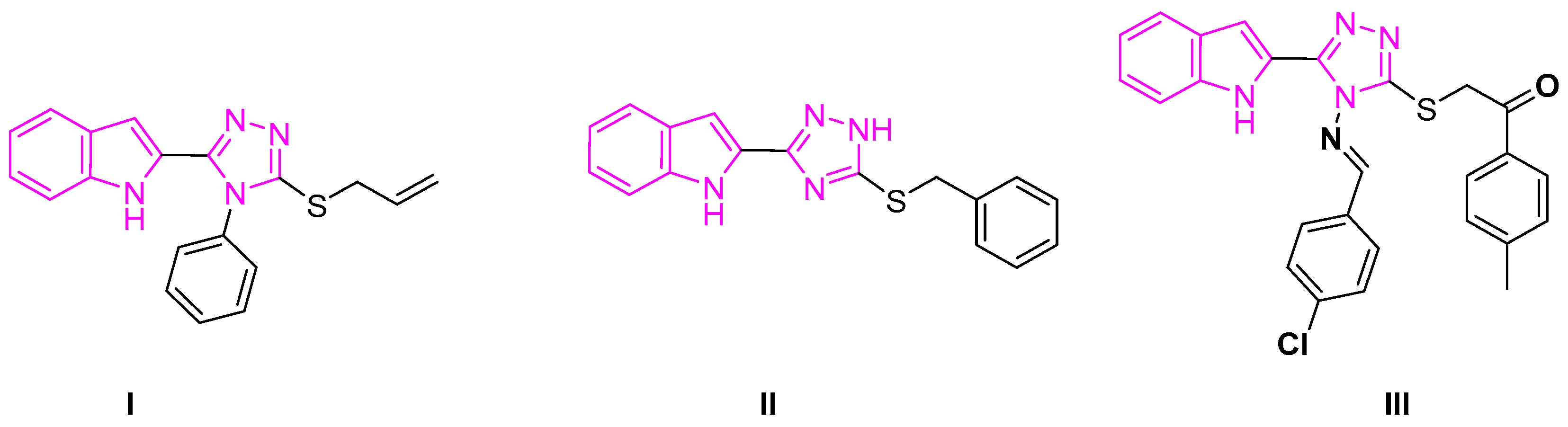
The alkylated indole-triazole system demonstrated strong anti-cancer activities (Figure 2). For instance, 3-(allylsulfanyl)-4-phenyl-5-(1H-indol-2-yl)-1,2,4-triazole I and its derivatives showed promising anti-breast cancer and antiproliferative effects [47]. The anti-proliferative effect of 3-benzylsulfenyl-5-(1H-indol-2-yl)–2H-1,2,4-triazole II against cancer cell lines of MCF-7 and HEPG-2 was encouraging [48]. Indolyl-triazole III and its derivatives showed a strong inhibition of VEGFR-2, which may have anti-renal cancer properties [49].
Building on our ongoing research into novel indole-based anticancer agents, we designed and synthesized a series of new S-alkylated derivatives derived from indole-1,2,4-triazoles. We assumed that the incorporation of S-alkyl groups, which enhance the lipophilicity and cellular permeability of these molecules, would thereby improve their anticancer efficacy. The synthesis strategy involved regioselective S-alkylation of the indole-1,2,4-triazole scaffold under the reaction conditions, which allowed a diverse range of alkyl substituents to be introduced. Structural characterization using 1H and 13C NMR, mass spectrometry, and elemental analysis unequivocally confirmed the successful formation of the target derivatives. The spectroscopic data were fully consistent with the proposed structures, and the elemental analyses were reasonable with the calculated values, thereby verifying both the identity and purity of the synthesized compounds.
Subsequent biological evaluations were then performed against breast cancer cell lines, revealing that several S-alkylated compounds exhibited significant cytotoxicity and induced cell cycle arrest. Notably, the promising derivatives inhibited cell proliferation as well as activated apoptotic pathways, as evidenced by increased caspase activity and modulation of the important key apoptotic markers. Our preliminary results highlight the potential of novel indolyl-1,2,4-triazole-based S-alkylated compounds as effective anticancer agents and warrant further investigation into their structure–activity relationships and in vivo efficacy.

2. Results and Discussion

2.1. Synthesis

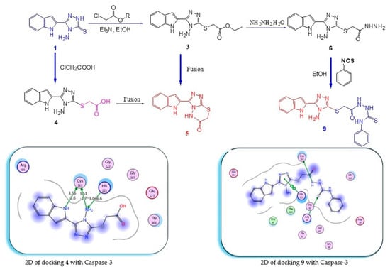
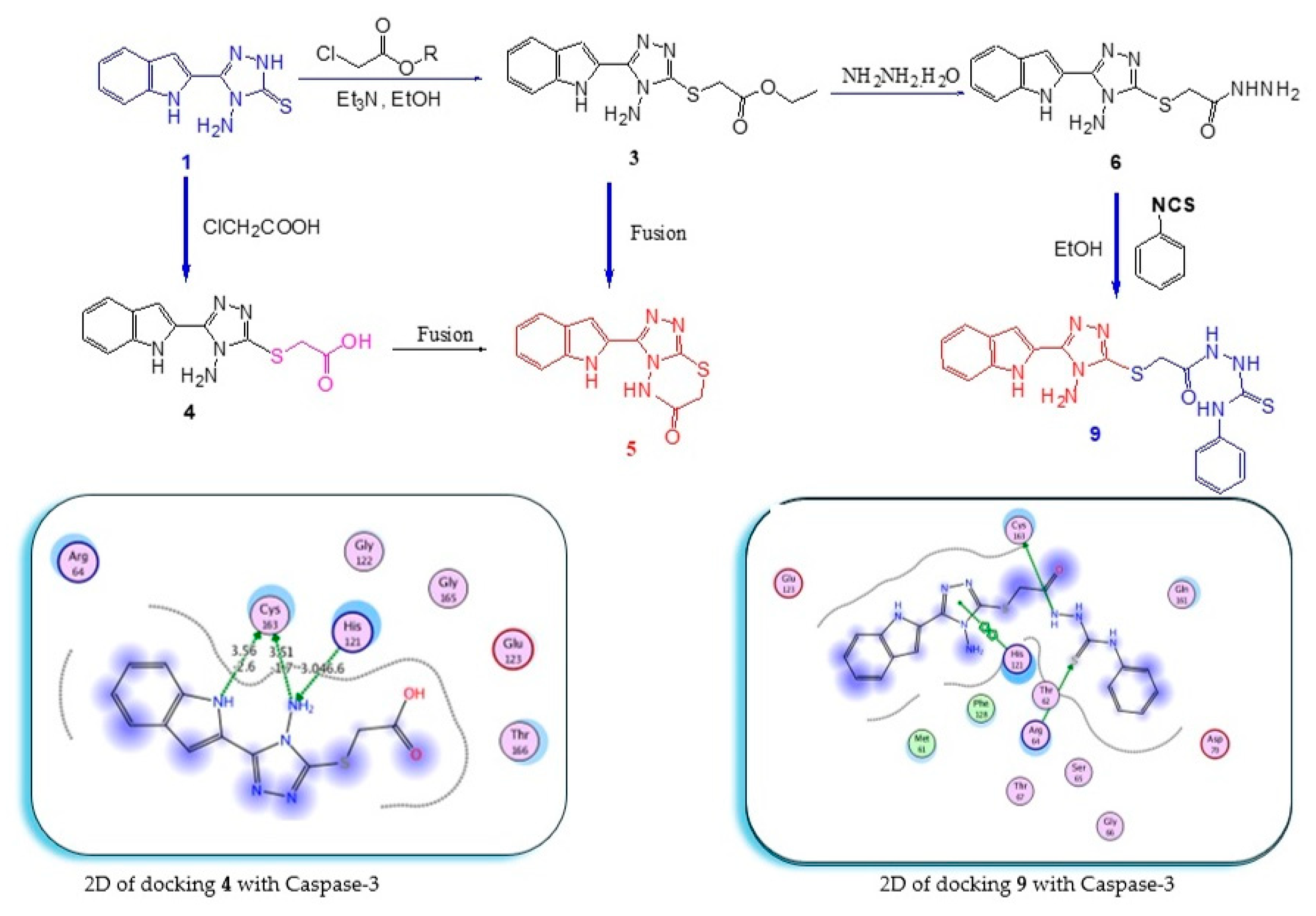
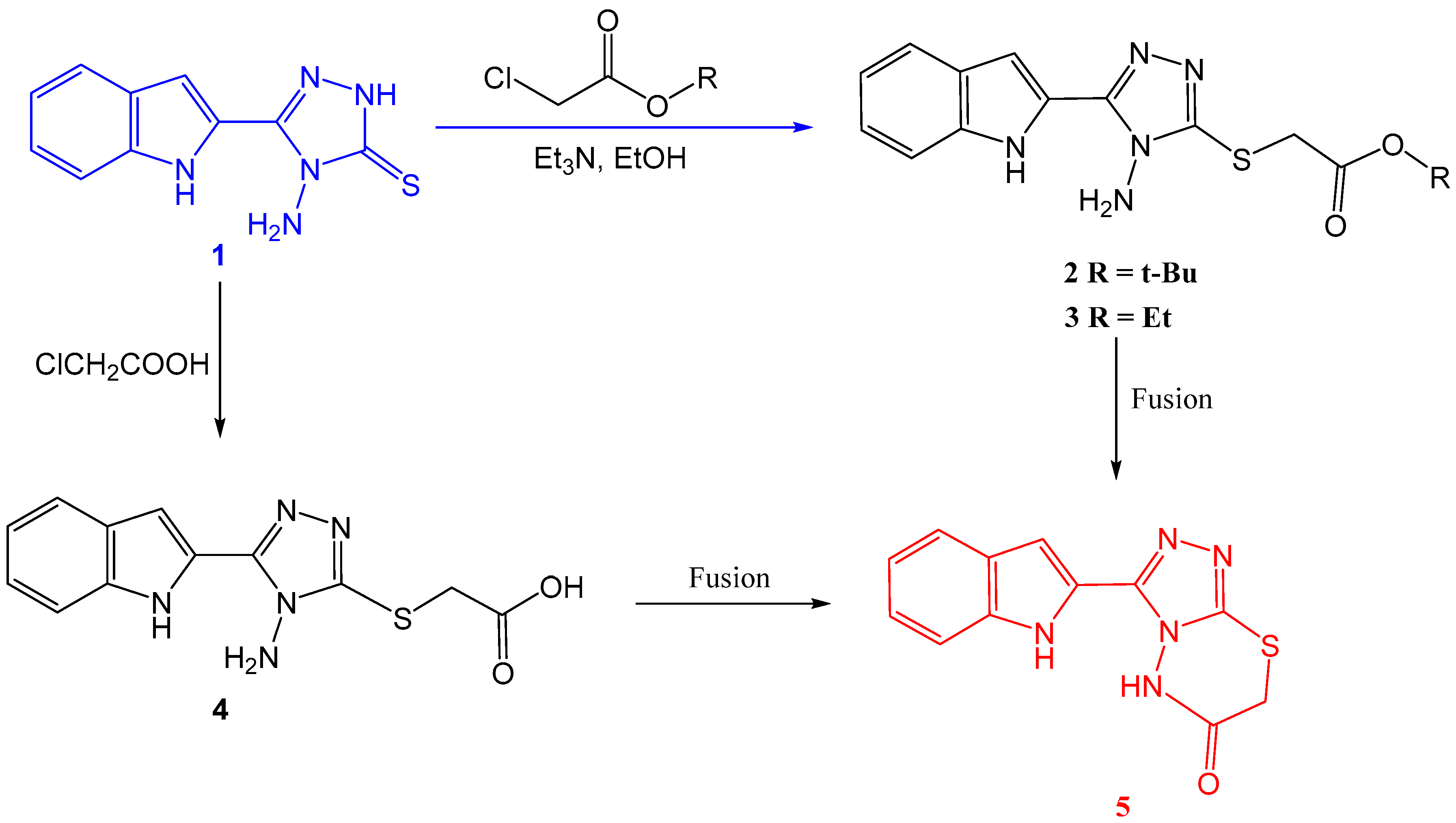
4-Amino-5-(1H-indol-2-yl)-2,4-dihydro-3H-1,2,4-triazole-3-thione 1 was alkylated by tert-butyl bromoacetate or ethyl chloroacetate in the presence of Et3N in ethanol, affording the S-alkylated product 3 [50]. Chloroacetic acid reacted with 1 in the presence of potassium carbonate, affording the acid 4. Fusion of esters 2 and 3, or of the acid 4, for three minutes, afforded 5 in excellent yield (Scheme 1). The structural confirmation of compounds 2, 3, and 5 agreed with the reported data [51]. The NMR of 4 displayed the Thio methylene protons (SCH2) at 4.04 ppm and the respective carbon at 33.45 ppm. Protons of NH2 appeared at 6.28 ppm. Protons of the indole NH and carboxylic group (COOH) showed at 11.72 and 12.87 ppm. The carbonyl carbon of the acid appeared at 169.8 ppm.
The single-crystal analysis of compounds 2, 3, and 5 was reported explicitly in our recent publications [51,52]. The crystal data showed that compounds 2 and 3 crystallized as a triclinic crystal system with primitive P 1 space group, while compound 5 was crystallized in the orthorhombic crystal system and Pbca space group. The unit cell parameters of crystals 2 and 3, respectively, were a = 5.43580(10) Å and 5.9924(2) Å, b = 10.4214(3) Å and 8.7769(3) Å, and c = 14.8371(3) Å and 29.2084(11) Å. Whereas, parameters of the unit cell 5 are a = 13.6036(3) Å, b = 7.8245(3) Å, and c = 21.3279(8) Å.
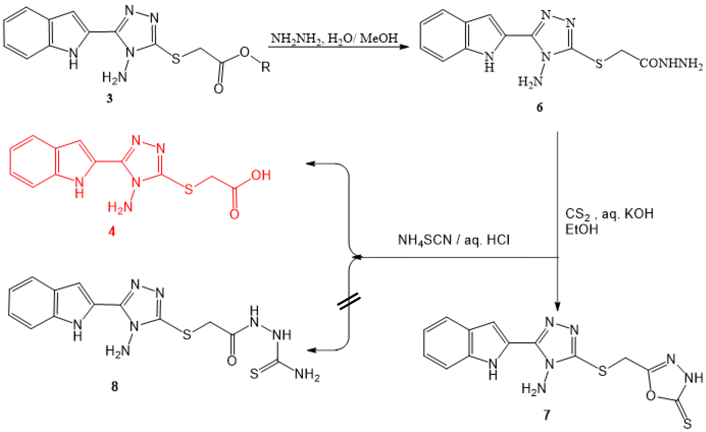
Hydrazide 6 is formed by the reaction of the esters 2 or 3 with NH2NH2·H2O in ethanol. Refluxing hydrazide 6 with ammonium thiocyanate in aqueous hydrochloric acid does not afford the thiosemicarbazide 8; it gives the acid 4. Refluxing the hydrazide 6 with CS2 in ethanol contains aqueous potassium hydroxide furnish 5-(4-Amino-5-(1H-indol-2-yl)-1,2,4-triazol-3-ylthiomethyl)-1,3,4-oxadiazol-2(3H)-thione 7 (Scheme 2). The NMR of 6 displayed the hydrazino-group (–NHNH2) at 4.32 and 9.34 ppm, the NH2 group at 6.28 ppm, and the indole NH protons at 11.74 ppm. A carbon of a carbonyl group appeared at 166.5 ppm [52]. NMR of compound 7 displayed a new proton signal at 14.49 ppm for NH of the new triazole ring. The thiocarbonyl carbon (C=S) of the formed triazoles-thione ring was found at 177.8 ppm.
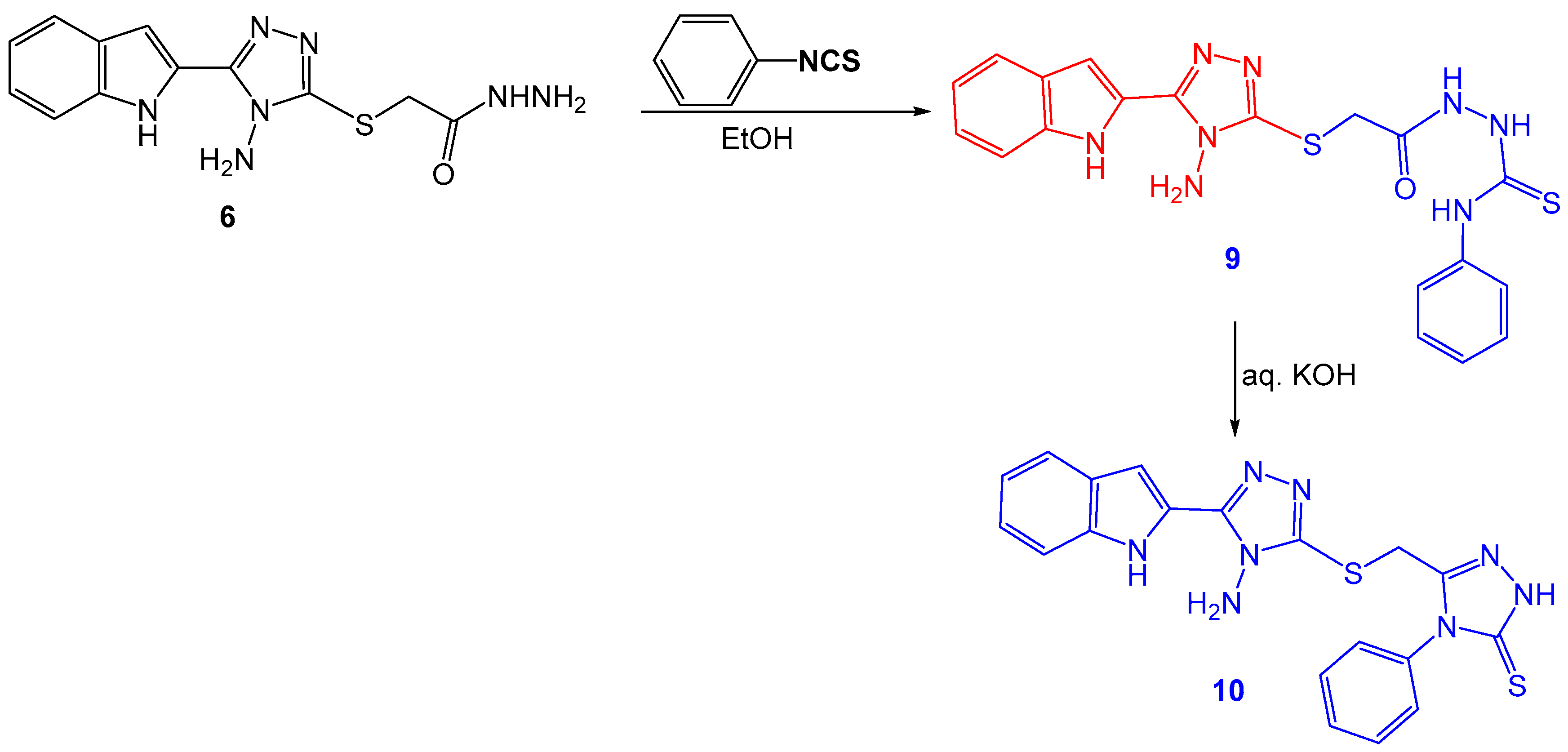
Reaction of phenyl isothiocyanate with hydrazide 6 afforded the thiosemicarbazide 9, which was cyclized to form compound 10, which contains indole and two 1,2,4-triazole rings in one aromatic system (Scheme 3). The thiosemicarbazide 9 NMR displayed the thiosemicarbazide proton signals (3 NH) at 9.34 and 10.37 ppm. The carbonyl carbon (C=O) and thiocarbonyl carbon (C=S) are found at 167.3 and 180.7 ppm. The NMR of the indolyl-triazolyl-triazole-thione 10 revealed the thiomethylene protons at 4.28 ppm, the protons of the amino group (NH2) at 6.23 ppm, and a new NH signal appeared at 13.85 ppm for the formed triazole-thione ring. The thiocarbonyl carbon (C=S) showed at 168.2 ppm.

2.2. Anticancer Activity Simulation Analysis

2.2.1. Docking Analysis

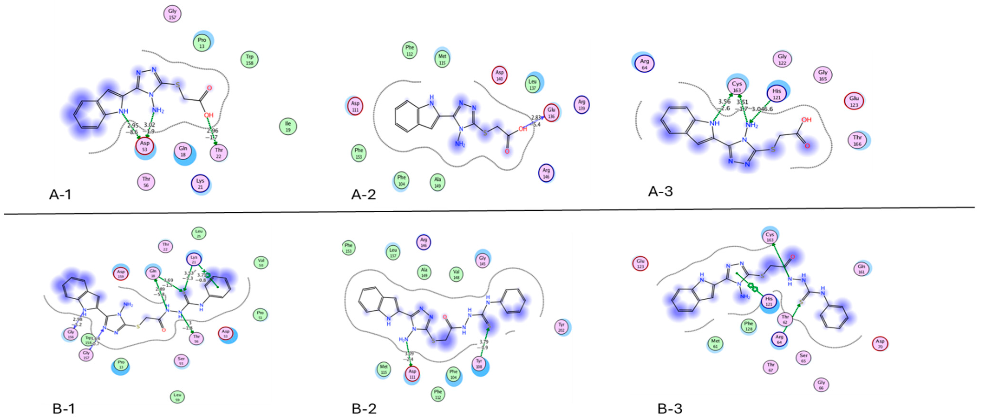
A molecular docking exploration was performed to investigate the purpose of investigating the binding disposition of synthetic chemicals. The docking results show that compounds 4 and 9 have strong binding towards the binding sites of Bax (PDB = 6EB6), Bcl2 (PDB = 6O0K), and Caspase-3 (PDB = 6BDV). The docking interaction of hit compounds 4 and 9 with Bax, Bcl2, and Caspase-3. Inside these figures, A represents the 2D interaction (Figure 3), B represents the 3D interaction, C represents the compound inside the protein pocket, and D represents the 3D compound on the surface of the protein. (Supplementary Figures S24–S29) Interestingly, compounds 4 and 9 form good interactive binding modes with the essential amino acids. The acid 4 interacts with Bax through three hydrogen bonds, two with Asp 53 and one with Thr 22 (bond lengths 2.95, 3.02, and 2.96 Å). The interaction of 4 with Bcl2 through hydrogen bonding with GLU 136 (bond length 2.83 Å). While the interaction of 4 with Caspase-3 is associated with two hydrogen bonds with CYS 163 and one hydrogen bond with HIS 121 (bond lengths 3.56, 3.51, and 3.04 Å). Hit compound 9 interacts with Bax through six hydrogen bonds with GLY 156, GLN 18, THR 56, GLY 157, GLN 18, LYS 21, and LYS 21 (bond lengths: 2.98, 2.89, 3.00, 3.64, 3.69, 3.53, and 3.71 Å, respectively). In this regard, compound 9 displays its interaction with Bcl2, the two hydrogen bonds with ASP 111 and TYR 108 (bond lengths: 3.09 and 3.79 Å). Moreover, compound 9 revealed the interaction with Caspase-3 through four hydrogen bonds with CYS 163, ARG 64, ARG 64, and HIS 121 (bond lengths: 3.67, 3.58, 4.36, and 3.97). As a result, compounds 4, 5, and 9 were nominated for the in vitro anticancer activity investigations.

2.2.2. Cytotoxicity Assay

Using the MTT test, the cytotoxic activity of the synthesized compounds was evaluated against MCF-7 and HepG2 cell lines, revealing significant dose-dependent inhibition of cancer cell viability. Among the tested derivatives, compounds 4, 5, and 9 displayed the highest cytotoxic effects against cells of HepG2 with IC50 values of 0.2715, 0.3167, and 0.4101 μM. Among the tested derivatives, compounds 1, 3, 5, 9, and 10 displayed the highest cytotoxic effects against MCF7 cells, with IC50 values of 0.2128, 0.3843, 0.2373, 0.4994, and 0.2506 μM. The potency of these compounds may be attributed to their enhanced lipophilicity and favorable electronic distribution, allowing for efficient cell membrane penetration and intracellular accumulation.
In Vitro Enzyme Assay (Inhibition%)
Measurement of VEGFR-2 kinase activity
Moreover, compounds 4 and 9 strongly inhibit VEGFR-2 by 97.9% and 96.4% at IC50 0.59 and 1.23 μM, respectively (Table 1). While compounds 1, 5, and 10 have inhibition activity, ranging from 83.9 to 87.8%.
In Vitro Analysis by Flow Cytometry
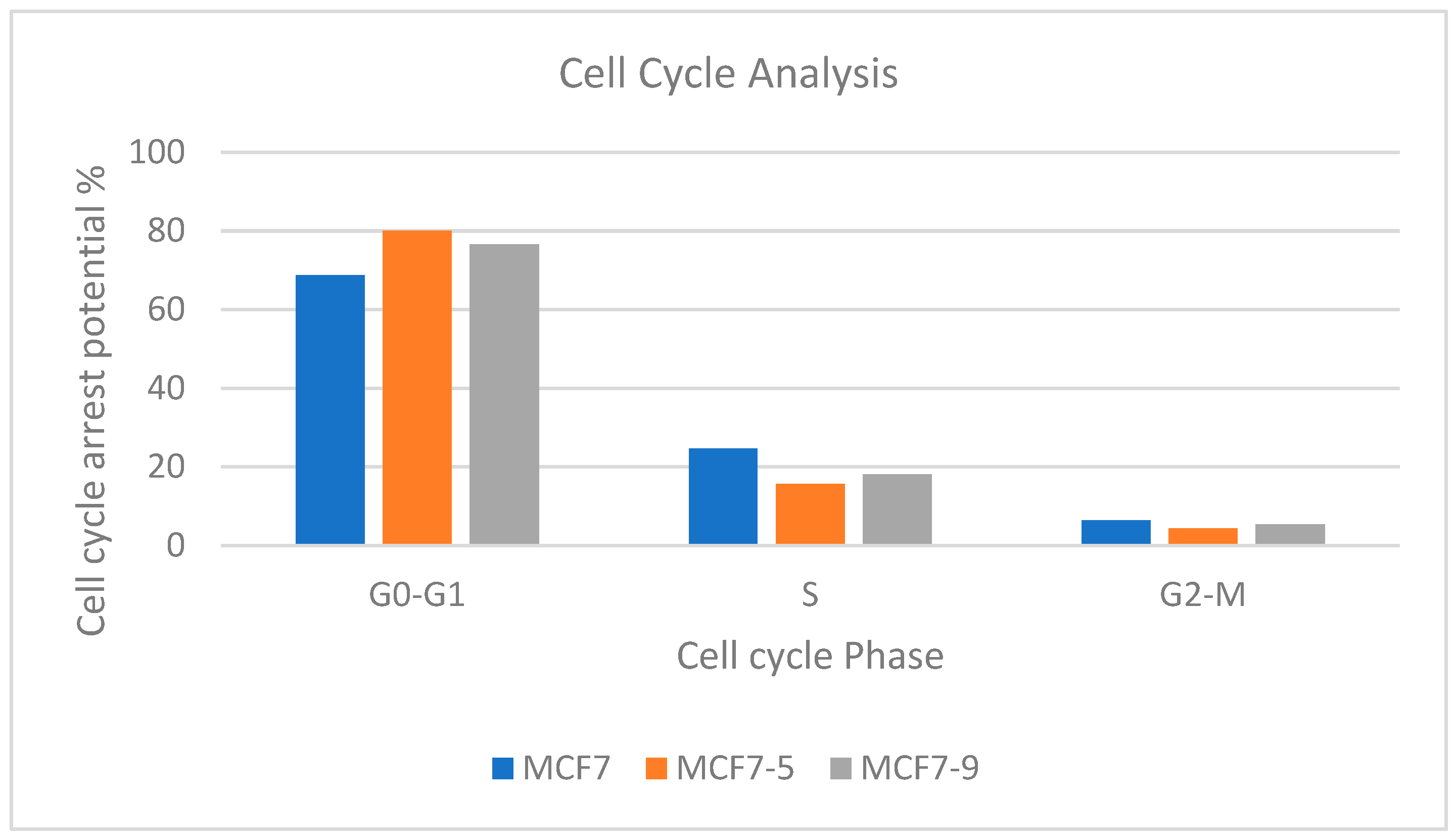
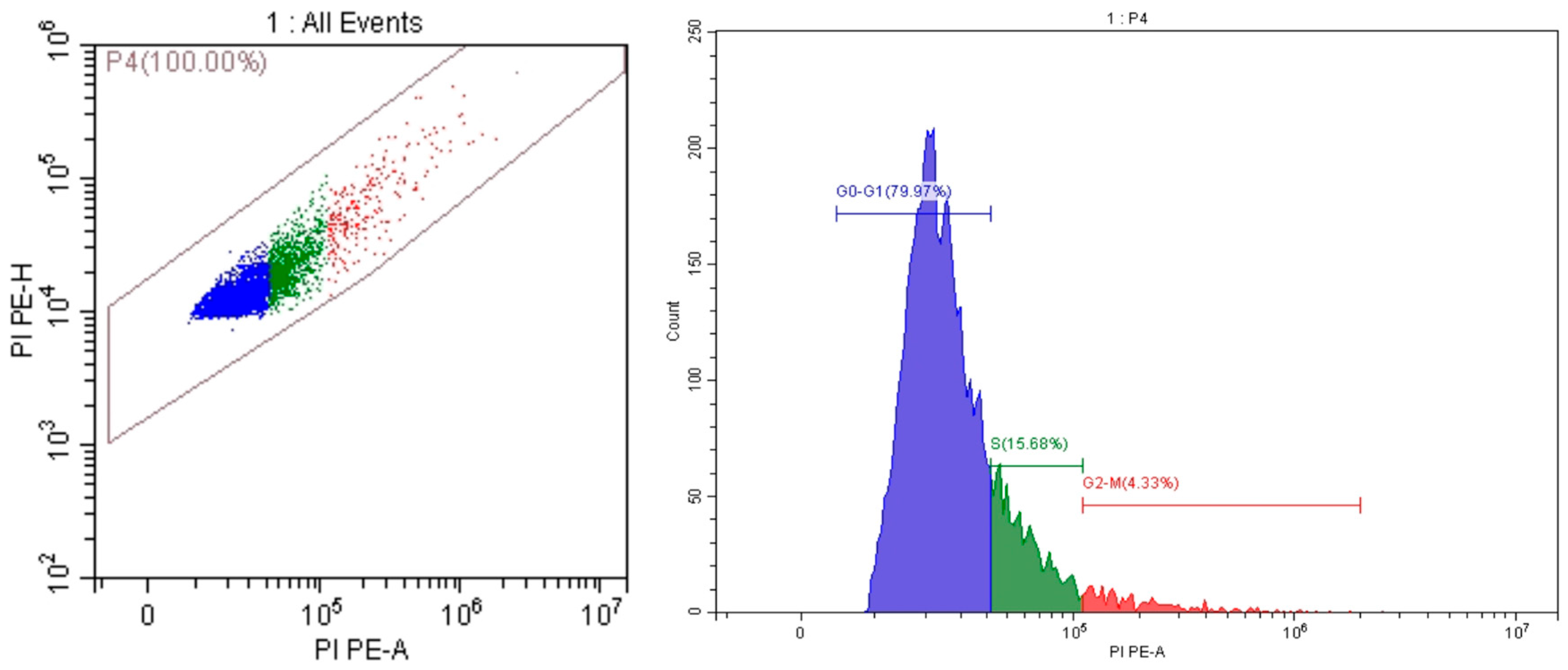
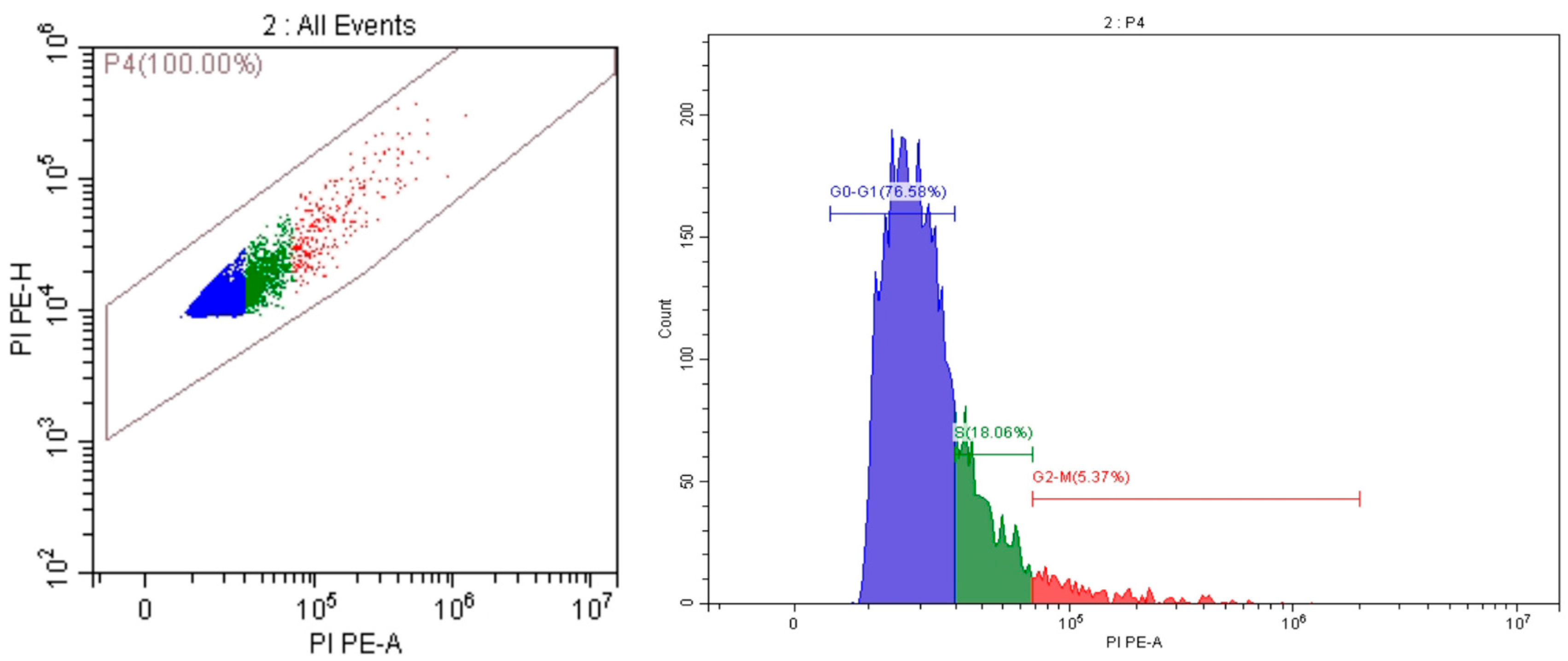
A series of events make up the cell cycle, which includes DNA replication, cell division, and ultimately the production of two daughter cells. To reduce the chance of generic medication side effects when treating cancer, it is attractive to build new anti-proliferative agents that can regulate the progression of the cell cycle and death. Consequently, the most effective anticancer substances (5) and (9), which showed notable activity on the cell line of MCF7, were also chosen for more research into the ability of these compounds to cause cell cycle arrest. Compounds 5 and 9 arrested the cell cycle at the S and G2/M phase, where the cell population levels of p53 wild-type cell lines MCF7 decreased from (24.74–6.48%) for the control to (15.68–4.33%) and (18.06–5.37%) for 5 and 9, respectively. However, the two compounds under investigation showed an extra arrest in the G0–G1 phase of the cell cycle, which raised the two compounds’ cell population levels (79.97, 76.58%), respectively, compared to the control (68.79%) (Figure 4, Table 2).
Table 2 summarizes the distribution of untreated MCF-7 cells and cells treated with compounds 5 and 9 across the G0/G1, S, and G2/M phases. Untreated cells showed 68.79% in G0/G1, 24.74% in the S phase, and 6.48% in G2/M. Treatment with compound 5 increased the G0/G1 population to 79.97% and reduced the S phase and G2/M populations to 15.68% and 4.33%, respectively. Similarly, compound 9 increased the G0/G1 fraction to 76.58%, with S and G2/M fractions of 18.06% and 5.37%. These shifts indicate that both compounds induce cell-cycle arrest, particularly enhancing the accumulation of cells in the G0/G1 phase, a typical indication of antiproliferative activity and the onset of apoptotic processes (as shown in Figure 5 and Figure 6).
The shift towards increased G0/G1-phase accumulation, coupled with the reduction in S and G2/M populations, suggests that the compounds affect cell cycle progression, which may lead to apoptosis and a reduction in overall cell proliferation. This evidence reinforces the potential of these compounds as promising anticancer agents.
Gene expression analysis:
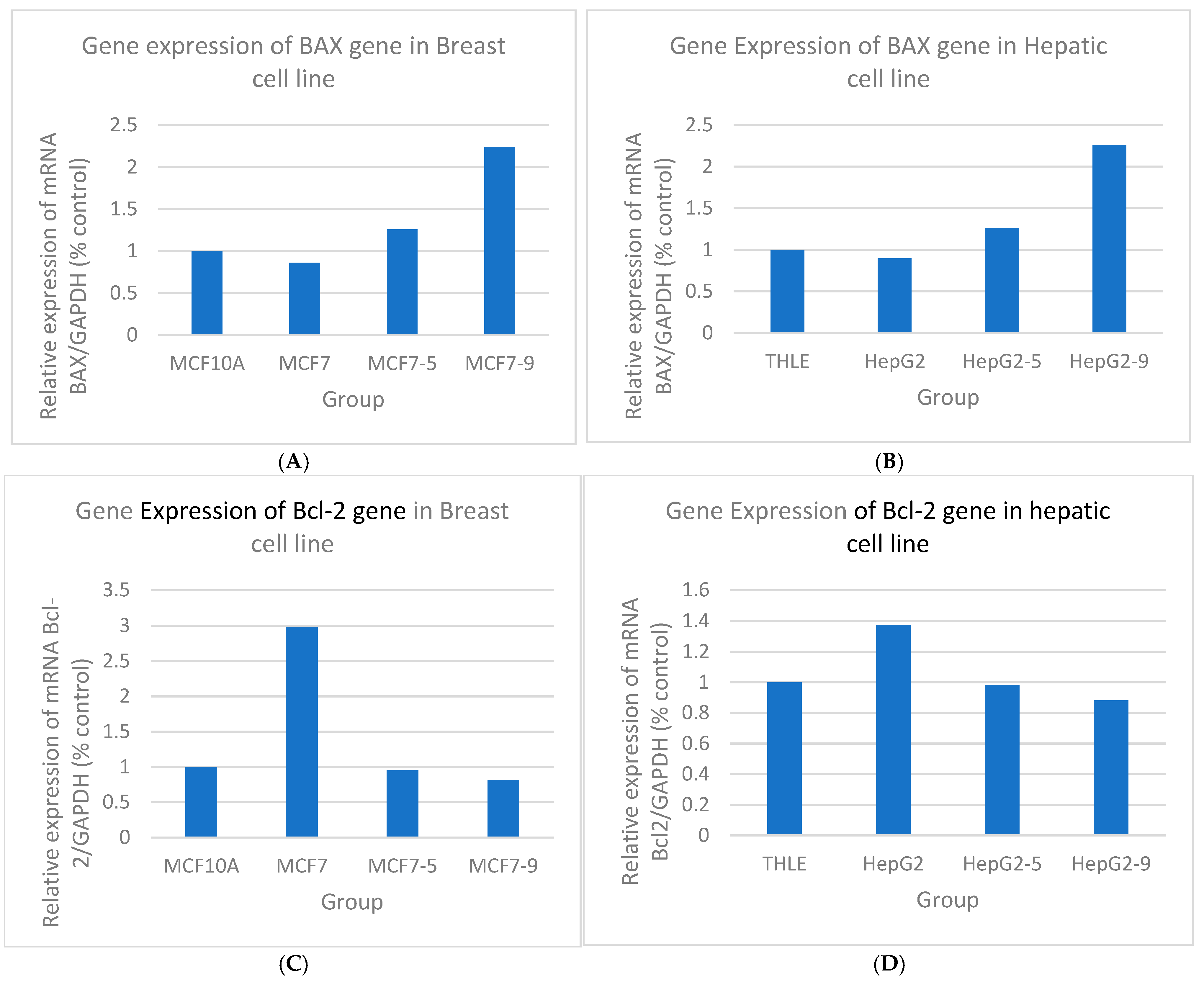
Figure 7 shows the relative mRNA expression of the apoptotic regulators BAX and Bcl-2 in MCF-7 and HepG2 cells after treatment with compounds 5 and 9. Compared with untreated cancer cells, both compounds significantly upregulated BAX and downregulated Bcl-2 (**** p < 0.0001), shifting the balance toward apoptosis.
Further investigation using quantitative PCR (qPCR) corroborated these findings, demonstrating that not only do compounds 5 and 9 restore BAX expression, but compound 4 similarly enhances the expression of this pro-apoptotic gene, while all treatments lead to a significant decrease in Bcl-2 expression. These molecular alterations suggest that these compounds exert their anticancer effects by directly influencing the intrinsic (mitochondrial) pathway of apoptosis. It is postulated that the structural features of these compounds enable strong hydrogen-bonding interactions with apoptotic regulatory proteins, thereby facilitating conformational changes that trigger mitochondrial outer membrane permeabilization.
The resultant release of cytochrome c from the mitochondria would then activate downstream Caspases, culminating in programmed cell death. This mechanism of action is particularly critical in drug-resistant malignancies, where evasion of apoptosis is a common survival strategy. By restoring the balance between pro- and anti-apoptotic factors, compounds 5, 9, and 4 not only promote cell death but also potentially overcome the resistance mechanisms that allow cancer cells to evade conventional therapies.
Thus, the ability of these compounds to modulate apoptosis underscores their potential as effective anticancer agents. Their action in reestablishing apoptotic sensitivity in cancer cells could be pivotal in the development of new therapeutic strategies aimed at targeting resistant tumors, reinforcing the notion that the modulation of apoptotic pathways is a defining characteristic of promising anticancer compounds.

3. Materials and Methods

3.1. Materials

All reagents and solvents were purchased from Sigma-Aldrich (St. Louis, MO, USA) and/or Al-Nasr Co. (Giza, Egypt) and used without further purification unless otherwise stated. Reaction progress was monitored by thin-layer chromatography (TLC) on silica gel 60 F254 plates and visualized under UV light (254/365 nm). Melting points were determined on an SMP10 melting point apparatus and are uncorrected. 1H and 13C NMR spectra were recorded on Bruker spectrometers (frequencies as indicated) in DMSO-d6 or CDCl3; chemical shifts (δ) are reported in ppm relative to residual solvent signals. Elemental analyses (C, H, N) were performed using a Flash EA-1112 analyzer (Thermo Fisher Scientific, Rodano, Italy). Mass spectra (EI/HRMS) were recorded as indicated in the compound characterization. Unless otherwise noted, products were purified by filtration and/or recrystallization from DMF/ethanol.

3.2. Experimental

3.2.1. Synthesis of Tert-Butyl 2-((4-Amino-5-(1H-indol-2-yl)-4H-1,2,4-triazol-3-yl)thio)acetate 2 and Ethyl 2-((4-Amino-5-(1H-indol-2-yl)-4H-1,2,4-triazol-3-yl)thio)acetate 3

Compounds 2 and 3 were synthesized as reported in our recent work [50]. Briefly, a mixture of 4-amino-5-(1H-indol-2-yl)-1,2,4-triazol-3(2H)-thione 1 (1.0 mmol) and triethylamine (1.2 mmol) in absolute ethanol was stirred at room temperature for 1 h. The appropriate halo ester (1.1 mmol) was then added, and the reaction mixture was stirred at room temperature overnight. After completion, ethanol was removed under vacuum using a rotary evaporator, and cold water (100 mL) was added to the residue. The resulting precipitate was collected by filtration, washed with water, and air-dried at room temperature. The crude product was purified by recrystallization from ethanol to afford compounds 2 and 3 as pure crystalline solids.

3.2.2. Synthesis of 4-Amino-3-(carboxymethylsulfanyl)-5-(1H-indol-2-yl)-1,2,4-triazole 4

A mixture of compound 1 (1.0 mmol) and K2CO3 (1.2 mmol) in ethanol (10 mL) was stirred at room temperature for 1 h. Chloroacetic acid (1.2 mmol) was added, and the reaction mixture was refluxed for 3 h. After cooling to room temperature, the solvent was removed under vacuum using a rotary evaporator. Ice water (50 mL) was added to the residue, and the mixture was acidified with 10% (v/v) aqueous HCl until the pH reached 3. The resulting precipitate was collected by filtration, washed with water, and recrystallized from DMF/EtOH to give compound 4. Yield: 78%, m.p. 240–241 °C. 1H NMR (DMSO-d6, 300 MHz) δ 4.04 (SCH2), 6.28 (s, 2 H, NH2, D2O exchangeable), 7.03 (dd, 1 H, J = 7.8, J = 7.5 Hz), 7.16 (dd, 1 H, J = 7.5, J = 8.1 Hz), 7.28 (d, 1 H, J = 1.5 Hz), 7.45 (d, 1 H, J = 8.1 Hz), 7.61 (d, 1 H, J = 7.8 Hz), 11.72 (br. s, 1 H, NH, D2O exchangeable), 12.87 (br. s, 1 H, COOH, D2O exchangeable); 13C NMR (DMSO-d6, 100 MHz) δ 33.5 (SCH2), 102.3, 111.9, 119.6, 120.7, 122.8, 123.9, 127.6, 136.5, 149.4, 152.9, 169.8 (C=O); HRMS (FAB + ve) calcd for C12H12N5O2S (M + 1): 290.0712 Found: 290.0690.

3.2.3. Synthesis of the 3-(1H-Indol-2-yl)-7H-[1,2,4]triazolo [3,4-b][1,3,4]thiadiazin-6-ol 5

Previously [51], compound 5 was obtained in 81% yield from reaction of 4-amino-5-(1H-indol-2-yl)-1,2,4-triazol-3(2H)-thione 1 (1.0 mmol) and ethyl chloroacetate or tert-butyl bromoacetate (1.2 mmol) in ethanol (10 mL) in the presence of K2CO3 (1.2 mmol) as base (Scheme 1), and single-crystal X-ray diffraction analysis showed that it was present in the enol form in the solid state.
In the present work, the alkylated esters (2 or 3) or the acid (4) (1.0 mmol) were fused on a hotplate in open air at atmospheric pressure for 3 min. After cooling to room temperature, the crude product was purified by recrystallization from DMF/ethanol to afford compound 5 as crystals. Yield: >92%; m.p. 291–292 °C. Full 1H and 13C NMR data are provided in the Supplementary Materials.

3.2.4. Synthesis of 2-((4-Amino-5-(1H-indol-2-yl)-4H-1,2,4-triazol-3-yl)thio)acetohydrazide 6

Compound 6 was synthesized according to our previously reported protocol [52], in which all characterization data and analytical details have been described. Briefly, ethyl 2-((4-amino-5-(1H-indol-2-yl)-4H-1,2,4-triazol-3-yl)thio)acetate 3 (2.0 mmol) was treated with hydrazine hydrate (3 mL) and refluxed for 1 h. After completion, excess solvent and unreacted hydrazine were removed under vacuum using a rotary evaporator, and the crude product was crystallized from ethanol to afford compound 6 as a pure solid. Yield: 87%; m.p. 248–250 °C. Full 1H and 13C NMR data are provided in the Supplementary Materials.

3.2.5. 5-(4-Amino-5-(1H-indol-2-yl)-1,2,4-triazol-3-ylthiomethyl)-1,3,4-oxadiazol-2(3H)-thione 7

Hydrazide 6 (2.0 mmol), carbon disulfide (5.0 mmol) and aqueous KOH (4.0 mmol) were refluxed in ethanol (30 mL) for 4 h. After cooling, the reaction mixture was concentrated by evaporation to approximately half of its original volume Ice water (50 mL) was added, and the mixture was acidified with 10% (v/v) aqueous HCl until pH equals 3. The precipitate was collected by filtration and recrystallized from DMF/EtOH to form and purify compound 7. Yield: 66%, m.p. 218–220 °C. 1H NMR (DMSO-d6, 300 MHz) δ 4.55 (s, 2 H, SCH2), 6.20 (br., 2 H, NH2), 7.04 (dd, 1 H, J = 7.8, J = 7.2 Hz), 7.17 (dd, 1 H, J = 7.2, J = 8.1 Hz), 7.31 (s, 1 H), 7.46 (d, 1 H, J = 8.1 Hz), 7.62 (d, 1 H, J = 7.8 Hz), 11.78 (br.s, 1 H, NH), 14.49 (br.s, H, NH); 13C NMR (DMSO-d6, 100 MHz) δ 25.1 (SCH2), 102.8, 111.9, 119.7, 120.9, 123.0 123.5, 127.1, 136.6, 149.7, 151.2, 160.7, 177.9 (C=S); HRMS (EI) calcd for C13H10N7OS2 (M+): 344.0388 Found: 344.0395.

3.2.6. Synthesis of 1-(4-Amino-5-(1H-indol-2-yl)-1,2,4-triazol-3-ylthioacety1)-4-phenylthiosemicarbazide 9

Hydrazide 6 (2.0 mmol) was refluxed with phenyl isothiocyanate (2.0 mmol) in ethanol (10 mL) for 3 h. After cooling to room temperature, the precipitate was collected by filtration and recrystallized from DMF/EtOH to afford compound 9. Yield: 92%, m.p. 227–228 °C. 1H NMR (DMSO-d6, 300 MHz) δ 3.87 (s, 2 H, SCH2), 6.28 (s, 2 H, NH2), 7.01–7.60 (m, 10 H), 9.34 (2br.s, 2 H, NHNH), 10.37 (br. s, 1 H, NH), 11.67 (br.s, 1 H, NH); 13C NMR (DMSO-d6, 100 MHz) δ 34.1 (SCH2), 102.6, 111.9, 119.7, 120.8, 122.9, 123.8, 125.1, 125.6, 127.5, 128.0, 136.5, 138.9, 149.3, 153.0, 167.4 (C=O), 180.72 (C=S); HRMS (FAB -ve) calcd for C19H17N8S2O (M-1): 437.0967 Found: 437.0953.

3.2.7. Synthesis of 4-Amino-5-(1H-indol-2-yl)-3-(4-phenyl-3-thioxo-1,2,4-triazol-5-ylmethylsulfanyl)-1,2,4-triazole 10

A mixture of thiosemicarbazide 9 (2.0 mmol) in aqueous KOH (4.0 N, 50 mL) was refluxed for 4 h, then allowed to cool to room temperature. The mixture was acidified with HCl (10%) until pH 3, and the formed precipitate was collected by filtration and recrystallized from DMF/EtOH to form compound 10. Yield: 71%, m.p. 253–255 °C. 1H NMR (DMSO-d6, 300 MHz) δ 4.28 (s, 2 H, SCH2), 6.23 (s, 2 H, NH2), 7.03 (dd, 1 H, J = 7.8, J = 7.2 Hz), 7.17 (dd, 1 H, J = 7.2, J = 8.1 Hz), 7.28 (s, 1 H), 7.45 (d, 1 H, J = 8.1 Hz), 7.50–7.57 (m, 5 H), 7.61 (d, 1 H, J = 7.8 Hz), 11.75 (br.s, 1 H, NH), 13.85 (br. s, 1 H, NH); 13C NMR (DMSO-d6, 75 MHz) δ 26.0 (SCH2), 102.5, 111.9, 119.7, 120.8, 122.9, 123.8, 127.6, 128.4, 129.4, 129.6, 133.3, 136.5, 148.7, 149.5, 151.2, 168.2 (C=S); HRMS (FAB -ve) calcd for C19H15N8S2 (M+): 419.0861 Found: 419.0899.

3.3. Anticancer Activity

3.3.1. Reagents and Chemicals

RPMI-1640 medium, fetal bovine serum (FBS), trypsin, and phosphate-buffered saline (PBS) were obtained from ICN Biomedicals Inc. (Aurora, OH, USA). Penicillin/streptomycin was purchased from Invitrogen (San Diego, CA, USA). The MTT reagent (CAS 298-93-1) was obtained from Yunbang Pharma (Changsha, Hunan, China). Doxorubicin was purchased from EBEWE Pharma (Unterach, Austria). Dimethyl sulfoxide (DMSO) was obtained from Merck (Darmstadt, Germany).

3.3.2. Cell Culture

Human HepG2 (liver cancer) and MCF-7 (breast cancer) cell lines were obtained from the Al-Azhar Virology Research Center (Faculty of Medicine, Cairo, Egypt). Cells were maintained in RPMI-1640 supplemented with 10% FBS and penicillin/streptomycin (100 U/mL and 100 µg/mL, respectively) and cultured as adherent monolayers at 37 °C in a humidified atmosphere containing 5% CO2.

3.3.3. Cell Viability (MTT) Assay

Cell viability was evaluated using the MTT reduction assay [53]. HepG2 and MCF-7 cells were seeded in 96-well plates (8000–10,000 cells/well; 100 µL complete medium) and allowed to attach overnight. The medium was then replaced with serum-free medium containing serial dilutions of the test compounds (0.195–25 µM; 1:2 dilution series). Stock solutions were prepared in DMSO, and the final DMSO concentration in all wells was kept below 0.5% (v/v). Control wells received vehicle only, and blank wells contained medium without cells for background subtraction.
After 24 h treatment, the medium was removed, and MTT reagent was added (0.5 mg/mL final concentration), followed by incubation for 4 h at 37 °C. Formazan crystals were dissolved in DMSO (150 µL/well), and absorbance was measured at 570 nm (reference 655 nm) using a STAT-Fax 2100 microplate reader (Awareness Technology, Inc., Palm City, FL, USA). Percentage cell viability was calculated relative to vehicle-treated controls. IC50 values were determined by nonlinear regression using GraphPad Prism (version 9). All experiments were performed in triplicate (n = 3), and data are reported as mean ± SD.
Assessment of In Vitro VEGFR-2 Inhibitory Activity
VEGFR-2 kinase inhibitory activity was assessed using a commercial VEGFR-2 kinase assay kit (Bioscience) according to the manufacturer’s instructions. Briefly, recombinant VEGFR-2 was incubated with the supplied substrate/ATP mixture in the presence of serial dilutions of the test compounds. Enzymatic control wells contained enzyme with inhibitor buffer only (no inhibitor), and blank wells contained all reagents except enzyme. Residual ATP was quantified using Kinase-Glo® MAX (Promega, Madison, WI, USA), and luminescence was recorded using a microplate reader. Percentage inhibition was calculated relative to the enzymatic control, and IC50 values were obtained from concentration–response curves (n = 3). Sunitinib was included as a reference inhibitor.
In Vitro Cell Cycle Analysis by Flow Cytometry
Cell cycle distribution was analyzed in MCF-7 cells after treatment with compounds 5 and 9. Following 24 h exposure, approximately 1 × 10⁶ cells were harvested by trypsinization, washed twice with ice-cold PBS (pH 7.4), and fixed in 60% ice-cold ethanol at 4 °C for 1 h. Fixed cells were washed with PBS and resuspended in PBS containing RNase A (50 µg/mL) and propidium iodide (PI; 10 µg/mL). After incubation in the dark for 30 min at 15–25 °C, samples were analyzed using a CytoFLEX™ flow cytometer (Beckman Coulter, Inc., Brea, CA, USA) with excitation/emission at 535/617 nm (FL2 channel). A minimum of 20,000 events was collected per sample, and cell-cycle distribution was calculated using Cytexpert™ v2.4.0.28 software.
Real-Time Quantitative PCR (RT-qPCR) Analysis
Total RNA was extracted from untreated and treated MCF-7 and HepG2 cells using QIAzol Lysis Reagent (QIAGEN, Hilden, Germany) according to the manufacturer’s protocol, and RNA concentration/purity were assessed using a DENOvix UV spectrophotometer (DeNovix Inc., Wilmington, DE, USA). First-strand cDNA was synthesized from equal amounts of RNA using the HiSenScript™ RH (-) cDNA Synthesis Kit (iNtRON Biotechnology, Seongnam, Republic of Korea). Quantitative real-time PCR (RT-qPCR) was performed on a CFX96 Deep Well Real-Time PCR System Bio-Rad Laboratories, Hercules, CA, USA). Primer sequences for BAX, Bcl-2 and GAPDH are listed in Table 3. Relative gene expression was calculated using the 2−ΔΔCt method and normalized to GAPDH. Each condition was analyzed in triplicate (n = 3).

3.3.4. Statistical Analysis

All quantitative data are presented as mean ± SD of three replicate measurements (n = 3) unless otherwise stated. IC50 values were obtained by nonlinear regression (four-parameter logistic model) using GraphPad Prism (version 9). For multi-group comparisons (e.g., RT-qPCR), statistical significance was assessed using one-way ANOVA followed by Tukey’s multiple comparisons test. A p-value < 0.05 was considered statistically significant.

4. Conclusions

A series of novel heterocyclic compounds incorporating indole, 1,2,4-triazole, thiadiazine, and 1,3,4-oxadiazole motifs was synthesized and evaluated for in vitro anticancer activity against MCF-7 and HepG2 cells. Several compounds showed low-micromolar cytotoxicity, induced cell-cycle perturbation, and modulated apoptotic markers (BAX upregulation and Bcl-2 downregulation). In addition, selected derivatives inhibited VEGFR-2, supporting a dual apoptosis/antiangiogenic profile. These findings warrant further optimization and mechanistic studies to assess their potential as anticancer leads. Structural analysis suggests that the unique arrangement of heterocyclic moieties facilitates strong hydrogen-bonding interactions with key regulatory proteins, thereby stabilizing conformational changes that favor apoptosis and impede survival signaling pathways. Such interactions likely contribute to the compounds’ ability to target the mitochondrial pathway, leading to the release of cytochrome c and subsequent activation of caspase cascades. The multifaceted mechanism of action of these heterocyclic scaffolds underscores their potential as next-generation anticancer agents. Their ability to simultaneously trigger intrinsic apoptotic pathways, modulate gene expression, and inhibit angiogenic processes presents a compelling therapeutic strategy for overcoming the limitations of current treatments, particularly in cases of drug-resistant cancers. These findings provide a strong rationale for further preclinical studies and optimization to translate these compounds into clinically viable therapies that could significantly improve outcomes for patients battling aggressive, resistant tumors.

Supplementary Materials

The following supporting information can be downloaded at: https://www.mdpi.com/article/10.3390/ph19030418/s1, Figures S1–S6: 1H NMR, 13C NMR, DEPT, COSY, HMQC, and EIMS of compound 4; Figures S7 and S8: 1H NMR and 13C NMR of compound 5; Figures S9–S11: 1H NMR, 13C NMR, and DEPT135 NMR of compound 6; Figures S12–S15: 1H NMR, 13C NMR, DEPT135 NMR, and HMQC NMR of compound 7; Figures S16–S19: 1H NMR, 13C NMR, DEPT, and HMQC of compound 9; Figures S20–S23: 1H NMR, 13C NMR, DEPT, and HMQC of compound 10; Figures S24–S29: docking interactions of compound 4 with Bax, Bcl2, and Caspase-3 and of compound 9 with Bax, Bcl2, and Caspase-3 (interaction views include: B = 3D interaction, C = compound inside the protein pocket, D = compound on the protein surface); Figures S30–S32: HepG2 IC50, MCF-7 IC50, and VEGFR-2 IC50 plots; Figures S33–S35: representative SDS-PAGE gel used prior to immunoblotting and uncropped western blots for BAX (20 kDa) and Bcl-2 (26 kDa) expression in control breast cells, untreated MCF-7 cells, and MCF-7 cells treated with compounds 5 and 9; Tables S1–S3: HepG2, MCF-7, and VEGFR-2 IC50 values (triplicate measurements) with calculated mean ± SD (n = 3).

Author Contributions

Conceptualization, E.E.S., A.T.A.B. and A.A.M.S.; Data curation, E.E.S., A.A.A., A.T.A.B., N.I.A. and H.K.; Formal analysis, S.E. and H.K.; Investigation, E.E.S., S.A., A.T.A.B., N.I.A. and A.A.M.S.; Methodology, E.E.S., A.A.A., A.T.A.B. and A.A.M.S.; Software, S.A. and K.S.E.-N.; Visualization, E.E.S., A.T.A.B. and N.I.A.; funding acquisition: E.E.S.; Project administration: E.E.S.; Supervision: E.E.S.; writing—original draft, E.E.S., H.K., S.A., A.T.A.B., N.I.A., K.S.E.-N. and S.E.; Data validation H.K.; and writing review and editing, E.E.S., A.A.A., A.T.A.B., A.A.M.S. and H.K. All authors have read and agreed to the published version of the manuscript.

Funding

This work was funded by the Deanship of Graduate Studies and Scientific Research at Jouf University under grant No. (DGSSR-2023-02-02475).

Institutional Review Board Statement

Not applicable.

Informed Consent Statement

Not applicable.

Data Availability Statement

The original contributions presented in this study are included in the article/Supplementary Material. Further inquiries can be directed to the corresponding author.

Conflicts of Interest

The authors declare no conflicts of interest.

References

  1. Sashidhara, K.V.; Kumar, A.; Kumar, M.; Sarkar, J.; Sinha, S. Synthesis and in vitro evaluation of novel coumarin–chalcone hybrids as potential anticancer agents. Bioorg. Med. Chem. Lett. 2010, 20, 7205–7211. [Google Scholar] [CrossRef] [PubMed]
  2. El-Shershaby, M.H.; Ghiaty, A.; Bayoumi, A.H.; Ahmed, H.E.; El-Zoghbi, M.S.; El-Adl, K.; Abulkhair, H.S. 1,2,4-Triazolo [4,3-c] quinazolines: A bioisosterism-guided approach towards the development of novel PCAF inhibitors with potential anticancer activity. New J. Chem. 2021, 45, 11136–11152. [Google Scholar] [CrossRef]
  3. Siegel, R.L.; Miller, K.D.; Wagle, N.S.; Jemal, A. Cancer statistics, 2023. CA Cancer J. Clin. 2023, 73, 17–48. [Google Scholar] [CrossRef]
  4. Aboshouk, D.R.; Youssef, M.A.; Bekheit, M.S.; Hamed, A.R.; Girgis, A.S. Antineoplastic indole-containing compounds with potential VEGFR inhibitory properties. RSC Adv. 2024, 14, 5690–5728. [Google Scholar] [CrossRef]
  5. International Agency for Research on Cancer, Cancer Tomorrow. Available online: https://gco.iarc.who.int/tomorrow/en (accessed on 30 May 2024).
  6. Rahib, L.; Wehner, M.R.; Matrisian, L.M.; Nead, K.T. Estimated projection of US cancer incidence and death to 2040. JAMA Netw. Open 2021, 4, e214708. [Google Scholar] [CrossRef]
  7. Bray, F.; Laversanne, M.; Weiderpass, E.; Soerjomataram, I. The ever-increasing importance of cancer as a leading cause of premature death worldwide. Cancer 2021, 127, 3029–3030. [Google Scholar] [CrossRef] [PubMed]
  8. Kelley, K.D.; Aronowitz, P. Cancer. Med. Clin. N. Am. 2022, 106, 411–422. [Google Scholar] [CrossRef]
  9. Hassan, S.M.; Farid, A.; Panda, S.S.; Bekheit, M.S.; Dinkins, H.; Fayad, W.; Girgis, A.S. Indole Compounds in Oncology: Therapeutic Potential and Mechanistic Insights. Pharmaceuticals 2024, 17, 922. [Google Scholar] [CrossRef]
  10. McPherson, K.; Steel, C.M.; Dixon, J.M. ABC of breast diseases. Breast cancerepidemiology, risk factors, and genetics. BMJ 2000, 321, 624–628. [Google Scholar] [CrossRef]
  11. DeSantis, C.; Bray, F.; Ferlay, J.; Lortet-Tieulent, J.; Anderson, B.O.; Jemal, A. International Variation in Female Breast Cancer Incidence and Mortality Rates. Cancer Epidemiol. Biomark. Prev. 2015, 24, 1495–1506. [Google Scholar] [CrossRef] [PubMed]
  12. Roaiah, H.M.; Ghannam, I.A.Y.; Ali, I.H.; El Kerdawy, A.M.; Ali, M.M.; Abbas, S.S.; El-Nakkady, S.S. Design, synthesis, and molecular docking of novel indole scaffoldbased VEGFR-2 inhibitors as targeted anticancer agents. Arch. Pharm. 2018, 351, 1700299. [Google Scholar] [CrossRef]
  13. Takahashi, S. Vascular endothelial growth factor (VEGF), VEGF receptors and their inhibitors for antiangiogenic tumor therapy. Biol. Pharm. Bull. 2011, 34, 1785–1788. [Google Scholar] [CrossRef]
  14. Liang, Y.; Brekken, R.A.; Hyder, S.M. Vascular endothelial growth factor induces proliferation of breast cancer cells and inhibits the antiproliferative activity of antihormones. Endocr. Relat. Cancer 2006, 13, 905–919. [Google Scholar] [CrossRef]
  15. Shibuya, M. Vascular Endothelial Growth Factor (VEGF) and Its Receptor (VEGFR) Signaling in Angiogenesis: A Crucial Target for Anti- and Pro-Angiogenic Therapies. Genes Cancer 2011, 2, 1097–1105. [Google Scholar] [CrossRef]
  16. Guo, S.; Colbert, L.S.; Fuller, M.; Zhang, Y.; Gonzalez-Perez, R.R. Vascular endothelial growth factor receptor-2 in breast cancer. Biochim. Biophys. Acta BBA Rev. Cancer 2010, 1806, 108–121. [Google Scholar] [CrossRef] [PubMed]
  17. Ferrara, N.; Gerber, H.-P.; LeCouter, J. The biology of VEGF and its receptors. Nat. Med. 2003, 9, 669–676. [Google Scholar] [CrossRef] [PubMed]
  18. Kerru, N.; Gummidi, L.; Maddila, S.; Gangu, K.K.; Jonnalagadda, S.B. A review on recent advances in nitrogen-containing molecules and their biological applications. Molecules 2020, 25, 1909. [Google Scholar] [CrossRef]
  19. Atukuri, D.; Gunjal, R.; Holagundi, N.; Korlahalli, B.; Gangannavar, S.; Akkasali, K. Contribution of N-heterocycles towards anti-tubercular drug discovery (2014–2019); predicted and reengineered molecular frameworks. Drug Dev. Res. 2021, 82, 767–783. [Google Scholar] [CrossRef] [PubMed]
  20. Patel, P.K.; Patel, R.V.; Mahajan, D.H.; Parikh, P.A.; Mehta, G.N.; Pannecouque, C.; De Clercq, E.; Chikhalia, K.H. Different heterocycles functionalized S-triazine analogues: Design, synthesis and in vitro antimicrobial, antituberculosis, and anti-HIV assessment. J. Heterocycl. Chem. 2014, 51, 1641–1658. [Google Scholar] [CrossRef]
  21. Zahoor, A.F.; Saeed, S.; Rasul, A.; Noreen, R.; Irfan, A.; Ahmad, S.; Faisal, S.; Al-Hussain, S.A.; Saeed, M.A.; Muhammed, M.T.; et al. Synthesis, Cytotoxic, and Computational Screening of Some Novel Indole–1,2,4-Triazole-Based S-Alkylated N-Aryl Acetamides. Biomedicines 2023, 11, 3078. [Google Scholar] [CrossRef]
  22. Mohammad, B.D.; Baig, M.S.; Bhandari, N.; Siddiqui, F.A.; Khan, S.L.; Ahmad, Z.; Khan, F.S.; Tagde, P.; Jeandet, P. Heterocyclic compounds as dipeptidyl peptidase-IV inhibitors with special emphasis on oxadiazoles as potent anti-diabetic agents. Molecules 2022, 27, 6001. [Google Scholar] [CrossRef]
  23. Othman, E.M.; Fayed, E.A.; Husseiny, E.M.; Abulkhair, H.S. The effect of novel synthetic semicarbazone- and thiosemicarbazonelinked 1,2,3-triazoles on the apoptotic markers, VEGFR-2, and cell cycle of myeloid leukemia. Bioorg. Chem. 2022, 127, 105968. [Google Scholar] [CrossRef]
  24. Al-Karmalawy, A.A.; Rashed, M.; Sharaky, M.; Abulkhair, H.S.; Hammouda, M.M.; Tawfik, H.O.; Shaldam, M.A. Novel fused imidazotriazines acting as promising top. II inhibitors and apoptotic inducers with greater selectivity against head and neck tumors: Design, synthesis, and biological assessments. Eur. J. Med. Chem. 2023, 259, 115661. [Google Scholar] [CrossRef]
  25. El-Shershaby, M.H.; El-Gamal, K.M.; Bayoumi, A.H.; El-Adl, K.; Ahmed, H.E.A.; Abulkhair, H.S. Synthesis, antimicrobial evaluation, DNA gyrase inhibition, and in silico pharmacokinetic studies of novel quinoline derivatives. Arch. Pharm. 2021, 354, 2000277. [Google Scholar] [CrossRef]
  26. Omar, A.M.; Ihmaid, S.; Habib, E.-S.E.; Althagfan, S.S.; Ahmed, S.; Abulkhair, H.S.; Ahmed, H.E.A. The rational design, synthesis, and antimicrobial investigation of 2-amino-4-methylthiazole analogues inhibitors of GlcN-6-P synthase. Bioorg. Chem. 2020, 99, 103781. [Google Scholar] [CrossRef]
  27. Baragaña, B.; Norcross, N.R.; Wilson, C.; Porzelle, A.; Hallyburton, I.; Grimaldi, R.; Osuna-Cabello, M.; Norval, S.; Riley, J.; Stojanovski, L.; et al. Discovery of a quinoline-4-carboxamide derivative with a novel mechanism of action, multistage antimalarial activity, and potent in vivo efficacy. J. Med. Chem. 2016, 59, 9672–9685. [Google Scholar] [CrossRef]
  28. Wan, Y.; Li, Y.; Yan, C.; Yan, M.; Tang, Z. Indole: A privileged scaffold for the design of anti-cancer agents. Eur. J. Med. Chem. 2019, 183, 111691. [Google Scholar] [CrossRef]
  29. Nisha; Singh, S.; Sharma, N.; Chandra, R. The indole nucleus as a selective COX-2 inhibitor and anti-inflammatory agent (2011–2022). Org. Chem. Front. 2022, 9, 3624–3639. [Google Scholar] [CrossRef]
  30. Nakhi, A.; Prasad, B.; Reddy, U.; Rao, R.M.; Sandra, S.; Kapavarapu, R.; Rambabu, D.; Krishna, G.R.; Reddy, C.M.; Ravada, K.; et al. A new route to indoles via in situ desilylation–Sonogashira strategy: Identification of novel small molecules as potential anti-tuberculosis agents. MedChemComm 2011, 2, 1006–1010. [Google Scholar] [CrossRef]
  31. Sears, J.E.; Boger, D.L. Total synthesis of vinblastine, related natural products, and key analogues and development of inspired methodology suitable for the systematic study of their structure—Function properties. Acc. Chem. Res. 2015, 48, 653–662. [Google Scholar] [CrossRef]
  32. Jagadeesan, S.; Karpagam, S. Novel series of N-acyl substituted indole based piperazine, thiazole and tetrazoles as potential antibacterial, anti-fungal, antioxidant and cytotoxic agents and their docking investigation as potential Mcl-1 inhibitors. J. Mol. Struct. 2023, 1271, 134013. [Google Scholar] [CrossRef]
  33. Salama, E.E.; Youssef, M.F.; Aboelmagd, A.; Boraei, A.T.A.; Nafie, M.S.; Haukka, M.; Barakat, A.; Sarhan, A.A.M. Discovery of Potent Indolyl-Hydrazones as Kinase Inhibitors for Breast Cancer: Synthesis, X-ray Single-Crystal Analysis, and In Vitro and In Vivo Anti-Cancer Activity Evaluation. Pharmaceuticals 2023, 16, 1724. [Google Scholar] [CrossRef]
  34. Kaur, K.; Jaitak, V. Recent Development in Indole Derivatives as Anticancer Agents for Breast Cancer. Anti-Cancer Agents Med. Chem. 2019, 19, 962–983. [Google Scholar] [CrossRef]
  35. Nussbaumer, S.; Bonnabry, P.; Veuthey, J.-L.; Fleury-Souverain, S. Analysis of anticancer drugs: A review. Talanta 2011, 85, 2265–2289. [Google Scholar] [CrossRef]
  36. Li, W.; Shuai, W.; Sun, H.; Xu, F.; Bi, Y.; Xu, J.; Ma, C.; Yao, H.; Zhu, Z.; Xu, S. Design, synthesis and biological evaluation of quinoline-indole derivatives as anti-tubulin agents targeting the colchicine binding site. Eur. J. Med. Chem. 2019, 163, 428–442. [Google Scholar] [CrossRef]
  37. Lockhart, A.C.; Cropp, G.F.; Berlin, J.D.; Donnelly, E.; Schumaker, R.D.; Schaaf, L.J.; Hande, K.; Fleischer, A.C.; Hannah, A.L.; Rothenberg, M.L. Phase I/pilot study of SU5416 (semaxinib) in combination with irinotecan/bolus 5-FU/LV (IFL) in patients with metastatic colorectal cancer. Am. J. Clin. Oncol. 2006, 29, 109–115. [Google Scholar] [CrossRef]
  38. Matsui, K.; Tsume, Y.; Amidon, G.E.; Amidon, G.L. In vitro dissolution of fluconazole and dipyridamole in gastrointestinal simulator (GIS), predicting in vivo dissolution and drug–drug interaction caused by acid-reducing agents. Mol. Pharm. 2015, 12, 2418–2428. [Google Scholar] [CrossRef]
  39. Foster, G.R.; Pianko, S.; Brown, A.; Forton, D.; Nahass, R.G.; George, J.; Barnes, E.; Brainard, D.M.; Massetto, B.; Lin, M.; et al. Efficacy of Sofosbuvir Plus Ribavirin with or Without Peginterferon-Alfa in Patients with Hepatitis C Virus Genotype 3 Infection and Treatment-Experienced Patients with Cirrhosis and Hepatitis C Virus Genotype 2 Infection. Gastroenterology 2015, 149, 1462–1470. [Google Scholar] [CrossRef]
  40. Shirinzadeh, H.; Ince, E.; Westwell, A.D.; Gurer-Orhan, H.; Suzen, S. Novel indole-based melatonin analogues substituted with triazole, thiadiazole and carbothioamides: Studies on their antioxidant, chemopreventive and cytotoxic activities. J. Enzym. Inhib. Med. Chem. 2016, 31, 1312–1321. [Google Scholar] [CrossRef]
  41. Blumenschein, G.R., Jr.; Kabbinavar, F.; Menon, H.; Mok, T.S.K.; Stephenson, J.; Beck, J.T.; Lakshmaiah, K.; Reckamp, K.; Hei, Y.-J.; Kracht, K.; et al. A phase II, multicenter, open-label randomized study of motesanib or bevacizumab in combination with paclitaxel and carboplatin for advanced nonsquamous nonsmall-cell lung cancer. Ann. Oncol. 2011, 22, 2057–2067. [Google Scholar] [CrossRef]
  42. Wedge, S.R.; Kendrew, J.; Hennequin, L.F.; Valentine, P.J.; Barry, S.T.; Brave, S.R.; Smith, N.R.; James, N.H.; Dukes, M.; Curwen, J.O.; et al. AZD2171: A highly potent, orally bioavailable, vascular endothelial growth factor receptor-2 tyrosine kinase inhibitor for the treatment of cancer. Cancer Res. 2005, 65, 4389–4400. [Google Scholar] [CrossRef]
  43. Goss, G.; Shepherd, F.A.; Laurie, S.; Gauthier, I.; Leighl, N.; Chen, E.; Feld, R.; Powers, J.; Seymour, L. A phase I and pharmacokinetic study of daily oral cediranib, an inhibitor of vascular endothelial growth factor tyrosine kinases, in combination with cisplatin and gemcitabine in patients with advanced non-small cell lung cancer: A study of the National Cancer Institute of Canada Clinical Trials Group. Eur. J. Cancer 2009, 45, 782–788. [Google Scholar] [CrossRef]
  44. Nikolinakos, P.; Heymach, J.V. The tyrosine kinase inhibitor cediranib for nonsmall cell lung cancer and other thoracic malignancies. J. Thorac. Oncol. 2008, 3, S131–S134. [Google Scholar] [CrossRef]
  45. Johnson, J.R.; Williams, G.; Pazdur, R. End points and United States food and drug administration approval of oncology drugs. J. Clin. Oncol. 2003, 21, 1404–1411. [Google Scholar] [CrossRef]
  46. Rashdan, H.R.M.; Shehadi, I.A. Triazoles Synthesis & Applications as Nonsteroidal Aromatase Inhibitors for Hormone-Dependent Breast Cancer Treatment. Heteroat. Chem. 2022, 2022, 5349279. [Google Scholar] [CrossRef]
  47. Boraei, A.T.A.; Gomaa, M.S.; El Ashry, E.S.H.; Duerkop, A. Design, selective alkylation and X-ray crystal structure determination of dihydro-indolyl-1,2,4-triazole-3-thione and its 3-benzylsulfanyl analogue as potent anticancer agents. Eur. J. Med. Chem. 2017, 125, 360–371. [Google Scholar] [CrossRef]
  48. Boraei, A.T.A.; Singh, P.K.; Sechi, M.; Satta, S. Discovery of novel functionalized 1,2,4-triazoles as PARP-1 inhibitors in breast cancer: Design, synthesis and antitumor activity evaluation. Eur. J. Med. Chem. 2019, 182, 111621. [Google Scholar] [CrossRef]
  49. Al-Hussain, S.A.; Farghaly, T.A.; Zaki, M.E.A.; Abdulwahab, H.G.; Al-Qurashi, N.T.; Muhammad, Z.A. Discovery of novel indolyl-1,2,4-triazole hybrids as potent vascular endothelial growth factor receptor-2 (VEGFR-2) inhibitors with potential anti-renal cancer activity. Bioorg. Chem. 2020, 105, 104330. [Google Scholar] [CrossRef]
  50. Boraei, A.T.A.; Soliman, S.M.; Haukka, M.; Barakat, A. X-Ray structure, Hirshfeld analysis and DFT studies of two new hits of triazolyl-indole bearing alkylsulfanyl moieties. J. Mol. Struct. 2021, 1225, 129302. [Google Scholar] [CrossRef]
  51. Boraei, A.T.A.; Soliman, S.M.; Haukka, M.; Barakat, A. Synthesis, X-ray structure, tautomerism aspect, and chemical insight of the 3-(1H-Indol-2-yl)-7H-[1,2,4]triazolo [3,4-b][1,3,4]thiadiazin-6-ol. J. Mol. Struct. 2021, 1227, 129429. [Google Scholar] [CrossRef]
  52. Boraei, A.T.A.; Nafie, M.S.; Barakat, A.; Tanaka, T.; Kawano, K.; Tojo, T.; Aoki, S.; Sarhan, A.A.M. Synthesis of new functionalized bioactive S-substituted indolyl-triazoles as cytotoxic and apoptotic agents through multi-targeted kinase inhibition. Bioorg. Chem. 2025, 156, 108154. [Google Scholar] [CrossRef]
  53. Berridge, M.V.; Tan, A.S. Characterization of the cellular reduction of 3-(4,5-dimethylthiazol-2-yl)-2,5-diphenyltetrazolium bromide (MTT): Subcellular localization, substrate dependence, and involvement of mitochondrial electron transport in MTT reduction. Arch Biochem. Biophys. 1993, 303, 474–482. [Google Scholar] [CrossRef] [PubMed]
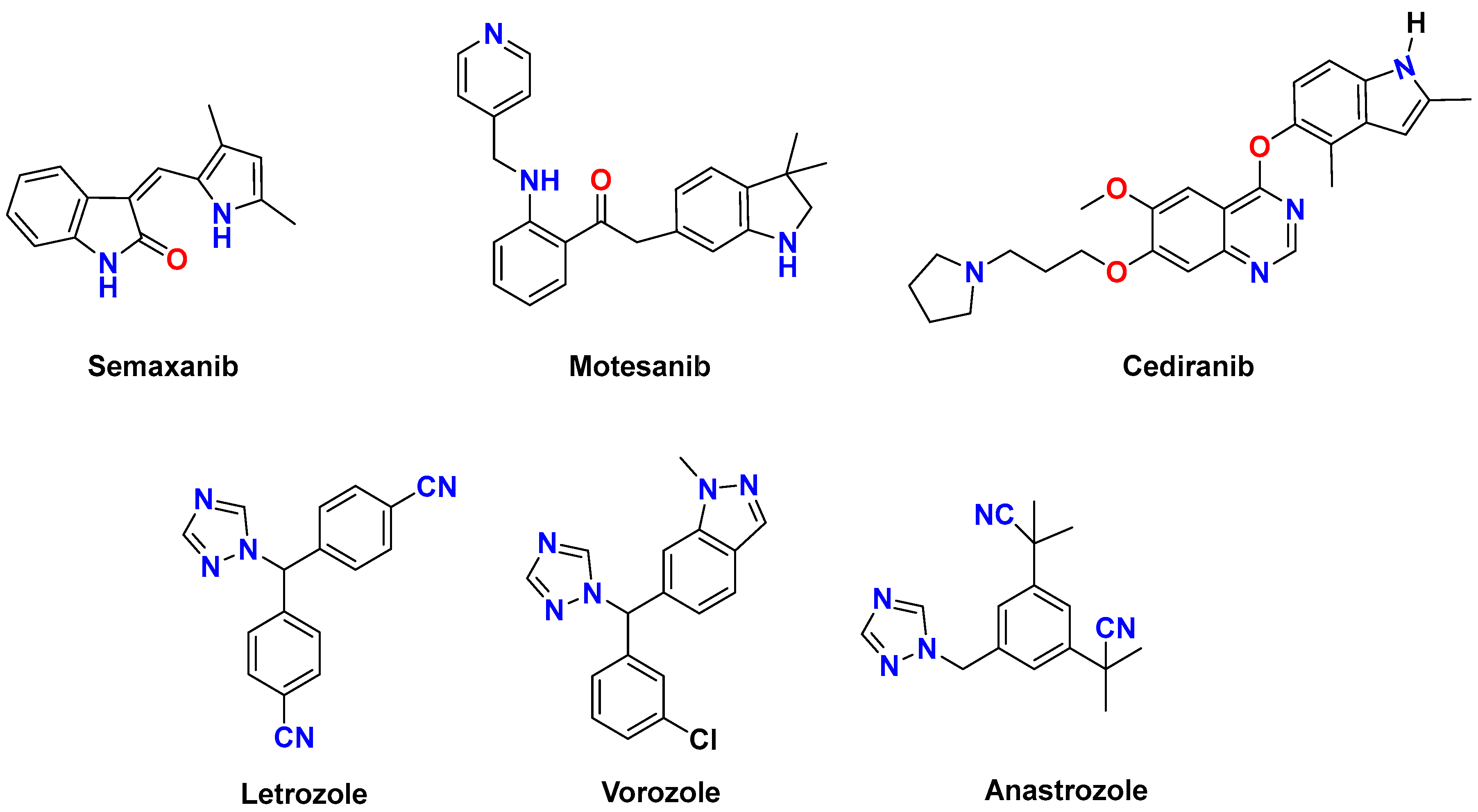
Figure 1. Typical pharmaceutical products with indole and 1,2,4-triazole components for the treatment of breast cancer.
Pharmaceuticals 19 00418 g001
Figure 2. The active alkylated indole-triazole derivatives.
Pharmaceuticals 19 00418 g002
Scheme 1. Alkylation of 4-amino-5-indolyl)-1,2,4-triazole-3-thione 1.
Pharmaceuticals 19 00418 sch001
Scheme 2. Synthesis of 5-(4-Amino-5-(indol-2-yl)-1,2,4-triazol-3-ylthiomethyl)-1,3,4-oxadiazol-2(3H)-thione 7.
Pharmaceuticals 19 00418 sch002
Scheme 3. Synthesis of 5-(((4-amino-5-(indol-2-yl)-triazol-3-yl)thio)methyl)-4-phenyl-2,4-dihydro-1,2,4-triazole-3-thione 10.
Pharmaceuticals 19 00418 sch003
Figure 3. 2D docking interaction diagrams of compounds 4 ((A-1): Bax, (A-2): Bcl-2, (A-3): Caspase-3) and compound 9 ((B-1): Bax, (B-2): Bcl-2, (B-3): Caspase-3), showing key hydrogen bonds and interacting amino acid residues in each binding site.
Pharmaceuticals 19 00418 g003
Figure 4. Cell-cycle distribution of MCF-7 cells after treatment with compounds 5 and 9. Cells were treated for 24 h with compound 5 (0.24 µM) or compound 9 (0.49 µM) (MCF-7 IC50 concentrations), stained with propidium iodide (PI), and analyzed by flow cytometry. Quantitative phase distributions (G0/G1, S, and G2/M) are presented as mean ± SD (n = 3) and summarized in Table 2. Statistical significance versus untreated control was assessed by one-way ANOVA with Tukey’s multiple comparisons test (p < 0.05).
Pharmaceuticals 19 00418 g004
Figure 5. Representative PI-based cell-cycle histogram of MCF-7 cells treated with compound 5. Cells were treated for 24 h with compound 5 (0.24 µM; MCF-7 IC50) and processed in parallel with untreated control cells. DNA content was assessed by PI staining and flow cytometry. The histogram shown is representative of three independent experiments (n = 3); the corresponding quantitative phase distributions and statistics are reported in Figure 4/Table 2 (one-way ANOVA with Tukey’s test).
Pharmaceuticals 19 00418 g005
Figure 6. Representative PI-based cell-cycle histogram of MCF-7 cells treated with compound 9. Cells were treated for 24 h with compound 9 (0.49 µM; MCF-7 IC50) and processed in parallel with untreated control cells. DNA content was assessed by PI staining and flow cytometry. The histogram shown is representative of three independent experiments (n = 3); the corresponding quantitative phase distributions and statistics are reported in Figure 4/Table 2 (one-way ANOVA with Tukey’s test).
Pharmaceuticals 19 00418 g006
Figure 7. Relative mRNA expression of BAX and Bcl-2 in (A,C) MCF-7 and (B,D) HepG2 cells after treatment with compounds 5 and 9. Expression levels were normalized to GAPDH and calculated using the 2−ΔΔCt method. Data are mean ± SD (n = 3). Statistical significance was assessed by one-way ANOVA with Tukey’s multiple comparisons test; p < 0.0001 versus untreated cancer cells.
Pharmaceuticals 19 00418 g007
Table 1. Cytotoxic activity of the synthesized compounds against HepG2 and MCF-7 cell lines and inhibitory activity against VEGFR-2. Values are reported as mean ± SD (n = 3). Individual replicate IC50 values are provided in the Supporting Information (Tables S1–S3).
Compound(HepG2)
(IC50 µM)
(MCF-7)
(IC50 µM)
VEGFR-2 (IC50 µM)VEGFR-2 Inhibition (%)
11.8 ± 0.00280.21 ± 0.000272.3 ± 0.01285.8
20.96 ± 0.00170.95 ± 0.0027
32.1 ± 0.00650.35 ± 0.0034
40.27 ± 0.0020.97 ± 0.00230.59 ± 0.00497.9
50.32 ± 0.00140.24 ± 0.000542.4 ± 0.01483.9
61.5 ± 0.0310.82 ± 0.00033
75.8 ± 0.0744.6 ± 0.0062
90.41 ± 0.000480.49 ± 0.00191.2 ± 0.02596.4
101.2 ± 0.00380.25 ± 0.000382.3 ± 0.01787.8
Doxo.0.16 ± 0.0000330.1 ± 0.00066
Sunitinib 0.32 ± 0.00698.9
Table 2. MCF7 = untreated/vehicle control cells; MCF7-5 = MCF-7 cells treated with compound 5; MCF7-9 = MCF-7 cells treated with compound 9. Cells were treated for 24 h and stained with PI prior to flow-cytometric analysis. Data are presented as mean ± SD (n = 3 independent experiments). Statistical analysis was performed using one-way ANOVA followed by Tukey’s multiple comparisons test (p < 0.05).
Group/PhaseG0-G1SG2-M
MCF768.79 ± 0.3424.74 ± 0.406.48 ± 0.16
MCF7-579.97 ± 0.5115.68 ± 0.464.33 ± 0.24
MCF7-976.58 ± 0.4618.06 ± 0.335.37 ± 0.30
Table 3. Primer Sequences for RT-qPCR.
GeneSequenceTm (°C)Product Size (bp)Accession Number
BAXF: 5′-GATTACAGACCCCAGGCAGG-3′ R: 5′-TGGCTCAAGTAGGACGGGTA-3′54130NM_001291428
Bcl-2F: 5′-GCAATGGGCACGAGTTTGTT-3′ R: 5′-AGTGTGTTCACCAGGCCAAA-3′60170NM_000633
GAPDHF: 5′-CCATCAACGACCCCTTCATT-3′ R: 5′-CACGACATACTCAGCACCAGC-3′58193NM_001256799
Disclaimer/Publisher’s Note: The statements, opinions and data contained in all publications are solely those of the individual author(s) and contributor(s) and not of MDPI and/or the editor(s). MDPI and/or the editor(s) disclaim responsibility for any injury to people or property resulting from any ideas, methods, instructions or products referred to in the content.