Article

Phytochemical Profiling, Antioxidant Activity, and In Vitro Cytotoxic Potential of Mangrove Avicennia marina

1
Department of Earth and Environmental Sciences DISAT, University of Milano-Bicocca, Piazza della Scienza 1, 20126 Milan, Italy
2
MaRHE Centre (Marine Research and Higher Education Center), Magoodhoo Island, Faafu Atoll 12030, Maldives
3
Department of Biotechnology and Biosciences, University of Milano-Bicocca, Piazza della Scienza 2, 20126 Milan, Italy
4
Nanomedicine Center NANOMIB, University of Milano-Bicocca, 20854 Vedano al Lambro, Italy
5
College of Marine Science and Aquatic Biology, University of Khorfakkan, Sharjah 18119, United Arab Emirates
*
Authors to whom correspondence should be addressed.
These authors contribute equally to this work.
Pharmaceuticals 2025, 18(9), 1308; https://doi.org/10.3390/ph18091308
Submission received: 21 July 2025 / Revised: 21 August 2025 / Accepted: 27 August 2025 / Published: 31 August 2025

Abstract

Background: Avicennia marina (Forsk.) Vierh., a widely distributed mangrove species, is known for its diverse secondary metabolites with potential pharmacological applications. Despite its dominance in the Arabian Gulf, where A. marina may have adapted to extreme environmental conditions with a distinct set of bioactive molecules, research in this region remains limited. Methods: This study investigates the phytochemical composition, antioxidant activity, and in vitro cytotoxicity of extracts from different plant parts, including roots, leaves, propagules, pericarps, and cotyledons, collected in the United Arab Emirates (UAE). Extracts were analyzed using ultra-pressure liquid chromatography coupled with high-resolution mass spectrometry (UPLC-HRMS). Antioxidant activity was assessed using DPPH and ABTS assays, while cytotoxicity was evaluated against human cancer and normal cell lines. Results: Analysis revealed 49 compounds, including iridoid glycosides, hydroxycinnamic acids, phenylethanoid glycosides, flavonoid glycosides, and triterpene saponins, several reported for the first time in A. marina and mangroves. The pericarp and root extracts exhibited the highest scavenging activity (DPPH: 187.14 ± 2.87 and 128.25 ± 1.12; ABTS: 217.16 ± 2.67 and 147.21 ± 2.42 μmol TE/g, respectively), correlating with phenylethanoid content. The root extract also displayed the highest cytotoxicity, with IC50 values of 58.46, 81.98, and 108.10 μg/mL against MDA-MB-231, SW480, and E705, respectively. In silico analysis identified triterpene saponins as potential contributors. Conclusions: These findings highlight the root extract of A. marina as a promising source of bioactive compounds with potential antioxidant and anticancer applications, supporting further exploration for novel therapeutic candidates.

Graphical Abstract

1. Introduction

Avicennia marina (Forsk.) Vierh, commonly known as the grey mangrove, is a true mangrove species [1] belonging to the Acanthaceae family. It is widely distributed across tropical and subtropical regions, including Africa; South, Southeast, and Southwest Asia; the Malay Archipelago; the North Island of New Zealand; Australia; the Southwest Pacific; and the Maldives [1,2,3].
The chemical profile of A. marina has been extensively studied, leading to the identification of diverse classes of bioactive molecules, including flavonoids, iridoid glycosides, terpenoids, alkaloids, and steroids, as well as a wide range of other metabolites [3,4,5,6,7,8].
Traditionally, A. marina has been utilized in folk medicine across different countries to treat several ailments and diseases [9,10]. Furthermore, its extracts and isolated compounds have demonstrated a wide range of biological activities, including antiviral, antimicrobial, anthelmintic and antimalarial, analgesic, antioxidant, antifouling, anticancer, antidiabetic, and anti-inflammatory [3,11]. In particular, extracts of this species have demonstrated promising in vitro anticancer activity [3]. However, research in this field remains relatively limited, especially in the context of the Arabian Gulf, where A. marina is the dominant mangrove species along the coasts of the United Arab Emirates (UAE), Saudi Arabia, Bahrain, Qatar, and Iran [12]. Ethnobotanical records for A. marina in the Arabian Gulf are scarce but include traditional uses in Iran for treatments of ulcers, rheumatism, and burns [9], and in the UAE for its use as an aphrodisiac, antifertility agent, and treatment for scabies and toothache [13]. A. marina remains largely unexplored here in terms of bioprospecting, with most studies focusing instead on its distribution, ecological significance, ecosystem services, and management and conservation [14,15]. Phytochemical investigations have only examined populations from China, India, Pakistan, Egypt, other Indo-Pacific locations, and the Red Sea coasts of Saudi Arabia [10,11,16,17,18].
Previous phytochemical studies on A. marina have generally examined only a few plant parts, typically aerial parts and primarily leaves, lacking a comprehensive analysis of plant-part-specific secondary metabolites. Biological activity also presents limitations, often focusing on few plant parts and, in the case of cytotoxicity, testing only a small number of cancer cell lines [9,10,11]. A major gap in existing research of A. marina is the absence of integrated studies combining detailed phytochemical profiling with antioxidant and cytotoxic assays across multiple parts of the plant, which is essential for linking bioactivities to tissue-specific metabolites. Such combined strategies are well established in plant research [19,20] and have been applied to mangroves [21] as they facilitate the prioritization of promising extracts and compounds, thereby enhancing efficiency in the early discovery of bioactive molecules. Moreover, the majority of chemical studies have relied on GC-MS, which biases detection towards volatile constituents [3,11,16]. Although GC-MS can also characterize phenolic compounds, abundant in mangroves [22], it requires a derivatization step that is both complex and time-consuming [23]. In contrast, ultra-high performance liquid chromatography coupled with high-resolution mass spectrometry (UPLC-HRMS) has emerged as a powerful platform for untargeted metabolomic profiling of complex plant matrices, offering superior sensitivity, selectivity, and mass accuracy, and enabling comprehensive detection and identification of secondary metabolites, especially phenolics [24,25,26,27].
In addition to these methodological gaps, the limited geographical scope of previous studies represents a crucial limitation in fully understanding the phytochemical diversity and bioactivity of A. marina. The Arabian Gulf is characterized by extreme environmental conditions, including elevated seawater temperatures, hypersalinity, and high turbidity, driven by its arid climate and shallow basin [28], and summer air temperatures can reach 50 °C [29], further stressing local ecosystems. Since the chemical composition of plants is influenced by geographical, environmental, and climate factors [30,31,32], plants exposed to such stresses often respond by increasing the accumulation of secondary metabolites, such as flavonoids, iridoid glycosides, and phenylethanoid glycosides, which enhance their tolerance to adverse conditions and also possess bioactivities of pharmacological interest, including antioxidant and anticancer [33,34,35,36,37,38,39].
It is plausible that A. marina in the Arabian Gulf may have adapted to extreme conditions through metabolic changes and the induction of antioxidant defense systems [40], potentially resulting in a distinct set of secondary metabolites with unique biological activities. Consequently, the objective of this study is to conduct a comprehensive investigation of A. marina grown in the UAE by characterizing the secondary metabolite composition of multiple parts of the plant, including roots, leaves, propagules (pericarps and internal tissues), and cotyledons, and evaluating their potential health benefits through antioxidant and cytotoxic activity assays in vitro.
Unlike previous studies, this work employs UPLC-HRMS to present a detailed phytochemical profile of each tissue type, allowing for the identification of specific compounds of A. marina and their localization within the plant. Furthermore, while the antioxidant and anticancer potentials of A. marina extracts have been previously reported [3,11], this study provides a comprehensive evaluation of antioxidant and cytotoxic activities of all plant parts, along with expanded cytotoxic screening in multiple cancer cell lines. In addition, in silico predictions of biological activities were applied to compounds identified in the extracts. This multi-level approach addresses existing regional and methodological gaps and lays the groundwork for the discovery of bioactive compounds from plants adapted to extreme environmental conditions.

2. Results

2.1. Characterization of Avicennia marina Extracts

Roots, leaves, cotyledons, pericarps, and propagules of A. marina displayed distinct metabolite profiles. The chromatographic profiles of the extracts are provided in Supplementary Material (Figures S1–S5), and the list of tentatively identified compounds is shown in Table 1, along with their corresponding identification level (IL), which reflects the confidence of compound annotation based on MSI guidelines (see Section 4.4). For compounds assigned to IL2, the identification relied on comparisons of MS/MS fragmentation data with published spectra from the literature or spectral databases. The specific references used to support each IL2 assignment are included directly in the table.
Analysis detected a total of 49 compounds across all plant parts. Triterpene saponins are a heterogeneous secondary metabolite consisting of a terpene-based aglycone linked to one or more sugar chains, commonly glucose (−162 Da), glucuronic acid (−176 Da), and pentoses (−146 Da) [72]. For example, compound 49, which has an m/z of 809.4342 and a molecular formula of C42H66O15, displayed characteristic MS/MS fragments at m/z 647.3797 [M-H-162] and 471.3469 [M-H-162-176], corresponding to sequential losses of sugar moieties. Based on this fragmentation and the molecular formula, it was identified as Azukisaponin III. Using similar fragmentation patterns, compounds 40, 43, 45, and 46 were also assigned as triterpene saponins [66,67,69,70,71].
Phenylethanoid glycosides are often based on β-D-glucosides of 2-phenylethanol, often with α-L-rhamnose (Rha) substitution at C-3′ of the glucose, resembling variants of verbascoside [73]. Simple phenylethanoid glycosides such as acteoside, isoacteoside, and plantamajoside exhibit similar fragmentation patterns in MS/MS experiments. These are characterized by neutral losses of 162, 152, or 146 m/z, which are associated with the presence of caffeic acid, glucose, rhamnose, and the phenethanol aglycone. Diagnostic fragment ions at m/z 179, 161, and 135 indicate the presence of caffeoyl, anhydroglucose, and anhydrophenethanol. Additionally, losses of water (−18 Da) or CO2 (−44 Da) are frequently observed [74]. Based on this information, the compounds 7, 13, 14, 17, 20, 22, 23, 24, and 39 belonged to the phenylethanoid glycosides group.
Flavonoid glycosides are a group of secondary metabolites that are widely distributed throughout the plant kingdom. Depending on the bond of the sugar portion, they are divided into O-glycosides or C-glycosides and can be distinguished by their unique MS/MS fragmentation spectra, which depend on the nature of the sugar fraction. Generally, C-glycosides exhibit neutral losses of 30, 90, and 120 Da for hexose sugars; 74 and 104 Da for deoxyhexose sugars; and 60 Da for pentose sugars. In contrast, O-glycosides exhibit neutral losses of 162 Da (hexose sugars), 176 Da (glucuronic acid), 146 Da (deoxyhexose sugars), and 132 Da (pentose sugars) [75]. Based on this information, compounds 12, 21, 25, 27, 28, 29, 31, and 37 were identified as O-glycosides.
Iridoid glycosides exhibit distinct fragmentation patterns that depend on the structure of the aglycone ring, the presence of functional groups, and the degree of unsaturation. Typically, a neutral loss of 162 Da is observed, corresponding to the breakage of the bond with the glucoside fraction. Subsequently, the formation of fragments due to the loss of water (18 Da) and the carboxyl group (44 Da) is also observed, together with characteristic fragments resulting from aglycone ring cleavage. Peak 3 with m/z 373.1139 and molecular formula C16H22O10 was identified as geniposidic acid based on its MS/MS spectrum. Fragment m/z 211.0605 corresponded to the loss of hexose sugar (162 Da), followed by the presence of fragments m/z 167.0700 and 149.0597, reflecting subsequent losses of H2O (−18 Da) and CO2 (−44 Da), respectively [76]. Furthermore, fragment 123.0440 is characteristic of the genistein ring. Based on the different fragmentation patterns, compounds 5, 6, 18, 34, and 35 were identified as iridoid glycosides.
Tissue-specific profiling revealed clear metabolic differentiation among parts, with the leaves containing the highest number of secondary metabolites (26), followed by the pericarps (23), roots (23), cotyledons (10), and propagules (6). Notably distinct distribution patterns were observed for specific classes of compounds across the different A. marina extracts. The results show that triterpene saponins occur almost exclusively in the root extract (five in roots and only one each in cotyledons, pericarps, and propagules; none in leaves). Phenylethanoid glycosides were predominantly found in root and pericarp extracts (seven in each), with only two detected in leaves and none in cotyledons and propagules. Flavonoid glycosides were mainly associated with leaf extract (six in leaves, two in roots, one in pericarps, and absent in cotyledons and propagules). In contrast, iridoid glycosides and hydroxycinnamic acid and derivatives showed a more uniform distribution across all the extracts.
Analysis confirmed several compounds previously reported in A. marina, including caffeoylquinic acid, geniposidic acid, marinoid A, C, and D, acteoside, quercetin 3-O-hexoside, kaempferol 3-O-glucuronide, isorhamnetin-3-O-rutinoside, diosmetin 7-glucuronide, and jionoside C [5,7,77,78,79,80]. Additionally, cistanoside F and kaempferol 3-O-glucoside were also detected, previously reported in other mangrove species but not in A. marina [81,82]. To our knowledge, several compounds such as mussaenosidic acid, (epi)loganic acid, caffeoylglucaric acid, icariside D1, suspensaside, grandifloroside, suspensaside methyl ether, suspensaside A, isorhamnetin glucuronide, isorhamnetin 7-glucoside, medicoside G, esculentoside C, and azukisaponin III have been newly reported in mangrove species.

2.2. Antioxidant Activity

2.2.1. DPPH and ABTS Assays

The antioxidant potential of A. marina extracts was evaluated using two spectrophotometric assays, ABTS and DPPH, which are widely used to assess the free radical scavenging activity of natural compounds. The results are shown in Figure 1.
The DPPH assay showed that the pericarp extract exhibited the highest radical scavenging activity, with a Trolox equivalent antioxidant capacity (TEAC) value of 187.14 ± 2.87 μmol TE/g. This was followed by the extracts of root (128.25 ± 1.12 μmol TE/g), cotyledon (58.23 ± 3.49 μmol TE/g), leaf (55.12 ± 1.52 μmol TE/g), and propagule (38.72 ± 6.96 μmol TE/g).
Similarly, the ABTS assay confirmed that the root and pericarp extracts exhibit high antioxidant activity compared to the other parts of the plant. In fact, the pericarp extracts again displayed the highest TEAC value (217.16 ± 2.67 μmol TE/g), followed by the root (147.21 ± 2.42 μmol TE/g), leaf (64.98 ± 0.84 μmol TE/g), cotyledon (54.46 ± 1.95 μmol TE/g), and propagule (32.23 ± 2.53 μmol TE/g) extracts.

2.2.2. Correlation Between Compound Classes and Antioxidant Activity

The pericarp and root extracts, which exhibited the highest antioxidant activity, are also the ones that contain a high number of phenylethanoid glycosides, compared to the other extracts, which may explain their higher activity. To explore the potential associations between the phytochemical composition of each extract and their antioxidant capacity, a Spearman correlation analysis was performed between the number of compounds in each major chemical class and the measured antioxidant activities (DPPH and ABTS assay) across the five plant-part extracts (n = 5) (Table 2). The analysis revealed a significant positive correlation between the number of phenylethanoid glycosides in the extracts and DPPH activity (ρ = 0.949; p = 0.014). ABTS activity showed a similar trend, though the correlation did not reach statistical significance (ρ = 0.791; p = 0.111). Antioxidant activity showed no statistically significant correlations with the number of other classes of compounds, including flavonoid glycosides, iridoid glycosides, hydroxycinnamic acids and derivatives, and triterpene saponins (all p > 0.05). Given the small sample size and the use of compound counts (not concentrations), these associations should be considered exploratory.

2.3. In Vitro Cytotoxic Activity

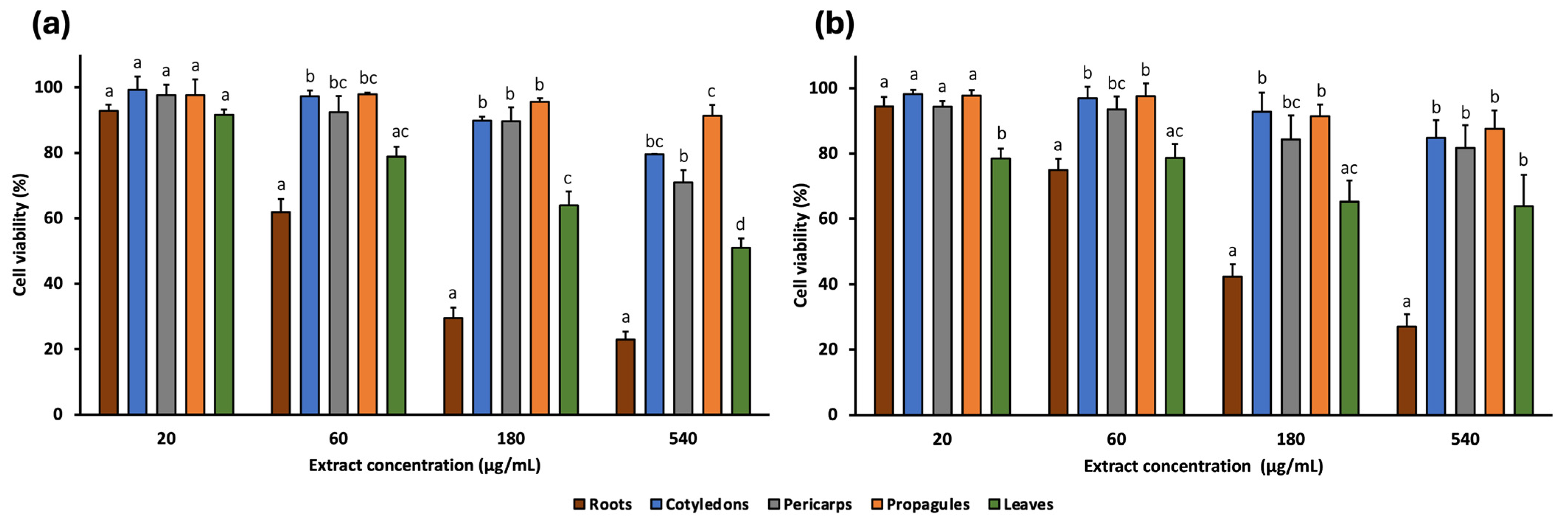
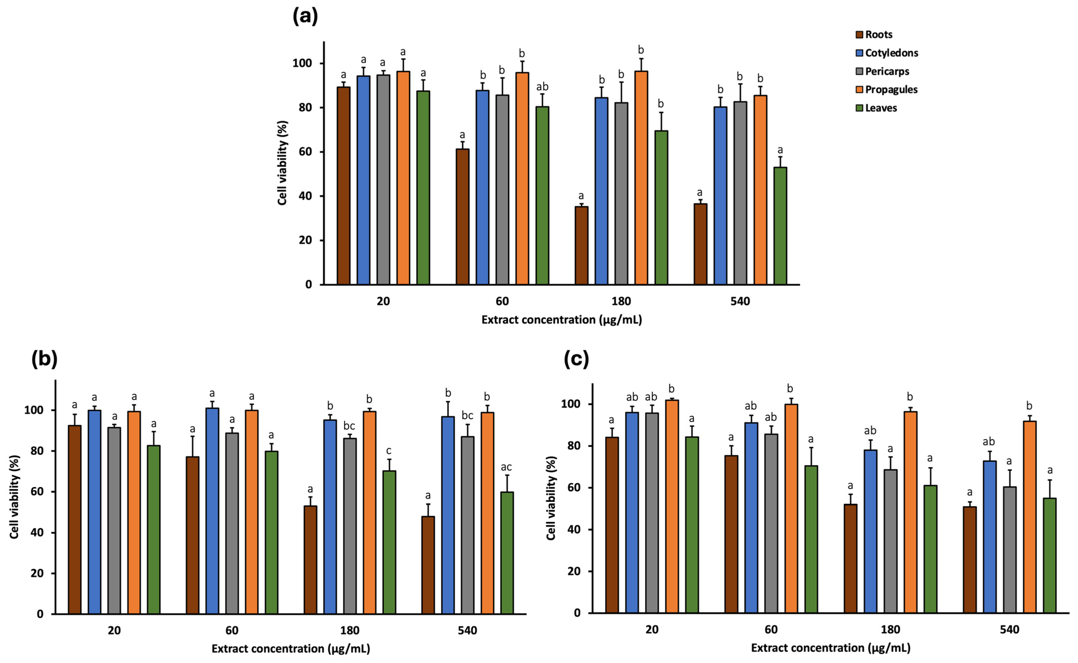
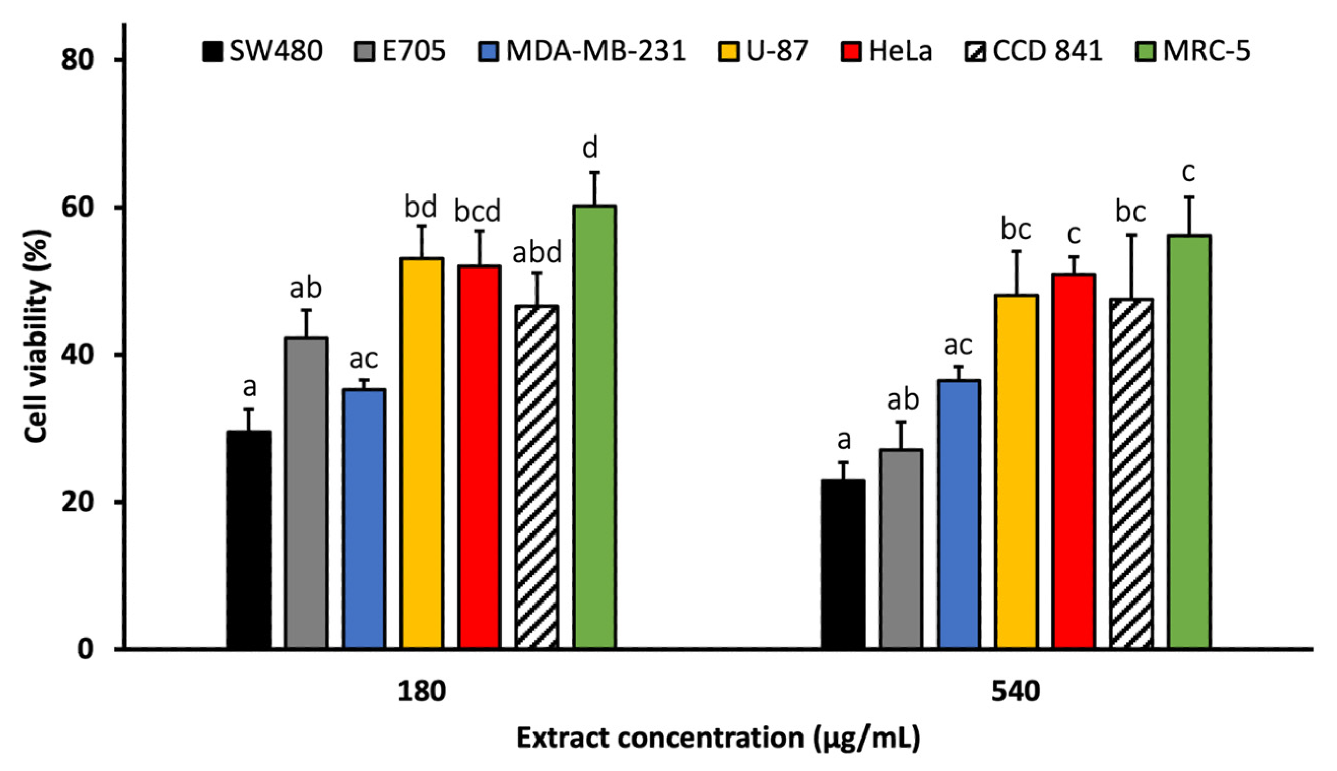
The cytotoxic effects of A. marina extracts (leaf, cotyledon, pericarp, propagule, and root) were evaluated against a panel of human cancer cell lines using the MTT assay. Four concentrations (20, 60, 180, and 540 μg/mL) were tested on two colorectal cancer cell lines (SW480 and E705) (Figure 2) and three additional cancer cell lines: MDA-MB-231 (triple-negative breast cancer), U-87 (glioblastoma), and HeLa (cervical cancer) (Figure 3). Furthermore, two non-cancerous cell lines, MRC-5 (normal human fibroblasts) and CCD 841 (healthy human mucosa), served as controls to assess extract selectivity (Figure 4). The complete data for all concentrations and cell lines are reported in Table S1.
Among the extracts, cotyledon, pericarp, and propagule generally exhibited the lowest cytotoxic activity, reducing cell viability by no more than 70% at the highest concentration (540 μg/mL) across all cell lines. The exception was the pericarp extract, which reduced viability of HeLa cells to 60.30%.
The leaf extract showed low cytotoxicity at lower concentrations. At 60 μg/mL, cell viability remains near 80% for SW480, E705, and MDA-MB-231 and was 70.44% for HeLa. In the non-cancerous cell lines, viability was even higher: 88.59% and 86.86% for CCD 841 and MRC-5, respectively. However, at 540 μg/mL, the extract reduced viability to 50–60% in most cell lines, particularly 50.98% for SW480, 63.91% for E705, 53.00% for MDA-MB-231, 59.77% for U-87, 54.96% for HeLa, and 61.01% for CCD 841, while the least reduction occurred in MRC-5 (71.26%).
Among all extracts, the root extract exhibited the highest cytotoxic activity. At 180 μg/mL, it reduced cell viability to 29.47% (SW480), 42.40% (E705), 35.26% (MDA-MB-231), 53.06% (U87), 52.00% (HeLa), 46.60% (CCD 841), and 60.15% (MRC-5). These reductions were statistically significant compared to all other extracts, except for E705, where differences with the leaf extract were not significant, and for HeLa. At 540 μg/mL, cytotoxicity remained similar across most lines, although viability dropped further in SW480 and E705 (22.93% and 27.03%, respectively).
These findings highlight the notable activity of the root extract, particularly at the highest concentrations, against SW480, E705, and MDA-MB-231 cell lines (Figure 5). Notably, at 180 μg/mL, the cytotoxic activity of the root extract in these cell lines was significantly greater than in non-cancerous MRC-5 cells, while no significant difference was observed compared to the healthy mucosa cell line CCD 841. Additionally, at 540 μg/mL, viability of SW480 cells was significantly lower than that of CCD 841.
Given this pronounced response, the analysis focused on dose–response effects in SW480, E705, and MDA-MB-231. Each cell line was treated with ten increasing concentrations of root extract (2 to 540 μg/mL; Figure S6), and the IC50 values were 81.98 μg/mL for SW480, 108.10 μg/mL for E705, and 57.93 μg/mL for MDA-MB-231.

2.4. In Silico Analysis

Given the strong cytotoxic activity observed for the A. marina root extract in vitro, an in silico analysis was conducted to evaluate the predicted biological activities of the compounds found exclusively or predominantly in this extract. PASS Online predicted potential cytotoxicity-related effects, including antineoplastic activity, apoptosis induction (caspase 3/8 stimulation and apoptosis agonism), TP53 expression enhancement, NF-κB stimulation, cytostatic activity, lipid peroxidase inhibition, and inhibition of ICAM-1 expression [83,84]. The complete list of predicted biological activities for compounds exclusive of the root extract is provided in Table S2 in the Supplementary Materials section.
Among the primary peaks detected in the root extract, the triterpene saponins medicoside G and azukisaponin III were found exclusively in the root extract, while esculentoside C was detected at high levels in the roots and in trace amounts in other extracts. These compounds showed high predicted probabilities (Pa) for antineoplastic activity (0.870, 0.905, and 0.908 for medicoside G, esculentoside C, and azukisaponin III, respectively), caspase 3/8 stimulation (0.994/0.984, 0.989/0.986, and 0.964/0.934), apoptosis agonism (0.901, 0.862, and 0.883), and NF-κB stimulation (0.965, 0.917, and 0.904). They were also predicted to inhibit ICAM-1 expression (0.908, 0.961, and 0.987) and lipid peroxidase activity (0.927, 0.952, and 0.991).
Additional compounds found exclusively in the root extract, including I, suspensaside A, kaempferol 3-O-glucoside, and quercetin 3-O-hexoside, were also predicted to exhibit antineoplastic activity with Pa values of 0.804, 0.863, 0.834, and 0.833, respectively. Kaempferol 3-O-glucoside and quercetin 3-O-hexoside also showed cytostatic activity (Pa: 0.811 and 0.825), enhancement of TP53 expression (Pa: 0.952 and 0.959) and lipid peroxidase inhibition (0.960 and 0.976, resp.).

3. Discussion

UHPLC-ESI/HRMS analyses enabled the characterization of A. marina extracts, revealing a total of 49 compounds, unevenly distributed across plant parts. The leaf extract contained the highest number of secondary metabolites, followed by pericarps, roots, cotyledons, and propagules. Notably, this study distinguishes between the external tissue of the propagule (here consistently called pericarp) and the internal tissues of the propagule (here consistently called simply propagule) [85], which are often analyzed as a single fruit unit in other studies. The propagule, which consists mainly of the embryo, contained few compounds, likely due to the focus on primary metabolites essential for germination [86]. In contrast, the pericarp, which serves a protective function, was significantly richer in secondary metabolites [87], particularly phenylethanoid glycosides. Similarly, cotyledons had low metabolite diversity and a profile similar to propagules but showed some additional peaks. These may reflect early biosynthesis of stress-related compounds in developing seedlings.
The compounds identified, including phenylethanoid glycosides, flavonoid glycosides, iridoid glycosides, hydroxycinnamic acid and derivatives, and triterpene saponins, are well known for their ecological roles in protecting plants from abiotic stressors such as drought, high salinity, intense sunlight, and elevated temperatures [33,34,35,36,38]. These classes are widely reported in A. marina from other regions, and some metabolites identified here have been documented previously, suggesting a shared core phytochemical profile with global populations [10].
However, differences from previous studies were observed. Firstly, a distinctive feature of A. marina elsewhere is the presence of naphthalene derivatives [10], which were not detected in our samples. This absence may reflect tissue specificity, as most of these compounds were extracted from branches, or differences in extraction methods [8]. More importantly, several compounds detected in this study have never been reported before in A. marina. These include one kaempferol-glycosides and two isorhamnetin glycosides. Notably, monohydroxy B-ring-substituted flavonoid glycosides (e.g., kaempferol-, diosmetin-, and isorhamnetin-glycosides) were more abundant than dihydroxy types, a pattern opposite to that expected under UV stress, where dihydroxy forms typically dominate due to their antioxidant potential [33,35,88]. This shift may reflect Gulf-specific regulation of flavonoid biosynthesis rather than species-specific traits, as it is not evident in previous reports [10]. The study identified novel compounds among the iridoid glycosides and hydroxycinnamic acids, but the most notable findings were within phenylethanoid glycosides and triterpene saponins, the latter all newly reported in A. marina. The high abundance of phenylethanoid glycosides in roots is consistent with their reported accumulation under water stress [36], while the accumulation of triterpene saponins is associated with osmotic stress response [38,89]. These trends suggest that UAE-grown A. marina may possess a distinctive phytochemical profile shaped by the extreme environmental conditions of the Arabian Gulf [40,90] Nonetheless, inter-regional comparisons are limited due to methodological differences. Future comparative studies using standardized LC-MS protocols would enhance our understanding of mangrove chemical ecology and support bioprospecting efforts.
The identified metabolite classes are associated with various biological activities, including antioxidant, anticancer, antimicrobial, and anti-inflammatory [91,92,93,94,95]. In particular, phenylethanoid glycosides are well-documented antioxidants, showing both DPPH and ABTS radical scavenging activity [91,96,97]. In this study, extracts of the pericarp and root were rich in these compounds and showed the highest antioxidant activity (187.14 ± 2.87 and 217.16 ± 2.67 μmol TE/g for the pericarps; 128.25 ± 1.12 and 147.21 ± 2.42 μmol TE/g for the roots). The strong positive Spearman correlation between the number of phenylethanoid glycosides and DPPH activity (ρ = 0.949; p = 0.014) supports the hypothesis that these compounds contribute to radical scavenging in the extracts. Key phenylethanoid glycosides identified in this study, such as cistanoside F, acteoside, and jionoside C, are known for their potent antioxidant activity [98,99,100], while less-studied molecules such as suspensaside and suspensaside A warrant further exploration. Our correlation findings are exploratory and do not prove causation. Definitive attribution will require targeted quantification of candidate phenylethanoid glycosides and subsequent activity testing.
In terms of cytotoxic activity, the cotyledon, pericarp, and propagule extracts showed negligible effects on cancer cell lines, even at the highest concentrations. In contrast, the leaf and root extracts displayed cytotoxicity at higher doses. The leaf extract showed limited cytotoxicity at lower concentrations but reduced viability (50–60%) at 540 μg/mL in several cancer cell lines. This agrees with Momtazi-Borojeni et al. [101], who reported no toxicity at low concentrations but moderate effects at higher doses (250 μg/mL). The root extract had the most promising cytotoxic profile, particularly against SW480, E705, and MDA-MB-231 cancer cell lines. At 540 μg/mL, it reduced cell viability below 40% but showed lower toxicity against normal cell lines (MRC-5 and CCD 841). These findings agree with previous studies showing selective cytotoxicity of root extracts towards cancer cell lines [102]. IC50 values for SW480, E705, and MDA-MB-231 were 81.98, 108.10, and 57.93 μg/mL, respectively. Based on the criteria established by the National Cancer Institute (NCI, USA) and the Geran protocol, which classified cytotoxicity as high when IC50 values are ≤ 20 μg/mL, moderate between 21 and 200 μg/mL, weak between 201 and 500 μg/mL, and absent above 500 μg/mL [103,104,105], this corresponds to moderate cytotoxic activity. While these IC50 values were not compared with a standard drug, they suggest the presence of active compounds. The values reported here are for crude extracts; further fractionation and isolation of active constituents are expected to yield more potent compounds, for which in vivo and clinical potential could be more realistically assessed through comparison with standard anticancer drugs. These are potentially triterpene saponins, which were only detected in the roots.
Triterpene saponins are gaining attention in cancer research due to their ability to target tumor-related pathways while maintaining low toxicity [106]. Although widespread in medicinal plants [107,108], their occurrence in mangroves is less documented, with only a few studies published on this topic [82,108]. In silico prediction using PASS software indicated a strong cytotoxic potential for several saponins identified in the root extract, including medicoside G, esculentoside C, and azukisaponin III. Additionally, two unidentified saponins suggest the presence of a potentially novel structure that merits further isolation and structural characterization. Other compounds specific to the root extract, such as suspensaside A and kaempferol 3-O-glucoside, showed high predicted probabilities for antineoplastic effects.
Given that the root extract exhibited the highest cytotoxicity, further work will focus on bioactivity-guided fractionation of this extract, with particular emphasis on isolating the triterpene saponin-rich fraction. These purified fractions will be tested for cytotoxicity alongside a standard anticancer drug (e.g., doxorubicin) to identify the compounds responsible for the observed activity. Mechanistic studies, including apoptosis assays, cell cycle analysis, and molecular pathway investigations, will be conducted to elucidate the modes of action. Such comprehensive analyses, together with the targeted quantification of candidate constituents, will clarify structure–activity correlations and enhance both therapeutic efficacy and selectivity. Importantly, these efforts, combined with further purification and structural elucidation, could identify promising novel lead compounds suitable for subsequent in vivo evaluation and development as potential anticancer agents.

4. Materials and Methods

4.1. Chemicals

Ethanol absolute, analytical-grade methanol, 1,1-diphenyl-2-picrylhydrazyl (DPPH), and 2,2-azinobis-(3-ethylbenzothiazoline-6-sulfonate) (ABTS•+) reagents were obtained from Sigma-Aldrich (Milan, Italy), while methanol and formic acid of LC-MS grade were sourced from Romil (Cambridge, UK). Ultrapure water (18 MΩ) was prepared by a Milli-Q purification system (Millipore, Bedford, MA, USA).

4.2. Plant Material

Samples were collected from different parts of A. marina, including leaves, roots, propagules, and cotyledons. The cotyledons were obtained from seedlings at an early growth stage, when the propagules had already opened and developed roots. All samples were harvested in September 2022 from multiple individual plants within the mangrove forest of Ajman Emirate, UAE. Although no herbarium voucher specimen was deposited, the plant material was identified based on morphological characteristics following established taxonomic keys [109]. This identification is supported by the fact that A. marina is the only mangrove species forming the evergreen coastal forests of the UAE [14], and its presence in the region has been validated by previous molecular analyses [110,111].
For more precise phytochemical characterization, propagules were separated into the pericarp, representing the external protective tissue, and the internal tissues. Thus, in the manuscript, the term pericarp (and pericarp extract) refers exclusively to the external part, while the generic term propagule (and propagule extract) refers to the internal tissues. Each type of plant material (e.g., all collected leaves, roots, pericarps, propagules, and cotyledons) was pooled by type and immediately freeze-dried after collection. The dried samples were homogenized using a Grindomix GM 200 knife mill (Retsch, Haan, Germany) and then sieved through a test sieve (Retsch AS 200, Haan, Germany) with a mesh size range of 300–600 μm to obtain powders with uniform particle size distribution.

4.3. Sample Preparation and Extraction

Root, leaf, cotyledon, pericarp, and propagule samples of A. marina underwent exhaustive ultrasound-assisted extraction using a thermostatically controlled ultrasonic bath (Sonorex TK 52; Bandelin electronic, Berlin, Germany). Each sample was extracted under controlled conditions (25 °C, 15 min) with 50% aqueous ethanol (v/v) at a solid-to-solvent ratio of 1:10 (w/v), which is commonly applied in metabolite profiling studies [112]; specifically, 1 g of powdered sample was mixed with 10 mL of solvent in a 50-mL polypropylene tube. A 50% aqueous ethanol solution was selected as the extraction solvent due to its effectiveness as a green, low-toxicity system, making it well-suited for bioactivity screening [113,114]. Ethanol–water mixtures offer a balanced polarity and are widely recognized for their ability to efficiently extract a broad range of bioactive compounds, particularly polyphenolic metabolites, which are well known for their antioxidant properties [115,116]. Moreover, this solvent was selected to ensure low toxicity in downstream biological assays, in case traces of solvent remain after evaporation, and because non-polar solvents or higher ethanol concentrations could reduce solubility in aqueous assay media, potentially compromising the suitability of the extracts for biological testing.
To ensure complete extraction, the process was repeated three times with fresh solvent. Following each extraction, the mixtures were centrifuged (13,000× g, 10 min), and the supernatants underwent filtration through Whatman No. 1 filter paper. The combined extracts were concentrated under pressure at 40 °C using a rotary evaporator to remove ethanol and subsequently lyophilized (Alpha 1-2 LD freeze dryer, Christ, Germany) to obtain dry residues for further analysis.
The extraction yields of A. marina were determined by calculating the ratio of the weight of dried extract obtained to the initial weight of dried plant material powder and expressed as a percentage. The yields were 34.23% for roots, 65.02% for cotyledons, 61.76% for pericarps, 38.55% for propagules, and 33.72% for leaves.

4.4. Characterization of Extracts

The chemical characterization of extracts was performed in negative mode using liquid chromatography coupled with electrospray ionization (ESI) and high-resolution mass spectrometry (UPLC-ESI/HRMS). A Waters ACQUITY UPLC system coupled with a Waters Xevo G2-XS QTof Mass Spectrometer (Waters Corp., Milford, MA, USA) was used. The extracts were dissolved in ultrapure water at a concentration of 100 μg/mL, and then 5 μL of each sample was injected into a Biphenyl column (100 mm × 2.1 mm, 2.6 μm; Phenomenex, Torrance, CA, USA). The chromatographic gradient was conducted with solvent A (0.1% formic acid in water) and solvent B (0.1% formic acid in methanol), starting with 95% A for 1 min, followed by a linear gradient to 95% B over 10 min, and 4 min of column washing at 95% B. The flow rate was maintained at 0.4 mL/min. The ESI source was operated under the following conditions: electrospray capillary voltage of 1.5 kV, source temperature of 140 °C, and desolvation temperature of 600 °C. MS spectra were acquired in full range mode, covering a mass range of 100–1000 m/z. MS/HRMS analysis was performed using data-dependent scan (DDA), selecting the two most intense ions from the HRMS scan for collision-induced dissociation (CID) with the following conditions: a minimum signal threshold of 500,000, isolation width at 2.0, and normalized collision energy of 30%. Metabolite identification followed the Metabolomics Standards Initiative (MSI) guidelines, which define three confidence levels indicated in the “IL” column of Table 1: Level 1 (IL1): compounds were unequivocally identified by comparison with authentic reference standards (retention time, MS/MS spectrum, and exact mass); Level 2 (IL2): tentative identifications were assigned based on matches between experimental MS/MS spectra and literature data or spectral libraries (e.g., GNPS, MassBank); Level 3 (IL3): compounds were classified by spectral similarity to known chemical families and supported by taxonomic evidence.
Novelty verification was performed using general literature databases (e.g., Google Scholar) and the chemical database SciFindern. In SciFindern, each tentatively identified compound was queried by chemical name, and all related references were investigated using keywords such as “Avicennia marina”, “Avicennia”, and “mangroves”. This process allowed determination of whether a compound had been previously reported in A. marina, other species within the genus Avicennia, or other mangrove species, or if it represents a first identification in mangroves.

4.5. Determination of Antioxidant Activity

The antioxidant capacities (AOCs) of the exhaustive extracts of A. marina (leaves, roots, pericarps, propagules, and cotyledons) were evaluated using 1,1-diphenyl-2-picrylhydrazyl (DPPH) and 2,2-azinobis-3-ethylbenzothiazoline-6-sulfonate (ABTS•+) assays according to Cannavacciuolo et al. [117]. The extracts were dissolved in ultrapure water and analyzed at a concentration of 0.5 mg/mL, with Trolox (0–500 μM) serving as a standard. The antioxidant activity was expressed as μmol Trolox equivalents per gram of sample matrix (TE/g MTX), representing the μmol of a standard Trolox solution exerting the same antioxidant capacity as 1 mg/mL of the tested extracts.
In the DPPH assay the stock solution of DPPH (5 mM) was prepared by dissolving 3.9 mg of DPPH in 100 mL of methanol and subsequently diluted to 100 μM to obtain the operating solution. This solution was prepared just before use and protected from light due to the photosensitivity of the reagent. The assay was set up in an Eppendorf by mixing 50 μL of sample with 950 μL of operative DPPH, and the mixture was incubated in the dark for 30 min. Subsequently, 200 μL of the solution was transferred to an absorbance reading plate at the 515 nm wavelength.
For the ABTS assay, the stock solution of ABTS (7 mM) was diluted with phosphate-buffered saline (PBS; 5 mM, pH 7.4) to achieve working concentrations. The assay was performed by combining 5 μL of diluted sample (or PBS control) and 500 μL of ABTS radical cation solution (0.1 mM in PBS). The reaction mixtures were protected from light and incubated at 30 °C for 60 min to allow complete radical scavenging. Absorbance measurements were then recorded at 734 nm using a microplate reader.

4.6. Cytotoxicity Evaluation

4.6.1. Cell Lines and Culture Conditions

Normal human fibroblasts (MRC-5), human glioblastoma (U-87), human triple-negative breast cancer (MDA-MB-231), human colorectal cancer (SW480), and human healthy mucosa (CCD841) cell lines were purchased from the American Type Culture Collection (Manassas, VA, USA). The human cervical cancer cell line (HeLa) was acquired from System Biosciences, and the human colon cancer cell line (E705) was provided by the Fondazione IRCCS Istituto Nazionale dei Tumori (Milan, Italy). The E705 cell line represents epithelial tissue cells of colorectal adenocarcinoma derived from a patient at the National Cancer Institute in Milan.
MRC-5, U-87, and HeLa cells were cultured in Dulbecco’s Modified Eagle Medium (DMEM) high glucose medium supplemented with 10% heat-inactivated fetal bovine serum (FBS), 1% penicillin/streptomycin (P/S), and 2 mM L-glutamine. MDA-MB-231 cells were maintained in Minimum Essential Medium (MEM) with Earl’s Salts supplemented with 10% heat-inactivated FBS, 1% P/S, 2 mM L-glutamine, and 0.1 mM MEM Non-Essential Amino Acids (MEM NEAA). E705 and SW480 cell lines were cultured in RPMI 1640 medium supplemented with 10% heat-inactivated FBS, 100 U/mL penicillin, 100 μg/mL streptomycin, and 2 mM L-glutamine. The CCD 841 cell line was grown in EMEM medium supplemented with 10% heat-inactivated FBS, 1% P/S, 2 mM L-glutamine, and 0.1 mM non-essential amino acids. All cell lines were incubated at 37 °C in a humidified atmosphere containing 5% CO2 and 95% air. Cell culture media and reagents were purchased from EuroClone (Pero, Italy).

4.6.2. Viability Assay

The cytotoxicity of A. marina extracts was evaluated using the MTT assay (CellTiter96®Non-Radioactive Cell Proliferation Assay, Promega, Madison, WI, USA) following the manufacturer’s protocol. The extract powders were solubilized in Milli-Q water, and four extract concentrations (20, 60, 180, and 540 μg/mL) were tested on all cell lines. Additionally, for the root extract, an extended dose-response analysis using ten concentrations (2, 10, 20, 40, 60, 100, 140, 180, 360, and 540 μg/mL) was performed on selected cell lines to enable IC50 determination. IC50 values were calculated only for extract–cell line combinations tested with this ten-point dilution series.
No positive control drugs were included in this preliminary screening, as the primary objective was to assess and compare the relative cytotoxicity of different A. marina extracts. Comparative analysis with standard anticancer agents will be incorporated in subsequent studies on purified fractions or isolated compounds.
Briefly, HeLa, MRC-5, U-87, and MDA-MB-231 cells were seeded into 96-well plates (from Euroclone, Pero, Italy) at a density of 5 × 10³ cells/well in 100 μL of growth medium, while E-705 and SW-480 cells were seeded at a density of 8 × 10³ cells/well. After 24 h of incubation at 37 °C in 5% CO2, the medium was replaced, and cells were treated with various concentrations of A. marina extracts. Following 48 h of treatment, the medium was replaced, and 15 μL of MTT solution was added to each well. After 3 h of incubation at 37 °C, formazan crystals were solubilized using 100 μL of stop solution and incubated under stirring for 1 h. Reduced MTT was quantified using a UV–vis plate reader (EnSight Multimode Microplate Reader, PerkinElmer, Waltham, MA, USA) at 570 nm with a reference wavelength of 630 nm.
Cell viability was expressed as a percentage relative to untreated cells (negative control), and medium with MilliQ water at equivalent concentrations (10% v/v) was used as a blank. Dose-response curves and the IC50 values, representing the extract concentration required to inhibit 50% of cell viability relative to untreated control cells, were generated using GraphPad Prism v10.5.0 software.

4.7. In Silico Prediction for Anticancer Activity

To identify potential bioactive compounds responsible for the cytotoxic effect, an in silico prediction of biological activity was performed using PASS Online software (https://www.way2drug.com/PASSOnline/index.php; accessed on 15 July 2025), a predictive tool from Way2Drug Services. The reliability of PASS for predicting in vitro cytotoxic activity has been demonstrated in previous studies [118,119], including those focusing on triterpene saponins [120].
The canonical Simplified Molecular Input Line Entry System (SMILES) of each compound was gathered from SciFindern and was used to run the software. The program independently calculates the estimated predictive biological activities based on structure–activity relationships, providing Pa (probability of activity) and Pi (probability of inactivity) values for each activity. Only activities with Pa > 0.7 were considered, as this threshold indicates a high likelihood that the substance will exhibit the predicted activity in experimental settings, although the probability of the compound being an analogue of a known pharmaceutical agent remains high [121]. Notably, when Pa > 0.9, as frequently observed in our study, the likelihood of false-positive predictions is insignificant [118].
The predicted anticancer-related activities included antineoplastic activity, apoptosis-related effects (apoptosis agonist, caspase 3/8 stimulation), TP53 expression enhancement, NF-kB modulation, cytostatic activity, lipid peroxidase inhibition, and ICAM-1 expression inhibition. These results were analyzed in relation to the cytotoxicity data of the MTT assay to establish potential correlations between the phytochemical composition and the observed cytotoxicity against cancer cells.

4.8. Statistical Analysis

Statistical analyses were conducted on data generated from three replicates. Cytotoxic activity results are presented as mean ± standard error of the mean (SEM). Antioxidant activity results are reported as mean ± standard deviation (SD). Before statistical analysis, the assumption of normality was assessed using the Shapiro–Wilk test. The homogeneity of the variances was evaluated using Levene’s test. When normality and homogeneity assumptions were met, a one-way analysis of variance (ANOVA) was performed, followed by Tukey’s honest significant difference (HSD) post hoc test to assess pairwise differences between the means of the group. In cases where the assumption of homogeneity of the variances was not met, Welch’s ANOVA was applied, followed by the Games–Howell post hoc test. Statistical significance was considered when p < 0.05.
The correlation between phytochemical composition and antioxidant activity was assessed using Spearman’s rank correlation coefficient (two-tailed). For each plant-part extract (n = 5), the number of tentatively identified compounds in each major chemical class (phenylethanoid glycosides, flavonoid glycosides, iridoid glycosides, hydroxycinnamic acids and derivatives, and triterpene saponins) was correlated with antioxidant activity (DPPH and ABTS, mean values). Statistical significance was set at p < 0.05.
All analyses were conducted with IBM SPSS Statistics v29.0.2.0.

5. Conclusions

The findings of this study highlight A. marina as a valuable source of bioactive compounds with promising therapeutic applications. The pericarp and root extracts exhibited the highest antioxidant activity, possibly due to the presence of phenylethanoid glycosides, which are known for their antioxidant activities. Among all extracts tested, the root extract displayed the strongest cytotoxicity, in particular against the triple-negative breast cancer cell line MDA-MB-231 and two colorectal cancer cell lines, SW480 and E705, with IC50 values of 58.46, 81.98, and 108.10 μg/mL, respectively. In silico predictions identified triterpene saponins, including medicoside G, esculentoside C, and azukisaponin III, as likely contributors to these effects.
The detection of triterpene saponins not previously reported from mangroves, together with several phenylethanoid glycosides and other compounds not earlier described in A. marina, is noteworthy. Plants adapted to extreme environments can accumulate distinctive secondary metabolites, and our plant-part-specific UPLC-HRMS analysis of UAE-grown A. marina provides region-specific evidence that complements existing phytochemical surveys. Moreover, combining this untargeted phytochemical investigation with biological activity screening and in silico analysis/statistical correlation offers a practicable approach to rapidly link observed activities to plant-part-specific compounds.
To advance these observations towards pharmacological relevance, future work should focus on bioactivity-guided fractionation of the extracts, structural elucidation, and targeted quantification of key compounds, along with mechanistic in vitro assays. These efforts will help clarify structure–activity correlations and potentially lead to the identification of a novel therapeutic candidate from this stress-adapted mangrove species.

Supplementary Materials

The following supporting information can be downloaded at: https://www.mdpi.com/article/10.3390/ph18091308/s1, Figures S1–S5: Representative chromatograms of A. marina extracts; Table S1: Cytotoxicity of Avicennia marina extracts on the tested cell lines at four concentrations (20–540 μg/mL); Figure S6: Cell viability of SW480 (a), E705 (b), and MDA-MB-231 (c) human cancer cell lines treated with root extract (2–540 μg/mL) for 48 h; Table S2: Probable cytotoxicity-related biological activities of 6 compounds tentatively identified in the root extract of A. marina by PASS (Prediction of Activity Spectra for Substances).

Author Contributions

Conceptualization, P.G., M.C. and L.C.; investigation, F.C. (cell line experiment on SW480, E705, and CCD 841 and chemical investigation), B.D.S. (cell line experiment on U-87 and MRC-5), F.S. (cell line experiment on MDA-MB-231 and HeLa), S.P. (chemical analysis); resources, P.F., P.G., M.C. and L.C.; writing—original draft preparation, F.C., B.D.S. and F.S.; writing—review and editing, F.C., S.P., M.F., M.G., L.S., P.G., M.C., L.C.; supervision, H.S., M.F., P.F., M.G., L.S., P.G., M.C. and L.C.; funding acquisition, P.G. All authors have read and agreed to the published version of the manuscript.

Funding

This research was funded under the National Recovery and Resilience Plan (NRRP), Mission 4, Component 2 Investment 1.3—Call for tender No. 3138, 16 December 2021, rectified by Decree n. 341 of 15 March 2022 of the Italian Ministry of University and Research funded by the European Union—NextGenerationEU; Project code PE0000003 ON FOODS—CUP:H43C22000820001—Spoke 6, Project title “ON Foods—Research and innovation network on food and nutrition Sustainability, Safety and Security—Working ON Foods”.

Data Availability Statement

Data presented in this study is contained within the article and Supplementary Materials. Further inquiries can be directed to the corresponding author.

Acknowledgments

The authors thank the National Biodiversity Future Center (NBFC), Palermo, Italy.

Conflicts of Interest

The authors declare no conflicts of interest.

References

  1. Kathiresan, K.; Bingham, B.L. Biology of Mangroves and Mangrove Ecosystems. Adv. Mar. Biol. 2001, 40, 81–251. [Google Scholar] [CrossRef]
  2. Cerri, F.; Louis, Y.D.; Fallati, L.; Siena, F.; Mazumdar, A.; Nicolai, R.; Zitouni, M.S.; Adam, A.S.; Mohamed, S.; Lavorano, S.; et al. Mangroves of the Maldives: A Review of Their Distribution, Diversity, Ecological Importance and Biodiversity of Associated Flora and Fauna. Aquat. Sci. 2024, 86, 44. [Google Scholar] [CrossRef]
  3. Cerri, F.; Giustra, M.; Anadol, Y.; Tomaino, G.; Galli, P.; Labra, M.; Campone, L.; Colombo, M. Natural Products from Mangroves: An Overview of the Anticancer Potential of Avicennia marina. Pharmaceutics 2022, 14, 2793. [Google Scholar] [CrossRef]
  4. Han, L.; Huang, X.; Dahse, H.M.; Moellmann, U.; Fu, H.; Grabley, S.; Sattler, I.; Lin, W. Unusual Naphthoquinone Derivatives from the Twigs of Avicennia marina. J. Nat. Prod. 2007, 70, 923–927. [Google Scholar] [CrossRef]
  5. Sharaf, M.; El-Ansari, M.A.; Saleh, N.A.M. New Flavonoids from Avicennia marina. Fitoterapia 2000, 71, 274–277. [Google Scholar] [CrossRef] [PubMed]
  6. Feng, Y.; Li, X.M.; Duan, X.J.; Wang, B.G. Iridoid Glucosides and Flavones from the Aerial Parts of Avicennia marina. Chem. Biodivers. 2006, 3, 799–806. [Google Scholar] [CrossRef]
  7. Sun, Y.; Ouyang, J.; Deng, Z.; Li, Q.; Lin, W. Structure Elucidation of Five New Iridoid Glucosides from the Leaves of Avicennia marina. Magn. Reson. Chem. 2008, 46, 638–642. [Google Scholar] [CrossRef]
  8. Han, L.; Huang, X.; Dahse, H.-M.; Moellmann, U.; Grabley, S.; Lin, W.; Sattler, I. New Abietane Diterpenoids from the Mangrove Avicennia marina. Planta Med. 2008, 74, 432–437. [Google Scholar] [CrossRef]
  9. Nabeelah Bibi, S.; Fawzi, M.M.; Gokhan, Z.; Rajesh, J.; Nadeem, N.; Rengasamy Kannan, R.R.; Albuquerque, R.D.D.G.; Pandian, S.K. Ethnopharmacology, Phytochemistry, and Global Distribution of Mangroves—A Comprehensive Review. Mar. Drugs 2019, 17, 231. [Google Scholar] [CrossRef]
  10. Zhou, P.; Hu, H.; Wu, X.; Feng, Z.; Li, X.; Tavakoli, S.; Wu, K.; Deng, L.; Luo, H. Botany, traditional uses, phytochemistry, pharmacological activities, and toxicity of the mangrove plant Avicennia marina: A comprehensive review. Phytochem. Rev. 2025, 1–36. [Google Scholar] [CrossRef]
  11. ElDohaji, L.M.; Hamoda, A.M.; Hamdy, R.; Soliman, S.S. Avicennia marina a natural reservoir of phytopharmaceuticals: Curative power and platform of medicines. J. Ethnopharmacol. 2020, 263, 113179. [Google Scholar] [CrossRef]
  12. El-Tarabily, K.A.; Sham, A.; Elbadawi, A.A.; Hassan, A.H.; Alhosani, B.K.K.; El-Esawi, M.A.; AlKhajeh, A.S.; AbuQamar, S.F. A Consortium of Rhizosphere-Competent Actinobacteria Exhibiting Multiple Plant Growth-Promoting Traits Improves the Growth of Avicennia marina in the United Arab Emirates. Front. Mar. Sci. 2021, 8, 715123. [Google Scholar] [CrossRef]
  13. Department of Health—Abu Dhabi. Encyclopedia of Medicine Plant of UAE. Available online: https://www.medicinalplants.doh.gov.ae/Encyclopedia-of-medicine-plant-of-UAE (accessed on 12 August 2025).
  14. Friis, G.; Killilea, M.E. Mangrove Ecosystems of the United Arab Emirates. In A Natural History of the Emirates; Burt, J.A., Ed.; Springer: Cham, Switzerland, 2023; pp. 217–240. [Google Scholar]
  15. Haseeba, K.P.; Aboobacker, V.M.; Vethamony, P.; Al-Khayat, J.A. Significance of Avicennia Marina in the Arabian Gulf Environment: A Review. Wetlands 2025, 45, 16. [Google Scholar] [CrossRef]
  16. Mitra, S.; Naskar, N.; Lahiri, S.; Chaudhuri, P. A study on phytochemical profiling of Avicennia marina mangrove leaves collected from Indian Sundarbans. Sustain. Chem. Environ. 2023, 4, 100041. [Google Scholar] [CrossRef]
  17. Khattab, R.A.; Temraz, T.A. Mangrove Avicennia marina of Yanbu, Saudi Arabia: GC-MS constituents and mosquito repellent activities. Egypt. J. Aquat. Biol. Fish. 2017, 21, 45–54. [Google Scholar] [CrossRef]
  18. Al-Mur, B.A. Biological activities of Avicennia marina roots and leaves regarding their chemical constituents. Arab. J. Sci. Eng. 2021, 46, 5407–5419. [Google Scholar] [CrossRef]
  19. Mohammed, H.A. Phytochemical Analysis, Antioxidant Potential, and Cytotoxicity Evaluation of Traditionally Used Artemisia Absinthium L. (Wormwood) Growing in the Central Region of Saudi Arabia. Plants 2022, 11, 1028. [Google Scholar] [CrossRef]
  20. Alzandi, A.A.; Taher, E.A.; Al-Sagheer, N.A.; Al-Khulaidi, A.W.; Azizi, M.; Naguib, D.M. Phytochemical components, antioxidant and anticancer activity of 18 major medicinal plants in Albaha region, Saudi Arabia. Biocatal. Agric. Biotechnol. 2021, 34, 102020. [Google Scholar] [CrossRef]
  21. Youssef, A.M.M.; Maaty, D.A.M.; Al-Saraireh, Y.M. Phytochemistry and Anticancer Effects of Mangrove (Rhizophora mucronata Lam.) Leaves and Stems Extract against Different Cancer Cell Lines. Pharmaceuticals 2022, 16, 4. [Google Scholar] [CrossRef]
  22. Botosoa, E.P.; Shahidi, F. Phenolics and polyphenolics in mangrove plants: Antioxidant activity and recent trends in food application—A review. Crit. Rev. Food Sci. Nutr. 2025, 1–35. [Google Scholar] [CrossRef]
  23. Wang, Y.; Xing, L.; Zhang, J.; Chen, Y.; Lu, S. Simultaneous Determination of 32 Polyphenolic Compounds in Berries via HPLC–MS/MS. Molecules 2025, 30, 2008. [Google Scholar] [CrossRef] [PubMed]
  24. Kodikara, C.; Netticadan, T.; Bandara, N.; Wijekoon, C.; Sura, S. A new UHPLC-HRMS metabolomics approach for the rapid and comprehensive analysis of phenolic compounds in blueberry, raspberry, blackberry, cranberry and cherry fruits. Food Chem. 2024, 445, 138778. [Google Scholar] [CrossRef] [PubMed]
  25. Singh, A.; Choudhary, K.K. Utilizing UHPLC-HRMS-metabolomic profiling to uncover enhanced bioactive potential and health benefits in chili (Capsicum annum L.) under salinity stress. Food Chem. 2025, 483, 144255. [Google Scholar] [CrossRef]
  26. Lee, I.Y.; Lee, D.H.; Park, J.H.; Joo, N. UHPLC-HRMS/MS–Based Metabolic Profiling and Quantification of Phytochemicals in Different Parts of Coccinia grandis (L.) Voigt. Food Sci. Nutr. 2025, 13, e70004. [Google Scholar] [CrossRef]
  27. Zanatta, A.C.; Vilegas, W.; Edrada-Ebel, R. UHPLC-(ESI)-HRMS and NMR-Based Metabolomics Approach to Access the Seasonality of Byrsonima intermedia and Serjania marginata From Brazilian Cerrado Flora Diversity. Front. Chem. 2021, 9, 534. [Google Scholar] [CrossRef]
  28. Mateos-Molina, D.; Ben Lamine, E.; Antonopoulou, M.; Burt, J.A.; Das, H.S.; Javed, S.; Judas, J.; Khan, S.B.; Muzaffar, S.B.; Pilcher, N.; et al. Synthesis and Evaluation of Coastal and Marine Biodiversity Spatial Information in the United Arab Emirates for Ecosystem-Based Management. Mar. Pollut. Bull. 2021, 167, 112319. [Google Scholar] [CrossRef]
  29. United Arab Emirates—Climatology | Climate Change Knowledge Portal. Available online: https://climateknowledgeportal.worldbank.org/country/united-arab-emirates/climate-data-historical#cckp-watershed-map (accessed on 1 April 2025).
  30. Jin, J.; Koroleva, O.A.; Gibson, T.; Swanston, J.; Maganj, J.; Zhang, Y.A.N.; Rowland, I.R.; Wagstaff, C. Analysis of Phytochemical Composition and Chemoprotective Capacity of Rocket (Eruca Sativa and Diplotaxis Tenuifolia) Leafy Salad Following Cultivation in Different Environments. J. Agric. Food Chem. 2009, 57, 5227–5234. [Google Scholar] [CrossRef]
  31. Abd-Elgawad, A.M.; Elshamy, A.I.; Al-Rowaily, S.L.; El-Amier, Y.A. Habitat Affects the Chemical Profile, Allelopathy, and Antioxidant Properties of Essential Oils and Phenolic Enriched Extracts of the Invasive Plant Heliotropium curassavicum. Plants 2019, 8, 482. [Google Scholar] [CrossRef]
  32. Rozirwan; Nugroho, R.Y.; Hendri, M.; Fauziyah; Putri, W.A.E.; Agussalim, A. Phytochemical Profile and Toxicity of Extracts from the Leaf of Avicennia marina (Forssk.) Vierh. Collected in Mangrove Areas Affected by Port Activities. S. Afr. J. Bot. 2022, 150, 903–919. [Google Scholar] [CrossRef]
  33. Neugart, S.; Krumbein, A.; Zrenner, R. Influence of Light and Temperature on Gene Expression Leading to Accumulation of Specific Flavonol Glycosides and Hydroxycinnamic Acid Derivatives in Kale (Brassica oleracea var. sabellica). Front. Plant Sci. 2016, 7, 181383. [Google Scholar] [CrossRef] [PubMed]
  34. Wang, D.H.; Du, F.; Liu, H.Y.; Liang, Z.S. Drought stress increases iridoid glycosides biosynthesis in the roots of Scrophularia ningpoensis seedlings. J. Med. Plants 2010, 4, 2691–2699. [Google Scholar] [CrossRef]
  35. Ferdinando, M.D.; Brunetti, C.; Fini, A.; Tattini, M. Flavonoids as antioxidants in plants under abiotic stresses. In Abiotic Stress Responses in Plants: Metabolism, Productivity and Sustainability; Ahmad, P., Prasad, M.N.V., Eds.; Springer: Berlin/Heidelberg, Germany, 2012. [Google Scholar]
  36. Falahi, H.; Sharifi, M.; Maivan, H.Z.; Chashmi, N.A. Phenylethanoid Glycosides Accumulation in Roots of Scrophularia Striata as a Response to Water Stress. Environ. Exp. Bot. 2018, 147, 13–21. [Google Scholar] [CrossRef]
  37. Franzoni, G.; Trivellini, A.; Bulgari, R.; Cocetta, G.; Ferrante, A. Bioactive molecules as regulatory signals in plant responses to abiotic stresses. In Plant Signaling Molecules; Khan, M.I.R., Reddy, P.S., Ferrante, A., Khan, N., Eds.; Woodhead Publishing: Duxford, UK, 2019; Volume 1, pp. 169–182. [Google Scholar]
  38. Sarri, E.; Termentzi, A.; Abraham, E.M.; Papadopoulos, G.K.; Baira, E.; Machera, K.; Loukas, V.; Komaitis, F.; Tani, E. Salinity Stress Alters the Secondary Metabolic Profile of M. sativa, M. arborea and Their Hybrid (Alborea). Int. J. Mol. Sci. 2021, 22, 4882. [Google Scholar] [CrossRef] [PubMed]
  39. Toscano, S.; Trivellini, A.; Cocetta, G.; Bulgari, R.; Francini, A.; Romano, D.; Ferrante, A. Effect of preharvest abiotic stresses on the accumulation of bioactive compounds in horticultural produce. Front. Plant Sci. 2019, 10, 1212. [Google Scholar] [CrossRef]
  40. Das, S.K.; Patra, J.K.; Thatoi, H. Antioxidative response to abiotic and biotic stresses in mangrove plants: A review. Int. Rev. Hydrobiol. 2015, 101, 3–19. [Google Scholar] [CrossRef]
  41. Galasso, S.; Pacifico, S.; Kretschmer, N.; Pan, S.P.; Marciano, S.; Piccolella, S.; Monaco, P.; Bauer, R. Influence of Seasonal Variation on Thymus longicaulis C. Presl Chemical Composition and Its Antioxidant and Anti-Inflammatory Properties. Phytochemistry 2014, 107, 80–90. [Google Scholar] [CrossRef]
  42. Fiori, J.; Amadesi, E.; Fanelli, F.; Tropeano, C.V.; Rugolo, M.; Gotti, R. Cellular and Mitochondrial Determination of Low Molecular Mass Organic Acids by LC–MS/MS. J. Pharm. Biomed. Anal. 2018, 150, 33–38. [Google Scholar] [CrossRef]
  43. Wang, D.-D.; Liang, J.; Yang, W.-Z.; Hou, J.j.; Yang, M.; Da, J.; Wang, Y.; Jiang, B.h.; Liu, X.; Wu, W.y.; et al. HPLC/QTOF-MS-Oriented Characteristic Components Data Set and Chemometric Analysis for the Holistic Quality Control of Complex TCM Preparations: Niuhuang Shangqing Pill as an Example. J. Pharm. Biomed. Anal. 2014, 89, 130–141. [Google Scholar] [CrossRef]
  44. Seo, O.N.; Kim, G.S.; Park, S.; Lee, J.H.; Kim, Y.H.; Lee, W.S.; Lee, S.J.; Kim, C.Y.; Jin, J.S.; Choi, S.K.; et al. Determination of Polyphenol Components of Lonicera japonica Thunb. Using Liquid Chromatography–Tandem Mass Spectrometry: Contribution to the Overall Antioxidant Activity. Food Chem. 2012, 134, 572–577. [Google Scholar] [CrossRef]
  45. Amessis-Ouchemoukh, N.; Abu-Reidah, I.M.; Quirantes-Piné, R.; Rodríguez-Pérez, C.; Madani, K.; Fernández-Gutiérrez, A.; Segura-Carretero, A. Tentative Characterisation of Iridoids, Phenylethanoid Glycosides and Flavonoid Derivatives from Globularia alypum L. (Globulariaceae) Leaves by LC-ESI-QTOF-MS. Phytochem. Analy 2014, 25, 389–398. [Google Scholar] [CrossRef] [PubMed]
  46. Xie, G.; Xu, Q.; Li, R.; Shi, L.; Han, Y.; Zhu, Y.; Wu, G.; Qin, M. Chemical Profiles and Quality Evaluation of Buddleja officinalis Flowers by HPLC-DAD and HPLC-Q-TOF-MS/MS. J. Pharm. Biomed. Anal. 2019, 164, 283–295. [Google Scholar] [CrossRef] [PubMed]
  47. Kiss, A.K.; Michalak, B.; Patyra, A.; Majdan, M. UHPLC-DAD-ESI-MS/MS and HPTLC Profiling of Ash Leaf Samples from Different Commercial and Natural Sources and Their in Vitro Effects on Mediators of Inflammation. Phytochem. Anal. 2020, 31, 57–67. [Google Scholar] [CrossRef]
  48. García-Villegas, A.; Fernández-Ochoa, Á.; Alañón, M.E.; Rojas-García, A.; Arráez-Román, D.; De La Luz Cádiz-Gurrea, M.; Segura-Carretero, A. Bioactive Compounds and Potential Health Benefits through Cosmetic Applications of Cherry Stem Extract. Mar. Drugs 2024, 25, 3723. [Google Scholar] [CrossRef] [PubMed]
  49. Han, J.; Ye, M.; Guo, H.; Yang, M.; Wang, B.-R.; Guo, D.-A. Analysis of Multiple Constituents in a Chinese Herbal Preparation Shuang-Huang-Lian Oral Liquid by HPLC-DAD-ESI-MSn. J. Pharm. Biomed. Anal. 2007, 44, 430–438. [Google Scholar] [CrossRef]
  50. Zhou, F.; Peng, J.; Zhao, Y.; Huang, W.; Jiang, Y.; Li, M.; Wu, X.; Lu, B. Varietal Classification and Antioxidant Activity Prediction of Osmanthus fragrans Lour. Flowers Using UPLC–PDA/QTOF–MS and Multivariable Analysis. Food Chem. 2017, 217, 490–497. [Google Scholar] [CrossRef] [PubMed]
  51. Zhang, Y.-D.; Huang, X.; Zhao, F.L.; Tang, Y.L.; Yin, L. Study on the Chemical Markers of Caulis Lonicerae japonicae for Quality Control by HPLC-QTOF/MS/MS and Chromatographic Fingerprints Combined with Chemometrics Methods. Anal. Methods 2015, 7, 2064–2076. [Google Scholar] [CrossRef]
  52. Petreska, J.; Stefova, M.; Ferreres, F.; Moreno, D.A.; Tomás-Barberán, F.A.; Stefkov, G.; Kulevanova, S.; Gil-Izquierdo, A. Potential Bioactive Phenolics of Macedonian sideritis Species Used for Medicinal “Mountain Tea”. Food Chem. 2011, 125, 13–20. [Google Scholar] [CrossRef]
  53. Hvattum, E. Determination of Phenolic Compounds in Rose Hip (Rosa canina) Using Liquid Chromatography Coupled to Electrospray Ionisation Tandem Mass Spectrometry and Diode-Array Detection. Rapid Commun. Mass Spectrom. 2002, 16, 655–662. [Google Scholar] [CrossRef]
  54. Kammerer, D.; Carle, R.; Schieber, A. Characterization of Phenolic Acids in Black Carrots (Daucus carota ssp. sativus var. atrorubens Alef.) by High-Performance Liquid Chromatography/Electrospray Ionization Mass Spectrometry. Rapid Commun. Mass Spectrom. 2004, 18, 1331–1340. [Google Scholar] [CrossRef]
  55. Gu, D.; Yang, Y.; Bakri, M.; Chen, Q.; Xin, X.; Aisa, H.A. A LC/QTOF–MS/MS Application to Investigate Chemical Compositions in a Fraction with Protein Tyrosine Phosphatase 1B Inhibitory Activity from Rosa rugosa Flowers. Phytochem. Anal. 2013, 24, 661–670. [Google Scholar] [CrossRef]
  56. Innocenti, M.; La Marca, G.; Malvagia, S.; Giaccherini, C.; Vincieri, F.F.; Mulinacci, N. Electrospray Ionisation Tandem Mass Spectrometric Investigation of Phenylpropanoids and Secoiridoids from Solid Olive Residue. Rapid Commun. Mass Spectrom. 2006, 20, 2013–2022. [Google Scholar] [CrossRef]
  57. Matos, P.; Figueirinha, A.; Paranhos, A.; Nunes, F.; Cruz, P.; Geraldes, C.F.G.C.; Cruz, M.T.; Batista, M.T. Bioactivity of Acanthus Mollis—Contribution of Benzoxazinoids and Phenylpropanoids. J. Ethnopharmacol. 2018, 227, 198–205. [Google Scholar] [CrossRef]
  58. Seeram, N.P.; Lee, R.; Scheuller, H.S.; Heber, D. Identification of Phenolic Compounds in Strawberries by Liquid Chromatography Electrospray Ionization Mass Spectroscopy. Food Chem. 2006, 97, 1–11. [Google Scholar] [CrossRef]
  59. Zhang, Y.; Shi, P.; Qu, H.; Cheng, Y. Characterization of Phenolic Compounds in Erigeron breviscapus by Liquid Chromatography Coupled to Electrospray Ionization Mass Spectrometry. Rapid Commun. Mass Spectrom. 2007, 21, 2971–2984. [Google Scholar] [CrossRef]
  60. Zhang, Y.; Liu, C.; Zhang, Z.; Wang, J.; Wu, G.; Li, S. Comprehensive Separation and Identification of Chemical Constituents from Apocynum venetum Leaves by High-Performance Counter-Current Chromatography and High Performance Liquid Chromatography Coupled with Mass Spectrometry. J. Chromatogr. B 2010, 878, 3149–3155. [Google Scholar] [CrossRef]
  61. Nijat, D.; Abdulla, R.; Liu, G.-Y.; Luo, Y.-Q.; Aisa, H.A. Identification and Quantification of Meiguihua Oral Solution Using Liquid Chromatography Combined with Hybrid Quadrupole-Orbitrap and Triple Quadrupole Mass Spectrometers. J. Chromatogr. B 2020, 1139, 121992. [Google Scholar] [CrossRef]
  62. Parejo, I.; Jauregui, O.; Sánchez-Rabaneda, F.; Viladomat, F.; Bastida, J.; Codina, C. Separation and Characterization of Phenolic Compounds in Fennel (Foeniculum vulgare) Using Liquid Chromatography-Negative Electrospray Ionization Tandem Mass Spectrometry. J. Agric. Food Chem. 2004, 52, 3679–3687. [Google Scholar] [CrossRef]
  63. Ibrahim, R.M.; Fayez, S.; Eltanany, B.M.; Abu-Elghait, M.; El-Demerdash, A.; Badawy, M.S.E.M.; Pont, L.; Benavente, F.; Saber, F.R. Agro-Byproduct Valorization of Radish and Turnip Leaves and Roots as New Sources of Antibacterial and Antivirulence Agents through Metabolomics and Molecular Networking. Sci. Hortic. 2024, 328, 112924. [Google Scholar] [CrossRef]
  64. Wang, Z.; Yao, M.; Ouyang, H.; He, M.; Zhao, W.; Wei, W.; Cui, Y.; Yang, S.; Zhong, G.; Feng, Y.; et al. Characterization of Chemical Constituents and Metabolites in Rat Plasma after Oral Administration of Ainsliaea fragrans Champ by Using UHPLC-QTOF-MS/MS. J. Chromatogr. B 2024, 1244, 124259. [Google Scholar] [CrossRef] [PubMed]
  65. Lei, H.; Xin, J.; Lv, Y.; Chen, W.; Xu, X.; Wang, J.; Tian, S.; Xie, B.; Shen, Y.; Zu, X. Effects of Processing on the Efficacy and Metabolites of Cistanche tubulosa Using Ultra-Performance Liquid Chromatography Coupled with Quadrupole Time-of-Flight Mass Spectrometry. Biomed. Chromatogr. 2023, 37, e5621. [Google Scholar] [CrossRef]
  66. Schliemann, W.; Ammer, C.; Strack, D. Metabolite Profiling of Mycorrhizal Roots of Medicago truncatula. Phytochemistry 2008, 69, 112–146. [Google Scholar] [CrossRef]
  67. Huhman, D.V.; Sumner, L.W. Metabolic Profiling of Saponins in Medicago Sativa and Medicago Truncatula Using HPLC Coupled to an Electrospray Ion-Trap Mass Spectrometer. Phytochemistry 2002, 59, 347–360. [Google Scholar] [CrossRef]
  68. Zhao, D.; Chen, X.; Wang, R.; Pang, H.; Wang, J.; Liu, L. Determining the Chemical Profile of Caragana jubata (Pall.) Poir. by UPLC–QTOF–MS Analysis and Evaluating Its Anti-Ischemic Stroke Effects. J. Ethnopharmacol. 2023, 309, 116275. [Google Scholar] [CrossRef] [PubMed]
  69. Saleri, F.D.; Chen, G.; Li, X.; Guo, M. Comparative Analysis of Saponins from Different Phytolaccaceae Species and Their Antiproliferative Activities. Molecules 2017, 22, 1077. [Google Scholar] [CrossRef] [PubMed]
  70. Tavares-Silva, C.; Holandino, C.; Homsani, F.; Luiz, R.R.; Prodestino, J.; Farah, A.; Lima, J.d.P.; Simas, R.C.; Castilho, C.V.V.; Leitão, S.G.; et al. Homeopathic Medicine of Melissa officinalis Combined or Not with Phytolacca decandra in the Treatment of Possible Sleep Bruxism in Children: A Crossover Randomized Triple-Blinded Controlled Clinical Trial. Phytomedicine 2019, 58, 152869. [Google Scholar] [CrossRef]
  71. Liu, R.; Cai, Z.; Xu, B. Characterization and Quantification of Flavonoids and Saponins in Adzuki Bean (Vigna angularis L.) by HPLC-DAD-ESI-MSn Analysis. Chem. Cent. J. 2017, 11, 93. [Google Scholar] [CrossRef] [PubMed]
  72. Pham, H.N.; Tran, C.A.; Trinh, T.D.; Nguyen Thi, N.L.; Tran Phan, H.N.; Le, V.N.; Le, N.H.; Phung, V.T. UHPLC-Q-TOF-MS/MS Dereplication to Identify Chemical Constituents of Hedera Helix Leaves in Vietnam. J. Anal. Methods Chem. 2022, 8, 1167265. [Google Scholar] [CrossRef]
  73. Kite, G.C. Characterization of phenylethanoid glycosides by multiple-stage mass spectrometry. Rapid Commun. Mass Spectrom. 2020, 34, e8563. [Google Scholar] [CrossRef]
  74. Qi, M.; Xiong, A.; Geng, F.; Yang, L.; Wang, Z. A Novel Strategy for Target Profiling Analysis of Bioactive Phenylethanoid Glycosides in Plantago Medicinal Plants Using Ultra-Performance Liquid Chromatography Coupled with Tandem Quadrupole Mass Spectrometry. J. Sep. Sci. 2012, 35, 1470–1478. [Google Scholar] [CrossRef]
  75. Pagliari, S.; Sicari, M.; Pansera, L.; Guidi Nissim, W.; Mhalhel, K.; Rastegar, S.; Germanà, A.; Cicero, N.; Labra, M.; Cannavacciuolo, C.; et al. A comparative metabolomic investigation of different sections of Sicilian Citrus x limon (L.) Osbeck, characterization of bioactive metabolites, and evaluation of in vivo toxicity on zebrafish embryo. J. Food Sci. 2024, 89, 3729–3744. [Google Scholar] [CrossRef]
  76. Fu, Z.; Xue, R.; Li, Z.; Chen, M.; Sun, Z.; Hu, Y.; Huang, C. Fragmentation patterns study of iridoid glycosides in Fructus Gardeniae by HPLC-Q/TOF-MS/MS. Biomed. Chromatogr. 2014, 28, 1795–1807. [Google Scholar] [CrossRef]
  77. Mohamed, A.S.; Elmi, A.; Spina, R.; Kordofani, M.A.Y.; Laurain-Mattar, D.; Nour, H.; Abchir, O.; Chtita, S. In Vitro and In Silico Analysis for Elucidation of Antioxidant Potential of Djiboutian Avicennia marina (Forsk.) Vierh. Phytochemicals. J. Biomol. Struct. Dyn. 2024, 42, 3410–3425. [Google Scholar] [CrossRef]
  78. Zhang, Y.C.; Zhuang, L.H.; Zhou, J.J.; Song, S.W.; Li, J.; Huang, H.Z.; Chi, B.J.; Zhong, Y.H.; Liu, J.W.; Zheng, H.L.; et al. Combined Metabolome and Transcriptome Analysis Reveals a Critical Role of Lignin Biosynthesis and Lignification in Stem-like Pneumatophore Development of the Mangrove Avicennia marina. Planta 2024, 259, 12. [Google Scholar] [CrossRef] [PubMed]
  79. Sun, Y.; Ding, Y.; Lin, W.H. Isolation and Identification of Compounds from Marine Mangrove Plant Avicennia marina. Beijing Da Xue Xue Bao Yi Xue Ban J. Peking Univ. Health Sci. 2009, 41, 221–225. [Google Scholar]
  80. Kartikaningsih, H.; Djamaludin, H.; Fauziyah, J.N.; Audina, N.; Noviyanti, L.; Saputra, D. Green extraction of Avicennia marina leaves by natural deep eutectic solvents: Phytochemical profile, antioxidant activity, molecular docking and admet analysis. Rasayan J. Chem. 1123, 17, 1123–1133. [Google Scholar] [CrossRef]
  81. Wu, J.; Huang, J.; Xiao, Q.; Zhang, S.; Xiao, Z.; Li, Q.; Long, L.; Huang, L. Complete Assignments of 1H and 13C NMR Data for 10 Phenylethanoid Glycosides. Magn. Reson. Chem. 2004, 42, 659–662. [Google Scholar] [CrossRef]
  82. Vinh, L.B.; Nguyet, N.T.M.; Yang, S.Y.; Kim, J.H.; Van Thanh, N.; Cuong, N.X.; Nam, N.H.; Van Minh, C.; Hwang, I.; Kim, Y.H. Cytotoxic Triterpene Saponins from the Mangrove Aegiceras corniculatum. Nat. Prod. Res. 2019, 33, 628–634. [Google Scholar] [CrossRef]
  83. Clemente, S.M.; Martínez-Costa, O.H.; Monsalve, M.; Samhan-Arias, A.K. Targeting Lipid Peroxidation for Cancer Treatment. Molecules 2020, 25, 5144. [Google Scholar] [CrossRef]
  84. Kang, J.H.; Uddin, N.; Kim, S.; Zhao, Y.; Yoo, K.C.; Kim, M.J.; Hong, S.A.; Bae, S.; Lee, J.Y.; Shin, I.; et al. Tumor-Intrinsic Role of ICAM-1 in Driving Metastatic Progression of Triple-Negative Breast Cancer through Direct Interaction with EGFR. Mol. Cancer 2024, 23, 230. [Google Scholar] [CrossRef] [PubMed]
  85. Tomlinson, P. The Botany of Mangroves; Cambridge University Press: Cambridge, UK, 2016; ISBN 1-316-79065-7. [Google Scholar]
  86. Rosental, L.; Nonogaki, H.; Fait, A. Activation and Regulation of Primary Metabolism during Seed Germination. Seed Sci. Res. 2014, 24, 1–15. [Google Scholar] [CrossRef]
  87. Sharif, Y.; Chen, H.; Deng, Y.; Ali, N.; Khan, S.A.; Zhang, C.; Xie, W.; Chen, K.; Cai, T.; Yang, Q.; et al. Cloning and Functional Characterization of a Pericarp Abundant Expression Promoter (AhGLP17-1P) From Peanut (Arachis hypogaea L.). Front. Genet. 2022, 12, 821281. [Google Scholar] [CrossRef] [PubMed]
  88. Zietz, M.; Weckmuller, A.; Schmidt, S.; Rohn, S.; Schreiner, M.; Krumbein, A.; Kroh, L.W. Genotypic and climatic influence on the antioxidant activity of flavonoids in kale (Brassica oleracea var. sabellica). J. Agric. Food Chem. 2010, 58, 2123–2130. [Google Scholar] [CrossRef] [PubMed]
  89. Wu, J.Y.; Wong, K.; Ho, K.P.; Zhou, L.G. Enhancement of saponin production in Panax ginseng cell culture by osmotic stress and nutrient feeding. Enzym. Microb. Technol. 2005, 36, 133–138. [Google Scholar] [CrossRef]
  90. Oku 2003Oku, H.; Baba, S.; Koga, H.; Takara, K.; Iwasaki, H. Lipid composition of mangrove and its relevance to salt tolerance. J. Plant Res. 2003, 116, 37–45. [Google Scholar] [CrossRef]
  91. Xue, Z.; Yang, B. Phenylethanoid Glycosides: Research Advances in Their Phytochemistry, Pharmacological Activity and Pharmacokinetics. Molecules 2016, 21, 991. [Google Scholar] [CrossRef] [PubMed]
  92. Sova, M.; Saso, L. Natural Sources, Pharmacokinetics, Biological Activities and Health Benefits of Hydroxycinnamic Acids and Their Metabolites. Nutrients 2020, 12, 2190. [Google Scholar] [CrossRef]
  93. Yang, B.; Liu, H.; Yang, J.; Gupta, V.K.; Jiang, Y. New Insights on Bioactivities and Biosynthesis of Flavonoid Glycosides. Trends Food Sci. Technol. 2018, 79, 116–124. [Google Scholar] [CrossRef]
  94. Wang, C.; Gong, X.; Bo, A.; Zhang, L.; Zhang, M.; Zang, E.; Zhang, C.; Li, M. Iridoids: Research Advances in Their Phytochemistry, Biological Activities, and Pharmacokinetics. Molecules 2020, 25, 287. [Google Scholar] [CrossRef]
  95. Sparg, S.G.; Light, M.E.; Van Staden, J. Biological Activities and Distribution of Plant Saponins. J. Ethnopharmacol. 2004, 94, 219–243. [Google Scholar] [CrossRef]
  96. De Marino, S.; Festa, C.; Zollo, F.; Incollingo, F.; Raimo, G.; Evangelista, G.; Iorizzi, M. Antioxidant activity of phenolic and phenylethanoid glycosides from Teucrium polium L. Food Chem. 2012, 133, 21–28. [Google Scholar] [CrossRef]
  97. Budzianowska, A.; Kikowska, M.; Budzianowski, J. Phenylethanoid glycosides accumulation and antiradical activity of fractionated extracts of Plantago ovata Forssk. callus cultures lines. Plant Cell Tissue Organ Cult. 2024, 156, 54. [Google Scholar] [CrossRef]
  98. Lu, S.-H.; Zuo, H.-J.; Huang, J.; Li, W.-N.; Huang, J.-L.; Li, X.-X. Chemical constituents from the leaves of Ligustrum robustum and their bioactivities. Molecules 2023, 28, 362. [Google Scholar] [CrossRef]
  99. Ji, S.L.; Cao, K.K.; Zhao, X.X.; Kang, N.X.; Zhang, Y.; Xu, Q.M.; Yang, S.L.; Liu, Y.L.; Wang, C. Antioxidant activity of phenylethanoid glycosides on glutamate-induced neurotoxicity. Biosci. Biotechnol. Biochem. 2019, 83, 2016–2026. [Google Scholar] [CrossRef]
  100. Wei, W.; Lan, X.B.; Liu, N.; Yang, J.M.; Du, J.; Ma, L.; Zhang, W.J.; Niu, J.G.; Sun, T.; Yu, J.Q. Echinacoside Alleviates Hypoxic-Ischemic Brain Injury in Neonatal Rat by Enhancing Antioxidant Capacity and Inhibiting Apoptosis. Neurochem. Res. 2019, 44, 1582–1592. [Google Scholar] [CrossRef]
  101. Momtazi-Borojeni, A.A.; Behbahani, M.; Sadeghi-Aliabadi, H. Antiproliferative Activity and Apoptosis Induction of Crude Extract and Fractions of Avicennia marina. Iran. J. Basic Med. Sci. 2013, 16, 1203. [Google Scholar]
  102. Tanjung, I.B.; Azizah, N.N.; Arsianti, A.; Anisa, A.S.; Audah, K.A. Evaluation of the Ethyl Acetate Extract of the Roots of Avicennia marina as Potential Anticancer Drug. In Proceedings of the 6th International Conference of Food, Agriculture, and Natural Resource (IC-FANRES 2021), Tangerang, Indonesia, 4–5 August 2021; Atlantis Press: Dordrecht, The Netherlands, 2022; Volume 16, pp. 75–81. [Google Scholar] [CrossRef]
  103. Geran, R.I.; Greenberg, N.H.; McDonald, M.M. Protocols for Screening Chemical Agents and Natural Products against Animal Tumors and Other Biological Systems. Cancer Chemother. Rep. 1972, 3, 1–103. [Google Scholar]
  104. Niksic, H.; Becic, F.; Koric, E.; Gusic, I.; Omeragic, E.; Muratovic, S.; Miladinovic, B.; Duric, K. Cytotoxicity screening of Thymus vulgaris L. essential oil in brine shrimp nauplii and cancer cell lines. Sci. Rep. 2021, 11, 13178. [Google Scholar] [CrossRef] [PubMed]
  105. Addy, B.S.; Firempong, C.K.; Komlaga, G.; Addo-Fordjour, P.; Domfeh, S.A.; Afolayan, O.; Emikpe, B.O. In vitro antiproliferative activities of some Ghanaian medicinal plants. Clin. Phytosci 2024, 10, 19. [Google Scholar] [CrossRef]
  106. Du, J.R.; Long, F.Y.; Chen, C. Research Progress on Natural Triterpenoid Saponins in the Chemoprevention and Chemotherapy of Cancer. Enzymes 2014, 36, 95–130. [Google Scholar] [CrossRef]
  107. da Silva Magedans, Y.V.; Phillips, M.A.; Fett-Neto, A.G. Production of Plant Bioactive Triterpenoid Saponins: From Metabolites to Genes and Back. Phytochem. Rev. 2020, 20, 461–482. [Google Scholar] [CrossRef]
  108. Yang, X.W.; Dai, Z.; Wang, B.; Liu, Y.P.; Zhao, X.D.; Luo, X.D. Antitumor Triterpenoid Saponin from the Fruits of Avicennia marina. Nat. Prod. Bioprospect 2018, 8, 347–353. [Google Scholar] [CrossRef]
  109. Duke, N.C. A Systematic Revision of the Mangrove Genus Avicennia (Avicenniaceae) in Australasia. Aust. Syst. Bot. 1991, 4, 299–324. [Google Scholar] [CrossRef]
  110. Friis, G.; Vizueta, J.; Smith, E.G.; Nelson, D.R.; Khraiwesh, B.; Qudeimat, E.; Salehi-Ashtiani, K.; Ortega, A.; Marshell, A.; Duarte, C.M.; et al. A high-quality genome assembly and annotation of the gray mangrove, Avicennia marina. G3 Genes Genomes Genet. 2021, 11, jkaa025. [Google Scholar] [CrossRef]
  111. Friis, G.; Smith, E.G.; Lovelock, C.E.; Ortega, A.; Marshell, A.; Duarte, C.M.; Burt, J.A. Rapid diversification of grey mangroves (Avicennia marina) driven by geographic isolation and extreme environmental conditions in the Arabian Peninsula. Mol. Ecol. 2024, 33, e17260. [Google Scholar] [CrossRef]
  112. Che Sulaiman, I.S.; Basri, M.; Fard Masoumi, H.R.; Chee, W.J.; Ashari, S.E.; Ismail, M. Effects of Temperature, Time, and Solvent Ratio on the Extraction of Phenolic Compounds and the Anti-Radical Activity of Clinacanthus nutans Lindau Leaves by Response Surface Methodology. Chem. Cent. J. 2017, 11, 54. [Google Scholar] [CrossRef]
  113. Lim, K.J.A.; Cabajar, A.A.; Lobarbio, C.F.Y.; Taboada, E.B.; Lacks, D.J. Extraction of Bioactive Compounds from Mango (Mangifera indica L. var. Carabao) Seed Kernel with Ethanol–Water Binary Solvent Systems. J. Food Sci. Technol. 2019, 56, 2536–2544. [Google Scholar] [CrossRef]
  114. Plaskova, A.; Mlcek, J. New Insights of the Application of Water or Ethanol-Water Plant Extract Rich in Active Compounds in Food. Front. Nutr. 2023, 10, 1118761. [Google Scholar] [CrossRef]
  115. Huamán-Castilla, N.L.; Díaz Huamaní, K.S.; Palomino Villegas, Y.C.; Allcca-Alca, E.E.; León-Calvo, N.C.; Colque Ayma, E.J.; Zirena Vilca, F.; Mariotti-Celis, M.S. Exploring a Sustainable Process for Polyphenol Extraction from Olive Leaves. Foods 2024, 13, 265. [Google Scholar] [CrossRef] [PubMed]
  116. Palaiogiannis, D.; Chatzimitakos, T.; Athanasiadis, V.; Bozinou, E.; Makris, D.P.; Lalas, S.I. Successive Solvent Extraction of Polyphenols and Flavonoids from Cistus creticus L. Leaves. Oxygen 2023, 3, 274–286. [Google Scholar] [CrossRef]
  117. Cannavacciuolo, C.; Pagliari, S.; Giustra, C.M.; Carabetta, S.; Guidi Nissim, W.; Russo, M.; Branduardi, P.; Labra, M.; Campone, L. LC-MS and GC-MS Data Fusion Metabolomics Profiling Coupled with Multivariate Analysis for the Discrimination of Different Parts of Faustrime Fruit and Evaluation of Their Antioxidant Activity. Antioxidants 2023, 12, 565. [Google Scholar] [CrossRef] [PubMed]
  118. Verbanac, D.; Jain, S.C.; Jain, N.; Chand, M.; Paljetak, H.C.; Matijašic, M.; Peric, M.; Stepanic, V.; Saso, L. An efficient and convenient microwave-assisted chemical synthesis of (thio)xanthones with additional in vitro and in silico characterization. Bioorg. Med. Chem. 2012, 20, 3180–3185. [Google Scholar] [CrossRef]
  119. Filimonov, D.A.; Lagunin, A.A.; Gloriozova, T.A.; Rudik, A.V.; Druzhilovskii, D.S.; Pogodin, P.V.; Poroikov, V.V. Prediction of the Biological Activity Spectra of Organic Compounds Using the PASS Online Web Resource. Chem. Heterocycl. Compd. 2014, 50, 444–457. [Google Scholar] [CrossRef]
  120. Desai, T.H.; Joshi, S.V. In Silico Evaluation of Apoptogenic Potential and Toxicological Profile of Triterpenoids. Indian J. Pharmacol. 2019, 51, 181–207. [Google Scholar] [CrossRef] [PubMed]
  121. Lagunin, A.; Stepanchikova, A.; Filimonov, D.; Poroikov, V. PASS: Prediction of Activity Spectra for Biologically Active Substances. Bioinformatics 2000, 16, 747–748. [Google Scholar] [CrossRef] [PubMed]
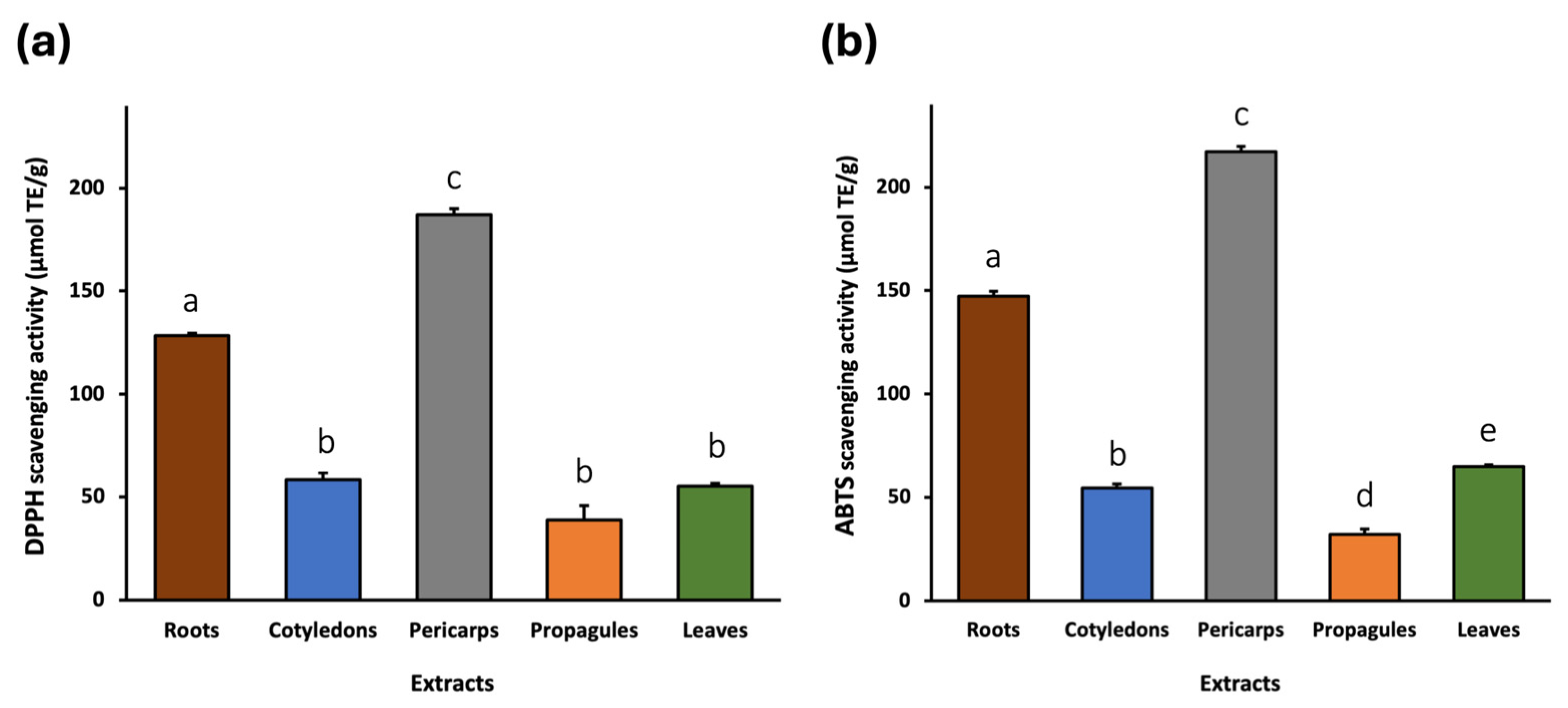
Figure 1. DPPH (a) and ABTS (b) radical scavenging activity of A. marina extracts expressed as μmol Trolox equivalents per gram of sample matrix (μmol TE/g). The bars represent the mean ± standard deviation (SD) from n = 3 independent experiments. Different lowercase letters indicate statistically significant differences between extracts (p < 0.05).
Pharmaceuticals 18 01308 g001
Figure 2. Cell viability (%) of SW480 (a) and E705 (b) human colorectal cancer cell lines treated with A. marina extracts (20–540 μg/mL) for 48 h. Bars represent mean ± standard error of the mean (SEM) from n = 3 independent experiments. Different lowercase letters indicate statistically significant differences between extracts (p < 0.05) and were assigned independently for each concentration.
Pharmaceuticals 18 01308 g002
Figure 3. Cell viability (%) of MDA-MB-231 (a), U-87 (b), and HeLa (c) human cancer cell lines treated with A. marina extracts (20–540 μg/mL) for 48 h. Bars represent mean ± standard error of the mean (SEM) from n = 3 independent experiments. Different lowercase letters indicate statistically significant differences between extracts (p < 0.05) and were assigned independently for each concentration.
Pharmaceuticals 18 01308 g003
Figure 4. Cell viability (%) of CCD 841 (a) and MRC-5 (b) healthy human cell lines treated with A. marina extracts (20–540 μg/mL) for 48 h. Bars represent mean ± standard error of the mean (SEM) from n = 3 independent experiments. Different lowercase letters indicate statistically significant differences between extracts (p < 0.05) and were assigned independently for each concentration.
Pharmaceuticals 18 01308 g004
Figure 5. Cell viability (%) of the cancer cell lines treated with A. marina root extract (180 and 540 μg/mL) for 48 h. Bars represent mean ± standard error of the mean (SEM) from n = 3 independent experiments. Different lowercase letters indicate statistically significant differences between extracts (p < 0.05) and were assigned independently for each concentration.
Pharmaceuticals 18 01308 g005
Table 1. UHPLC-ESI/HRMS data of compounds detected in Avicennia marina extracts. The main fragment ion for each compound is indicated in bold.
No.RT (min)[M − H]FormulaΔ ppmMS/MSNameClassPartILRef.
10.58701.1893
[M + Cl]
C24H42O21−2.1677665.2134, 485.1499, 443.1393, 383.1182, 341.1075, 179.0549StachyoseTetrasaccharidesCotyledons/pericarps/propagules/rootsIL2[41]
20.99191.0188C6H8O76.3850111.0073Citric acidTricarboxylic acidsCotyledons/pericarpsIL2[42]
33.87373.1139C16H22O100.3221211.0605, 167.0700, 149.0597, 123.0440, 105.0333Geniposidic acidIridoid glycosidesLeaves/cotyledons/pericarps/propagules/rootsIL2[43]
44.03353.0875C16H18O90.8633191.0551, 179.0339, 161.0233, 135.0439Caffeoylquinic acid isomerHydroxycinnamic acids and derivativesRootsIL2[44]
54.05375.1291C16H24O101.5167213.0756, 169.0857, 151.0753, 133.0644, 125.0595, 107.0490Mussaenosidic acidIridoid glycosidesLeaves/cotyledons/pericarps/propagulesIL2[45]
64.33375.1285C16H24O103.1119213.0747, 169.0854, 151.0748, 133.0644, 125.0591, 113.0230, 107.0484(Epi)loganic acidIridoid glycosidesLeavesIL2[45]
74.53487.1451C21H28O131.2588179.0334, 161.0228, 135.0435Cistanoside FPhenylethanoid glycosidesPericarpsIL2[46]
84.80327.0715C14H16O91.9983179.0335, 165.0389, 147.0283, 135.0434, 105.0178Unidentified -Leaves
95.09353.0871C16H18O9−3.0904191.0550, 179.0337, 173.0442, 161.0232, 135.0439,Caffeoylquinic acid isomer Hydroxycinnamic acids and derivativesLeaves/cotyledons/pericarps/propagules/rootsIL2[44]
105.47371.0982C15H16O11−8.7858209.0635, 179.0337, 161.0228, 135.0435, 129.0178Caffeoyl hexaric acidHydroxycinnamic acids and derivativesLeavesIL2[47]
115.60443.0655 C18H20O11S−0.3243 275.0218, 167.0338, 152.0105, 123.0440, 108.0204Unidentified-Roots
125.65415.1603C19H28O101.6114235.0963, 191.1062, 173.0958, 149.0953, 137.0590, 101.0226Icariside D1Flavonoid glycosidesLeavesIL2[48]
135.93639.1964C29H36O16−5.2193621.1807, 529.1554, 459.1488, 251.0549, 179.0337, 161.0232, 151.0387Suspensaside isomerPhenylethanoid glycosidesPericarps/rootsIL2[49,50]
145.95639.1964C29H36O16−5.2193621.1807, 529.1554, 459.1488, 251.0549, 179.0337, 161.0232, 151.0387Suspensaside isomerPhenylethanoid glycosidesPericarps/rootsIL2[49,50]
156.14537.1628C25H30O13−2.6672493.1708, 375.1275, 323.0758, 213.0752, 179.0334, 169.0854, 161.0230, 151.0750, 135.0435, 125.0593, 107.0486GrandiflorosideHydroxycinnamic acid and derivativesLeavesIL2[51]
166.33619.1644C29H32O153.9407383.0758, 311.0549, 267.0646Unidentified-Pericarps/roots
176.41639.1929C29H36O160.2477621.1817, 529.1554, 459.1493, 251.0549 179.0338, 161.0236, 151.0385Suspensaside isomerPhenylethanoid glycosidesRootsIL2[49,50]
186.65521.1658C25H30O121.2446357.1176, 169.0854, 163.0385, 151.0749, 145.0280, 125.0591, 119.0486, 117.0329, 107.0486Marinoid CIridoid glycosidesLeaves/cotyledons/pericarpsIL3[7]
196.65653.2091C29H34O17 9.55333621.1822, 459.1499, 179.0338, 161.0234, 151.0388, 135.0437Suspensaside methyl etherPhenylethanoid glycosides RootsIL2[49,50]
206.79623.1981C29H36O150.0705461.1657, 161.0233, 113.0283Verbascoside (acteoside) isomerPhenylethanoid glycosidesLeaves/pericarps/rootsIL2[52]
216.9463.0874C21H20O121.7229301.0324, 300.0264, 271.0235, 255.0285Quercetin 3-O-hexosideFlavonoid glycosides RootsIL2[53,54,55]
227.02667.2239C31H40O16 0.6864621.1824, 459.1499, 179.0338, 161.0235, 151.0386, 135.0436β-ethyl-OH-verbascosidePhenylethanoid glycosides PericarpsIL2[56,57]
237.11623.2001C29H36O15−3.1336461.1661, 161.0235,Verbascoside (acteoside) isomerPhenylethanoid glycosidesLeaves/pericarps/rootsIL2[52]
247.21621.1838C29H34O15−2.0992461.1652, 179.0337, 161.0233, 151.0387Suspensaside APhenylethanoid glycosidesRootsIL2[49,50]
257.21461.0718C21H18O121.6222285.0391Kaempferol-3-O-glucuronideFlavonoid glycosidesLeavesIL2[58]
267.21681.2063C31H38O17−3.9236519.1708, 490.1321, 181.0129, 179.0334, 161.0230Unidentified-Pericarps
277.3447.0926C21H20O111.5287327.0494, 285.0648, 284.0315, 255.0288, 227.0338, 151.0013Kaempferol 3-O-glucosideFlavonoid glycosidesRootsIL2[55,59,60,61]
287.40623.1658C28H32O16−6.4750315.0494, 314.0421, 300.0258, 299.0187, 271.0234Isorhamnetin-3-O-rutinosideFlavonoid glycosidesLeaves/pericarpsIL2[62]
297.40491.0828C22H20O130.6385315.0499, 300.0264Isorhamnetin glucuronideFlavonoid glycosidesLeavesIL2[63]
307.51535.1477C25H28O13 −3.7032329.1021, 179.0338, 161.0232, 149.0595, 135.0438Unidentified-Leaves/Pericarps
317.51477.1036 C22H22O12−5.1250315.0467, 314.0420, 285.0392, 271.0236, 257.0441, 243.0286, Isorhamnetin 7-glucosideFlavonoid glycosidesLeavesIL2[63]
327.61471.1874C22H32O11−0.4545287.1273, 263.1278, 219.1379, 201.1273, 186.1036, 147.1166Unidentified-Pericarps
337.86519.1143C24H24O130.2199315.0472, 314.0423, 299.0186, 285.0383, 271.0236, 257.0443, 243.0286Unidentified -Leaves
347.90553.1556C25H30O141.2256329.1021, 197.0445, 182.0206, 153.0454, 149.0596, 131.0489, Marinoid D Iridoid glycosidesCotyledons/pericarps/propagules/rootsIL3[7]
357.95505.1757C25H29O11 0.2000357.1184, 213.0757, 195.0650, 169.0857, 151.0753, 147.0439, 125.0596, 113.0230, 107.0487, 103.0539Marinoid A Iridoid glycosidesLeavesIL3[7]
368.03519.1505C25H28O12 0.5766313.1072, 295.0961, 163.0388, 149.0596, 145.0282, 131.0490, 119.0487Unidentified-Leaves/cotyledons/pericarps/roots
378.03475.0887C22H20O12−1.0510300.0589, 299.0554, 285.0358, 284.0318. Diosmetin 7-glucuronideFlavonoid glycosidesLeavesIL2[64]
388.24549.1616C26H30O13−0.4279343.1176, 325.1064, 193.0495, 175.0387, 149.0595, 134.0360, 131.0489Unidentified-Leaves/cotyledons/pericarps/roots
398.36591.2119C29H36O13−6.0539179.0333, 161.0234, 133.0282, 113.0228Jionoside C Phenylethanoid glycosidesPericarpsIL2 [65]
408.62825.4276C44H66O160.2536663.3744, 601.3735Unknown triterpene saponin Triterpene saponinsRootsIL3[66,67]
418.70539.2152C26H36O12−3.3318193.0485, 183.1010, 175.0382, 149.0591, 131.0485, 121.0642Unidentified-Leaves
428.80541.2285C26H38O121.0147193.0485, 185.1166, 175.0382, 149.0591, 131.0485, 121.0642Unidentified-Leaves
438.88825.4285C42H66O16−0.8354663.3744, 601.3735, 487.3421Unknown triterpene saponinTriterpene saponinsRootsIL3[66,67]
448.97299.0546C16H12O65.0379285.0345, 284.0313, 256.0363, 227.0334Trihydroxy-methoxyflavoneFlavonesLeavesIL2[68]
458.99825.4273C42H66O160.6166663.3744, 601.3735,Medicoside G (medicagenic acid 3,28-di-glucoside)Triterpene saponinsRootsIL2[66,67]
469.08809.4316C42H66O151.5979689.3884, 647.3788, 629.3680, 585.3786Esculentoside C (phycolaccoside D)Triterpene saponinsCotyledons/pericarps/propagules/rootsIL2[69,70]
479.30505.1711C25H30O110.8600281.1170, 195.0649, 151.0750, 147.0438, 133.0645, 107.0486Unidentified-Leaves
489.38503.1572C25H28O11−2.6077279.1010, 253,0854, 209.0954, 195.0647, 147.0437, 131.0486, 103.0536Unidentified-Leaves/pericarps
499.54809.4342C42H66O15−1.6102647.3797, 471.3469Azukisaponin IIITriterpene saponinsRootsIL2 [71]
Table 2. Spearman correlation coefficients (ρ) between the number of compounds per chemical class and the antioxidant activity (DPPH and ABTS assays). Statistically significant correlations are indicated in bold (p value < 0.05).
Compound ClassDPPHABTS
ρ-Valuep-Valuep-Valuep-Value
Iridoid glycosides−0.1030.8700.5100.935
Hydroxycinnamic acid and derivatives−0.1120.8580.2240.718
Phenylethanoid glycosides0.7910.1110.9490.014
Flavonoid glycosides0.2050.7410.5740.322
Triterpene saponins0.4470.4500.2240.718
Disclaimer/Publisher’s Note: The statements, opinions and data contained in all publications are solely those of the individual author(s) and contributor(s) and not of MDPI and/or the editor(s). MDPI and/or the editor(s) disclaim responsibility for any injury to people or property resulting from any ideas, methods, instructions or products referred to in the content.