Article

New Nitrogen, Oxygen and Sulfur-Containing Heterocyclic Compounds as Anti-colon Cancer Agents: Synthesis, Multitargeted Evaluations, Molecular Docking Simulations and ADMET Predictions

by
Nahed Nasser Eid El-Sayed
1,*,
Najeh Krayem
2,
Hamed Ahmed Derbala
3,
Shimaa Kamal
3,
Syde Nasir Abbas Bukhari
4,
Mohamed K. El-Ashrey
5,6,
Zainab M. Almarhoon
7,
Seham Soliman Alterary
7 and
Abir Ben Bacha
8,*
1
Egyptian Drug Authority, 51 Wezaret El-Zerra St., Giza 35521, Egypt
2
Laboratoire de Biochimie et de Génie Enzymatique des Lipases, ENIS, Université de Sfax, Route de Soukra 3038, Sfax BP 1173, Tunisia
3
Chemistry Department, Faculty of Science, Ain Shams University, Abbassia, Cairo 11566, Egypt
4
Department of Pharmaceutical Chemistry, College of Pharmacy, Jouf University, Sakaka 72388, Saudi Arabia
5
Pharmaceutical Chemistry Department, Faculty of Pharmacy, Cairo University, Kasr Elini St., Cairo 11562, Egypt
6
Medicinal Chemistry Department, Faculty of Pharmacy, King Salman International University, South Sinai 46612, Egypt
7
Department of Chemistry, College of Science, King Saud University, P.O. Box 2455, Riyadh 11451, Saudi Arabia
8
Biochemistry Department, College of Sciences, King Saud University, P.O. Box 22452, Riyadh 11495, Saudi Arabia
*
Authors to whom correspondence should be addressed.
Pharmaceuticals 2025, 18(6), 801; https://doi.org/10.3390/ph18060801 (registering DOI)
Submission received: 8 April 2025 / Revised: 11 May 2025 / Accepted: 19 May 2025 / Published: 27 May 2025
(This article belongs to the Special Issue Heterocyclic Compounds in Medicinal Chemistry, 2nd Edition)

Abstract

:
Background/Objectives: Oxidative stress, the Warburg effect, and resistance to apoptosis are key hallmarks driving colorectal tumorigenesis. This study aimed to develop novel multi-target compounds capable of modulating these pathways. Methods: A library of 24 newly synthesized compounds—incorporating annulated thiophene, thiazole, quinazolinone, 2-oxoindoline, and 1,2,3-oxadiazole scaffolds, as well as N-(1-(4-hydroxy-3-methoxyphenyl)-3-oxo-3-(2-(phenylcarbamothioyl)hydrazineyl) prop-1-en-2-yl)benzamide—was evaluated for antioxidant activity (DPPH assay), PDK-1 and LDHA inhibition, cytotoxic effects against LoVo and HCT-116 colon carcinoma cells, with parallel assessment of safety profiles on normal HUVECs. The underlying anticancer mechanism of the most active compound was investigated through analysis of cell cycle distribution, apoptosis induction, intracellular reactive oxygen species levels, mitochondrial membrane potential disruption, and expression levels of apoptosis-related genes. Molecular docking assessed binding interactions within LDHA and PDK-1 active sites. The physicochemical, drug-likeness, and ADMET properties of the multi-bioactive candidates were predicted in silico. Results: Among the synthesized compounds, thiophenes 3b and 3d exhibited superior PDK-1/LDHA and DPPH/LDHA inhibitions along with significant cytotoxic effects on LoVo and HCT-116 cells (IC50 in µM: 190.3/170.2 and 161.0/156.6), respectively, and minimal cytotoxicity toward HUVECs. Molecular docking revealed favorable interactions with key amino acid residues within the LDHA and/or PDK-1 active sites. Compound 3d notably induced G2/M (LoVo) and G1 (HCT-116) arrest and promoted apoptosis via enhancing ROS generation, modulating Bax/Bcl-2 expressions, disrupting mitochondrial membrane potential, and ultimately activating caspses-3. In silico predictions indicated their promising drug-likeness and pharmacokinetics, though high lipophilicity, poor solubility (especially for 3b), and potential toxicity risks were identified as limitations. Conclusions: Thiophenes 3b and 3d emerged as promising multi-target candidates; however, structural optimization is warranted to enhance their solubility, bioavailability, and safety to support further development as lead anti-colon cancer agents.

Graphical Abstract

1. Introduction

Colorectal cancer (CRC) ranks as the third most commonly diagnosed malignancy in men and the second in women, and it remains the second-leading cause of cancer-related deaths worldwide [1]. Approximately 25% of patients are diagnosed at an advanced stage (stage IV), where the five-year survival rate drops below 14% [2]. Despite the availability of treatments such as surgery, chemotherapy, and targeted therapies, clinical outcomes remain suboptimal due to systemic toxicity, therapeutic resistance, poor long-term survival, and high recurrence rates [3]. These challenges highlight the need for novel strategies targeting the complex and multifactorial nature of CRC pathogenesis.
CRC arises from the dysregulation of multiple intracellular signaling pathways [4], enabling normal cells to acquire abnormal capabilities—or hallmarks—associated with malignancy [5].
One major contributor to CRC initiation, progression, and maintenance is oxidative stress (OS), a pathological condition marked by excessive production of reactive oxygen species (ROS) that overwhelm the cell’s antioxidant defenses. In intestinal mucosal cells, this ROS overproduction leads to oxidative DNA damage and promotes chronic inflammation [6], both of which contribute to malignant transformation. Moreover, OS supports tumor development through several mechanisms, including resistance to cell death, stimulation of proliferation, stress tolerance, angiogenesis, invasion, migration, and recurrence [7]. Accordingly, antioxidant compounds have garnered attention as potential chemopreventive agents against CRC [8].
CRC is also characterized by a distinct metabolic hallmark known as the Warburg effect, wherein cancer cells favor glycolysis over oxidative phosphorylation for ATP production—even under normoxic conditions [5]. This metabolic shift is driven largely by the upregulation of key glycolytic regulators such as lactate dehydrogenase A (LDHA) and pyruvate dehydrogenase kinase-1 (PDK-1) [9]. These enzymes facilitate the conversion of pyruvate to lactate, leading to promoting its accumulation. Lactate, an oncogenic metabolite, acidifies the tumor microenvironment, resulting in immune evasion, tumor progression, metastasis, and resistance to chemotherapy [10]. Consequently, impeding lactate production via the inhibition of LDHA and PDK-1 has emerged as a promising anticancer strategy [11].
Furthermore, CRC cells often harbor mutations in tumor suppressor genes such as APC, TP53, KRAS, and CDKN2A [5,12,13], which enable them to bypass cell cycle checkpoints, leading to uncontrolled proliferation [5]. In parallel, evasion of apoptosis is frequently observed, facilitated by dysregulation of pro-apoptotic (e.g., Bax, Bak) and anti-apoptotic (e.g., Bcl-xL, Bcl-2) proteins in the intrinsic pathway or by disruption of death-inducing signaling complexes in the extrinsic cascade [5,14]. This imbalance between proliferation and apoptosis plays a central role in tumor development, drives disease progression, and contributes to resistance to chemotherapy and radiotherapy [15]. As such, therapeutic approaches that simultaneously target cell cycle regulation and apoptotic pathways are being increasingly explored for effective CRC management [16].
Given these interconnected pathogenic features, the development of multitarget agents capable of modulating several cancer hallmarks represents a compelling strategy [17]. In this context, heterocyclic compounds containing nitrogen, oxygen, or sulfur have emerged as versatile scaffolds capable of modulating diverse cellular targets implicated in carcinogenesis [18,19,20,21,22,23].
In continuation of our previous efforts targeting CRC [24], we report the design and synthesis of four novel series of nitrogen-, oxygen-, and sulfur-containing five- and six-membered heterocycles as potential multitarget therapeutic agents. These compounds were evaluated for their antioxidant activity, inhibition of PDK-1 and LDHA, as well as their antiproliferative effects against HCT-116 and LoVo colon carcinoma cell lines. To assess selectivity and safety, cytotoxicity was also tested against normal human umbilical vein endothelial cells (HUVECs).
The lead antiproliferative compound was further investigated for its ability to disrupt cell cycle progression and induce apoptosis in CRC cells. Additionally, molecular docking simulations were performed to elucidate the interactions of active compounds with their enzymatic targets. Finally, in silico predictions of physicochemical properties, drug-likeness, ADME, and toxicity risks were conducted to assess the drug development potential of the lead multitarget candidates.

2. Results and Discussion

2.1. Chemistry

The synthetic routes leading to the new target compounds 3ae, 8, 9ac, 13, 14, 17, 18ah, 20, 22, 23, and 25 are depicted in Scheme 1, Scheme 2 and Scheme 3. The starting heterocyclic compounds used in this study—including 2-aminothiophenes 1ac [25,26,27], 2-aminothiazole 4 [28], 4-(benzo[d]thiazol-2-yl)aniline 11 [29], oxazinones 10a,b and the corresponding 3-aminoquinazolinones 15a,b [30,31], as well as azlactone 19 and its hydrazide derivative 23 [32]—were synthesized according to previously reported procedures.
Initially, equimolar amounts of the appropriate fused thiophene-based amine 1ac and aromatic aldehyde 2ad were heated under reflux in absolute ethanol containing a catalytic amount of glacial acetic acid for 12 h, yielding the thiophene–Schiff base-conjugates 3ae (Scheme 1). Meanwhile, the reaction of 2-aminothiazole 4 with chloroacetyl chloride 5 in dry chloroform and in the presence of Et3N yielded chloroacetamide intermediate 6, which subsequently condensed with thioglycolic acid 7 in pyridine to furnish ethyl 2-(3,5-dioxothiomorpholino)-4-methylthiazole-5-carboxylate 8. Additionally, the condensation of amine 4 with aromatic aldehydes 2a, 2c, and 2d in absolute ethanol containing a catalytic amount of glacial acetic acid afforded Schiff bases 9ac (Scheme 1).
The fusion of oxazinone 10a with 4-(benzo[d]thiazol-2-yl)aniline 11 or cyclohexyl amine 12 led to the formation of 3-substituted-6,7-dimethoxy-2-methyl quinazolin-4(3H)-one derivatives 13 and 14, respectively. Also, the reaction of oxazinones 10a,b with excess hydrazine hydrate afforded the corresponding 3-aminoquinazolinones 15a,b. Next, refluxing 3-aminoquinazolinone 15a with excess ethyl chloroformate 16 yielded the carbamate derivative 17, and eight new Schiff bases, 18ah, were synthesized through the condensation of 3-aminoquinazolinones 15a,b with the appropriate aryl and/or heteroaryl aldehydes 2b,2dh in absolute ethanol containing a catalytic amount of glacial acetic acid (Scheme 2).
In a separate sequence (Scheme 3), (Z)-2-methoxy-4-((5-oxo-2-phenyloxazol-4(5H)-ylidene)methyl) phenyl acetate 19 underwent ring opening upon reaction with ethyl 2-amino-4-methylthiazole-5-carboxylate 4, affording the corresponding thiazolyl benzamidoacrylamide 20. Meanwhile, treatment of compound 19 with excess hydrazine hydrate at room temperature led to ring opening and deacylation of the ester group, yielding hydrazide 21. Cyclocondensation of the latter compound with carbon disulfide in the presence of KOH afforded 2-thioxo-1,3,4-oxadiazole derivative 22. Furthermore, hydrazide 21 was converted into the N-phenylthiosemicarbazide derivative 23 through reaction with phenyl isothiocyanate. Finally, condensation of hydrazide 21 with isatin derivative 24 produced 2-oxoindolyl hydrazone 25 (Scheme 3).
The structures of all newly synthesized compounds were confirmed by FT-IR, 1H and 13C-NMR and MS analyses as detailed in Section 3; representative spectra are provided in the Supplementary Materials.
Notably, due to the restricted rotation around the imine (C=N) group, two geometric isomers—E and Z—are theoretically possible for the aldimines 3ae, 9ac, and 18ah. However, these compounds were obtained as single geometrical isomers, evidenced by their 1H-NMR spectra. Additionally, energy minimization and total energy calculations (Table S1) revealed that the Z-isomers possessed higher total energies and were therefore less stable than their E counterparts. This reduced stability is likely due to steric repulsion between the bulky heterocyclic and aryl groups across the C=N bond [33], thus favoring the E-configuration for these aldimines.
Considering compounds 20, 21, 22, 23, and 25, the configuration of the olefinic double bond within the conjugated side chains was assigned as Z, consistent with the geometry of the parent 4-arylidene-2-phenyloxazol-5(4H)-one [34].
In addition, the geometry around the double bond of the 2-oxoindolin-3-ylidene)hydrazineyl fragment in compound 25 was assigned a Z-configuration based on previous studies of related compounds, which reported that the hydrazide-NH and the 2-oxoindoline carbonyl group form an intramolecular hydrogen bond that enhances stabilization [35]. This assignment is further supported by energy minimization data, which indicate that the Z,Z isomer possesses lower energy than the Z,E isomer (Table S1).

2.2. In Vitro Biological Evaluations

The results of various biochemical assays for the newly synthesized compounds, 3ae, 8, 9ac, 13, 14, 17, 18ah, 20, 2223, and 25, are summarized in Tables S2–S6.
Notably, in the enzyme inhibition and viability assays, the initial screening concentration of (100 µg/mL) was selected based on our standard practice in early-stage multitarget drug discovery [24]. This approach facilitates the identification of hit compounds by enabling rapid and consistent comparison across structurally diverse candidates prior to detailed dose-response analysis.

2.2.1. Antioxidant Activity

The antioxidant activities of the studied compounds were assessed using the 1,1-diphenyl-2-picrylhydrazyl (DPPH) radical scavenging method as detailed by Bersuder et al. [36]. The results were expressed as the concentration of the test compound required to neutralize 50% of DPPH radicals (IC50, mM). As shown in Table S2, the compounds exhibited variable degrees of scavenging power, with IC50 values spanning from 0.164 ± 0.008 to 1.811 ± 0.115 as compared to 0.245 ± 0.027 mM for the reference antioxidant, butylated hydroxytoluene (BHT). In detail, the thiophenes 3ae, thiazoles 8 and 9ac, and quinazolinones 13, 14, 17, and 18ah, along with benzamide derivatives 20, 22, 23, and 25, demonstrated IC50 values ranging from 0.164 ± 0.008 to 0.890 ± 0.039, 0.881 ± 0.041 to 1.811 ± 0.115, 0.188 ± 0.010 to 1.820 ± 0.188 and 0.162 ± 0.023 to 1.211 ± 0.030 mM, respectively. Among them, compounds 3a, 3d, 18b, and 18g demonstrated better DPPH radical scavenging activity than BHT, with lower IC50 values of 0.214 ± 0.027, 0.164 ± 0.008, 0.188 ± 0.010, and 0.238 ± 0.006 mM, respectively. In contrast, none of the thiazole-based derivatives 8, 9ac and 20 exhibited significant antioxidant activity, despite the fact that the Schiff bases 9ac were derived from the same aldehydes as the most potent scavengers 3a, 3d and 18b, respectively.

2.2.2. PDK-1 and LDHA Inhibitory Activities

The same set of compounds was initially screened for their inhibitory effects on PDK-1 and LDHA at a concentration of 100 µg/mL, and the results are listed in Table S3.
In the PDK-1 assay, thiophene 3b along with quinazolinones 14 and 17 showed the highest inhibition percentages, with values of 94.65 ± 2.33, 80.50 ± 4.95, and 91.00 ± 2.83% of inhibition, respectively, compared to 100% inhibition by the reference compound sodium dichloroacetate (SDA) at 150.92 µg/mL. The rest of the compounds exhibited weak to moderate inhibition, with percentages ranging from 7.60 ± 0.85 to 72.67 ± 2.08.
Similarly, in the LDHA assay, thiophenes 3b and 3d were the most potent inhibitors, with inhibition rates of 82.75 ± 3.18 and 85.90 ± 2.97, respectively, compared to 100% inhibition achieved by the standard inhibitor sodium oxamate (SO) at 111.03 µg/mL. The rest of the compounds showed inhibition ranging from 5.75 ± 0.35 to 67.35 ± 3.32%.
For compounds showing >80% inhibition of either enzyme, IC50 values (µM) were determined (Table S3). Thus, IC50 values against PDK-1 for compounds 3b, 3d, 14, and 17 were 147.29 ± 5.95, 305.38 ± 11.07, 192.08 ± 5.57, and 168.86 ± 1.81, respectively, as compared to 170.62 ± 7.02 µM for SDA. Against LDHA, their IC50 (µM) values were 166.79 ± 10.10, 149.166 ± 7.94, 378.01 ± 3.98, and 1059.71 ± 7.64, respectively, as compared to 177.19 ± 7.66 for SO.
Overall, among all the studied compounds, thiophene derivative 3b was identified as the most promising dual inhibitor of PDK-1 and LDHA. Although thiophene 3d exhibited weak PDK-1 inhibition, it was the most potent LDHA inhibitor. These findings are notable, as several studies highlighted a strong correlation between PDK-1 inhibition and the reduction of cellular proliferative tumorigenic potential as well as enhanced radiosensitization in various tumor cell lines [37,38]. Similarly, previous in vitro and in vivo xenograft models and preclinical studies have shown that the reduction of LDHA expression and inhibition of its activity in cancer cells stimulates mitochondrial OXPHOS, leading to increased mitochondrial ROS production, which in turn triggers apoptosis-mediated cell death [39,40] and enhances the efficacy of radiotherapy [41].

2.2.3. Cytotoxic Activities

The studied compounds were also screened for their cytotoxic effects on HCT-116 and LoVo colon carcinoma cells, as well as normal HUVECs, using the standard LDH assay [42] at a concentration of 100 µg/mL. Results are reported as the mean percentage of viable cells after 48 h of treatment (Table S4). Notably, compounds 3b, 3d, 14, 17, 18d, 18h, and 22 induced significant cytotoxicity in LoVo cells, reducing cell viability to 28.33 ± 1.41, 19.67 ± 2.83, 13.00 ± 1.14, 10.50 ± 0.71, 30.67 ± 4.24, 33.00 ± 4.24, and 29.50 ± 2.12%, respectively, compared to 99.50 ± 0.71% (assay medium, negative control) and 0% (0.1% Triton X-100, positive control). Against HCT-116 cells, compounds 3b, 3d, 8, 14, 17, 18d, 18h, and 23 reduced cell viability to 20.67 ± 2.08, 21.67 ± 2.08, 26.5 ± 2.12, 12.50 ± 0.71, 8.50 ± 0.71, 38.00 ± 2.00, 37.00 ± 2.00, and 23.00 ± 2.83%, respectively, compared to 100.00% (negative control) and 0% (positive control). The remaining compounds exhibited moderate to weak cytotoxic activities, with viabilities ranging from 48.33 ± 2.83 to 95.67 ± 1.53% (LoVo cells) and 40.50 ± 3.54 to 89.33 ± 4.16% (HCT-116 cells). Notably, all tested compounds showed limited cytotoxicity toward normal HUVECs, with cell viabilities ranging from 96.67 ± 1.16 to 70.33 ± 1.53%.
Based on these results, the mean IC50 values (µM) were determined for the ten most active compounds (i.e., those reducing viability to ≤38%) and are presented in Table S5. These compounds demonstrated variable sensitivity, with IC50 values spanning from 132.23 ± 7.40 to 479.17 ± 19.18 µM (LoVo) and from 160.96 ± 5.43 to 510.84 ± 5.61 µM (HCT-116), compared to 19.99 ± 3.21 and 26.98 ± 1.87 µM, respectively, for 5-fluorouracil. Among all the tested compounds, quinazolinone 18a was the most effective against LoVo cells (IC50 = 132.23 ± 7.40 µM), while thiophene 3d was the most potent against HCT-116 (IC50 = 160.96 ± 5.43 µM) and the second most active compound against LoVo cells (IC50 = 156.60 ± 5.22 µM).

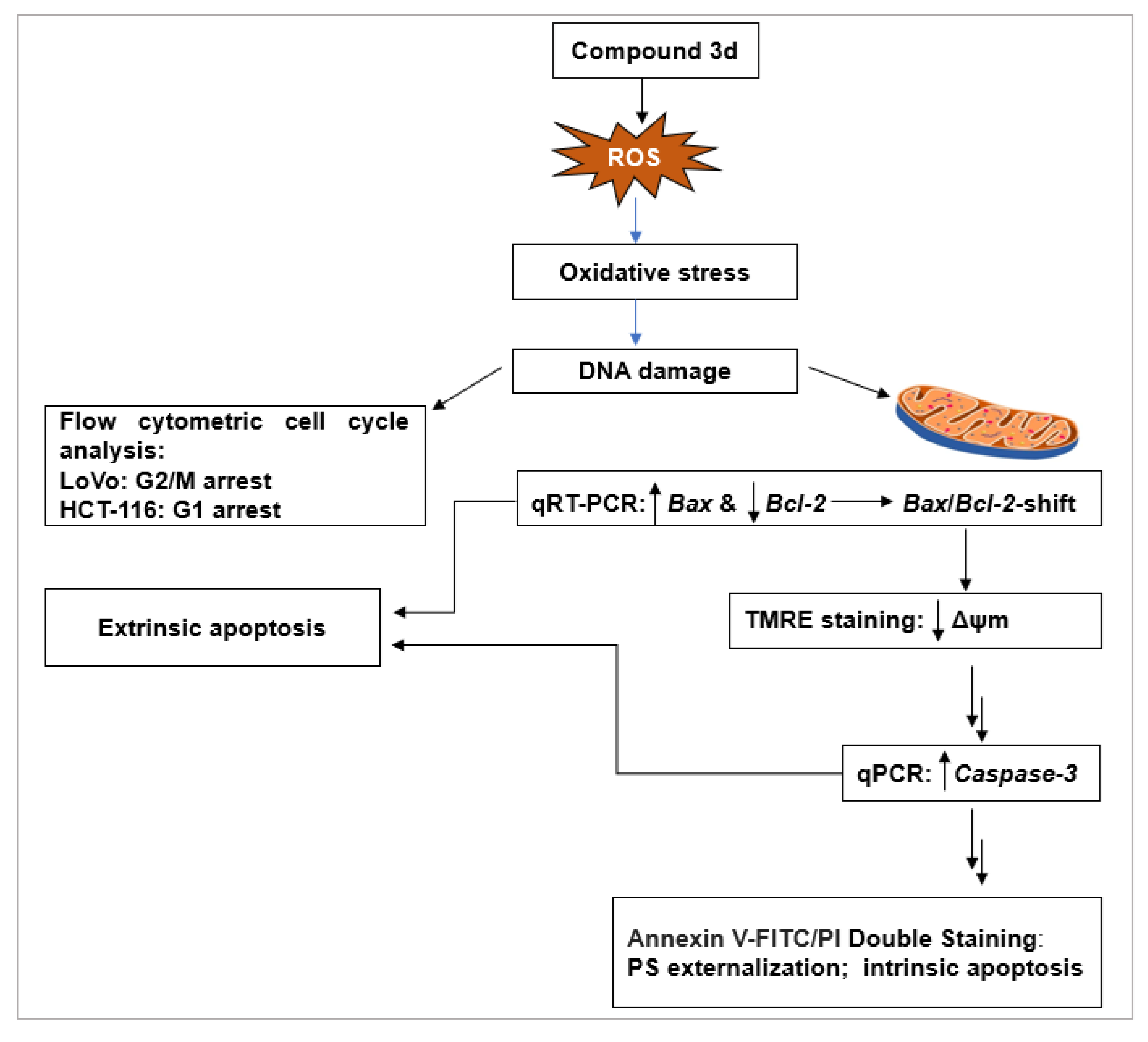
2.2.4. Investigation of Anticancer Mechanism of 3d

Cell cycle regulation and apoptosis are closely interconnected processes that play critical roles in carcinogenesis. Disruption of genomic integrity through severe or irreparable DNA damage can lead to the unchecked proliferation of abnormal cells, thereby contributing to tumor development. Conversely, the activation of cell cycle checkpoints and arrest, along with the induction of apoptosis, functions as a crucial negative regulatory mechanism to eliminate damaged cells and prevent malignant transformation [43]. Accordingly, these mechanisms are key targets in cancer therapy and active areas of research, leading to the identification of many chemotherapeutic agents and candidates, whose anticancer effects rely on inhibiting cell proliferation, inducing cell cycle arrest, and/or promoting apoptotic cell death [15,16,17]. On this basis, and owing to its significant cytotoxic activity against both LoVo and HCT-116 colorectal cancer cell lines, compound 3d was selected for further mechanistic investigation to elucidate its mode of action by evaluating its ability to induce cell cycle arrest and/or trigger apoptosis.

Effect of Compound 3d on Cell Cycle Progression

Cell proliferation depends on orderly progression through the phases of the cell cycle (G1−S−G2−M). Cytotoxic agents can impair the integrity of DNA, leading to checkpoint arrest at G1/S or G2/M, which subsequently blocks proliferation [43]. To evaluate the impact of compound 3d on cell cycle distribution, colon carcinoma cells were treated with 30 µg/mL of 3d for 48 h, followed by DNA content analysis using flow cytometry. As shown in DNA histograms (Figure 1A–D) and a bar chart (Figure 1E), treatment with 3d increased the proportion of LoVo cells in the G2/M phase to 24.06 ± 1.32%, compared to 11.51 ± 1.24% in untreated control cells. In contrast, in HCT-116 cells, 3d caused an accumulation in the G1 phase, reaching 60.17 ± 1.44% versus 47.37 ± 1.68% in untreated cells. These results suggest that compound 3d inhibits proliferation through the induction of G2/M arrest in LoVo cells and G1 arrest in HCT-116 cells.

Induction of Apoptosis by Compound 3d

Apoptosis is a tightly regulated, multistep process orchestrated through two main signaling pathways: the intrinsic (mitochondrial) and extrinsic (death receptor-mediated) [44,45]. Both pathways are tightly regulated by molecular checkpoints, most notably the B-cell lymphoma 2 (BCL-2) protein family and caspases. The B-cell lymphoma 2 (BCL-2) family of proteins comprises pro-apoptotic (e.g., Bax, Bak, and Bok) and anti-apoptotic (e.g., Bcl-2 and Bcl-xL) members [46]. The second pivotal regulatory family, caspases—cysteine-aspartic proteases—are broadly categorized into initiator caspases (Casp-2, -8, -9, and -10) and executioner caspases (Casp-3, -6, and -7) [47]. It is well established that cancer cells have a compromised antioxidant defense system and require higher levels of reactive oxygen species (ROS) to promote mitogenic effects and cellular proliferation compared to normal cells [48]. However, when ROS accumulated beyond a critical threshold, oxidative stress arises—a key initiator of both apoptotic cell mechanisms.
In the intrinsic pathway, ROS levels are augmented in mitochondria (due to external agents such as cytotoxic agents and UV radiation). This disrupts cellular redox homeostasis and damages mitochondrial components, which, in turn, promotes the activation of pro-apoptotic BCL-2 family proteins such as Bax. Activated Bax induces mitochondrial outer membrane permeabilization (MOMP), facilitating the release of apoptogenic factors like cytochrome c and apoptosis-inducing factor (AIF) into the cytosol. Cytochrome c then associates with apoptotic protease-activating factor 1 (Apaf-1) to activate initiator Casp-9, which subsequently activates executioner Casp-3. Activated Casp-3 cleaves cellular substrates, driving the morphological and biochemical hallmarks of apoptosis [45]. Conversely, anti-apoptotic proteins such as Bcl-2 can preserve mitochondrial integrity to inhibit this cascade [49]. Therefore, the Bax/Bcl-2 ratio is a critical determinant of mitochondrial-mediated apoptosis.
In the extrinsic pathway, ROS modulates signaling molecules involved in this cascade [3,50], thereby enhancing the expression or activation of death receptors and promoting the binding of extracellular death ligands to their receptors on the cell surface, forming the death-inducing signaling complex (DISC). This complex activates initiators Casp-8 and -10, which regulate the expression of executioner Casp-3, -6 and -7. Therefore, both apoptosis pathways converge at Caps-3, the central executioner caspase.
In light of these findings, to elucidate whether the cytotoxic activity of compound 3d is associated with apoptosis induction, we investigated multiple apoptotic markers [51,52].
  • ROS Generation as an Upstream Apoptotic Trigger
To determine whether compound 3d induces oxidative stress, intracellular ROS levels were quantified after treatment (30 µg/mL, 48 h) using a human ROS ELISA kit. As shown in Table S6, compound 3d significantly increased ROS levels to 434.5 pg/mL in LoVo cells and 290.1 pg/mL in HCT-116 cells, compared to 168.6 pg/mL and 143.5 pg/mL in the respective controls. These results suggest that ROS accumulation serves as a likely upstream trigger of apoptosis.
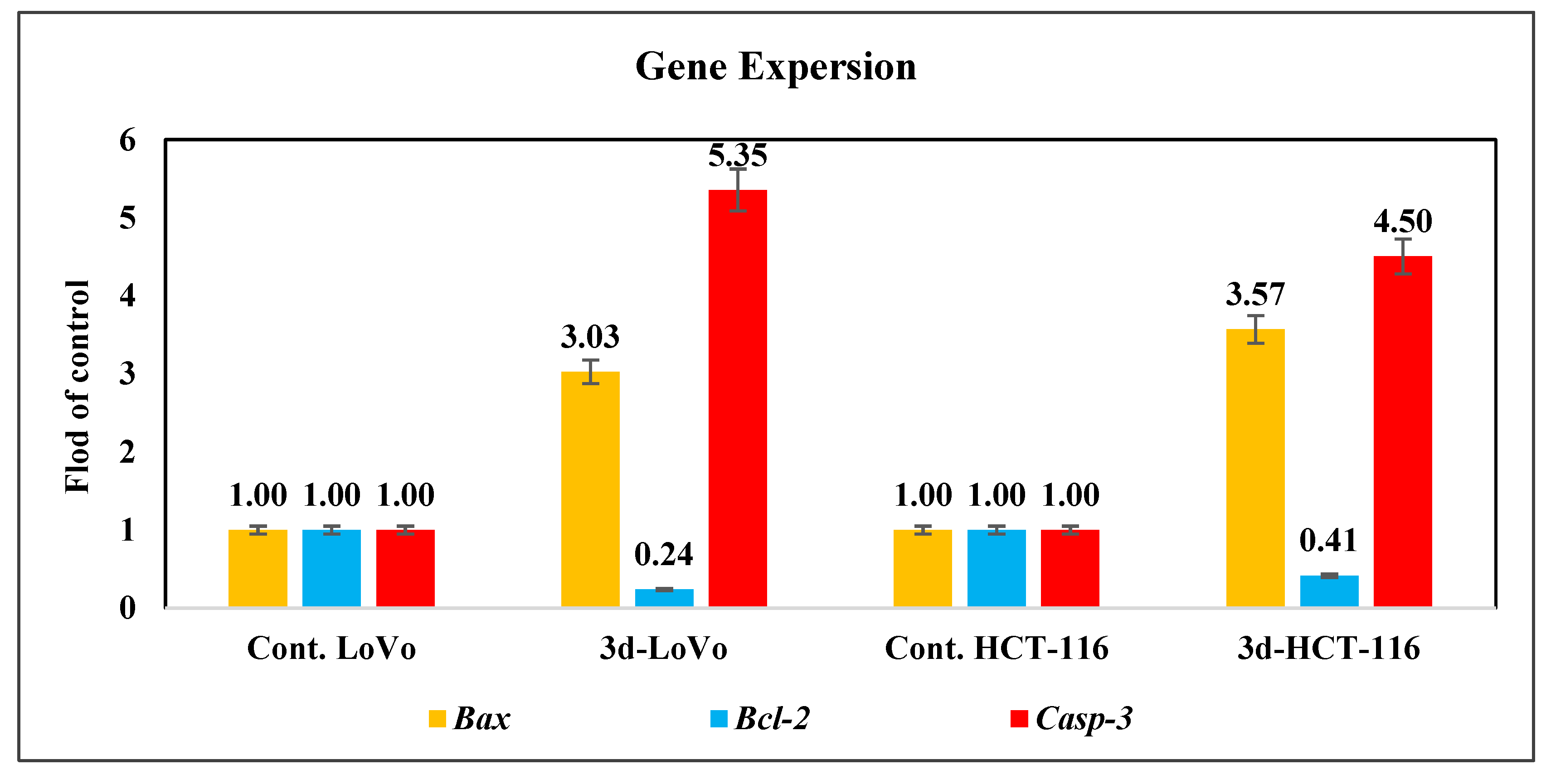
  • Modulation of Apoptosis-Related Gene Expression
To investigate molecular events downstream of ROS generation, we evaluated mRNA expression levels of key regulators of mitochondrial apoptosis—Bax and Bcl-2—using qRT-PCR. This analysis revealed that compound 3d significantly upregulated Bax mRNA levels by 3.03 ± 0.41-fold in LoVo cells and 3.57 ± 0.18-fold in HCT-116 cells. Simultaneously, Bcl-2 expression was significantly downregulated to 0.24 ± 0.03-fold and 0.41 ± 0.04-fold, respectively (Figure 2). As a result, the Bax/Bcl-2 ratios were calculated to be 12.63 in LoVo cells and 8.71 in HCT-116 cells, reflecting a pronounced pro-apoptotic shift. This alteration in the Bax/Bcl-2 ratio strongly supports the activation of the mitochondrial apoptotic pathway.
  • Loss of Mitochondrial Membrane Potential (ΔΨm)
To assess mitochondrial integrity, TMRE staining was used to measure ΔΨm. Treatment with compound 3d (30 µg/mL, 48 h) significantly reduced ΔΨm by 60.23 ± 1.87% in LoVo cells and 43.47 ± 0.81% in HCT-116 cells, supporting mitochondrial depolarization as a key mechanistic event consistent with the observed increase in ROS levels.
  • Upregulation of Caspase-3 Expression
To confirm activation of the execution phase of apoptosis, Casp-3 mRNA levels were evaluated by qRT-PCR. Treatment with compound 3d significantly upregulated Casp-3 expression by 5.35 ± 0.33-fold in LoVo cells and 4.50 ± 0.40-fold in HCT-116 cells (Figure 2), verifying the engagement of caspase-mediated apoptotic execution.
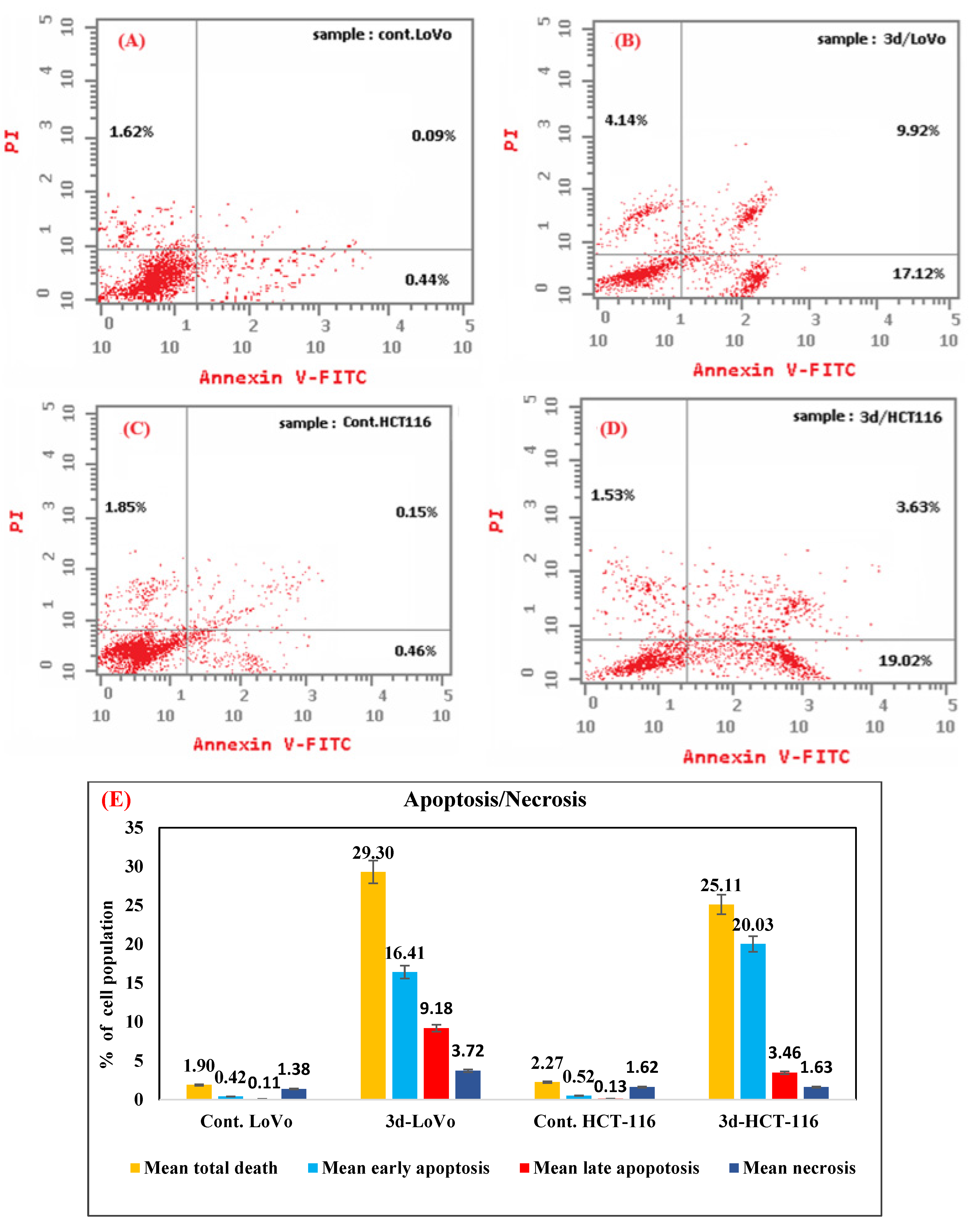
  • Phosphatidylserine Externalization by Annexin V-FITC/PI Staining
Translocation of phosphatidylserine (PS) residues on the cell surface is an early apoptosis indicator, detectable through flow cytometric analysis and using double staining with propidium iodide (PI) dye and fluorescein isothiocyanate (FITC)-conjugated Annexin V (Annexin V-FITC) [53].
The results of this assay (Figure 3A–E) revealed increased apoptotic populations. In LoVo cells, early (Annexin-V-FITC positive and PI negative) and late apoptotic cells (both Annexin-V and PI positive) increased to 17.12% and 9.92%, respectively, compared to 0.44% and 0.09% in the controls (Figure 3B vs. Figure 3A). Similarly, HCT-116 cells exhibited 19.02% early and 3.63% late apoptotic populations, compared to 0.46 and 0.15, respectively, in untreated controls (Figure 3D vs. Figure 3C). Meanwhile, the necrotic population (Annexin-V−/PI+) accounted for 4.14% and 1.53% of cell death in LoVo and HCT-116 cells, respectively (Figure 3B and 3D). Additionally, total apoptosis reached 29.30% and 25.11% in LoVo and HCT-116 cells, respectively, compared to necrotic percentages of 1.38 and 1.63, (Figure 3E), confirming apoptosis as the predominant form of cell death.
Collectively, the anticancer effects of compound 3d are mediated through dual complementary mechanisms. First, it suppresses cell proliferation by inducing G2/M arrest in LoVo cells and G1 arrest in HCT-116 cells. Second, it promotes cell death primarily through apoptosis, with minimal necrosis observed. Apoptosis was characterized by hallmark features consistent with the intrinsic (mitochondrial) pathway, including increased ROS generation, mitochondrial membrane depolarization, upregulation of caspase-3, and a pronounced pro-apoptotic shift in the Bax/Bcl-2 ratio—calculated as 12.63 in LoVo and 8.71 in HCT-116 cells. The concurrent upregulation of Bax and caspase-3 may also suggest potential crosstalk with the extrinsic pathway [54]. Overall, the significant increase in Annexin V-positive cells, in conjunction with other apoptotic markers, confirms that apoptosis as the predominant mode of cell death induced by compound 3d in LoVo and HCT-116 cells (Figure 4).

2.3. In Silico Studies

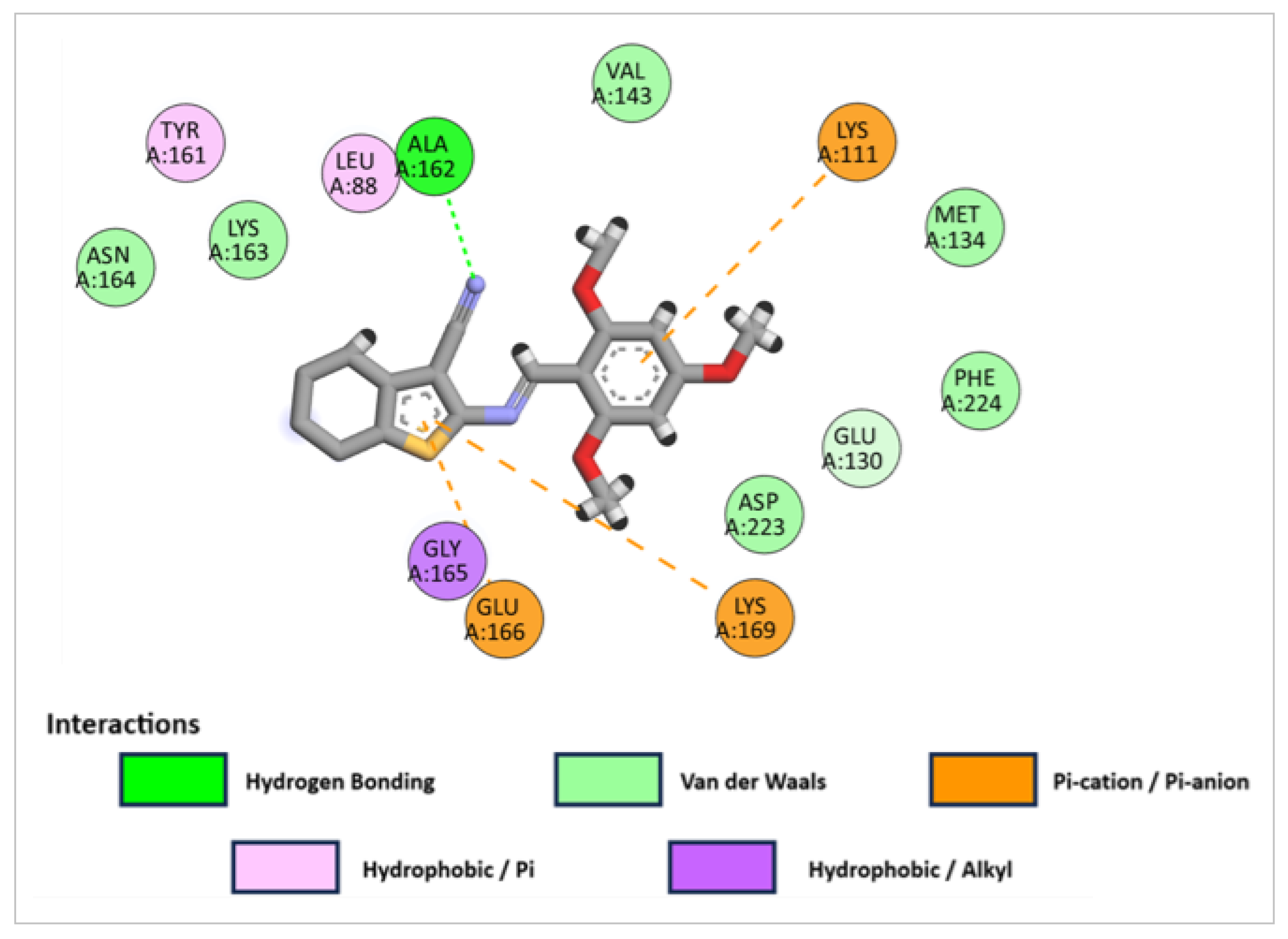
2.3.1. Molecular Docking Studies

The in vitro inhibitory efficiencies of compounds 3b and 3d against LDHA, and 3b against PDK-1, as revealed by enzymatic assays were further substantiated through molecular docking simulations, which enabled the estimation of binding affinities and prediction of the binding modes within the active sites of the target enzymes. To validate the docking protocol, redocking experiments were performed using the co-crystallized ligands: GN0 for LDHA (PDB ID: 4ZVV) [55] and AZX for PDK-1 (PDB ID: 2Q8G) [56]. The computed root mean square deviation (RMSD) values were 0.79 Å for LDHA and 1.65 Å for PDK-1, both falling within the acceptable threshold (<2.0 Å) for reliable docking performance [57], thus confirming the suitability of the protocol (Figure 5).
Docking results against LDHA revealed that both 3b and 3d (Figure 6) exhibited favorable binding energies of −8.1 and −7.9 kcal/mol, respectively, comparable to that of the native ligand GN0 (−8.8 kcal/mol). These compounds formed key interactions with residues critical to LDHA inhibition, including Asn137 and Arg168.
Compound 3b formed a hydrogen bond between one of its methoxy oxygen atoms with Arg168, while its imino nitrogen and thienyl sulfur participated in polar interactions with Asn137 and His192.
In the case of 3d, the imino nitrogen at position-3 of the thienopyrimidine ring interacted via hydrogen bonding with Arg168. Additional intermolecular bonding through π-anion and hydrophobic interactions were observed involving Asn137, His192, and Asp165. These interactions suggest that both compounds occupy a similar binding pocket to GN0, engaging residues crucial for LDHA inhibition and complex stability. These results align with findings by Sharma, who identified the pharmacophoric features (aromatic rings and H-bond acceptors) and the residues essential for LDHA inhibition [58].
In the case of PDK-1, compound 3b also exhibited a favorable docking score (−7.9 kcal/mol), though slightly higher than that of the reference ligand AZX (−8.4 kcal/mol). It established a hydrogen bond between its methoxy oxygen and Ala162 residue; meanwhile its aryl rings engaged in pi-cation interactions with the basic amino acids Lys111 and Lys169. These main interactions are supported by additional hydrophobic contacts (Figure 7).

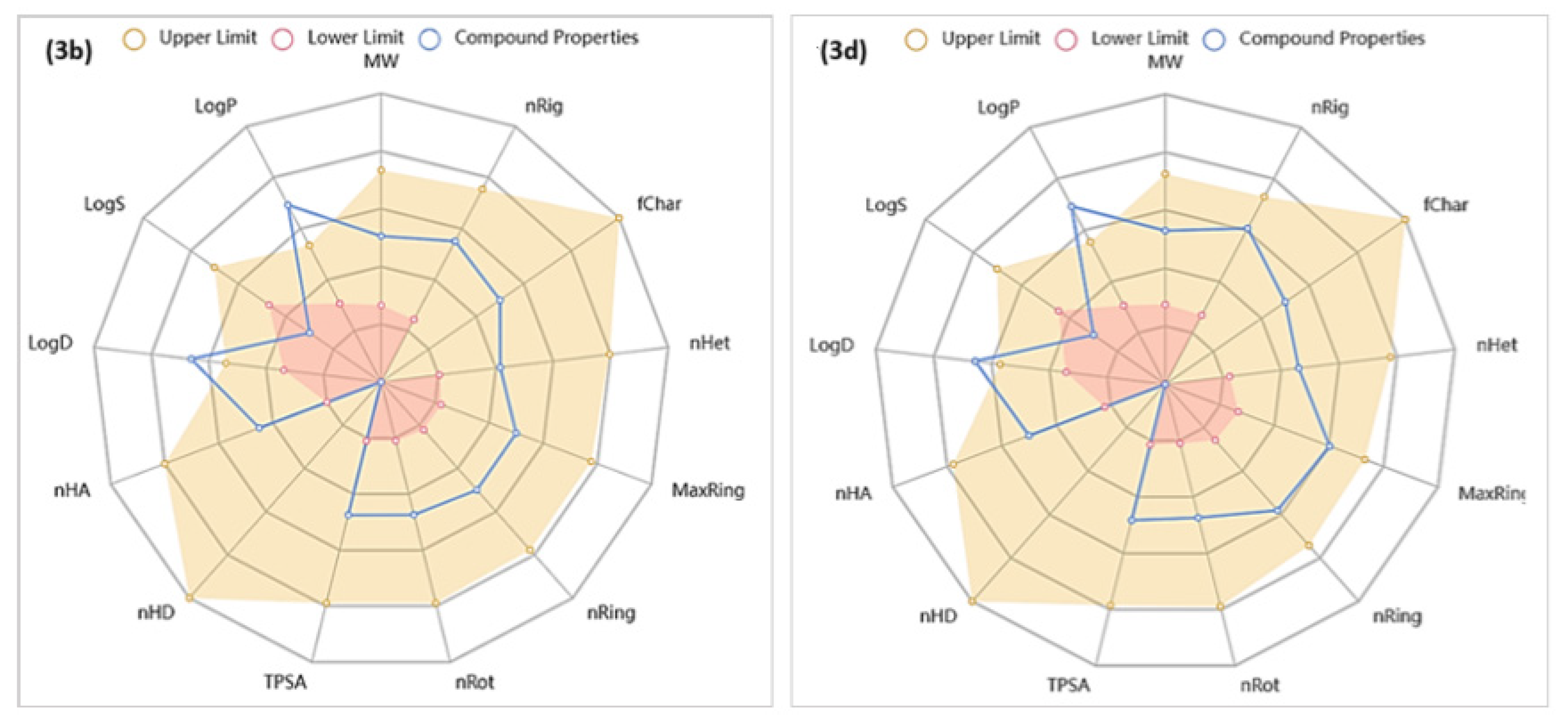
2.3.2. In Silico Predictions of the Physicochemical, Drug-Likeness and ADMET Properties of Compounds 3b and 3d

The physicochemical properties, drug-likeness, absorption, distribution, metabolism, excretion and toxicity (ADMET) characteristics of compounds 3b and 3d were predicted using the free webserver ADMETlab 2.0 (https://admetmesh.scbdd.com/, accessed on 7 February 2024) [59].

Analyses of Physicochemical Characteristics

The physicochemical properties influencing the drug’s capability to penetrate membranes for transport throughout the body were generated. As shown in the radar charts (Figure 8), both compounds satisfied the number of hydrogen bond acceptor groups (nHA), number of hydrogen bond donating groups (nHD), topological polar surface area (TPSA), number of rotatable bonds (nRot), number of rings (nRing), number of atoms in the biggest ring (MaxRing), number of heteroatoms (nHet), formal charge (fChar), number of rigid bonds (nRig), and molecular weight (MW); however, they violated lipophilicity and solubility parameters.
The calculated LogP values were 4.03 (3b) and 3.21 (3d), whereas the acceptable range is 0 to 3 mol/L. This suggests that both compounds possess moderate to high lipophilicity, with 3b being more lipophilic. It is reported that high lipophilicity increases the risk of poor aqueous solubility, poor absorption, in vivo toxicity, reduced in vitro receptor selectivity [60], and metabolic instability. The computed values for solubility descriptors LogS and LogD were −6.05/−4.24 (optimal range −4 to 0.5 mol/L) and 3.68/3.021 (proper range 1 to 3 log mol/L) for 3b/3d, respectively. Based on these values, 3b may be considered a poorly soluble compound, which may result in poor absorption from the gastrointestinal tract. Also, 3d is still a poorly soluble candidate, but it is comparatively better than 3b, suggesting that it may have more favorable absorption characteristics. Overall, the high lipophilicity of compounds 3b and 3d may favor membrane permeation, but it can also lead to poor solubility, which may negatively impact their oral bioavailability and overall pharmacokinetic profiles. Therefore, optimizing solubility without compromising permeability is essential for minimizing potential drawbacks and enhancing their drug-likeness.

Analyses of Drug-Likeness Characteristics for Compounds 3b and 3d

The likelihood of the studied derivatives being developed into drugs was assessed using a number of medicinal chemistry filters. According to quantitative estimate of drug-likeness (QED) scores [61], 3b was predicted to be an attractive candidate, while 3d was ranked as an unattractive molecule. Calculated synthetic accessibility scores (SA score) implied the ease of the synthesis of both compounds [62]. With respect to the medicinal chemistry evolution in 2018 (MCE-18) filter, which is a measure for scoring molecules by novelty according to their sp3 complexity [63], compound 3d showed a suitable value, whereas 3b did not. The four physicochemical parameters of Lipinski’s rule of five [64]—including LogP (less than 5), MW (less than 500), nHA (not more than 10), and nHD (not more than 5)—which determine the suitability of a drug candidate to be orally administrated, were satisfied by both compounds except for the LogP values. Therefore, both compounds are expected to be absorbed through the intestinal walls. Finally, the pan assay interference compounds (PAINS) filter [65], which flags suspicious compounds by identifying substructural alters that may give false positive results in biochemical assays, showed that both derivatives had no PAINS chemotypes.

Analyses of ADMET Characteristics for Compounds 3b and 3d

  • Absorption
The Caco-2 (the human colon adenocarcinoma cell lines) permeability [66] parameter is used to predict the human intestinal absorption (HIA) of a compound, which is an important feature for a drug to be properly absorbed through intestinal membrane. The estimated values for 3b and 3d were optimal. The human oral bioavailability 30% (F30%) values were also predicted to be good for both compounds. Screening both compounds for permeability glycoprotein (P-gp) [67], the efflux protein that recognizes the xenobiotics and is considered a cause of drug resistance, indicated that both compounds had high probabilities of being P-gp substrates; thus, significant drug–drug interactions (DDIs) would be expected if co-administrated with other P-gp inhibitors.
  • Distribution
Prediction of plasma protein binding (PPB) indicated that both derivatives had high abilities to bind to plasma proteins. Although this finding indicates their narrow therapeutic index, it can also be advantageous as it indicates the prolonged duration of the drugs’ action [68]. The computed volume of distribution (VD) values were optimal for both compounds, indicating favorable distribution throughout the body’s tissues rather than plasma confinement. Also, the studied compounds are expected to penetrate the blood–brain barrier (BBB) due to their high lipophilicity; thus, they could cause central nervous system (CNS) side effects.
  • Metabolism
Drug metabolism includes two phases: phase I (functionalization reactions; oxidation, reduction and hydrolysis) and phase II (conjugation reactions). The human cytochrome P450 enzyme (CYPs) family is responsible for the phase I metabolism of about two thirds of the know drugs [69], especially five isoenzymes: CYP1A2, CYP3A4, CYP2C9, CYP2C19 and CYP2D6. Prediction of the affinities of 3b and 3d for these five CYP isoenzymes showed that they would be substrates and might act as inhibitors; thus, both compounds are expected to cause DDIs.
  • Excretion
Total clearance (CL) parameters predict the removal rate of a drug from systemic circulation by all means in mL/min/kg. CL is an important pharmacokinetic parameter since it is a determinant of half-life time (t1/2), oral bioavailability and the efficacious dose of a drug [70]. The predicted values for both derivatives were modest. The elimination half-life time of the studied compounds was predicted to be excellent, with t1/2 values exceeding 3 h.
  • Toxicity
Some toxicity properties were assessed, such as blocking of the human ether-a-go-go gene (hERG) [71], which plays a major role in regulating cardiac action potential. According to predictions, both compounds could possibly block this gene, and so they might cause prolongation of QT interval and palpitation. Hepatotoxicity probability was predicted to be intermediate for both derivatives. The predicted FDA maximum daily dose (FDAMDD) value [72], which estimates the toxic dose threshold of chemicals in humans, indicates a high and a medium probability of compounds 3b and 3d being toxic, respectively.

3. Materials and Methods

3.1. Chemistry

3.1.1. Instrumentation

Melting points (°C) were recorded on a Gallen Kamp melting point apparatus. The IR spectra were recorded on a Perkin Elmer FT spectrophotometer, Spectrum BX 1000 (PerkinElmer, Waltham, MA, USA) in wave number ν (cm−1) using KBr discs. Nuclear magnetic resonance (NMR) analyses were performed using an Eclipse 300 FT NMR spectrometer (Tokyo, Japan) operating at 300 MHz for 1H-NMR and at 75 MHz for 13C-NMR, using DMSO-d6 or CDCl3 as solvents. Chemical shifts were expressed in (ppm), and coupling constants (J) were expressed in Hz. Splitting patterns were designated as s (singlet), br. s (broad singlet), d (doublet), dd (doublet of doublet), t (triplet), q (quartet) and m (multiplet). Mass spectra were carried out on a direct probe controller inlet part of a single quadrupole mass analyzer (Thermo Scientific GCMS), model ISO Lt with Thermo X-Calibur Software (Thermo Fisher Scientific, Waltham, MA, USA).

General Procedure A for Synthesis of Various Schiff Bases

An equimolar mixture (0.01 mol) of the appropriate heterocyclic aminenamely, ethyl 2-amino-6-phenyl-4,5,6,7-tetrahydrobenzo[b]thiophene-3-carboxylate 1a [25]; 2-amino-4,5,6,7-tetrahydrobenzo[b]thiophene-3-carbonitrile 1b [26]; 5,6,7,8-tetrahydrobenzo[4,5]thieno[2,3-d]pyrimidin-4-amine 1c [27]; ethyl 2-amino-4-methylthiazole-5-carboxylate 4 [28]; 4-(benzo[d]thiazol-2-yl)aniline 11 [29]; 3-amino-6,7-dimethoxy-2-methylquinazolin-4(3H)-one 15a [30]; or 3-amino-6-fluoro-2-methylquinazolin-4(3H)-one 15b [31]and the corresponding aldehyde 2ah: 3-bromo-4-hydroxy-5-methoxy benzaldehyde; 2,4,6-trimethoxybenzaldehyde; 3,4,5-trimethoxybenzaldehyde; 2-hydroxy-1-naphthaldehyde, 4-(trifluoro-methyl)benzaldehyde; 4-hydroxy-3-methoxybenzaldehyde; isovanillin; or thiophene-2-carbaldehyde, respectively, in absolute ethanol (30 mL) containing a catalytic amount of glacial acetic acid (3 mL) was heated under reflux for 12 h. Afterwards, the excess solvent was evaporated, and the resulting solid was recrystallized from ethanol to afford the pure products.

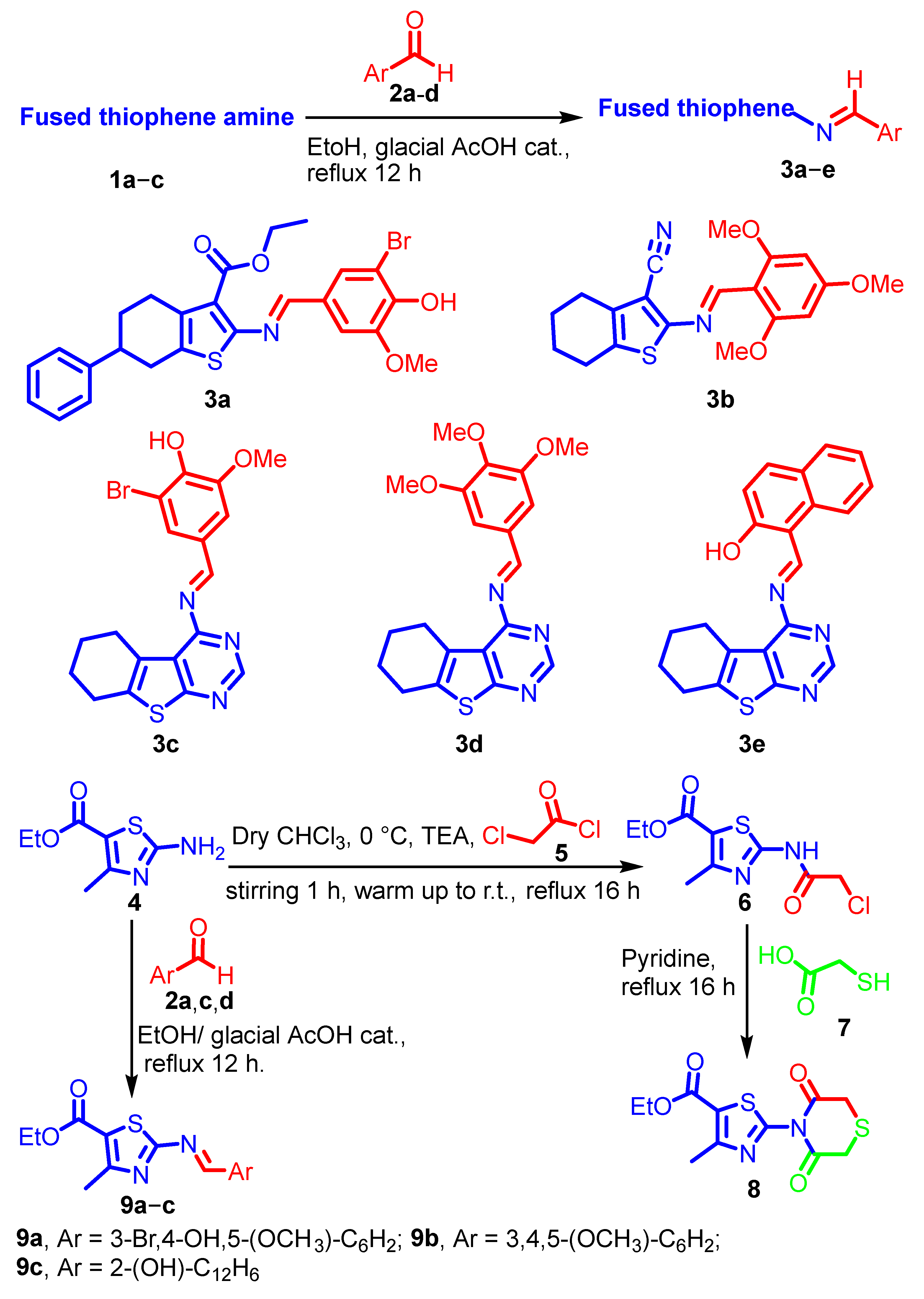
3.1.2. Synthesis of Thiophene Schiff Bases 3ae

They were prepared according to general procedure A using amines 1ac and aldehydes 2ad.
  • (E)-Ethyl2-((3-bromo-4-hydroxy-5-methoxybenzylidene)amino)-6-phenyl-4,5,6,7-tetrahydrobenzo[b]thiophene-3-carboxylate 3a
Yellow powder; yield (65%); m.p.107–109 °C; IR (KBr): νmax (cm−1) 3315 (OH), 2975 (CH-Ar), 2929 and 2847 (CH-aliphatic), 1676 (C=O), 1586 (C=N), 1500, 1436 and 1426 (C=C), 1355, 1289, 1156, 1044, 971, 855, 831, 791, 755, 678, 537; 1H-NMR (300 MHz, DMSO-d6): δH 1.24 (t, 3H, J = 7.1 Hz, CH3), 1.80–1.95 (m, 2H, CH2), 2.55–2.65 (m, 3H, CH2 and CH), 2.80–2.87 (m, 2H, CH2), 3.90 (s, 3H, OCH3), 4.14 (q, 2H, J = 7.1 Hz, CH2O), 7.19–7.29 (m, 6H, 6 × CH-Ar), 7.41 (s, 2H, CH-Ar), 7.71 (s, 1H, CH=N), 9.77 (s, 1H, OH); 13C-NMR (75 MHz, DMSO-d6): δC 14.60 (CH3), 27.09, 29.96, 31.95 (3 × CH2 and CH), 56.52 (OCH3), 58.91 (OCH2), 102.74, 109.44, 109.77, 115.29, 126.32, 127.01, 128.53, 128.87, 129.13, 131.34, 146.17 (7 × CH-Ar and 7 × Cq-Ar), 148.85, 150.03 (2 × Cq-O), 163.40 (CH=N), 165.30 (C=O); MS (EI, 70 eV): m/z (%) [M+ + 4] 517.61 (17.30) for C25H24BrNO4S, 498.40 (11.48), 485.56 (15.43), 454.41 (11.30), 410.36 (29.63), 382.13 (15.12), 321.27 (14.23), 292.62 (11.09), 270.15 (13.27), 235.09 (32.14), 223.32 (62.59), 196.15 (17.23), 131.13 (30.73), 102.23 (32.52), 76.18 (48.12), 64.19 (100.00), 54.70 (37.78), 44.24 (17.52).
  • (E)-2-((2,4,6-Trimethoxybenzylidene)amino)-4,5,6,7-tetrahydrobenzo[b]thiophene-3-carbonitrile 3b
Yellowish green powder; yield (52%); m.p. 173–175 °C; IR (KBr): νmax (cm−1) 3055 (CH-Ar), 2930 and 2830 (CH-aliphatic), 2214 (CN), 1599 (C=N), 1563, 1502 and14656 (C=C), 1413, 1331, 1230, 1209, 1157, 1118, 1033, 954, 888, 811, 640, 511; 1H-NMR (300 MHz, DMSO-d6): δH 1.78 (apparent s, 4H, 2 × CH2), 2.64 (apparent s, 4H, 2 × CH2), 3.87 (s, 9H, 3 × OCH3), 6.33 (s, 2H, 2 × Ar-H), 8.70 (s, 1H, CH=N); 13C-NMR (75 MHz, DMSO-d6): δC 21.95, 22.96, 24.18, 24.91 (4 × CH2), 56.12 and 56.60 (3 × OCH3), 91.63 (2 × CH-trimethoxy phenyl), 103.28, 105.31 (Cq and Cq-CN), 114.96, 117.13 (CN and Cq-cyclohexene), 134.30 (Cq-S), 154.67 (N-Cq-S), 162.99, 163.57, 165.56 (CH=N and 3 × Cq-OCH3); MS (EI; m/z; %): [M+ + 2] 358.74 (58.73) for C19H20N2O3S, 342.57 (40.45), 322.03 (52.87), 297.42 (39.05), 278.53 (43.18), 245.48 (22.12), 231.42 (25.53), 217.35 (15.72), 201.59 (17.75), 188.78 (51.62), 172.39 (75.41), 146.71 (48.47), 93.66 (100.00), 76.39 (90.52), 65.90 (58.85), 41.03 (52.33).
  • (E)-2-Bromo-6-methoxy-4-(((5,6,7,8-tetrahydrobenzo[4,5]thieno[2,3-d]pyrimidin-4-yl) imino)methyl)phenol 3c
Brown powder; yield (60%); m.p. 130–133 °C; IR (KBr): νmax (cm−1) 3317 (OH), 3024 (CH-Ar), 2934 and 2854 (CH-aliphatic), 1675 (N-Cq=N), 1587 (2 × HC=N), 1500, 1461, (C=C), 1427, 1358, 1289, 1158, 1044, 988, 911, 854, 830, 792, 791, 677 (C-Br), 578, 488; 1H-NMR (300 MHz, DMSO-d6): δH 1.72–1.74 (m, 4H, 2 × CH2), 2.65–2.85 (m, 4H, 2 × CH2), 3.88 (s, 3H, OCH3), 6.74 (br. s, 1H, OH), 7.36 (s, 1H, CH-Ar), 7.64 (s, 1H, CH-Ar), 7.95 (s, 1H, CH=N-azomethine), 9.72 (s, 1H, CH=N-pyrimidine); 13C-NMR (75 MHz, DMSO-d6) δC: 21.78, 22.48, 25.33, 25.46 (4 × CH2), 56.33 (OCH3), 115.09, 126.85, 128.57, 128.91, 130.79, 131.10, 132.08, (2 × CH-Ar, Cq-Br and 4 × Cq-Ar), 144.69, 148.62, 149.84, 157.67, 158.04, 162.41 (2 × Cq-O3, CH=N-azomethine, CH=N-pyrimidine, N-Cq=N and S-Cq=N); MS (EI, 70 eV): m/z (%) [M+ + 4] 421.03 (12.56) for C18H16BrN3O2S, 338.21 (25.20), 318.08 (35.19), 298.60 (89.08), 283.16 (23.51), 225.28 (35.19), 197.33 (100.00), 180.88 (48.71), 153.21 (71.21), 138.29 (64.60), 106.57 (65.13), 64.51 (77.28), 45.36 (41.56).
  • (E)-N-(5,6,7,8-Tetrahydrobenzo[4,5]thieno[2,3-d]pyrimidin-4-yl)-1-(3,4,5-trimethoxyphenyl)methanimine 3d
Yellow powder; yield (82%); m.p. 153–155 °C; IR (KBr): νmax (cm−1) 3013 (CH-Ar), 2939, 2844 and 2754 (CH-aliphatic), 1683 (HC=N), 1587, 1505, 1462 (C=C), 1426, 1392, 1329, 1236, 1127, 728, 989, 845, 785, 756, 728, 626, 591, 521, 461; 1H-NMR (300 MHz, DMSO-d6): δH 1.72–1.74 (m, 4H, 2 × CH2), 2.65–2.85 (m, 4H, 2 × CH2), 3.75 (s, 3H, OCH3), 3.84 (s, 6H, 2 × OCH3), 7.23 (s, 2H, 2 × CH-trimethoxy phenyl), 8.16 (s, 1H, CH=N), 9.85 (s, 1H, CH=N-pyrimidine); 13C-NMR (75 MHz, DMSO-d6): δC 21.49, 22.07, 22.33, 24.89 (4 × CH2), 56.12 and 60.28 (3 × OCH3), 106.80, 115.16, 126.97, 131.12, 131.76, 142.97 (2 × CH-Ar and 6 × Cq-Ar), 152.95, 153.44, 158.20, 165.56, 171.70 (3 × Cq-OCH3, HC=N-azomethine, HC=N-pyrimidine and Cq=N); MS (EI, 70 eV): m/z (%) [M+ + 2] 385.34 (21.36) for C20H21N3O3S 369.39 (16.45), 359.93 (51.25), 334.14 (22.35), 319.90 (25.82), 292.99 (16.64), 270.28 (14.19), 242.16 (24.80), 216.23 (23.12), 200.21 (38.44), 183.18 (24.69), 156.44 (63.94), 130.69 (68.76), 115.02 (46.24), 98.15 (61.09), 62.99 (57.78), 51.16 (100.00).
  • (E)-1-(((5,6,7,8-Tetrahydrobenzo[4,5]thieno[2,3-d]pyrimidin-4-yl)imino)methyl) naphthalene-2-ol 3e
Yellowish green powder; yield (78%); m.p. 237–240 °C; IR (KBr): νmax (cm−1) 3331 (br., OH), 3043 (CH-Ar), 2927 (CH-aliphatic), 2369, 2342, 1655 (N-Cq=N), 1590 (2 × HC=N), 1512, 1463 and 1436 (C=C), 1374, 1311, 1247, 1176 (Cq-O), 1076, 990, 965, 861, 785, 745, 713, 652, 559; 1H-NMR (300 MHz, DMSO-d6): δH 1.56–1.76 (m, 4H, 2 × CH2), 2.59–2.87 (m, 4H, 2 × CH2), 5.35 (br. s, 1H, OH), 7.18 (d, 1H, J = 8.7 Hz, CH-Ar), 7.34 (t, 1H, J = 7.2 Hz, CH-Ar), 7.55 (t, 1H, J = 7.2 Hz, CH-Ar), 7.79 (d, 1H, J = 7.8 Hz, CH-Ar), 7.98 (s, 1H, CH=N-azomethine), 8.04 (d, 1H, J = 9.0 Hz, CH-Ar), 8.19 (s, 1H, CH=N-pyrimidine), 8.94 (d, 1H, J = 8.1 Hz, CH-Ar); 13C-NMR (75 MHz, DMSO-d6): δC 21.65, 21.89, 24.49, 25.00 (4 × CH2), 112.19, 114.72, 119.61, 121.75, 122.37, 123.35, 126.79, 128.70, 130.64, 131.77, 137.82, 144.46 (6 × CH-Ar and 7 × Cq-Ar), 152.54, 157.76, 162.09, 165.60 (Cq-OH, N-Cq=N, CH=N-azomethine and HC=N-pyrimidine); MS (EI, 70 eV): m/z (%) [M+ + 2] 361.40 (0.80), [M+ + 1] 360.09 (8.56), [M+] 359.20 (28.10) for C21H17N3OS, 345.60 (21.16), 327.49 (16.06), 318.40 (89.72), 295.51 (26.68), 259.58 (16.79), 245.01 (12.03), 216.59 (64.13), 196.96 (11.58), 172.34 (15.17), 147.56 (22.36), 124.51 (85.69), 109.97 (100.00), 104.71 (55.63), 76.72 (60.02), 62.73 (27.47), 48.30 (7.51).

3.1.3. Synthesis of Ethyl 2-(3,5-Dioxothiomorpholino)-4-methylthiazole-5-carboxylate 8

To a stirred, ice-cooled solution of ethyl 2-amino-4-methylthiazole-5-carboxylate 4 (3.6 g, 0.02 mol) in dry CHCl3 (30 mL), triethylamine (2.8 mL, 0.02 mol) was added, followed by the dropwise addition of chloroacetyl chloride 5 (1.6 mL, 0.02 mol). The reaction mixture was stirred further at 0 °C for one hour, then allowed to warm to room temperature, and subsequently refluxed for 16 h. After evaporation of the solvent under reduced pressure, the resulting solid was washed with a saturated solution of NaHCO3, followed by water, and air-dried. Recrystallization from ethanol afforded the pure chloroacetamide intermediate 6.
An equimolar mixture of chloroacetamide intermediate 6 and thioglycolic acid 7 (0.004 mol) in dry pyridine (15 mL) was refluxed for 16 h. After cooling to room temperature, the precipitated solid was filtered, dissolved in diethyl ether and triturated with aqueous ethanol until precipitation occurred. The solid was collected by filtration and recrystallized from ethanol to afford compound 8 as a buff-colored powder; yield (55%); m.p. 180–183 °C; IR (KBr): νmax (cm−1) 2926 (CH-aliphatic), 1705 (br. 3 × CO), 1547 (C=N), 1464 (C=C), 1372, 1318, 1266, 1161, 1095, 948, 760, 683, 592, 449; 1H-NMR (300 MHz, DMSO-d6): δH 1.27 (t, 3H, J = 7.1 Hz, CH3-CH2), 2.57 (s, 3H, CH3), 3.58 (s, 4H, 2 × CH2S), 4.24 (q, 2H, J = 7.1 Hz, CH2O); 13C-NMR (75 MHz, DMSO-d6): δC 14.20, 16.99 (2 × CH3), 33.79, 34.34 (2 × CH2S), 60.55 (CH2O), 114.25, 156.18, 159.40, 162.08, 168.57, 170.80 (3 × Cq and 3 × C=O); MS (EI, 70 eV): m/z (%) [M+ + 4] 304.55 (38.60) for C11H12N2O4S2, 232.06 (63.58), 222.22 (100.00), 206.85 (60.18), 132.17 (55.79).

3.1.4. Synthesis of Thiazolyl Schiff Bases 9ac

These were prepared according to general procedure A using amine 4 and aldehydes 2a, 2c and 2d, respectively.
  • Ethyl (E)-2-((3-bromo-4-hydroxy-5-methoxybenzylidene)amino)-4-methyl thiazole-5-carboxylate 9a
Yellow Powder; yield (75%); m.p. 143–145 °C; IR (KBr): νmax (cm−1) 3376 (OH), 3098 (CH-Ar), 2979 and 2933 (CH-aliphatic), 1680 (C=O), 1587 (CH=N and S-Cq=N), 1504, 1462, 1428 (C=C), 1373, 1279 (C-O), 1157, 1100, 1043, 972, 853, 828, 791, 759, 677 (C-Br), 588, 493; 1H-NMR (300 MHz, DMSO-d6): δH 1.18 (t, 3H, J = 7.1 Hz, CH3-CH2-O), 2.37 (s, 3H, CH3), 3.86 (s, 3H, OCH3), 4.14 (q, 2H, J = 7.1 Hz, CH3-CH2-O), 7.38 (d, 1H, J = 1.8 Hz, CH-Ar), 7.65 (apparent d, 1H, J = 1.5 Hz, CH-Ar), 7.72 (s, 1H, CH=N), 9.74 (s, 1H, OH); 13C-NMR (75 MHz, DMSO-d6): δC 14.33, 17.01 (2 × CH3), 56.36 (OCH3), 59.86.(OCH2), 107.63, 109.29, 109.67, 128.54, 128.94 (2 × CH-Ar, Cq-S, Cq-Ar and Cq-Br), 148.62, 149.80, 158.73, 158.86, 161.93, 170.21 (N-Cq-CH3, 2 × Cq-O, CH=N, N=Cq-S and C=O); MS (EI, 70 eV): m/z (%) [M+ + 4] 401.94 (8.33) for C15H15BrN2O4S, 384.87 (12.31), 355.44 (25.54), 290.12 (26.20), 243.66 (54.01), 193.16 (28.22), 159.36 (40.18), 144.21 (84.24), 129.80 (69.64), 113.32 (100.00), 91.38 (86.76), 84.04 (94.60), 51.27 (71.52), 43.14 (45.23).
  • Ethyl(E)-4-methyl-2-((3,4,5-trimethoxybenzylidene)amino)thiazole-5-carboxylate 9b
Yellow Powder; yield (60%); m.p. 110–113 °C; IR (KBr): νmax (cm−1) 3086 (CH-Ar), 2977, 2939 (CH-aliphatic), 1680 (C=O), 1586 (CH=N and Cq=N), 1514, 1466, 1426 (C=C), 1328, 1279 (C-O), 1125, 1094, 990, 844, 755, 628, 524, 423; 1H-NMR (300 MHz, DMSO-d6): δH 1.22 (t, 3H, J = 7.1 Hz, CH3-CH2-O), 2.37 (s, 3H, CH3), 3.77 (s, 3H, OCH3), 3.86 (s, 6H, 2 × OCH3), 4.15 (q, 2H, J = 7.1 Hz, CH3-CH2-O), 7.23 (s, 2H, 2 × CH-Ar), 7.70 (s, 1H, CH=N); 13C-NMR (75 MHz, DMSO-d6): δC 14.24, 17.21 (2 × CH3), 55.97 (2 × OCH3), 59.92, 60.12 (OCH3 and OCH2), 106.67 (2 × CH-Ar), 107.42 (Cq-S), 131.62, 142.84, 153.29, 159.24, 161.91, 170.21 (Cq-Ar, N-Cq-CH3, 3 × Cq-OCH3, CH=N, S-Cq=N and C=O); MS (EI, 70 eV): m/z (%) [M+ + 2] 366.33 (4.87) for C17H20N2O5S, 336.86 (19.60), 361.41 (17.33), 292. (8.97), 270.38 (15.79), 255.85 (16.42), 219.69 (7.39), 193.86 (11.61), 177.30 (16.45), 139.85 (24.95), 125.16 (19.41), 92.02 (24.62), 77.62 (49.20), 64.14 (100), 50.33 (34.70).
  • Ethyl(E)-2-(((2-hydroxynaphthalen-1-yl)methylene)amino)-4-methylthiazole-5-carboxylate 9c
Buff powder; yield (60%); m.p. 174–176 °C; IR (KBr): νmax (cm−1) 3375 (OH), 3088 (CH-Ar), 2982, 2923 (CH-aliphatic), 1707 (C=O), 1676, 1624 (CH=N and S-Cq=N), 1549, 1515, 1463 (C=C), 1372, 1318, 1265 (C-O), 1210, 1151, 1090, 813, 752, 713, 589, 530, 421; 1H-NMR (300 MHz, DMSO-d6): δH 1.23 (t, 3H, J = 7.1 Hz, CH3-CH2-O), 2.36 (s, 3H, CH3), 4.11 (q, 2H, J = 7.1 Hz, CH3-CH2-O), 7.15–7.26 (m, 1H, CH-Ar), 7.38–7.46 (m, 1H, CH-Ar), 7.60 (t, 1H, J = 7.2 Hz, CH-Ar), 7.70 (s, 2H, CH=N and OH), 7.86 (d, 1H, J = 7.8 Hz, CH-Ar), 8.10 (d, 1H, J = 9.0 Hz, CH-Ar), 8.92 (d, 1H, J = 8.7 Hz, CH-Ar); 13C-NMR (75 MHz, DMSO-d6): δC 14.27, 17.05 (2 × CH3), 59.71 (OCH2), 107.43, 112.35, 118.67, 121.99, 124.16, 127.53, 128.77, 129.19, 131.72, 138.35 (6 × CH-Ar, Cq-S and 3 × Cq-Ar), 159.14, 161.91, 163.88, 170.19 (CH=N, N-Cq-CH3, N=Cq-S, C=O and Cq-OH); MS (EI, 70 eV): m/z (%) [M+ + 3] 343.61 (10.13), [M+ + 2] 342.35 (36.41), [M+ + 1] (35.12) for C18H16N2O3S, 321.81 (74.11), 301.97 (34.88), 271.94 (30.74), 252.88 (41.26), 238.88 (59.64), 226.69 (78.45), 194.41 (37.70), 167.62 (42.94), 147.92 (78.10), 139.90 (100.00), 119.61 (40.05), 87.81 (70.39), 60.36 (84.63), 42.49 (90.30).

3.1.5. General Procedure B for the Synthesis of 3-Substituted-6,7-dimethoxy-2-methyl quinazolin-4(3H)-one Derivatives 13 and 14

Equimolar quantities (0.001 mol) of 6,7-dimethoxy-2-methyl-4H-benzo[d][1,3]oxazin-4-one 10a and 4-(benzo[d]thiazol-2-yl)aniline 11 or cyclohexylamine 12 were fused in an oil bath at 200 °C for 2 h. The reaction mixture was allowed to cool to 100 °C, then concentrated hydrochloric acid (3 mL, 37%) was added. After further cooling to room temperature, the mixture was neutralized with aqueous sodium hydroxide (25%). The resulting precipitate was collected by filtration, washed with water, air dried, and recrystallized from ethanol to afford compounds 13 and 14, respectively.
  • 3-(4-(Benzo[d]thiazol-2-yl)phenyl)-6,7-dimethoxy-2-methylquinazolin-4(3H)-one 13
Green powder; yield (60%); m.p. 154–157 °C; IR (KBr): νmax (cm−1) 3058, 3002 (CH-Ar), 2927, 2833 (CH-aliphatic), 1668 (C=O), 1605 (C=N), 1497 and 1465 (C=C), 1435, 1390, 1266, 1104, 1025, 969, 848, 758, 679, 523, 509; 1H-NMR (300 MHz, DMSO-d6): δH 2.23 (s, 3H, CH3), 3.94 (s, 3H, OCH3), 3.98 (s, 3H, OCH3), 6.82–6.94 (m, 1H, Ar-H), 7.08 (s, 1H, CH-quinazoline), 7.36–7.50 (m, 4H, 4 × Ar-H), 7.56 (s, 1H, CH-quinazoline), 7.88 (d, 1H, J = 8.1 Hz, Ar-H), 8.08 (d, 1H, J = 7.8 Hz, Ar-H), 8.26 (d, 2H, J = 8.7 Hz, 2 × Ar-H); 13C-NMR (75 MHz, DMSO-d6): δC 23.70 (CH3), 55.82 (2 × OCH3), 105.43, 106.89 (2 × CH-quinazolinone), 113.29, 121.27, 122.14, 123.00, 125.13, 125.72, 126.09, 128.51, 134.02, 134.73, 143.21, 148.53 (8 × CH-Ar and 6 × Cq-Ar), 151.91, 153.57, 154.81, 161.13, 165.90 (2 × Cq-OCH3, C=O and 2 × Cq=N); MS (EI, 70 eV): m/z (%) [M++ 2] 431.86 (19.55) for C24H19N3O3S, 337.23 (51.58), 269.57 (84.30), 91.49 (40.71), 78.25 (100.00), 51.63 (83.87).
  • 3-Cyclohexyl-6,7-dimethoxy-2-methylquinazolin-4(3H)-one 14
White powder; yield (70%); m.p. 100–103 °C; IR (KBr): νmax (cm−1) 3025 (CH-Ar), 2926, 2852 (CH-aliphatic), 1659 (C=O), 1614 (C=N), 1572, 1502 (C=C), 1457, 1398, 1321, 1246, 1166, 1026, 978, 845, 781, 609, 453; 1H-NMR (300 MHz, CDCl3): δH 1.12–1.18 (m, 2H, CH2), 1.33–1.49 (m, 4H, 2 × CH2), 1.72–1.76 (m, 4H, 2 × CH2), 2.58 (s, 3H, CH3), 3.42 (app. s, 1H, CH-N), 3.84 (s, 3H, OCH3), 3.87 (s, 3H, OCH3), 6.97 (s, 1H, Ar-H), 7.35 (s, 1H, Ar-H); 13C-NMR (75 MHz, CDCl3): δC 23.78 (CH3), 25.09, 25.92, 28.21 (3 × CH2), 55.60, 55.89 (5 × OCH3), 59.83 (N-CH), 105.04, 106.96, 114.55, 142.89 (2 × CH-Ar and 2 × Cq-Ar), 148.21, 153.38, 154.45, 161.04 (2 × Cq-OCH3, Cq=N and C=O); MS (EI, 70 eV): m/z (%) [M+] 301.91 (17.37) for C17H22N2O3, 239.47 (66.06), 107.94 (91.96), 100.29 (80.20), 75.25 (90.44), 69.08 (86.71), 53.83 (100.00).

3.1.6. Synthesis of Ethyl (6,7-Dimethoxy-2-methyl-4-oxoquinazolin-3(4H)-yl)carbamate 17

A mixture of 3-amino-6,7-dimethoxy-2-methylquinazolin-4(3H)-one 10a (0.0028 mol) and ethyl chloroformate 16 (0.038 mol) was heated under reflux for 10 h. The excess reagent was removed under reduced pressure, and the resulting residue was triturated with diethyl ether to induce solidification. The precipitated solid was filtered, washed with water, air-dried and recrystallized from benzene to afford the title compound as off-white powder; yield (80%); m.p. 130–133 °C; IR (KBr): νmax (cm−1) 3520 (NH), 3066 (CH-Ar), 2980, 2930 (CH-aliphatic), 1733, 1689 (2 × C=O), 1611 (C=N), 1505, 1464 (C=C), 1387, 1273, 1154, 1099, 864, 776, 688, 538; 1H-NMR (300 MHz, DMSO-d6) δH: 1.21 (t, 3H, J = 7.1 Hz, CH2-CH3), 2.40 (s, 3H, CH3), 3.87 (s, 3H, OCH3), 3.90 (s, 3H, OCH3), 4.22 (q, 2H, J = 7.1 Hz, O-CH2-CH3), 7.05–7.16 (m, 1H, Ar-H), 7.40–7.42 (m, 1H, Ar-H), 10.39 (s, 1H, NH); 13C-NMR (75 MHz, DMSO-d6) δC: 14.14 (CH3-CH2-O), 20.82 (CH3), 55.53, 55.91 (2 × OCH3), 61.56 (CH2O), 105.35, 107.95, 113.22, 142.55(2 × CH-Ar and 2 × Cq-Ar), 148.60, 152.66, 154.76 (2 × Cq-O and Cq=N), 155.48, 158.55 (2 × C=O); MS (EI, 70 eV): m/z (%) [M+] 307.28 (5.96) for C14H17N3O5], 271.00 (35.41), 256.06 (35.00), 76.35 (42.35), 74.59 (100.00).

3.1.7. Synthesis of Quinazolinone Based-Schiff Bases 18ah

These were prepared according general procedure A using amines 10a,b and aldehydes 2b and 2dh.
  • (E)-6,7-Dimethoxy-2-methyl-3-((2,4,6-trimethoxybenzylidene)amino)quinazolin-4(3H)-one 18a
Yellow powder; yield (73%); m.p. 200–203 °C; IR (KBr): νmax (cm−1) 3035 (CH-Ar), 2927 (CH-aliphatic), 2369, 1669 (C=O), 1610 (CH=N and Cq=N), 1503 and 1458 (C=C), 1336, 1245, 1207 (C-O), 1131, 1027, 861, 802, 775, 637, 469; 1H-NMR (300 MHz, DMSO-d6): δH 2.44 (s, 3H, CH3), 3.63 (s, 9H, 3 × OCH3), 3.88 (s, 6H, 2 × OCH3), 6.22 (s, 2H, 2 × CH-trimethoxy phenyl), 7.09 (s, 1H, CH-quinazolinone), 7.40 (s, 1H, CH-quinazolinone), 8.84 (s, 1H, CH=N); 13C-NMR (75 MHz, DMSO-d6): δC 24.40 (CH3), 55.31, 55.68, 55.88 and 56.22 (5 × OCH3), 91.31(2 × CH-quinazolinone), 104.70, 107.45, 108.81, 113.61 (2 × CH-Ar, Cq-ipso and Cq-C=O), 144.08, 147.92, 153.88, 155.53, 156.72, 159.01, 160.78 (Cq=N, Cq-N, -CH=N and 5 × O-Cq), 161.73 (C=O); MS (EI, 70 eV): m/z (%) [M+] 413.27 (17.47) for C21H23N3O6, 406.96 (69.19), 398.89 (56.11), 386.69 (31.89), 377.44 (49.14), 337.29 (54.02), 350.60 (30.03), 337.29 (54.02), 325.76 (25.79), 297.19 (27.65) 272.82(17.65), 251.33 (59.99), 226.28 (100.00), 208.35 (31.73), 187.44 (97.92), 147.89 (49.86), 101.62 (48.17), 82.10 (38.26), 66.59 (30.50), 57.17 (28.28), 46.22 (24.82).
  • (E)-3-(((2-Hydroxynaphthalen-1-yl)methylene)amino)-6,7-dimethoxy-2-methylquin- azolin-4(3H)-one 18b
Yellowish green powder; yield (75%); m.p. 205–207 °C; IR (KBr): νmax (cm−1) 3308 (OH), 3020 (CH-Ar), 2927 (CH-aliphatic), 2371, 2341, 1667 (C=O), 1615 (CH=N and Cq=N), 1600, 1502, 1464 (C=C), 1396, 1331, 1277, 1245, 1207 (C-O), 1021, 973, 864, 826, 798, 772, 739, 684, 605, 552, 428; 1H-NMR (300 MHz, DMSO-d6): δH 2.54 (s, 3H, CH3), 3.87 (s, 6H, 2 × OCH3), 7.02 (s, 1H, CH-quinazolinone), 7.09–7.48 (m, 4H, CH-quinazolinone, 2 × CH-Ar and CH=N), 7.59 (t, 1H, J = 7.4 Hz, CH-Ar), 7.89 (d, 1H, J = 7.2 Hz, CH-Ar), 8.04 (d, 1H, J = 9.0 Hz, CH-Ar), 8.80 (d, 1H, J = 8.1 Hz, CH-Ar), 9.67 (s, 1H, OH); 13C-NMR (75 MHz, DMSO-d6): δC 21.64 (CH3), 55.68 and 55.85 (2 × OCH3), 99.22, 104.74, 107.28, 112.76, 113.81, 119.18, 123.14, 128.89, 135.81, 139.16, 142.88, 148.30, 151.35, (8 × CH-Ar and 5 × Cq-Ar), 154.58, 157.10, 159.43, 161.93, 169.00, 172.27 (3 × Cq-O, CH=N, Cq=N and C=O); MS (EI, 70 eV): m/z (%) [M+ + 3] 392.49 (3.60) for C22H19N3O4, 387.24 (70.29), 378.84 (8.91), 367.80 (21.98), 346.55 (23.79), 331.82 (22.02), 297.63 (12.21), 280.96 (11.59), 244.77 (8.58), 220.18 (63.36), 204.47 (13.31), 168.99 (31.47), 151.80 (20.15), 136.21 (81.96), 127.13 (100.00), 105.86 (22.75), 92.17 (47.14), 64.22 (87.35), 52.80 (73.61).
  • (E)-6,7-Dimethoxy-2-methyl-3-((4-(trifluoromethyl)benzylidene)amino)quinazolin-4(3H)-one 18c
Yellow powder; yield (70%); m.p. 217–219 °C; IR (KBr): νmax (cm−1) 3074 and 3012 (CH-Ar), 2936 (CH-aliphatic), 2371, 2341, 1670 (C=O), 1612 (CH=N and Cq=N), 1509, 1471 and 1437 (C=C), 1394, 1325 (C-F), 1278, 1246, 1212, 1169, 1111, 1067, 1019, 985, 958, 870, 842, 806, 771, 680, 652, 601, 552, 515, 478; 1H-NMR (300 MHz, CDCl3): δH 2.65 (s, 3H, CH3), 3.98 (s, 6H, 2 × OCH3), 7.08 (s, 1H, CH-quinazolinone), 7.56 (s, 1H, CH-quinazolinone), 7.75 (d, 2H, J = 8.1 Hz, 2 × Ar-H), 7.97 (d, 2H, J = 7.8 Hz, 2 × Ar-H), 9.27 (s, 1H, CH=N); 13C-NMR (75 MHz, CDCl3): δC 22.57 (CH3), 56.12, 56.74 (2 × OCH3), 99.11, 105.91, 107.28, 114.43, 121.71, 125.72, 128.71, 133.48, 136.27, 142.48, 148.78 (6 × CH-Ar and 5 × Cq-Ar), 152.69, 155.07, 158.22 (Cq=N, CH=N and 2 × Cq-O), 163.10 (C=O); MS (EI, 70 eV): m/z (%) [M+] 391.03 (4.37) for C19H16F3N3O3, 384.43 (10.21), 366.31 (5.93), 343.19 (100.00), 323.16 (13.07), 296.26 (12.21), 286.20 (9.93), 245.18 (14.02), 233.18 (10.08), 206.17 (20.75), 189.16 (38.35), 148.14 (93.61), 115.13 (17.48), 59.12 (13.13).
  • (E)-3-((4-Hydroxy-3-methoxybenzylidene)amino)-6,7-dimethoxy-2-methylquinazoli-N-4(3H)-one 18d
Dark orange powder; yield (85%); m.p. > 300 °C; IR (KBr): νmax (cm−1) 3451 (OH), 3142 and 3002 (CH-Ar), 2989 (CH-aliphatic), 2367, 1649 (C=O), 1574 (CH=N and Cq=N), 1505, 1462 (C=C), 1399, 1334, 1300, 1251, 1210, 1128 (C-O), 1027, 975, 842, 810, 770, 675, 612, 548, 436; 1H-NMR (300 MHz, DMSO-d6): δH 2.45 (s, 3H, CH3), 3.90 (s, 9H, 3 × OCH3), 6.94–6.97 (m, 1H, CH-Ar), 7.07 (s, 1H, CH-quinazolinone), 7.30–7.44 (m, 2H, 2 × CH-Ar), 7.55 (s, 1H, CH-quinazolinone), 8.68 (s, 1H, CH=N), 10.04 (br. s, 1H, OH); 13C-NMR (75 MHz, DMSO-d6): δC 21.69 (CH3), 55.35, 55.61 (3 × OCH3), 105.10, 106.98, 107.15, 110.21, 113.57, 115.31, 123.42, 124.18 (5 × CH-Ar and 3 × Cq-Ar), 142.21, 147.91, 151.09, 151.38, 154.18, 156.57 (4 × Cq-O, Cq=N and CH=N), 168.59 (C=O); MS (EI, 70 eV): m/z (%) [M+ + 3] 372.04 (1.85), [M+ + 2] 371.04 (10.91), [M+] 369.03 (32.42) for C19H19N3O5, 327.05 (30.05), 259.07 (4.35), 217.04 (100.00), 176.05 (6.34), 146.03 (1.78), 120.05 (3.28), 42.05 (17.41).
  • (E)-6-Fluoro-2-methyl-3-((2,4,6-trimethoxybenzylidene)amino)quinazolin-4(3H)-one 18e
Off-white powder; yield (80%); m.p. 220–222 °C; IR (KBr): νmax (cm−1) 3072 (CH-Ar), 2926 (CH-aliphatic), 1674 (C=O), 1610 (CH=N and Cq=N), 1480, 1413 (C=C), 1333, 1227 (C-O), 1133 (C-F), 1037, 950, 876, 827, 799, 641, 486; 1H-NMR (300 MHz, CDCl3): δH 2.57 (s, 3H, CH3), 3.89 (s, 9H, 3 × OCH3), 6.13 (s, 2H, 2 × CH-trimethoxy phenyl), 7.40 (dd, 1H, J = 8.1, 3.1 Hz, CH-Ar), 7.60–7.65 (apparent m, 1H, CH-Ar), 7.86–7.91 (apparent m, 1H, CH-Ar), 8.93 (s, 1H, CH=N); 13C-NMR (75 MHz, CDCl3): δC 22.53 (CH3), 55.33 and 55.99 (3 × OCH3), 90.53 (2 × CH-trimethoxy phenyl), 102.82, 111.79, 122.36, 122.68, 128.95, 143.33 (3 × CH-Ar and 3 × Cq-Ar), 153.15, 157.60, 158.52, 161.79, 162.17, 164.94 (3 × Cq-O, Cq=N, CH=N, C=O and C-F); MS (EI, 70 eV): m/z (%) [M+] 371.21 (20.10) for C19H18FN3O4, 356.84 (16.73), 342.27 (23,12), 326.27 (21.85), 297.94 (11.17), 281.57 (25.34), 252.84 (86.17), 235.23 (28.44), 215.02 (23.88), 195.68 (27.22), 151.81 (45.41), 124.28 (21.12), 104.74 (100.00), 77.07 (89.61), 48.16 (47.46).
  • (E)-6-Fluoro-3-(((2-hydroxynaphthalen-1-yl)methylene)amino)-2-methylquinazolin-4(3H)-one 18f
Yellowish orange powder; yield (88%); m.p. 207–209 °C; IR (KBr): νmax (cm−1) 3446 (OH), 3071 (CH-Ar), 2370, 2341, 2925 (CH-aliphatic), 2369, 2341, 1679 (C=O), 1599 (CH=N and Cq=N), 1485 (C=C), 1319 (C-F), 1276, 1242, 1187 (C-O), 1096, 1035, 961, 892, 824, 771, 741, 672, 594, 560, 531, 428; 1H-NMR (300 MHz, DMSO-d6): δH 2.51 (s, 3H, CH3), 7.33 (d, 1H, J = 9.0 Hz, CH-Ar), 7.45 (t, 1H, J = 7.5 Hz, CH-Ar), 7.63 (t, 1H, J = 7.8 Hz, CH-Ar), 7.69–7.86 (m, 3H, 2 × CH-Ar and CH=N), 7.85 (apparent dd, 1H, J = 8.7, 2.4 Hz, CH-Ar), 7.93 (d, 1H, J = 7.8 Hz, Ar-H), 8.11 (d, 1H, J = 9.0 Hz, Ar-H), 8.82 (d, 1H, J = 8.4 Hz, Ar-H), 9.67 (s, 1H, OH); 13C-NMR (75 MHz, DMSO-d6): δC 22.26 (CH3), 108.30, 111.06, 111.37, 118.55, 123.31, 124.07, 128.03, 128.72, 129.04, 129.37, 132.05, 136.29, 143.02 (9 × CH-Ar and 5 × Cq-Ar), 152.83, 156.98, 158.23, 160.66, 169.70 (CH=N, Cq=N, Cq-O, Cq-F and C=O); MS (EI, 70 eV): m/z (%) [M+] 347.00 (16.59) for C20H14FN3O2, 320.34 (54.51), 298.98 (29.25), 275.72 (27.16), 267.46 (23.16), 244.82 (24.16), 222.86 (20.75), 190.87 (100.00), 186.10 (68.87), 177.91 (33.65), 165.55 (22.57), 150.57 (45.77), 125.90 (46.98), 102.68 (47.54), 84.16 (19.41), 51.82 (54.71).
  • (E)-6-Fluoro-3-((3-hydroxy-4-methoxybenzylidene)amino)-2-methylquinazolin-4(3H)-one 18g
Yellowish white powder; yield (72%); m.p. 183–185 °C; IR (KBr): νmax (cm−1) 3301 (OH), 30,188 and 3071 (CH-Ar), 2930 (CH-aliphatic), 2371, 1675 (C=O), 1637 (CH=N and Cq=N), 1598, 1513, 1483 (C=C), 1442, 1380, 1350, 1279 (C-F), 1240, 1194, 1158 (C-O), 1109, 1027, 964, 993, 841, 7729, 706, 644, 601, 565, 474; 1H-NMR (300 MHz, DMSO-d6): δH 2.48 (s, 3H, CH3), 3.86 (s, 3H, OCH3), 7.10 (apparent dd, 1H, J = 8.4, 2.4 Hz, CH-Ar), 7.30 (apparent dd, 1H, J = 8.3, 2.0 Hz, CH-Ar), 7.47 (d, 1H, J = 8.3, 2.1 Hz, CH-Ar), 7.68–7.88 (m, 3H, 3 × CH-Ar), 8.72 (s, 1H, CH=N), 9.58 (s, 1H, OH); 13C-NMR (75 MHz, DMSO-d6): δC 22.47 (CH3), 56.12 (OCH3), 111.21, 111.51, 112.22, 113.66, 123.02, 123.34, 123.58, 125.24, 129.95 (6 × CH-Ar and 3 × Cq-Ar) 143.67, 147.40, 153.27, 152.47, 161.64, 170.07 (CH=N, Cq-F, Cq=N, 2 × Cq-O and C=O); MS (EI, 70 eV): m/z (%) [M+ + 1] 327.90 (11.78) for C17H14FN3O3, 311.93 (76.65), 281.67 (33.96), 228.95 (6.91), 207.57 (69.90), 177.71 (12.14), 138.21 (50.05), 101.14 (40.93), 83.47 (42.48), 66.09 (100.00).
  • (E)-6-Fluoro-2-methyl-3-((thiophen-2-ylmethylene)amino)quinazolin-4(3H)-one 18h
Off-white powder; yield (75%); m.p. 159–160 °C; IR (KBr): νmax (cm−1) 3002 (CH-Ar), 2925 (CH-aliphatic), 2372, 2342, 1676 (C=O), 1600 (CH=N and Cq=N), 1486, 1430 (C=C), 1374, 1342 (C-F), 1302, 1272, 1243, 1134, 1188, 1134, 1040, 981, 886, 837, 814, 780, 711, 625, 596, 557, 497, 461; 1H-NMR (300 MHz, DMSO-d6): δH 2.51 (s, 3H, CH3), 7.30 (dd, 1H, J = 4.8, 3.6 Hz, CH-Ar), 7.69–7.84 (m, 4H, 4 × CH-Ar), 7.98 (d, 1H, J = 5.1 Hz, CH-Ar), 9.13 (s, 1H, CH=N); 13C-NMR (75 MHz, DMSO-d6): δC 22.39 (CH3), 111.56, 122.55, 123.09, 123.40, 128.94, 130.06, 133.43, 136.50, 143.53, 153.18, 157.27 (6 × CH-Ar, 3 × Cq-Ar, Cq=N and CH=N), 161.75, 163.72 (C=O and Cq-F); MS (EI, 70 eV): m/z (%) [M+ + 2] 289.54 (28.27) for C14H10FN3OS, 264.19 (77.05), 258.21 (42.35), 235.92 (63.12), 218.42 (91.49), 210.31 (57.75), 198.35 (63.28), 186.87 (31.56), 171.33 (71.67), 162.99 (100.00), 150.01 (25.26), 138.23 (23.79), 78.89 (31.41), 41.62 (21.99).

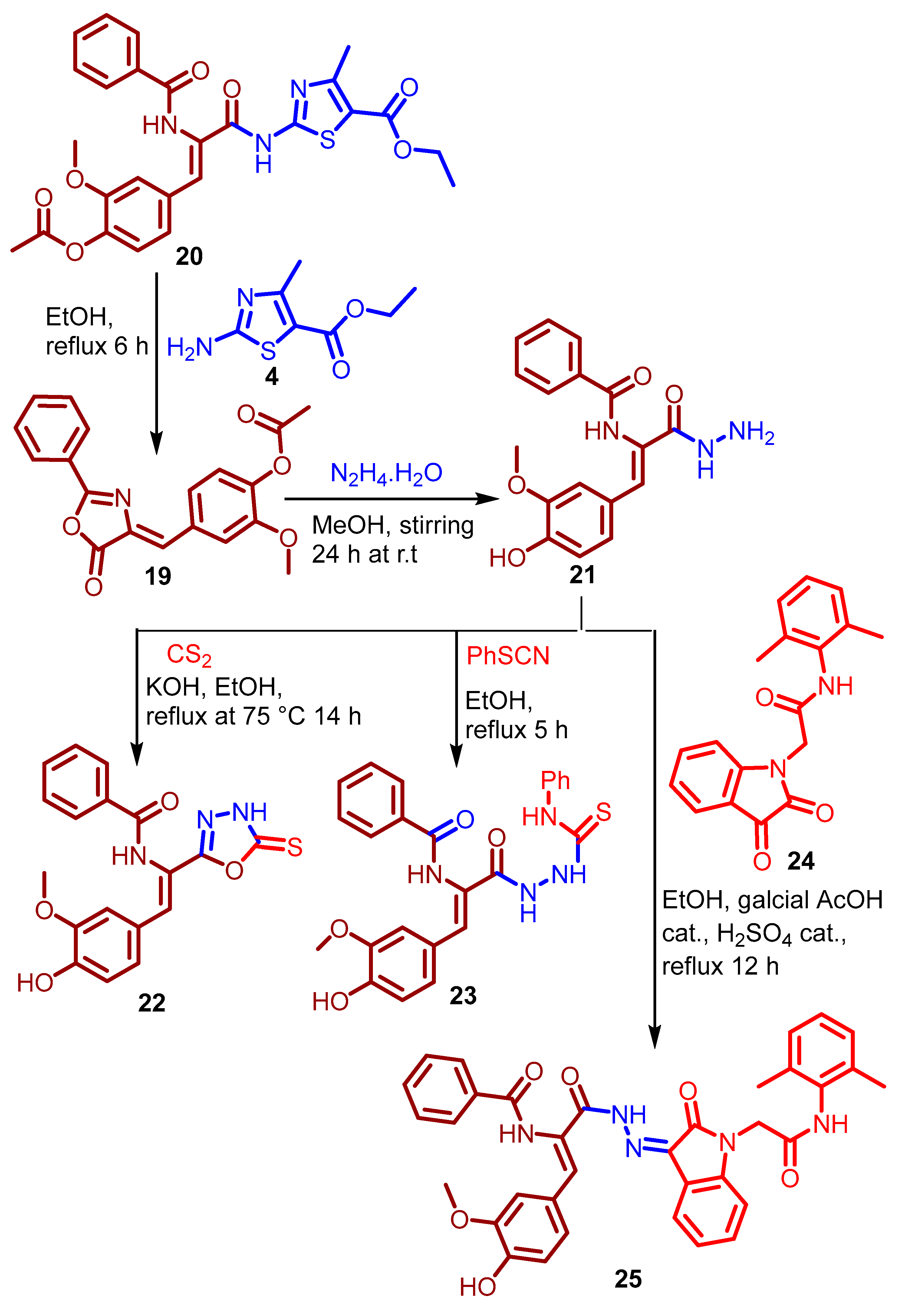
3.1.8. Synthesis of Ethyl (Z)-2-(3-(4-Acetoxy-3-methoxyphenyl)-2-benzamido-acrylamido)- 4-methylthiazole-5-carboxylate 20

Equimolar quantities of (Z)-2-methoxy-4-((5-oxo-2-phenyloxazol-4(5H)-ylidene)methyl)phenyl acetate 19 [32] and ethyl 2-amino-4-methylthiazole-5-carboxylate 4 (0.01 mol) were dissolved in ethanol (30 mL), and the mixture was refluxed for 6 h. After evaporation of excess solvent under reduced pressure, the resulting solid was recrystallized from ethanol to give the title compound as buff-colored powder; yield (55%); m.p. 138–140 °C; IR (KBr): νmax (cm−1) 3372, 3297 (2 × NH and OH), 3079 (CH-Ar), 2980 (CH-aliphatic), 1764 (COO), 1670 (br. 2 × CONH), 1598 (C=N), 1514 (br. C=C), 1373, 1278, 1123, 1092, 913, 831, 716, 589, 525, 424; 1H-NMR (300 MHz, DMSO-d6): δH 1.23 (t, 3H, J = 7.1 Hz, CH3-CH2), 2.26 (s, 3H, CH3), 3.68 (s, 3H, OCH3), 4.15 (AB q, 2H, J = 7.1 Hz, CH2O), 7.15 (d, 1H, J = 8.0 Hz, Ar-H), 7.32 (dd, 1H, J = 8.0, 4.0 Hz, Ar-H), 7.44 (s, 1H, C=CH), 7.49–7.62 (m, 4H, 4 × Ar-H), 7.75 (s, 1H, OH, D2O-exchangeable), 8.02 (d, 2H, J = 8.0 Hz, 2 × Ar-H), 10.17 (s, 2H, 2 × NH, D2O-exchangeable); 13C-NMR (75 MHz, DMSO-d6): δC 14.36, 20.35 (2 × CH3), 55.62 (OCH3), 60.65.(CH2), 107.54, 114.01, 122.67, 123.08, 127.02, 127.68, 128.54, 129.00, 132.00, 132.38, 132.44, 133.50, 140.18, 150.73 (9 × CH-Ar and 6 × Cq-Ar), 162.06, 164.98, 166.31, 168.40, 170.37 (2 × Cq-O, and 3 × C=O); MS (EI, 70 eV): m/z (%) [M+ + 2] 482.25 (29.12) for C24H23N3O6S], 413.96 (47.16), 376.30 (38.77), 340.42 (69.10), 70.38 (100.00).

3.1.9. Synthesis of N-(2-(4-Hydroxy-3-methoxyphenyl)-1-(5-thioxo-4,5-dihydro-1,3,4-oxa-diazol-2-yl)vinyl)benzamide 22

Carbon disulfide (1.25 mmol) and potassium hydroxide (1 mmol) were added to a solution of hydrazide 21 (1 mmol) [32] in ethanol (15 mL). The resulting reaction mixture was heated under reflux at 75 °C for 14 h. After evaporation of excess ethanol, the remaining residue was acidified to pH 2 with HCl (37%), and the precipitated solid was collected by filtration, washed with water and recrystallized from ethanol to afford the title compound as pale yellow powder; yield (55%); m.p. 190–193 °C; IR (KBr): νmax (cm−1) 3245 (OH), 3061 (CH-Ar), 2931 (CH-aliphatic), 1652 (C=O), 1578 (C=N), 1511, 1480 (C=C), 1281 (C=S), 1169, 1030, 942, 808, 708, 616; 1H-NMR (300 MHz, DMSO-d6): δH 3.58 (s, 3H, OCH3), 6.82 (dd, 1H, J = 8.1, 4.2 Hz, CH-Ar), 7.21 (app. dd, 1H, J = 6.9, 1.5 Hz, CH-Ar), 7.31 (s, 1H, CH=C), 7.39 (s, 1H, CH-Ar), 7.53–7.62 (m, 3H, 3 × Ar-H), 8.03–8.19 (m, 2H, 2 × CH-Ar), 9.67, 10.26 (2s, 3H, OH and 2 × NH, D2O-exchangeable); 13C-NMR (75 MHz, DMSO-d6): δC 55.46 (OCH3), 113.64, 115.83, 116.10, 124.73, 124.85, 127.87, 128.51, 128.88, 129.44, 132.45, 133.20 (8 × CH-Ar, CH=Cq-arylidene and 3 × Cq-Ar), 147.61, 148.92, 160.67 (Cq-OH, Cq-OCH3 and N=Cq-O), 166.32 (C=O), 177.49 (C=S); MS (EI, 70 eV): m/z (%) [M+ + 2] 371.00 (28.12) for C18H15N3O4S, 352.47 (58.99), 349.42 (83.16), 297.41 (98.07), 264.22 (57.68), 137.40 (63.64), 101.49 (100.00).

3.1.10. Synthesis of N-(1-(4-Hydroxy-3-methoxyphenyl)-3-oxo-3-(2-(phenylcarbamothionyl)hydrazinyl)prop-1-en-2-yl)benzamide 23

Equimolar quantities of hydrazide 21 and phenyl isothiocyanate (0.003 mol) were dissolved in ethanol (15 mL) and heated under reflux for 5 h. After evaporation of excess solvent under reduced pressure, the resulting solid was recrystallized from ethanol to afford the title compound as yellow powder; yield (88%); m.p. 175–177 °C; IR (KBr): νmax (cm−1) 3350 and 3228 (br. 4 × NH and OH), 3060 (CH-Ar), 2929 (CH-aliphatic), 1650 (2 × C=O), 1597, 1513 (C=C), 1285, 1130, 1033, 931, 814, 742, 694, 615, 499; 1H-NMR (300 MHz, DMSO-d6): δH 3.57 (s, 3H, OCH3), 4.50 (br. s, 1H, OH), 6.81 (d, 1H, J = 8.4 Hz, Ar-H), 7.08–7.17 (m, 3H, 3 × Ar-H), 7.27–7.38 (m, 5H, 5 × Ar-H), 7.50–7.63 (m, 3H, 3 × Ar-H), 7.82 (d, 1H, J = 7.6 Hz, Ar-H), 8.11 (d, 1H, J = 8.0 Hz, Ar-H), 9.32, 9.56, 9.84, 11.05 (4s, 4H, 4 × NH, D2O-exchangeable); 13C-NMR (75 MHz, DMSO-d6): δC 55.19 (OCH3), 113.26, 115.52, 123.51, 124.42, 124.68, 124.86, 125.03, 127.99, 128.16, 128.37, 130.00, 132.00, 132.50, 139.10 (13 × CH-Ar, CH-arylidene and 4 × Cq-Ar), 147.32, 148.11 (Cq-OH, Cq-OCH3), 164.46 (C=O), 167.36 (C=O), 179.98 (C=S); MS (EI, 70 eV): m/z (%) [M+ + 2] 464.59 (72.07) for C24H22N4O4S, 452.30 (75.28), 437.99 (89.10), 170.48 (88.65), 164.22 (100.00), 116.83 (76.92), 99.56 (82.08).

3.1.11. Synthesis of N-(3-(2-(1-(2-((2,4-Dimethylphenyl)amino)-2-oxoethyl)-2-oxoindolin-3-ylidene)hydrazinyl)-1-(4-hydroxy-3-methoxyphenyl)-3-oxoprop-1-en-2-yl)benzamide 25

Equimolar quantities of hydrazide 21 and N-(2,4-dimethyl phenyl)-2-(2,3-dioxoindolin-1-yl)acetamide 24 (0.01 mol) were dissolved in absolute ethanol (30 mL) containing catalytic amounts of glacial AcOH (3 mL) and conc. H2SO4 acid (0.5 mL), and the mixture was refluxed for 16 h. After, evaporation of excess solvent, the resulting solid was recrystallized from ethanol to afford the title compound as orange powder; yield (70%); m.p. 173–175 °C; IR (KBr): νmax (cm−1) 3384 and 3246 (3 × NH and OH), 3055 (CH-Ar), 2928, 2850 (CH-aliphatic), 1720 (CO), 1661 (3 × CO-NH), 1590 (C=N), 1515, 1466 (C=C), 1381, 1218, 1165, 1031, 917, 806, 742, 693, 616, 540; 1H-NMR (300 MHz, DMSO-d6): δH 2.17 (s, 3H, CH3), 2.22 (s, 3H, CH3), 3.76 (br. s, 1H, OH), 3.86 (s, 3H, OCH3), 4.65 (s, 2H, CH2N), 6.89–7.30 (m, 9H, 8 × Ar-H and CH=C), 7.44–7.63 (m, 3H, 3 × Ar-H), 7.90–8.20 (m, 3H, 3 × Ar-H), 8.39 (d, 1H, J = 008.4 Hz, Ar-H), 9.66 (s br., 2H, 2 × NH), 10.48 (s, 1H, NH); 13C-NMR (75 MHz, DMSO-d6): δC 17.89, 20.58 (2 × CH3), 42.02 (CH2N), 55.59 (OCH3), 109.03, 115.77, 115.98, 117.45, 122.00, 122.15, 125.19, 125.50, 126.00, 126.62, 127.09, 127.50, 128.39, 128.80, 129.32, 131.00, 132.00, 133.30, 134.50, 134.70, 139.75, 143.45 (15 × CH-Ar, CH-arylidene and 8 × Cq-Ar), 147.91, 150.31 (Cq-OH, Cq-OCH3), 159.12, 161.06, 165.64, 170.15 (4 × C=O); MS (EI, 70 eV): m/z (%) [M+] 617.31 (20.41) for C35H31N5O6 435.23 (39.63), 332.70 (71.87), 216.41 (100.00), 140.10 (46.12), 112.55 (54.91), 102.09 (53.22).

3.2. Evaluation of Biological Activities

Detailed procedures are available in Supplementary Materials.

3.2.1. In Vitro DPPH Radical Scavenging Assay

The antioxidant efficiencies of target compounds were determined using the 1,1-diphenyl-2-picrylhydrazyl (DPPH) radical scavenging method as detailed by Bersuder et al. [36].

3.2.2. In Vitro PDK-1 Inhibition Assay

Principally, the PDK-1 inhibitory activities were evaluated using the non-radioactive Kinase-Glo™ Luminescent Kinase Kit (Promega Corporation, Madison, WI, USA) following the instructions of the manufacturer.

3.2.3. In Vitro LDHA Inhibitory Assay

The LDHA inhibitory efficiencies were investigated on Spectramax M2 spectrofluorometer via measuring the amounts of the consumed NADH40 using an NAD+/NADH quantification kit.

3.2.4. Cell Culture and Viability Assay

The antiproliferative activities of the newly synthesized compounds 3a–e, 8, 9a–c, 13, 14, 17, 18a–h, 20, 2223 and 25 were examined on human colon HCT-116 and LoVo cell lines as well as HUVEC (American Type Culture Collection, Manassas, VA, USA).

3.2.5. Cell Cycle Assay

Flow cytometric analysis using a Propidium Iodide Flow Cytometry Kit for Cell Cycle Analysis (Abcam, Cambridge, UK), was used in order to analyze the changes in cell cycle distribution induced in HCT 116 and LoVo cultures upon treatment with compound 3d.

3.2.6. Annexin-V-FITC Assay

An Annexin V-FITC Apoptosis Detection Kit (BioVision Research Products, Mountain View, CA, USA) was used to analyze the distribution of early and late apoptotic cells, as well as necrotic cells after treatment with compound 3d.

3.2.7. Analysis of Reactive Oxygen Species (ROS) Levels

A Human ROS ELISA Kit was used to measure the intracellular accumulation of ROS in the 3d-treated LoVo and HCT-116 cells.

3.2.8. Mitochondrial Transmembrane Potential (MMP) Measurement

The mitochondrial transmembrane potential was measured using a tetramethylrhodamine, ethyl ester (TMRE) mitochondrial membrane potential assay kit.

3.2.9. Quantification of the Expression Levels of Bax, Bcl-2 and Caspase-3 Genes

The expression levels of these genes were determined using a quantitative real-time PCR technique.

3.3. In Silico Studies

3.3.1. Molecular Docking Simulations

Protein structure preparation: Molecular docking simulations were conducted using AutoDock Vina 1.1.2. [73,74]. The required protein files in PDB format were downloaded from the Protein Data Bank ((https://www.rcsb.org/) [75]. The crystal structure of lactate dehydrogenase A (LDHA) was obtained using PDB ID 4ZVV [55], which is co-crystallized with the ligand GN0 ((2R)-5-(2-chlorophenyl)sulfanyl-2-(4-morpholin-4-ylphenyl)-4-hydroxy-2-thiophen-3-yl-1,3-dihydropyridin-6-one). For pyruvate dehydrogenase kinase isoform 1 (PDK-1), the structure with PDB ID 2Q8G was selected, co-crystallized with AZX ligand; 4-[3-chloro-4-[[(2R)-3,3,3-trifluoro-2-hydroxy-2-methyl-propanoyl]amino] phenyl]sulfonyl-N,N-dimethyl-benz-amide) was used [56]. For both structures, protein preparation was performed using MGLTools 1.5.7. as follows: All water molecules were removed from the protein structures to eliminate potential interference with the docking process. Polar hydrogen atoms were added to account for potential hydrogen bonding interactions. Kollman united atom charges were assigned to all atoms in the protein structures.
The co-crystallized ligands were removed to define and expose the binding site location. Non-polar hydrogens were merged, and Gasteiger partial atomic charges were calculated and added. The prepared protein structures were then saved in PDBQT format as required for AutoDock Vina calculations [76].
Ligand preparation: Ligand structures were initially sketched using MarvinSketch 19.17.0 software (ChemAxon, Budapest, Hungary) and then saved in MOL format. Geometry optimization and energy minimization of ligands were performed using MMFF94 force field in Open Babel v3.1.1 before conversion to PDBQT format using MGLTools 1.5.7. Each ligand was optimized as follows: All non-polar hydrogen atoms were merged, and Gasteiger partial charges were calculated and assigned to each atom. Rotatable bonds were defined to allow flexibility during the docking process. Energy minimization was performed using the universal force field (UFF) with 5000 steps of steepest descent followed by conjugate gradient minimization until the energy difference between steps was less than 0.1 kcal/mol. The optimized ligands’ structures were saved in the required pdbqt format for subsequent docking studies.
Docking calculations: Molecular docking simulations were performed using AutoDock Vina 1.1.2. [73,74]. For LDHA (4ZVV). The grid box was centered on the GN0 binding site (cocrystal ligand) with dimensions of 25 × 25 × 25 Å to ensure complete coverage of the binding pocket. Similarly, for PDK-1 (2Q8G), the grid box was centered on the binding site of AZX with identical dimensions. The grid spacing was set to 0.375 Å for both proteins. The docking parameters were configured as follows: Exhaustiveness was set to 32 to balance accuracy and computational time, energy range was set to a maximum of 4 kcal/mol, and the number of binding modes was set to 9. The search algorithm was set to employ the default Broyden–Fletcher–Goldfarb–Shanno (BFGS) method for local optimization. For each ligand–protein pair, a minimum of three independent docking runs were performed to ensure reproducibility of the results.
Visualization of analysis: Docked poses and protein–ligand interactions were visualized and analyzed using BIOVIA Discovery Studio Visualizer v21.1.0.20298 [77].

3.3.2. Physicochemical, Drug-Likeness and ADMET Studies

The physicochemical, drug-likeness and ADMET predictions were carried out by using the free webserver [59] ADMETlab 2.0 (https://admetmesh.scbdd.com/), which was accessed on 7 February 2024; the tested compounds were imported through their coding smiles.

4. Conclusions

A series of 24 novel N,O,S-containing heterocyclic derivatives were synthesized and evaluated for their antioxidant potential, PDK-1 and LDHA inhibitory activity, and cytotoxic effects against LoVo and HCT-116 colon carcinoma cell lines, alongside safety profiling in normal HUVECs. Among them, compounds 3b and 3d emerged as the most promising multi-target agents. Compound 3b exhibited strong dual inhibition of PDK-1 and LDHA, while 3d showed potent DPPH radical scavenging and LDHA inhibition, with IC50 values lower than those of the reference standards. Molecular docking simulations confirmed the high binding affinities of both compounds to the active sites of LDHA and/or PDK-1, supporting the observed biochemical inhibition. In cytotoxicity assays conducted at 100 µg/mL for 48 h, compounds 3b and 3d significantly reduced the viability of LoVo and HCT-116 colon cancer cells to below 29%, while preserving approximately 90% viability in normal HUVECs. Their IC50 values ranged from 156.60 to 190.30 µM across the cancer cell lines. The antitumor effects of compound 3d (30 µg/mL, 48 h) were mechanistically linked to cell cycle arrest (G2/M in LoVo, G1 in HCT-116) and apoptosis induction, as evidenced by increased ROS generation, mitochondrial membrane potential disruption, Bcl-2 downregulation, and upregulation of Bax and caspase-3. In silico ADMET predictions revealed moderate to high lipophilicity and poor aqueous solubility for both compounds, particularly 3b, suggesting the need for structural optimization to improve solubility and oral bioavailability. Notably, both 3b and 3d showed only one violation of Lipinski’s rule of five (LogP), and neither contained PAINS-associated substructures, supporting their drug-likeness and the validity of their bioactivities. Taken together, these findings position compounds 3b and 3d as promising lead candidates for further development as multi-target, anti-colorectal cancer agents.

Supplementary Materials

The following supporting information can be downloaded at: https://www.mdpi.com/article/10.3390/ph18060801/s1, Table S1: Energy minimization calculations for compounds 3as, 9ac, 18ah, 20, 21, 22, 23 and 25; Table S2: Evaluation of DPPH radical scavenging activity of the newly synthesized compounds 3ae, 8, 9ac, 13, 14, 17, 18ah, 20, 2223 and 25. Data expressed are as IC50 values (mM) ± SD from three independent experiments. BHT was used as reference antioxidant; Table S3. The mean inhibitory efficiencies (%) of the newly synthesized compounds 3ae, 8, 9ac, 13, 14, 17, 18ah, 20, 2223 and 25; against PDK-1 and LDHA, determined at a concentration of 100 µg/mL ± SD from three replicates. Sodium oxamate (SO), and sodium dichloroacetate (SDA), both at 1000 µM, were used as the reference inhibitors for LDHA and PDK-1, respectively. The mean half-maximal inhibitory concentrations were calculated for compounds showing >80% inhibition of LDHA and/or PDK-1. Results are expressed in µM ± SD from three independent experiments; Table S4: Cytotoxic effects of the newly synthesized compounds 3ae, 8, 9ac, 13, 14, 17, 18ah, 20, 2223, and 25 on LoVo and HCT-116 colon carcinoma cells, as well as HUVECs. Results are presented as the mean percentage of viable cells remained after 48 h exposure to 100 µg/mL of each compound, based on LDH release assay data from three independent determinations ± SD. Assay medium and 0.1% Triton X-100 were used as negative and positive controls, respectively; Table S5: Mean half-maximal inhibitory concentrations (IC50 values) of compounds that reduced cell viability to less than 50% in HCT-116 and/or LoVo colon carcinoma cells. Results are expressed in µM ± SD from three independent determinations. 5-Fluorouracil (5-FU) was used as the reference drug; Table S6: Reactive oxygen species (ROS) production levels in HCT-116 and LoVo colon carcinoma cells following treatment with 30 µg/mL of compound 3d for 48 h. Results are expressed as mean values (pg/mL) ± SD from two independent experiments. Table S7: Primer sequences for Bax, Bcl-2, Caspase-3, and GAPDH (control). Section S3.2. Evaluation of Biological Activities. S3.2.1. In vitro DPPH radical scavenging assay; S3.2.2. In vitro PDK-1 inhibition assay; S3.2.3. In vitro LDHA inhibitory assay; S3.2.4. Cell culture and viability assay; S3.2.5. Cell cycle assay; S3.2.6. Annexin-V-FITC assay; S3.2.7. Analysis of reactive oxygen species (ROS) levels; S3.2.8. Mitochondrial transmembrane potential (MMP) measurement; S3.2.9. Quantification of the expression levels of Bax, Bcl-2 and Casp-3 genes, and 1H-NMR and 13C-NMR spectra of some selected Examples.

Author Contributions

N.N.E.E.-S., conceptualization, interpretation of spectroscopic data, writing—original draft of the manuscript, editing, resources and supervision of the student. S.K., Ph.D student, synthesis and interpreting spectroscopic data. H.A.D. and S.S.A., editing and supervision of the student. A.B.B. and N.K., biological analyses. M.K.E.-A., docking and physicochemical, drug-likeness and ADMET analyses. S.N.A.B. and Z.M.A., editing. All authors have read and agreed to the published version of the manuscript.

Funding

This research received no external funding.

Institutional Review Board Statement

Not applicable.

Informed Consent Statement

Not applicable.

Data Availability Statement

The data presented in this study are available in the Section 3 and the Supplementary Materials.

Conflicts of Interest

The authors declare no conflicts of interest.

References

  1. Xi, Y.; Xu, P. Global colorectal cancer burden in 2020 and projections to 2040. Trans. Oncol. 2021, 14, 101174. [Google Scholar] [CrossRef] [PubMed]
  2. Bray, F.; Ferlay, J.; Soerjomataram, I.; Siegel, R.L.; Torre, L.A.; Jemal, A. Global cancer statistics 2018: GLOBOCAN estimates of incidence and mortality worldwide for 36 cancers in 185 countries. CA Cancer J. Clin. 2018, 68, 394–424. [Google Scholar] [CrossRef] [PubMed]
  3. Nelson, V.K.; Nuli, M.V.; Mastanaiah, J.; Saleem, T.S.M.; Birudala, G.; Jamous, Y.F.; Alshargi, O.; Kotha, K.K.; Sudhan, H.H.; Mani, R.R.; et al. Reactive oxygen species mediated apoptotic death of colon cancer cells: Therapeutic potential of plant derived alkaloids. Front. Endocrinol. 2023, 14, 1201198. [Google Scholar] [CrossRef]
  4. Wan, M.L.; Wang, Y.; Zeng, Z.; Deng, B.; Zhu, B.S.; Cao, T.; Li, Y.K.; Xiao, J.; Han, Q.; Wu, Q. Colorectal cancer (CRC) as a multifactorial disease and its causal correlations with multiple signaling pathways. Biosci. Rep. 2020, 40, BSR20200265. [Google Scholar] [CrossRef] [PubMed]
  5. Li, J.; Ma, X.; Chakravarti, D.; Shalapour, S.; DePinho, R.A. Genetic and biological hallmarks of colorectal cancer. Genes Dev. 2021, 35, 787–820. [Google Scholar] [CrossRef]
  6. Bardelčíková, A.; Šoltys, J.; Mojžiš, J. Oxidative Stress, Inflammation and Colorectal Cancer: An Overview. Antioxidants 2023, 12, 901. [Google Scholar] [CrossRef]
  7. Catalano, T.; Selvaggi, F.; Cotellese, R.; Aceto, G.M. The Role of Reactive Oxygen Species in Colorectal Cancer Initiation and Progression: Perspectives on Theranostic Approaches. Cancers 2025, 17, 752. [Google Scholar] [CrossRef]
  8. Guzmán-Carrasco, A.; Mesas, C.; Doello, K.; Porres, J.M.; García-Beltrán, A.; Martínez, R.; Bermúdez, F.; Peña, M.; Melguizo, C.; Prados, J. The Antioxidant and Chemopreventive Activity of a Nutraceutical Derived from Brassicaceae Seed Extracts for Colorectal Cancer. Nutrients 2025, 17, 1358. [Google Scholar] [CrossRef]
  9. Fang, S.; Fang, X. Advances in glucose metabolism research in colorectal cancer. Biomed. Rep. 2016, 5, 289–295. [Google Scholar] [CrossRef]
  10. Pérez-Tomás, R.; Pérez-Guillén, I. Lactate in the Tumor Microenvironment: An Essential Molecule in Cancer Progression and Treatment. Cancers 2020, 12, 3244. [Google Scholar] [CrossRef]
  11. Angulo-Elizari, E.; Gaviria-Soteras, L.; Zubiri, I.; Ramos-Inza, S.; Sanmartin, C.; Plano, D. Unmasking the Warburg Effect: Unleashing the Power of Enzyme Inhibitors for Cancer Therapy. Drugs Drug Candidates 2023, 2, 728–769. [Google Scholar] [CrossRef]
  12. Alvarado-Ortiz, E.; Ortiz-Sánchez, E.; Sarabia-Sánchez, M.A.; de la Cruz-López, K.G.; García-Carrancá, A.; Robles-Flores, M. Mutant p53 gain-of-function stimulates canonical Wnt signaling via PI3K/AKT pathway in colon cancer. J. Cell Commun. Signal. 2023, 17, 1389–1403. [Google Scholar] [CrossRef] [PubMed]
  13. Morin, P.J. Colorectal cancer: The APC-lncRNA link. J. Clin. Investig. 2019, 129, 503–505. [Google Scholar] [CrossRef]
  14. Yang, S.Y.; Sales, K.M.; Fuller, B.; Seifalian, A.M.; Winslet, M.C. Apoptosis and colorectal cancer: Implications for therapy. Trends Mol. Med. 2009, 15, 225–233. [Google Scholar] [CrossRef] [PubMed]
  15. Carneiro, B.A.; El-Deiry, W.S. Targeting apoptosis in cancer therapy. Nat. Rev. Clin. Oncol. 2020, 17, 395–417. [Google Scholar] [CrossRef]
  16. Zhang, X.; He, L.; Li, Y.; Qiu, Y.; Hu, W.; Lu, W.; Du, H.; Yang, D. Compound 225# inhibits the proliferation of human colorectal cancer cells by promoting cell cycle arrest and apoptosis induction. Oncol. Rep. 2024, 51, 70. [Google Scholar] [CrossRef]
  17. Doostmohammadi, A.; Jooya, H.; Ghorbanian, K.; Gohari, S.; Dadashpour, M. Potentials and future perspectives of multi-target drugs in cancer treatment: The next generation anti-cancer agents. Cell. Commun. Signal. 2024, 22, 228. [Google Scholar] [CrossRef]
  18. Kumar, A.; Singh, A.K.; Singh, H.; Vijayan, V.; Kumar, D.; Naik, J.; Thareja, S.; Yadav, J.P.; Pathak, P.; Grishina, M.; et al. Nitrogen Containing Heterocycles as Anticancer Agents: A Medicinal Chemistry Perspective. Pharmaceuticals 2023, 16, 299. [Google Scholar] [CrossRef]
  19. Bansal, R.; Malhotra, A. Therapeutic progression of quinazolines as targeted chemotherapeutic agents. Eur. J. Med. Chem. 2021, 211, 113016. [Google Scholar] [CrossRef]
  20. Abdel-Wahab, S.M.; Yassen, A.S.; Elrayessb, R.A.; Attiaa, K.M.; Elshihawy, H.A. 4,5,6,7-Tetrahydro benzo[b]thiophene-Based Derivatives as Anticancer Agents: An overview. Rec. Pharm. Biomed. Sci. 2023, 7, 173–180. [Google Scholar] [CrossRef]
  21. Sayed, M.T.M.; Hassan, R.A.; Halim, P.A.; El-Ansary, A.K. Recent updates on thienopyrimidine derivatives as anticancer agents. Med. Chem. Res. 2023, 32, 659–681. [Google Scholar] [CrossRef]
  22. Stecoza, C.E.; Nitulescu, G.M.; Draghici, C.; Caproiu, M.T.; Olaru, O.T.; Bostan, M.; Mihaila, M. Synthesis and Anticancer Evaluation of New 1,3,4-Oxadiazole Derivatives. Pharmaceuticals 2021, 14, 438. [Google Scholar] [CrossRef]
  23. Carradori, S.; Guglielmi, P.; Luisi, G.; Secci, D. Nitrogen- and Sulfur-Containing Heterocycles as Dual Anti-oxidant and Anti-cancer Agents. In Handbook of Oxidative Stress in Cancer: Mechanistic Aspects; Chakraborti, S., Ray, B.K., Roychowdhury, S., Eds.; Springer: Singapore, 2021; pp. 1–18. [Google Scholar] [CrossRef]
  24. El-Sayed, N.N.E.; Al-Otaibi, T.M.; Alonazi, M.; Masand, V.H.; Barakat, A.; Almarhoon, Z.M.; Ben Bacha, A. Synthesis and Characterization of Some New Quinoxalin-2(1H)one and 2-Methyl-3H-quinazolin-4-one Derivatives Targeting the Onset and Progression of CRC with SAR, Molecular Docking, and ADMET Analyses. Molecules 2021, 26, 3121. [Google Scholar] [CrossRef]
  25. Briel, D.; Rybak, A.; Kronbach, C.; Unverferth, K. Substituted 2-aminothiopen-derivatives: A potential new class of GluR6-antagonists. Eur. J. Med. Chem. 2010, 45, 69–77. [Google Scholar] [CrossRef]
  26. Fondjo, E.S.; Döpp, D.; Henkel, G. Reactions of some anellated 2-aminothiophenes with electron poor acetylenes. Tetrahedron 2006, 62, 7121–7131. [Google Scholar] [CrossRef]
  27. Pfeiffer, W.D.; Dollinger, H.; Langer, P. Synthesis of 5-Thioxo-hexahydrobenzo[b]thiopheno[2,3-d]-1,2,4-triazolo[1,5-c]pyrimidines and Related Compounds Based on Cyclocondensations of 2-Isothiocyanato-3-cyano-4,5,6,7-tetrahydrobenzo[b]thiophene. Phosphorus Sulfur Silicon Relat. Elem. 2009, 184, 626–637. [Google Scholar] [CrossRef]
  28. King, L.C.; Hlavacek, R.J. The reaction of ketones with iodine and thiourea. J. Am. Chem. Soc. 1950, 72, 3722–3725. [Google Scholar] [CrossRef]
  29. Lin, G.W.; Wang, Y.; Jin, Q.M.; Yang, T.T.; Song, J.M.; Lu, Y.; Huang, Q.J.; Song, K.; Zhou, J.; Lu, T. Synthesis, structures and the biological activity study on the metal complexes of 2-(4-aminophenyl) benzothiazole derivative. Inorg. Chim. Acta 2012, 382, 35–42. [Google Scholar] [CrossRef]
  30. Osarodion, O.P.; Orile, E.M.; Odianosen, U.C. Electron impact ionization mass spectra of 3-amino 5,6-dimethoxyl-2-methylquinazolin-4-(3H)-one derivative. Am. J. Mater. Synth. Process. 2019, 4, 62–67. [Google Scholar] [CrossRef]
  31. Al-Maneai, N.M. Synthesis and Pharmacological Evaluation of Some Novel 4-(3H)-Quinazolinone Derivatives. Master’s Thesis, King Saud University, Riyadh, Saudi Arabia, 2017. [Google Scholar]
  32. Soujanya, M.; Rajitha, G.; Umamaheswari, A.; Kumar, K.S. Synthesis, Biological Evaluation and Docking Studies of N-(2-benzamido feruloyl) Aryl Hydrazone Analogues. Lett. Drug Des. Discov. 2018, 15, 875–886. [Google Scholar] [CrossRef]
  33. Mansour, M.A.; AboulMagd, A.M.; Abdel-Rahman, H.M. Quinazoline-Schiff base conjugates: In silico study and ADMET predictions as multi-target inhibitors of coronavirus (SARS-CoV-2) proteins. RSC Adv. 2020, 10, 34033–34045. [Google Scholar] [CrossRef] [PubMed]
  34. Prokof’ev, E.P.; Karpeiskaya, E.I. The proton coupled 13C NMR direct determination of Z-, E-configuration of 4-benzyliden-2-phenyl(methyl)-Δ2-oxazolin-5-ones and products of their solvolysis. Tetrahedron Lett. 1979, 20, 737–740. [Google Scholar] [CrossRef]
  35. Belskaya, N.P.; Dehaen, W.; Bakulev, V.A. Synthesis and properties of hydrazones bearing amide, thioamide and amidine functions. ARKIVOC 2010, 2010, 275–322. [Google Scholar] [CrossRef]
  36. Bersuder, P.; Hole, M.; Smith, G. Antioxidants from a heated histidine-glucose model system. I: Investigation of the antioxidant role of histidine and isolation of antioxidants by high-performance liquid chromatography. J. Am. Oil Chem. Soc. 1998, 75, 181–187. [Google Scholar] [CrossRef]
  37. Xu, B.; Yu, Z.; Xiang, S.; Li, Y.; Zhang, S.L.; He, Y. Rational design of mitochondria-targeted pyruvate dehydrogenase kinase 1 inhibitors with improved selectivity and antiproliferative activity. Eur. J. Med. Chem. 2018, 155, 275–284. [Google Scholar] [CrossRef]
  38. Zhang, S.L.; Yang, Z.; Hu, X.; Tam, K.Y. Dichloroacetophenones targeting at pyruvate dehydrogenase kinase 1 with improved selectivity and antiproliferative activity: Synthesis and structure-activity relationships. Bioorg. Med. Chem. Lett. 2018, 28, 3441–3445. [Google Scholar] [CrossRef]
  39. Al-Salam, S.; Kandhan, K.; Sudhadevi, M. Down regulation of lactate dehydrogenase initiates apoptosis in HeLa and MCF-7 cancer cells through increased voltage-dependent anion channel protein and inhibition of BCL2. Oncotarget 2021, 12, 923–935. [Google Scholar] [CrossRef] [PubMed]
  40. Zhang, W.; Wang, C.; Hu, X.; Lian, Y.; Ding, C.; Ming, L. Inhibition of LDHA suppresses cell proliferation and increases mitochondrial apoptosis via the JNK signaling pathway in cervical cancer cells. Oncol. Rep. 2022, 47, 77. [Google Scholar] [CrossRef]
  41. Kozal, K.; Jóźwiak, P.; Krześlak, A. Contemporary Perspectives on the Warburg Effect Inhibition in Cancer Therapy. Cancer Control 2021, 28, 10732748211041243. [Google Scholar] [CrossRef]
  42. Kim, E.Y.; Choi, H.J.; Park, M.J.; Jung, Y.S.; Lee, S.O.; Kim, K.J.; Choi, J.H.; Chung, T.W.; Ha, K.T. Myristica fragrans Suppresses Tumor Growth and Metabolism by Inhibiting Lactate Dehydrogenase A. Am. J. Chin. Med. 2016, 44, 1063–1079. [Google Scholar] [CrossRef]
  43. Basu, A.; Haldar, S. The relationship between BcI2, Bax and p53: Consequences for cell cycle progression and cell death. Mol. Hum. Reprod. 1998, 4, 1099–1109. [Google Scholar] [CrossRef] [PubMed]
  44. Mustafa, M.; Ahmad, R.; Tantry, I.Q.; Ahmad, W.; Siddiqui, S.; Alam, M.; Abbas, K.; Hassan, M.I.; Habib, S.; Islam, S. Apoptosis: A Comprehensive Overview of Signaling Pathways, Morphological Changes, and Physiological Significance and Therapeutic Implications. Cells 2024, 13, 1838. [Google Scholar] [CrossRef] [PubMed]
  45. Eskandari, E.; Eaves, C.J. Paradoxical roles of caspase-3 in regulating cell survival, proliferation, and tumorigenesis. J. Cell Biol. 2022, 221, e202201159. [Google Scholar] [CrossRef]
  46. Hussar, P. Apoptosis Regulators Bcl-2 and Caspase-3. Encyclopedia 2022, 2, 1624–1636. [Google Scholar] [CrossRef]
  47. Zhra, M.; Qasem, R.J.; Aldossari, F.; Saleem, R.; Aljada, A. A Comprehensive Exploration of Caspase Detection Methods: From Classical Approaches to Cutting-Edge Innovations. Int. J. Mol. Sci. 2024, 25, 5460. [Google Scholar] [CrossRef]
  48. Idelchik, M.D.P.S.; Begley, U.; Begley, T.J.; Melendez, J.A. Mitochondrial ROS control of cancer. Semin. Cancer Biol. 2017, 47, 57–66. [Google Scholar] [CrossRef] [PubMed]
  49. Fernández-Lázaro, D.; Sanz, B.; Seco-Calvo, J. Mechanisms of programmed cell death: Structural and functional pathways. A narrative review. Investig. Clín. 2024, 65, 230–252. [Google Scholar] [CrossRef]
  50. Tsuzuki, T.; Kambe, T.; Shibata, A.; Kawakami, Y.; Nakagawa, K.; Miyazawa, T. Conjugated EPA Activates Mutant p53 via Lipid Peroxidation and Induces p53-Dependent Apoptosis in DLD-1 Colorectal Adenocarcinoma Human Cells. Biochim. Biophys. Acta Mol. Cell Biol. Lipids 2007, 1771, 20–30. [Google Scholar] [CrossRef]
  51. Sheikh, B.Y.; Sarker, M.M.R.; Kamarudin, M.N.A.; Mohan, G. Antiproliferative and apoptosis inducing effects of citral via p53 and ROS-induced mitochondrial-mediated apoptosis in human colorectal HCT116 and HT29 cell lines. Biomed. Pharmacother. 2017, 96, 834–846. [Google Scholar] [CrossRef]
  52. Meng, X.; Zhang, J.; Wu, H.; Yu, D.; Fang, X. Akkermansia muciniphila Aspartic Protease Amuc_1434* Inhibits Human Colorectal Cancer LS174T Cell Viability via TRAIL-Mediated Apoptosis Pathway. Int. J. Mol. Sci. 2020, 21, 3385. [Google Scholar] [CrossRef]
  53. van Engeland, M.; Nieland, L.J.; Ramaekers, F.C.; Schutte, B.; Reutelingsperger, C.P. Annexin V-affinity assay: A review on an apoptosis detection system based on phosphatidylserine exposure. Cytometry 1998, 31, 1–9. [Google Scholar] [CrossRef]
  54. Zhu, S.; Li, T.; Tan, J.; Yan, X.; Zhang, D.; Zheng, C.; Chen, Y.; Xiang, Z.; Cui, H. Bax is essential for death receptor-mediated apoptosis in human colon cancer cells. Cancer Biother. Radiopharm. 2012, 27, 577–581. [Google Scholar] [CrossRef] [PubMed]
  55. Boudreau, A.; Purkey, H.E.; Hitz, A.; Robarge, K.; Peterson, D.; Labadie, S.; Kwong, M.; Hong, R.; Gao, M.; Del Nagro, C.; et al. Metabolic plasticity underpins innate and acquired resistance to LDHA inhibition. Nat. Chem. Biol. 2016, 12, 779–786. [Google Scholar] [CrossRef]
  56. Kato, M.; Li, J.; Chuang, J.L.; Chuang, D.T. Distinct structural mechanisms for inhibition of pyruvate dehydrogenase kinase isoforms by AZD7545, dichloroacetate, and radicicol. Structure 2007, 15, 992–1004. [Google Scholar] [CrossRef]
  57. Rimac, H.; Grishina, M.; Potemkin, V. Use of the Complementarity Principle in Docking Procedures: A New Approach for Evaluating the Correctness of Binding Poses. J. Chem. Inf. Model. 2021, 61, 1801–1813. [Google Scholar] [CrossRef]
  58. Sharma, H.; Sharma, P.; Urquiza, U.; Chastain, L.R.; Ihnat, M.A. Exploration of a Large Virtual Chemical Space: Identification of Potent Inhibitors of Lactate Dehydrogenase-A against Pancreatic Cancer. J. Chem. Inf. Model. 2023, 63, 1028–1043. [Google Scholar] [CrossRef] [PubMed]
  59. Xiong, G.; Wu, Z.; Yi, J.; Fu, L.; Yang, Z.; Hsieh, C.; Yin, M.; Zeng, X.; Wu, C.; Lu, A.; et al. ADMETlab 2.0: An integrated online platform for accurate and comprehensive predictions of ADMET properties. Nucleic Acids Res. 2021, 49, W5–W14. [Google Scholar] [CrossRef]
  60. Hernandez, R.D.; Genio, F.A.F.; Casanova, J.R.; Conato, M.T.; Paderes, M.C. Antiproliferative activities and SwissADME predictions of physicochemical properties of carbonyl group-modified rotenone analogues. ChemistryOpen 2024, 13, e202300087. [Google Scholar] [CrossRef]
  61. Bickerton, G.R.; Paolini, G.V.; Besnard, J.; Muresan, S.; Hopkins, A.L. Quantifying the chemical beauty of drugs. Nat. Chem. 2012, 4, 90–98. [Google Scholar] [CrossRef] [PubMed]
  62. Skoraczyński, G.; Kitlas, M.; Miasojedow, B.; Gambin, A. Critical assessment of synthetic accessibility scores in computer-assisted synthesis planning. J. Cheminform. 2023, 15, 6. [Google Scholar] [CrossRef]
  63. Ivanenkov, Y.A.; Zagribelnyy, B.A.; Aladinskiy, V.A. Are We Opening the Door to a New Era of Medicinal Chemistry or Being Collapsed to a Chemical Singularity? J. Med. Chem. 2019, 62, 10026–10043. [Google Scholar] [CrossRef]
  64. Lipinski, C.A. Lead- and drug-like compounds: The rule-of-five revolution. Drug Discov. Today Technol. 2004, 1, 337–341. [Google Scholar] [CrossRef] [PubMed]
  65. Baell, J.B.; Nissink, J.W.M. Seven Year Itch: Pan-Assay Interference Compounds (PAINS) in 2017-Utility and Limitations. ACS Chem. Biol. 2018, 13, 36–44. [Google Scholar] [CrossRef] [PubMed]
  66. Lee, J.B.; Zgair, A.; Taha, D.A.; Zang, X.; Kagan, L.; Kim, T.H.; Kim, M.G.; Yun, H.Y.; Fischer, P.M.; Gershkovich, P. Quantitative analysis of lab-to-lab variability in Caco-2 permeability assays. Eur. J. Pharm. Biopharm. 2017, 114, 38–42. [Google Scholar] [CrossRef]
  67. Kim, Y.; Chen, J. Molecular structure of human P-glycoprotein in the ATP-bound, outward-facing conformation. Science 2018, 359, 915–919. [Google Scholar] [CrossRef]
  68. Kratochwil, N.A.; Huber, W.; Müller, F.; Kansy, M.; Gerber, P.R. Predicting plasma protein binding of drugs: A new approach. Biochem. Pharmacol. 2002, 64, 1355–1374. [Google Scholar] [CrossRef]
  69. Crettol, S.; Petrovic, N.; Murray, M. Pharmacogenetics of phase I and phase II drug metabolism. Curr. Pharm. Des. 2010, 16, 204–219. [Google Scholar] [CrossRef]
  70. Smith, D.A.; Beaumont, K.; Maurer, T.S.; Di, L. Clearance in Drug Design. J. Med. Chem. 2019, 62, 2245–2255. [Google Scholar] [CrossRef] [PubMed]
  71. Garrido, A.; Lepailleur, A.; Mignani, S.M.; Dallemagne, P.; Rochais, C. hERG toxicity assessment: Useful guidelines for drug design. Eur. J. Med. Chem. 2020, 195, 112290. [Google Scholar] [CrossRef]
  72. Gozalbes, R.; Jacewicz, M.; Annand, R.; Tsaioun, K.; Pineda-Lucena, A. QSAR-based permeability model for drug-like compounds. Bioorg. Med. Chem. 2011, 19, 2615–2624. [Google Scholar] [CrossRef]
  73. Eberhardt, J.; Santos-Martins, D.; Tillack, A.F.; Forli, S. AutoDock Vina 1.2.0: New Docking Methods, Expanded Force Field, and Python Bindings. J. Chem. Inf. Model. 2021, 61, 3891–3898. [Google Scholar] [CrossRef]
  74. Trott, O.; Olson, A.J. AutoDock Vina: Improving the speed and accuracy of docking with a new scoring function, efficient optimization, and multithreading. J. Comput. Chem. 2010, 31, 455–461. [Google Scholar] [CrossRef] [PubMed]
  75. Berman, H.M.; Westbrook, J.; Feng, Z.; Gilliland, G.; Bhat, T.N.; Weissig, H.; Shindyalov, I.N.; Bourne, P.E. The Protein Data Bank. Nucleic Acids Res. 2000, 28, 235–242. [Google Scholar] [CrossRef] [PubMed]
  76. Morris, G.M.; Huey, R.; Lindstrom, W.; Sanner, M.F.; Belew, R.K.; Goodsell, D.S.; Olson, A.J. AutoDock4 and AutoDockTools4: Automated docking with selective receptor flexibility. J. Comput. Chem. 2009, 30, 2785–2791. [Google Scholar] [CrossRef] [PubMed]
  77. BIOVIA; Dassault Systèmes. Discovery Studio Visualizer, v21.1.0.20298; Dassault Systemes: San Diego, CA, USA, 2005.
Scheme 1. Synthesis of compounds 3ae, 8, and 9.
Pharmaceuticals 18 00801 sch001
Scheme 2. Synthesis of compounds 13, 14, 17, and 18ah.
Pharmaceuticals 18 00801 sch002
Scheme 3. Synthesis of compounds 20, 22, 23, and 25.
Pharmaceuticals 18 00801 sch003
Figure 1. Effects of compound 3d on the cell cycle distribution of LoVo and HCT-116 cells: (A) negative control LoVo cells, (B) 3d-treated LoVo cells, (C) negative control HCT-116 cells, (D) 3d-treated HCT-116 cells, (E) bar chart depicting mean cell cycle distribution (% ± SD, n = 2).
Pharmaceuticals 18 00801 g001
Figure 2. qRT-PCR quantification of Bax, Bcl2 and Casp-3 genes expression in LoVo and HCT-116 cells treated with compound 3d (30 µg/mL, 48 h). Data are expressed as fold changes relative to untreated controls and normalized to GAPDH.
Pharmaceuticals 18 00801 g002
Figure 3. Flow cytometric analysis of apoptosis in LoVo and HCT-116 cells treated with compound 3d (30 µg/mL, 48 h): (A) untreated LoVo cells (control), (B) 3d-treated LoVo cells, (C) untreated HCT-116 cells (control), (D) 3d-treated HCT-116 cells. (E) bar chart quantifying early apoptosis, late apoptosis, total apoptosis, and necrosis expressed as mean percentages ± SD of two independent experiments, compared.
Pharmaceuticals 18 00801 g003
Figure 4. Mechanistic flowchart summarizing the sequential events by which compound 3d exerts anticancer effects in colorectal carcinoma cells. Arrows indicate; pathway flow and potential crosstalk with extrinsic apoptosis. Abbreviations: ROS, reactive oxygen species; ΔΨm, mitochondrial membrane potential.
Pharmaceuticals 18 00801 g004
Figure 5. Validation of the docking process against LDHA and PDK-1 enzymes. Superimposition of the co-crystallized ligands (purple) and re-docked ligands (cyan).
Pharmaceuticals 18 00801 g005
Figure 6. A 2D representation of the interactions with amino acid residues in the active binding site of LDHA: (a) Compound 3b, (b) compound 3d.
Pharmaceuticals 18 00801 g006
Figure 7. A 2D representation of the interactions between compound 3b and amino acid residues in the active binding site of PDK-1.
Pharmaceuticals 18 00801 g007
Figure 8. Radar chart representations of compounds 3b and 3d, exhibiting their physicochemical properties.
Pharmaceuticals 18 00801 g008
Disclaimer/Publisher’s Note: The statements, opinions and data contained in all publications are solely those of the individual author(s) and contributor(s) and not of MDPI and/or the editor(s). MDPI and/or the editor(s) disclaim responsibility for any injury to people or property resulting from any ideas, methods, instructions or products referred to in the content.

