Article

Borylated 5-Membered Ring Iminosugars: Synthesis and Biological Evaluation for Glycosidase Inhibition and Anticancer Properties for Application in Boron Neutron Capture Therapy (BNCT)—Part 2

1
Discipline of Chemistry, University of Newcastle, Callaghan, NSW 2308, Australia
2
Department of Hospital Pharmacy, University of Toyama, 2630 Sugitani, Toyama 930-0194, Japan
3
PhytoQuest Ltd., Plas Gogerddan, Aberystwyth, Ceredigion SY23 3EB, UK
4
Department of Medical Oncology, Calvary Mater Newcastle Hospital, Waratah, NSW 2298, Australia
*
Author to whom correspondence should be addressed.
Current address: Newcastle CSIRO Energy Centre, 10 Murray Dwyer Circuit, Mayfield West, NSW 2304, Australia.
Pharmaceuticals 2025, 18(11), 1739; https://doi.org/10.3390/ph18111739
Submission received: 2 September 2025 / Revised: 28 October 2025 / Accepted: 7 November 2025 / Published: 17 November 2025

Abstract

Background: The synthesis and biological investigation of pyrrolidine (L-gulo) iminosugars bearing an organic boron pharmacophore in ortho and meta positions of an N-benzyl group is reported. This paper completes the structure–activity relationship data for this novel family of boron-bearing iminosugars. These can establish reversible intramolecular interactions via dative bonding from nucleophilic amino acid side chains to the empty p-orbital of the boron atom. Methods: Inhibitory activities against two panels of glycosidases and cancer cell lines were investigated to ascertain structure–activity relationship profiles for these novel iminosugar drug leads. Results: These iminosugars display selective, moderate-to-weak inhibitions (IC50s = 116–617 μM) of β-D-galactosidase (bovine liver), and indications of inhibition of β-D-glucosidases (almond, bovine liver) (IC50s = 633 and 710 μM) and α-D-glucosidases (rice, yeast, rat intestinal maltase) (IC50s = 106–784 μM). The boronic acid group emerges as a useful pharmacophore for management of lysosomal storage disorders via the chaperone-mediated therapy approach. The cancer assays revealed that the A2780 ovarian carcinoma cell line is selectively inhibited by all compounds screened and the MIA-Pa-Ca2 pancreatic carcinoma cell line is selectively inhibited by most compounds. Growth inhibition and GI50 values were most potent for the meta 7 side-product. Conclusions: Beyond the cancer cell line inhibition and dose-response capabilities, the real therapeutic potential of these borylated drugs lies in their switch on/switch off activation under boron neutron capture therapy (BNCT) radiotherapeutic conditions, thus providing an important area of application for borylated monosaccharides.

1. Introduction

From a historical perspective, groundbreaking applications have been achieved in boron chemistry in several areas, spanning boranes and organoboranes—leading to the development of the first general asymmetric synthesis of pure enantiomers -, cross-coupling reactions and semiconductors [1,2,3,4]. Currently, boron-10 is a promising isotope in boron neutron capture therapy (BNCT); metal borides possess exceptional physical properties, such as hardness, high melting and boiling points, light-weightness, chemical inertness, and superconductivity, which make them amenable to high-performance environments, including space applications, nuclear reactor safety operations, as rocket/aircraft propellant, component of tank armours, and next-generation electrolytic and fuel-generation systems, to name a few [5,6,7,8,9].
Boron’s chemistry is wonderfully rich, displaying complex chemical equilibria [10,11,12,13,14,15], which make it amenable to varied applications [16,17,18,19,20]. In medicinal chemistry, installation of organic boron pharmacophores on high Fsp3 index substrates (e.g., monosaccharides, which contain heteroatoms, many consecutive stereocentres, and anomeric forms) expands the interactions between borylated drugs and biological systems to reversible covalent bonds, changes in shape and electronics at the boron and neighbouring sites. Organic B atoms thus introduce significant structural and electronic refinements to the induced fit model [21,22,23] by providing drug molecules that are imbued with conformational and electronic flexibility to adapt into enzyme active sites and neighbouring sites. Drugs and enzymes team up to work in unison and adapt to each other.
A high Fsp3 index [24,25] has been identified as one of the main factors propelling drug leads through to clinical success. We believe that the expansion of the palette of interactions between drug and enzyme is another important factor [26,27,28,29,30]. This paper completes the discussion on the synthesis and biological evaluation of a novel family of borylated iminosugars [30].
Our group has worked in the areas of carbohydrate chemistry [27] and iminosugar [31] research for many years. Here, we communicate the synthesis to borylated 5-membered ring iminosugars of l-gulo absolute stereochemical configuration via development of synthetic and purification protocols to several selective, up to moderately potent glycosidase inhibitors and perspective BNCT agents.
This type of drug leads and previous ones we have communicated [27,28,29,30,32,33] are quite unique in the medicinal chemical literature. They introduce a proof-of-concept and open up a new area in medicinal chemistry, namely borylated monosaccharides and derivatives, imbued with low toxicities (thanks to the organic boron moiety), the capacity to mimic carbohydrates and exploit the same entry mechanisms across membranes [34], glycosidase modulation and switch on/switch off BNCT capabilities that allows cancer management via a milder radiation therapy methodology.

2. Results and Discussion

2.1. Synthesis

Divergent intermediate 1 is available via a four-step synthetic protocol [35], starting from reagent 1,2:5,6-di-O-isopropylidene-α-d-allofuranose (Scheme 1).
N-alkylation, steps (i) and (iv): This reaction (i) required optimisation as initial attempts employing a modified method [36] successfully employed with 6-membered ring systems, was found to be insufficient to drive to completion the reaction of the 5-membered ring. After a thorough investigation of the relevant chemical literature, several strategies can be employed, including SN2 [37,38] and reductive amination [39,40,41] on protected [37,39] and unprotected [36,42] iminosugars.
Several strategies were trialled, including a 31 h reaction time at a temperature of 50 °C, after which product formation was confirmed. Acetone was used as reaction solvent in another strategy, requiring a longer reaction time and increased equivalents of potassium carbonate and benzylating agent 3-/2-bromomethylphenylboronic acid pinacol ester. This was likely due to the low solubility of potassium carbonate in this solvent system.
Additional N-alkylation strategies trialled, involved one-pot global deprotection of the crude N-alkylated product (sequence: steps (i) and (ii)), or global deprotection of divergent iminosugar 1 followed by N-alkylation (sequence: steps (ii), (iii) and (i), or steps (ii), (i) and (iii). Neither of these strategies resulted in simplified purification protocols.
The optimised method employs a slight excess of the iminosugar starting material 1 (1.1 eq) compared to the base (K2CO3) and benzylating agent (2-/3-bromomethylphenyl boronic acid pinacol esters), both utilised in 1.0 equivalent masses. DMF was found to be the best solvent, using a concentration of 10 mg of iminosugar 1/mL DMF. The resultant crude was dissolved in chloroform and washed with NaOH (aq. 0.1 M), requiring no flash-column purification. This method resulted in high purity products in yields of 72% and 71%, respectively, for meta 2, ortho 2 and meta 5.
Purification of boron-bearing organic molecules: Initially, purifications were carried out via flash chromatography (e.g., silica gel) and resin columns (e.g., DOWEX). However, organic boron esters and boronic acids are notoriously difficult to purify due to the Lewis acidity of the B atom and the chemical instability of organic B groups [4]. Purification strategies required optimisation. In our experience, the presence of boronic acid and boronate ester groups greatly widens the range of solvent solubility of molecules they are bonded to. Inevitably, during partitions, several desired intermediates/products tend to travel to both aqueous and organic layers, or to both organic layers (if the partition is carried out solely with organic solvents). Crystallisation, recrystallisations and judicious utilisation of partitioning strategies are powerful tools to maximise yields and minimise chemical instability of boron-bearing molecules.
Global deprotection, step (ii): This was effected by employing HCl (aq. 1 M) at 55 °C to give access to the corresponding lactols meta 3 and ortho 3. Access to meta 3 required a 4 h reaction time, whereas 18 h were needed for ortho 3. Pinacol removal proved difficult and a minor fraction permained.
Reduction, step (iii): The final borylated iminosugars were afforded by a sodium borohydride reduction in lactols ortho/meta 3. The optimised reaction conditions were a 1 h reaction time at room temperature with NaBH4 (1.2 eq) added at the start of the reaction and an additional portion (1.2 eq) after 30 min. Quenching with glacial acetic acid, followed by filtration and evaporation to dryness gave the final products.
Oxidation side-reaction, steps (v) and (vi): Phenol meta 6 arose from slow oxidation of meta 5 over several months, at room temperature. meta 7 arose when the reaction mixture of meta 4 was purified by prop t.l.c. The reaction mixture was concentrated in vacuo and triturated, then filtered with methanol/ethanol (1:1). Prep t.l.c. (toluene/acetone/acetic acid, 10:15:1) resulted in oxidation of meta 4 to meta 7.
The detailed 11B-NMR, 1H-, 13C- and 2D NMR data analysis and spectroscopic method for study of mutarotation and borarotation equilibria are available in these publications [30,32,33].
SN2 reaction between 1 and 3-bromomethylphenylboronic acid afforded the pinacol-deprotected meta 5 in excellent yield. For NMR data and analysis, see [33]. Care must be taken in avoiding loss of product in the aqueous layer during the work-up stage. meta 6 arises from slow oxidation on the bench or in aqueous solution. ortho 5 arose when a strongly basic aqueous layer (NaOH, 1 M) was utilised to wash the reaction mixture. For NMR data and analysis, see [33].

2.2. Biological Assays

2.2.1. Glycosidase Inhibition—Background

Glycosidase inhibition data for N-benzyl-1,4-dideoxy-1,4-imino derivatives are shown in Table 1 and Figure 1.
N-Benzyl-1,4-dideoxy-1,4-imino-d-allitol displays selectivity towards α-l-fucosidase (76%) [43], and N-benzyl-1,4-dideoxy-1,4-imino-d-galactitol towards α-d-glucosidase (93.2%, IC50 = 40.6 μM) [44]. When N-benzyl-1,4-dideoxy-1,4-imino-d-mannitol.HCl [45,46,47] is N-arylalkylated with short alkyl chains, no glycosidase inhibition is observed [46]. However, lengthening of the N-alkyl chain results in increasing inhibition of β-d-glucosidase, β-d-galactosidase and β-d-glucuronidase [45]. Inhibition of α-d-mannosidases is greatest for lysosomal acidic (34%), neutral (44%) and Golgi II (72%) [46], fruit fly lysosomal acidic (IC50 = 1.5 × 10⁻³ M) and fruit fly Golgi II (IC50 = 6.9 × 10⁻⁴ M) [47]. N-Benzyl-1,4-dideoxy-1,4-imino-d-talitol.HCl poorly inhibited β-d-galactosidase (E. coli), α-d-galactosidase (coffee bean) and α-d-mannosidase (Jack bean) [48]. However, the N-(2-methylphenyl boronic acid) derivative shows significant inhibition of β-d-galactosidase (E. coli) (44–55%), and an increase in inhibition of α-d-galactosidase (coffee bean) (<5%) and α-d-mannosidase (Jack bean) (10%) [48].
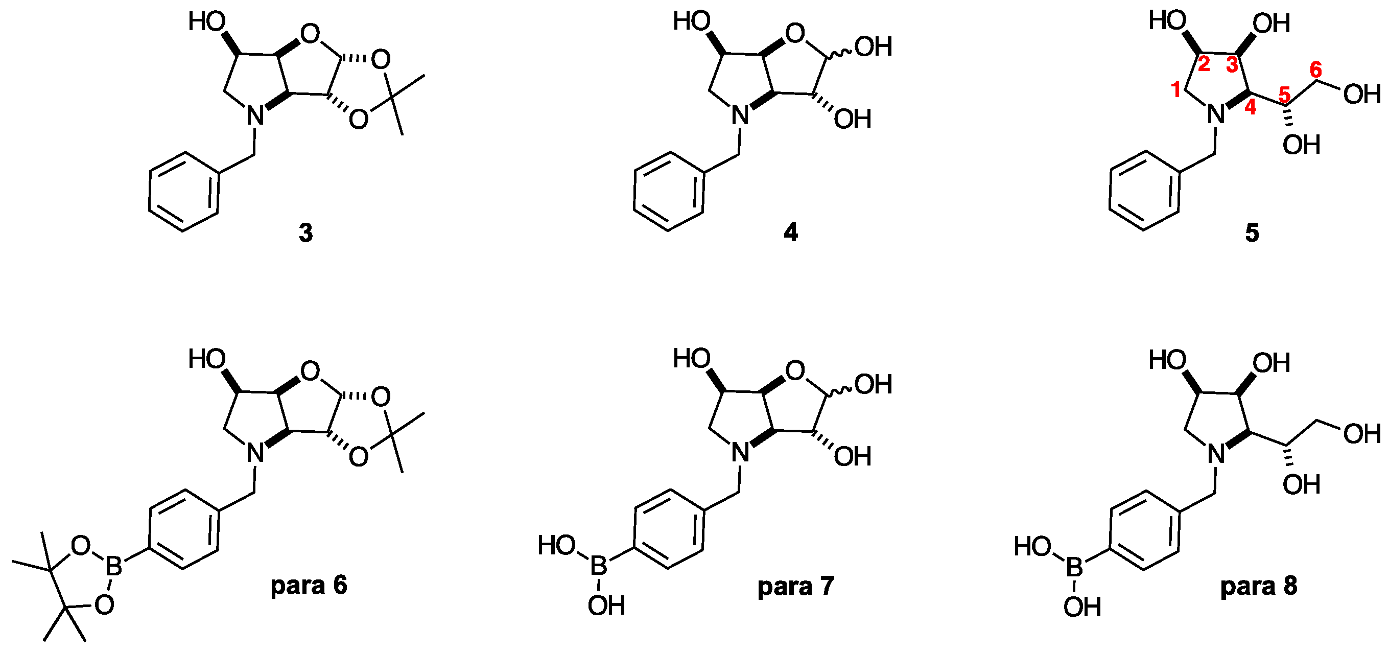
Recently, we reported on a family of borylated iminosugars (para 6, para 7 and para 8) and their parent compounds (3, 4 and 5) (Figure 1) displaying selective inhibitions (>30%) for β-d-glycosidases (two glucosidases and one galactosidase).
N-benzyl-3,6-dideoxy-3,6-imino-d-gulofuranose 4, N-(4-methylphenyl boronic acid)-3,6-dideoxy-3,6-imino-d-gulofuranose para 7 and N-(4-methylphenyl boronic acid)-1,4-dideoxy-1,4-imino-l-gulitol para 8 selectively inhibited β-d-galactosidase (bovine liver) with, respectively, IC50 values of 133 μM (moderate), 218 μM (moderate) and 501 μM (weak). These iminosugars also display an indication of inhibition towards β-d-glucosidases (almond, bovine liver). β-d-glucosidase (EC 3.2.1.21) and β-d-galactosidase (EC 3.2.1.23) are associated with lysosomal storage disorders, such as Gaucher disease [49,50,51] and GM1 gangliosidosis (linked to β-d-glucosidase activity), and Morquio syndrome B (linked to β-d-galactosidase activity) [52]. Rationalisation of inhibition profiles is possible through a systematic postulated framework, such as this one [53].

2.2.2. Glycosidase Inhibitions (Table 2, Scheme 1, Figure 1)

Controls: Borocaptate sodium (BSH) and 4-borono-l-phenylalanine (BPA) are clinically used for BNCT. BSH, BPA, and their 10B-isotopomers 10B-BSH and 10B-BPA were utilised as controls. Under the conditions screened, none of them significantly inhibits the glycosidases in the panel at 100 or 1000 μM. Percent inhibitions range from a minimum value of 0 to a maximum value of 19.6.
Table 2. Glycosidase inhibition. Percentage inhibition data for controls (BSH, 10B-BSH, BPA, 10B-BPA), intermediates ortho 2, ortho 3, meta 2 and meta 3, and target compounds ortho 4 and meta 4, and side products ortho 5 and meta 5–7 (drugs) in a panel of 15 glycosidases. IC50 values are on green coloured backgrounds (μM): light green denotes weak inhibition, dark green denotes moderate inhibition. NA = not available. Blue background denotes biological activity >30% under conditions screened. In the “Drug” column, on the orange background, the assay concentration legend is as follows: aNI: no inhibition (less than 50% inhibition at 1000 μM), b( ): inhibition % at 1000 μM. In the “Drug” column, on the white background, the assay concentration legend is as follows: cNI: no inhibition (less than 50% inhibition at 100 μM), d( ): inhibition % at 100 μM.
Drugα-d-Glucosidaseβ-d-Glucosidaseα-d-
Galactosidase
β-d-Galactosidase
RiceYeastRat Intestinal MaltaseHuman
Lysosome
AlmondBovine LiverHuman
Lysosome
Coffee
Beans
Bovine LiverE. coli
BSHaNI b(0%)aNI b(6.9%)aNI b(0%)NAaNI b(0%)aNI b(15%)NAaNI b(12.3%)aNI b(0%)NA
10B-BSHaNI b(0%)aNI b(5.6%)aNI b(0%)NAaNI b(0%)aNI b(11.9%)NAaNI b(3.9%)aNI b(22.3%)NA
BPAcNI d(0%)cNI d(0%)cNI d(0%)NAcNI d(0%)cNI d(0%)NAcNI d(4.3%)cNI d(10.9%)NA
10B-BPAcNI d(0%)cNI d(0%)cNI d(0%)NAcNI d(0%)cNI d(14.2%)NAcNI d(1.2%)cNI d(0%)NA
ortho 2cNI d(0%)cNI d(1%)cNI d(15.5%)cNI d(3.3%)cNI d(35%)cNI d(4.9%)cNI d(5.6%)cNI d(0%)362cNI d(8.8%)
ortho 2aNI b(0%)aNI b(0%)aNI b(0%)NAaNI b(7.2%)aNI b(19.7%)NAaNI b(2.9%)aNI b(21.8%)NA
ortho 3cNI d(0%)cNI d(2.9%)cNI d(0%)cNI d(0.3%)cNI d(13.7%)cNI d(0%)cNI d(0%)cNI d(0%)617cNI d(4.4%)
ortho 3aNI b(0%)aNI b(1%)aNI b(0.7%)NAaNI b(38.8%)710NAaNI b(0%)141NA
ortho 4cNI d(0%)cNI d(0%)cNI d(0%)cNI d(2.2%)cNI d(2.6%)cNI d(3.3%)cNI d(0%)cNI d(0%)cNI d(3.3%)cNI d(14%)
ortho 4622784aNI b(29.4%)NAaNI b(40.1%)aNI b(25.7%)NAaNI b(2.7%)533NA
ortho 5cNI d(6.7%)cNI d(0%)cNI d(0%)cNI d(0%)cNI d(2.7%)cNI d(13.4%)cNI d(3.5%)cNI d(8.5%)cNI d(6.1%)cNI d(0%)
meta 2cNI d(0%)cNI d(8.8%)cNI d(5.8%)cNI d(3.9%)cNI d(6.8%)cNI d(0%)cNI d(0%)cNI d(0%)cNI d(2.2%)cNI d(0%)
meta 2aNI b(2.9%)aNI b(0%)aNI b(12.4%)NAaNI b(12.6%)aNI b(32.7%)NAaNI b(0%)aNI b(41.8%)NA
meta 3cNI d(0%)cNI (1.4%)cNI d(12.3%)cNI d(3.5%)cNI d(24.8%)cNI d(8.2%)cNI d(6.6%)cNI d(0%)546cNI d(0.2%)
meta 3aNI b(5.9%)aNI b(37%)aNI b(2.0%)NAaNI b(45.8%)633NAaNI b(0.7%)116NA
meta 4cNI d(8%)cNI d(8.1%)cNI d(22.8%)cNI d(7.2%)cNI d(0.6%)cNI d(0%)cNI d(13.6%)cNI d(1.1%)cNI d(24.6%)cNI d(4.9%)
meta 4106aNI b(4.9%)588NAaNI b(43%)aNI b(37.9%)NAaNI b(3.7%)322NA
meta 5cNI d(0%)cNI d(7.8%)cNI d(0%)cNI d(0.3%)cNI d(0%)cNI d(23%)cNI d(0%)cNI d(1.5%)cNI d(0%)cNI d(0%)
meta 6cNI d(0%)cNI d(0%)cNI d(4.7%)cNI d(3.3%)cNI d(6.3%)cNI d(15.8%)cNI d(0%)cNI d(0%)cNI d(15.5%)cNI d(0.8%)
meta 7cNI d(0%)cNI d(3.4%)cNI d(0%)cNI d(0%)cNI d(0%)cNI d(0%)cNI d(9.9%)cNI d(1.8%)cNI d(17.5%)cNI d(0%)
meta 7aNI b(5.0%)aNI b(4.3%)aNI b(7.2%)NAaNI b(9.6%)aNI b(13.6%)NAaNI b(1.7%)aNI b(8.1%)NA
Drugα-d-
Mannosidase
β-d-
Mannosidase
α-l-
Rhamnosidase
α-l-
Fucosidase
β-d-GlucuronidaseTrehalaseβ-d-
Glucanase
Amyloglucosidase
Jack BeanSnailP. decumbensBovine
Kidney
E. coliBovine LiverPorcine
Kidney
T.
Longibrachiatum
A. niger
BSHaNI b(7.9%)aNI b(19.2%)aNI b(0.2%)aNI b(0%)aNI b(6%)aNI b(19.6%)aNI b(4.2%)NAaNI b(0%)
10B-BSHaNI b(7.7%)aNI b(15%)aNI b(0%)aNI b(6.5%)aNI b(3.2%)aNI b(12.5%)aNI b(2.3%)NAaNI b(0%)
BPAcNI d(1.1%)cNI d(2.1%)cNI d(0%)cNI d(0%)cNI d(0.5%)cNI d(7.4%)cNI d(0%)NAcNI d(0%)
10B-BPAcNI d(0%)cNI d(0.3%)cNI d(0%)cNI d(0%)cNI d(2.4%)cNI d(0%)cNI d(0%)NAcNI d(0%)
ortho 2cNI d(9.4%)cNI d(0%)cNI d(1.5%)NAcNI d(4.1%)cNI d(2.2%)cNI d(5.6%)cNI d(21%)cNI d(0%)
ortho 2aNI b(1.3%)aNI b(7.8%)aNI b(2.0%)aNI b(7.8%)aNI b(5.2%)aNI b(8.4%)aNI b(1.8%)NAaNI b(3.2%)
ortho 3cNI d(3.8%)cNI d(0%)cNI d(0%)NAcNI d(0%)cNI d(2.9%)cNI d(0%)cNI d(0%)cNI d(0%)
ortho 3aNI b(0%)aNI b(0%)aNI b(3.2%)aNI b(2.1%)aNI b(8.4%)aNI b(6.6%)aNI b(0%)NAaNI b(2.1%)
ortho 4cNI d(5.6%)cNI d(5.3%)cNI d(4.9%)NAcNI d(0%)cNI d(0%)cNI d(0%)cNI d(0%)cNI d(0%)
ortho 4aNI b(0.4%)aNI b(2.3%)aNI b(9.5%)aNI b(1.3%)aNI b(31.8%)aNI b(0.9%)aNI b(0%)NAaNI b(0%)
ortho 5cNI d(12.9%)cNI d(7.0%)cNI d(0%)NAcNI d(1.2%)cNI d(0%)cNI d(4.9%)cNI d(5.2%)cNI d(4.9%)
meta 2cNI d(7.5%)cNI d(8.4%)cNI d(1.5%)NAcNI d(13.7%)cNI d(1.4%)cNI d(0%)cNI d(3.9%)cNI d(1.4%)
meta 2aNI b(0%)aNI b(3.7%)aNI b(0%)aNI b(3.8%)aNI b(5.6%)aNI b(10.7%)aNI b(5.8%)NAaNI b(0%)
meta 3cNI d(0%)cNI d(0%)cNI d(1.2%)NAcNI d(16.2%)cNI d(1.2%)cNI d(20.9%)cNI d(0%)cNI d(24.6%)
meta 3aNI b(0%)aNI b(0%)aNI b(3.2%)aNI b(3.8%)aNI b(36.9%)aNI b(7.2%)aNI b(0%)NAaNI b(1.5%)
meta 4cNI d(0%)cNI d(0%)cNI d(8.3%)NAcNI d(3.5%)cNI d(0%)cNI d(0.8%)cNI d(1.2%)cNI d(6.6%)
meta 4aNI b(0%)aNI b(0%)aNI b(14.7%)aNI b(3.0%)aNI b(20.6%)aNI b(14.7%)aNI b(3.4%)NAaNI b(3.6%)
meta 5cNI d(6.3%)cNI d(2.3%)cNI d(0.6%)NAcNI d(4.1%)cNI d(2.9%)cNI d(0%)cNI d(0%)cNI d(0%)
meta 6cNI d(9.5%)cNI d(0.6%)cNI d(6.5%)NAcNI d(0%)cNI d(1.6%)cNI d(0%)cNI d(3.1%)cNI d(4.8%)
meta 7cNI d(0%)cNI d(0%)cNI d(3.8%)NAcNI d(0.5%)cNI d(1.2%)cNI d(0%)cNI d(0%)cNI d(5.1%)
meta 7aNI b(0%)aNI b(0%)aNI b(2.1%)aNI b(2.1%)aNI b(7.5%)aNI b(0%)aNI b(1.9%)NAaNI b(2.1%)
Glycosidase Inhibitions: For quantitative comparison of glycosidase inhibition, these ranges are used: weak (IC50 > 250 μm), moderate (IC50 = 100–249 μm), good (IC50 = 10–99 μm), potent (IC50 = 0.1–9 μm) and very potent (IC50 < 0.1 μm) inhibition [53]. Table 2 is packed with glycosidase inhibition and structure–activity relationships data. Table 2 needs to be analysed in conjunction with glycosidase inhibition data in [30].
Top-down overview of the inhibition data shows two stark features dominating Table 2: the selectivity towards β-d-galactosidase (bovine liver), and the emerging inhibition towards β-d-glucosidases (almond, bovine liver). The top inhibitions are for meta 4 towards α-d-glucosidase (rice) with an IC50 = 106 μm, for meta 3 towards β-d-galactosidase (bovine liver) with an IC50 = 116 μm, and for ortho 3 towards β-d-galactosidase (bovine liver) with an IC50 = 141 μm.
Comparison of protected intermediates 3, para 6 [30], meta 2 and ortho 2. These protected intermediates would be expected to have minimal or no activity. In this context ortho 2 fares remarkably well towards β-d-galactosidase (bovine liver) with an IC50 = 362 μM and an emerging inhibition profile for β-d-glucosidases (almond, bovine liver) for 5 (31.9%), ortho 2 (35%) and meta 2 (32.7%).
Comparison of deprotected lactol intermediates 4, para 7 [30], meta 3 and ortho 3. The inhibitory capability of these intermediates increases significantly compared to their protected precursors. The boronic acid is more accessible to interact with glycosidases. The lactol group tends to display poor metabolic stability [54,55,56,57]. In this context ortho 3 fares remarkably well towards β-d-galactosidase (bovine liver) with an IC50 = 617 μM and meta 3 inhibits the same enzyme with an IC50 = 546 μM. At 1000 μM concentration, selectivity is retained towards β-d-galactosidase (bovine liver). An emerging inhibition profile for β-d-glucosidases (almond, bovine liver) can be seen for para 7 (43.6% and 38.8%, respectively) and ortho 3 (38.8%). An emerging inhibition of β-d-glucuronidase (E. coli) can be seen for meta 3 (36.9%). meta 3 and ortho 3 inhibit β-d-glucosidases (bovine liver) with IC50s = 633 and 710 μM, respectively. Moderate IC50s are displayed by all compounds towards β-d-galactosidase (bovine liver) with IC50s = 133 μM (for 4), 218 μM (for para 7), 116 μM (for meta 3) and 141 μM (for ortho 3). This indicates the iminosugar absolute stereochemistry is important in selecting the enzyme.
Comparison of target compounds 5, para 8 [30], meta 4, meta 7 and ortho 4. Comparison of meta 7 with meta 4 shows complete abrogation of inhibition when the boronic acid group is replaced with an OH group. This indicates establishment of favourable interactions between the boronic acid group with several glycosidases: β-d-glucosidases (almond) 43%, (bovine liver) 37.9%, α-d-glucosidase (rat intestinal maltase) IC50 = 588 μm, β-d-galactosidase (bovine liver) IC50 = 322 μm, culminating with inhibition of α-d-glucosidase (rice) IC50 = 106 μm. Comparison of meta 4 and ortho 4 shows they have similar inhibition profiles towards α-d-glucosidase (rice), β-d-glucosidase (almond), and β-d-galactosidase (bovine liver) albeit with reduced potency for ortho 4. ortho 4 additionally weakly inhibits α-d-glucosidase (yeast) with an IC50 = 784 μm. Comparison to their non-borylated analogue 5 [30] shows biological activity is abrogated, pointing to a preponderant role for the boronic acid group in its interactions with glycosidases.
Comparison to their para 8 congener shows inhibition of β-d-galactosidase (bovine liver) IC50 = 501 μm, again indicating a role for the boronic acid in interactions with the same family of glycosidases, but further reduced.
Activity of side-products meta 5, meta 6, meta 7 and ortho 5. The presence of the acetonide protecting group abrogates inhibitory activity in meta 5, meta 6 and ortho 5.
Lack of boronic acid group in meta 7 also results in abrogation of biological activity. These data confirm that both the iminosugar absolute stereochemical configuration and the presence of the boronic acid group are both important to activity.
For comparison, other structurally related glycosidase inhibitors include the natural product 1-DNJ, which is a good inhibitor of α-galactosidase (green coffee beans, IC50 16 µM), α-glucosidase (Saccharomyces cerevisiae, IC50 35 µM), β-glucosidase (almond, IC50 71 µM) [58], and amyloglucosidase (IC50 2.1 µM) [59]. These are comparable potencies to the best inhibitors presented here. However, 1-DNJ lacks specificity of inhibition. N-Butyl-DNJ, marketed as Zavesca® (miglustat) for the management of Gaucher Disease inhibits glucocerebrosidase with an IC50 5–50 µM [60]. It also shows inhibition of α-glucosidases (endoplasmatic reticulum I and II, IC50 0.68 µM and 10.8 µM, respectively) [61]. Basen® (voglibose), a clinical agent for the management of diabetes, inhibits α-glucosidases (rat maltase, rat isomaltase and rat sucrase with IC50 0.11, 0.16 and 0.07 µM, respectively).

2.2.3. Glycosidase Inhibitions (Table 3, Scheme 1, Figure 1)

Controls: Under the assay conditions, BSH and its 10B-BSH inhibited several glycosidases (31.6–65.9%). BPA and 10B-BPA inhibitions were all <30%.
Table 3. Glycosidase inhibition. Percentage inhibition data for controls (BSH, 10B-BSH, BPA, 10B-BPA), and borylated intermediates, target compounds and side-products (drugs) in a panel of five-eight glycosidases. NA = not available. The pale blue background denotes inhibitory activity 30–60%, and the blue background inhibitory activity >60%, under the conditions screened. Sample preparation: 1 mL of water was added to 1 mg solid. In bold are highlighted the % inhibitions >13 as significant.
DrugSample
Appearance
α-d-Glucosidaseβ-d-Glucosidaseα-d-MannosidaseN-Acetyl-β-d-glucosaminidaseN-Acetyl-β-d-Hexosaminidaseβ-d-Glucuronidase
YeastBacillusRat
Intestine
AlmondJack BeanBovine KidneyRat IntestineBovine Liver
BSHIn solution5948.1NA47.3−18.737.1NA31.6
10B-BSHIn solution65.953NA49.9−16.840.9NA44.1
BPASome in solution with undissolved sediment2.919.9NA3.90.66.7NA−0.7
10B-BPA3.419.5NA3−0.76.3NA−1
ortho 2In solution−2NA−6.773.3NANA13.8−1.8
ortho 3In solution4.1NA−1.468.6NANA19.21.3
ortho 4In solution−6.7NA17.449.8NANA−0.22.2
ortho 5In solution8.7/11NA4.28.7NANA5.21.4
meta 2In solution9.3/7.9NA16.813.2NANA11.59.9
meta 3In solution−16.7NA12.1/8.961.7NANA8.713.3
meta 4In solution23.5NA41.147.9NANA−2.2−2.7
meta 5In solution14.8/13.7NA11.67.7NANA5−0.9
meta 6In solution6.6NA4.80.7NANA−0.73.6
meta 7In solution19.5NA−10.72.9NANA−2.5−3
Glycosidase Inhibitions: Table 3 needs to be analysed in conjunction with glycosidase inhibition data in [30]. The predominant feature in Table 3 is represented by the inhibition of β-d-glucosidase (almond) and an emerging inhibition towards α-d-glucosidases.
Inhibition of β-d-glucosidase (almond) was selective and strong for para 7 (68.1%), meta 3 (61.7%), ortho 2 (73.3%) and ortho 3 (68.6%). β-d-glucosidase (almond) is also inhibited by meta 4 (47.9%) and ortho 4 (49.8%). ortho 3 also inhibits N-acetyl-β-d-hexosaminidase (rat intestine) (19.2%), meta 4 shows activity also against α-d-glucosidase (rat intestine) (41.1%) and α-d-glucosidase (yeast) (23.5%), meta 7 inhibits weakly α-d-glucosidase (yeast) (19.5%), ortho 4 shows an emerging inhibition of α-d-glucosidase (rat intestine) (17.4%).
The strongest inhibition was seen for para 7 which also inhibits β-d-glucosidase (almond) in a significant amount (68.1%, strong inhibition), mirroring the measurement in Table 2.

2.2.4. Cancer Screening (Table 4, Scheme 1, Figure 1)

The assays are the dose screen to ascertain cell growth inhibition in response to 25 µM of drug (on the blue background) and the dose response (GI50) (on the green background). For a discussion of BNCT as a radiotherapeutic modality, a rationalisation is provided here [27].
Table 4. Cancer screening. On the blue background DOSE SCREEN: percentage (%) cell growth inhibition in response to 25 µM of drug (the higher the value, the greater the growth inhibition) and inhibition value ranges: 35–59% (green), 10–34% (blue) and 0–9% (black). On the green background DOSE RESPONSE: GI50 = concentration (µM) that inhibits cell growth by 50% (the lower the value, the greater the growth inhibition). In bold are highlighted the values of the most potent inhibition. * n = 3–4, otherwise n = 2.
CarcinomasNormal
DrugHT29
Colon
A2780
Ovarian
H460
Lung
A431
Skin
MIA-Pa-Ca2
Pancreatic
MCF10A
Breast
BSH* 3 ± 2* 2 ± 5* 8 ± 2* <0* 2 ± 6* 8 ± 3
10B-BSH* 5 ± 1* 5 ± 4* 4 ± 2* <0* 2 ± 4* 13 ± 4
BPA* 14 ± 0* 4 ± 1* 7 ± 8* 4 ± 6* 3 ± 3* 4 ± 1
10B-BPA* 15 ± 4* 8 ± 4* 8 ± 5* 4 ± 4* 11 ± 3* <0
ortho 27 ± 07 ± 211 ± 410 ± 59 ± 79 ± 4
>50>50>50>50>50>50
ortho 3<017 ± 22 ± 13 ± 15 ± 59 ± 7
>50>50>50>50>50>50
ortho 4<016 ± 88 ± 217 ± 27 ± 28 ± 4
>50>50>50>50>50>50
ortho 54 ± 419 ± 22 ± 29 ± 412 ± 37 ± 6
>50>50>50>50>50>50
meta 21 ± 1025 ± 25 ± 28 ± 619 ± 86 ± 4
>50>50>50>50>50>50
meta 3<018 ± 12 ± 23 ± 017 ± 46 ± 4
>50>50>50>50>50>50
meta 4<018 ± 57 ± 16 ± 116 ± 514 ± 13
>50>50>50>50>50>50
meta 5<032 ± 710 ± 28 ± 816 ± 17 ± 4
>50>50>50>50>50>50
meta 64 ± 323 ± 66 ± 34 ± 912 ± 44 ± 2
>50>50>50>50>50>50
meta 729 ± 148 ± 737 ± 356 ± 639 ± 231 ± 16
50 ± 0.030 ± 2047 ± 7.017 ± 8.551 ± 1.543 ± 17
Controls: The controls do not inhibit cell growth at 25 µM with cell growth inhibitions ranging from < 0% to 19% in our panel of five cancer cell lines and a normal cell line. Only six out of 24 entries for the controls were 10%—or greater—percent inhibitions. Three entries have values of 0% or < 0%. The remaining inhibitions ranged between 0.01% and 9.99%.
Cancer Screening: Table 4 needs to be analysed in conjunction with cancer screening data in [30]. From a structure–activity relationship perspective, the A2780 ovarian carcinoma cell line is inhibited by all compounds apart from ortho 2. A second main feature is inhibition of the MIA-Pa-Ca2 pancreatic carcinoma cell line.
Overall, weak/moderate cell growth inhibitions are displayed with most between 10% and 32% (blue), and between 39% and 56% for meta 7, which inhibits all cancer cell lines, including the MCF10A breast (normal) cell line. GI50s for meta 7 were also significant with values ranging from 51 µM to 17 µM. meta 7 also inhibits the MCF10A breast (normal) cell line with a GI50 = 43 µM.
Addition of the boron groups tempers the potency of inhibition. More important than the potency of inhibition and dose-response capabilities, is the capability for switch on/switch off activation of these borylated drugs under BNCT radiotherapeutic conditions. This is where their real therapeutic potential resides. This is when they become destructive to the cancer molecules, provided they can selectively accumulate in them versus healthy cells. We have demonstrated in earlier work that this is possible [29,34,62].

3. Materials and Methods

3.1. Glycosidase Inhibition for Table 2

The enzymes α-d-glucosidase (yeast), β-d-glucosidases (almond, bovine liver), α-d-galactosidase (coffee beans), β-d-galactosidase (bovine liver), α-d-mannosidase (Jack bean), β-d-mannosidase (snail), α-l-rhamnosidase (P. decumbens), α-l-fucosidase (bovine kidney), trehalase (porcine kidney), β-d-glucuronidases (E. coli, bovine liver), amyloglucosidase (A. niger), para-nitrophenyl glycosides, and various disaccharides were purchased from Sigma-Aldrich Co (St. Louis, MO, USA).
Brush border membranes were prepared from the rat small intestine according to the method of Kessler et al. [63], and were assayed at pH 6.8 for rat intestinal maltase using maltose. For rat intestinal maltase, porcine kidney trehalase, and A. niger amyloglucosidase activities, the reaction mixture contained maltose (25 μM) and the appropriate amount of enzyme, and the incubations were performed for 10–30 min at 37 °C. The reaction was stopped by heating at 100 °C for 3 min. After centrifugation (600 g; 10 min), the resulting reaction mixture was added to the Glucose CII-test Wako (Wako Pure Chemical Ind., Osaka, Japan). The absorbance at 505 nm was measured to determine the amount of the released d-glucose. Other glycosidase activities were determined using an appropriate para-nitrophenyl glycoside as substrate at the optimum pH of each enzyme. The reaction mixture contained the substrate (2 μM) and the appropriate amount of enzyme. The reaction was stopped by the addition of Na2CO3 (400 μM). The released para-nitrophenol was measured spectrometrically at 400 nm. All reactions were run in methanol.

3.2. Glycosidase Inhibition for Table 3

All enzymes and para-nitrophenyl substrates were purchased from Sigma. Enzymes were assayed at 27 °C in citric acid (0.1 M)/disodium hydrogen phosphate (0.2 M) buffers at the optimum pH for the enzyme. The incubation mixture consisted of 10 mL enzyme solution, 10 mL of 1 mg/mL aqueous solution of extract and 50 mL of the appropriate 5 μM para-nitrophenyl substrate made up in buffer at the optimum pH for the enzyme. The reactions were stopped by addition of 70 mL 0.4 M glycine (pH 10.4) during the exponential phase of the reaction, which had been determined at the beginning using uninhibited assays in which water replaced the inhibitor. Final absorbances were read at 405 nm using a Versamax microplate reader (Molecular Devices, Wokingham, UK). Assays were carried out in triplicate, and the values given are means of the three replicates per assay. All reactions were run in water.

3.3. Cancer Screening

For Table 4, all test agents were prepared as stock solutions (20 μM) in dimethyl sulfoxide (DMSO) and stored at −20 °C. Cell lines used in the study included HT29 (colorectal carcinoma), A2780 (ovarian carcinoma), H460 (lung carcinoma), A431 (skin carcinoma), MiaPaCa-2 (pancreatic carcinoma), and SMA560 (spontaneous murine astrocytoma), together with MCF10A (non-tumour derived normal breast cell line). Cell line sources: the HT29, H460, and MiaPaCa-2, together with MCF10A were purchased from ATCC. The A2780 and A431 were purchased from the European Collection of Cell Culture (ECACC). All cell lines were incubated in a humidified atmosphere 5% CO2 at 37 °C. The cancer cell lines were maintained in Dulbecco’s modified Eagle’s medium (DMEM; Sigma, Truganina, Australia) supplemented with foetal bovine serum (10%), sodium pyruvate (10 μM), penicillin (100 IUmL−1), streptomycin (100 µg mL−1), and l-glutamine (2 μM).
The non-cancer MCF10A cell line was maintained in DMEM:F12 (1:1) cell culture media, 5% heat inactivated horse serum, supplemented with penicillin (50 IUmL−1), streptomycin (50 µg mL−1), HEPES (20 μM), l-glutamine (2 μM), epidermal growth factor (20 ng mL−1), hydrocortisone (500 ng mL−1), cholera toxin (100 ng mL−1), and insulin (10 mg mL−1).
Growth inhibition was determined by plating cells in duplicate in medium (100 µL) at a density of 2500–4000 cells per well in 96-well plates. On day 0 (24 h after plating), when the cells are in logarithmic growth, medium (100 µL) with or without the test agent was added to each well. After 72 h drug exposure, growth inhibitory effects were evaluated using the MTT (3-(4,5-dimethyltiazol-2-yl)-2,5-diphenyltetrazolium bromide) assay and absorbance read at 540 nm. The percentage growth inhibition was calculated at a fixed concentration of 25 µM, based on the difference between the optical density values on day 0 and those at the end of drug exposure. Each data point is the mean ± the standard error of the mean (SEM) calculated from three replicates which were performed on separate occasions and separate cell line passages.

3.4. Numbering System

Spectroscopic data for all compounds are assigned based on a numbering system derived from systematic naming of materials according to IUPAC recommendations on carbohydrate nomenclature [64]. The numbering is given in Scheme 1 by the red numbers and letters on selected structures.

3.5. General Chemical Characterisation Methods

Spectra were recorded on a Bruker AscendTM 400 in deuterated solvent as stated. Chemical shifts (δ) are quoted in ppm and coupling constants (J) in Hz. Residual signals from the CDCl3 (7.26 ppm in 1H-NMR, 77.16 ppm in 13C-NMR), deuterated acetone (2.05 ppm in 1H-NMR, 29.84 ppm in 13C-NMR), deuterated methanol (3.31 ppm in 1H-NMR, 49.00 ppm in 13C-NMR), deuterated acetic acid (2.04 ppm and 11.65 ppm in 1H-NMR, 178.0 ppm and 20.0 ppm in 13C-NMR) and deuterium oxide (4.79 ppm in 1H-NMR) were used as an internal reference [65]. NMR spectra in the Supplementary Information were produced utilising TopSpin 4.2.0 [66]. The boron hump is visible between ~10 and ~−40 ppm in the 11B-NMR spectra.
Infrared (IR) Spectroscopy: IR spectra were obtained on a PerkinElmer Spectrum Two Spectrometer and on a PerkinElmer Spectrum 2 with UATR. Only characteristic peaks are quoted and in units of cm−1. Vibrations relating to boron-containing groups were derived and assigned from [67,68,69,70,71,72].
Low Resolution Mass Spectrometry (LRMS) spectra were obtained on an Agilent Technologies 1260 Infinity UPLC system with a 6120 Quadrupole LC/MS in electrospray ionisation (ESI) positive and negative modes. All LCMS methods used a mobile phase A of 100% water with 0.1% formic acid and mobile phase B of 9:1 v/v ACN/water with 0.1% formic acid.
Optical rotations were carried out on a Jasco P-2000 polarimeter with a length of 1.0 dm. Concentrations are quoted in g/100 mL.

3.6. Reagents and Solvents

Solvents: Dichloromethane, N,N-dimethylformamide (DMF), acetic acid and pyridine were purchased from the Aldrich Chemical Company in sure-sealTM reagent bottles. Reverse osmosis water was used. All other solvents (analytical or HPLC grade) were used as supplied without further purification. The 2-/3-bromomethylphenyl boronic acid pinacol esters were purchased from Boron Molecular. Deuterated solvents were purchased from Cambridge Isotope Laboratories.
Reagents: The reagents were used as provided without further purification, with NMR analysis confirming an acceptable degree of purity and correct structural identity.
Purification via prep t.l.c. were performed on Davisil 40–63-micron silica gel. Thin layer chromatography (t.l.c.) was performed on aluminium sheets coated with 60 F254 silica by Merck and visualised using UVG-11 Compact UV lamp (254 nm) or stained with a solution of ammonium molybdate (12 g) and ceric ammonium molybdate (0.5 g) in concentrated sulfuric acid (15 mL) and distilled water (235 mL) and heated until development.

3.7. Chemistry Experimental Method

3.7.1. N-(3-Methylphenyl boronic acid pinacol ester)-3,6-dideoxy-3,6-imino-1,2-O-isopropylidene-α-d-gulofuranose meta 2 (Scheme 1, Step (i))

Pharmaceuticals 18 01739 i001
3,6-Dideoxy-3,6-imino-1,2-O-isopropylidene-α-d-gulofuranose p-toluenesulfonate 1 (194 mg, 0.520 mmol, 1.1 eq) was dissolved and stirred in DMF (20 mL). K2CO3 (67 mg, 0.487 mmol, 1.0 eq) and 3-bromomethylphenyl boronic acid pinacol ester (145 mg, 0.243 mmol, 1.0 eq) were added portionwise. The reaction was heated to 100˚C and allowed to stir for 19 h. T.l.c. analysis (ethyl acetate/hexane, 1:4) showed presence of one product (Rf 0.34) and consumption of the starting material (Rf 0.00). The solvent was evaporated to dryness, the crude residue gently triturated in chloroform (60 mL) and washed with NaOH (aqueous 0.1 M, 20 mL). The organic layer was evaporated to dryness to give the desired product N-(3-methylphenyl boronic acid pinacol ester)-3,6-dideoxy-3,6-imino-1,2-O-isopropylidene-α-d-gulofuranose meta 2 as a brown oil (156 mg, 0.374 mmol, 72%). m/z (HRMS ES+): found mass 418.2 [M+H+]+, required mass 418.2 [M+H+]+; [α]28919 0.11 (c 0.037 in MeOH); νmax (thin film, cm−1): 3391 (m, broad, OH), 3056 (w, ArCH), 2981, 2938 (m), 2876, 2805 (w, alkyl CH), 1652 (w), 1608 (w), 1488 (w), 1457 (w), 1430 (w, ArC=C), 1372 (m, B-O), 1356 (s, B-O), 1322 (m, C-O), 1274 (m), 1214 (m, B-O-H bend), 1164 (m, OH bend), 1143 (s), 1069 (s, B-C), 1013 (m), 965 (m), 922 (m), 881, (w, meta-subst), 852 (s), 814 (m, meta-subst), 710 (s), 672 (m, meta-subst), 621 (m), 567 (m), 516 (w), 439 (w), 412 (w). For NMR data and analysis, see [33].

3.7.2. N-(3-Methylphenyl boronic acid)-3,6-dideoxy-3,6-imino-d-gulofuranose meta 3 (Scheme 1, Step (ii))

Pharmaceuticals 18 01739 i002
N-(3-Methylphenyl boronic acid pinacol ester)-3,6-dideoxy-3,6-imino-1,2-O-isopropylidene-α-d-glucofuranose meta 2 (117 mg, 0.279 mmol) was dissolved in HCl (aqueous 1 M, 12 mL) and allowed to stir at 55˚C for four hours. T.l.c. analysis (ethyl acetate/hexane, 1:4) indicated complete consumption of the starting material (Rf 0.29) and appearance of a single spot (Rf 0.00). The solvent was evaporated to dryness to give product N-(3-methylphenyl boronic acid)-3,6-dideoxy-3,6-imino-d-gulofuranose meta 3 and the free pinacol as a brown oil (quant). m/z (HRMS ES+): found mass 296.1 [M+H+]+, required mass 296.1 [M+H+]+; [α]28919 0.34 (c 0.035 in MeOH); νmax (thin film, cm−1): 3318 (s, broad, OH), 2980 (m, with shoulders), 1639 (m, with shoulder), 1610 (m), 1525 (w), 1433 (m, ArC=C), 1359 (s, B-O), 1278 (m, C-O), 1212 (m, B-O-H bend), 1193 (s, OH bend), 1143 (s), 1123 (s), 1109 (s), 1083 (s, B-C), 1035 (s), 1011 (s), 980 (m), 964 (m), 952 (m), 890 (m, meta-subst), 852 (m), 813 (m), 786 (m, meta-subst), 708 (m), 682 (m), 665 (s, meta-subst), 565 (s), 555 (s). For NMR data and analysis, see [33].

3.7.3. N-(3-Methylphenyl boronic acid)-1,4-dideoxy-1,4-imino-l-gulitol meta 4 (Scheme 1, Step (iii))

Pharmaceuticals 18 01739 i003
N-(3-Methylphenyl boronic acid)-3,6-dideoxy-3,6-imino-d-gulofuranose meta 3 (61 mg, 0.205 mmol) was dissolved in ethanol (aqueous 50%, 10 mL) prior to the addition of NaBH4 (9.4 mg, 0.246 mmol, 1.2 eq) and the reaction was allowed to stir at r.t. After 30 min, an additional portion of NaBH4 (11 mg, 0.298 mmol, 1.5 eq) was carefully added and the reaction stirred for one more hour. T.l.c. analysis (toluene/acetone/acetic acid, 5.0:4.5:0.5) showed the reaction had not yet gone to completion, so an additional portion of NaBH4 (9 mg, 0.246 mmol, 1.2 eq) was added and stirred at r.t. for 18 h. T.l.c. analysis (toluene/acetone/acetic acid, 5.0:4.5:0.5) showed formation of one product (Rf 0.00) and consumption of the starting material (Rf 0.65). After quenching with glacial acetic acid (a few drops), the mixture was evaporated to dryness to give a crude residue (112 mg). This crude residue (50 mg) was triturated with methanol/ethanol (1:1), filtered and dried to give product N-(3-methylphenyl boronic acid)-1,4-dideoxy-1,4-imino-l-gulitol meta 4 as a brown residue (23 mg, 0.077 mmol, 84%). [α]28919 −0.09 (c 0.033 in H2O); νmax (thin film, cm−1): 3330 (s, broad, OH), 2956 (m, with shoulders, alkyl CH), 2737, 2619, 1603 (m, with shoulder), 1593 (m), 1459 (m, ArC=C), 1368 (m, B-O), 1285 (m, C-O), 1213 (m, B-O-H bend), 1171 (m, OH), 1107 (s, B-C), 1036 (s), 1012 (m), 959 (m, with shoulder, meta-subst), 817 (w, ), 792 (w, meta-subst), 756 (w), 703 (w), 686 (w, meta-subst), 568 (w), 533 (w). For NMR data and analysis, see [33].

3.7.4. N-(2-Methylphenyl boronic acid pinacol ester)-3,6-dideoxy-3,6-imino-1,2-O-isopropylidene-α-d-gulofuranose ortho 2 (Scheme 1, Step (i))

Pharmaceuticals 18 01739 i004
3,6-Dideoxy-3,6-imino-1,2-O-isopropylidene-α-d-gulofuranose p-toluenesulfonate 1 (100 mg, 0.268 mmol, 1.1 eq) was dissolved and stirred in DMF (10 mL). K2CO3 (34 mg, 0.243 mmol, 1.0 eq) and 2-bromomethylphenyl boronic acid pinacol ester (72 mg, 0.243 mmol, 1.0 eq) were added portionwise. The reaction was heated to 100˚C and allowed to stir for 19 h. T.l.c. analysis (ethyl acetate/hexane, 1:4) showed presence of one product (Rf 0.35) and consumption of the starting material (Rf 0.00). The solvent was evaporated to dryness and the crude dissolved in chloroform (30 mL) and washed with NaOH (aqueous 0.1 M, 10 mL). The organic layer was evaporated to dryness to give product N-(2-methylphenyl boronic acid pinacol ester)-3,6-dideoxy-3,6-imino-1,2-O-isopropylidene-α-d-gulofuranose ortho 2 as a brown oil (79 mg, 0.190 mmol, 71%). m/z (HRMS ES+): found mass 418.2 [M+H+]+, required mass 418.2 [M+H+]+; [α]29825 0.15 (c 0.08 in MeOH); νmax (thin film, cm−1): 3505 (broad, w, OH), 3047 (w, ArCH), 2983 (m) 2930 (m) 2868 (w) 2803 (w, alkyl CH), 1723, 1669 (w, ortho-subst, overtones), 1601 (m), 1570 (w), 1492 (m), 1455 (m), 1444 (m, ArC=C), 1381 (s, B-O), 1372 (s, B-N), 1346 (s, B-O), 1313 (s, C-O), 1262 (m), 1216 (s, B-O-H bend), 1163 (s, OH bend), 1144 (s), 1084 (s, B-C), 1066 (s) and 1040 (s, C-N bend and stretch), 1019 (s), 963 (m), 883 (m), 861 (s), 751 (s, ortho-subst), 662 (s), 578 (w), 565 (w), 555 (w), 515 (w), 493 (w). For NMR data and analysis, see [33].

3.7.5. N-(2-Methylphenyl boronic acid)-3,6-dideoxy-3,6-imino-d-gulofuranose ortho 3 (Scheme 1, Step (ii))

Pharmaceuticals 18 01739 i005
N-(2-Methylphenyl boronic acid pinacol ester)-3,6-dideoxy-3,6-imino-1,2-O-isopropylidene-α-d-gulofuranose ortho 2 (69 mg, 0.164 mmol) was dissolved in HCl (aqueous 1 M, 7 mL) and allowed to stir at 55˚C for 18 h. T.l.c. analysis (ethyl acetate/hexane, 1:4) indicated complete consumption of the starting material (Rf 0.29) and appearance of a single spot (Rf 0.0). The solvent was evaporated to dryness to give the desired product N-(2-methylphenyl boronic acid)-3,6-dideoxy-3,6-imino-d-gulofuranose ortho 3 and the remaining free pinacol as a brown residue (quant). m/z (HRMS ES+): found mass 296.1 [M+H+]+, required mass 296.1 [M+H+]+; [α]28925 0.18 (c 0.065 in MeOH); νmax (thin film, cm−1): 3329 (broad, s, OH), 3081 (w, ArCH), 2982, 2947 (m, alkyl CH), 2700, 2593, 1717 (w, ortho-subst, overtones), 1639 (w), 1602 (w, sharp), 1573 (w), 1496 (w), 1447 (m, ArC=C), 1352 (s, with shoulder, B-O), 1325 (s, C-O), 1273 (m), 1210 (m, B-O-H bend), 1178 (m, OH bend), 1142 (s), 1124 (s), 1072 (s, B-C), 1044 (s), 981 (m), 964 (m), 858 (m), 767 (m, ortho subst), 702 (m), 658 (m), 633 (m), 566 (m), 556 (m), 512 (m). For NMR data and analysis, see [33].

3.7.6. N-(2-Methylphenyl boronic acid)-1,4-dideoxy-1,4-imino-l-gulitol ortho 4 (Scheme 1, Step (iii))

Pharmaceuticals 18 01739 i006
N-(2-Methylphenyl boronic acid)-3,6-dideoxy-3,6-imino-d-glucofuranose ortho 3 (34 mg, 0.114 mmol) was dissolved in ethanol (aqueous 50%, 20 mL) prior to the addition of NaBH4 (5.5 mg, 0.137 mmol, 1.2 eq). The reaction was then allowed to stir at room temperature for 30 min. One more portion of NaBH4 (6 mg, 0.137 mmol, 1.2 eq) was carefully added and the reaction stirred for an additional 30 min. After quenching with glacial acetic acid (a few drops) and filtered, the solvent was removed to give product N-(2-methylphenyl boronic acid)-1,4-dideoxy-1,4-imino-l-gulitol ortho 4 (quant.) as an off-white residue. [α]28919 0.06 (c 0.031 in H2O); νmax (thin film, cm−1): 3201 (broad s, OH), 2987, 2929 and 2861 (w, alkyl CH), 1738 (w, ortho-subst, overtones), 1654 (sharp, s), 1602 (w, ArC=C), 1414 (w) and 1387 (m, B-O), 1294 (m, C-O), 1253 (m), 1143 (w), 1099 (m, B-C), 1049 and 1028 (m, C-N bend and stretch), 981, 895, 870, 772, 730 (m, ortho-subst), 662, 580 (m). For NMR data and analysis, see [33].

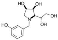
3.7.7. N-(3-Hydroxyphenyl)-1,4-dideoxy-1,4-imino-l-gulitol meta 7 (Scheme 1, step (vi))
Pharmaceuticals 18 01739 i007
N-(3-Methylphenyl boronic acid)-3,6-dideoxy-3,6-imino-d-glucofuranose meta 3 (61 mg, 0.205 mmol) was dissolved in EtOH (aq 50%, 10 mL) prior to the addition of NaBH4 (9 mg, 0.246 mmol, 1.2 eq) and the reaction was allowed to stir at room temperature for 30 minutes. Additional NaBH4 (11 mg, 0.298 mmol, 1.5 eq) was added and the reaction stirred for an additional 1 hour. the reaction was observed to be incomplete so additional NaBH4 (9 mg, 0.246 mmol, 1.2 eq) was added and the reaction stirred at room temperature for 18 hours, after which the reaction was quenched with glacial acetic acid (a few drops) and dried to give a crude mass (112 mg). A portion (52 mg) was purified by prep t.l.c. (toluene/acetone/acetic acid, 10:15:1) which resulted in oxidation to give meta 7 (39 mg, 0.144 mmol, quant.) as a yellow residue. [α]28919 24.65 (c 0.052 in H2O); νmax (thin film, cm-1): 3377 (s, broad, OH), 2964 (w, alkyl CH), 1560 (s, 1489 (w), 1413 (s, ArC=C), 1347 (w), 1285 (w, C-O), 1206 (w), 1179 (m, OH bend), 1125 (s), 1038 (m), 1014 (w), 963 (w), 816 (w), 755 (w, meta-subst), 687 (w), 653 (w, meta-subst), 620 (w), 568 (w), 471 (w), 416 (w); δH (Acetic acid-d4, 400MHz): 7.39 (1H, ddd, partially obscured, JHE,HD 7.2 Hz, JHE,HC 2.5, JHE,HA 0.8 Hz, ArHE), 7.09 (1H, dt, JHD,HC 7.8, JHD,HA 1.0 Hz, ArHD), 7.02 (1H, app-d, J 1.1 Hz, ArHA), 7.01 (1H, ddd, partially obscured, JHC,HD 7.5 Hz, JHC,HE 2.5 Hz, JHC,HA 0.8 Hz, ArHD), 4.66 (1H, d, JHa,Hb 13.0 Hz, ArCHaHb), 4.49 (1H, ddd, JH-2,H-1 8.7, JH-2,H-1′ 7.0, JH-2,H-3 4.0 Hz, H-2), 4.39 (1H, app-t, JH-3,H-2/4 4.3 Hz, H-3), 4.32 (1H, ddd, JH-5,H-4 8.8, JH-5,H-6 5.2, JH-5,H-6′ 3.3 Hz, H-5), 4.17 (1H, d, JHb,Ha 13.0 Hz, ArCHaHb), 3.82 (1H, dd, JH-6′,H-6 12.2, JH-6′,H-5 3.2 Hz, H-6′), 3.74 (1H, dd, JH-4,H-5 9.1, JH-4,H-3 4.7 Hz, H-4), 3.68 (1H, dd, JH-6,H-6′ 12.1, JH-6,H-5 5.2 Hz, H-6), 3.48 (1H, dd, JH-1′,H-1 12.0, JH-1,H-2 7.0 Hz, H-1′), 3.23 (1H, dd, partially obscured, JH-1,H-1′ 12.0, JH-1,H-2 8.4 Hz, H-1); δC (Acetic acid-d4, 100MHz): 152.8 (ArCquat-OH), 127.4 (CE), 126.7 (ArCquat), 119.5 (CD), 114.3 (CA), 113.5 (CC), 67.1 (C-3), 66.3 (C-4), 65.8 (C-2), 65.5 (C-5), 59.8 (C-6), 57.4-58.2 (ArCH2, 81 Hz), 50.3 (C-1). δB (Acetic acid-d4, 128 MHz): No signal.

3.7.8. N-(3-Methylphenyl boronic acid)-3,6-dideoxy-3,6-imino-1,2-O-isopropylidene-α-d-glucofuranose meta 5 (Scheme 1, step (iv)) and N-(3-hydroxyphenyl)-3,6-dideoxy-3,6-imino-1,2-O-isopropylidene-α-d-gulofuranose meta 6 (Scheme 1, step (v))

Pharmaceuticals 18 01739 i008
3,6-Dideoxy-3,6-imino-1,2-O-isopropylidene-α-d-glucofuranose-p-toluenesulfonate 1 (98 mg, 0.268 mmol, 1.1 eq) was dissolved in DMF (10 mL) prior to the addition of K2CO3 (34 mg, 0.243 mmol, 1.0 eq) and 3-bromomethylphenyl boronic acid (52 mg, 0.243 mmol, 1.0 eq). The reaction was heated to 100 °C and allowed to stir for 19 hours. T.l.c. analysis (EtOAc) showed presence of one product (Rf 0.68) and consumption of the starting material (Rf 0.00). The solvent was evaporated and the crude dissolved in CHCl3 (60 mL) and washed with NaOH (aq. 0.1 M, 20 mL) to give N-(3-methylphenyl boronic acid)-3,6-dideoxy-3,6-imino-1,2-O-isopropylidene-α-d-glucofuranose meta 5 in the organic layer as a brown oil (88 mg, 0.263 mmol, 98%). [α]28919 0.13 (c 0.072 in MeOH). νmax (thin film, cm-1): 3441, 3415, 3398, 3309, 3283 (s, broad), 1681, 1656 (m), 1601, 1508, 1473, 1458, 1450, 1436 (w, ArC=C), 1385 (w, B-O), 1185 (s, OH bend), 1126 (s), 1040 (s), 1012 (s), 814, 719, 710, 683 (m), 617 (m), 566 (s), 514, 495, 454, 424 (m). (Scheme 1, step (v)). When meta 5 is left on the bench or aqueous solution, it slowly oxidises to N-(3-hydroxyphenyl)-3,6-dideoxy-3,6-imino-1,2-O-isopropylidene-α-d-gulofuranose meta 6. δH (Acetic acid-d4, 400 MHz): 8.09 (1H, s, ArHA), 7.94 (1H, d, JHC,HD 7.5 Hz, ArHC), 7.76 (1H, d, JHE,HD 7.8 Hz, ArHE), 7.48 (1H, app-t, JHD,HE/HC 7.6 Hz, ArHD), 6.15 (1H, d, J 2.5 Hz, H-1), 5.09-4.96 (2H, app-s, H-2 and H-4), 4.68-4.58 (1H, obscured, H-5), 4.62 (1H, d, JHa,Hb 11.8 Hz, ArCHaHb), 4.55 (1H, d, JHb,Ha 11.8 Hz, ArCHaHb), 4.28 (1H, d, J 4.3 Hz, H-3), 3.74 (1H, dd, JH-6,H-6′ 12.6 Hz, JH-6,H-5 5.2 Hz, H-6), 3.54 (1H, dd, JH-6′,H-6 12.2 Hz, JH-6′,H-5 5.1 Hz, H-6′), 1.45, 1.27 (6Hs, 2 × s, 2 × acetonide CH3); δC (Acetic acid-d4, 100 MHz): 148.5 (ArCquat-OH), 138.1 (ArCA), 136.8 (ArCC), 134.6 (ArCE), 129.6 (ArCD), 114.4 (acetonide Cquat), 108.5 (C-1), 82.9 (C-4 and C-2), 74.0 (C-3), 69.7 (C-5), 60.0 (ArCH2), 59.2 (C-6), 27.3, 26.8 (2 × CH3 acetonide). ArCquat not discernible. δB (Acetic acid-d4, 128 MHz): No signal.

4. Conclusions

BNCT is the least destructive radiation therapy to the human body, provided BNCT agents can be selectively delivered to cancer cells. Current clinical agents based on inorganic boron moieties do not yet possess an ideal profile in terms of toxicity, membrane-crossing capabilities and accumulation into cancer cells. We provide the first examples of monosaccharides (and their congeners) borylated (new B-C bond) with organic boron moieties to improve toxicity and accumulation profiles, with a view to improved ADME profiles and a more targeted/controllable radiation delivery. This paper communicates another synthetic development and biological investigation to borylated pyrrolidine iminosugars, and continues to cement the opening of the field of borylated carbohydrates for controllable switch on/switch off cancer radiotherapies. It is envisaged this type of cancer therapy technology could in the future be incorporated in long-term manned space travels.
This paper provides a complete set of structure–activity relationship data for this novel family of boron-bearing iminosugars displaying selective, moderate-to-weak inhibitions (IC50s = 116–617 μM) of β-d-galactosidase (bovine liver), and emerging inhibition of β-d-glucosidases (almond, bovine liver) (IC50s = 633 and 710 μM) and α-d-glucosidases (rice, yeast, rat intestinal maltase) (IC50s = 106–784 μM). This indicates the boronic acid pharmacophore may form suitable interactions for management of lysosomal storage disorders to support restoration of biological activity of mutant enzymes via the chaperone-mediated therapy approach. From a structure–activity perspective, the cancer screening revealed that the A2780 ovarian carcinoma cell line is selectively inhibited by all compounds screened and the MIA-Pa-Ca2 pancreatic carcinoma cell line is also selectively inhibited. The real therapeutic potential of these borylated drugs lies in their controllable switch on/switch off activation under BNCT radiotherapeutic conditions.

Supplementary Materials

The following supporting information can be downloaded at https://www.mdpi.com/article/10.3390/ph18111739/s1, Figure S1: 1H- (400 MHz), 13C-NMR (100 MHz), DEPT, COSY and HSQC spectra of N-(3-hydroxyphenyl)-3,6-dideoxy-3,6-imino-1,2-O-isopropylidene-α-d-gulofuranose meta 6 in Acetic acid-d6.; Figure S2: 1H- (400 MHz), 13C-NMR (100 MHz), DEPT, COSY, HSQC and HMBC spectra of N-(3-hydroxyphenyl)-1,4-dideoxy-1,4-imino-l-gulitol meta 7 in Acetic acid-d6.

Author Contributions

Manuscript conception, M.S.; writing and original draft preparation, M.S.; synthesis of derivatives, M.S. and K.P.; characterisation of derivatives, M.S.; biological assays, S.Y., K.Y., A.T., A.K., R.N., B.B., J.G. and J.S.; editing, data analysis, and interpretation, M.S., A.K., R.N., J.S. All authors have read and agreed to the published version of the manuscript.

Funding

Financial support for this project provided by the B18 Project and—at the University of Newcastle—by the Priority Research Centre for Drug Development and by the Faculty of Science is gratefully acknowledged. This work was supported by JST SPRING (Grant Number JPMJSP2145) (K.Y.).

Institutional Review Board Statement

Not applicable.

Informed Consent Statement

Not applicable.

Data Availability Statement

Data presented in this study is contained within the article and Supplementary Material. Further inquiries can be directed to the corresponding author.

Conflicts of Interest

Authors R.N. and B.B. were employed by the company PhytoQuest Ltd. The remaining authors declare that the research was conducted in the absence of any commercial or financial relationships that could be construed as a potential conflict of interest.

References

  1. Brown, H.C. Organic Syntheses via Boranes; John Wiley & Sons, Inc.: New York, NY, USA, 1975. [Google Scholar]
  2. Miyaura, N.; Yamada, K.; Suzuki, A. A new stereospecific cross-coupling by the palladium-catalyzed reaction of 1-alkenylboranes with 1-alkenyl or 1-alkynyl halides. Tetrahedron Lett. 1979, 20, 3437–3440. [Google Scholar] [CrossRef]
  3. Brown, H.C.; Kulkarni, S.U. Organoboranes: XXV. Hydridation of dialkylhaloboranes. New practical syntheses of dialkylboranes under mild conditions. J. Organomet. Chem. 1981, 2018, 299. [Google Scholar] [CrossRef]
  4. Hall, D.G. Chapter 1: Structure, properties, and preparation of boronic acid derivatives. Overview of their reactions and applications. In Boronic Acids: Preparation and Applications in Organic Synthesis, Medicine and Materials; Wiley—VCH Verlag GmbH & Co. KGaA: Weinheim, Germany, 2011. [Google Scholar]
  5. Martin, J.E. Physics for Radiation Protection: A Handbook; WILEY—VCH Verlag GmbH & Co.KGaA: Weinheim, Germany, 2008. [Google Scholar]
  6. Hampshire, S. 2.01 Fundamental Aspects of Hard Ceramics. In Comprehensive Hard Materials; Materials Science and Materials Engineering; Elsevier: Amsterdam, The Netherlands, 2014; Volume 2. [Google Scholar]
  7. Gupta, S.; Chunduri, A.; Spreitzer, M.; Kržmanc, M.M.; Patel, N. 3—Synthesis and catalytic applications of metal boride ceramics. In Ceramic Catalysts, Materials, Synthesis, and Applications; Elsevier: Amsterdam, The Netherlands, 2023. [Google Scholar]
  8. Hong, J.; Mutalik, S.; Pescarmona, P.P.; Protesescu, L. Metal Borides: From Industrial Classics to Versatile Colloidal Nanocrystals for Energy, Catalysis, and Hard Coatings Applications. Chem. Mater. 2024, 36, 2147–2164. [Google Scholar] [CrossRef]
  9. Zhang, J.; Yan, X.; Cheng, Z.; Han, Y.; Zhang, Y.; Dong, Y. Applications, prospects and challenges of metal borides in lithium sulfur batteries. J. Colloid Interface Sci. 2024, 657, 511–528. [Google Scholar] [CrossRef] [PubMed]
  10. DeFrancesco, H.; Dudley, J.; Coca, A. Chapter 1: Boron Chemistry: An Overview. In Boron Reagents in Synthesis; ACS Symposium Series eBooks; ACS Publications: Washington, DC, USA, 2016; Volume 1236. [Google Scholar]
  11. Matsuda, I.; Wu, K. 2D Boron: Boraphene, Borophene, Boronene; Springer: Cham, Switzerland, 2021. [Google Scholar]
  12. Reyes, H.; García, M.C.; Flores, B.M.; López-Rebolledo, H.; Santillán, R.; Farfán, N. Synthesis, NMR and X-Ray Diffraction Analysis of Boron Complexes Derived from Hydroxychalcones. J. Mex. Chem. Soc. 2006, 50, 106–113. [Google Scholar]
  13. Yamashita, M.; Yamamoto, Y.; Akiba, K.-Y.; Hashizume, D.; Iwasaki, F.; Takagi, N.; Nagase, S. Syntheses and Structures of Hypervalent Pentacoordinate Carbon and Boron Compounds Bearing an Anthracene Skeleton- Elucidation of Hypervalent Interaction Based on X-ray Analysis and DFT Calculation. J. Am. Chem. Soc. 2005, 127, 4354–4371. [Google Scholar] [CrossRef]
  14. Woods, W.G. An Introduction to Boron: History, Sources, Uses, and Chemistry. Environ. Health Perspect 1994, 102, 5–11. [Google Scholar] [CrossRef] [PubMed]
  15. Zhu, Y.; Cai, J.; Hosmane, N.S.; Zhang, Y. Chapter 1: Introduction: Basic concept of boron and its physical and chemical properties. In Fundamentals and Applications of Boron Chemistry; Elsevier: Amsterdam, The Netherlands, 2022. [Google Scholar]
  16. Chatterjee, S.; Tripathi, N.M.; Bandyopadhyay, A. The modern role of boron as a ‘magic element’ in biomedical science: Chemistry perspective. Chem. Commun. 2021, 100, 13629–13640. [Google Scholar] [CrossRef]
  17. Légareé, M.-A.; Pranckevicius, C.; Braunschweig, H. Metallomimetic Chemistry of Boron. Chem. Rev. 2019, 119, 8231–8261. [Google Scholar] [CrossRef]
  18. Duret, G.; Quinlan, R.; Bisseret, P.; Blanchard, N. Boron chemistry in a new light. Chem. Sci. 2015, 10, 5366–5382. [Google Scholar] [CrossRef]
  19. Rubanov, Z.M.; Supranovich, V.I.; Levin, V.V.; Dilman, A.D. BF2-Chelates of N-Acylhydrazones as Versatile Coupling Partners in Photoredox Promoted Reactions. Eur. J. Org. Chem. 2023, 26, e202300247. [Google Scholar] [CrossRef]
  20. Yan, Q.-Q.; Zhao, X.Y.; Zhang, T.; Li, S.-D. Perfect Core-Shell Octahedral B@B38+, Be@B38, and Zn@B38 with an Octa-Coordinate Center as Superatoms Following the Octet Rule. ChemPhysChem 2023, 24, e202200947. [Google Scholar] [CrossRef]
  21. Selvaraj, C.; Rudhra, O.; Alothaim, A.S.; Alkhanani, M.; Singh, S.K. Structure and chemistry of enzymatic active sites that play a role in the switch and conformation mechanism. In Advances in Protein Chemistry and Structural Biology; Elsevier Inc.: Amsterdam, The Netherlands, 2022; Volume 130. [Google Scholar]
  22. Goh, C.-S.; Milburn, D.; Gerstein, M. Conformational changes associated with protein–protein interactions. Curr. Opin. Struct. Biol. 2004, 14, 104–109. [Google Scholar] [CrossRef] [PubMed]
  23. Belorkar, S.A.; Jogaiah, S. Chapter 1—Enzymes—Past, present, and future. In Protocols and Applications in Enzymology; Academic Press: London, UK, 2022. [Google Scholar]
  24. Lowering, F.; Bikker, J.; Humblet, C. Escape from Flatland: Increasing Saturation as an Approach to Improving Clinical Success. J. Med. Chem. 2009, 52, 6752–6756. [Google Scholar] [CrossRef] [PubMed]
  25. Lovering, F. Escape from Flatland 2: Complexity and promiscuity. Med. Chem. Commun. 2013, 4, 515–519. [Google Scholar] [CrossRef]
  26. Jenkinson, S.F.; Thompson, A.L.; Simone, M. Methyl 2-(5,5-dimethyl-1,3,2-dioxaborinan-2-yl)-4-nitrobenzoate. Acta Cryst. 2012, E68, o2699–o2700. [Google Scholar] [CrossRef]
  27. Simone, M. Diastereoselective Synthesis of the Borylated D-Galactose Monosaccharide 3-Boronic-3-Deoxy-D-Galactose and Biological Evaluation in Glycosidase Inhibition and in Cancer for Boron Neutron Capture Therapy (BNCT). Molecules 2023, 28, 4321. [Google Scholar] [CrossRef]
  28. Legge, W.J.; Shimadate, Y.; Sakoff, J.; Houston, T.A.; Kato, A.; Bernhardt, P.V.; Simone, M. Borylated methyl cinnamates: Green synthesis, characterization, crystallographic analysis and biological activities—In glycosidase inhibition and in cancer cells lines. Beilstein Arch. 2021, 2021, 20214. [Google Scholar] [CrossRef]
  29. Campkin, D.M.; Shimadate, Y.; Bartholomew, B.; Bernhardt, P.V.; Nash, R.J.; Sakoff, J.A.; Kato, A.; Simone, M. Borylated 2,3,4,5-Tetrachlorophthalimide and Their 2,3,4,5-Tetrachlorobenzamide Analogues: Synthesis, Their Glycosidase Inhibition and Anticancer Properties in View to Boron Neutron Capture Therapy. Molecules 2022, 27, 3447. [Google Scholar] [CrossRef]
  30. Prichard, K.; Yamamoto, S.; Shimadate, Y.; Yoshimura, K.; Bartholomew, B.; Gilbert, J.; Sakoff, J.; Nash, R.; Kato, A.; Simone, M. Borylated 5-Membered Ring Iminosugars: Synthesis, Spectroscopic Analysis and Biological Evaluation for Glycosidase Inhibition and Anticancer Properties for Application in Boron Neutron Capture Therapy (BNCT)—Part 1. Pharmaceuticals 2025, 18, 1302. [Google Scholar] [CrossRef]
  31. Simone, M.; Soengas, R.G.; Jenkinson, S.F.; Evinson, E.L.; Nash, R.J.; Fleet, G.W.J. Synthesis of three branched iminosugars [(3R,4R,5S)-3-(hydroxymethyl)piperidine-3,4,5-triol, (3R,4R,5R)-3-(hydroxymethyl)piperidine-3,4,5-triol and (3S,4R,5R)-3-(hydroxymethyl)piperidine-3,4,5-triol] and a branched trihydroxynipecotic acid [(3R,4R,5R)-3,4,5-trihydroxypiperidine-3-carboxylic acid] from sugar lactones with a carbon substituent at C-2. Tetrahedron: Asymm. 2012, 23, 401–408. [Google Scholar] [CrossRef]
  32. Simone, M. Borylated Monosaccharide 3-Boronic-3-deoxy-D-galactose: Detailed NMR Spectroscopic Characterisation, and Method for Spectroscopic Analysis of Anomeric and Boron Equilibria. Int. J. Mol. Sci. 2024, 25, 12396. [Google Scholar] [CrossRef]
  33. Simone, M. Borylated 5-Membered Ring Iminosugars: Detailed Nuclear Magnetic Resonance Spectroscopic Characterisation, and Method for Analysis of Anomeric and Boron Equilibria. Molecules 2025, 30, 1402. [Google Scholar] [CrossRef] [PubMed]
  34. Glenister, A.; Simone, M.; Hambley, T.W. A Warburg effect targeting vector designed to increase the uptake of compounds by cancer cells demonstrates glucose and hypoxia dependent uptake. PLoS ONE 2019, 14, e0217712. [Google Scholar] [CrossRef]
  35. Fleet, G.W.J.; Peach, J.M.; Smith, P.W.; Austin, G.N.; Baird, P.D.; Watkin, D.J. 3,6-Dideoxy-3,6-imino-1,2-O-isopropylidene-α-D-glucofuranose as a divergent intermediate for the synthesis of hydroxylated pyrrolidines: Synthesis of 1,4-dideoxy-1,4-imino-L-gulitol, 1,4-dideoxy-1,4-imino-D-lyxitol, (2S,3S,4R)-3,4-dihydroxyproline and (1S,2R,8S,8aR)-1,2,8-trihydroxyoctahydroindolizine [8-episwainsonine]. X-ray crystal structure of (1S,2R,8S,8aR)-1,2,8-trihydroxy-5-oxooctahydroindolizine. Tetrahedron 1987, 43, 3095–3108. [Google Scholar] [CrossRef]
  36. Boshagen, H.; Heiker, F.-R.; Schuller, M. The chemistry of the 1-deoxynojirimycin system. Synthesis of 2-acetamido-1,2-dideoxynojirimycin from 1-deoxynojirimycin. Carbohydr. Res. 1987, 164, 141–148. [Google Scholar] [CrossRef]
  37. Zinner, H.; Wulf, G.; Heinatz, R. Derivate der Zucker-mercaptale, XXXV. Darstellung und Mercaptalbildung der 2-Desoxy-D-xylose. Chem. Ber. 1964, 97, 3536–3540. [Google Scholar] [CrossRef]
  38. Moore, J.L.; Taylor, S.M.; Soloshonok, V.A. An efficient and operationally convenient general synthesis of tertiary amines by direct alkylation of secondary amines with alkyl halides in the presence of Huenig’s base. Arkivoc 2005, vi, 287–292. [Google Scholar] [CrossRef]
  39. Wang, G.-N.; Twigg, G.; Butters, T.D.; Zhang, S.; Zhang, L.; Zhang, L.-H.; Ye, X.-S. Synthesis of N-substituted ε-hexonolactams as pharmacological chaperones for the treatment of N370S mutant Gaucher disease. Org. Biomol. Chem. 2012, 10, 2923–2927. [Google Scholar] [CrossRef]
  40. Zhang, G.-L.; Chen, C.; Xiong, Y.; Zhang, L.-H.; Ye, J.; Ye, X.-S. Synthesis of N-substituted iminosugar derivatives and their immunosuppressive activities. Carbohydr. Res. 2010, 345, 780–786. [Google Scholar] [CrossRef]
  41. Zhou, J.; Zhang, Y.; Zhou, X.; Zhou, J.; Zhang, L.-H.; Yea, X.-S.; Zhang, X.-L. An expeditious one-pot synthesis of 1,6-dideoxy-N-alkylated nojirimycin derivatives and their inhibitory effects on the secretion of IFN-g and IL-4. Bioorg. Med. Chem. 2008, 16, 1605–1612. [Google Scholar] [CrossRef] [PubMed]
  42. Zhou, Y.; Zhao, Y.; Boyle, K.M.O.; Murphy, P.V. Hybrid angiogenesis inhibitors: Synthesis and biological evaluation of bifunctional compounds based on 1-deoxynojirimycin and aryl-1,2,3-triazoles. Bioorg. Med. Chem. Lett. 2008, 18, 954–958. [Google Scholar] [CrossRef]
  43. Daher, S.A.; Fleet, G.; Namgoong, S.K.; Winchester, B. Change in specificity of glycosidase inhibition by N-alkylation of amino sugars. Biochem. J. 1989, 258, 613–615. [Google Scholar] [CrossRef]
  44. Guazzelli, L.; D’Andrea, F.; Sartini, S.; Giorgelli, F.; Confini, G.; Quattrini, L.; Piano, I.; Gargini, C.; Motta, C.L. Synthesis and investigation of polyhydroxylated pyrrolidine derivatives as novel chemotypes showing dual activity as glucosidase and aldose reductase inhibitors. Bioorg. Chem. 2019, 92, 103298. [Google Scholar] [CrossRef]
  45. Yang, L.-F.; Shimadate, Y.; Kato, A.; Li, Y.-X.; Jia, Y.-M.; Fleet, G.W.J.; Yu, C.-Y. Synthesis and glycosidase inhibition of N-substituted derivatives of 1,4-dideoxy-1,4-imino-D-mannitol (DIM). Org. Biomol. Chem. 2020, 18, 999–1011. [Google Scholar] [CrossRef]
  46. Winchester, B.; Daher, S.A.; Carpenter, N.C.; Bello, I.C.D.; Choi, S.S.; Fairbanks, A.J.; Fleet, G.W.J. The structural basis of the inhibition of human α-mannosidases by azafuranose analogues of mannose. Biochem. J. 1993, 290, 743–749. [Google Scholar] [CrossRef] [PubMed]
  47. Šesták, S.; Bella, M.; Klunda, T.; Gurská, S.; Dzubáck, P.; Wöls, F.; Wilson, I.B.H.; Sladek, V.; Hajdúch, M.; Poláková, M.; et al. N-Benzyl Substitution of Polyhydroxypyrrolidines: The Way to Selective Inhibitors of Golgi α-Mannosidase II. ChemMedChem 2018, 13, 373–383. [Google Scholar] [CrossRef] [PubMed]
  48. Johnson, L.L.; Houston, T.A. A drug targeting motif for glycosidase inhibitors: An iminosugar–boronate shows unexpectedly selective β-galactosidase inhibition. Tetrahedron Lett. 2002, 43, 8905–8908. [Google Scholar] [CrossRef]
  49. Cox, T.M. Gaucher’s Disease—An exemplary monogenic disorder. QJM Int. J. Med. 2001, 94, 399–402. [Google Scholar] [CrossRef]
  50. Mena-Barragán, T.; García-Moreno, M.I.; Sevšek, A.; Okazaki, T.; Nanba, E.; Higaki, K.; Martin, N.I.; Pieters, R.J.; Fernández, J.M.G.; Mellet, C.O. Probing the Inhibitor versus Chaperone Properties of sp2-Iminosugars towards Human β-Glucocerebrosidase: A Picomolar Chaperone for Gaucher Disease. Molecules 2018, 23, 927. [Google Scholar] [CrossRef]
  51. Martín-Banderas, L.; Holgado, M.A.; Durán-Lobato, M.; Infante, J.J.; Álvarez-Fuentes, J.; Fernández-Arévalo, M. Role of Nanotechnology for Enzyme Replacement Therapy in Lysosomal Diseases. A Focus on Gaucher’s Disease. Curr. Med. Chem. 2016, 23, 929–952. [Google Scholar] [CrossRef] [PubMed]
  52. Hinek, A.; Zhang, S.; Smith, A.C.; Callahan, J.W. Impaired Elastic-Fiber Assembly by Fibroblasts from Patients with Either Morquio B Disease or Infantile GM1-Gangliosidosis Is Linked to Deficiency in the 67-kD Spliced Variant of β-Galactosidase. Am. J. Hum. Genet. 2000, 67, 23–26. [Google Scholar] [CrossRef]
  53. Prichard, K.; Campkin, D.; O’Brien, N.; Kato, A.; Fleet, G.W.J.; Simone, M. Biological Activities of 3,4,5-Trihydroxypiperidines and their O- and N-Alkylated Derivatives. Chem. Biol. Drug Des. 2018, 92, 1171–1197. [Google Scholar] [CrossRef] [PubMed]
  54. Nicoll-Griffith, D.A.; Falgueyret, J.P.; Silva, J.M.; Morin, P.E.; Trimble, L.; Chan, C.C.; Clas, S.; Leger, S.; Wang, Z.; Yergey, J.A.; et al. Oxidative bioactivation of the lactol prodrug of a lactone cyclooxygenase-2 inhibitor. Drug. Metab. Dispos. 1999, 27, 403–409. [Google Scholar] [CrossRef]
  55. Gajula, S.N.R.; Nadimpalli, N.; Sonti, R. Drug metabolic stability in early drug discovery to develop potential lead compounds. Drug Metabolism Rev. 2021, 53, 459–477. [Google Scholar] [CrossRef]
  56. Li, L.; Chenna, B.C.; Yang, K.S.; Cole, T.R.; Goodall, Z.T.; Giardini, M.; Moghadamchargari, Z.; Hernandez, E.A.; Gomez, J.; Calvet, C.M.; et al. Self-Masked Aldehyde Inhibitors: A Novel Strategy for Inhibiting Cysteine Proteases. J. Med. Chem. 2021, 64, 11267–11287. [Google Scholar] [CrossRef]
  57. Volle, J.-N.; Filippini, D.; Krawczy, B.; Kaloyanov, N.; Lee, A.V.d.; Maurice, T.; Pirat, J.-L.; Virieux, D. Drug discovery: Phosphinolactone, in vivo bioisostere of the lactol group. Org. Biomol. Chem. 2010, 8, 1438–1444. [Google Scholar] [CrossRef]
  58. Ahuja-Casarín, A.I.; Merino-Montiel, P.; Vega-Baez, J.L.; Montiel-Smith, S.; Fernandes, M.X.; Lagunes, I.; Maya, I.; Padrón, J.M.; López, Ó.; Fernández-Bolaños, J.G. Tuning the activity of iminosugars: Novel N-alkylated deoxynojirimycin derivatives as strong BuChE inhibitors. J. Enzyme Inhib. Med. Chem. 2020, 36, 138–146. [Google Scholar] [CrossRef]
  59. Iftikhar, M.; Lu, Y.; Zhou, M. An overview of therapeutic potential of N-alkylated 1-deoxynojirimycin congeners. Carbohydr. Res. 2021, 504, 108317. [Google Scholar] [CrossRef] [PubMed]
  60. Fujii, T.; Tanaka, Y.; Oki, H.; Sato, S.; Shibata, S.; Maru, T.; Tanaka, Y.; Tanaka, M.; Onishi, T. A new brain-penetrant glucosylceramide synthase inhibitor as potential Therapeutics for Gaucher disease. J. Neurochem. 2021, 159, 543–553. [Google Scholar] [CrossRef]
  61. Rawlings, A.J.; Lomas, H.; Pilling, A.W.; Lee, M.J.-R.; Alonzi, D.S.; Rountree, J.S.S.; Jenkinson, S.F.; Fleet, G.W.J.; Dwek, R.A.; Jones, J.H.; et al. Synthesis and Biological Characterisation of Novel N-Alkyl-Deoxynojirimycin a-Glucosidase Inhibitors. ChemBioChem 2009, 10, 1101–1105. [Google Scholar] [CrossRef] [PubMed]
  62. Glenister, A.; Chen, C.K.J.; Paterson, D.J.; Renfrew, A.K.; Simone, M.; Hambley, T.W. Warburg Effect Targeting Co(III) Cytotoxin Chaperone Complexes. J. Med. Chem. 2021, 64, 2678–2690. [Google Scholar] [CrossRef]
  63. Kessler, M.; Acuto, O.; Storelli, C.; Murer, H.; Müller, M.; Semenza, G. A modified procedure for the rapid preparation of efficiently transporting vesicles from small intestinal brush border membranes. Their use in investigating some properties of D-glucose and choline transport systems. Biochim. Biophys. Acta 1978, 506, 136–154. [Google Scholar] [CrossRef] [PubMed]
  64. IUPAC and IUBMB Joint Commission on Biochemical Nomenclature; McNaught, A.D. Nomenclature of Carbohydrates. Pure Appl. Chem. 1996, 68, 1919–2008. [Google Scholar] [CrossRef]
  65. Gottlieb, H.E.; Kotlyar, V.; Nudelman, A. NMR Chemical Shifts of Common Laboratory Solvents as Trace Impurities. J. Org. Chem. 1997, 62, 7512–7515. [Google Scholar] [CrossRef]
  66. Bruker. NMR Software TopSpin. Available online: https://www.bruker.com/en/products-and-solutions/mr/nmr-software/topspin.html?utm_source=Advertising&utm_medium=GoogleAd&utm_campaign=BBIO-Software-Cross-All-TopSpin-H2-2025&utm_source=Advertising&utm_medium=GoogleAd&utm_campaign=BBIO-Software-Cross-All-H2-2022&gad_source=1&gad_campaignid=15149563659&gbraid=0AAAAADsq6H1Plj_hwRuSXc2BkFGqPxpxq&gclid=EAIaIQobChMI6qmQ4oXojgMVLaNmAh3L9Q0jEAAYASABEgJxrfD_BwE (accessed on 20 September 2025).
  67. Peak, D.; Luther, G.W., III; Sparks, D.L. ATR-FTIR spectroscopic studies of boric acid adsorption on hydrous ferric oxide. Geochim. Cosmochim. Acta 2003, 67, 2551–2560. [Google Scholar] [CrossRef]
  68. Faniran, J.A.; Shurvell, H.F. Infrared spectra of phenylboronic acid (normal and deuterated) and diphenyl phenylboronate. Can. J. Chem. 1968, 46, 2089–2095. [Google Scholar] [CrossRef]
  69. Balachander, L.; Ramadevudu, G.; Shareefuddina, M.; Sayanna, R.; Venudhar, Y.C. IR analysis of borate glasses containing three alkali oxides. ScienceAsia 2013, 39, 278–283. [Google Scholar] [CrossRef]
  70. Chaplin, M. Structure and Properties of Water in its Various States Fundamentals of Water, Chemistry, Particles, and Ecology. In Encyclopedia of Water: Science, Technology, and Society; John Wiley & Sons, Inc.: Hoboken, NJ, USA, 2019. [Google Scholar] [CrossRef]
  71. Finch, A.; Pearn, E.J. Boron-oxygen stretching modes in cyclic systems. Spectrochim. Acta 1963, 19, 1621–1623. [Google Scholar] [CrossRef]
  72. Stuart, B.H. Chapter 4: Organic Molecules. In Infrared Spectroscopy: Fundamentals and Applications; John Wiley & Sons, Ltd.: Hoboken, NJ, USA, 2004; pp. 71–93. [Google Scholar]
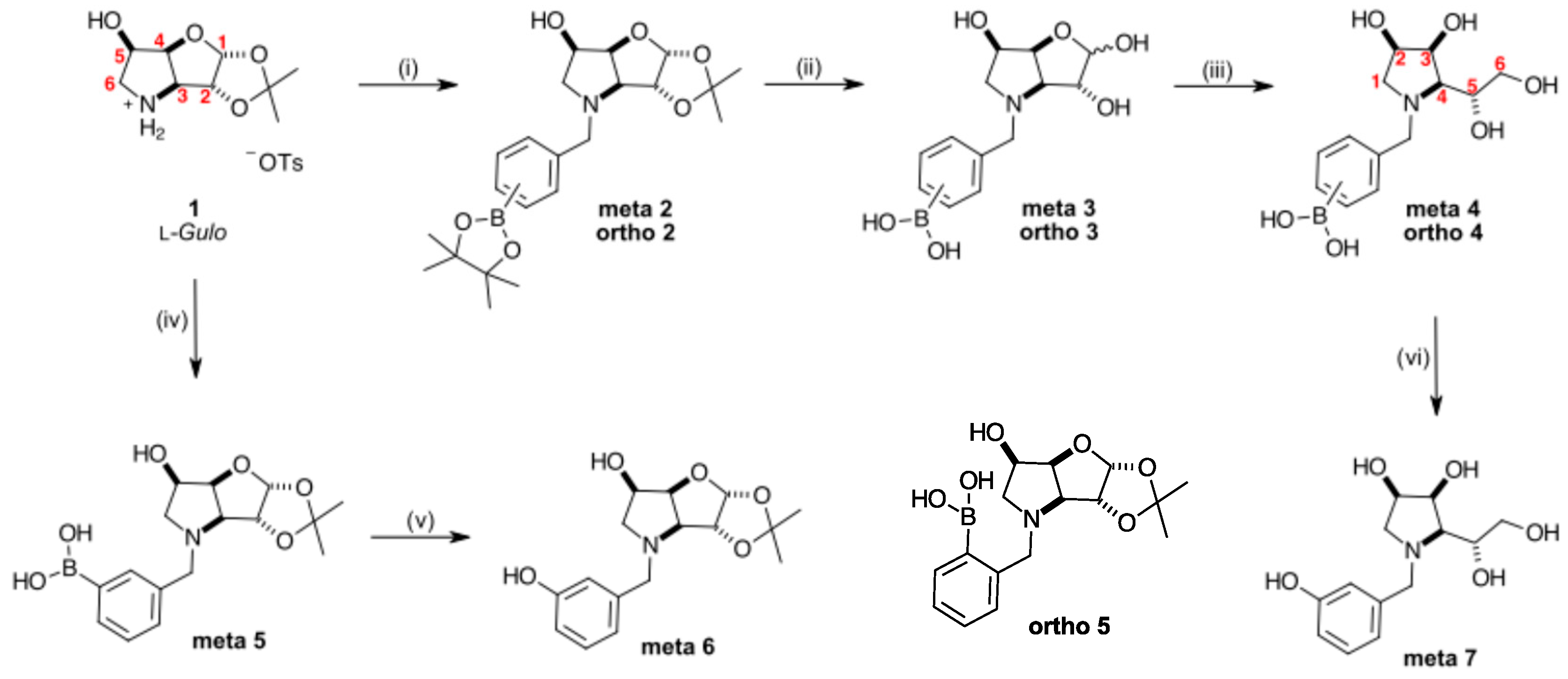
Scheme 1. Reaction conditions by synthetic steps (i) 1 (1.1 eq), 3-/2-bromomethylphenylboronic acid pinacol ester (1.0 eq), K2CO3 (1.0 eq), DMF, 100 °C, 19 h, 72%/71% (respectively). (ii) For meta 3: meta 2, HCl (aq 1 M), 55 °C, 4 h, quant.; for ortho 3: ortho 2, HCl (aq 1 M), 55 °C, 18 h, quant. (iii) For meta 4: meta 3 (1.0 eq), NaBH4 (3.9 eq), 50% aq EtOH, r.t., 20 h, 84%; for ortho 4: ortho 3 (1.0 eq), NaBH4 (2.4 eq), 50% aq EtOH, r.t., 1 h, quant.; (iv) 1 (1.1 eq), 3-bromomethylphenylboronic acid (1.0 eq), K2CO3 (1.0 eq), DMF, 100 °C, 19 h, 98%; (v) slow oxidation; (vi) meta 4, prep t.l.c. (toluene/acetone/acetic acid, 10:15:1), quant.
Pharmaceuticals 18 01739 sch001
Figure 1. Borylated iminosugars and parent congeners from [30].
Pharmaceuticals 18 01739 g001
Table 1. Summary of glycosidase inhibitions by N-benzyl-1,4-dideoxy-1,4-imino derivatives.
IminosugarMost Inhibited Glycosidase/s
N-Benzyl-1,4-dideoxy-1,4-imino-
   d-allitol [43]α-l-Fucosidase (76%)
   d-galactitol [44]α-d-Glucosidase (93.2%), IC50 = 40.6 μM
   l-gulitol 5 [30]NI
   d-mannitol.HCl [45,46,47]α-d-Mannosidases (lysosomal acidic, 34%), (neutral, 44%) and (Golgi II, 72%)
   d-talitol.HCl [48]NI
N-(2-Methylphenyl boronic acid)-1,4-dideoxy-1,4-imino-d-talitol.HCl [48]β-d-Galactosidase (E. coli) (44–55%)
N-Benzyl-3,6-dideoxy-3,6-imino-d-gulofuranose 4 [30]β-d-Galactosidase (bovine liver), IC50 = 133 μM
N-(4-Methylphenyl boronic acid)-3,6-dideoxy-3,6-imino-d-gulofuranose para 7 [30]β-d-Galactosidase (bovine liver), IC50 = 218 μM
N-(4-Methylphenyl boronic acid)-1,4-dideoxy-1,4-imino-l-gulitol para 8 [30]β-d-Galactosidase (bovine liver), IC50 = 501 μM
Disclaimer/Publisher’s Note: The statements, opinions and data contained in all publications are solely those of the individual author(s) and contributor(s) and not of MDPI and/or the editor(s). MDPI and/or the editor(s) disclaim responsibility for any injury to people or property resulting from any ideas, methods, instructions or products referred to in the content.