Review

Therapeutic Potential of Molecular Hydrogen in Metabolic Diseases from Bench to Bedside

1
Faculty of Environment and Life, Beijing University of Technology, Beijing 100124, China
2
Beijing Molecular Hydrogen Research Center, Beijing 100124, China
*
Author to whom correspondence should be addressed.
These authors contributed equally to this work.
Pharmaceuticals 2023, 16(4), 541; https://doi.org/10.3390/ph16040541
Submission received: 8 March 2023 / Revised: 26 March 2023 / Accepted: 29 March 2023 / Published: 4 April 2023
(This article belongs to the Special Issue Therapeutic Potential of Molecular Hydrogen)

Abstract

:
Oxidative stress and chronic inflammation have been implicated in the pathophysiology of metabolic diseases, including diabetes mellitus (DM), metabolic syndrome (MS), fatty liver (FL), atherosclerosis (AS), and obesity. Molecular hydrogen (H2) has long been considered a physiologically inert gas. In the last two decades, accumulating evidence from pre-clinical and clinical studies has indicated that H2 may act as an antioxidant to exert therapeutic and preventive effects on various disorders, including metabolic diseases. However, the mechanisms underlying the action of H2 remain unclear. The purpose of this review was to (1) provide an overview of the current research on the potential effects of H2 on metabolic diseases; (2) discuss the possible mechanisms underlying these effects, including the canonical anti-oxidative, anti-inflammatory, and anti-apoptotic effects, as well as suppression of ER stress, activation of autophagy, improvement of mitochondrial function, regulation of gut microbiota, and other possible mechanisms. The potential target molecules of H2 will also be discussed. With more high-quality clinical trials and in-depth mechanism research, it is believed that H2 will eventually be applied to clinical practice in the future, to benefit more patients with metabolic disease.

1. Introduction

Molecular hydrogen (H2) is a colorless, tasteless, and odorless gas, which has long been considered physiologically inert. Its special properties, including small size, low mass, neutral charge, nonpolarity, and high rate of diffusion, enable it to easily penetrate cellular biomembranes and rapidly diffuse into the cytosol, mitochondria, nucleus, and other organelles [1], which stimulates the scientist to explore its potential biological function. In 1975, Dole et al. first reported that hyperbaric H2 therapy (absolute pressure: 8 atm, 2.5% O2 and 97.5% H2) for 2 weeks could induce significant tumor regression in mice with squamous cell carcinoma [2]. In 2001, Gharib et al. provided evidence that hyperbaric treatment (absolute pressure: 8 atm) for 2 weeks could significantly alleviate parasite-induced liver inflammation in mice [3]. However, due to the high demand for hyperbaric H2 therapy and the potential explosion hazards of H2, these studies did not attract much attention. The turnaround occurred in 2007, when Ohta’s group reported that inhalation of low concentration of H2 (2%) at normal atmospheric pressure could markedly attenuate cerebral ischemia–reperfusion injury in rats by selectively scavenging hydroxyl radicals and peroxynitrite [4]. Subsequently, further studies have been conducted to explore the potential biological effects of H2 in a wide range of disease models, including metabolic diseases, neurodegeneration, mitochondrial diseases, inflammation, and cancer [5]. Among these studies, H2 has been administered by multiple routes, including oral intake of hydrogen-rich water (HRW), injection of hydrogen-rich saline (HRS), inhalation of H2 gas, HRW baths, and hydrogen-producing materials. Although the therapeutic and preventive potentials of H2 have been extensively reported in both pre-clinical and clinical studies, inconsistencies of intervention effects across studies may exist due to the influence of various factors, including the H2 concentration, treatment duration, and the time point of H2 intervention. In addition, the mechanism underlying the action of H2 has not been clearly elucidated as yet.
Metabolic diseases, including diabetes mellitus (DM), metabolic syndrome (MS), fatty liver (FL), atherosclerosis (AS), and obesity, are characterized by dyslipidemia, insulin resistance, defective insulin secretion, glucose intolerance, and chronic inflammation. Oxidative stress has been implicated in the pathophysiology of metabolic diseases [6]. A considerable amount of literature has been published on the potential beneficial effects of H2 intervention in metabolic diseases (Figure 1), including improving glucose and lipid metabolism, alleviating pancreatic beta-cell damage, attenuating hepatic steatosis, promoting atherosclerotic plaque stabilization, etc. In this review, we aim to summarize the current knowledge on the protective effects of H2 against metabolic diseases and the potential mechanisms involved.

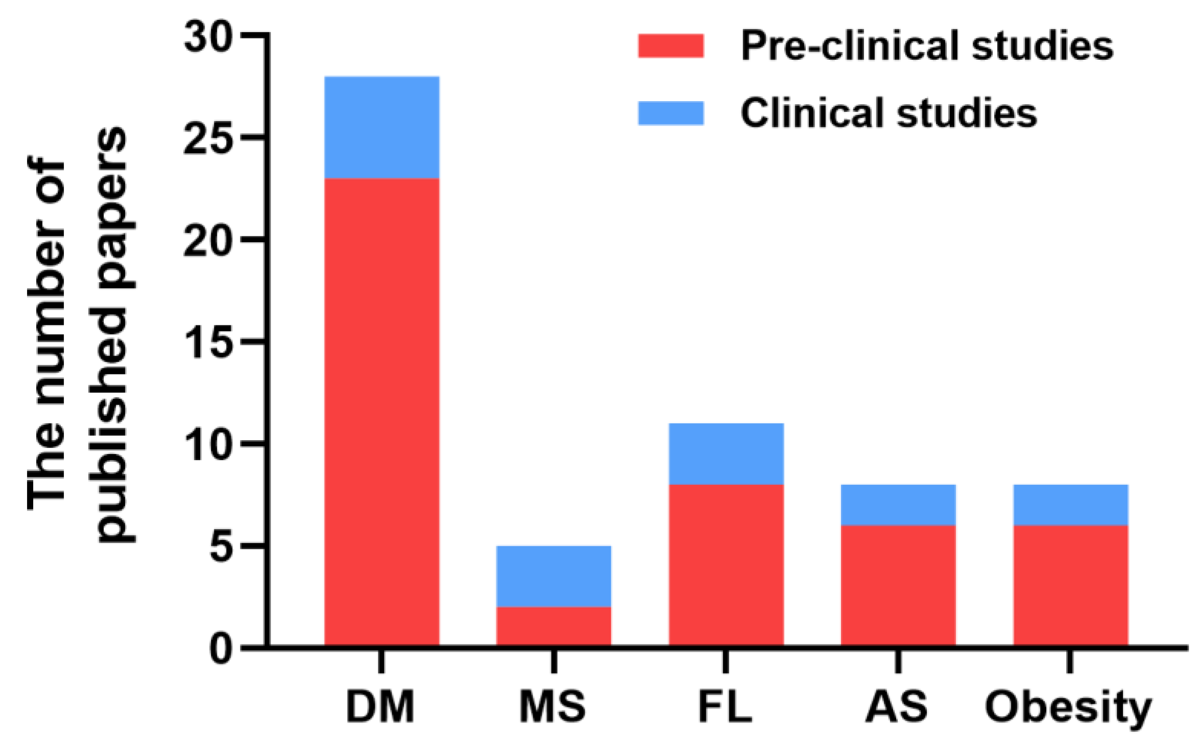
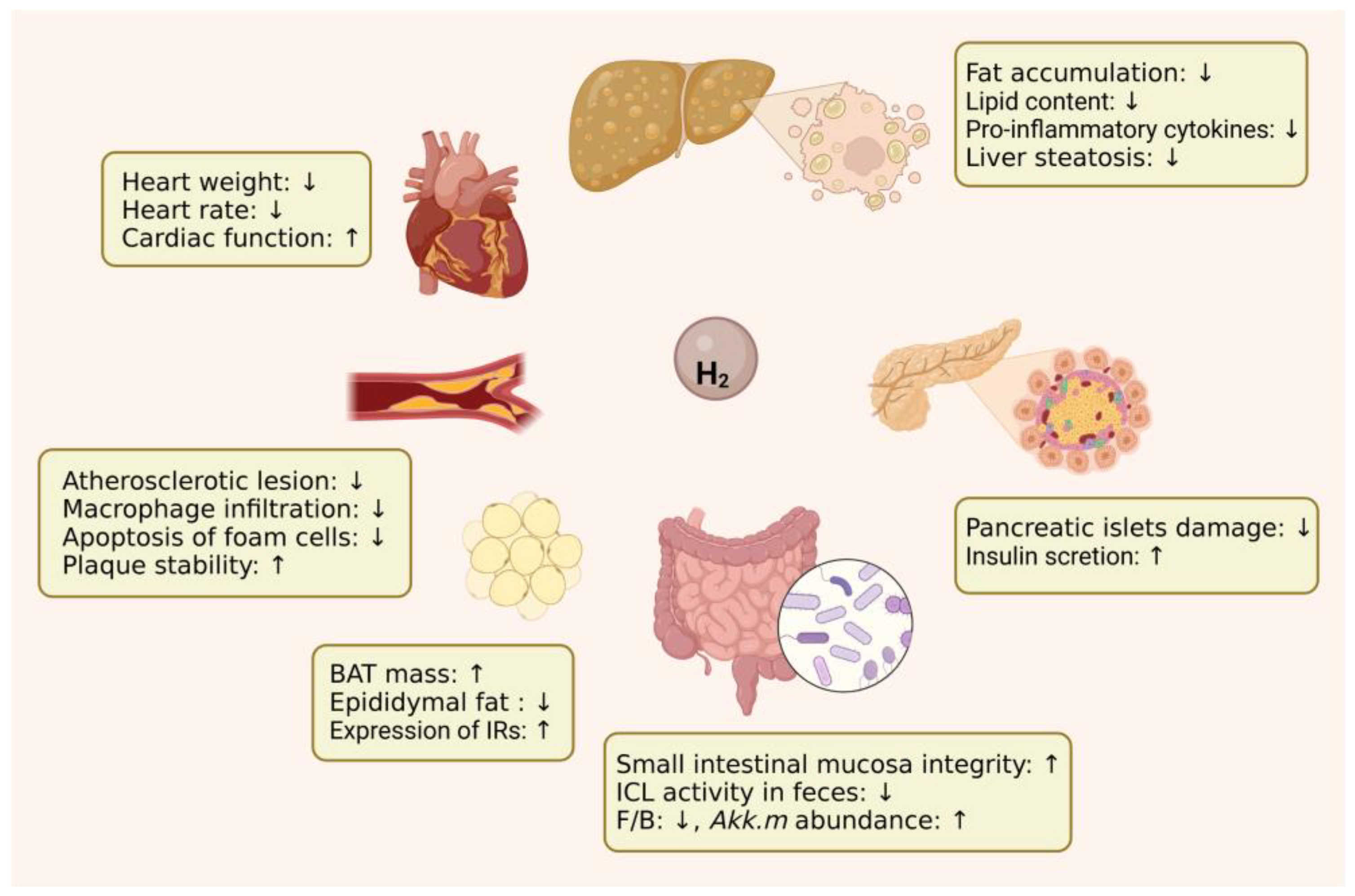
2. Protective Effects of H2 against Metabolic Diseases

2.1. Diabetes Mellitus

DM is a chronic metabolic disorder characterized by chronic hyperglycemia, resulting from defects in insulin secretion and/or insulin function. DM is mainly categorized into two sub-types: type 1 DM (T1DM) and type 2 DM (T2DM). T1DM is an autoimmune disorder that attacks the insulin-producing pancreatic beta cells with T cells, resulting in an absolute insulin deficiency [7]. As the most prevalent form of DM, T2DM, which is also known as non-insulin-dependent diabetes, is mainly caused by impaired insulin secretion by pancreatic beta cells and resistance of insulin-sensitive tissues to insulin action, resulting in a relative lack of insulin [8]. DM can cause multiple complications, including macrovascular complications (such as coronary heart disease, stroke, and peripheral vascular disease) and microvascular complications (such as end-stage renal disease (ESRD), retinopathy, and neuropathy) [9]. Although considerable research has been devoted to the development of diabetes medications, their efficacies are often hampered by various side-effects, thus necessitating the search for novel therapeutic agents for the treatment of DM.
A growing body of evidence has demonstrated that excessive reactive oxygen species (ROS) generation, largely due to hyperglycemia, causes oxidative stress, which further promotes the development and progression of DM and its complications [10]. Pancreatic beta cells are highly vulnerable to oxidative stress, due to their high endogenous production of ROS and low levels of antioxidant enzyme expression [11]. The destruction of beta cells is a pathological component of T1DM and T2DM. As a novel antioxidant agent, the potential therapeutic effects of molecular hydrogen on DM and its complications have been extensively investigated in both pre-clinical and clinical studies, as summarized in Table 1. The attenuation of oxidative stress and enhancement of an anti-oxidant response, as evidenced by decreased malondialdehyde (MDA) levels and increased glutathione (GSH) levels and superoxide dismutase (SOD) and catalase (CAT) activities, is at least partially responsible for the antidiabetic effect of H2 on DM [12,13,14,15,16,17].
To investigate the effect of molecular hydrogen on T2DM, two types of animal models have been used in pre-clinical research: (1) insulin resistance obesity and diabetes models, including Leprdb/db mice and HFD-induced obesity mice or rats. These animals exhibited a significant increase in body weight, and FBG, insulin, TG, and TC levels. (2) HFD + STZ-induced mice or rat models. As HFD induces insulin resistance and STZ causes partial beta cell destruction, these models closely mimic natural disease progression (from insulin resistance to beta cell dysfunction), as well as the metabolic characteristics of T2DM [21]. Based on these models, hydrogen was administered to animals via four routes, including drinking HRW, oral gavage of HRS, injection of HRS, and injection of hydrogen gas, for different durations (from 2 weeks to 3 months). Until now, no research has been conducted to compare the effect of hydrogen delivery methods, treatment duration, and hydrogen dosage on the FBG-lowing effect in detail, although several studies have provided some clues. Different hydrogen intervention methods may present different change patterns in FBG levels; for example, the FBG levels dropped quickly during the first two weeks of subcutaneous injection of hydrogen gas, followed by a slow reduction during the next two weeks [13], while a significant decrease in FBG levels did not occur until the third week of HRW intake [12]. Low-concentration HRW has been shown to have a smaller FBG-lowering effect to high-concentration HRW [17]; however, the dosage effects of H2 on DM need to be further investigated. It is worth noting that the FBG-lowering effect with higher initial FBG levels prior to intervention seems to be more apparent, especially in HFD + STZ-induced DM models.
Insulin resistance and beta-cell dysfunction play an important role in the pathogenesis and evolution of T2DM [22]. The anti-T2DM effect of H2 has been associated with improved insulin sensitivity, evidenced by increased insulin sensitivity index (ISI), decreased insulin resistance index (IRI) and homeostasis model assessment-insulin resistance (HOMA-IR) index, and improved glucose and insulin tolerance [13,14,16]. The insulin-sensitizing effect of H2 is even slightly better than pioglitazone [14]. H2-induced upregulation of insulin and insulin receptors in both the adipose and skeletal muscle tissues may partially contribute to the improved insulin sensitivity [15]. H2 also showed a protective effect with respect to STZ-induced beta-cell damage, although the evidence was not definitive. A histopathological examination revealed a significant attenuation of STZ-induced pancreatic islet morphological changes after H2 intervention [16]. STZ-induced decreases in insulin levels could also be ameliorated by H2 [15], which indirectly confirmed the protective role of H2 in beta-cell dysfunction. It is worth pointing out that in some obesity and HFD + STZ-induced animals, hyperinsulinemia secondary to hyperglycemia and insulin resistance could also be restored by H2 [13,14,17].
The modulating role on metabolism may also contribute to the anti-T2DM effect of H2. Hydrogen-induced alterations in hepatic glucose and lipid metabolism have been reported in several studies. Kamimura et al. [17] found that drinking HRW may stimulate energy metabolism, through upregulation of hepatic fibroblast growth factor 21 (FGF21) expression. It has been established that the liver-derived metabolic hormone FGF21 plays a key role in the regulation of energy homeostasis [23]. The induction of FGF21 expression may be responsible for the decrease in plasma triglyceride and glucose induced by hydrogen administration. It has been reported that increased hepatic FGF21 expression can also be induced by dietary manipulation, such as fasting or ketogenic diet [23]. Consistently, diet restriction and HRW consumption showed a similar downregulatory effect on plasma glucose, insulin, and triglycerides levels. Hydrogen administration has also been found to promote the synthesis of hepatic glycogen, whilst improving the utilization of glucose in the liver [16]. In addition, a reduction of hepatic fat accumulation can also be observed, even with short-term hydrogen interventions (1–2 weeks), suggesting a significant effect of hydrogen on hepatic lipid metabolism [12,17]. The reduced serum levels of TG, TC, and LDL-c, and increased HDL-c levels, also support the regulatory effect of hydrogen on lipid metabolism [12,13,14,16,17].
Unlike T2DM, research on the effect of H2 on T1DM is very limited. Amitani et al. [18] established an STZ-induced type 1 diabetic mice model and found that chronic intraperitoneal and oral administration of H2 significantly improved glycemic control. In fact, as early as 2006, Kim et al. [19] provided the first in vivo evidence of the antidiabetic effect of electrolyzed reduced water (ERW) in both T1DM and T2DM, as evidenced by decreased FBG levels and improved glucose tolerance. ERW, also called “alkaline ionized water”, is characterized by alkaline pH, low dissolved oxygen gas concentration, high dissolved hydrogen gas concentration, and a negative oxidation–reduction potential (ORP) [24]. The anti-T1DM effect of ERW has also been observed in an alloxan-induced insulin-dependent diabetic mice model [20]. Increasing evidence demonstrates that the dissolved hydrogen in ERW is exclusively responsible for any observed therapeutic effects [24]. From current research, the inhibition of oxidative stress and beta-cell apoptosis and promotion of glucose uptake into skeletal muscle and insulin sensitivity in peripheral tissues may contribute to the anti-T1DM effects of H2 [18,19,20].
The protective effects of H2 against the diabetic complications, including peripheral neuropathy [25,26,27,28,29], retinopathy [30,31], cardiomyopathy [32,33], wound healing [34,35,36], bone loss [37], and erectile dysfunction [38], have been extensively reported. STZ-induced mice or rats models were used in the above in vivo studies for diabetic complications. In addition to conventional hydrogen delivery routes, some novel administration methods, such as photo-driven nanoreactor, microneedle patch, and algae–bacteria gel patch, were used, which allowed in situ continuous hydrogen production to treat chronic diabetic wounds. The inhibition of oxidative stress, inflammation, endoplasmic reticulum stress, apoptosis, and pyroptosis, as well as parthanatos (a particular type of programmed cell death that is different from the common apoptosis, necrosis, and autophagy) and activation of the mitochondrial ATP-sensitive potassium (Mito-K-ATP) pathway may be involved in the protective effects of H2 on diabetic complications [25,26,27,28,29,30,31,32,33,34,35,36,37,38].
To date, three clinical trials have been conducted to explore the potentials of H2 in the treatment of DM (Table 2). The first clinical trial was conducted in 2008 by Kajiyama et al. [39], showing a potential role of HRW in the prevention of T2DM and insulin resistance, as evidenced by normalized glucose tolerance in four of six patients with impaired glucose tolerance (IGT); however, no changes were observed in the levels of FBG and insulin. HRW intake also induced decreased levels of modified LDL, especially small dense LDL and urinary 8-isoprostanes; however, additional research is needed to clarify the regulation of these biomarkers. The other two clinical trials were both aimed at exploring the potential effects of ERW on T2DM. One of the two clinical studies was a multicenter, prospective, double-blind, randomized controlled trial led by Tohoku University [40]. Drinking ERW for three months induced a significant reduction in serum lactate levels, but no changes were observed in HOMA-IR and levels of FBG and fasting plasma insulin (FPI). Further in-depth analysis revealed that in the HOMA-IR ≥1.73 group, the rate of change in lactate levels was positively correlated with the rate of change in HOMA-IR, FBG and FPI levels; in the FBG ≥6.1 mM group, the FPI levels dropped significantly and FBG levels reduced with a marginal significance (p = 0.067). These findings indicated that patients with more serious conditions, such as more insulin resistant or higher FBG levels, may obtain more benefits from hydrogen treatment, which is consistent with pre-clinical studies. Another clinical study showed that oral intake ERW for 12 days decreased total cholesterol and improved other lipid profiles in patients with T2DM accompanied by dyslipidemia [41]. An FBG-lowering effect was also observed in the ERW group compared with the controls, although the difference was not significant. In conclusion, potential beneficial effects of H2 for T2DM were observed in recent clinical studies. However, due to the small number of participants and short periods of intervention, the therapeutic effect of H2 on T2DM is not obvious. Further high-quality clinical trials, such as large-scale, multicenter, prospective, double-blind, randomized control clinical trials of H2 for the prevention and treatment of T2DM, T1DM, and DM-related complications are needed in the future.

2.2. Metabolic Syndrome

MS is typically characterized by the simultaneous occurrence of at least three of the following medical conditions: obesity, hyperglycemia, hypertension or dyslipidemia, which predispose individuals to cardiovascular disease and T2DM [52]. The pre-clinical studies conducted by Hashimoto et al. [53] showed that intake of HRW for 16 weeks had no effect on TG, TC, FBG, and blood pressure in SHR.Cg-Leprcp/NDmcr (SHR-cp) rats; however, HRW could exert beneficial effects against renal abnormalities, as evidenced by preventing glomerulosclerosis and ameliorating creatinine clearance. The enhancement in antioxidative activity and inhibition ROS production in the kidneys may be responsible for the effect of H2 on MS-related renal dysfunction. Zhao et al. [54] provided indirect evidence that oral administration of L-arabinose, a naturally occurring plant pentose, can induce hydrogen gas production through gut fermentation and alleviate HFD-induced MS, including gain in body weight and fat, impaired insulin sensitivity, liver steatosis, dyslipidemia, and elevated inflammatory cytokines, indicating that increasing endogenous hydrogen gas release through modulating gut microbial composition by administration of prebiotic nutrients or hydrogen-producing probiotics might be a promising approach for the treatment of MS. In conclusion, these pre-clinical findings revealed the potential beneficial effects of H2 on MS (Table 3).
To further characterize the effects of H2 on patient with MS, three clinical studies have been conducted to date (Table 2). Nakao et al. first performed an open label pilot study on 20 subjects with potential MS and demonstrated that drinking HRW (~1 mmol H2/day) for 8 weeks could increase HDL-c levels by 8% and decrease the total cholesterol-to-HDL ratio by 13% [42]. These effects may have been caused by the antioxidant effect of H2, as evidenced by SOD levels increased by 39% and decreased TBARs by 43%. Similarly, song et al. reported that HRW intake (0.5 mmol H2/day) for 10 weeks in patients with potential MS decreased TC and LDL-C levels, improved HDL function, and alleviated oxidative stress and inflammation [43]. Recently, LeBaron et al. carried out a randomized, double-blinded, placebo-controlled trial in patients with MS [44]. Compared with the previous two studies, this trial included more subjects (60 MS patients), and, importantly, higher concentration HRW (>5.5 mmol H2/day) was administered for a longer duration (24 weeks). Consistently with previous studies, HRW intake decreased the levels of TG and LDL-c, TC/HDL-c ratio, inflammation, and lipid peroxidation. However, this study also found some different effects from previous studies: (1) The body mass index (BMI), waist–hip circumference, and resting heart rate were significantly decreased, all these changes were absent in the previous two studies. (2) FBG levels decreased after a 24-week HRW intervention with an accompanying 12% reduction in HbA1C, while this glucose-lowering effect was not observed in previous studies. (3) The levels of HDL-c were significantly decreased, whereas in the open-label study, there was an increase in HDL levels. A reduction in HDL-c was also observed in T2DM rats after HRW intake [12]. This was probably due to the dramatic reduction in TC levels induced by H2, resulting in a lower TC/HDL-c ratio, which is associated with a decreased risk of cardiovascular disease. The longer duration with high-concentration of HRW may have been responsible for these different effects. In conclusion, HRW administration, especially long-term treatment with a high concentration of HRW, may be a promising adjuvant therapy to decrease the features of MS.

2.3. Fatty Liver

FL is characterized by a wide histological spectrum, ranging from steatosis, steatohepatitis (SH), to fibrosis/cirrhosis, and has traditionally been divided into alcoholic liver disease (ALD) and non-alcoholic fatty liver disease (NAFLD) [75]. ALD is caused by chronic and excessive alcohol consumption, while NAFLD is defined by evidence of FL without alcohol abuse. NAFLD has been considered a continuum from obesity to MS and DM [76]. Due to the close association of NAFLD with metabolic risks, it has recently been proposed to change its name to metabolic (dysfunction)-associated fatty liver disease (MAFLD) [77]. Oxidative stress has been considered to play a key role in the pathogenesis of both ALD and NAFLD [75]. The beneficial effects of H2 on both ALD and NAFLD were reported in previous pre-clinical studies. Lin et al. investigated the potential effect of HRW on ALD in chronic-binge ethanol-fed mice model [56]. The results showed that HRW protected against early-stage chronic EtOH-induced liver injury, possibly by reversing EtOH-induced anorexia via upregulation of acyl ghrelin expression, and suppressing inflammation and oxidative stress, which indicates the potential of HRW for prevention and clinical complementary treatment of ALD. Compared with ALD, more detailed and in-depth studies exploring the effects of H2 on NAFLD have been reported. As shown in Table 3, the evidence for H2-induced improvement of NAFLD includes (1) alleviation of hepatic steatosis, as evidenced by reduced hepatic lipid contents, hepatocellular ballooning degeneration, liver weight, and plasma ALT and AST activities; (2) reversion of steatohepatitis and fibrosis, supported by inhibition of hepatic inflammatory cell infiltration and hepatic stellate cell (HSC) activation, reduced necrotic areas and Sirius red-positive area in liver histopathology, downregulation of pro-inflammatory cytokines, and upregulation of anti-inflammatory cytokines; (3) attenuation of dyslipidemia, as evidenced by reduced body weight gain, BMI, WAT mass, and serum TC and TG; (4) improvement of glycemic control, as demonstrated by decreased FBG and fasting insulin (FINS) levels, and improved insulin sensitivity and glucose tolerance. In addition, it has also been found that H2 could ameliorate ischemia reperfusion injury in FL and prevent hepatocarcinogenesis [59,63]. Some of these studies investigated the dose effect of H2 and showed that higher doses of H2 administration demonstrated better therapeutic effects in most cases [60,62], even though a low dose of H2 showed better effects for some indices (e.g., plasma ALT and AST levels) [60]. Moreover, the study by Li et al. demonstrated that a longer duration of H2 intervention exhibited more obvious improvements [61]. Consistently, prolonged hydrogen release from a nanocapsule exerted more profound effects [62]. These studies also indicated that antioxidant, anti-inflammatory, and anti-apoptotic effects may be involved in the mechanism of action of H2 in FL (Table 3).
The currently published clinical studies mainly focused on the effects of H2 on NALFD (Table 2). A preliminary clinical study involving 12 overweight outpatients with NALFD was conducted by Korovljev et al. in 2019 [45]. The results showed that drinking HRW (3 mmol/day) significantly reduced liver fat accumulation and serum AST levels; however, there was no significant effect on the body weight, BMI, body composition, lipid profiles, and glucose levels. Recently, the effects of HRW were investigated in another clinical trial with 30 subjects [46]. HRW intervention was performed with a higher dose (>6 mmol/day) and longer duration (8 weeks), resulting in a favorable trend in body weight, BMI, SBP (reduced by ~2 mmHg), TG/HDL ratio, and LDH; however, most of the observed trends were not statistically significant. The lack of statistically significant changes may be due to either the values already being in homeostasis/range or a shorter duration of H2 intervention. Another recent study investigated the effect of 13-week hydrogen/oxygen (66% H2, 33% O2) inhalation on 43 subjects with NALFD [47]. Hydrogen/oxygen inhalation showed no effect on liver steatosis in mild cases; however, the liver fat content measured by ultrasound and CT scans was significantly improved in moderate–severe cases. Hydrogen/oxygen inhalation also significantly decreased LDL-c, AST, ALT, and biomarkers of oxidative stress and inflammation. In conclusion, the present studies have shown beneficial effects of H2 on NAFLD, especially in moderate–severe cases, a while higher dose of H2 intervention for a longer period had a tendency to be more effective.

2.4. Atherosclerosis

AS is a chronic progressive disease triggered mainly by abundant accumulation of apoB-containing lipoproteins and vascular inflammation in medium-sized to large arteries and that eventually leads to compromised blood flow [78]. One possible pathological mechanism underlying atherogenesis is that chronic stress causes endothelial injury, inducing the entry of monocytes into the subendothelium and their subsequent differentiation into macrophages, promoting foam cell formation and resulting in the formation of atherosclerotic plaque [79]. The potential effects of H2 on AS were investigated in apoE or LDL receptor (LDLR)-deficient mice. As demonstrated in Table 3, on the one hand, H2 intervention could downregulate apoB protein levels in plasma and in the liver, alleviate serum lipid oxidation, improve HDL functionality, and reduce inflammatory processes in the arterial wall, which may contribute to the inhibition of the initiation and development of atherosclerosis. On the other hand, H2 could increase plaque collagen content; reduce macrophage infiltration, lipid deposition, vascular smooth muscle cell (VSMC) content, cell apoptosis, and MMP-9 expression in atherosclerotic plaques; and alleviate the formation and apoptosis of macrophage-derived foam cells, which resulted in plaque stabilization. Higher doses of hydrogen intervention showed better anti-AS effects [66,67,69]. These studies indicated the potential role of H2 in preventing and treating atherosclerotic cardiovascular and cerebrovascular disease. The suppression of oxidative stress, inflammation, apoptosis, and ER stress, and activation of autophagy may be involved in the anti-AS effects of H2.
To date, no published clinical trials have directly revealed the effect of H2 on patients with AS; however, several clinical studies have provided evidence that H2 may exert inhibitory effects on the initiation and progression of AS, such as improving vascular endothelial function, and preventing and relieving peripheral arterial disease (PAD) (Table 2). Sakai et al. investigated the potential effects of high-concentration (3.5 mM) HRW on vascular endothelial function in 34 health subjects [48]. The endothelial function was assessed by measuring the flow-mediated dilation (FMD) of the brachial artery (BA) 30 min after drinking HRW. Compared with the placebo group, the relative ratio to the baseline in the changes of FMD after consumption of HRW was significantly improved, indicating the protective potential of H2 toward the endothelium. The improved endothelial function induced by H2 was corroborated by comparable observations in a recent clinical study from the same group [49]. The peripheral endothelial function in the small arteries of the fingers was evaluated using another non-invasive approach, reactive hyperemia-peripheral arterial tonometry (RH-PAT), in 68 healthy subjects. A significant improvement in natural logarithmic transformation of the reactive hyperemia index (Ln-RHI) was observed 24 h after the first consumption and continuous consumption of HRW for 2 weeks. To investigate the influence of Ln_RHI values before HRW intake on the effects of H2, the subjects were then divided into low and high Ln-RHI groups, for further analysis. Compared with the placebo group, daily intake of HRW for 2 weeks significantly improved the Ln_RHI value in the low Ln_RHI group, while no significant improvement was observed in the high Ln_RHI group, indicating that patients with higher risk for vascular events may benefit more from H2 interventions. Since endothelial dysfunction is an early event of AS, these results indicated the inhibitory effects of H2 on the initiation of AS. In conclusion, the present studies have provided preliminary evidence of the potential benefits of H2 for AS-related disease, and further clinical studies should be performed to confirm the anti-AS role of H2.

2.5. Obesity

Obesity is characterized by excessive body fat accumulation and chronic low-grade inflammation, and is often associated with increased risk of hypertension, T2DM, dyslipidemia, NAFLD, and cardiovascular disease (CVD) [80]. Many factors are involved in the pathogenesis of obesity, such as genetics, viruses, insulin resistance, inflammation, gut microbiome, circadian rhythms, and hormones [80]. Anti-obesity effects have been extensively reported in pre-clinical studies. These studies provided evidence that H2 could suppress weight gain and fat accumulation in the liver, alleviate plasma dyslipidemia, improve glycemic control, and extended the average of lifespan of HFD-induced-obesity mice (Table 3). Research by Masuda et al. showed that a short H2 intervention (2 weeks) had no effect on the blood sugar level in HFD-induced-obesity mice but changed the anti-metabolic phenotype of both WAT and BAT adipocytes, from hypertrophic to hyperplastic [73]. The findings of H2-induced rapid regulation of lipid metabolism in this study, along with evidence from other studies, such as the reduction in hepatic fat accumulation and the promotion of fatty acid metabolic gene expression in the liver found in obese mice after 1–2 weeks of HRW intake [17,71], indicated that H2 could restore lipid metabolism toward energy consumption more favorably than glucose metabolism. In addition to the suppression of oxidative stress and inflammation, the regulation of gut microbiota [72], the alleviation of oxidized phospholipid (OxPLs) accumulation by enhancing the anti-oxidative capacity of HDL [74], as well as the acceleration of BAT activation [73] may be involved in the anti-obesity effects of H2.
To date, the clinical research published on the anti-obesity effects of H2 has been very limited (Table 2). Korovljev et al. evaluated the effects of H2 intervention on body composition, hormonal status, and mitochondrial function in ten middle-aged overweight women [50]. Orally administration of hydrogen-generating minerals for 4 weeks resulted in a significant reduction in body fat, arm fat index, serum TG, and insulin levels, accompanied by a strong increasing trend of ghrelin secretion. However, H2 administration had no significant effect on BW, serum glucose, and other lipid parameters, which may have been caused by the relatively short treatment duration. It has been reported that fasting blood lactate accumulation reflects mitochondrial dysfunction, which in turn influences metabolic disease risk [81]. It is noteworthy that after 4 weeks of H2 intervention, the blood lactate levels were significantly lower than the placebo group (p = 0.01), indicating that an improvement of mitochondrial function may be involved in the anti-obesity effect of H2. Asada et al. also provided evidence that taking a 10 min warm (41 °C) HRW bath every day for 1–6 month could decrease the visceral fat accumulation and blood LDL-c levels in some subjects [51]. Although the current clinical studies have provided some evidence of the anti-obesity effects of H2, the reliability of the evidence is limited, due to the small number of subjects. Further large-scale trails with long-term H2 interventions should be performed, to investigate the anti-obesity effects of H2 in detail.
In conclusion, the results from current pre-clinical and clinical studies indicated the potential beneficial effects of H2 on metabolic diseases. The main effects of H2 on various tissues in metabolic diseases are illustrated in Figure 2.

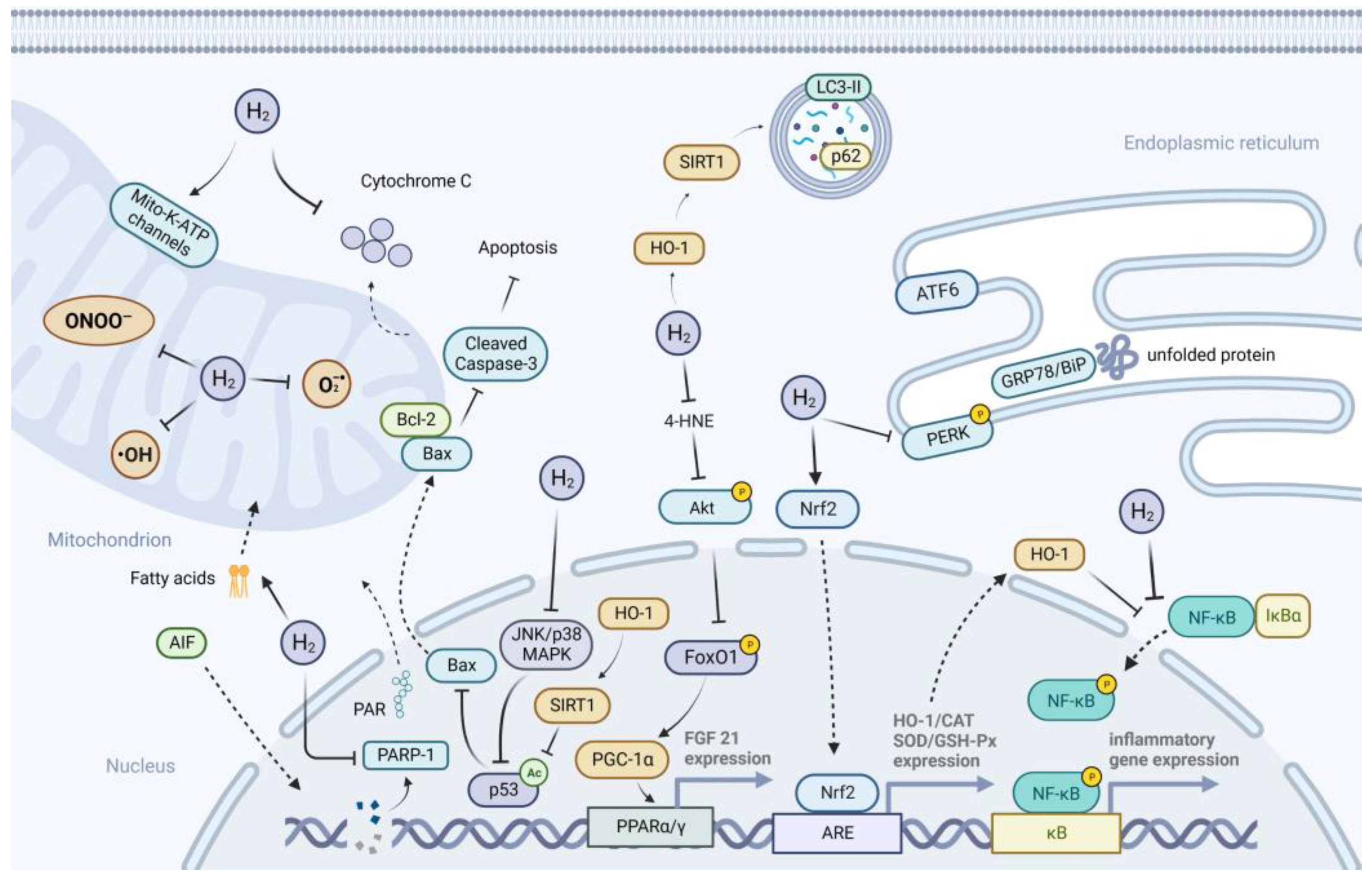
3. Possible Mechanisms of H2 Action on Metabolic Diseases

The canonical mechanisms of H2 action, including anti-oxidative, anti-inflammatory, and anti-apoptotic effects, have been extensively reported in various diseases. In addition to these canonical effects, suppression of ER stress, and activation of autophagy, preservation of mitochondrial function, and regulation of gut microbiota may also be involved in the action of H2 on metabolic diseases. The possible mechanisms underlying the effects of H2 on metabolic diseases are illustrated in Figure 3.

3.1. Anti-Oxidative Effects of H2

The anti-oxidative effect of H2 has been confirmed in numerous disease models of metabolic diseases. H2 may exert its antioxidant activity in three ways, including directly scavenging free radicals, inhibiting the ROS generation, and enhancing antioxidant enzyme activity. Ohta’s group first proposed that H2 may play an antioxidant role by selectively scavenging hydroxyl radicals and peroxynitrite [4]; however, the increasing evidence of H2-induced biological effects cannot be fully explained by its radical scavenging properties [82]. Based on the theory of metal-free hydrogen activation by Lewis acid and base, known as the frustrated Lewis pair (FLP) mechanism, Ishibashi proposed that H2 could be activated in the Q-chamber of mitochondria, providing electrons and protons to the ubiquinone (UQ) species, including highly-reactive semiquinone intermediates, preventing the premature leakage of electrons from the ETC and leading to the suppression of ROS generation [82]. Ishihara et al. provided in vitro evidence that H2 mainly suppress superoxide generation in complex I [83]. In addition, Ma’s group demonstrated the existence of H2-evolving activity in eukaryotic mitochondria, which is closely related to complex I and could be promoted by hypoxia [84]. They proposed that hypoxia-induced oxidative stress could be inhibited by H2 production. In this process, hypoxia induced mitochondrial succinate accumulation and reverse electron transfer, leading to a reduction in the UQ pool, and protons then have the chance to compete with UQ for electrons, which eventually results in the release of H2. This evidence indicated that eukaryotic mitochondria, especially the complex I, may partially retain the activity of hydrogenase, allowing the conversion of dihydrogen into protons and electrons, and the generation of dihydrogen, which may play an important role in the regulation of ROS generation. In addition to directly reducing ROS, H2 could also indirectly suppress oxidative stress by increasing antioxidant capacity via regulating anti-oxidative signal transduction. H2 has been reported to activate the nuclear factor erythroid 2-related factor 2 (Nrf2)-mediated anti-oxidative pathway, leading to an increased expression of downstream target enzymes, including heme oxygenase-1 (HO-1), catalase (CAT), SOD, and GSH-Px [29,34,67]. Although H2 has been reported to ameliorate lipid accumulation by regulating the miR-136/MEG3/Nrf2 pathway in NAFLD [58], it is unclear whether H2 could also regulate Nrf2 activity through the canonical Keap1-Nrf2-ARE pathway or other non-canonical pathways in metabolic diseases [85].
In addition, previous studies also provided further evidence to explain how H2 exerts protective effects against metabolic diseases through antioxidation. Ohta’s group performed a microarray analysis to examine the changes in hepatic gene expression at different time points of H2 treatment in obesity mice [71]. Combined with their previous findings, they speculated that H2 reduced hydroxyl radicals, resulting in a decrease of peroxides and their end products, including 4-HNE, and thus activating PGC-1α through suppression of Akt/FoxO1 phosphorylation, followed by stimulation of the PPARα pathway, upregulation of FGF21 gene expression, and finally enhancing the fatty acid metabolism. PPARγ, another member of the PPAR nuclear receptor superfamily, was also upregulated after H2 treatment, which may synergistically improve glucose and lipid metabolism with PPARα [57]. In conclusion, H2 could suppress oxidative stress via directly scavenging free radicals, inhibiting the ROS generation, or increasing the cellular antioxidant capacity, leading to the alleviation of oxidative damage in various organs, including the liver and pancreas, as well as the improvement of glucose and lipid metabolism.

3.2. Anti-Inflammatory Effects of H2

The occurrence and development of metabolic diseases are closely associated with an uncontrolled immune response. Whereas in T1DM, pancreatic beta cells are destroyed by an autoimmune response, the onset of T2DM, MS, FL, AS, and obesity are accompanied by chronic low-grade inflammation. It has been extensively reported that H2 could alleviate inflammation in metabolic diseases through downregulation of pro-inflammatory cytokines, and upregulation of anti-inflammatory cytokines. The anti-inflammatory effects of H2 are always paralleled by its antioxidant effects. HO-1 is an important mediator of the cross-talk between anti-inflammatory and anti-oxidative pathways. It has been reported that H2 could inhibit the lipopolysaccharide (LPS)-induced production of TNF-α and other inflammatory cytokines via an HO-1/interleukin 10 (IL-10)-independent pathway [61]. As an inducible isoform of heme oxygenases, HO-1 catalyzes the oxidative conversion of heme to carbon monoxide (CO), ferrous ions, and biliverdin (BV), which is reduced to bilirubin-IXα (BR) by biliverdin reductase (BVR) [86]. Since heme can catalyze membrane lipid peroxidation and oxidize low-density lipoprotein, both BV and BR act as powerful antioxidants, and the removal of heme and production of BV and BR may responsible for the anti-oxidative effects of HO-1 [86]. The HO-1/CO system has been considered as a modulator of inflammation, apoptosis, and autophagy [87]; however, the involvement of HO-1-derived CO in the immunomodulatory effects of H2 needs to be further investigated.
Excessive ROS generation under abnormal metabolic conditions not only causes damage to cellular macromolecules, but also activates redox sensitive transcription factors, such as nuclear factor-kappaB (NF-kB), resulting in the release of pro-inflammatory mediators. Deregulated NF-kB activation is a hallmark of chronic inflammatory diseases [88]. Previous studies have provided evidence that H2 could alleviate inflammation via inhibition of NF-kB activation in metabolic diseases [16,32,65]. Since the induction of HO-1 expression has also been related to the inhibition of NF-kB activation [89], the activation of the Nrf2/HO-1 pathway induced by H2 may contributed to its anti-inflammatory effects. Thus, it is reasonable that the anti-inflammatory activities of H2 may be attributed to its antioxidant effects.

3.3. Anti-Apoptotic Effects of H2

Apoptosis or programmed cell death plays an important role in the maintenance of tissue homeostasis. Excess apoptosis has been implicated in the initiation and development of metabolic diseases, such as DM and its complications [90]. Extensive studies have demonstrated the anti-apoptotic effects of H2 in metabolic diseases, as evidenced by the upregulation of anti-apoptotic Bcl-2, downregulation of pro-apoptotic Bax and cleaved caspase-3, and inhibition of caspase activity, as well as reduced TUNEL positive cells [20,25,28,31,32,59,67]. Oxidative stress-induced DNA breakage can rapidly activate poly (ADP-ribose) polymerase-1 (PARP-1), a DNA nick sensing enzyme, leading to the induction of either caspase-independent apoptosis via translocation of apoptosis-inducing factor (AIF) from mitochondria to the nucleus or caspase-dependent apoptosis by activation of caspase-3. H2 has been reported to inhibit the activation of PARP-1, leading to a reduction in apoptosis of Schwann cells induced by high glucose-mediated oxidative stress via the caspase-independent and caspase-dependent pathways [25]. The inhibition of apoptosis by H2 could also be mediated by elevating HO-1-induced expression of an NAD-dependent protein deacetylase, Sirtuin1 (SIRT1) [59]. This elevated SIRT1 expression could further reduce the expression of Bax, acetylation of p53, and cleaved caspase-3, and subsequently suppress the hepatocyte apoptosis. In addition, Wu et al. reported that H2 could suppress the activation of the Jun NH2-terminal kinase (JNK) and p38 mitogen activated protein kinase (MAPK) signaling pathways, leading to a reduction of the Bax/Bcl-2 ratio and caspase-3 activity, and finally inhibiting apoptosis [32]. In fact, p53 is considered as a major JNK/p38 MAPK substrate in promoting apoptosis [91], thus the JNK/p38 MAPK/p53 pathway could also be involved in the suppression of apoptosis by H2. In addition, H2 could also inhibit apoptosis by suppressing the activation of the ER stress pathway [32,67]. In conclusion, H2 could inhibit the apoptosis induced by oxidative stress or other stimulus in metabolic diseases via multiple signaling pathways, including but not limited to the PARP-1-mediated caspase-dependent and caspase-independent pathways, and HO-1/SIRT1/p53, JNK/p38 MAPK, and ER stress pathways.

3.4. Suppression of ER Stress

Endoplasmic reticulum stress (ER stress) is a transient state of functional imbalance characterized by the accumulation of unfolded and misfolded proteins in the ER lumen, which is usually resolved by the activation of multiple adaptive mechanisms, such as the unfolded protein response (UPR), ER-associated protein degradation (ERAD), or autophagy, and that eventually induces two conflicting responses: survival or apoptosis [92]. As the “canonical ER stress response”, the UPR pathway is regulated by ER chaperone protein glucose-regulated protein 78 (GRP78), also known as binding immunoglobulin protein (BiP), and three traditional UPR sensors, including proteins R (PKR)-like endoplasmic reticulum kinase (PERK), inositol-requiring enzyme1α (IREα), and activating transcription factor 6 (ATF6) [93]. ER stress has been associated with a number of metabolic disorders, including T2DM, FL, AS, and obesity [92]. Previous studies have shown that H2 could alleviate ER stress, as evidenced by a reduction in the expression of C/EBP homologous protein (CHOP), X-box protein1 (XBP1), ATF6, phosphorylated PERK (P-PERK), and GRP78/BiP [DM-17, AS-4]. These results indicated that H2 may exert anti-ER stress effects via regulation of the UPR pathway. Whether the regulation of “non-canonical ER stress responses”, including integrated stress response (ISR), ER translocation and ERK reactivation, endoplasmic-reticulum-associated protein degradation (ERAD), and ER-phagy [93], is also involved in the suppression of ER stress by H2 requires further investigation. Furthermore, Nrf2 could also be activated by PERK-mediated phosphorylation, which coordinates the convergence of ER stress with oxidative stress signaling [94], and thus Nrf2 may serve as an important cross-talk mediator of the anti-oxidative pathways with the anti-ER stress pathways induced by H2.

3.5. Activation of Autophagy

Autophagy is an evolutionarily conserved intracellular lysosome-dependent catabolic process, involving turnover of long-lived proteins and damaged organelles, and thus contributing to maintenance of cellular homeostasis, clearance of damaged or excess cellular components, and adaptation to environmental challenges. Nutrient-sensing pathways, including the mechanistic target of rapamycin complex 1 (mTORC1), AMP-activated kinase (AMPK), and SIRT1, are involved in the regulation of autophagic flux, depending on nutrient status [95]. Insufficient levels of autophagy cause the accumulation of damaged organelles or lipid droplets, which is crucial for the pathogenesis of metabolic diseases [95]. H2 has been shown to activate autophagy in macrophages, to prevent AS development, as evidenced by an increased expression of LC3-II and autophagy-related (Atg) proteins Atg3 and reduced expression of p62 [68,69]. Yang et al. also provided evidence that SIRT1 is essential for the upregulation of autophagic flux by H2 in ox-LDL-treated macrophages [68]. Although defective autophagy has been widely implicated in the pathogenesis of metabolic diseases, upregulated autophagy in subcutaneous and visceral adipose tissue has also been observed in patients with obesity or T2DM [95]; whether H2 could inhibit autophagy responses in these conditions needs to be further investigated. In conclusion, the activation of autophagy by H2 may contribute to its beneficial effects in metabolic diseases; however, the detailed mechanism underlying the regulation of autophagic flux by H2 requires further investigation.

3.6. Improvement of Mitochondrial Function

Mitochondria, the cell powerhouses, are involved in essential cellular functions, including ATP production, intracellular Ca2+ regulation, production and modulation of ROS, and apoptosis [96]. Mitochondrial dysfunction leads to reduced ATP generation and mitochondrial biogenesis, and increased ROS and cytosolic Ca2+ imbalances, which have been linked to the pathogenesis of several metabolic disorders, including MS, DM, and obesity [96]. H2 has been widely demonstrated to inhibit ROS generation, directly scavenge ROS or indirectly removing ROS via enhancing antioxidant enzyme activity, which may be responsible for improvements of mitochondrial function in metabolic diseases. It has been shown that the Mito-K-ATP channels play a homeostatic role in blood glucose regulation in organisms, and their dysfunction has been associated with DM and its complications [97]. The activation of Mito-K-ATP channels could effectively prevent mitochondrial ROS production [98]. Jiao et al. demonstrated that 5-hydroxydecanoate, a selective Mito-K-ATP channel blocker, could eliminate the neuroprotective effects of H2 in diabetic peripheral neuropathy, indicating the activation of Mito-K-ATP channels by H2 [26]. In addition, Ma’s group provided evidence that H2 could promote fatty acid oxidation, probably by inducing their transport to mitochondria and subsequent catabolism to ketone bodies, as evidenced by an increased level of acylcarnitines, the mitochondrial fatty acid transporters, and acetoacetate, a marker of ketogenesis in the liver, which may be mediated by the NADP/NADPH redox pathway [99]. In conclusion, improvement of mitochondrial function by H2, including but not limited to the activation of Mito-K-ATP channels and promotion of mitochondrial fatty acids oxidation, may be involved in the beneficial effects of H2 in metabolic diseases.

3.7. Regulation of Gut Microbiota

The gut microbiota is a dense and diverse microbial ecosystem dominated by bacteria, which mainly comprise four main phyla: Actinobacteria, Bacteroidetes, Firmicutes, and Proteobacteria in mammalians [100]. Gut microbiota-derived metabolites can interact with the host and serve as central regulators in metabolic disorders, including T2DM, NAFLD, and obesity [101]. Qiu et al. reported that HRS could alleviate HFD-induced dysbacteriosis by inhibiting the glyoxylic acid cycle of the intestinal flora [72]. He’s group found that sustained and ultrahigh H2 intervention in MAFLD mice drastically changed the composition of gut microbiota, reduced the ratio of Firmicutes/Bacteroidetes (F/B), and increased the abundance of Akkermansia muciniphila (Akk.m) [62]. The Akk.m has been considered a gut probiotic that can improve metabolic diseases, and its genome contains genes encoding hydrogenases, enabling it to catalytically decompose and utilize H2. It is likely that the utilization of H2 contributes to the outgrowth of Akk.m, finally leading to the improvement of MAFLD. It is worth noting that H2 could improve glucose metabolism but not lipid and energy metabolism via reprogrammed gut microbiota, because antibiotic treatment abolished the blood glucose-lowering effect but did not affect lipid parameters. In conclusion, bacterial hydrogenases enable the utilization of exogenous H2, leading to an altered gut microbiome profile, which may eventually regulate the host metabolism, especially the glucose metabolism, through gut microbiota-derived metabolites.

3.8. Other Possible Mechanisms and Potential Molecular Targets of H2

In addition to the above mechanisms underlying the effects of H2 on metabolic diseases, previous studies have also explored the possibility of other potential mechanisms. Qin’s group demonstrated that H2 could improve the protein composition and functionality of HDL by modulating the oxidized phospholipids (OxPLs)-related enzymes binding to HDL [74]. Zou et al. Provided evidence that H2 could reduce pyroptosis, a type of programmed cell death that occurs in the presence of inflammation, via inhibition of the AMPK/mTOR/NLRP3 signaling pathway in diabetic cardiomyopathy [33]. Li et al. reported that H2 could protect Schwann cells from high glucose-induced parthanatos, which is a type of PARP-1-dependent programmed cell death [27]. Although many possible mechanisms of H2 have been proposed, the target molecule of H2 is still unclear. Ohta’s group first considered hydroxyl radicals and peroxynitrite as the potential targets of H2; however, this view has been debated, due to the considerably low reaction rate of H2 with hydroxyl radicals. Later, several research groups proposed that some protein molecules may be potential targets of H2. Ma’s group first provided evidence that H2 could directly interact with acetylcholinesterase (AChE) and enhance its activity [102]. They later found that horseradish peroxidase (HRP) might also be the target molecule of H2. H2 might induce alterations in the microenvironment of histidine, tryptophan, and aspartate in HRP, and reconstruct the hydrogen bond network with the “H2O-H2O” bridge, thus increasing the efficiency of energy transfer to the heme center, and finally enhancing the enzyme activity [103]. Another potential molecular target of H2 was identified by He’s group. They reported that Fe-porphyrin may serve as a redox-related biosensor of H2, making it the upstream signaling molecular of CO through Fe-porphyrin-catalytic reduction of CO2 into CO in the hypoxic microenvironment [104]. Fe-porphyrin is a common heme cofactor in the active sites of many proteins, including hemoglobin (Hb) and cytochromes (Cyt), as well as HRP. They also provided evidence of the catalytic hydrogenation activity of Fe-porphyrin on the levels of proteins and cells, such as the catalytic activity of Hb for the hydrogenation of hydroxyl radicals and the reduction of CO2 by H2 into CO through Fe-porphyrin–catalytic hydrogenation. Heme proteins can also interact with other gaseous molecules, such as CO, NO, O2, and H2S, which play vital roles in many biological processes [105,106]. Thus, it is likely that H2 competes with them to interact with heme proteins, which ultimately affects the physiological functions of these gaseous molecules. Ohta recently proposed that the oxidative potential of hydroxyl radicals may be alleviated by H2-targeting porphyrin, which contributes to the activation of Nrf2 as a hormesis-like effect [107]. In conclusion, H2 may interact with multiple target molecules, including but not limited to hydroxyl radicals, peroxynitrite, AChE, HRP, and Fe-porphyrin, which finally contribute to the extensive biological effects of H2.

4. Conclusions and Perspectives

To date, both pre-clinical and clinical studies have provided evidence that H2 interventions might exert beneficial effects on metabolic diseases, with no reported adverse effects. However, the efficacy of intervention varied among the different studies, with some studies even showing no therapeutic effects. There are serval factors that may contribute to these differences: (1) H2 concentration—the dose-dependent effect of H2 has been observed in certain studies, which showed that higher doses of H2 exhibited better therapeutic effects. Different H2 delivery methods may cause different H2 concentrations in organs related to the initiation and development of metabolic diseases, and ultimately lead to different treatment effects. (2) Treatment duration—the response time to H2 may vary in different diseases, such as the relative faster regulation of hyperlipemia than hyperglycemia by H2. Prolonged H2 treatment may have a more significant impact on metabolic diseases. (3) The time point of H2 intervention—the effectiveness of H2 therapy may differ in different stage of metabolic diseases. (4) The severity of metabolic disorders— pre-clinical studies have shown that higher initial FBG levels prior to intervention seem to have a more apparent FBG-lowering effect. However, the current research only provides preliminary experimental evidence of the influence of the above factors on the effect of H2 intervention, and more in-depth and detailed research should be carried out in the future. Furthermore, an increasing number of clinical trials have been designed to evaluate the therapeutic effect of H2 on metabolic diseases; however, high-quality clinical studies have been relatively limited, and large-scale, randomized, controlled, multicentered, double-blind, and long-term interventions should be considered in future clinical trial designs.
The mechanism of action of H2 has long been a focal point and challenge for research. Although many hypotheses of H2 action have been proposed, most can be attributed to the antioxidative effect of H2. Free radicals have been considered as a double-edged sword in various disease treatments [108]; even hydroxyl radicals, the most cytotoxic of ROS, do not always have negative effects on cells. For example, the superoxide anion and its derivative hydroxyl radicals are essential for human phagocytes, to kill invading bacteria [109]. Thus, the selective hydroxyl radical-scavenging activity of H2 cannot explain its extensive beneficial effects. From the current research, H2 may have multiple target molecules, and the phenotypes induced by H2 intervention may be a comprehensive result of the interaction between molecular hydrogen and its target molecules. Future mechanistic studies should be focused on the identification of the potential targets of H2 and on exploring how H2 interacts with target molecules to induce cellular responses.
In conclusion, the evidence from the current studies suggests the potential application of H2 in the treatment of metabolic diseases. It is believed that, with further clinical and mechanism research, H2 will eventually be applied to clinical practice in the future, to benefit more patients.

Author Contributions

Writing-original draft preparation, F.X., Y.S. and Y.Y.; writing-review and editing, X.J., S.M., M.L. and P.Z.; visualization, C.M., J.L. and Z.Z.; funding acquisition, X.M. All authors have read and agreed to the published version of the manuscript.

Funding

This research was funded by the Military Logistics Key Open Research Projects (BHJ17L018) and National Natural Science Foundation of China (81602408).

Institutional Review Board Statement

Not applicable.

Informed Consent Statement

Not applicable.

Data Availability Statement

Data presented in this study are available on request from the corresponding author.

Conflicts of Interest

The authors declare no conflict of interest.

References

  1. Ohta, S. Molecular hydrogen as a novel antioxidant: Overview of the advantages of hydrogen for medical applications. Methods Enzymol. 2015, 555, 289–317. [Google Scholar] [CrossRef] [PubMed]
  2. Dole, M.; Wilson, F.R.; Fife, W.P. Hyperbaric hydrogen therapy: A possible treatment for cancer. Science 1975, 190, 152–154. [Google Scholar] [CrossRef] [PubMed]
  3. Gharib, B.; Hanna, S.; Abdallahi, O.M.; Lepidi, H.; Gardette, B.; De Reggi, M. Anti-inflammatory properties of molecular hydrogen: Investigation on parasite-induced liver inflammation. Comptes Rendus Acad. Sci. III 2001, 324, 719–724. [Google Scholar] [CrossRef] [PubMed]
  4. Ohsawa, I.; Ishikawa, M.; Takahashi, K.; Watanabe, M.; Nishimaki, K.; Yamagata, K.; Katsura, K.; Katayama, Y.; Asoh, S.; Ohta, S. Hydrogen acts as a therapeutic antioxidant by selectively reducing cytotoxic oxygen radicals. Nat. Med. 2007, 13, 688–694. [Google Scholar] [CrossRef] [PubMed]
  5. Ohta, S. Molecular hydrogen as a preventive and therapeutic medical gas: Initiation, development and potential of hydrogen medicine. Pharmacol. Ther. 2014, 144, 1–11. [Google Scholar] [CrossRef] [Green Version]
  6. Yadav, U.C.; Rani, V.; Deep, G.; Singh, R.K.; Palle, K. Oxidative stress in metabolic disorders: Pathogenesis, prevention, and therapeutics. Oxid. Med. Cell. Longev. 2016, 2016, 9137629. [Google Scholar] [CrossRef] [Green Version]
  7. Katsarou, A.; Gudbjörnsdottir, S.; Rawshani, A.; Dabelea, D.; Bonifacio, E.; Anderson, B.J.; Jacobsen, L.M.; Schatz, D.A.; Lernmark, Å. Type 1 diabetes mellitus. Nat. Rev. Dis. Prim. 2017, 3, 17016. [Google Scholar] [CrossRef]
  8. Galicia-Garcia, U.; Benito-Vicente, A.; Jebari, S.; Larrea-Sebal, A.; Siddiqi, H.; Uribe, K.B.; Ostolaza, H.; Martin, C. Pathophysiology of Type 2 Diabetes Mellitus. Int. J. Mol. Sci. 2020, 21, 6275. [Google Scholar] [CrossRef]
  9. Harding, J.L.; Pavkov, M.E.; Magliano, D.J.; Shaw, J.E.; Gregg, E.W. Global trends in diabetes complications: A review of current evidence. Diabetologia 2019, 62, 3–16. [Google Scholar] [CrossRef] [Green Version]
  10. Darenskaya, M.A.; Kolesnikova, L.I.; Kolesnikov, S.I. Oxidative stress: Pathogenetic role in diabetes mellitus and its complications and therapeutic approaches to correction. Bull. Exp. Biol. Med. 2021, 171, 179–189. [Google Scholar] [CrossRef]
  11. Eguchi, N.; Vaziri, N.D.; Dafoe, D.C.; Ichii, H. The role of oxidative stress in pancreatic beta cell dysfunction in diabetes. Int. J. Mol. Sci. 2021, 22, 1509. [Google Scholar] [CrossRef]
  12. Zheng, M.; Yu, H.; Xue, Y.; Yang, T.; Tu, Q.; Xiong, K.; Deng, D.; Lu, L.; Huang, N. The protective effect of hydrogen-rich water on rats with type 2 diabetes mellitus. Mol. Cell. Biochem. 2021, 476, 3089–3097. [Google Scholar] [CrossRef]
  13. Zhang, X.; Liu, J.; Jin, K.; Xu, H.; Wang, C.; Zhang, Z.; Kong, M.; Zhang, Z.; Wang, Q.; Wang, F. Subcutaneous injection of hydrogen gas is a novel effective treatment for type 2 diabetes. J. Diabetes Investig. 2018, 9, 83–90. [Google Scholar] [CrossRef]
  14. Wang, Q.; Zha, X.; Kang, Z.; Xu, M.; Huang, Q.; Zou, D. Therapeutic effects of hydrogen saturated saline on rat diabetic model and insulin resistant model via reduction of oxidative stress. Chin. Med. J. 2012, 125, 1633–1637. [Google Scholar] [CrossRef]
  15. Retnaningtyas, E.; Susatia, B.; Arifah, S.N.; Rahayu Lestari, S. The improvement of insulin level after hydrogen-rich water therapy in streptozotocin-induced diabetic rats. Vet. World 2022, 15, 182–187. [Google Scholar] [CrossRef]
  16. Ming, Y.; Ma, Q.H.; Han, X.L.; Li, H.Y. Molecular hydrogen improves type 2 diabetes through inhibiting oxidative stress. Exp. Ther. Med. 2020, 20, 359–366. [Google Scholar] [CrossRef]
  17. Kamimura, N.; Nishimaki, K.; Ohsawa, I.; Ohta, S. Molecular hydrogen improves obesity and diabetes by inducing hepatic FGF21 and stimulating energy metabolism in db/db mice. Obesity 2011, 19, 1396–1403. [Google Scholar] [CrossRef]
  18. Amitani, H.; Asakawa, A.; Cheng, K.; Amitani, M.; Kaimoto, K.; Nakano, M.; Ushikai, M.; Li, Y.; Tsai, M.; Li, J.B.; et al. Hydrogen improves glycemic control in type1 diabetic animal model by promoting glucose uptake into skeletal muscle. PLoS ONE 2013, 8, e53913. [Google Scholar] [CrossRef]
  19. Kim, M.J.; Kim, H.K. Anti-diabetic effects of electrolyzed reduced water in streptozotocin-induced and genetic diabetic mice. Life Sci. 2006, 79, 2288–2292. [Google Scholar] [CrossRef]
  20. Li, Y.; Hamasaki, T.; Nakamichi, N.; Kashiwagi, T.; Komatsu, T.; Ye, J.; Teruya, K.; Abe, M.; Yan, H.; Kinjo, T.; et al. Suppressive effects of electrolyzed reduced water on alloxan-induced apoptosis and type 1 diabetes mellitus. Cytotechnology 2011, 63, 119–131. [Google Scholar] [CrossRef] [Green Version]
  21. Zhang, M.; Lv, X.Y.; Li, J.; Xu, Z.G.; Chen, L. The characterization of high-fat diet and multiple low-dose streptozotocin induced type 2 diabetes rat model. Exp. Diabetes Res. 2008, 2008, 704045. [Google Scholar] [CrossRef] [PubMed] [Green Version]
  22. Keane, K.N.; Cruzat, V.F.; Carlessi, R.; de Bittencourt, P.I., Jr.; Newsholme, P. Molecular events linking oxidative stress and inflammation to insulin resistance and beta-cell dysfunction. Oxid. Med. Cell. Longev. 2015, 2015, 181643. [Google Scholar] [CrossRef] [PubMed] [Green Version]
  23. Lewis, J.E.; Ebling, F.J.P.; Samms, R.J.; Tsintzas, K. Going back to the biology of FGF21: New insights. Trends Endocrinol. Metab. 2019, 30, 491–504. [Google Scholar] [CrossRef] [PubMed]
  24. LeBaron, T.W.; Sharpe, R.; Ohno, K. Electrolyzed-reduced water: Review I. molecular hydrogen is the exclusive agent responsible for the therapeutic effects. Int. J. Mol. Sci. 2022, 23, 14750. [Google Scholar] [CrossRef]
  25. Yu, Y.; Ma, X.; Yang, T.A.O.; Li, B.O.; Xie, K.; Liu, D.; Wang, G.; Yu, Y. Protective effect of hydrogen-rich medium against high glucose-induced apoptosis of Schwann cells in vitro. Mol. Med. Rep. 2015, 12, 3986–3992. [Google Scholar] [CrossRef] [Green Version]
  26. Jiao, Y.; Yu, Y.; Li, B.; Gu, X.; Xie, K.; Wang, G.; Yu, Y. Protective effects of hydrogen-rich saline against experimental diabetic peripheral neuropathy via activation of the mitochondrial ATP-sensitive potassium channel channels in rats. Mol. Med. Rep. 2019, 21, 282–290. [Google Scholar] [CrossRef] [Green Version]
  27. Li, Q.; Jiao, Y.; Yu, Y.; Wang, G.; Yu, Y. Hydrogen-rich medium alleviates high glucose-induced oxidative stress and parthanatos in rat Schwann cells in vitro. Mol. Med. Rep. 2019, 19, 338–344. [Google Scholar] [CrossRef] [Green Version]
  28. Wang, P.; You, L.; Li, Q.; Wang, H.; Long, Y.; Chen, W. Hydrogen relieves neuropathic pain in diabetic rats by inhibiting MCP1 and CCR2 expressions. Kafkas Univ. Vet. Fak. Derg. 2022, 22, 169–176. [Google Scholar] [CrossRef]
  29. Han, X.C.; Ye, Z.H.; Hu, H.J.; Sun, Q.; Fan, D.F. Hydrogen exerts neuroprotective effects by inhibiting oxidative stress in experimental diabetic peripheral neuropathy rats. Med. Gas Res. 2023, 13, 72–77. [Google Scholar] [CrossRef]
  30. Feng, Y.; Wang, R.; Xu, J.; Sun, J.; Xu, T.; Gu, Q.; Wu, X. Hydrogen-rich saline prevents early neurovascular dysfunction resulting from inhibition of oxidative stress in STZ-diabetic Rats. Curr. Eye Res. 2012, 38, 396–404. [Google Scholar] [CrossRef]
  31. Xiao, X.; Cai, J.; Xu, J.; Wang, R.; Cai, J.; Liu, Y.; Xu, W.; Sun, X.; Li, R. Protective effects of hydrogen saline on diabetic retinopathy in a streptozotocin-induced diabetic rat model. J. Ocul. Pharmacol. Ther. 2012, 28, 76–82. [Google Scholar] [CrossRef] [Green Version]
  32. Wu, F.; Qiu, Y.; Ye, G.; Luo, H.; Jiang, J.; Yu, F.; Zhou, W.; Zhang, S.; Feng, J. Treatment with hydrogen molecule attenuates cardiac dysfunction in streptozotocin-induced diabetic mice. Cardiovasc. Pathol. 2015, 24, 294–303. [Google Scholar] [CrossRef]
  33. Zou, R.; Nie, C.; Pan, S.; Wang, B.; Hong, X.; Xi, S.; Bai, J.; Yu, M.; Liu, J.; Yang, W. Co-administration of hydrogen and metformin exerts cardioprotective effects by inhibiting pyroptosis and fibrosis in diabetic cardiomyopathy. Free Radic. Biol. Med. 2022, 183, 35–50. [Google Scholar] [CrossRef]
  34. Zheng, Y.; Zhang, Z.; Wang, T.; Zhang, J.; Tian, D.; Zhang, X.; Wu, Z. Photodriven nanoreactor with a hydrogen-insulin double act repairs diabetic wounds through Nrf2 pathway activation. Chem. Eng. J. 2021, 425, 131800. [Google Scholar] [CrossRef]
  35. Chen, H.; Guo, Y.; Zhang, Z.; Mao, W.; Shen, C.; Xiong, W.; Yao, Y.; Zhao, X.; Hu, Y.; Zou, Z.; et al. Symbiotic algae-bacteria dressing for producing hydrogen to accelerate diabetic wound healing. Nano Lett. 2022, 22, 229–237. [Google Scholar] [CrossRef]
  36. Wang, P.; Wu, J.; Yang, H.; Liu, H.; Yao, T.; Liu, C.; Gong, Y.; Wang, M.; Ji, G.; Huang, P.; et al. Intelligent microneedle patch with prolonged local release of hydrogen and magnesium ions for diabetic wound healing. Bioact. Mater. 2023, 24, 463–476. [Google Scholar] [CrossRef]
  37. Guo, J.; Dong, W.; Jin, L.; Wang, P.; Hou, Z.; Zhang, Y. Hydrogen-rich saline prevents bone loss in diabetic rats induced by streptozotocin. Int. Orthop. 2017, 41, 2119–2128. [Google Scholar] [CrossRef]
  38. Fan, M.; Xu, X.; He, X.; Chen, L.; Qian, L.; Liu, J.; Qing, J.; Chao, Z.; Sun, X. Protective effects of hydrogen-rich saline against erectile dysfunction in a streptozotocin induced diabetic rat model. J. Urol. 2013, 190, 350–356. [Google Scholar] [CrossRef]
  39. Kajiyama, S.; Hasegawa, G.; Asano, M.; Hosoda, H.; Fukui, M.; Nakamura, N.; Kitawaki, J.; Imai, S.; Nakano, K.; Ohta, M.; et al. Supplementation of hydrogen-rich water improves lipid and glucose metabolism in patients with type 2 diabetes or impaired glucose tolerance. Nutr. Res. 2008, 28, 137–143. [Google Scholar] [CrossRef]
  40. Ogawa, S.; Ohsaki, Y.; Shimizu, M.; Nako, K.; Okamura, M.; Kabayama, S.; Tabata, K.; Tanaka, Y.; Ito, S. Electrolyzed hydrogen-rich water for oxidative stress suppression and improvement of insulin resistance: A multicenter prospective double-blind randomized control trial. Diabetol. Int. 2022, 13, 209–219. [Google Scholar] [CrossRef]
  41. Ramadhan, A.; Wicaksono, S.A.; Nugroho, T.E.; Utami, S.B. The effects of alkaline ionized water administration to the total cholesterol levels in patients with type 2 diabetes mellitus accompanied by dyslipidemia. Pak. J. Med. Health Sci. 2021, 15, 1449–1455. [Google Scholar] [CrossRef]
  42. Nakao, A.; Toyoda, Y.; Sharma, P.; Evans, M.; Guthrie, N. Effectiveness of hydrogen rich water on antioxidant status of subjects with potential metabolic syndrome-an open label pilot study. J. Clin. Biochem. Nutr. 2010, 46, 140–149. [Google Scholar] [CrossRef] [PubMed] [Green Version]
  43. Song, G.; Li, M.; Sang, H.; Zhang, L.; Li, X.; Yao, S.; Yu, Y.; Zong, C.; Xue, Y.; Qin, S. Hydrogen-rich water decreases serum LDL-cholesterol levels and improves HDL function in patients with potential metabolic syndrome. J. Lipid Res. 2013, 54, 1884–1893. [Google Scholar] [CrossRef] [PubMed] [Green Version]
  44. LeBaron, T.W.; Singh, R.B.; Fatima, G.; Kartikey, K.; Sharma, J.P.; Ostojic, S.M.; Gvozdjakova, A.; Kura, B.; Noda, M.; Mojto, V.; et al. The effects of 24-Week, high-concentration hydrogen-rich water on body composition, blood lipid profiles and inflammation biomarkers in men and women with metabolic syndrome: A randomized controlled trial. Diabetes Metab. Syndr. Obes. 2020, 13, 889–896. [Google Scholar] [CrossRef] [PubMed] [Green Version]
  45. Korovljev, D.; Stajer, V.; Ostojic, J.; LeBaron, T.W.; Ostojic, S.M. Hydrogen-rich water reduces liver fat accumulation and improves liver enzyme profiles in patients with non-alcoholic fatty liver disease: A randomized controlled pilot trial. Clin. Res. Hepatol. Gastroenterol. 2019, 43, 688–693. [Google Scholar] [CrossRef]
  46. Kura, B.; Szantova, M.; LeBaron, T.W.; Mojto, V.; Barancik, M.; Szeiffova Bacova, B.; Kalocayova, B.; Sykora, M.; Okruhlicova, L.; Tribulova, N.; et al. Biological effects of hydrogen water on subjects with NAFLD: A randomized, placebo-controlled trial. Antioxidants 2022, 11, 1935. [Google Scholar] [CrossRef]
  47. Tao, G.; Zhang, G.; Chen, W.; Yang, C.; Xue, Y.; Song, G.; Qin, S. A randomized, placebo-controlled clinical trial of hydrogen/oxygen inhalation for non-alcoholic fatty liver disease. J. Cell. Mol. Med. 2022, 26, 4113–4123. [Google Scholar] [CrossRef]
  48. Sakai, T.; Sato, B.; Hara, K.; Hara, Y.; Naritomi, Y.; Koyanagi, S.; Hara, H.; Nagao, T.; Ishibashi, T. Consumption of water containing over 3.5 mg of dissolved hydrogen could improve vascular endothelial function. Vasc. Health Risk Manag. 2014, 10, 591–597. [Google Scholar] [CrossRef] [Green Version]
  49. Ishibashi, T.; Kawamoto, K.; Matsuno, K.; Ishihara, G.; Baba, T.; Komori, N. Peripheral endothelial function can be improved by daily consumption of water containing over 7 ppm of dissolved hydrogen: A randomized controlled trial. PLoS ONE 2020, 15, e0233484. [Google Scholar] [CrossRef]
  50. Korovljev, D.; Trivic, T.; Drid, P.; Ostojic, S.M. Molecular hydrogen affects body composition, metabolic profiles, and mitochondrial function in middle-aged overweight women. Ir. J. Med. Sci. 2018, 187, 85–89. [Google Scholar] [CrossRef]
  51. Asada, R.; Saitoh, Y.; Miwa, N. Effects of hydrogen-rich water bath on visceral fat and skin blotch, with boiling-resistant hydrogen bubbles. Med. Gas Res. 2019, 9, 68–73. [Google Scholar] [CrossRef]
  52. Hashimoto, M.; Katakura, M.; Nabika, T.; Tanabe, Y.; Hossain, S.; Tsuchikura, S.; Shido, O. Effects of hydrogen-rich water on abnormalities in a SHR.Cg-Leprcp/NDmcr rat—A metabolic syndrome rat model. Med. Gas Res. 2011, 1, 26. [Google Scholar] [CrossRef] [Green Version]
  53. Katakura, M.; Hashimoto, M.; Tanabe, Y.; Shido, O. Hydrogen-rich water inhibits glucose and α,β -dicarbonyl compound-induced reactive oxygen species production in the SHR.Cg-Leprcp/NDmcr rat kidney. Med. Gas Res. 2012, 2, 18. [Google Scholar] [CrossRef] [Green Version]
  54. Lin, C.P.; Chuang, W.C.; Lu, F.J.; Chen, C.Y. Anti-oxidant and anti-inflammatory effects of hydrogen-rich water alleviate ethanol-induced fatty liver in mice. World J. Gastroenterol. 2017, 23, 4920–4934. [Google Scholar] [CrossRef]
  55. Zhai, X.; Chen, X.; Lu, J.; Zhang, Y.; Sun, X.; Huang, Q.; Wang, Q. Hydrogen-rich saline improves non-alcoholic fatty liver disease by alleviating oxidative stress and activating hepatic PPARalpha and PPARgamma. Mol. Med. Rep. 2017, 15, 1305–1312. [Google Scholar] [CrossRef] [Green Version]
  56. Wang, X.; Wang, J. High-content hydrogen water-induced downregulation of miR-136 alleviates non-alcoholic fatty liver disease by regulating Nrf2 via targeting MEG3. Biol. Chem. 2018, 399, 397–406. [Google Scholar] [CrossRef]
  57. Li, S.; Fujino, M.; Ichimaru, N.; Kurokawa, R.; Hirano, S.; Mou, L.; Takahara, S.; Takahara, T.; Li, X.K. Molecular hydrogen protects against ischemia-reperfusion injury in a mouse fatty liver model via regulating HO-1 and Sirt1 expression. Sci. Rep. 2018, 8, 14019. [Google Scholar] [CrossRef] [Green Version]
  58. Liu, B.; Xue, J.; Zhang, M.; Wang, M.; Ma, T.; Zhao, M.; Gu, Q.; Qin, S. Hydrogen inhalation alleviates nonalcoholic fatty liver disease in metabolic syndrome rats. Mol. Med. Rep. 2020, 22, 2860–2868. [Google Scholar] [CrossRef]
  59. Li, S.W.; Takahara, T.; Que, W.; Fujino, M.; Guo, W.Z.; Hirano, S.I.; Ye, L.P.; Li, X.K. Hydrogen-rich water protects against liver injury in nonalcoholic steatohepatitis through HO-1 enhancement via IL-10 and Sirt 1 signaling. Am. J. Physiol. Gastrointest. Liver Physiol. 2021, 320, G450–G463. [Google Scholar] [CrossRef]
  60. Jin, Z.; Sun, Y.; Yang, T.; Tan, L.; Lv, P.; Xu, Q.; Tao, G.; Qin, S.; Lu, X.; He, Q. Nanocapsule-mediated sustained H2 release in the gut ameliorates metabolic dysfunction-associated fatty liver disease. Biomaterials 2021, 276, 121030. [Google Scholar] [CrossRef]
  61. Kawai, D.; Takaki, A.; Nakatsuka, A.; Wada, J.; Tamaki, N.; Yasunaka, T.; Koike, K.; Tsuzaki, R.; Matsumoto, K.; Miyake, Y.; et al. Hydrogen-rich water prevents progression of nonalcoholic steatohepatitis and accompanying hepatocarcinogenesis in mice. Hepatology 2012, 56, 912–921. [Google Scholar] [CrossRef]
  62. Ohsawa, I.; Nishimaki, K.; Yamagata, K.; Ishikawa, M.; Ohta, S. Consumption of hydrogen water prevents atherosclerosis in apolipoprotein E knockout mice. Biochem. Biophys. Res. Commun. 2008, 377, 1195–1198. [Google Scholar] [CrossRef] [PubMed]
  63. Song, G.; Tian, H.; Liu, J.; Zhang, H.; Sun, X.; Qin, S. H2 inhibits TNF-alpha-induced lectin-like oxidized LDL receptor-1 expression by inhibiting nuclear factor kappaB activation in endothelial cells. Biotechnol. Lett. 2011, 33, 1715–1722. [Google Scholar] [CrossRef] [PubMed]
  64. Song, G.; Tian, H.; Qin, S.; Sun, X.; Yao, S.; Zong, C.; Luo, Y.; Liu, J.; Yu, Y.; Sang, H.; et al. Hydrogen decreases athero-susceptibility in apolipoprotein B-containing lipoproteins and aorta of apolipoprotein E knockout mice. Atherosclerosis 2012, 221, 55–65. [Google Scholar] [CrossRef] [PubMed]
  65. Song, G.; Zong, C.; Zhang, Z.; Yu, Y.; Yao, S.; Jiao, P.; Tian, H.; Zhai, L.; Zhao, H.; Tian, S.; et al. Molecular hydrogen stabilizes atherosclerotic plaque in low-density lipoprotein receptor-knockout mice. Free Radic. Biol. Med. 2015, 87, 58–68. [Google Scholar] [CrossRef]
  66. Yang, S.; He, J.; Li, X.; Liu, H.; Zhao, J.; Liu, M. Hydrogen attenuated oxidized low-density lipoprotein-induced inflammation through the stimulation of autophagy via sirtuin 1. Exp. Ther. Med. 2018, 16, 4042–4048. [Google Scholar] [CrossRef]
  67. Hu, R.; Dai, C.; Dong, C.; Ding, L.; Huang, H.; Chen, Y.; Zhang, B. Living macrophage-delivered tetrapod PdH nanoenzyme for targeted atherosclerosis management by ROS scavenging, hydrogen anti-inflammation, and autophagy activation. ACS Nano 2022, 16, 15959–15976. [Google Scholar] [CrossRef]
  68. Ignacio, R.M.; Kang, T.Y.; Kim, C.S.; Kim, S.K.; Yang, Y.C.; Sohn, J.H.; Lee, K.J. Anti-obesity effect of alkaline reduced water in high fat-fed obese mice. Biol. Pharm. Bull. 2013, 36, 1052–1059. [Google Scholar] [CrossRef] [Green Version]
  69. Kamimura, N.; Ichimiya, H.; Iuchi, K.; Ohta, S. Molecular hydrogen stimulates the gene expression of transcriptional coactivator PGC-1alpha to enhance fatty acid metabolism. NPJ Aging Mech. Dis. 2016, 2, 16008. [Google Scholar] [CrossRef] [Green Version]
  70. Qiu, X.; Ye, Q.; Sun, M.; Wang, L.; Tan, Y.; Wu, G. Saturated hydrogen improves lipid metabolism disorders and dysbacteriosis induced by a high-fat diet. Exp. Biol. Med. 2020, 245, 512–521. [Google Scholar] [CrossRef]
  71. Masuda, H.; Sato, A.; Miyata, K.; Shizuno, T.; Oyamada, A.; Ishiwata, K.; Nakagawa, Y.; Asahara, T. Drinking molecular hydrogen water is beneficial to cardiovascular function in diet-induced obesity mice. Biology 2021, 10, 364. [Google Scholar] [CrossRef]
  72. Liu, B.; Zhao, M.; Xue, J.; Gu, Q.; Zhang, X.; Qin, S. Hydrogen influences HDL-associated enzymes and reduces oxidized phospholipids levels in rats fed with a high-fat diet. Life Sci. 2021, 267, 118945. [Google Scholar] [CrossRef]
  73. Chan, K.L.; Cathomas, F.; Russo, S.J. Central and peripheral inflammation link metabolic syndrome and major depressive disorder. Physiology 2019, 34, 123–133. [Google Scholar] [CrossRef]
  74. Zhao, L.; Wang, Y.; Zhang, G.; Zhang, T.; Lou, J.; Liu, J. L-Arabinose elicits gut-derived hydrogen production and ameliorates metabolic syndrome in C57BL/6J mice on high-fat-diet. Nutrients 2019, 11, 3054. [Google Scholar] [CrossRef] [Green Version]
  75. Garcia-Ruiz, C.; Fernandez-Checa, J.C. Mitochondrial oxidative stress and antioxidants balance in fatty liver disease. Hepatol. Commun. 2018, 2, 1425–1439. [Google Scholar] [CrossRef] [Green Version]
  76. Toshikuni, N.; Tsutsumi, M.; Arisawa, T. Clinical differences between alcoholic liver disease and nonalcoholic fatty liver disease. World J. Gastroenterol. 2014, 20, 8393–8406. [Google Scholar] [CrossRef]
  77. Eslam, M.; Newsome, P.N.; Sarin, S.K.; Anstee, Q.M.; Targher, G.; Romero-Gomez, M.; Zelber-Sagi, S.; Wai-Sun Wong, V.; Dufour, J.-F.; Schattenberg, J.M.; et al. A new definition for metabolic dysfunction-associated fatty liver disease: An international expert consensus statement. J. Hepatol. 2020, 73, 202–209. [Google Scholar] [CrossRef]
  78. Makinen, P.; Ruotsalainen, A.K.; Yla-Herttuala, S. Nucleic acid-based therapies for atherosclerosis. Curr. Atheroscler. Rep. 2020, 22, 10. [Google Scholar] [CrossRef] [Green Version]
  79. Yao, B.C.; Meng, L.B.; Hao, M.L.; Zhang, Y.M.; Gong, T.; Guo, Z.G. Chronic stress: A critical risk factor for atherosclerosis. J. Int. Med. Res. 2019, 47, 1429–1440. [Google Scholar] [CrossRef]
  80. Lustig, R.H.; Collier, D.; Kassotis, C.; Roepke, T.A.; Kim, M.J.; Blanc, E.; Barouki, R.; Bansal, A.; Cave, M.C.; Chatterjee, S.; et al. Obesity I: Overview and molecular and biochemical mechanisms. Biochem. Pharmacol. 2022, 199, 115012. [Google Scholar] [CrossRef]
  81. Broskey, N.T.; Zou, K.; Dohm, G.L.; Houmard, J.A. Plasma lactate as a marker for metabolic health. Exerc. Sport Sci. Rev. 2020, 48, 119–124. [Google Scholar] [CrossRef] [PubMed]
  82. Ishibashi, T. Therapeutic efficacy of molecular hydrogen: A new mechanistic insight. Curr. Pharm. Des. 2019, 25, 946–955. [Google Scholar] [CrossRef] [PubMed]
  83. Ishihara, G.; Kawamoto, K.; Komori, N.; Ishibashi, T. Molecular hydrogen suppresses superoxide generation in the mitochondrial complex I and reduced mitochondrial membrane potential. Biochem. Biophys. Res. Commun. 2020, 522, 965–970. [Google Scholar] [CrossRef] [PubMed]
  84. Zhang, X.; Zhang, Z.; Wei, Y.; Li, M.; Zhao, P.; Adzavon, Y.M.; Liu, M.; Zhang, X.; Xie, F.; Wang, A. Mitochondria in higher plants possess H2 evolving activity which is closely related to complex I. arXiv 2020, arXiv:2001.02132. [Google Scholar]
  85. Matzinger, M.; Fischhuber, K.; Heiss, E.H. Activation of Nrf2 signaling by natural products-can it alleviate diabetes? Biotechnol. Adv. 2018, 36, 1738–1767. [Google Scholar] [CrossRef]
  86. Ryter, S.W. Heme oxygenase-1: An anti-inflammatory effector in cardiovascular, lung, and related metabolic disorders. Antioxidants 2022, 11, 555. [Google Scholar] [CrossRef]
  87. Ryter, S.W. Heme oxygenase-1/carbon monoxide as modulators of autophagy and inflammation. Arch. Biochem. Biophys. 2019, 678, 108186. [Google Scholar] [CrossRef]
  88. Liu, T.; Zhang, L.; Joo, D.; Sun, S.C. NF-kappaB signaling in inflammation. Signal Transduct. Target Ther. 2017, 2, 17023. [Google Scholar] [CrossRef] [Green Version]
  89. Wardyn, J.D.; Ponsford, A.H.; Sanderson, C.M. Dissecting molecular cross-talk between Nrf2 and NF-kappaB response pathways. Biochem. Soc. Trans. 2015, 43, 621–626. [Google Scholar] [CrossRef] [Green Version]
  90. You, S.; Zheng, J.; Chen, Y.; Huang, H. Research progress on the mechanism of beta-cell apoptosis in type 2 diabetes mellitus. Front. Endocrinol. 2022, 13, 976465. [Google Scholar] [CrossRef]
  91. Yue, J.; Lopez, J.M. Understanding MAPK signaling pathways in apoptosis. Int. J. Mol. Sci. 2020, 21, 2346. [Google Scholar] [CrossRef] [Green Version]
  92. Lemmer, I.L.; Willemsen, N.; Hilal, N.; Bartelt, A. A guide to understanding endoplasmic reticulum stress in metabolic disorders. Mol. Metab. 2021, 47, 101169. [Google Scholar] [CrossRef]
  93. Bhardwaj, M.; Leli, N.M.; Koumenis, C.; Amaravadi, R.K. Regulation of autophagy by canonical and non-canonical ER stress responses. Semin. Cancer Biol. 2020, 66, 116–128. [Google Scholar] [CrossRef]
  94. Cullinan, S.B.; Diehl, J.A. Coordination of ER and oxidative stress signaling: The PERK/Nrf2 signaling pathway. Int. J. Biochem. Cell Biol. 2006, 38, 317–332. [Google Scholar] [CrossRef]
  95. Kitada, M.; Koya, D. Autophagy in metabolic disease and ageing. Nat. Rev. Endocrinol. 2021, 17, 647–661. [Google Scholar] [CrossRef]
  96. Bhatti, J.S.; Bhatti, G.K.; Reddy, P.H. Mitochondrial dysfunction and oxidative stress in metabolic disorders—A step towards mitochondria based therapeutic strategies. Biochim. Biophys. Acta Mol. Basis Dis. 2017, 1863, 1066–1077. [Google Scholar] [CrossRef]
  97. Liang, W.; Chen, M.; Zheng, D.; Li, J.; Song, M.; Zhang, W.; Feng, J.; Lan, J. The opening of ATP-sensitive K+ channels protects H9c2 cardiac cells against the high glucose-induced injury and inflammation by inhibiting the ROS-TLR4-necroptosis pathway. Cell. Physiol. Biochem. 2017, 41, 1020–1034. [Google Scholar] [CrossRef]
  98. Facundo, H.T.; de Paula, J.G.; Kowaltowski, A.J. Mitochondrial ATP-sensitive K+ channels are redox-sensitive pathways that control reactive oxygen species production. Free Radic. Biol. Med. 2007, 42, 1039–1048. [Google Scholar] [CrossRef]
  99. Adzavon, Y.M.; Xie, F.; Yi, Y.; Jiang, X.; Zhang, X.; He, J.; Zhao, P.; Liu, M.; Ma, S.; Ma, X. Long-term and daily use of molecular hydrogen induces reprogramming of liver metabolism in rats by modulating NADP/NADPH redox pathways. Sci. Rep. 2022, 12, 3904. [Google Scholar] [CrossRef]
  100. Aw, W.; Fukuda, S. Understanding the role of the gut ecosystem in diabetes mellitus. J. Diabetes Investig. 2018, 9, 5–12. [Google Scholar] [CrossRef] [Green Version]
  101. Agus, A.; Clement, K.; Sokol, H. Gut microbiota-derived metabolites as central regulators in metabolic disorders. Gut 2021, 70, 1174–1182. [Google Scholar] [CrossRef] [PubMed]
  102. Wang, T.; Zhao, L.; Liu, M.; Xie, F.; Ma, X.; Zhao, P.; Liu, Y.; Li, J.; Wang, M.; Yang, Z.; et al. Oral intake of hydrogen-rich water ameliorated chlorpyrifos-induced neurotoxicity in rats. Toxicol. Appl. Pharmacol. 2014, 280, 169–176. [Google Scholar] [CrossRef] [PubMed] [Green Version]
  103. Yi, Y.; Zhang, X.; Guo, B.; Xie, F.; Zhang, Z.; Ma, S.; Zhao, P.; Li, y.; Ma, X. Effect of hydrogen on horseradish peroxidase activity and its mechanism. Chin. J. Biochem. Mol. Biol. 2020, 36, 811–819. [Google Scholar]
  104. Jin, Z.; Zhao, P.; Gong, W.; Ding, W.; He, Q. Fe-porphyrin: A redox-related biosensor of hydrogen molecule. Nano Res. 2022, 16, 2020–2025. [Google Scholar] [CrossRef]
  105. Spiro, T.G.; Soldatova, A.V.; Balakrishnan, G. CO, NO and O2 as Vibrational Probes of Heme Protein Interactions. Coord. Chem. Rev. 2013, 257, 511–527. [Google Scholar] [CrossRef] [Green Version]
  106. Bieza, S.; Mazzeo, A.; Pellegrino, J.; Doctorovich, F. H2S/Thiols, NO, and NO/HNO: Interactions with Iron Porphyrins. ACS Omega 2022, 7, 1602–1611. [Google Scholar] [CrossRef]
  107. Ohta, S. Molecular hydrogen may activate the transcription factor Nrf2 to alleviate oxidative stress through the hydrogen targeted porphyrin. Aging Pathobiol. Ther. 2023, 5, 25–32. [Google Scholar] [CrossRef]
  108. Zhu, L.; Luo, M.; Zhang, Y.; Fang, F.; Li, M.; An, F.; Zhao, D.; Zhang, J. Free radical as a double-edged sword in disease: Deriving strategic opportunities for nanotherapeutics. Coord. Chem. Rev. 2023, 475, 214875. [Google Scholar] [CrossRef]
  109. Andres, C.M.C.; Perez de la Lastra, J.M.; Andres Juan, C.; Plou, F.J.; Perez-Lebena, E. Superoxide Anion Chemistry—Its Role at the Core of the Innate Immunity. Int. J. Mol. Sci. 2023, 24, 1841. [Google Scholar] [CrossRef]
Figure 1. The number of papers published on the effects of H2 on metabolic diseases.
Pharmaceuticals 16 00541 g001
Figure 2. The main effects of H2 on various tissues in metabolic diseases. Abbreviations: BAT, brown adipose tissue; IRs: insulin receptors; ICL: isocitrate lyase; F/B, Firmicutes/Bacteroidetes; Akk.m, Akkermansia muciniphila.
Pharmaceuticals 16 00541 g002
Figure 3. The possible mechanisms underlying the effects of H2 on metabolic diseases. The figure demonstrates the potential mechanisms underlying the effects of H2 on metabolic diseases, including scavenging or inhibiting generation of ROS; activating Mito-K-ATP channels; promoting the transport of fatty acids into mitochondria; as well as the pathways involved in the anti-oxidative, anti-inflammatory, and anti-apoptotic effects, and suppression of ER stress and activation of autophagy, including the Nrf2/ARE/HO-1, PARP-1/AIF, HO-1/SIRT1/p53, JNK/p38 MAPK/p53, Akt/FoxO1/PGC-1α/PPARα/γ, NF-kB-mediated, and PERK-mediated UPR pathways. Abbreviations: Mito-K-ATP, mitochondrial ATP-sensitive potassium; SIRT1, Sirtuin1; LC3-II, microtubule-associated protein light chain 3-II; HO-1, heme oxygenase-1; ATF6, activating transcription factor 6; GRP78/BiP, glucose-regulated protein 78/binding immunoglobulin protein; PERK, proteins R (PKR)-like endoplasmic reticulum kinase; 4-HNE, 4-hydoxy-2-nonenal; FoxO1, forkhead box O1; PGC-1α, peroxisome proliferator-activated receptor gamma coactivator-1 alpha; PPARα/γ, peroxisome proliferator-activated receptor alpha/gamma; Nrf2, nuclear factor erythroid 2-related factor 2; ARE, antioxidant responsive element; NF-kB, nuclear factor-kappaB; IkBα, inhibitory subunit of NF-kB alpha; JNK/p38 MAPK, c-Jun N-terminal kinase/p38 mitogen-activated protein kinase; Bcl-2, B-cell lymphoma 2; Bax, Bcl-2-associated X protein; PARP-1, poly (ADP-ribose) polymerase-1; PAR, poly(ADP-ribose); AIF, apoptosis-inducing factor; CAT, catalase; SOD, superoxide dismutase; GSH-Px, glutathione peroxidase; FGF21, fibroblast growth factor 21.
Pharmaceuticals 16 00541 g003
Table 1. The effects of H2 intervention on DMs.
Disease TypeModel SystemRoutes of H2 DeliveryH2 ConcentrationDurationInitial FBG Levels (mmol/L)Reduction (%)Main EffectsReference
T2DMHFD (4 weeks) and low-dose STZ (25 mg/kg)-induced DM ratdrinking HRW>0.6 mM3 weeks~22~14FBG, TG, TC, LDL-c, HDL-c, IL-1β, hepatic fat, renal and spleen tissue damage: ↓
GHb: NSC
[12]
T2DMHFD (4 weeks) and STZ (100 mg/kg)-induced DM micesubcutaneous injection of H2 gas~0.8 mM4 weeks~24~40FBG, insulin, TG, LDL-c: ↓
HDL-c: ↑
glucose tolerance, insulin sensitivity: ↑
diabetic renal injury: ↓
[13]
T2DMHGHFD-induced (8 weeks) insulin resistance rat model, high glucose and HFD (4 weeks) and low-dose STZ (20 mg/kg)-induced DM ratoral gavage of HRSNA8 weeksNAIR: 12.50%; DM: 19.63%IR: FBG, insulin, TG, TC, LDL-c: ↓, insulin sensitivity: ↑
DM: FBG, TG, TC, LDL-c: ↓
[14]
T2DMHFD (30 days) and low-dose STZ (35 mg/kg)-induced DM ratdrinking HRW0.5 mM2 weeksNANAinsulin: ↑
IRs expression in the adipose and skeletal muscle tissues: ↑
[15]
T2DMHFD (4 weeks) and low-dose STZ (30 mg/kg)-induced DM ratintravenous injection of HRS>0.6 mM80 days~28~50FBG, TG, TC, LDL-c: ↓
HDL-c: ↑
insulin resistance, pancreatic islets and glomeruli damage: ↓
insulin: NSC
[16]
T2DMLeprdb/db micedrinking HRW0.8 mM3 monthsNA20.75FBG, insulin, TG, hepatic fat: ↓[17]
T2DMHFD-induced mice, Leprdb/db micedrinking HRW0.8 mMHFD-induced mice: 25 weeks; db/db mice: 18 weeksHFD-induced mice): 9.61; db/db mice: 16.56NAFBG, GA, lipid parameters: NSC;[18]
T1DMSTZ (50 mg/kg/day for 5 days)-induced T1DM micedrinking HRW or intraperitoneal injection of HRS0.8 mMHRW: 18 weeks;
HRS: 4 weeks
NAHRS: 19.31; HRW: NSCHRS: FBG, GHb, TG: ↓
HRW: FBG: NSC; GHb, TG: ↓
[18]
T2DMLeprdb/db micedrinking ERW0.15–0.3 mM4 weeks~9~41FBG: ↓
insulin: ↑;
glucose tolerance: NSC
[19]
T1DMSTZ (60 mg/kg/day for 5 days)-induced T1DM micedrinking ERW0.15–0.3 mM6 weeksNA38.98FBG: ↓
insulin: NSC
glucose tolerance: ↑
[19]
T1DMAlloxan (100 mg/kg/injection, 3 injections)-induced T1DM micedrinking ERW~0.5 mM9 weeks5.3956.35FBG: ↓
insulin: ↑
[20]
Abbreviations: HFD, high-fat diet; STZ, streptozotocin; FBG, fast blood glucose; TG, total triglyceride; TC, total cholesterol; LDL-c, low-density lipoprotein cholesterol; HDL-c, high-density lipoprotein cholesterol; IL-1β, interleukin-1 beta; GHb, glycated hemoglobin; HGHFD, high glucose and high fat diet; IR, insulin resistance; IRs, insulin receptors; Leprdb/db, leptin receptor-deficient db/db; GA, glycated albumin; ERW, electrolyzed-reduced water; NSC, no significant change; NA, not available.
Table 2. The effects of H2 intervention on metabolic diseases in clinical trials.
Disease/ObjectiveSubjects No.Routes of H2 DeliveryH2 ConcentrationDurationMain EffectsClinical Registration No.Reference
MaleFemale
T2DM/IGT1818drinking HRW~0.6 mM8 weekssdLDL, emLDL, u-IsoP: ↓, oxLDL: ⇓
glucose tolerance in 4 of 6 IGT patients: ↑
TG, TC, LDL, HDL, FBG, HbA1C, insulin: NSC
NA[39]
T2DM2520drinking ERWNA12 weekslactate: ↓, FBG, insulin, HOMA-IR: NSC
the rate of change in lactate was positively correlated with the rate of change in HOMA-IR, FBG and insulin in the HOMA-IR ≥1.73 group,
in the FBG ≥6.1 mM group: insulin: ↓, FBG: ⇓
UMIN000019032[40]
T2DM1515drinking ERWNA12 daysTC, TG, LDL: ↓, HDL: ↑
FBG: ⇓
NA[41]
MS1010drinking HRW0.55–0.65 mM8 weeksSOD: ↑, TBARS in urine: ↓
HDL: ↑ (week 4)
LDL, TC/HDL: ↓ (week 4)
TC, TG, FBG: NSC
NA[42]
MS128drinking HRW0.2–0.25 mM10 weeksTC, LDL, apoB100, apoE: ↓
TG, HDL, FBG: NSC
MDA: ↓, SOD: ↑
TNF-α, IL-6: NSC
NA[43]
MS3030drinking high-concentration HRW (>5.5 mM)>5.5 mmol H2 per day24 weeksBMI, WHC, TG, TC, LDL, HDL, FBG, HbA1c: ↓
TNF-α, IL-6, CRP: ↓
MDA: ↓, TBARS: NSC
Vitamin E, Vitamin C: ↑
NA[44]
NAFLD57drinking HRW3 mM4 weeksliver fat, AST: ↓
BW, BMI, body composition, lipid profiles, FBG: NSC
NCT03625362[45]
NAFLD1317drinking HRW>2 mM8 weeksBW, BMI, SBP, AST, ALT, LDH, NF-κB, HSP70, MMP-9: ⇓
CRP, ALB, ALP, TG, TC, HDL, LDL, TG/HDL ratio, 8-OHdG, MDA: ⇑
NCT05325398[46]
NAFLD2419Hydrogen/oxygen inhalation66%13 weeksTC, TG, HDL: NSC
LDL, AST, ALT, MDA, TNF-α, IL-6: ↓
SOD: ↑
liver fat in moderate–severe cases: ↓
ChiCTR-IIR-16009114[47]
Vascular
endothelial function
1816drinking high-concentration HRW (3.5 mM)>3.5 mM1 timethe ratio of the changes in FMD of the BA: ↑NA[48]
Vascular
endothelial function
2444drinking high-concentration HRW (3.5 mM)>3.5 mM2 weeksLn_RHI value in the low Ln_RHI group: ↑
Ln_RHI value in the high Ln_RHI group: NSC
NA[49]
Obesity010oral intake of H2
generating minerals
~6 ppm of H2 per day4 weeksbody fatness, arm fat index, TG, insulin: ↓
ghrelin: ⇑, lactate: ↓
BW, FBG, other lipid parameters: NSC
NCT02832219[50]
Obesity22HRW bath (41 °C, 0.15 mM)~0.15 mM4–24 weeksvisceral fat, LDL in two women: ↓NA[51]
Abbreviations: IGT, impaired glucose tolerance; sdLDL, small dense LDL; emLDL, electronegative charge of modified LDL; u-IsoP, urinary 8-isoprostanes; oxLDL, oxidized LDL; HbA1C, hemoglobin A1c; HOMA-IR, homeostasis model assessment of insulin resistance; TBARS, thiobarbituric acid reactive substances; apoB100, apolipoprotein B100; apoE, apolipoprotein E; TNF-α, tumor necrosis factor alpha; IL-6, interleukin-6; BMI, body mass index; WHC, waist–hip circumference; CRP, C-reactive protein; MDA, malondialdehyde; AST, aspartate aminotransferase; BW, body weight; SBP, systolic blood pressure; ALT, alanine transferase; LDH, lactate dehydrogenase; NF-κB, nuclear factor kappa B; HSP70, heat shock protein 70; MMP-9, matrix metalloproteinase-9; ALB, albumin; ALP, alkaline phosphatase; 8-OHdG, 8-hydroxydeoxyguanosine; FMD, flow-mediated dilation; BA, brachial artery; Ln_RHI, the natural logarithmic transformed value of the RH-PAT (reactive hyperemia-peripheral arterial tonometry) index; ⇓, decrease without significance; ⇑, increase without significance; NSC, no significant change; NA, not available.
Table 3. The effects of H2 intervention on MS, FL, AS, and obesity.
Disease TypeModel SystemRoutes of H2 DeliveryH2 ConcentrationDurationMain EffectsReference
MSSHR-cp ratsdrinking HRW0.15–0.2 mM16 weeksplasma BUN, creatinine:↓, BAP: ↑
plasma TG, TC, FBG, 8-OHdG: NSC,
glomerulosclerosis score: ↓
24 h water intake and urine flow: ↑
urinary albumin to creatinine ratio: ↓
[53]
MSSHR-cp ratsdrinking HRW0.15–0.2 mM16 weeksrenal glyoxal, methylglyoxal, and 3-deoxyglucosone levels: ↓
ROS production in kidney: ↓
[55]
ALDchronic-binge ethanol-fed mice modeldrinking HRW (0.25 mM)0.25–0.3 mM13 weeksEtOH-induced anorexia and liver enlargement: ↓
serum TG, TC, ALT, TNF-α, IL-6,
oxidative stress: ↓, IL-10, IL-22: ↑
hepatic TG, TC, TNF-α, IL-6: ↓
[56]
NAFLDSTZ (25 mg/kg, single dose) + HFHSD (8 weeks)-induced rats modelintraperitoneal injection of HRS>0.6 mM8 weeksserum ALT, TBIL, TC, TG, FBG, insulin: ↓
insulin sensitivity, glucose tolerance: ↑
hepatocyte apoptosis: ↓
Inflammation, oxidative stress: ↓
[57]
NAFLDHFD-induced mice modeldrinking HRW0.8 mM8 weeksserum ALT, AST, TC, TG: ↓
hepatic expression of MEG3, Nrf2: ↑, miR-136: ↓
[58]
NAFLDMCDHF diet-induced mice modelHRS (3.5 mM) spread in the liver after clipping hepatic portal vein with a microvascular clamp for ischemia3.5 mM1 timeserum ALT, AST: ↓
ischemia–reperfusion injury in fatty liver: ↓
hepatic expression of TNF-α, IL-6, TLR-4, Nlrp3: ↓
[59]
NAFLDHFFD-induced rats modelhydrogen inhalation4% or 67%10 weeksBW, BMI, abdominal fat index, liver index: ↓
glucose tolerance: ↓
serum TG, ALT, AST, LDH: ↓, TC: NSC
hepatic fat: ↓
[60]
NASH and fibrosisCDAA diet (20 weeks)-induced mice modeldrinking HRW3.5 mM4, 8, or 20 weeksserum AST, ALT, TBARS: ↓
hepatic fibrosis, macrophage recruitment, HSC activation, expression of inflammatory cytokines: ↓
[61]
NAFLDHFD (16 weeks)-induced mice model and db/db mice (6 weeks)oral intake of hydrogen nanocapsule-mixed HFD>104 times higher than that of HRW16 or 6 weeksBW, WAT mass, liver weight, liver steatosis: ↓
plasma TC, FBG, insulin: ↓, glucose tolerance: ↑
food intake, plasma TG: NSC
[62]
NASH and HCCMCD diet (8 weeks)-induced mice model and HFD (8 weeks) induced-STAM® mice modeldrinking HRW0.175–0.225 mM8 weeksplasma ALT, hepatic TC, plasma and hepatic oxidative stress, hepatocyte apoptosis, NAS: ↓
hepatic expression of free fatty acid uptake-related enzymes, inflammatory cytokines, PPARα: ↓
hepatocarcinogenesis: ↓
[63]
ASapoE knockout mice modeldrinking HRW>0.6 mM8 weeksatherosclerotic lesion: ↓
oxidative stress level of aorta: ↓
[64]
ASapoE knockout mice modelintraperitoneal injection of HRS>0.6 mM8 weeksLOX-1 expression and NF-κB activation in thoracic aorta: ↓[65]
ASapoE knockout mice modelintraperitoneal injection of HRS>0.6 mM8 weeksplasma TC, non-HDL-C, MDA, SAA: ↓, PON-1: ↑
plasma and hepatic apoB: ↓
aortic inflammation: ↓
RCT-related genes expression: ↑
oxidation of non-HDL: ↓, HDL quality: ↑
[66]
ASLeprdb/db mice fed an atherogenic dietintraperitoneal injection of HRS>0.6 mM28 weeksatherosclerotic plaque stability: ↑
the formation and apoptosis of macrophage-derived foam cells: ↓
serum ox-LDL, ROS in aorta, peritoneal macrophages: ↓
[67]
ASmouse macrophage-like cell linecultured in HRM0.6 mM24 hox-LDL-induced inflammation: ↓
autophagic flux: ↑
[68]
ASapoE knockout mice modelintravenously injected with tetrapod needle-like PdH nanozyme loaded by macrophagesNA8 weeksplaque area and the necrotic cores in aortic sinus sections: ↓
macrophage infiltration and MMP-9 expression in atherosclerotic plaques: ↓
stability of atherosclerotic plaques: ↑
[69]
obesityLeprdb/db mice, HFD-induced obesity micedrinking HRW0.8 mM12 weekshepatic oxidative stress and lipid accumulation: ↓
BW, serum FBG, TG, insulin: ↓
TC, LDL, HDL: NSC
[17]
obesityHFD-induced obesity micedrinking ERW0.05 mM12 weeksBW, epididymal fat, liver fat: ↓
blood neutrophils and lymphocytes: ↑
serum adiponectin: ↑
[70]
obesityHFD-induced obesity micegavage of MgH2 suspensionNA74 weeksplasma TG: ↓
the average of lifespan: ↑
[71]
obesityHFD-induced micegavage of HRS>0.6 mM2, 4, 6 weeksBW, blood TC, TG, LDL: ↓, HDL: ↑
intestinal integrity: ↑
abundance of Bacteroides, Bifidobacteria, Lactobacillus in feces: ↑, Enterobacter cloacae: ↓, ICL activity: ↓
[72]
obesityHFD-induced micedrinking HRW~0.45 mM2 weeksheart weight: ↓, BAT mass: ↑
cardiac function, vascular bioactivity: ↑
FBG, BW, WAT mass, HR: NSC
[73]
obesityHFD-induced ratshydrogen inhalation4%10 weekshepatic FFA, MDA, 4-HNE, PGPC, PONPC, PAzPC: ↓, TC, TG: ⇓
plasma PONPC: ↓, TC, TG, FFA, MDA, 4-HNE: NSC
plasma HDL antioxidant activity: ↑, Lp-PLA2 activity: ↓, PON-1 activity, LCAT expression: ↑
[74]
Abbreviations: SHR-cp, SHR.Cg-Leprcp/NDmcr; BUN, blood urea nitrogen; BAP, biological antioxidant potential; EtOH, ethyl alcohol; IL-10, interleukin-10; IL-22, interleukin-22; HFHSD, high fat and high sugar diet; TBIL, total bilirubin; MEG3, maternally expressed gene 3; Nrf2, nuclear factor erythroid 2-related factor 2; miR-136, microRNA-136; MCDHF, methionine and choline-deficient plus high fat; TLR-4, toll-like receptor-4; Nlrp3, nucleotide-binding oligomerization domain (NOD)-like receptor 3; HFFD, high fat and fructose diet; NASH, nonalcoholic steatohepatitis; CDAA, choline-deficient, L-amino acid-defined; HSC, hepatic stellate cell; WAT, white adipose tissue; MCD, methionine and choline-deficient; NAS, NAFLD activity score; PPARα, peroxisome proliferator-activated receptor alpha; LOX-1, lectin-like oxidized LDL receptor-1; SAA, serum amyloid A; PON-1, paraoxonase-1; RCT, reverse cholesterol transport; HRM, hydrogen-rich medium; ICL, isocitrate lyase; BAT, brown adipose tissue; HR, heart rate; FFA, free fatty acids; 4-HNE, 4-hydoxy-2-nonenal; PGPC, 1-palmitoyl-2-glutaroyl-sn-glycero-3-phosphatidylcholine; PONPC, 1-palmitoyl-2-(9-oxo-nonanoyl)-sn-glycero-3-phosphatidylcholine; PAzPC, 1-palmitoyl-2-azelaoyl-sn-glycero-3-phosphatidylcholine; Lp-PLA2, lipoprotein-associated phospholipase A2; LCAT, lecithin:cholesterol acyltransferase; ⇓, decrease without significance; NSC, no significant change.
Disclaimer/Publisher’s Note: The statements, opinions and data contained in all publications are solely those of the individual author(s) and contributor(s) and not of MDPI and/or the editor(s). MDPI and/or the editor(s) disclaim responsibility for any injury to people or property resulting from any ideas, methods, instructions or products referred to in the content.

