Review

Drug Discovery Based on Oxygen and Nitrogen (Non-)Heterocyclic Compounds Developed @LAQV–REQUIMTE/Aveiro
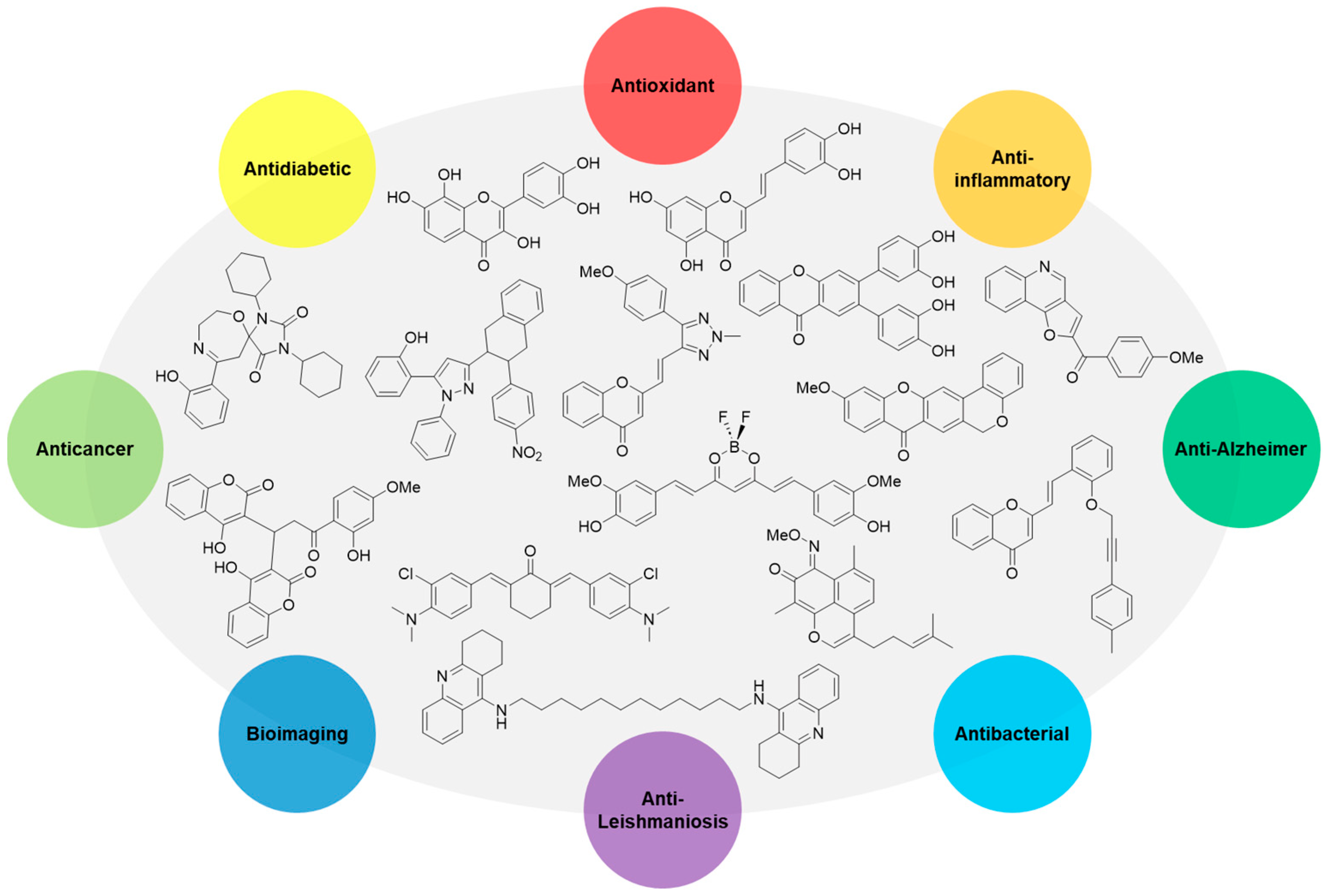
by
Joana L. C. Sousa
,
Hélio M. T. Albuquerque
and
Artur M. S. Silva
*
LAQV-REQUIMTE, Department of Chemistry, University of Aveiro, 3810-193 Aveiro, Portugal
*
Author to whom correspondence should be addressed.
These authors contributed equally to this work.
Pharmaceuticals 2023, 16(12), 1668; https://doi.org/10.3390/ph16121668
Submission received: 19 October 2023 / Revised: 18 November 2023 / Accepted: 27 November 2023 / Published: 30 November 2023
(This article belongs to the Special Issue State of the Art of Medicinal Chemistry in Portugal)

Abstract

:
Artur Silva’s research group has a long history in the field of medicinal chemistry. The development of new synthetic methods for oxygen (mostly polyphenols, e.g., 2- and 3-styrylchromones, xanthones, flavones) and nitrogen (e.g., pyrazoles, triazoles, acridones, 4-quinolones) heterocyclic compounds in order to be assessed as antioxidant, anti-inflammatory, antidiabetic, and anticancer agents has been the main core work of our research interests. Additionally, the synthesis of steroid-type compounds as anti-Alzheimer drugs as well as of several chromophores as important dyes for cellular imaging broadened our research scope. In this review article, we intend to provide an enlightened appraisal of all the bioactive compounds and their biological properties that were synthesized and studied by our research group in the last two decades.

1. Introduction

Medicinal chemistry is a chemistry-based discipline, which has the primary objective of designing and discovering new compounds suitable for use as drugs [1]. However, this simplistic definition ignores the larger historical context of medicinal chemistry and its ongoing evolution. In 1998, IUPAC proposed a more holistic definition for medicinal chemistry—“Medicinal Chemistry is a chemistry-based discipline, also involving aspects of biological, medical, and pharmaceutical sciences. It is concerned with the invention, discovery, design, identification and preparation of biologically active compounds, the study of their metabolism, the interpretation of their mode of action at the molecular level and the construction of structure-activity relationships (SARs)”. Medicinal chemistry is a highly interdisciplinary science combining several fields such as organic chemistry, biochemistry, computational chemistry, pharmacology, molecular biology, statistics, and physical chemistry, among others. Medicinal chemists play a crucial role in the drug discovery process, especially in its early stages (Figure 1). This requires a thorough understanding of modern organic chemistry, essential to prepare new chemical entities [2]. The role of organic chemistry is not limited to the early stages of drug development. In recent years, with the advent of C-H activation, medicinal chemists were driven to use late-stage functionalization (LSF) strategies, which enable a rapid exploration of SARs, the generation of oxidized metabolites, the blocking of metabolic hot spots, and the preparation of biological probes (Figure 1). LSF strategies created the possibility of exploring the chemical space more effectively than the conventional synthetic approaches [3,4].
Heterocyclic compounds are defined as cyclic structures with at least one heteroatom. Among all the possible heteroatoms, nitrogen, oxygen, and sulfur are most prevalent ones in heterocyclic compounds. In the medicinal chemistry field, the role of heterocycles is quite indisputable as they are often present not only as fragments in a number of active pharmaceutical ingredients (APIs) but also as excipients. The importance of heterocycles in medicinal chemistry is often related to their capacity to modify physicochemical properties (solubility, lipophilicity, polarity, and hydrogen bonding capacity of biologically active agents), essential for the optimization of the absorption, distribution, metabolism, and excretion–toxicity (ADMET) properties of drugs and drug candidates.
It is estimated that up to 85% of biologically active compounds have in their structure a heterocyclic fragment. In terms of approved drugs, 84% have at least one nitrogen atom in their scaffolds, and about 60% have some sort of nitrogen heterocycle such as piperidine, pyridine, pyrrolidine, thiazole, imidazole, indole, and tetrazole [5,6]. Oxygen heterocycles are the second most prevalent type of heterocycle in approved drugs, with pyranoses, furanoses, macrolactones, morpholines, and dioxolanes being placed in the top five examples [7].
In this personal account, around twenty years of development of bioactive compounds will be reviewed, focusing not only on traditional biological activities like antioxidant, anti-inflammatory, antidiabetic, and anticancer properties, but also more recent interests such as Alzheimer’s disease-targeting compounds and bioimaging tools (Figure 2).

2. Antioxidant Activity

2.1. ROS/RNS Scavenging Activity

The main contributing factor to the oxidative stress-related pathologies, such as inflammation, atherosclerosis, cancer, and aging, is the overproduction of reactive oxygen species (ROS) and reactive nitrogen species (RNS). The maintenance of the ROS/RNS balance is carried out by endogenous enzymatic antioxidant defenses such as superoxide dismutase, glutathione peroxidase, thioredoxin reductase, and catalase, and by non-enzymatic compounds such as glutathione, uric acid, and coenzyme Q. If the internal production of antioxidants is not enough to neutralize all the ROS/RNS produced, a series of exogenous non-enzymatic antioxidants can be provided from the human diet that includes carotenoids, phenolic compounds, and flavonoids, among others.
Based on the demand for new antioxidant agents, the design and synthesis of more effective scavengers of ROS and RNS was the core of our research for many years [8,9,10,11,12,13]. In this context, we synthesized novel polyhydroxylated 2-[styryl or (4-arylbutadienyl)]chromones 14, xanthones 5,6, flavones 7,8, chalcones 9, and flavonols 10 (Figure 3), which were assessed for their in vitro scavenging capability against the most physiologically relevant ROS (superoxide radical (O2•−), hydrogen peroxide (H2O2), hypochlorous acid (HOCl), singlet oxygen (1O2), and peroxyl radical (ROO)) and RNS (nitric oxide (NO) and peroxynitrite anion (ONOO)).
In the Figure 3, the active derivatives of each family to scavenge the referred to ROS and RNS are summarized in order to show which type of structures are more suitable for scavenging each reactive species. Thus, among the active compounds 1,4,5 to scavenge H2O2, the 2-styrylchromone (2-SC) derivatives 1a,b, presenting a catechol moiety at the B-ring in conjugation with the Cα=Cβ double bond of the styryl moiety, and free OH groups at C-5 and C-7, emerged as lead compounds with IC50 values of 48.9–50.3 µM (in comparison with ascorbic acid used as positive control, IC50 = 625.5 ± 49.6 µM) [13]. On the other hand, the 2-SC derivative 2, possessing two catechol units at the A- and B-rings, was more effective for scavenging 1O2 (IC50 = 4.69 ± 0.64 µM) than ascorbic acid, used as a positive control (IC50 = 10.2 ± 1.5 µM) [12], and the flavones 7. In addition, the xanthone 6, bearing two catechol units at C-2 and C-3, is a potent scavenger of O2•− (IC50 = 10.4 ± 0.8 µM) vs. tiron, used as a positive control (IC50 = 273 ± 32 µM), and compounds 2,3,8,10. The xanthone 6 was also an effective scavenger of ONOO, showing an IC50 value of 0.17 ± 0.01 µM vs. ebselen, used as a positive control (IC50 = 0.50 ± 0.03 µM) [11], and compounds 35,7,8. Regarding the scavenging activity against HOCl, the chalcones 9 were the most active scavengers, specifically, the derivative bearing a catechol moiety at the B-ring and free OH groups at C-2′, C-4′, and C-6′ was the most potent compound (IC50 = 1.0 ± 0.1 µM) in comparison with ascorbic acid, used as a positive control (IC50 = 11 ± 1 µM) [8]. Finally, to scavenge NO, the flavonols 10a,b were definitely the most potent compounds, as they are active in the nanomolar range (IC50 = 55–58 nM) compared to quercetin, used as a positive control (IC50 = 1.3 ± 0.1 µM) [9], and to compounds 13,6.

2.2. Free Radicals Scavenging Activity

Using betulinic acid (BA), which is a lupane-type pentacyclic triterpenoid commonly isolated from the bark of birch trees, as a raw material to produce amphiphilic antioxidants, we synthesized the polyhydroxylated 19,28-epoxyoleanane-3,28-dione-type 11ad and 12, and methyl betulonate-type 13 compounds (Figure 4) [14]. We found that the derivative 12, bearing a catechol moiety and an extended π-conjugated carbonyl system, emerged as a lead compound, since it was revealed to be the most efficient scavenger of the 2,2′-azino-bis(3-ethylbenzothiazoline-6-sulfonic acid) radical cation (ABTS•+) (IC50 = 15.9 ± 0.2 µM). In fact, it was more active than α-tocopherol (IC50 = 19.2 ± 0.1 µM), used as a positive control and the pristine BA (no activity was found up to the highest tested concentration—800 µM).
The antioxidant activity of xanthenediones 14af (Figure 5) was evaluated by their ability to scavenge the 2,2-diphenyl-1-picrylhydrazyl free radical (DPPH) and to reduce Fe(III) [15]. The compounds were tested at different concentrations and, naturally, the reducing ability increased with concentration. Among the tested compounds, the derivative 14e reduced considerably the ferric ion, being more efficient than 2,6-bis(1,1-dimethylethyl)-4-methylphenol (BHT) and similar to quercetin, used as positive controls. This result confirmed that a catechol moiety in a molecule is important for the reducing power activity. In addition, this derivative was also the most potent scavenger of DPPH, presenting an EC50 value of 3.79 ± 0.06 µM. Furthermore, these compounds were also evaluated for their anti-acetylcholinesterase activity, which will be discussed later in Section 6.
The antioxidant activity of the synthesized 10-(4-hydroxy-6-methyl-2-oxo-2H-pyran-3-yl)-3-methyl-1H,10H-pyrano [4,3-b]chromen-1-ones 15ak and 3,3′-[(2-hydroxy-3,4-dimethoxyphenyl)methylene]bis(4-hydroxy-6-methyl-2H-pyran-2-one) 16 (Figure 6) was evaluated using the DPPH scavenging assay [16]. Compound 15j, bearing a catechol moiety at the A-ring, was the most active compound, presenting an IC50 value of 0.364 mg/mL.

2.3. Enzymatic Inhibition Activity

Xanthine oxidase (XO) is a highly versatile enzyme with a broad range of activities in reducing substrates. As a consequence, XO is considered an important biological source of ROS, inducing oxidative stress, and being involved in many pathological processes. Therefore, the development of XO inhibitors is expected to be therapeutically useful for the treatment of the aforementioned pathological states.
In this context, a series of 2-SCs developed in our group were evaluated as potential XO inhibitors [17]. From a library of ten 2-SCs, the results showed a concentration-dependent and non-competitive inhibition effect, with derivative 1a (Figure 3) being the most potent compound (IC50 = 0.55 ± 0.03 μM), 10-fold more effective than the positive control, allopurinol (IC50 = 5.43 ± 0.80 μM).

3. Anti-Inflammatory Activity

3.1. Flavones

Methylated flavones 17ad (Figure 7) have revealed the ability to modulate the neutrophils’ oxidative burst [18]. Thus, the study of the modulatory effect of 17ad on the stimulation of neutrophils by the phorbol-12-myristate-13-acetate (PMA) involved fluorescent [using amplex red and aminophenylfluorescein (APF) as probes] and chemiluminescent (using luminol and lucigenin as probes) techniques. The flavone 17b was the most potent derivative (IC50 = 0.4–8.6 µM) in inhibiting PMA-induced neutrophils’ oxidative burst assessed by both chemiluminescent and fluorescent techniques (Figure 7). These results suggest that the 3′-methoxy group in the B-ring is an important chemical feature that contributed to its higher activity in comparison to luteolin (IC50 = 0.9–9.5 µM).
These flavone derivatives were also evaluated as cyclooxygenase (COX) and lipoxygenase (LOX) inhibitors [19,20]. However, these compounds did not show any promising results when compared to other flavonoid-type compounds.
The novel chlorinated flavones 18ae (Figure 8) were investigated for their anti-inflammatory properties in comparison with the parent non-chlorinated flavonoids [21,22].
Firstly, their effect in neutrophils’ oxidative burst and lifespan was studied [21]. The obtained results demonstrate that chlorinated flavonoids were more efficient than their parent compounds in modulating neutrophils’ oxidative burst in PMA-activated neutrophils. Some of the tested flavonoids drive neutrophil apoptosis in a caspase 3-dependent fashion. The present data showed that 8-chloro-3′,4′,5,7-tetrahydroxyflavone (18c) constitutes an alternative anti-inflammatory therapy, due to the proven ability to suppress mechanisms engaged at the onset and progression of inflammation (Figure 8).
Then, the anti-inflammatory potential of these chlorinated flavones 18ae (Figure 8) were also tested in the activity of COX-1 and COX-2, and in the production of cytokines (interleukins (IL-6 and IL-1β) tumor necrosis factor (TNF)), and the chemokine IL-8, as well as in the production of reactive species, using human whole blood as a representative in vitro model [22]. None of the chlorinated flavones 18ae were able to inhibit COX at the highest tested concentration (100 μM). However, 6-chloro-3′,4′,5,7-tetrahydroxyflavone (18b) was able to reduce the production of reactive species, even in hyperthermic conditions, and also modulated the production of the cytokines IL-1β, IL-6, TNF, and the chemokine IL-8b (Figure 8).

3.2. Xanthones

With the demand for new dual-acting anti-inflammatory agents, a range of 2,3-diarylxanthones 1921 (Figure 9) were tested through their ability to interact in arachidonic acid metabolism [23]. The in vitro anti-inflammatory activity was evaluated through the inhibition of 5-LOX-catalyzed leukotriene B4 (LTB4) formation in human neutrophils and the inhibition of COX-1- and COX-2-catalyzed prostaglandin E2 (PGE2) formation in human whole blood.
The xanthone 19c with a 2-catechol group was the most active one (IC50~9 μM) in preventing LTB4 production in human neutrophils. The more effective arylxanthones in preventing COX-1-catalyzed PGE2 production presented IC50 values from 1 to 7 μM, exhibiting a structural feature with at least one non-substituted aryl group. All the studied arylxanthones were ineffective at preventing the formation of PGE2 catalyzed by COX-2, up to the maximum concentration of 100 μM. The ability of the tested 2,3-diarylxanthones to interact with both 5-LOX and COX-1 pathways constitutes an important step in the research of novel dual-acting anti-inflammatory drugs (Figure 9).

3.3. 2-Styrylchromones

The anti-inflammatory potential of 2-SCs was evaluated by studying their COX-1 and COX-2 inhibitory capacity as well as their effects on the LTB4 production using stimulated human polymorphonuclear leukocytes (PMNL) [24].
Some of the tested 2-SCs were able to inhibit both COX-1 activity and LTB4 production, which makes them dual inhibitors of the COX and 5-LOX pathways. The most effective compound in this study was derivative 1a (Figure 3), which has structural moieties with proven antioxidant activity (free OH groups at C-5 and C-7 and a 3′,4′-catechol-substituted B-ring in conjugation with the Cα=Cβ double bond of the 2-styryl moiety).
This type of compound may exhibit anti-inflammatory activity with a wider spectrum than that of classical non-steroidal anti-inflammatory drugs (NSAIDs) by inhibiting 5-LOX product-mediated inflammatory reactions, towards which NSAIDs are ineffective.

4. Antidiabetic Activity

Diabetes mellitus (DM) is one of the most significant public health concerns worldwide. According to the International Diabetes Federation, 10.5% of the adult population (20–79 years) has diabetes according to 2021 data, and will probably reach 783 million (1 in 8 adults) in 2045, an increase of 46% [25]. DM is a multifactorial metabolic disorder, characterized by chronic hyperglycemia and can be primarily classified as type 1 (T1DM, insulin-dependent DM) and type 2 (T2DM, non-insulin-dependent DM) [26]. T2DM is the most common form of DM, accounting for more than 90% of all diabetic patients, and results from the interaction between behavioral, environmental, and genetic risk factors [27,28].
Novel therapeutic druggable targets for the management of T2DM have been emerging, namely carbohydrate-hydrolyzing enzymes α-amylase and α-glucosidase, fructose-1,6-bisphosphatase (FBPase), protein tyrosine phosphatase 1B (PTP-1B), dipeptidyl peptidase-4 (DPP-4) and glycogen phosphorylase (GP). The discovery of new inhibitors of these targets represents an alternative for the currently used antidiabetic agents. In the next subsections, we will show the potential of synthetic flavonoid, 2-(styryl or 4-arylbutadienyl)chromone, xanthone, and pyrazole derivatives as antidiabetic agents through inhibiting some of the referred to targets.

4.1. Flavonoids

4.1.1. Flavonols

The flavonols 10ae were designed to possess catechol moieties at the A-ring and/or B-ring (Figure 10) [9], allowing the assessment of SAR studies involving these compounds as antidiabetic agents, namely, as DPP-4, PTP1B, FBPase, and α-glucosidase inhibitors.
The flavonols 10ae were found to be effective PTP1B inhibitors for the treatment of T2DM [26]. The most active derivatives were 10d (IC50 = 10 ± 1 µM), followed by 10c (IC50 = 16 ± 2 µM), showing that the presence of both -OBn and -OMe groups in the flavonol structure significantly increases their in vitro PTP1B inhibition (Figure 10). In comparison with the flavonol 10e, which possesses five -OH groups in their place, its ability to inhibit PTP1B was much lower (%inhibition = 36 ± 3% at the highest tested concentration of 200 μM).
The flavonols 10ae were studied concerning their ability to inhibit DPP-4 enzyme [27]. This study was based on in vitro fluorometric and colorimetric methods using the human isolated enzyme. The fluorometric method was revealed to be more sensitive and was applied in the evaluation of DPP-4 activity in an ex vivo assay, using human blood and plasma. The only effective flavonol based on the fluorometric method was 10b (IC50 = 73 ± 2 µM) (Figure 10), displaying no inhibitory activity in the ex vivo method, probably due to its high affinity for plasma proteins, notably, human serum albumin.
The same flavonols 10ae were also tested as FBPase inhibitors [28]. However, none of these derivatives performed as effective FBPase inhibitors, presenting a %inhibition <20% at the highest tested concentration of 200 µM.
The flavonols 10ae were screened for their in vitro inhibitory activity of α-glucosidase [29]. In this case, flavonol 10e was the most active derivative (Figure 10), presenting an IC50 (7.6 ± 0.4 µM) much lower than the one found for the most widely prescribed α-glucosidase inhibitor, acarbose (IC50 = 607 ± 56 µM). Due to this promising result, the flavonol 10e was further studied for its inhibitory activity against human sucrase-isomaltase, the α-glucosidase found in Caco-2/TC7 cells [30], being the most active compound among the tested flavonoids (Figure 10), presenting an IC50 value of 2.2 ± 0.2 μM and 2.5 ± 0.2 μM when using sucrose or maltose as substrates, respectively.

4.1.2. Chlorinated Flavones

The chlorinated flavones 18ae (Figure 8) were explored for their inhibitory effect against α-glucosidase for the first time [29]. All of them were active, except for 18b, presenting IC50 values of 21 ± 2 to 55 ± 2 µM, being more potent than acarbose, used as a positive control (IC50 607 ± 56 µM). Among this group of compounds, the 3-chlorinated derivative 18a was the most active compound (Figure 11), presenting an IC50 (21 ± 2 µM) similar to the one obtained for quercetin (15 ± 3 µM), showing that the presence of a 3-OH group or a 3-Cl atom at the C-ring is almost indifferent for the inhibitory effect.
This group of chlorinated flavones 18ae was also addressed for their inhibitory pancreatic α-amylase activity [31]. The 3-chlorinated derivative 18a was once again the most active compound (Figure 11), presenting an IC50 value of 44 ± 3 µM. In this case, the presence of a 3-Cl atom at the C-ring seems to be crucial for its α-amylase inhibition, since, in comparison with quercetin, which presents a 3-OH group, its α-amylase inhibition significantly decreased (IC50 = 138 ± 5 µM). Also, the absence of the 3-Cl atom led to a decrease in the inhibitory activity of flavones, as it is possible to verify through comparing 18a with luteolin (IC50 = 78 ± 3 µM).
The compounds 18ae also did not inhibit human liver FBPase up to the maximum tested concentration of 200 μM [28], despite their promising activities against α-amylase and α-glucosidase. These results highlight the importance of finding a detailed potential antidiabetic mechanism of action of each flavonoid and, in this way, the possible finding of a specific target-directed therapeutic.
The in vitro inhibitory capacity of the chlorinated flavones 18ae against DPP-4 was also evaluated [27]. The most effective compounds were 18b and 18e (Figure 11), showing similar activities (IC50 = 170 ± 10 and 171 ± 8 µM, respectively). Structurally, the presence of an additional 8-Cl substituent in the derivative 18e does not affect the inhibitory effect of this type of flavones.

4.2. 2-(Styryl or 4-Arylbutadienyl)chromones

Several chromone derivatives bearing a styryl or a 4-arylbutadienyl group at C-2 were evaluated for their in vitro ability to inhibit GP [32]. The SAR study indicates that the presence and the position of free hydroxy groups at A and B rings is determinant for the inhibitory activity of these two families. Among the 2-SCs, compound 1a, bearing hydroxy groups at C-5 and C-7 of the A-ring and a catechol moiety on the B-ring (Figure 12), was the most active compound, with an IC50 value of 31.7 ± 2.4 µM. Moreover, the 2-(4-arylbutadienyl)chromone 4a bearing a hydroxy group at C-7 of the A-ring and a catechol moiety on the B ring (Figure 12), and compound 4b with a hydroxy group at C-5 and a methoxy group at C-7 of the A-ring, and a catechol moiety on the B-ring (Figure 12), were the most active derivatives, with similar IC50 values of 16.7 ± 1.5 µM and 15.9 ± 1.1 µM, respectively.

4.3. Xanthones

A series of nine hydroxylated xanthones 1921 (Figure 9) were evaluated as dual-target antidiabetic agents, acting in the inhibition of both α-amylase and α-glucosidase enzymes [33]. The results showed that the xanthones 1921 exhibited a stronger inhibition of α-glucosidase rather than of α-amylase, since all of them (except for the derivative 19a) present IC50 values (8.6–27.4 µM) lower than acarbose, used as a positive control (IC50 = 515 ± 19 µM). Particularly, the derivatives 20c (IC50 = 27 ± 1 µM), 21a (IC50 = 23 ± 1 µM), and 21b (IC50 = 27 ± 1 µM), bearing one catechol moiety, were the most active inhibitors of α-amylase, while the xanthones 20c (IC50 = 8.9 ± 0.3 µM), 21b (IC50 = 8.6 ± 0.3 µM), and 21c (IC50 = 9.2 ± 0.4 µM) were the most active against α-glucosidase activity, with IC50 values lower than 10 μM. The polyhydroxylated xanthones 20c and 21b can be considered as lead dual-target inhibitors of both the α-amylase and α-glucosidase enzymes (Figure 13).

4.4. Pyrazoles

A library of twenty-two pyrazoles was evaluated for the first time as human PTP1B inhibitors [34]. The pyrazole 22, bearing a phenyl group at N-1, a 2-(4-nitrophenyl)tetralin at C-3 and a phenol at C-5 (Figure 14); pyrazole 23, holding a phenol at C-3 and a 2-(4-methoxyphenyl)naphthalene at C-5 (Figure 14); and pyrazole 24, presenting a phenyl group at N-1, a 3-(4-methoxyphenyl)naphthalen-2-yl at C-3 and a phenol at C-5 (Figure 14), were the most active compounds, with IC50 values of 27–40 µM, respectively. These findings suggest that the presence of additional benzene rings as functional groups in the pyrazole moiety increases their ability to inhibit PTP1B. The most active compounds showed selectivity over the homologous T-cell protein tyrosine phosphatase (TCPTP).
The inhibitory activity of a group of 4- and 5-styrylpyrazoles 2539 (Figure 15) was evaluated against GP [32]. However, no relevant inhibitory activities were observed up to the highest tested concentration of 50 µM.

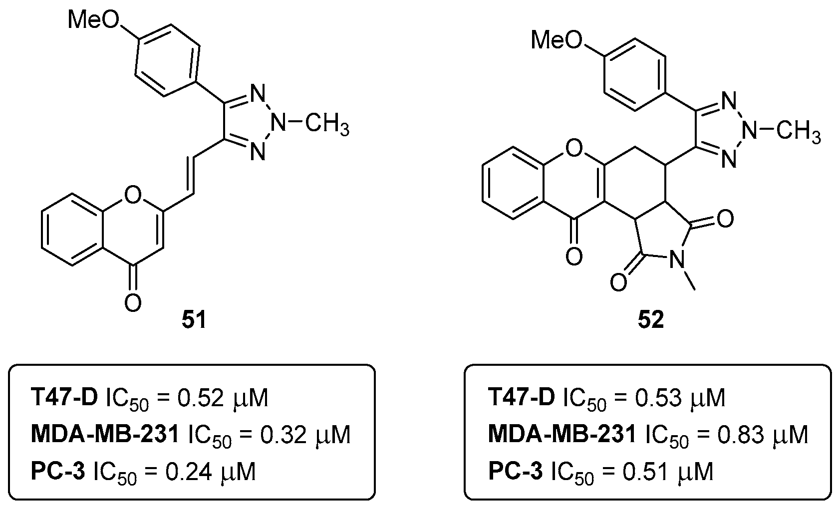
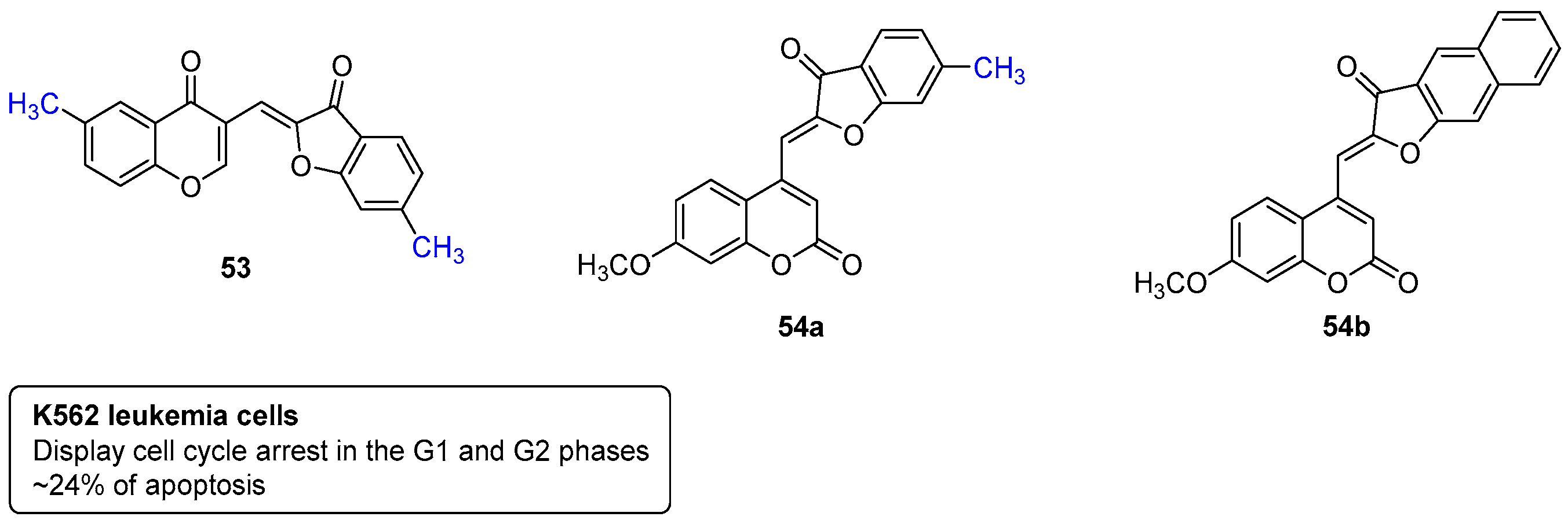
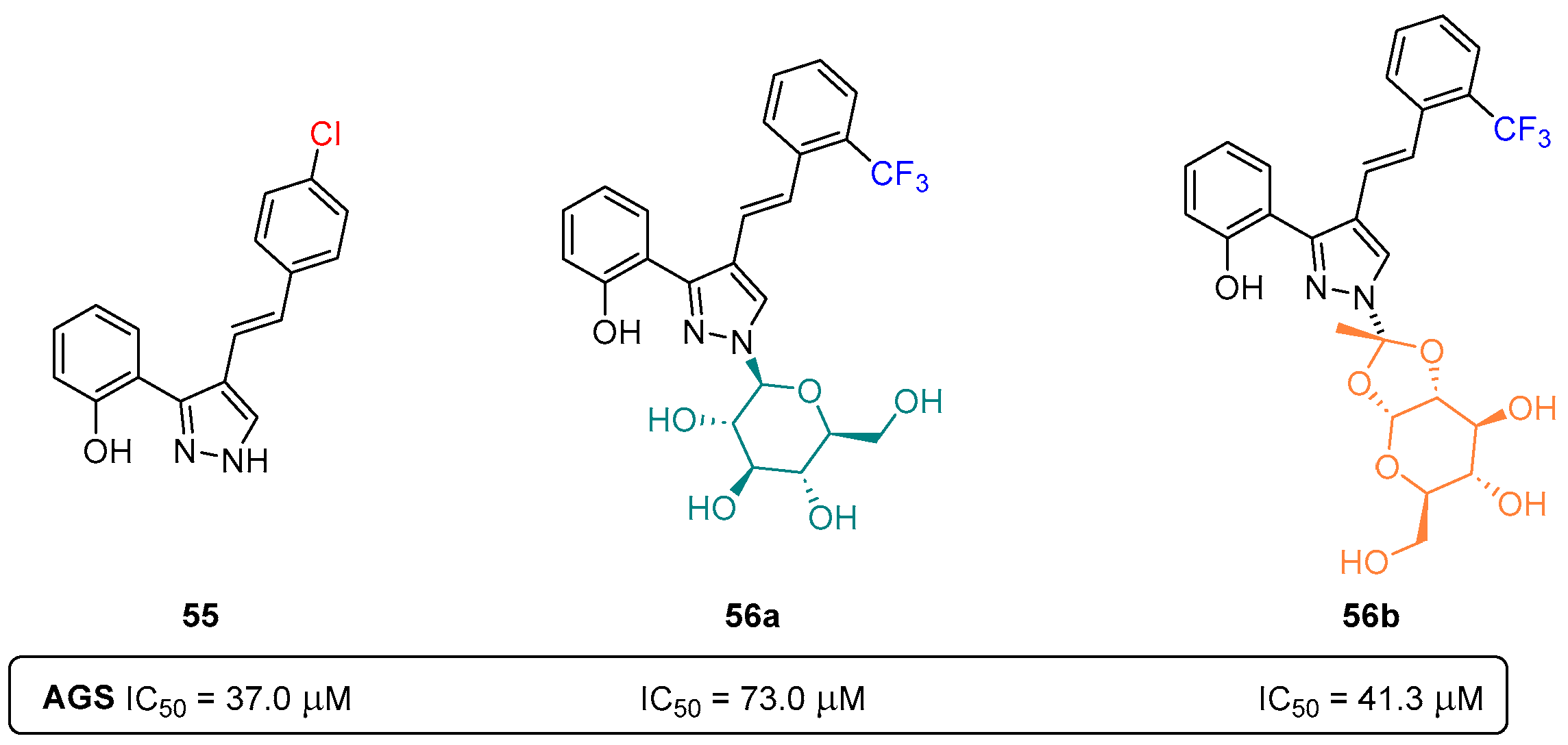
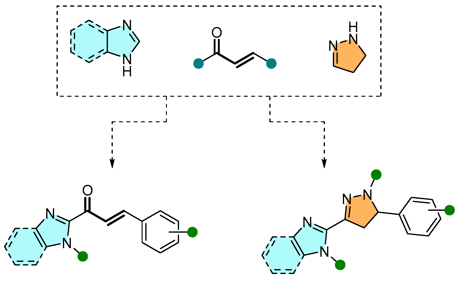
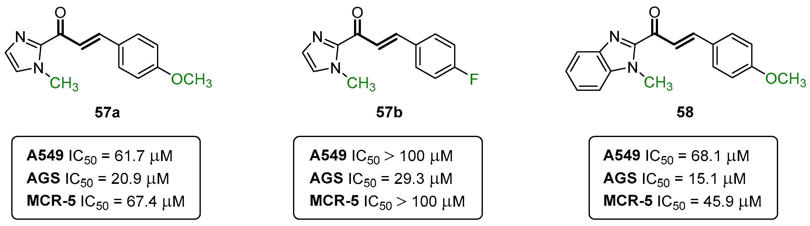
5. Anticancer Activity

Cancer is one of the main causes of death worldwide, accounting for nearly 10 million deaths in 2020 [35]. Through the decades, the cure rate of patients has increased due to improved early diagnosis and more personalized treatments. Among them, radiation therapy, surgery, immunotherapy, endocrine therapy, gene therapy, and chemotherapy should be highlighted, with the latter being the most widely used either as monotherapy or in combination with other treatments. Despite the overall success of most types of chemotherapies, resistance in most aggressive cancers has increased, which, together with the adverse effects of chemotherapy, has led to the need for the development of new anticancer agents.
One of the main focuses of our research group’s efforts in medicinal chemistry is in the early-stage drug discovery of anticancer small molecules. Two seminal works reported by our research group are focused on synthetic bis-coumarin compounds, more specifically, the 40a,b, and their effects in lung cancer cells with KRAS mutations and myeloid leukemia (CML) cell models (Figure 16).
After a thorough investigation, 40a was found to strongly inhibit the proliferation of non-small cell lung cancer cells with KRAS mutations through the reduction in aldehyde dehydrogenase expression and abrogated spheroid formation. Subsequent mechanistic investigations showed that 40a triggers cellular stress, including metabolic catastrophe, mitochondrial stress, and ER/Golgi stress preceded by STAT3 inactivation, inhibiting the STAT3 transactivation and expression of its target genes linked to cell proliferation. It was also demonstrated that 40a activates sensitization against BH3 mimetics in NSCLC, leading to immunogenic cancer cell death mechanisms. Overall, this compelling evidence supports the potential of bis-coumarin templates as novel candidates for future drug investigation in lung cancer [36].
Driven by the stimulating results of 40a triggering ER stress in lung cancer, the potential of its chlorinated analogue 40b (Figure 16) was further investigated against various chronic myeloid leukemia (CML) cell models. Once more, 40b was proved to trigger ER stress leading to canonical, caspase-dependent apoptosis and the release of danger-associated molecular patterns. The compound 40b was also capable of inhibiting tumor necrosis factor α-induced activation of nuclear factor-κB, producing synergistic effects upon combination with imatinib, inhibiting the colony formation in vitro and Bcr-Abl+ patient blast xenograft growth in zebrafish [37]. An additional synergistic capacity of 40b with omacetaxine was observed in imatinib-resistant KBM-5 R cells, resulting in the inhibition of the expression of Mcl-1 and triggering apoptosis [37].
Both these seminal publications on the bis-coumarin compounds 40a,b had their starting point many years ago, back into 2014. One of the first studies involving our own synthetic bis-coumarins described their effects on leukemic cell line proliferation and NF-kB regulation [38]. The investigation carried out, in collaboration with Professor Marc Diederich (College of Pharmacy, Seoul National University), started with the synthesis of the template bis-coumarin 40c (Figure 17). The anti-proliferative effects of compound 40c on human K-562 (chronic myeloid leukemia) and JURKAT (acute T-cell leukemia) cell lines were assessed, and the results revealed the inhibition of TNFa-induced NF-kB activation in K-562 (IC50 = 17.5 μM) and JURKAT (IC50 = 19.0 μM) cell lines. Of note, the compound 40c did not affect the viability of peripheral blood mononuclear cells (PBMCs) from healthy donors, even at concentrations above 100 μM [38]. The bis-coumarin molecule 40c was deconstructed into fragments as substructures of the bis-coumarin-type compound, 20-hydroxyphenylpropione and 4-hydroxycoumarin, which were completely inactive [38].
Cdc25 phosphatases are key enzymes regulating the cell cycle, being a valuable target for cancer treatment. To assess the inhibitory potential of small-molecules towards Cdc25 phosphatases, human glutathione-S-transferase (GST)-Cdc25 recombinant enzymes are usually the ideal choice. As demonstrated in previous reports, coumarin derivatives enclose a great potential in the development of alternative cancer therapies. As such, a library of coumarin-based polycycles, like the ones in Figure 18, was screened for their Cdc25 phosphatase-inhibition activity. The screening showed that the coumarins 41a,b (Figure 18) were the most potent phosphatase inhibitors with low micromolar IC50 values. More specifically, the benzylated compound 41a presented IC50 (Cdc25A) = 13.2 μM, IC50 (Cdc25B) = 46.1 μM, and IC50 (Cdc25C) = 9.0 μM, while the derivative 41b showed IC50 (Cdc25A) = 5.8 μM, IC50 (Cdc25B) = 14.4 μM, and IC50 (Cdc25C) = 2.3 μM [39].
Covalent inhibitors are being increasingly recognized as an important component in drug discovery and therapeutics. Generally, covalent inhibitors are designed to form a covalent bond with a specific molecular target. The covalent bond can be either reversible or irreversible, depending on the chosen warhead. A number of different warheads have been exploited to target specific amino acid residues, including, among others, cysteine, serine, threonine, tyrosine, and lysine. One of the most commonly used warheads is the α,β-unsaturated carbonyl moiety (Figure 19), which can be found in several covalent inhibitors in clinical trials [40,41,42]. Some variations in these Michael acceptor moieties have also been reported, such as the (2-hydroxyphenyl)-3-oxoprop-1-enyl (HOPO) and the cinnamoyl (CINA) (Figure 19). In this sense, the chromanones 42a,b, as well as the 2-SC 43 were specifically designed to have the HOPO and CINA pharmacophoric moieties (Figure 20).
The chromanones 42a,b revealed a C5 value of 2.5 μM [C5 value, concentration causing a five-fold induction of luciferase activity (= Nrf2 activity)], being more potent than the positive controls xanthohumol (XN) C5 = 7.8 μM and sulforaphane (SFN) C5 = 4.8 μM [43]. The 2-SC 43 showed a slightly higher C5 value of 2.9 μM [43]. In an additional assay, the chromanone 42b and 2-SC 43 were also shown to be the more potent compounds inhibiting leukemia K562 cell proliferation, with IC50 values of 7.9 ± 2.6 μM and 4.5 ± 1.9 μM, respectively, with no appreciable effect on normal peripheral blood mononuclear cells [43]. Despite the encouraging results of the compounds 42a,b and 43, it still unknown if the mechanism of action is indeed covalent or non-covalent, and, therefore, further mechanistic studies are still required.
It is commonly accepted that the small-molecule inhibition of nuclear factor-ĸB (NF-ĸB) is quite an interesting strategy to improve cancer chemotherapy. In this field of research, our group reported collaborative work in the anti-proliferative, cytotoxic, and NF-ĸB inhibitory properties of spiro(lactone-cyclohexanone) synthetic compounds in human leukemia [44]. The spiro(lactone-cyclohexanone) derivatives 44 and 45 (Figure 21) were revealed as the most effective in the inhibition of the proliferation of the human leukemia cell lines K562 and U937, with IC50 values of 74.02 ± 4.10 μM (K562) and 51.6 ± 4.2 μM (U937) for the derivative 44, and 58.6 ± 4.2 μM (K562) and 43.7 ± 1.5 μM (U937) for compound 45 [44]. Additionally, the spiro(lactone-cyclohexanone) 44 was also capable of reducing TNFα-stimulated NF-ĸB activation, with an IC50 of 15.9 ± 4.0 μM [44].
Within the spirocyclic chemotype compounds, the spiro-heterocyclic surrogates have also been showing promising results in the chemotherapy of various cancer types. Back in 2016, a series of oxygen and nitrogen spiro-bisheterocycles were studied for their effects on the in vitro proliferation and apoptosis of human breast cancer cell lines (MCF-7 and MDA-MB-231) [45]. From the screened library, the three compounds 4648 stood out as the most effective ones (Figure 22).
Compounds 46 and 48 showed a dose-dependent decreasing effect in cell proliferation and induced apoptosis in the MCF-7 and MDA-MB-231 cell lines, while the derivative 47 was only active towards MDA-MB-231 [45]. In this study, it was also reported that the compounds 4648 cause apoptosis by targeting p53–MDM2 interaction. It was also investigated and concluded that the spiro-heterocyclic compounds 4648 promote apoptosis via the p53-independent pathway(s) [45].
More recently, other collaborative projects have led to the expansion of the chemical space of anticancer agents, namely, towards 1,2,3-triazole-xanthenediones and triazole-benzimidazole-chalcones (Figure 23) [46,47]. The reported library of 1,2,3-triazole-xanthenediones was screened in breast cancer (T47-D and MDA-MB-231) and prostate cancer (PC3) cell lines. The compound 49 (Figure 23) was the most generally effective compound, with IC50 (T47-D) = 15.50 ± 1.59 μM, IC50 (MDA-MB-231) = 20.88 ± 0.20 μM, and IC50 (PC3) = 10.20 ± 0.22 μM [46].
The library of triazole-benzimidazole-chalcones was also screened in the same types of cancer cells. Interestingly, the chlorinated derivative 50 (Figure 23) was the best-performing compound with IC50 (T47-D) = 6.23 ± 1,03 μM, IC50 (MDA-MB-231) = 5.89 ± 1.35 μM and IC50 (PC3) = 10.70 ± 1.25 μM [47].
The common structural motif among the compounds 49 and 50 is the 1,2,3-triazole. Actually, the 1,2,3-triazole ring is a major pharmacophore system among nitrogen-containing heterocycles, displaying relevant roles either as key pharmacophores or linkers [48].
In a recently published study (2023), eight in-house chromone-based compounds were screened for their effects in breast (T-47D and MDA-MB-231) and prostate (PC3) cancer cell lines, and in non-cancerous human mammary epithelial cells (HuMECs). Interestingly, the compounds with better performance in cancer cells were those with the 1,2,3-triazole moiety, 51 and 52 (Figure 24) [49].
Both compounds presented very good potencies in three of the tested cell lines, i.e., 51, IC50 (T-47D) = 0.52 μM, IC50 (MDA-MB-231) = 0.32 μM, and IC50 (PC3) = 0.24 μM; 52, IC50 (T-47D) = 0.53 μM; IC50 (MDA-MB-231) = 0.83 μM and IC50 (PC3) = 0.51 μM [49]. In breast (MDA-MB-231) and prostate (PC3) cell lines, the compounds 51 and 52 were actually more potent than the positive control (doxorubicin IC50 1.51 μM for MDA-MB-231 and IC50 = 0.73 μM for PC3). Notably, the IC50 of the compounds 51 and 52 was 24–388 times lower in non-cancerous HuMECs when compared to the IC50 of doxorubicin (0.57 μM) [49]. The compounds 51 and 52 have different mechanisms of action; while the chromone-1,2,3-triazole 51 induces G2/M arrest in T-47D, MDA-MB-231, and PC3 cells, compound 52 had no effect on the cell cycle [49]. In terms of the structure–activity relationships (SARs), the main conclusion is that the installation of a tetrahydroisoindole-1,3-dione moiety (Figure 24) is irrelevant for potency in all the tested cancer cell lines, but it is important in lowering the cytotoxic effect in HuMECs (IC50 = 221.35 μM) [49]. Of note, the hybrid compounds 51 and 52 are the most potent anticancer agents in the more than twenty years of medicinal chemistry research in our lab, and, therefore, the most promising drug candidates.
Years ago, chromones were also combined with additional frameworks such as aurones (2-benzylidene-benzofuran-3(2H)-ones) (Figure 25). In the same project, the aurones were further combined with coumarins (Figure 25), and both libraries of compounds were screened for their anti-cancer activities in K562 human leukemia cells [50]. Most of the screened compounds were capable of blocking the K562 cell cycle in the G1, S, or G2 phase, with the compounds 53 and 54a,b also being capable of inducing a high apoptosis rate (~24%) [50].
Another important class of azoles are the pyrazoles, five-membered heterocyclic aromatic rings with three carbons and two adjacent nitrogen atoms. Charting the long history of our research group in the chemistry of pyrazoles, in 2019, a series of styrylpyrazole-glucosides (Figure 26) was reported [51]. This new class of compounds, as well as their non-glycosylated precursors, were further evaluated for their cytotoxic potential against human gastric adenocarcinoma AGS cells and healthy MRC-5 lung fibroblasts.
The preliminary cytotoxic evaluation human gastric adenocarcinoma AGS cells revealed the non-glycosylated pyrazole 55 (Figure 26) as the most potent compound among the tested analogues (IC50 = 37.0 μM), while the glycosylated compounds 56a,b were less potent (56a IC50 = 73.0 μM; 56b IC50 = 41.4 μM). This suggests the more significant role of Cl-substitution over either the CF3-substitution or the glycosyl moiety (Figure 26). As for the non-cancer cell line (MRC-5 lung fibroblasts), the most potent pyrazole 55 also displays a certain degree of toxicity (IC50 = 45.0 μM), being only slightly more active towards the tumor cell line, AGS [51].
Within the large family of azoles, the 2-pyrazolines and (benz)imidazoles also show important biological relevance. In a recent collaborative effort, our research group reported the design of chalcone-type and 2-pyrazoline derivatives bearing an (benz)imidazole moiety (Figure 27), as well as their effects on human lung (A549) and stomach (AGS) cancer cell lines, being additionally evaluated in the non-cancer human lung fibroblast (MRC-5) cell line [52].
An appreciable library of fifteen compounds covering several substitution patterns were synthesized and screened. While 2-pyrazoline derivatives were devoid of toxicity in all cell lines used, the chalcones bearing the (benz)imidazole ring in the β position of the carbonyl group 57a,b and 58 (IC50 = 15.1–29.3 μM) were found to be toxic toward the AGS cell line (Figure 28), while only compounds 57a and 58 were toxic for A549 (IC50 = 61.7–68.1 μM). Importantly, these compounds were considerably less toxic toward non-cancer cells. Further mechanistic investigations on compounds 57a,b and 58 revealed the trigger loss of cell viability and mitochondrial membrane potential, while eliciting morphological traits compatible with refractory cancer disease (RCD). All three compounds were less toxic when incubated in the presence of a pan-caspase inhibitor and compounds 57b and 58 were shown to increase the activity of caspase-3 [52].
Another important class of anti-cancer agents are the benzophenones. Several examples of both natural and synthetic analogues can be found in the literature. In 2019, our research group reported additional examples of anticancer aryl-benzophenones targeting breast and prostate cancer cell lines [53]. The most prominent compound was the benzophenone 59 (Figure 29), showing IC50 values of 26.49 ± 0.95, 12.09 ± 1.07, and 23.32 ± 0.05 μM, for the MDA-MB-231, T47-D, and PC-3 cell lines, respectively [53].
Of note, the benzophenone 59 showed induced cell cycle retardation only in prostate cancer cells, suggesting cell type-dependent effects that may be related to the compound acting on molecular targets which are differentially expressed in breast and prostate cancer cells. Although, more detailed molecular studies are required to provide clarifications on the use of benzophenone 59 as a pharmacophore and to understand its specific action on mitochondrial membrane, protein, and glycoprotein interactions [53].
In 2014, one of the most interesting research projects from our group in medicinal chemistry targeting cancer was published. In this work, the synthesis of benzo[b]acridin-12(7H)-ones with tethered carboranyl moieties was described (Figure 30), as well as their potential as boron neutron capture therapy (BNCT) agents in cancer treatment [54].
Remarkably, compound 60 (Figure 30) showed no cytotoxicity in U87 human glioma cells, a clinically relevant tumor cell line for BNCT, in concentrations ranging 5–200 μM. This is an important parameter in BNCT so that boron concentrations within tumors can be maximized. The subsequent evaluation of benzo[b]acridin-12(7H)-one 60 as a new BNCT agent demonstrated that it enters the cells and deposits an adequate amount of B atoms (2.8 × 10¹⁰ 10B atoms per cell) superior to the recommended concentration of 108–109 10B atoms, demonstrating considerably high activity in the U87 cells upon neutron irradiation [54].

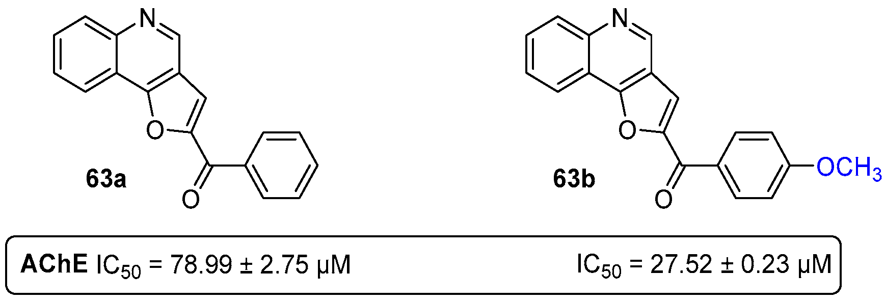
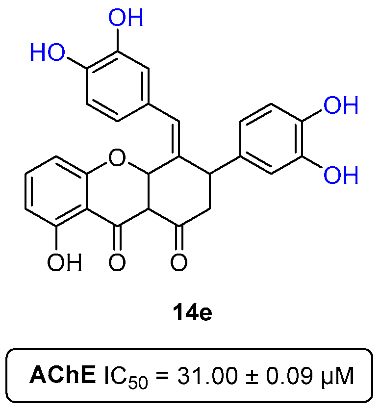
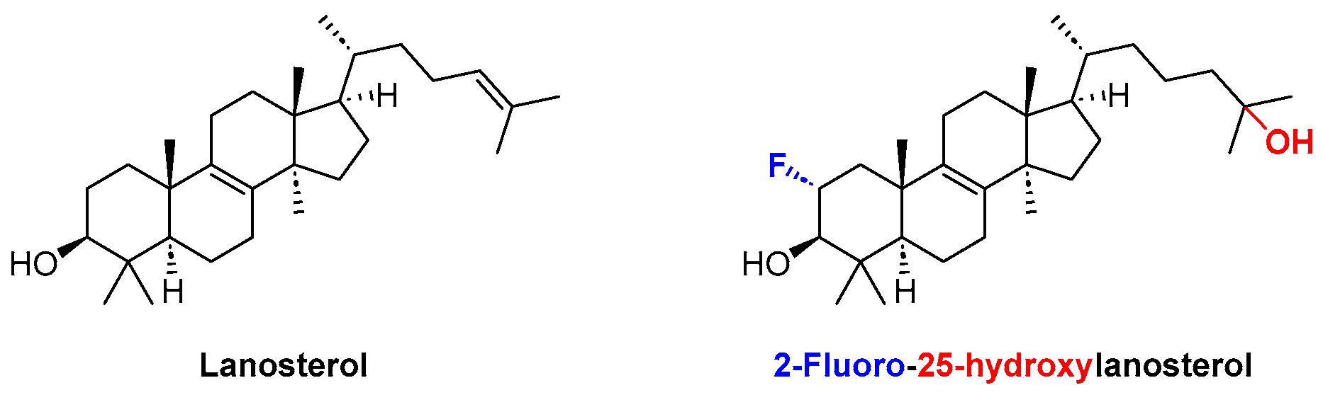
6. Anti-Alzheimer Activity

Alzheimer’s disease (AD) is the most prevalent neurodegenerative disorder, quickly becoming a major healthcare and economic problem to deal with in modern societies. As for the majority of neurodegenerative diseases, AD is age-related and mainly affects people over 65 years old. In 2022, an estimated 32 million people worldwide suffered with AD, and this figure is expected to rise to 131.5 million in 2050 [55].
The multifactorial nature of AD has led to the discovery of several targets or target combinations in order to find effective treatments. Among many theories behind the disease pointing to glutamate excitotoxicity [56], oxidative stress [57], and biometal dyshomeostasis [58] as causative triggers of the underlying neurodegeneration (reviewed elsewhere [59]), the amyloid-β (Aβ) plaques and tau fibrils are hypothesized as the major contributing factors [60,61]. The mainstream concept though is clearly the amyloid cascade hypothesis (formulated by Hardy and Higgins), which explains that the aggregation of Aβ species causes neural death and eventually progression to AD [62,63].
The “cholinergic hypothesis” [64], which was the first and most studied approach describing AD pathophysiology, is based on compelling evidence mainly regarding three aspects: (i) the nucleus basalis of Meynert in the basal forebrain undergoes severe neurodegeneration in AD; (ii) the presynaptic cholinergic markers in the cerebral cortex are depleted seriously in AD; and (iii) cholinergic antagonists impair memory, whereas agonists alleviate cognitive deficits [65]. The theory behind the “cholinergic hypothesis” led to the development of not only several acetylcholinesterase inhibitors (AChEIs), but also many examples of dual- or multi-target compounds combining inhibitory effects in AChE with other AD-related targets (the so-called multi-target cholinesterase inhibitors) [65,66]. One of the most common strategies to develop multi-target directed ligands (MTDLs) envisioning anti-AD agents, is based on the design of AChE and Aβ inhibitors [66,67]. On the basis of this, in 2021, we described first-in-class AChE and Aβ aggregation dual-inhibitors based on the chromeno[3,4-b]xanthone scaffold (Figure 31) [68]. Also, the (E)-2-[2-(propargyloxy)styryl]chromone precursors were screened, and, from this study, three lead compounds were identified (Figure 31).
The non-substituted chromeno[3,4-b]xanthone 61a emerged as a lead single-target AChEI, with an IC50 value of 2.1 μM (Figure 31). Upon the installation of a methoxy group (compound 61b), the AChE inhibition slightly decreased to IC50 = 3.9 μM, while the anti-aggregation activity increased up to 70% inhibition at 20 μM (Figure 31). Concerning the (E)-2-[2-(propargyloxy)styryl]chromone precursors, their overall activity was limited, with the exception of compound 62, which presented an IC50 (AChE) = 2.9 μM and 66% of Aβ aggregation inhibition at 20 μM (Figure 31) [68].
In a different research project, 2-aroylfuro[3,2-c]quinolines (Figure 32) showed reasonable potential as AChE inhibitors, although much less potent than the above mentioned chromeno[3,4-b]xanthones [69]. The non-substituted 2-aroylfuro[3,2-c]quinoline 63a showed an IC50 = 78.99 μM, while its methoxylated analogue 63b was 2.9-fold more potent (IC50 = 27.52 μM) as an AChEI (Figure 32) [69].
Back in 2014, our group reported a different type of compound with decent potency against AChE, the xanthenediones. Within a small library of six tested compounds (Figure 5), xanthenedione 14e (Figure 33), having two catechol moieties, showed an IC50 value of 31.0 ± 0.09 μM [15].
When it comes to AD-targeting compounds, steroids are perhaps the last class of compounds that comes to mind. It happens that steroids enclose a great unexplored potential to develop alternative therapies for AD, in two ways: (i) they have protein aggregation inhibition capacity, Aβ above all [70]; and (ii) they can be used as an anchor to the neuronal cell membrane−lipid rafts enhancing the blood–brain barrier (BBB) permeability [71].
It all started in 2015, when lanosterol (Figure 34) was reported as an efficient inhibitor of crystallin aggregation in vivo, due to its amphiphilic nature which enables it to intercalate into and coat hydrophobic core areas of large protein aggregates, allowing these aggregations to gradually become water-soluble again [72,73]. The efficacy of lanosterol against crystallin aggregation was further enhanced through the chemical installation of a fluorine and a hydroxy group in positions C-2 and C-25 of the basic lanosterol scaffold, respectively (Figure 34) [74].
The potential of lanosterol as an inhibitor of general protein aggregation was demonstrated later against the Aβ peptide aggregation [70]. Molecular dynamics simulations with the amyloidogenic segment core (KLVFFA) of Aβ showed two types of inhibition mechanisms: (i) lanosterol entangles with peptides to establish a hydrophobic moiety with residues Phe-19 and Phe-20; and (ii) it can also interfere with the steric zipper interaction at the β-sheet−β-sheet interface [70]. Nevertheless, the thioflavin (ThT) fluorescence assay and AFM imaging showed that the minimum lanosterol concentration necessary to effectively inhibit the Aβ peptide self-aggregation in vitro must be 200 μM (Aβ/lanosterol ratio = 1:8), which is a relatively high value for potential use as a drug [70].
The most curious conclusion of all these studies is that cholesterol (Figure 35) was always a much weaker inhibitor of either crystallin or Aβ aggregation (in the latter case, 500 μM are necessary to achieve reasonable inhibition) [70,72]. Despite the structural similarities between lanosterol and cholesterol (both are amphiphilic compounds), the later one is less hydrophobic and therefore less effective against protein aggregation. Thus, it is conceivable that, if the structure of cholesterol was appropriately modified, we might achieve even better effectiveness than lanosterol towards the inhibition of Aβ aggregation.
Having that in mind, through a “framework combination” design strategy, we prepared two types of quinoline–cholesterol hybrid compounds 64 and 65 (Figure 36) which demonstrated high capacity in the inhibition and reversion of Aβ aggregation in vitro and in cell models [75]. At 20 μM (Aβ/inhibitor concentration ratio = 1:2), compounds 64 and 65 showed a 15–60% inhibition range of ThT-monitored Aβ1-42 in vitro self-aggregation [75].
In cell models, namely, SH-SY5Y neuronal-like cells incubated with pre-aggregated Aβ1-42 synthetic peptide and HeLa cells expressing the protein aggregation sensor HSP27:GFP [76] with nilotinib (NTB) as a proteostasis impairment inducer, the hybrid compounds 64 and 65 have demonstrated a disaggregation capacity of ∼50–75% [75].

7. Bioimaging

Bioimaging is generically defined as a noninvasive process of visualizing biological activity in a specific period of time. Over the years, several imaging techniques, such as positron emission tomography (PET), magnetic resonance imaging (MRI), and single photon emission computed tomography (SPECT), have been developed both for clinical applications and fundamental research. In particular, fluorescence-based techniques have been useful to study several types of protein aggregates [77,78,79], and developing improved fluorophores for protein aggregation is of the utmost importance, for example, in the aging population. In this context, colleagues in our research group developed an azine-based probe 66, decorated with pyridine cationic groups which assure its solubility in water and biological media (Figure 37) [80].
The probe 66 presented an absorption and emission wavelength of 360 and 510 nm, respectively, and a very large Stokes shift (150 nm), with almost no overlap between the absorption and emission bands. Additionally, the azine-based probe 66 showed an enhancement of its emission intensity upon interaction with DNA as well as upon complexation with cyclodextrins. Moreover, co-staining experiments with Proteostat® in HeLa cells revealed 54% of the co-localized population with probe 66 [80].
Fungi are eukaryotic organisms that morphologically can be assigned to two main groups: unicellular fungi (yeasts) and filamentous fungi (molds). The typical fungal cell has a rigid cell wall containing chitin, glucans, or chitosan, which serves as a protective layer. As consequence, it is quite difficult for imaging probes to cross this wall and, therefore, the fluorescence imaging of fungi is hard, and the synthetic organic dyes available to do so are limited [81]. Very recently, our research group developed additional curcumin-based molecular probes 6769, for the staining of a filamentous fungus (Fusarium oxysporum) (Figure 38), based either in the donor–acceptor (DA) or donor–acceptor–donor (DAD) architectures [82].
The native fluorescent properties of the represented compounds 6769, i.e., absorption (λabs) and emission (λem) wavelengths ranged between 424 and 588 nm and 472 and 696 nm, respectively, with a very small shift between the different solvents [82]. The reported probes were found to be non-toxic and capable of entering the fungi cells. Also, they selectively accumulate in sub-cellular compartments, and their visualization is enabled through using different filter sets, depending on the dye used [82].
It is commonly known that lipids are an important class of biomolecules, and are extremely important in metabolic and signaling processes. Despite their biological relevance, the abnormal accumulation in cells and tissues can lead to the development of the so-called lipid-based disorders (or lipidosis) [83]. As such, the early detection of these lipid aggregates is so important for diagnosis and drug screening purposes. One of the best ways to detect lipid aggregates is using fluorescent probes, which are not widely available at a low cost nor are they easy to use. Relying on the group’s experience in the design and synthesis of fluorophores, the so-called liprobe 70 (Figure 39) [84], with a DAD architecture, was reported in 2022 [85].
The cyclic-core fluorophore liprobe 70 absorbs at 381 nm and emits at 578 nm, with a quantum yield of 1% in dichloromethane. Liprobe 70 was shown to be physiologically inert in cells, not toxic to living zebrafish embryos, and differentially stained the muscle and bone tissues. Furthermore, confocal and cell-based high content screens revealed that liprobe 70 is able to selectively detect lipid droplets and ceramide loads in normal and Farber’s disease human fibroblasts, respectively. All this evidence demonstrates the potential of liprobe 70 for the establishment of a gold standard to be used in high content screening assays for the preliminary diagnosis of Farber’s disease and, potentially, of other lipidoses [85].

8. Other Biological Activities

Infectious diseases have had a profound impact not only on human health but also on the course of history. Despite the continuous discovery and development of anti-infective agents, dating back to the discovery of penicillin, infectious diseases of many kinds still chase even the most developed societies. In this century, the World Health Organization (WHO) declared fourteen infectious disease-related public health emergencies, including SARS, Influenza H1N1, Ebola, MRSA, MERS, Zika, SARS-CoV-2, and monkeypox. On the other hand, these health emergency declarations do not include the chronic infectious diseases, such as HIV, HCV, HBV, HSV, and not to mention bacterial infections or even neglected infectious diseases.
Leishmaniosis is a major health problem, mainly affecting people from developing countries from the tropics, subtropics, and the Mediterranean basin, with an estimated world prevalence of 12 million cases [86]. Recently, our research group extended its medicinal chemistry efforts to the development of antileishmanial agents based on the 1,2,3,4-tetrahydroacridine scaffold [87]. The approach started with a virtual screening (VS) campaign of natural compounds (99 K) against a specific enzyme of the parasite, the S-adenosylmethionine decarboxylase (AdoMet DC). A large number of hit compounds were obtained, and upon the retrosynthetic analysis of their complexity, two tetrahydroacridine templates 71 and 72 were selected for the further preparation of analogues (Figure 40) [87].
A library of forty compounds was screened for its antileishmanial activity against L. infantum promastigotes. The screened compounds showed activities in the low micromolar range (IC50 = 0.37–14.17 μM). Compounds 73 (IC50 = 0.37 ± 0.06 μM) and 74 (IC50 = 0.60 ± 0.11 μM) emerged as the most potent analogues against the parasite (Figure 41). However, compound 73 showed a high level of cytotoxicity (99.7% at 10 μM), while the quinoline analogue 74 was much less toxic (EC50 = 11.69 ± 3.96 μM), presenting a therapeutic index (TI) of 19.48. This means that the replacement of the tetrahydroacridine for a chloro-quinoline surrogate resulted in much lower toxicity levels, not compromising the antileishmanial activity [87].
Bacterial infections are recognized by WHO as one of the top 10 global public health threats due to ever-increasing anti-microbial resistance. According to a recent report on antimicrobial resistance, about 0.7 million deaths are caused by AMR annually, and this number is expected to increase to 10 million by 2050 [88,89]. Therefore, the discovery of new antibacterial agents is urgent to combat increasing bacterial resistance.
In this context, a series of nitrogenated derivatives of biflorin (Figure 42) were synthesized through a reaction with hydrazines and hydroxylamines, and screened for their antibacterial activity against six Gram-positive and Gram-negative bacterial strains [90].
All the screened nitrogen analogues demonstrated satisfactory minimum inhibitory concentration (MIC) values. The most promising compounds were the hydrazone 75, with an MIC value of 256 μg/mL for Staphylococcus aureus; and oximes 76a,b, with MIC values of 32 μg/mL and 16 μg/mL for Enterococcus faecalis, respectively; and Staphylococcus aureus, with MIC values 32 μg/mL, both (Figure 43) [90].
Among several viral infections known by mankind, the human noroviruses (NoV) are recognized as the most frequent cause of outbreaks and sporadic cases of acute gastroenteritis. Despite some developments in vaccine development clinical trials, there is still no available vaccine against noroviruses [91]. Therefore, the discovery of anti-norovirus drugs is an unmet medical need. In this field, back in 2010, our research group reported a series of (E)-2-SC with promising anti-norovirus activity [92]. From the twelve screened compounds, the (E)-2-SCs 77a,b (Figure 44) were the most potent antiviral agents, with IC50 values of 7.0 ± 0.7 μM and 7.4 ± 1.3 μM, respectively [92].
The presence of two specific substituents (the OH and OCH3) was crucial for potent antiviral activity. A first insight into the mechanism of action of both (E)-2-SCs was proposed. The addition of the compounds at different timepoints revealed that compounds 77a,b interfere more with the steps of the viral life cycle that follow the entrance of the virus in cells [92].

9. Conclusions

In this review, we took a survey on more than twenty years of research in medicinal chemistry, developed in Artur Silva’s research group. Several types of compounds were included, such as flavonoids, chalcones, chromones, coumarins, xanthones, quinolines, acridine and acridones, spirocyclic compounds, chromeno[3,4-b]xanthones, steroids, and curcumin analogues. All these chemically diverse scaffolds displayed promising biological activities ranging from the traditional antioxidant, anti-inflammatory, and anticancer to the more recent antidiabetic, anti-Alzheimer, and anti-leishmaniosis, passing by the development of imaging tools.
From our point of view, three lead compounds developed by our research group de-serve individual highlights.
The first is the flavonol 10e (having two catechol moieties at the A- and B-rings), which is the most advanced in-house compound in the antidiabetic drug discovery pipeline. Actually, flavonol 10e was initially designed as an antioxidant, but repurposed later as an antidiabetic lead, due to its high inhibition of α-glucosidase in vitro as well as in a Caco-2/TC7 cellular model. In 2021, this compound entered preclinical animal model studies.
The second well-deserved highlight goes to the chromone-triazole compound 51, which was the most potent anticancer compound ever produced in our laboratory. Compound 51 showed IC50 values ranging from 0.24 to 0.52 µM, for breast (T-47D and MDA-MB-231) and prostate (PC3) cancer cell lines, being more potent than doxorubicin in the MDA-MB-231 and PC3 cell lines.
The third and final highlight should be given to liprobe 70, which is the first imaging tool ever patented by our research group. Liprobe 70 holds a European Patent (WO/2017/182945) and shows potential to be a gold standard to be used in the preliminary diagnosis of Farber’s disease and, potentially, of other lipidoses.

Author Contributions

Conceptualization and writing—review and editing, J.L.C.S., H.M.T.A. and A.M.S.S.; investigation and writing—original draft preparation, J.L.C.S. and H.M.T.A.; supervision, project administration and funding acquisition, A.M.S.S. All authors have read and agreed to the published version of the manuscript.

Funding

This work received financial support from PT national funds (FCT/MCTES, Fundação para a Ciência e a Tecnologia and Ministério da Ciência, Tecnologia e Ensino Superior) through the project LAQV-REQUIMTE (UIDB/50006/2020 and UIDP/50006/2020), as well as the Portuguese NMR Network. Joana Sousa gratefully acknowledges LAQV-REQUIMTE for her researcher contract. Hélio Albuquerque gratefully acknowledges MuTaTher-AD—“Multi-target theranostics for Alzheimer’s disease” (10.54499/2022.06064.PTDC) for his researcher contract.

Institutional Review Board Statement

Not applicable.

Informed Consent Statement

Not applicable.

Data Availability Statement

Data sharing is not applicable.

Conflicts of Interest

The authors declare no conflict of interest.

References

  1. Hughes, J.P.; Rees, S.; Kalindjian, S.B.; Philpott, K.L. Principles of early drug discovery. Br. J. Pharmacol. 2011, 162, 1239–1249. [Google Scholar] [CrossRef]
  2. Lombardino, J.G.; Lowe, J.A. The role of the medicinal chemist in drug discovery—Then and now. Nat. Rev. Drug Discov. 2004, 3, 853–862. [Google Scholar] [CrossRef]
  3. Moir, M.; Danon, J.J.; Reekie, T.A.; Kassiou, M. An overview of late-stage functionalization in today’s drug discovery. Expert Opin. Drug Discov. 2019, 14, 1137–1149. [Google Scholar] [CrossRef]
  4. Cernak, T.; Dykstra, K.D.; Tyagarajan, S.; Vachal, P.; Krska, S.W. The medicinal chemist’s toolbox for late stage functionalization of drug-like molecules. Chem. Soc. Rev. 2016, 45, 546–576. [Google Scholar] [CrossRef] [PubMed]
  5. Heravi, M.M.; Zadsirjan, V. Prescribed drugs containing nitrogen heterocycles: An overview. RSC Adv. 2020, 10, 44247–44311. [Google Scholar] [CrossRef]
  6. Chaudhry, F.; Munir, R.; Malik, N. N-Heterocycles as Privileged Scaffolds in FDA Approved Different NMEs of 2021: A Review. Lett. Org. Chem. 2023, 20, 287–299. [Google Scholar] [CrossRef]
  7. Delost, M.D.; Smith, D.T.; Anderson, B.J.; Njardarson, J.T. From Oxiranes to Oligomers: Architectures of U.S. FDA Approved Pharmaceuticals Containing Oxygen Heterocycles. J. Med. Chem. 2018, 61, 10996–11020. [Google Scholar] [CrossRef] [PubMed]
  8. Martins, T.; Silva, V.L.M.; Silva, A.M.S.; Lima, J.L.F.C.; Fernandes, E.; Ribeiro, D. Chalcones as Scavengers of HOCl and Inhibitors of Oxidative Burst: Structure-Activity Relationship Studies. Med. Chem. 2022, 18, 88–96. [Google Scholar] [CrossRef]
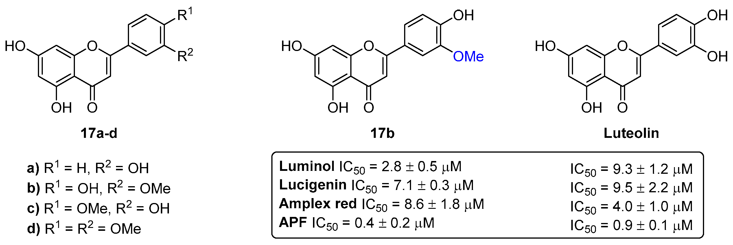
  9. Sousa, J.L.C.; Proença, C.; Freitas, M.; Fernandes, E.; Silva, A.M.S. New polyhydroxylated flavon-3-ols and 3-hydroxy-2-styrylchromones: Synthesis and ROS/RNS scavenging activities. Eur. J. Med. Chem. 2016, 119, 250–259. [Google Scholar] [CrossRef] [PubMed]
  10. Proença, C.; Albuquerque, H.M.T.; Ribeiro, D.; Freitas, M.; Santos, C.M.M.; Silva, A.M.S.; Fernandes, E. Novel chromone and xanthone derivatives: Synthesis and ROS/RNS scavenging activities. Eur. J. Med. Chem. 2016, 115, 381–392. [Google Scholar] [CrossRef]
  11. Santos, C.M.M.; Freitas, M.; Ribeiro, D.; Gomes, A.; Silva, A.M.S.; Cavaleiro, J.A.S.; Fernandes, E. 2,3-Diarylxanthones as strong scavengers of reactive oxygen and nitrogen species: A structure-activity relationship study. Bioorganic Med. Chem. 2010, 18, 6776–6784. [Google Scholar] [CrossRef]
  12. Gomes, A.; Neuwirth, O.; Freitas, M.; Couto, D.; Ribeiro, D.; Figueiredo, A.G.P.R.; Silva, A.M.S.; Seixas, R.S.G.R.; Pinto, D.C.G.A.; Tomé, A.C.; et al. Synthesis and antioxidant properties of new chromone derivatives. Bioorganic Med. Chem. 2009, 17, 7218–7226. [Google Scholar] [CrossRef]
  13. Gomes, A.; Fernandes, E.; Silva, A.M.S.; Santos, C.M.M.; Pinto, D.C.G.A.; Cavaleiro, J.A.S.; Lima, J.L.F.C. 2-Styrylchromones: Novel strong scavengers of reactive oxygen and nitrogen species. Bioorganic Med. Chem. 2007, 15, 6027–6036. [Google Scholar] [CrossRef]
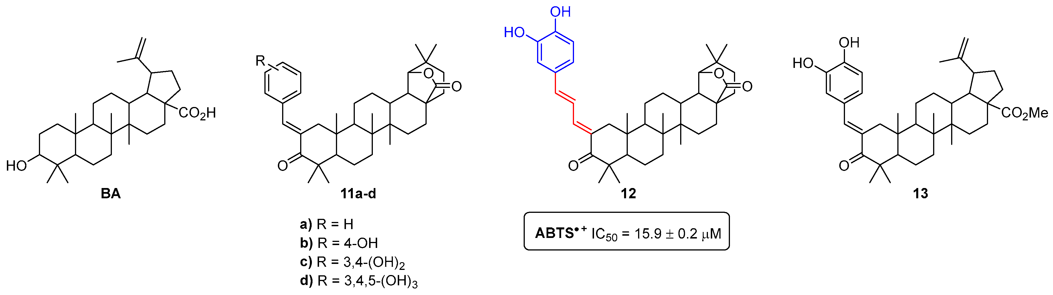
  14. Sousa, J.L.C.; Gonçalves, C.; Ferreira, R.M.; Cardoso, S.M.; Freire, C.S.R.; Silvestre, A.J.D.; Silva, A.M.S. Functionalization of betulinic acid with polyphenolic fragments for the development of new amphiphilic antioxidants. Antioxidants 2021, 10, 148. [Google Scholar] [CrossRef] [PubMed]
  15. Seca, A.M.L.; Leal, S.B.; Pinto, D.C.G.A.; Barreto, M.C.; Silva, A.M.S. Xanthenedione Derivatives, New Promising Antioxidant and Acetylcholinesterase Inhibitor Agents. Molecules 2014, 19, 8317–8333. [Google Scholar] [CrossRef] [PubMed]
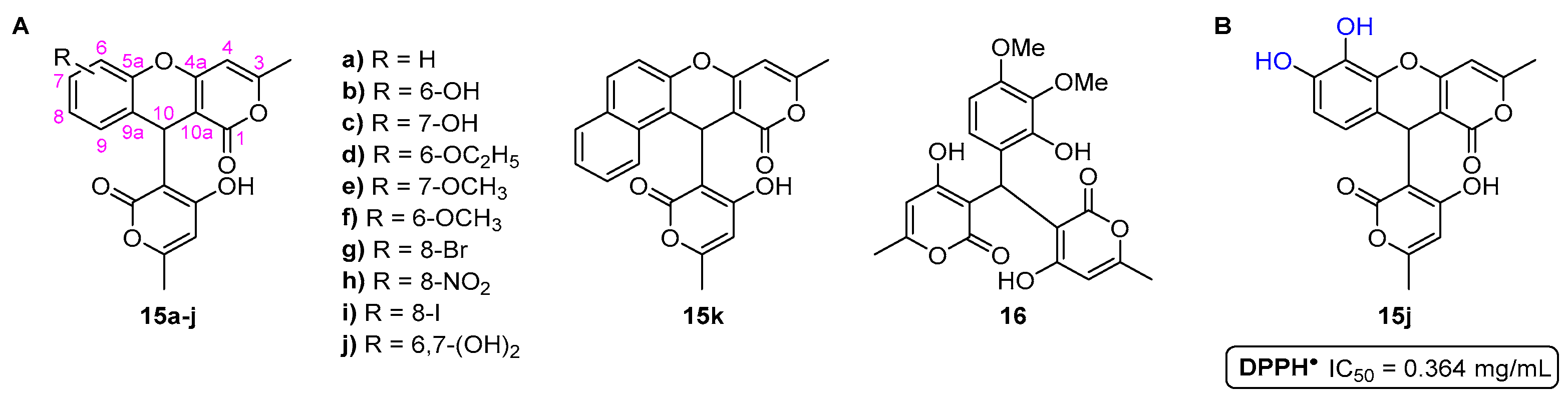
  16. Saher, L.; Makhloufi-Chebli, M.; Dermeche, L.; Dermeche, S.; Boutemeur-Khedis, B.; Rabia, C.; Hamdi, M.; Silva, A.M.S. 10-(4-Hydroxy-6-methyl-2-oxo-2H-pyran-3-yl)-3-methyl-1H,10H-pyrano [4,3-b] chromen-1-ones from a pseudo-multicomponent reaction and evaluation of their antioxidant activity. Tetrahedron 2018, 74, 872–879. [Google Scholar] [CrossRef]
  17. Fernandes, E.; Carvalho, F.; Silva, A.M.S.; Santos, C.M.M.; Pinto, D.C.G.A.; Cavaleiro, J.A.S.; Bastos, M.D.L. 2-Styrylchromones as novel inhibitors of xanthine oxidase. A structure-activity study. J. Enzym. Inhib. Med. Chem. 2002, 17, 45–48. [Google Scholar] [CrossRef]
  18. Ribeiro, D.; Freitas, M.; Tomé, S.M.; Silva, A.M.S.; Porto, G.; Fernandes, E. Modulation of human neutrophils’ oxidative burst by flavonoids. Eur. J. Med. Chem. 2013, 67, 280–292. [Google Scholar] [CrossRef]
  19. Ribeiro, D.; Freitas, M.; Tomé, S.M.; Silva, A.M.S.; Laufer, S.; Lima, J.L.F.C.; Fernandes, E. Flavonoids Inhibit COX-1 and COX-2 Enzymes and Cytokine/Chemokine Production in Human Whole Blood. Inflammation 2015, 38, 858–870. [Google Scholar] [CrossRef]
  20. Ribeiro, D.; Freitas, M.; Tomé, S.M.; Silva, A.M.S.; Porto, G.; Cabrita, E.J.; Marques, M.M.B.; Fernandes, E. Inhibition of LOX by flavonoids: A structure-activity relationship study. Eur. J. Med. Chem. 2014, 72, 137–145. [Google Scholar] [CrossRef]
  21. Freitas, M.; Ribeiro, D.; Tomé, S.M.; Silva, A.M.S.; Fernandes, E. Synthesis of chlorinated flavonoids with anti-inflammatory and pro-apoptotic activities in human neutrophils. Eur. J. Med. Chem. 2014, 86, 153–164. [Google Scholar] [CrossRef] [PubMed]
  22. Proença, C.; Ribeiro, D.; Soares, T.; Tomé, S.M.; Silva, A.M.S.; Lima, J.L.F.C.; Fernandes, E.; Freitas, M. Chlorinated Flavonoids Modulate the Inflammatory Process in Human Blood. Inflammation 2017, 40, 1155–1165. [Google Scholar] [CrossRef]
  23. Santos, C.M.M.; Ribeiro, D.; Silva, A.M.S.; Fernandes, E. 2,3-Diarylxanthones as potential inhibitors of arachidonic acid metabolic pathways. Inflammation 2017, 40, 956–964. [Google Scholar] [CrossRef] [PubMed]
  24. Gomes, A.; Fernandes, E.; Silva, A.M.S.; Pinto, D.C.G.A.; Santos, C.M.M.; Cavaleiro, J.A.S.; Lima, J.L.F.C. Anti-inflammatory potential of 2-styrylchromones regarding their interference with arachidonic acid metabolic pathways. Biochem. Pharmacol. 2009, 78, 171–177. [Google Scholar] [CrossRef] [PubMed]
  25. Available online: https://idf.org/about-diabetes/diabetes-facts-figures/ (accessed on 15 November 2023).
  26. Proença, C.; Freitas, M.; Ribeiro, D.; Sousa, J.L.C.; Carvalho, F.; Silva, A.M.S.; Fernandes, P.A.; Fernandes, E. Inhibition of protein tyrosine phosphatase 1B by flavonoids: A structure–activity relationship study. Food Chem. Toxicol. 2018, 111, 474–481. [Google Scholar] [CrossRef]
  27. Proença, C.; Freitas, M.; Ribeiro, D.; Tomé, S.M.; Araújo, A.N.; Silva, A.M.S.; Fernandes, P.A.; Fernandes, E. The dipeptidyl peptidase-4 inhibitory effect of flavonoids is hindered in protein rich environments. Food Funct. 2019, 10, 5718–5731. [Google Scholar] [CrossRef] [PubMed]
  28. Proença, C.; Oliveira, A.; Freitas, M.; Ribeiro, D.; Sousa, J.L.C.; Ramos, M.J.; Silva, A.M.S.; Fernandes, P.A.; Fernandes, E. Structural Specificity of Flavonoids in the Inhibition of Human Fructose 1,6-Bisphosphatase. J. Nat. Prod. 2020, 83, 1541–1552. [Google Scholar] [CrossRef]
  29. Proença, C.; Freitas, M.; Ribeiro, D.; Oliveira, E.F.T.; Sousa, J.L.C.; Tomé, S.M.; Ramos, M.J.; Silva, A.M.S.; Fernandes, P.A.; Fernandes, E. α-Glucosidase inhibition by flavonoids: An in vitro and in silico structure–activity relationship study. J. Enzym. Inhib. Med. Chem. 2017, 32, 1216–1228. [Google Scholar] [CrossRef]
  30. Proença, C.; Rufino, A.T.; Ferreira de Oliveira, J.M.P.; Freitas, M.; Fernandes, P.A.; Silva, A.M.S.; Fernandes, E. Inhibitory activity of flavonoids against human sucrase-isomaltase (α-glucosidase) activity in a Caco-2/TC7 cellular model. Food Funct. 2022, 13, 1108–1118. [Google Scholar] [CrossRef]
  31. Proença, C.; Freitas, M.; Ribeiro, D.; Tomé, S.M.; Oliveira, E.F.T.; Viegas, M.F.; Araújo, A.N.; Ramos, M.J.; Silva, A.M.S.; Fernandes, P.A.; et al. Evaluation of a flavonoids library for inhibition of pancreatic α-amylase towards a structure–activity relationship. J. Enzyme Inhib. Med. Chem. 2019, 34, 577–588. [Google Scholar] [CrossRef]
  32. Rocha, S.; Aniceto, N.; Guedes, R.C.; Albuquerque, H.M.T.; Silva, V.L.M.; Silva, A.M.S.; Corvo, M.L.; Fernandes, E.; Freitas, M. An In Silico and an In Vitro Inhibition Analysis of Glycogen Phosphorylase by Flavonoids, Styrylchromones, and Pyrazoles. Nutrients 2022, 14, 306. [Google Scholar] [CrossRef]
  33. Santos, C.M.M.; Proença, C.; Freitas, M.; Araújo, A.N.; Silva, A.M.S.; Fernandes, E. Inhibition of the carbohydrate-hydrolyzing enzymes α-amylase and α-glucosidase by hydroxylated xanthones. Food Funct. 2022, 13, 7930–7941. [Google Scholar] [CrossRef]
  34. Rocha, S.; Lucas, M.; Silva, V.L.M.; Gomes, P.M.O.; Silva, A.M.S.; Araújo, A.N.; Aniceto, N.; Guedes, R.C.; Corvo, M.L.; Fernandes, E.; et al. Pyrazoles as novel protein tyrosine phosphatase 1B (PTP1B) inhibitors: An in vitro and in silico study. Int. J. Biol. Macromol. 2021, 181, 1171–1182. [Google Scholar] [CrossRef]
  35. Ferlay, J.; Colombet, M.; Soerjomataram, I.; Parkin, D.M.; Piñeros, M.; Znaor, A.; Bray, F. Cancer statistics for the year 2020: An overview. Int. J. Cancer 2021, 149, 778–789. [Google Scholar] [CrossRef]
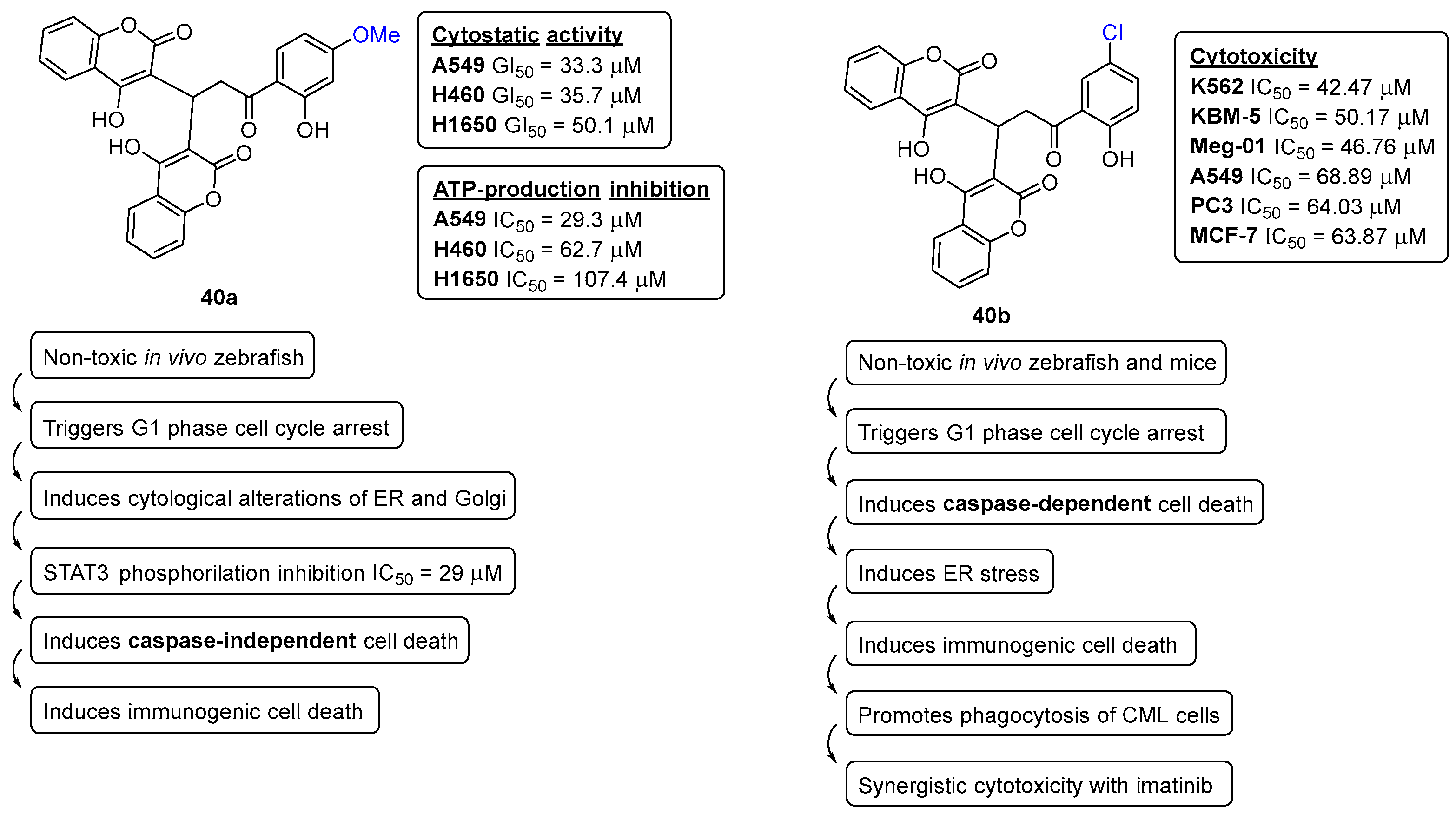
  36. Lee, J.-Y.; Talhi, O.; Jang, D.; Cerella, C.; Gaigneaux, A.; Kim, K.-W.; Lee, J.W.; Dicato, M.; Bachari, K.; Han, B.W.; et al. Cytostatic hydroxycoumarin OT52 induces ER/Golgi stress and STAT3 inhibition triggering non-canonical cell death and synergy with BH3 mimetics in lung cancer. Cancer Lett. 2018, 416, 94–108. [Google Scholar] [CrossRef]
  37. Mazumder, A.; Lee, J.-Y.; Talhi, O.; Cerella, C.; Chateauvieux, S.; Gaigneaux, A.; Hong, C.R.; Kang, H.J.; Lee, Y.; Kim, K.-W.; et al. Hydroxycoumarin OT-55 kills CML cells alone or in synergy with imatinib or Synribo: Involvement of ER stress and DAMP release. Cancer Lett. 2018, 438, 197–218. [Google Scholar] [CrossRef] [PubMed]
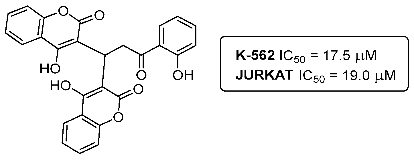
  38. Talhi, O.; Schnekenburger, M.; Panning, J.; Pinto, D.G.C.; Fernandes, J.A.; Almeida Paz, F.A.; Jacob, C.; Diederich, M.; Silva, A.M.S. Bis(4-hydroxy-2H-chromen-2-one): Synthesis and effects on leukemic cell lines proliferation and NF-κB regulation. Bioorganic Med. Chem. 2014, 22, 3008–3015. [Google Scholar] [CrossRef] [PubMed]
  39. Valente, S.; Xu, Z.; Bana, E.; Zwergel, C.; Mai, A.; Jacob, C.; Meiser, P.; Bagrel, D.; Silva, A.M.S.; Kirsch, G. Reactivity of 4-Vinyl-2H-1-benzopyran-2-ones in Diels–Alder Cycloaddition Reactions: Access to Coumarin-Based Polycycles with Cdc25 Phosphatase-Inhibiting Activity. Eur. J. Org. Chem. 2013, 2013, 2869–2877. [Google Scholar] [CrossRef]
  40. Sutanto, F.; Konstantinidou, M.; Dömling, A. Covalent inhibitors: A rational approach to drug discovery. RSC Med. Chem. 2020, 11, 876–884. [Google Scholar] [CrossRef] [PubMed]
  41. Gehringer, M.; Laufer, S.A. Emerging and Re-Emerging Warheads for Targeted Covalent Inhibitors: Applications in Medicinal Chemistry and Chemical Biology. J. Med. Chem. 2019, 62, 5673–5724. [Google Scholar] [CrossRef]
  42. Shindo, N.; Ojida, A. Recent progress in covalent warheads for in vivo targeting of endogenous proteins. Bioorganic Med. Chem. 2021, 47, 116386. [Google Scholar] [CrossRef]
  43. Talhi, O.; Brodziak-Jarosz, L.; Panning, J.; Orlikova, B.; Zwergel, C.; Tzanova, T.; Philippot, S.; Pinto, D.C.G.A.; Paz, F.A.A.; Gerhäuser, C.; et al. One-Pot Synthesis of Benzopyran-4-ones with Cancer Preventive and Therapeutic Potential. Eur. J. Org. Chem. 2016, 2016, 965–975. [Google Scholar] [CrossRef]
  44. Bouhenna, M.M.; Orlikova, B.; Talhi, O.; Schram, B.; Pinto, D.C.G.A.; Taibi, N.; Bachari, K.; Diederich, M.; Silva, A.M.S.; Mameri, N. Anti-proliferative, Cytotoxic and NF-ĸB Inhibitory Properties of Spiro(Lactone-Cyclohexanone) Compounds in Human Leukemia. Anticancer Res. 2017, 37, 5225–5233. [Google Scholar]
  45. Lamia Hamdan, R.; Oualid, T.; Nadia, T.; Laetitia, D.; Caroline, D.; Artur, S.; Khaldoun, B.; Marie-Paule, V.; Florence, C.-C. Effects of Spiro-bisheterocycles on Proliferation and Apoptosis in Human Breast Cancer Cell Lines. Anticancer Res. 2016, 36, 6399. [Google Scholar]
  46. Naouri, A.; Djemoui, A.; Ouahrani, M.R.; Lahrech, M.B.; Lemouari, N.; Rocha, D.H.A.; Albuquerque, H.; Mendes, R.F.; Almeida Paz, F.A.; Helguero, L.A.; et al. Multicomponent and 1,3-dipolar cycloaddition synthesis of triazole- and isoxazole-acridinedione/xanthenedione heterocyclic hybrids: Cytotoxic effects on human cancer cells. J. Mol. Struct. 2020, 1217, 128325. [Google Scholar] [CrossRef]
  47. Djemoui, A.; Naouri, A.; Ouahrani, M.R.; Djemoui, D.; Lahcene, S.; Lahrech, M.B.; Boukenna, L.; Albuquerque, H.M.T.; Saher, L.; Rocha, D.H.A.; et al. A step-by-step synthesis of triazole-benzimidazole-chalcone hybrids: Anticancer activity in human cells+. J. Mol. Struct. 2020, 1204, 127487. [Google Scholar] [CrossRef]
  48. Bozorov, K.; Zhao, J.; Aisa, H.A. 1,2,3-Triazole-containing hybrids as leads in medicinal chemistry: A recent overview. Bioorganic Med. Chem. 2019, 27, 3511–3531. [Google Scholar] [CrossRef] [PubMed]
  49. Saher, L.; Monteiro, F.L.; Rocha, D.H.A.; Albuquerque, H.M.T.; Silva, A.M.S.; Helguero, L.A. Selective Induction of DNA Damage and Cell Cycle Arrest Mediated by Chromone-Triazole Dyads Derivatives: Effects on Breast and Prostate Cancer Cells. Chem. Biodivers. 2023, 20, e202300251. [Google Scholar] [CrossRef] [PubMed]
  50. Zwergel, C.; Valente, S.; Salvato, A.; Xu, Z.; Talhi, O.; Mai, A.; Silva, A.; Altucci, L.; Kirsch, G. Novel benzofuran–chromone and –coumarin derivatives: Synthesis and biological activity in K562 human leukemia cells. MedChemComm 2013, 4, 1571–1579. [Google Scholar] [CrossRef]
  51. Carreira, A.R.F.; Pereira, D.M.; Andrade, P.B.; Valentão, P.; Silva, A.M.S.; Braga, S.S.; Silva, V.L.M. Novel styrylpyrazole-glucosides and their dioxolo-bridged doppelgangers: Synthesis and cytotoxicity. New J. Chem. 2019, 43, 8299–8310. [Google Scholar] [CrossRef]
  52. Chouiter, M.I.; Boulebd, H.; Pereira, D.M.; Valentão, P.; Andrade, P.B.; Belfaitah, A.; Silva, A.M.S. New chalcone-type compounds and 2-pyrazoline derivatives: Synthesis and caspase-dependent anticancer activity. Future Med. Chem. 2020, 12, 493–509. [Google Scholar] [CrossRef] [PubMed]
  53. Saidi, L.; Rocha, D.H.A.; Talhi, O.; Bentarzi, Y.; Nedjar-Kolli, B.; Bachari, K.; Almeida Paz, F.A.; Helguero, L.A.; Silva, A.M.S. Synthesis of Benzophenones and in vitro Evaluation of Their Anticancer Potential in Breast and Prostate Cancer Cells. ChemMedChem 2019, 14, 1041–1048. [Google Scholar] [CrossRef] [PubMed]
  54. da Silva, A.F.F.; Seixas, R.S.G.R.; Silva, A.M.S.; Coimbra, J.; Fernandes, A.C.; Santos, J.P.; Matos, A.; Rino, J.; Santos, I.; Marques, F. Synthesis, characterization and biological evaluation of carboranylmethylbenzo[b]acridones as novel agents for boron neutron capture therapy. Org. Biomol. Chem. 2014, 12, 5201–5211. [Google Scholar] [CrossRef] [PubMed]
  55. Gustavsson, A.; Norton, N.; Fast, T.; Frölich, L.; Georges, J.; Holzapfel, D.; Kirabali, T.; Krolak-Salmon, P.; Rossini, P.M.; Ferretti, M.T.; et al. Global estimates on the number of persons across the Alzheimer’s disease continuum. Alzheimer’s Dement. 2023, 19, 658–670. [Google Scholar] [CrossRef]
  56. Horak, M.; Holubova, K.; Nepovimova, E.; Krusek, J.; Kaniakova, M.; Korabecny, J.; Vyklicky, L.; Kuca, K.; Stuchlik, A.; Ricny, J.; et al. The pharmacology of tacrine at N-methyl-d-aspartate receptors. Prog. Neuro-Psychopharmacol. Biol. Psychiatry 2017, 75, 54–62. [Google Scholar] [CrossRef] [PubMed]
  57. Praticò, D. Evidence of Oxidative Stress in Alzheimer’s Disease Brain and Antioxidant Therapy. Ann. N. Y. Acad. Sci. 2008, 1147, 70–78. [Google Scholar] [CrossRef]
  58. Sharma, A.; Pachauri, V.; Flora, S.J.S. Advances in Multi-Functional Ligands and the Need for Metal-Related Pharmacology for the Management of Alzheimer Disease. Front. Pharmacol. 2018, 9, 1247. [Google Scholar] [CrossRef]
  59. Du, X.; Wang, X.; Geng, M. Alzheimer’s disease hypothesis and related therapies. Transl. Neurodegener. 2018, 7, 2. [Google Scholar] [CrossRef]
  60. Hamley, I.W. The Amyloid Beta Peptide: A Chemist’s Perspective. Role in Alzheimer’s and Fibrillization. Chem. Rev. 2012, 112, 5147–5192. [Google Scholar] [CrossRef]
  61. Malafaia, D.; Albuquerque, H.M.T.; Silva, A.M.S. Amyloid-β and tau aggregation dual-inhibitors: A synthetic and structure-activity relationship focused review. Eur. J. Med. Chem. 2021, 214, 113209. [Google Scholar] [CrossRef]
  62. Reitz, C. Alzheimer’s Disease and the Amyloid Cascade Hypothesis: A Critical Review. Int. J. Alzheimer’s Dis. 2012, 2012, 369808. [Google Scholar] [CrossRef] [PubMed]
  63. Mohamed, T.; Shakeri, A.; Rao, P.P.N. Amyloid cascade in Alzheimer’s disease: Recent advances in medicinal chemistry. Eur. J. Med. Chem. 2016, 113, 258–272. [Google Scholar] [CrossRef] [PubMed]
  64. Coyle, J.T.; Price, D.L.; DeLong, M.R. Alzheimer’s disease: A disorder of cortical cholinergic innervation. Science 1983, 219, 1184–1190. [Google Scholar] [CrossRef] [PubMed]
  65. Wang, H.; Zhang, H. Reconsideration of Anticholinesterase Therapeutic Strategies against Alzheimer’s Disease. ACS Chem. Neurosci. 2019, 10, 852–862. [Google Scholar] [CrossRef] [PubMed]
  66. Gorecki, L.; Uliassi, E.; Bartolini, M.; Janockova, J.; Hrabinova, M.; Hepnarova, V.; Prchal, L.; Muckova, L.; Pejchal, J.; Karasova, J.Z.; et al. Phenothiazine-Tacrine Heterodimers: Pursuing Multitarget Directed Approach in Alzheimer’s Disease. ACS Chem. Neurosci. 2021, 12, 1698–1715. [Google Scholar] [CrossRef] [PubMed]
  67. Blaikie, L.; Kay, G.; Kong Thoo Lin, P. Current and emerging therapeutic targets of alzheimer’s disease for the design of multi-target directed ligands. MedChemComm 2019, 10, 2052–2072. [Google Scholar] [CrossRef] [PubMed]
  68. Malafaia, D.; Oliveira, A.; Fernandes, P.A.; Ramos, M.J.; Albuquerque, H.M.T.; Silva, A.M.S. Chromeno[3,4-b]xanthones as First-in-Class AChE and Aβ Aggregation Dual-Inhibitors. Int. J. Mol. Sci. 2021, 22, 4145. [Google Scholar] [CrossRef]
  69. Ferreira, J.P.S.; Cardoso, S.M.; Almeida Paz, F.A.; Silva, A.M.S.; Silva, V.L.M. Synthesis of 2-aroylfuro[3,2-c]quinolines from quinolone-based chalcones and evaluation of their antioxidant and anticholinesterase activities. New J. Chem. 2020, 44, 6501–6509. [Google Scholar] [CrossRef]
  70. Zhou, H.; Yang, Z.; Tian, X.; Chen, L.; Lee, S.; Huynh, T.; Ge, C.; Zhou, R. Lanosterol Disrupts the Aggregation of Amyloid-β Peptides. ACS Chem. Neurosci. 2019, 10, 4051–4060. [Google Scholar] [CrossRef]
  71. Liu, K.; Guo, T.L.; Chojnacki, J.; Lee, H.-G.; Wang, X.; Siedlak, S.L.; Rao, W.; Zhu, X.; Zhang, S. Bivalent Ligand Containing Curcumin and Cholesterol as a Fluorescence Probe for Aβ Plaques in Alzheimer’s Disease. ACS Chem. Neurosci. 2012, 3, 141–146. [Google Scholar] [CrossRef]
  72. Zhao, L.; Chen, X.-J.; Zhu, J.; Xi, Y.-B.; Yang, X.; Hu, L.-D.; Ouyang, H.; Patel, S.H.; Jin, X.; Lin, D.; et al. Lanosterol reverses protein aggregation in cataracts. Nature 2015, 523, 607–611. [Google Scholar] [CrossRef] [PubMed]
  73. Kang, H.; Yang, Z.; Zhou, R. Lanosterol Disrupts Aggregation of Human γD-Crystallin by Binding to the Hydrophobic Dimerization Interface. J. Am. Chem. Soc. 2018, 140, 8479–8486. [Google Scholar] [CrossRef] [PubMed]
  74. Yang, X.; Chen, X.-J.; Yang, Z.; Xi, Y.-B.; Wang, L.; Wu, Y.; Yan, Y.-B.; Rao, Y. Synthesis, Evaluation, and Structure–Activity Relationship Study of Lanosterol Derivatives To Reverse Mutant-Crystallin-Induced Protein Aggregation. J. Med. Chem. 2018, 61, 8693–8706. [Google Scholar] [CrossRef] [PubMed]
  75. Albuquerque, H.M.T.; Nunes da Silva, R.; Pereira, M.; Maia, A.; Guieu, S.; Soares, A.R.; Santos, C.M.M.; Vieira, S.I.; Silva, A.M.S. Steroid–Quinoline Hybrids for Disruption and Reversion of Protein Aggregation Processes. ACS Med. Chem. Lett. 2022, 13, 443–448. [Google Scholar] [CrossRef]
  76. Pereira, M.; Tomé, D.; Domingues, A.S.; Varanda, A.S.; Paulo, C.; Santos, M.A.S.; Soares, A.R. A Fluorescence-Based Sensor Assay that Monitors General Protein Aggregation in Human Cells. Biotechnol. J. 2018, 13, 1700676. [Google Scholar] [CrossRef]
  77. Hong, Y.; Meng, L.; Chen, S.; Leung, C.W.T.; Da, L.-T.; Faisal, M.; Silva, D.-A.; Liu, J.; Lam, J.W.Y.; Huang, X.; et al. Monitoring and Inhibition of Insulin Fibrillation by a Small Organic Fluorogen with Aggregation-Induced Emission Characteristics. J. Am. Chem. Soc. 2012, 134, 1680–1689. [Google Scholar] [CrossRef]
  78. Bolognesi, M.L.; Gandini, A.; Prati, F.; Uliassi, E. From Companion Diagnostics to Theranostics: A New Avenue for Alzheimer’s Disease? J. Med. Chem. 2016, 59, 7759–7770. [Google Scholar] [CrossRef]
  79. Aliyan, A.; Cook, N.P.; Martí, A.A. Interrogating Amyloid Aggregates using Fluorescent Probes. Chem. Rev. 2019, 119, 11819–11856. [Google Scholar] [CrossRef]
  80. Nunes da Silva, R.; Costa, C.C.; Santos, M.J.G.; Alves, M.Q.; Braga, S.S.; Vieira, S.I.; Rocha, J.; Silva, A.M.S.; Guieu, S. Fluorescent Light-up Probe for the Detection of Protein Aggregates. Chem. Asian J. 2019, 14, 859–863. [Google Scholar] [CrossRef]
  81. Brul, S.; Nussbaum, J.; Dielbandhoesing, S.K. Fluorescent probes for wall porosity and membrane integrity in filamentous fungi. J. Microbiol. Methods 1997, 28, 169–178. [Google Scholar] [CrossRef]
  82. Ferreira, J.R.M.; Alves, M.; Sousa, B.; Vieira, S.I.; Silva, A.M.S.; Guieu, S.; Cunha, Â.; Nunes da Silva, R. Curcumin-based molecular probes for fluorescence imaging of fungi. Org. Biomol. Chem. 2023, 21, 1531–1536. [Google Scholar] [CrossRef]
  83. Gross, D.A.; Silver, D.L. Cytosolic lipid droplets: From mechanisms of fat storage to disease. Crit. Rev. Biochem. Mol. Biol. 2014, 49, 304–326. [Google Scholar] [CrossRef] [PubMed]
  84. Soares da Silva, A.M.; André Guieu, S.J.; Matias Celestino Gomes da Rocha, J.C.; Moreira Pinto Vieira, S.I.; da Cruz E Silva, O.; Dos Santos Dias, R.A.; Frade Ruivo, R. Fluorescent Compounds, Production Methods and Uses Thereof. WO/2017/182945, 26 October 2017. [Google Scholar]
  85. Vieira, S.I.; Nunes da Silva, R.; Alves, M.; Dias, R.A.; Meireles Sousa, A.M.; Camões, F.; Maia, A.; Almeida, M.; Rocha, J.; Silva, A.M.; et al. Liprobe, a vital dye for lipid aggregates detection in imaging and high-content screens. Front. Photonics 2022, 3, 963778. [Google Scholar] [CrossRef]
  86. Oryan, A.; Akbari, M. Worldwide risk factors in leishmaniasis. Asian Pac. J. Trop. Med. 2016, 9, 925–932. [Google Scholar] [CrossRef] [PubMed]
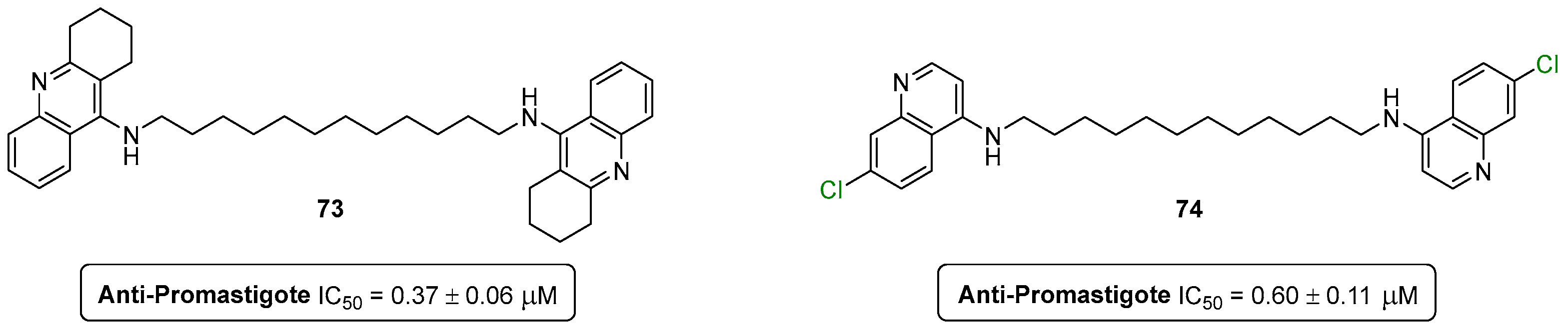
  87. Silva, C.F.M.; Leão, T.; Dias, F.; Tomás, A.M.; Pinto, D.C.G.A.; Oliveira, E.F.T.; Oliveira, A.; Fernandes, P.A.; Silva, A.M.S. Structure & ndash; Activity Relationship Studies of 9-Alkylamino-1,2,3,4-tetrahydroacridines against Leishmania (Leishmania) infantum Promastigotes. Pharmaceutics 2023, 15, 669. [Google Scholar]
  88. Dhanda, G.; Acharya, Y.; Haldar, J. Antibiotic Adjuvants: A Versatile Approach to Combat Antibiotic Resistance. ACS Omega 2023, 8, 10757–10783. [Google Scholar] [CrossRef]
  89. Almeida, M.C.; da Costa, P.M.; Sousa, E.; Resende, D.I.S.P. Emerging Target-Directed Approaches for the Treatment and Diagnosis of Microbial Infections. J. Med. Chem. 2023, 66, 32–70. [Google Scholar] [CrossRef] [PubMed]
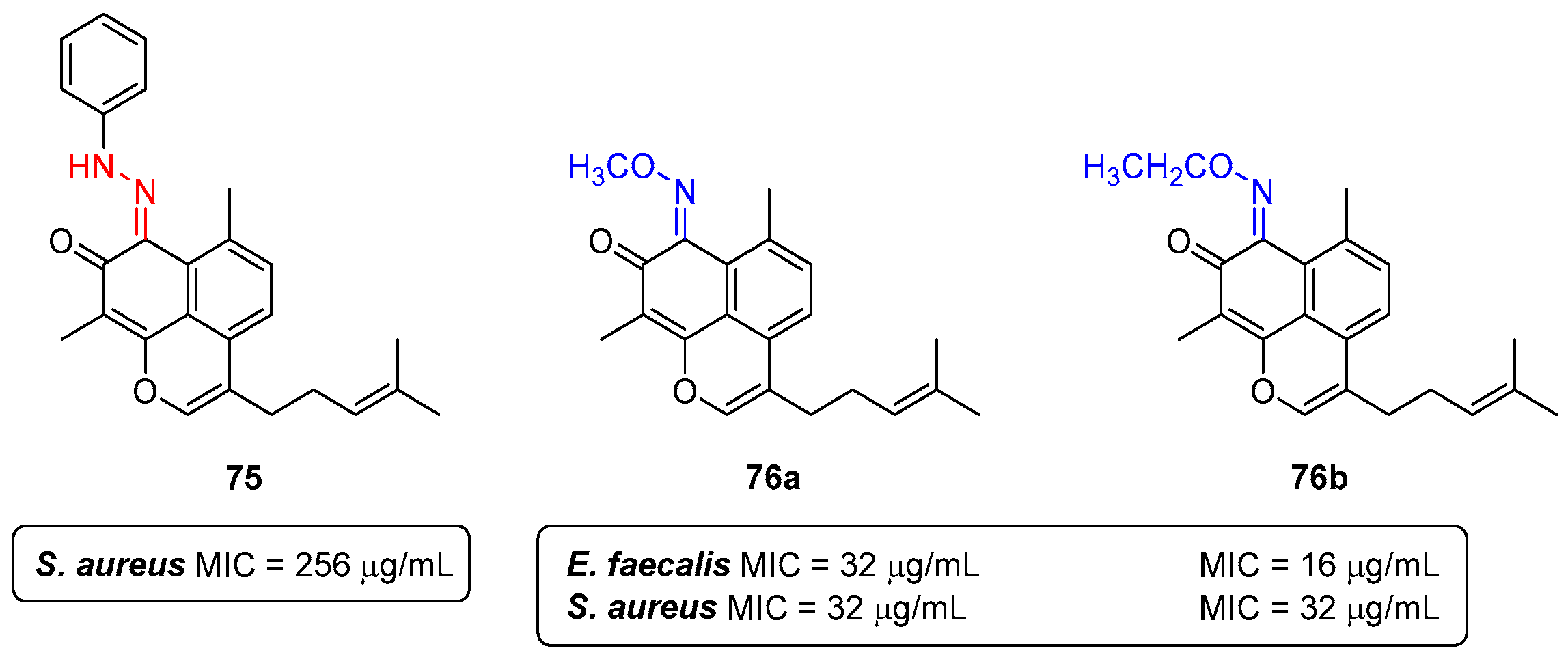
  90. Souza, L.G.d.S.; Almeida, M.C.S.; Lemos, T.L.G.; Ribeiro, P.R.V.; de Brito, E.S.; Silva, V.L.M.; Silva, A.M.S.; Braz-Filho, R.; Costa, J.G.M.; Rodrigues, F.F.G.; et al. Synthesis, antibacterial and cytotoxic activities of new biflorin-based hydrazones and oximes. Bioorganic Med. Chem. Lett. 2016, 26, 435–439. [Google Scholar] [CrossRef]
  91. Zhang, M.; Fu, M.; Hu, Q. Advances in Human Norovirus Vaccine Research. Vaccines 2021, 9, 732. [Google Scholar] [CrossRef]
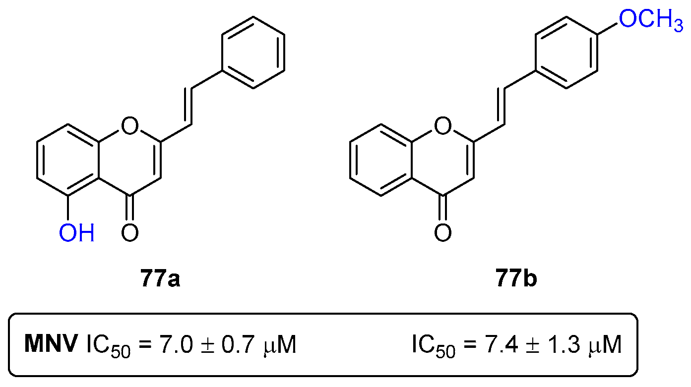
  92. Rocha-Pereira, J.; Cunha, R.; Pinto, D.C.G.A.; Silva, A.M.S.; Nascimento, M.S.J. (E)-2-Styrylchromones as potential anti-norovirus agents. Bioorganic Med. Chem. 2010, 18, 4195–4201. [Google Scholar] [CrossRef]
Figure 1. Schematic depiction of drug discovery process. TA: target assessment; LI: lead identification; LO: lead optimization; CCS: clinical candidate selection; LSF: late-stage functionalization.
Pharmaceuticals 16 01668 g001
Figure 2. Schematic overview of some developed bioactive compounds as antioxidant, anti-inflammatory, antidiabetic, anticancer, anti-Alzheimer, bioimaging, anti-Leishmaniosis and antibacterial agents.
Pharmaceuticals 16 01668 g002
Figure 3. Most efficient scavengers of ROS and RNS based on polyhydroxylated 2-[styryl or (4-arylbutadienyl)]chromones 14, xanthones 5,6, flavones 7,8, chalcones 9, and flavonols 10.
Pharmaceuticals 16 01668 g003
Figure 4. Chemical structures of the betulinic acid (BA) and the 19,28-epoxyoleanane-3,28-dione and methyl betulonate-derived polyhydroxylated compounds 1113.
Pharmaceuticals 16 01668 g004
Figure 5. Chemical structures of the xanthene-1,9(2H)-diones 14af and the most active derivative 14e in reducing the ferric ion and scavenging DPPH.
Pharmaceuticals 16 01668 g005
Figure 6. Chemical structures of the 10-(4-hydroxy-6-methyl-2-oxo-2H-pyran-3-yl)-3-methyl-1H,10H-pyrano[4,3-b]chromen-1-ones 15ak and 3,3′-[(2-hydroxy-3,4-dimethoxyphenyl)methylene]bis(4-hydroxy-6-methyl-2H-pyran-2-one) (16) (A) and the most active compound 15j in the scavenging of DPPH (B).
Pharmaceuticals 16 01668 g006
Figure 7. Chemical structures of the methylated flavones 17ad and the most effective derivative 17b in inhibiting PMA-induced neutrophils’ oxidative burst.
Pharmaceuticals 16 01668 g007
Figure 8. Chemical structures of the chlorinated flavones 18ae (A) and the lead chlorinated flavones as anti-inflammatory agents (B). DCFH-DA = 2′,7′-Dichlorodihydrofluorescein diacetate.
Pharmaceuticals 16 01668 g008
Figure 9. Chemical structures of the hydroxylated 2,3-diarylxanthones 1921 (A) and the emerged dual-acting anti-inflammatory agents 19b,c and 21a (B).
Pharmaceuticals 16 01668 g009
Figure 10. Chemical structures of flavonols 10ae (A) and the most active flavonols for the in vitro inhibition of PTP1B, DPP-4, and α-glucosidase (B).
Pharmaceuticals 16 01668 g010
Figure 11. The most active chlorinated flavones as α-glucosidase, α-amylase, and DPP-4 inhibitors.
Pharmaceuticals 16 01668 g011
Figure 12. Chemical structures of the most active 2-SC 1a and 2-[4-(3,4-dihydroxyphenyl)butadienyl]chromones 4a,b as GP inhibitors.
Pharmaceuticals 16 01668 g012
Figure 13. Chemical structures of the lead compounds 20c and 21b as dual-target inhibitors of both α-glucosidase and α-amylase.
Pharmaceuticals 16 01668 g013
Figure 14. Chemical structures of the most active pyrazoles 2224 as PTP1B inhibitors.
Pharmaceuticals 16 01668 g014
Figure 15. Chemical structures of the tested (E)-4-styrylpyrazoles 2531, (Z)-4-styrylpyrazoles 32,33, and 5-styrylpyrazoles 3439 as GP inhibitors.
Pharmaceuticals 16 01668 g015
Figure 16. Chemical structures of bis-coumarins 40a,b and their anticancer profile.
Pharmaceuticals 16 01668 g016
Figure 17. Bis-coumarin 40c as template for the development of anticancer agents.
Pharmaceuticals 16 01668 g017
Figure 18. Coumarin derivatives as Cdc25 phosphatase inhibitors.
Pharmaceuticals 16 01668 g018
Figure 19. α,β-Unsaturated carbonyl moiety as warhead of covalent inhibitors.
Pharmaceuticals 16 01668 g019
Figure 20. Chromanones 42a,b and 2-SC 43 as potent inducers of the cytoprotective Keap1-Nrf2 signaling pathway.
Pharmaceuticals 16 01668 g020
Figure 21. Spiro(lactone-cyclohexanones) as antiproliferative agents in leukemia cell lines.
Pharmaceuticals 16 01668 g021
Figure 22. Effective spiro-bisheterocyclic compounds 4648 against proliferation (A) and apoptosis (B) of human breast cancer cell lines.
Pharmaceuticals 16 01668 g022
Figure 23. 1,2,3-Triazole-xanthenediones and triazole-benzimidazole-chalcones as anticancer agents.
Pharmaceuticals 16 01668 g023
Figure 24. Chromone-1,2,3-triazole hybrid compounds targeting breast and prostate cancer cells.
Pharmaceuticals 16 01668 g024
Figure 25. Chromone– and coumarin–aurone hybrids targeting K562 human leukemia cells.
Pharmaceuticals 16 01668 g025
Figure 26. Glycosylated- and non-glycosylated pyrazoles as anticancer agents.
Pharmaceuticals 16 01668 g026
Figure 27. Design strategy of chalcone-type and 2-pyrazoline compounds incorporating an (benz)imidazole moiety.
Pharmaceuticals 16 01668 g027
Figure 28. Chalcone-type compounds identified as promising anticancer agents toward the AGS cell line.
Pharmaceuticals 16 01668 g028
Figure 29. Structure of aryl-benzophenone 59.
Pharmaceuticals 16 01668 g029
Figure 30. Benzo[b]acridin-12(7H)-one 60 as lead structure for BNCT.
Pharmaceuticals 16 01668 g030
Figure 31. Single- and dual-target lead compounds towards AChE and Aβ aggregation.
Pharmaceuticals 16 01668 g031
Figure 32. 2-Aroylfuro[3,2-c]quinolines as AChE inhibitors.
Pharmaceuticals 16 01668 g032
Figure 33. Xanthenedione with two catechol moieties as AChE inhibitor.
Pharmaceuticals 16 01668 g033
Figure 34. Structures of lanosterol and 2-fluoro-25-hydroxylanosterol.
Pharmaceuticals 16 01668 g034
Figure 35. Structures of lanosterol and cholesterol and their Aβ/steroid ratios for aggregation inhibition.
Pharmaceuticals 16 01668 g035
Figure 36. Quinoline–cholesterol hybrids as effective inhibitors of Aβ and random protein aggregation.
Pharmaceuticals 16 01668 g036
Figure 37. Fluorescent light-up probe 66 for detection of protein aggregates.
Pharmaceuticals 16 01668 g037
Figure 38. Curcumin-based molecular probes for staining Fusarium oxysporum.
Pharmaceuticals 16 01668 g038
Figure 39. Chemical structure of liprobe with DAD.
Pharmaceuticals 16 01668 g039
Figure 40. VS hit templates based on the tetrahydroacridine scaffold.
Pharmaceuticals 16 01668 g040
Figure 41. Antileishmanial lead compounds based on tetrahydroacridine and quinoline scaffolds.
Pharmaceuticals 16 01668 g041
Figure 42. Hydrazone and oxime analogues of biflorin.
Pharmaceuticals 16 01668 g042
Figure 43. Best performing antibacterial compounds, hydrazone 75 and oximes 76a,b.
Pharmaceuticals 16 01668 g043
Figure 44. Most effective anti-norovirus (E)-2-SC 77a,b. MNV = murine norovirus.
Pharmaceuticals 16 01668 g044
Disclaimer/Publisher’s Note: The statements, opinions and data contained in all publications are solely those of the individual author(s) and contributor(s) and not of MDPI and/or the editor(s). MDPI and/or the editor(s) disclaim responsibility for any injury to people or property resulting from any ideas, methods, instructions or products referred to in the content.

