Article

Influence of the Molar Activity of 203/212Pb-PSC-PEG2-TOC on Somatostatin Receptor Type 2-Binding and Cell Uptake

1
Department of Nuclear Medicine, University Hospital Carl Gustav Carus, Technical University Dresden, Fetscherstr. 74, 01307 Dresden, Germany
2
Department of Radiology, University of Iowa, Iowa City, IA 52240, USA
3
Viewpoint Molecular Targeting, Inc. (DBA Perspective Therapeutics), Coralville, IA 52241, USA
4
Department of Chemistry, University of Iowa, Iowa City, IA 52241, USA
5
Department of Radiation Oncology, University of Iowa, Iowa City, IA 52242, USA
*
Author to whom correspondence should be addressed.
These authors contributed equally to this work.
Pharmaceuticals 2023, 16(11), 1605; https://doi.org/10.3390/ph16111605
Submission received: 10 October 2023 / Revised: 6 November 2023 / Accepted: 10 November 2023 / Published: 14 November 2023
(This article belongs to the Special Issue Therapeutic Radionuclides in Nuclear Medicine)

Abstract

:
(1) Background: In neuroendocrine tumors (NETs), somatostatin receptor subtype 2 is highly expressed, which can be targeted by a radioactive ligand such as [177Lu]Lu-1,4,7,10-tetraazacyclododecane-N,N′,N″,N‴-tetraacetic acid-[Tyr3,Thr8]-octreotide (177Lu-DOTA-TOC) and, more recently, by a lead specific chelator (PSC) containing 203/212Pb-PSC-PEG2-TOC (PSC-TOC). The molar activity (AM) can play a crucial role in tumor uptake, especially in receptor-mediated uptake, such as in NETs. Therefore, an investigation of the influence of different molar activities of 203/212Pb-PSC-TOC on cell uptake was investigated. (2) Methods: Optimized radiolabeling of 203/212Pb-PSC-TOC was performed with 50 µg of precursor in a NaAc/AcOH buffer at pH 5.3–5.5 within 15–45 min at 95° C. Cell uptake was studied in AR42 J, HEK293 sst2, and ZR75-1 cells. (3) Results: 203/212Pb-PSC-TOC was radiolabeled with high radiochemical purity >95% and high radiochemical yield >95%, with AM ranging from 0.2 to 61.6 MBq/nmol. The cell uptake of 203Pb-PSC-TOC (AM = 38 MBq/nmol) was highest in AR42 J (17.9%), moderate in HEK293 sstr (9.1%) and lowest in ZR75-1 (0.6%). Cell uptake increased with the level of AM. (4) Conclusions: A moderate AM of 15–40 MBq/nmol showed the highest cell uptake. No uptake limitation was found in the first 24–48 h. Further escalation experiments with even higher AM should be performed in the future. It was shown that AM plays an important role because of its direct dependence on the cellular uptake levels, possibly due to less receptor saturation with non-radioactive ligands at higher AM.

1. Introduction

Recently, receptor-targeted α-therapy (TAT) has gained importance in nuclear medicine clinical routine, especially for tumor patients who develop resistance to β-therapy [1,2]. Typically, patients receive multiple doses of 90Y or 177Lu (dose of 5–8 GBq per patient) at periodic intervals of administration (e.g., 8-week intervals) [3]. Unfortunately, a significant proportion of these patients will eventually experience progressive disease and discontinue therapy. On the other hand, it has been observed that further responses and prolonged survival can be achieved by initiating α-therapy after disease progression. For example, the use of 225Ac (dose: 100 kBq/kg, four α-particles per decay) can dramatically reduce the required level of administered radioactivity (by a factor of about 1000 compared to 177Lu). However, the behavior of α-particles in tumor cells is complicated by α-emitting radionuclide progeny in the 225Ac series. Of particular interest are 221Fr, 217At, 213Bi, and 213Po. A key issue is the biological fate of 213Bi (t1/2 46 min), which is transported out of the tumor cells (by its own escape or by the escape of 221Fr or 217At) and accumulates mainly in the kidneys, delivering an α-emitting dose of 213Po, which, in turn, could have a higher negative impact on renal function [4]. Therefore, it is often suggested that only patients with an efficient renal function can be considered eligible for 225Ac therapy, while patients with impaired renal function may not be eligible for these therapies.
212Pb is a promising radionuclide for targeted alpha particle therapy that emits only one α-particle per β-decay of 212Pb to 212Bi and then either 64% to 212Po or 36% to 208Tl [5,6]. Therefore, a 212Pb-labeled radiopharmaceutical, once accumulated in the tumor tissue, will deposit its highest dose in the form of the α-particle specifically in the tumor cells, with a lower probability of further α-decay occurring in healthy organs. Thus, 212Pb represents a more favorable choice for cancer α-therapy patients who are naïve to (or who have progressed on) β-therapy, including patients with reduced renal function. Ongoing preclinical and clinical studies are investigating the potential of 212Pb-labeled peptides and antibodies (at a dose of approximately 2 MBq/kg) [4] or approximately between 177Lu and 225Ac administered doses of radioactivity. Lead specific chelator-PEG2-[Tyr3,Thr8]-octreotide (PSC-PEG2-TOC) (VMT-α-NET) is a somatostatin subtype 2 (SST2) receptor targeting peptide for the treatment of neuroendocrine tumors (NETs) that exhibits rapid tumor accumulation, high tumor retention, and rapid renal excretion [7]. It carries the chelator PSC [7], which forms highly stable complexes with 203/212Pb and, in contrast to less stable 1,4,7,10-tetraazacyclododecane-N,N′,N″,N‴-tetraacetic acid (DOTA) complexes, remains intact even after β conversion to 212Bi [8]. Recently, the true matched pair 203/212Pb has come into focus through several first in-human theranostic applications [9,10,11]. While 203Pb (t1/2 = 52 h; 279 keV gamma ray; 81% intensity) represents an ideal elementally matched imaging surrogate, 212Pb itself can be used for SPECT imaging [12]. A true matched pair could finally overcome the differential pharmacokinetic/pharmacological properties observed between diagnostic and therapeutic radiotracers with unmatched radionuclides pairs [13].
In addition to somatostatin analogs, such as PSC-TOC, other radiopharmaceuticals targeting other receptors (e.g., PSMA derivatives) for radiolabeling with 203/212Pb are under preclinical investigation and may soon be translated into clinical use [4,14,15,16]. Emerging evidence suggests that α-particles have the potential to overcome resistance to β-therapy and could lead to further therapeutic options for patients with palliative effects and, in some cases, even complete remission [17]. Furthermore, the appearance of Cherenkov light due to the decay of 212Pb may be useful not only for diagnostics but also for further treatment options [18]. The optimal mass of the precursor peptide for radiopharmaceuticals is an important parameter in radiopharmaceutical development to ensure that the highest degree of tumor targeting with the lowest accumulation and retention in normal organs and healthy tissues is achieved. This parameter is also important in achieving a formulation that results in smooth radiometallation and a high radiochemical yield and purity. In general, this parameter is known as the molar activity (AM), which plays a crucial role for all PET radiotracers targeting saturable binding sites (e.g., receptors) but is secondary or negligible for many metabolic PET radiotracers, where the endogenous levels of the compound are in great excess of the radiotracer itself due to saturation of the receptors at the tumor site [19].
In this work, the influence of the AM-to-cell uptake of 203/212Pb-PSC-TOC was investigated in different cell lines (AR42 J, HEK293 sst2, and ZR75-1) to develop a more detailed understanding of the tracer in preparation for clinical use.

2. Results

2.1. Radiochemistry

The radiochemical yield (RCY) was 74–97% for 203Pb-PSC-TOC (Table 1) and 22–99% for 212Pb-PSC-TOC, depending on the reaction conditions (Table 2 and Table 3). The radiochemical purity (RCP) was always >95%. Stabilities greater than 95% of the formulated 203/212Pb-PSC-TOC were found up to 16 h at r.t. and for 203Pb-PSC-TOC up to 11 d at 4 °C by RP-HPLC. For 212Pb-PSC-TOC, HPLC is very useful for determining the identity of the product but not for the direct determination of RCY, according to the approach used here. A free 208Tl daughter nuclide was observed up to 20% in the chromatogram 4 h after radiosynthesis. Isothermal titration calorimetry (ITC) experiments confirmed the stability of the 212Bi daughter radionuclide in the PSC chelator to >95% at 2 h after radiolabeling. Importantly, the contribution of free 208Tl to the total body dose was shown to be negligible [20].
The optimization of the radiochemical labeling procedure included steps to overcome the loss of activity due to several purification steps: The freshly eluted activity from the 224Ra/212Pb generator is partitioned into Pb resin 10–12%, waste ~2%, and product 84–86%. The purification of 212Pb by Pb resin removes potential traces of the parent nuclide 224Ra, as well as the daughter nuclides 212Bi and 208Tl [21]. Therefore, activity measurements with a dose calibrator (ISOMED 2010, Nuvia Instruments GmbH, Dresden, Germany) in equilibrium of the mother and daughter nuclides show different values before and after Pb resin purification. Therefore, a normalization factor for the dose calibrator is required immediately after purification (Table 4). Further purification with C18 or Maxi-Clean (MC) SPE cartridges results in a further loss of product activity. The C18 contained 6–10% of the product activity, and the MC cartridge contained up to 40% of the product activity. It appears that the chelated 212Pb-PSC complex has strong binding competition with the sorbent material of the MC cartridges. Therefore, these cartridges are not suitable for purification. However, the cartridge purifications were later found to be obsolete due to further development of optimized radiolabeling conditions.

2.2. Cell Uptake Studies with Different Molar Activities

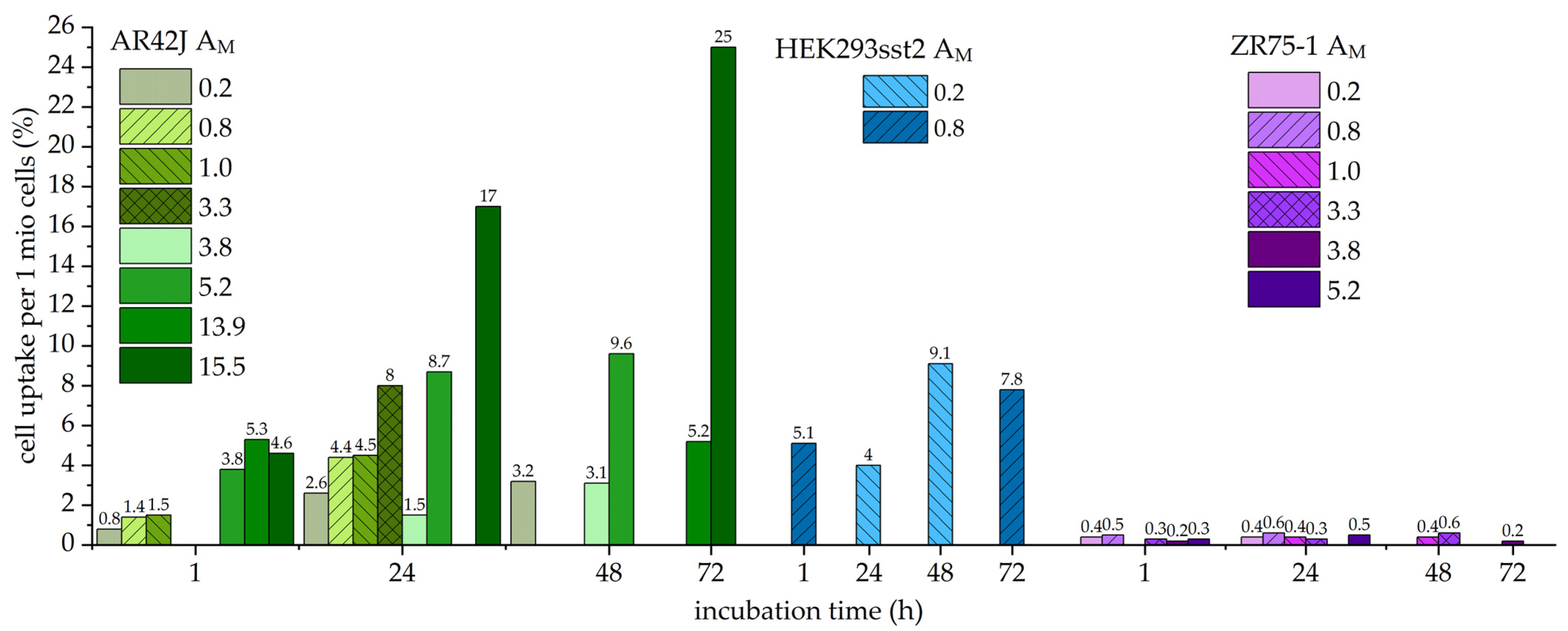
During the studies with 203Pb-PSC-TOC, it was found that SST2-transfected HEK293 cells showed a moderate cell uptake compared to endogenously SST2-positive AR42J cells, which showed a higher cell uptake. Compared to the routinely used 68Ga-DOTA-[Tyr3,Thr8]-octreotate (TATE) with a cell uptake in AR42J of about 8.7 ± 0.4% after 1 h, the uptake of 203/212Pb-PSC-TOC reached a comparable level 24 h after incubation only at the highest AM > 7.9 MBq/nmol (203Pb-PSC-TOC) and AM > 3.3 MBq/nmol (212Pb-PSC-TOC). ZR75-1 cells were used as a negative control and showed no significant uptake for all radiotracers used in this work.
It was found that cell uptake was strongly influenced by the number of cells seeded 1–2 days prior to the uptake experiments. With 100,000 cells seeded, very low cell uptake was observed, and the cell uptake increased with the number of cells seeded. The optimal number of cells for uptake was 1.0 million cells per well. Seeding cells three days prior to uptake had no further effect on cell uptake.
The optimum activity of 203Pb-PSC-TOC for incubation was found to be 100 kBq per well on a six-well plate, and cell viability was good for over 72 h. For 212Pb-PSC-TOC, activities greater than 50 kBq per well lead to a decrease in cell uptake and even lower cell viability (11% viability after 72 h), which affected the credibility of the cell uptake values. Therefore, activities of 25 kBq per well were used for 212Pb-PSC-TOC.
Figure 1 shows the cell uptake for 203Pb-PSC-TOC, and Figure 2 shows the cell uptake for 212Pb-PSC-TOC. It was found that the uptake increased with the AM for both radioligands. In most of the experiments, only timeframes between 1 and 24 h were considered, because, for diagnostic purposes, patients are measured only within a timeframe of 24 h, and, for therapeutic purposes, most of the dose is already deposited (two half-lives = 75% of the dose, t1/2 = 10.64 h). In the case of the highest produced AM of 203/212Pb-PSC-TOC, the timeframe was extended to 72 h in order to find a possible plateau or decrease in cell uptake. 203Pb-PSC-TOC showed an increase of relative cell uptake per 1 million cells up to 24.0 ± 0.8% even 149 h after incubation (2.9 × t1/2 of 203Pb). For 212Pb-PSC-TOC, the cell uptake still increased up to 25.0 ± 0.5% 72 h after incubation. Increased uptake values were found at the lowest excess of cold peptide (25%). These results could support the hypothesis that free binding sites of SST2 receptors are still available (Figure 2).
Table 5 and Table 6 show the statistical evaluation of the cell accumulation of 203/212Pb-PSC-TOC. Each cell line was incubated with the respective AM in triplicate. The standard deviation of the mean accumulation was less than 10%, confirming that the data were consistent with the conclusions. The standard deviation for ZR75-1 uptake was not shown, because it was less than 0.04% in each experiment.

3. Discussion

3.1. Radiochemistry

Radiolabeling of PSC-TOC with 203Pb proceeded smoothly under standard labeling conditions. Challenges were observed with 212Pb when using the same conditions as for 203Pb that needed to be overcome. However, the addition of a higher mass of peptide precursor and slightly longer reaction times solved this problem [21]. The quality control showed an RCY >95% in most cases for 203/212Pb-PSC-TOC. It is noteworthy that, when the RCP for 212Pb-PSC-TOC was only >90%, this was due to ejected daughter nuclides that transferred to a solvent form [22]. When the developed TLC was remeasured after 2 h, the RCP increased to >95% (24 h later, the RCP was even >99%), as the free daughter nuclides decayed on the TLC within this time, confirming the radiochemical purity of 212Pb-PSC-TOC.

3.2. Influence of Molar Activity on Cell Uptake

For the cell uptake experiments performed in this work, it was observed that, the higher the AM, the higher the cell uptake. An uptake limit was not found in these experiments, but there may be one, as can be found in the literature [19]. Furthermore, neither PSMA nor TATE peptides reached tumor saturation in the patients [23]. AR42J cells always showed the highest cell uptake for each AM compared to HEK293 sst2 cells, which showed lower cell uptake. ZR75-1 cells as a negative control did not show significant cell uptake due to the lack of SST2 receptor. Therefore, AR42J cells may have the highest SST2 receptor expression of the cells used in this study. For further studies in this direction, transfected cells with even higher SST2 receptor expression than in AR42J cells could be used [24].
The high uptake of SST2-specific radioligands has been well examined for agonists and antagonists. It is known that agonists like TOC effectuate the internalization of SST2 receptors after stimulation in vivo and that this is the reason for the high and long-lasting uptake of SST2 radioligands [25]. However, this high accumulation seems to reduce after several treatments with β-radioligands, due to differentiation of the SST2 expressing tumor cells. With only low SST2 expression at the tumor site, this could be one reason for the “β resistance” when only low amounts of radioligands reach the tumor site. The lower expression of SST2 receptor may merely be overcome by TAT, which seem to have a similar impact in tumor therapy at much lower doses compared to β-therapy [26]. Therefore, antagonists like JR11, which have a similar accumulation to the agonistic radioligands, even after several treatments, may be a solution for this issue, since the SST2 expressing tumors do not differentiate when blocked by antagonists [27]. Another instrument to overcome the lowering SST2-specific tumor accumulation may be epigenetic stimulation before treatment or during a series of treatments. Experimental data have shown higher uptake rates in HEK293 sst2 cells up to 28 times versus untreated cells [28].
In radioligand therapy, a medium AM tracer could reduce radiation exposure to dose-limiting organs with only a limited effect on radionuclide accumulation in the tumor. In this case, high AM is considered to be 200 MBq/nmol, and low AM is considered to be 2 MBq/nmol. Therefore, it can be assumed that the moderate AM level of 15–40 MBq/nmol (Table 1 and Table 3) achieved in this work may be the optimal amount for application. Nevertheless, further cell studies with even higher AM should be performed to confirm this. One way to achieve higher AM is HPLC purification, which has been shown to increase the AM of a 68Ga ligand by a factor of 10,000 [29]. In addition, a pilot therapy study with the optimal AM can be performed against low AM and high AM to validate the observations from the cell studies. This experimental comparison may provide evidence to support the idea that moderate molar activities may indeed provide superior tumor uptake while minimizing radiation exposure to dose-limiting organs.
The amount of somatostatin analog administered varies with the analog used and the intended purpose of the administered drug. The TATE analog is administered at a high tracer level (100 nmol) in α- and β-therapy, which is in the range of physiologically applied concentrations. This could lead to a lower tumor uptake if tumor receptors are blocked with an unlabeled peptide. Conversely, a higher AM could negatively affect the tumor-to-organ ratio by saturating the physiologically expressing organs. For 212Pb therapy, this would mean that AM = 1 MBq/nmol is closer to the amount of, e.g., Luthathera (50 MBq/nmol, 148 nmol for 7.4 GBq [30]) than AM = 10 MBq/nmol.
A comparable study on the influence of the AM applied with 68Ga or 177Lu was not found in the scientific literature. The results of an Al18F-labeled PSMA-11 study were presented [31], where it was found that the administration of a high AM tracer increased the detection of low expression tumors while also increasing uptake in PSMA-expressing tissues, potentially leading to false-positive findings.
Taken together, variations in AM may have different implications for diagnostic and therapeutic use. However, the moderate AM of 15–40 MBq/nmol investigated in this work showed the highest cell uptake and should be used when administered to patients. For patients, a lower amount of peptide could increase tumor uptake while decreasing side effects.

4. Materials and Methods

All reagents and solvents were purchased from commercial suppliers at the highest purity and used without further purification. PSC-PEG2-TOC (VMT-α-NET) and 224Ra/212Pb generator (VMT-α-GEN) were obtained from Perspective Therapeutics Inc., Coralville, IA, USA. 203Pb solution in 8 M HCl was obtained from Cross Cancer Institute, Edmonton, AB, Canada. RCP was monitored by thin-layer chromatography (TLC) on iTLC-SG plates (Agilent, Santa Clara, CA, USA). Measurement of the radionuclide purity (RNP) and evaluation of the radio-TLC was performed with a thin-layer scanner (MiniScanPRO+, Eckert&Ziegler Eurotope GmbH, Berlin, Germany) equipped with a Model 43-2 alpha detector ZnS(Ag) scintillator (Ludlum Measurements, Sweetwater, TX, USA) and a build-in multi-channel analyzer (MCA) for gamma spectroscopy. Radio-HPLC was performed on a Shimadzu HPLC system (Thermo Scientific, Dreieich, Germany), equipped with a reverse-phase column (Merck Chromolith RP-18e; 100 × 4.6 mm plus a 5 × 4.6 mm guard column, Darmstadt, Germany) and a UV diode array detector (220 nm). The solvent system used was a gradient of acetonitrile:water (containing 0.05% TFA) (0–13 min: 0–60% MeCN) at a flow rate of 1.6 mL/min, unless otherwise stated. The pH was measured using a reflectance photometer (QUANTOFIX Relax, Macherey-Nagel GmbH & Co. KG, Düren, Germany).

4.1. Radiochemistry

Radiolabeling of the DOTA and PSC conjugates was performed according to standard protocols for these chelators [7]. Briefly, 50 µg precursor (DOTA-TATE (M = 1435.6 g/mol) or PSC-PEG2-TOC (PSC-PEG2-TOC, M = 1578.7 g/mol)) in H2 Osuprapure was added to a 10 mL reaction vial together with 100 µL EtOHabsolute, 290 µL 1 M NaAc/AcOH buffer (pH 4, 99.99% trace metal), and 2 mg sodium ascorbate (Ph.Eur.).
203/212Pb in 5–10 mL 1.6 M HClsuprapure was trapped on a custom-made Pb resin cartridge (100 mg PB-B10-F, Triskem, Bruz, France) preconditioned with 1 mL 2 M HClsuprapure. The captured activity was rinsed with 1 mL 2 M HClsuprapure. The activity was eluted with 2 mL NaAc/AcOH buffer (pH 6, 99.99% trace metal) directly into the reaction vial. The solution was heated at 95 °C for 15 min for 203Pb and 15 or 45 min for 212Pb. The reaction solution was then diluted with 4 mL of 0.9% NaCl solution and cooled.
Finally, the product was purified by using a C18 Plus light cartridge (WAT023501, Waters, Eschborn, Germany) preconditioned with 1 mL EtOH and 3 mL H2 O (wet condition). The cooled and diluted product solution (4 mL 0.9% NaCl) was slowly passed through the C18 cartridge. The C18 cartridge containing the product was rinsed with 2 mL of 0.9% NaCl solution and was directly eluted with 1 mL of 50% EtOH for injection directly through a vented sterile filter (0.22 µm, SLGVV255F, Millex-GV, Merck-Millipore, Darmstadt, Germany) into a product vial. Finally, the product was diluted with 7 mL of 0.9% NaCl solution.
Another purification method was performed using a Maxi-Clean (MC) SPE 0.5 mL IC-Chelate cartridge (5122565, S*Pure, Mainz, Germany) preconditioned with 5 mL H2 Osuprapure (wet condition). The cooled and diluted product solution (4 mL 0.9% NaCl) was slowly transferred through a vented sterile filter into the product vial via the MC cartridge and washed with an additional 2 mL 0.9% NaCl into the product vial.

4.2. Quality Control of Radiotracer

Quality control included several standard tests established in the clinical manufacturing:
  • TLC with eluent 0.1 M Na-citrate pH 5 (Start: 203/212Pb-PSC-TOC and particles (Rf < 0.4), end: 203/212Pb-chloride) (Figure 3).
  • TLC with eluent 1MNH4 Ac: MeOH1: 1 (Start: 203/212Pb-particles, end: 203/212Pb-PSC-TOC and 203/212Pb-chloride) (Figure 4).
  • HPLC 203/212Pb-PSC-TOC tR = 7.4 min. 203/212Pb-DOTA-TATE tR = 7.1 min.
  • pH value: 5.3 ± 0.5.
  • RNP: 212Pb: 75 and 238 keV; 212Bi: 727 keV (6.7%); and 208Tl: 510 (22.6%), 583 (85.0%), and 860 (12.5%) keV (Figure 5).

4.3. Cell Uptake Experiments

Cell uptake of 203/212Pb-PSC-TOC was tested against our gold standard 68Ga-DOTATATE. AR42J (CRL-1492, ATCC®, Manassas, VA, USA), HEK293 sst2 (stably SST2 receptor transfected cells derived from Andrea Kliewer, University Hospital, Jena, Germany), and ZR75-1 (CRL-1500, ATCC®, Manassas, VA, USA); cells were seeded 1–2 days prior to the assay in 6-well plates to reach 0.5 × 10⁶ cells per well in a humidified atmosphere containing 5% CO2.
Each cell line was grown in its own medium:
  • AR42J: Gibco RPMI 1640 medium (ATCC-Modification) supplemented with 10% fetal calf serum (FCS)
  • HEK293 sst2 (stably transfected HEK293 cells): Dulbecco’s modified Eagle’s medium supplemented with 10% fetal calf serum (FCS), l-glutamine (2 mM = 1%), G-418 (50 mg/mL)
  • ZR75-1: Gibco RPMI 1640 medium (w/o glutamine) supplemented with 10% fetal calf serum (FCS), 1% NEAA, 1 mM sodium pyruvate (1%), and 2 mM N-acetyl-alanyl-l-glutamine (1%)
After incubation with the respective radiotracer for 1 h, 24 h, 48 h, and 72 h, the medium, wash fraction (2 × 1 mL cold PBS 4 °C), and cell fraction (lysed with 1 mL 0.1 M NaOH and cell scraper) were collected, and the remaining activity in the fractions was measured with a gamma counter (HIDEX). The relative cell uptake in percent per 1 million cells was calculated between the medium, wash fraction, and lysate. Each cell incubation was performed in triplicate.

5. Conclusions

It was shown that the AM of 203/212Pb-PSC-TOC has an effect on the cell uptake. No saturation was found in this work, but it is known from the literature that higher AM > 100 MBq/nmol could have a negative effect on tumor uptake due to non-specific binding of the radioligand. It is noteworthy that higher AM leads to higher cell uptake due to receptor-specific binding, and the values found in this work can be used as a reference for clinical application.
Another interesting radiochemical observation of this investigation is that the 212Pb labeling benefited from a longer reaction time and a larger amount of precursor to achieve a high RCY. Since 203Pb and 212Pb are elementally identical, the apparent differences must necessarily be due to the differences in stable Pb found in the solutions provided (generator eluant versus 203Pb solution), and thus, further investigation of the relative specific activity of the solutions is required.
In conclusion, for high tumor uptake, radiolabeling of 50 µg of PSC-TOC precursor should be performed with >1000 MBq 203Pb and with >500 MBq 212Pb.

Author Contributions

Conceptualization, M.P., E.M., R.R. and J.K.; methodology, M.P., R.R., K.W. and K.T.; software, R.R.; validation, M.P. and R.R.; formal analysis, R.R.; investigation, M.P. and R.R.; resources, M.K.S. and J.K.; data curation, M.P., K.W. and R.R.; writing—original draft preparation, M.P. and F.B.; writing—review and editing, M.P., E.M., R.R., K.W., K.T., M.K.S. and J.K.; visualization, M.P.; supervision, M.P. and R.R.; project administration, E.M. and J.K.; funding acquisition, M.K.S. and J.K. All authors have read and agreed to the published version of the manuscript.

Funding

This research received no external funding.

Data Availability Statement

All data can be referred to on request to the corresponding author.

Acknowledgments

The authors would like to thank Andrea Kliewer from University Hospital Jena for providing the transfected HEKsst2 cells. Moreover, we thank Fiorenza Ianzini and Lisa Hübinger for the extensive proofreading of the manuscript.

Conflicts of Interest

The MiniScanPRO+ was provided by Eckert&Ziegler especially for the evaluation of α-particles on TLC. M.K.S. is employed by Perspective Therapeutics, which has financial interest in the VMT-α-NET radiopharmaceutical. The authors declare no further conflict of interest.

References

  1. Kratochwil, C.; Bruchertseifer, F.; Giesel, F.L.; Weis, M.; Verburg, F.A.; Mottaghy, F.; Kopka, K.; Apostolidis, C.; Haberkorn, U.; Morgenstern, A. 225Ac-PSMA-617 for PSMA-targeted α-radiation therapy of metastatic castration-resistant prostate cancer. J. Nucl. Med. 2016, 57, 1941–1944. [Google Scholar] [CrossRef] [PubMed]
  2. Guerra Liberal, F.D.C.; O’Sullivan, J.M.; McMahon, S.J.; Prise, K.M. Targeted alpha therapy: Current clinical applications. Cancer Biother. Radiopharm. 2020, 35, 404–417. [Google Scholar] [CrossRef] [PubMed]
  3. Brogsitter, C.; Hartmann, H.; Wunderlich, G.; Schottelius, M.; Wester, H.J.; Kotzerke, J. Twins in spirit part IV—[177Lu] high affinity DOTATATE. A promising new tracer for peptide receptor radiotherapy? Nuklearmedizin 2017, 56, 1–8. [Google Scholar] [CrossRef] [PubMed]
  4. Dos Santos, J.C.; Schäfer, M.; Bauder-Wüst, U.; Lehnert, W.; Leotta, K.; Morgenstern, A.; Kopka, K.; Haberkorn, U.; Mier, W.; Kratochwil, C. Development and dosimetry of 203Pb/212Pb-labelled PSMA ligands: Bringing “the lead” into PSMA-targeted alpha therapy? Eur. J. Nucl. Med. Mol. Imaging 2019, 46, 1081–1091. [Google Scholar] [CrossRef]
  5. Li, M.; Sagastume, E.A.; Lee, D.; McAlister, D.; DeGraffenreid, A.J.; Olewine, K.R.; Graves, S.; Copping, R.; Mirzadeh, S.; Zimmerman, B.E.; et al. 203/212Pb theranostic radiopharmaceuticals for image-guided radionuclide therapy for cancer. Curr. Med. Chem. 2020, 27, 7003–7031. [Google Scholar] [CrossRef]
  6. McNeil, B.L.; Mastroianni, S.A.; McNeil, S.W.; Zeisler, S.; Kumlin, J.; Borjian, S.; McDonagh, A.W.; Cross, M.; Schaffer, P.; Ramogida, C.F. Optimized production, purification, and radiolabeling of the 203Pb/212Pb theranostic pair for nuclear medicine. Sci. Rep. 2023, 13, 10623. [Google Scholar] [CrossRef]
  7. Li, M.; Baumhover, N.J.; Liu, D.; Cagle, B.S.; Boschetti, F.; Paulin, G.; Lee, D.; Dai, Z.; Obot, E.R.; Marks, B.M.; et al. Preclinical evaluation of a lead specific chelator (PSC) conjugated to radiopeptides for 203Pb and 212Pb-Based theranostics. Pharmaceutics 2023, 15, 414. [Google Scholar] [CrossRef]
  8. Mirzadeh, S.; Kumar, K.; Gansow, O.A. The chemical fate of 212Bi-DOTA formed by β- decay of 212Pb(DOTA)2-. Radiochim. Acta 1993, 60, 1–10. [Google Scholar] [CrossRef]
  9. Meredith, R.; Torgue, J.; Shen, S.; Fisher, D.R.; Banaga, E.; Bunch, P.; Morgan, D.; Fan, J.; Straughn, J.M., Jr. Dose escalation and dosimetry of first-in-human alpha radioimmunotherapy with 212Pb-TCMC-trastuzumab. J. Nucl. Med. 2014, 55, 1636–1642. [Google Scholar] [CrossRef]
  10. Delpassand, E.S.; Tworowska, I.; Esfandiari, R.; Torgue, J.; Hurt, J.; Shafie, A.; Núnez, R. Targeted α-emitter therapy with 212Pb-DOTAMTATE for the treatment of metastatic SSTR-expressing neuroendocrine tumors: First-in-humans dose-escalation clinical trial. J. Nucl. Med. 2022, 63, 1326–1333. [Google Scholar] [CrossRef]
  11. Müller, D.; Herrmann, H.; Schultz, M.K.; Solbach, C.; Ettrich, T.; Prasad, V. 203Pb-VMT-α-NET scintigraphy of a patient with neuroendocrine tumor. Clin. Nucl. Med. 2023, 48, 54–55. [Google Scholar] [CrossRef] [PubMed]
  12. Mikalsen, L.T.G.; Kvassheim, M.; Stokke, C. Optimized SPECT Imaging of 224Ra α-particle therapy by 212Pb photon emissions. J. Nucl. Med. 2023, 64, 1131–1137. [Google Scholar] [CrossRef]
  13. Kotzerke, J.; Runge, R.; Braune, A.; Wunderlich, G. Different Radionuclides in DOTA-EB-TATE Effect Different Uptake in Somatostatin Receptor-Positive HEK293 Cells. J. Nucl. Med. 2019, 60, 436. [Google Scholar] [CrossRef] [PubMed]
  14. Stenberg, V.Y.; Juzeniene, A.; Chen, Q.; Yang, X.; Bruland, O.S.; Larsen, R.H. Preparation of the alpha-emitting prostate-specific membrane antigen targeted radioligand [212Pb]Pb-NG001 for prostate cancer. J. Label. Comp. Radiopharm. 2020, 63, 129–143. [Google Scholar] [CrossRef]
  15. Durand-Panteix, S.; Monteil, J.; Sage, M.; Garot, A.; Clavel, M.; Saidi, A.; Torgue, J.; Cogne, M.; Quelven, I. Preclinical study of 212Pb alpha-radioimmunotherapy targeting CD20 in non-Hodgkin lymphoma. Br. J. Cancer 2021, 125, 1657–1665. [Google Scholar] [CrossRef]
  16. Li, J.; Huang, T.; Hua, J.; Wang, Q.; Su, Y.; Chen, P.; Bidlingmaier, S.; Li, A.; Xie, Z.; Bidkar, A.P.; et al. CD46 targeted 212Pb alpha particle radioimmunotherapy for prostate cancer treatment. J. Exp. Clin. Cancer Res. 2023, 42, 61. [Google Scholar] [CrossRef] [PubMed]
  17. Rathke, H.; Bruchertseifer, F.; Kratochwil, C.; Keller, H.; Giesel, F.L.; Apostolidis, C.; Haberkorn, U.; Morgenstern, A. First patient exceeding 5-year complete remission after 225Ac-PSMA-TAT. Eur. J. Nucl. Med. Mol. Imaging 2021, 48, 311–312. [Google Scholar] [CrossRef]
  18. Wood, V.; Ackerman, N.L. Cherenkov light production from the alpha-emitting decay chains of 223Ra, 212Pb, and 149Tb for Cherenkov luminescence imaging. Appl. Radiat. Isot. 2016, 118, 354–360. [Google Scholar] [CrossRef] [PubMed]
  19. Luurtsema, G.; Pichler, V.; Bongarzone, S.; Seimbille, Y.; Elsinga, P.; Gee, A.; Vercouillie, J. EANM guideline for harmonisation on molar activity or specific activity of radiopharmaceuticals: Impact on safety and imaging quality. EJNMMI Radiopharm. Chem. 2021, 6, 34. [Google Scholar] [CrossRef]
  20. Orcutt, K.D.; Henry, K.E.; Habjan, C.; Palmer, K.; Heimann, J.; Cupido, J.M.; Gottumukkala, V.; Cissell, D.D.; Lyon, M.C.; Hussein, A.I.; et al. Dosimetry of [212Pb]VMT01, a MC1R-targeted alpha therapeutic compound, and effect of free 208Tl on tissue absorbed doses. Molecules 2022, 27, 5831. [Google Scholar] [CrossRef]
  21. Li, M.; Zhang, X.; Quinn, T.P.; Lee, D.; Liu, D.; Kunkel, F.; Zimmerman, B.E.; McAlister, D.; Olewein, K.R.; Menda, Y.; et al. Automated cassette-based production of high specific activity [203/212Pb] peptide-based theranostic radiopharmaceuticals for image-guided radionuclide therapy for cancer. Appl. Radiat. Isot. 2017, 127, 52–60. [Google Scholar] [CrossRef] [PubMed]
  22. Bobba, K.N.; Bidkar, A.P.; Meher, N.; Fong, C.; Wadhwa, A.; Dhrona, S.; Sorlin, A.; Bidlingmaier, S.; Shuere, B.; He, J.; et al. Evaluation of 134Ce/134La as a PET imaging theranostic pair for 225Ac α-radiotherapeutics. J. Nucl. Med. 2023, 64, 1076–1082. [Google Scholar] [CrossRef] [PubMed]
  23. Sabet, A.; Nagarajah, J.; Dogan, A.S.; Biersack, H.-J.; Sabet, A.; Guhlke, S.; Ezziddin, S. Does PRRT with standard activities of 177Lu-octreotate really achieve relevant somatostatin receptor saturation in target tumor lesions?: Insights from intra-therapeutic receptor imaging in patients with metastatic gastroenteropancreatic neuroendocrine tumors. EJNMMI Res. 2013, 3, 82. [Google Scholar] [PubMed]
  24. Tian, R.; Jacobson, O.; Niu, G.; Kiesewetter, D.O.; Wang, Z.; Zhu, G.; Ma, Y.; Liu, G.; Chen, X. Evans Blue attachment enhances somatostatin receptor subtype-2 imaging and radiotherapy. Theranostics 2018, 8, 735–745. [Google Scholar] [CrossRef]
  25. Waser, B.; Tamma, M.L.; Cescato, R.; Maecke, H.R.; Reubi, J.C. Highly efficient in vivo agonist-induced internalization of sst2 receptors in somatostatin target tissues. J. Nucl. Med. 2009, 50, 936–941. [Google Scholar] [CrossRef]
  26. Kratochwil, C.; Apostolidis, L.; Rathke, H.; Apostolidis, C.; Bicu, F.; Bruchertseifer, F.; Choyke, P.L.; Haberkorn, U.; Giesel, F.L.; Morgenstern, A. Dosing 225Ac-DOTATOC in patients with somatostatin-receptor-positive solid tumors: 5-year follow-up of hematological and renal toxicity. Eur. J. Nucl. Med. Mol. Imaging 2021, 49, 54–63. [Google Scholar] [CrossRef]
  27. Nicolas, G.P.; Schreiter, N.; Kaul, F.; Uiters, J.; Bouterfa, H.; Kaufmann, J.; Erlanger, T.E.; Cathomas, R.; Christ, E.; Fani, M.; et al. Sensitivity Comparison of 68Ga-OPS202 and 68Ga-DOTATOC PET/CT in patients with gastroenteropancreatic neuroendocrine tumors: A prospective phase II imaging study. J. Nucl. Med. 2018, 59, 915–921. [Google Scholar] [CrossRef]
  28. Kotzerke, J.; Buesser, D.; Naumann, A.; Runge, R.; Huebinger, L.; Kliewer, A.; Freudenberg, R.; Brogsitter, C. Epigenetic-like stimulation of receptor expression in SSTR2 transfected HEK293 cells as a new therapeutic strategy. Cancers 2022, 14, 2513. [Google Scholar] [CrossRef]
  29. von Hacht, J.L.; Erdmann, S.; Niederstadt, L.; Prasad, S.; Wagener, A.; Exner, S.; Beindorff, N.; Brenner, W.; Grötzinger, C. Increasing molar activity by HPLC purification improves 68Ga-DOTA-NAPamide tumor accumulation in a B16/F1 melanoma xenograft model. PLoS ONE 2019, 14, e0217883. [Google Scholar] [CrossRef]
  30. Hennrich, U.; Kopka, K. Lutathera®: The First FDA- and EMA-approved radiopharmaceutical for peptide receptor radionuclide therapy. Pharmaceuticals 2019, 12, 114. [Google Scholar] [CrossRef]
  31. Piron, S.; Verhoeven, J.; De Coster, E.; Descamps, B.; Kersemans, K.; Pieters, L.; Vral, A.; Vanhove, C.; De Vos, F. Impact of the molar activity and PSMA expression level on [18F]AlF-PSMA-11 uptake in prostate cancer. Sci. Rep. 2021, 11, 22623. [Google Scholar] [CrossRef] [PubMed]
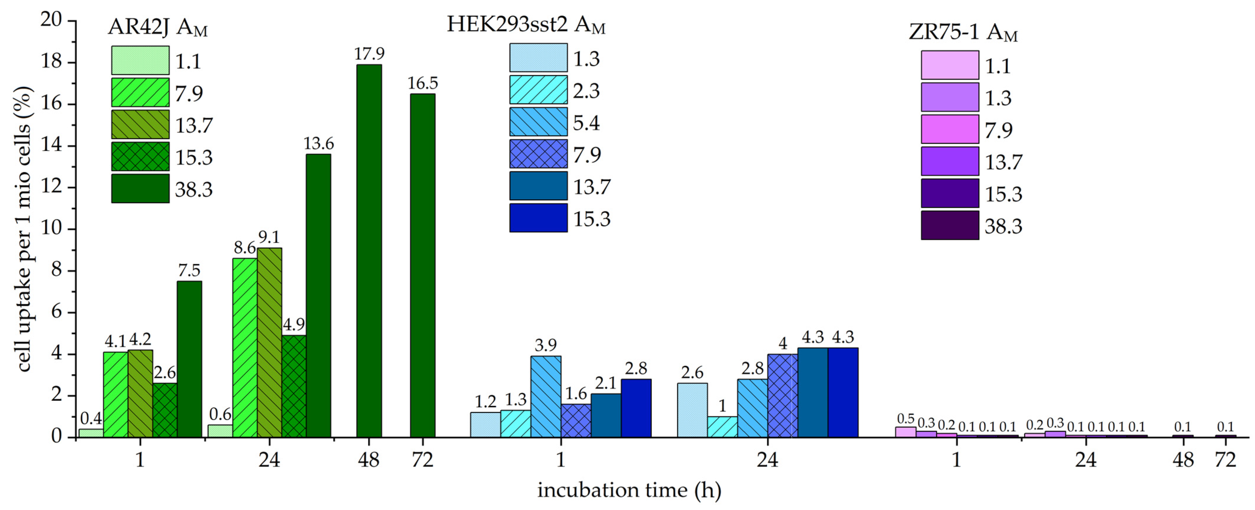
Figure 1. The cell uptake of 203Pb-PSC-TOC in different cell lines is shown. The values above the columns represent the relative mean cell uptake in percent. AM is given in MBq/nmol. Each column represents the mean value of a triplicate. Shades of green: AR42J; shades of blue: HEK293 sst2; shades of purple: ZR75-1.
Pharmaceuticals 16 01605 g001
Figure 2. Cell uptake of 212Pb-PSC-TOC in different cell lines is shown. The values above the columns represent the relative mean cell uptake in percent. AM is given in MBq/nmol. Each column represents the mean value of a triplicate. Shades of green: AR42J; shades of blue: HEK293 sst2; shades of purple: ZR75-1.
Pharmaceuticals 16 01605 g002
Figure 3. Corresponding TLC with Na-citrate as the eluent for the detection of unlabeled 203/212Pb at the front (position 115 mm, 3.9%) of the TLC. 212Pb-TOC runs at the origin (position 25 mm, 96.1%).
Pharmaceuticals 16 01605 g003
Figure 4. Corresponding TLC with NH4 Az:MeOH as the eluent for the detection of 203/212Pb-particles at the origin (position 20 mm, 0.6%) of the TLC. 212Pb-TOC runs at the front (position 80 mm, 99.4%.)
Pharmaceuticals 16 01605 g004
Figure 5. Corresponding gamma spectrum via MCA for verification of the RNP. Gamma lines found: 212Pb: 75, 238 keV; 208Tl: 510, 583, 860 keV; 212Bi: 727 keV.
Pharmaceuticals 16 01605 g005
Table 1. 203Pb-PSC-TOC reactions.
Reactions 1Starting Activity (MBq)Product Activity (MBq)RCY (%)AM (MBq/nmol)
17472972.3
2174128745.4
34036901.3
4302249827.9
55364889115.3
64554389613.7
7210519729461.6
8 2125109871.1
9130112269438.3
1 Reactions were always performed with 50 µg (31.7 nmol) VTM-α-NET for 15 min at 95 °C. 2 Exception: here, it is 156 µg (98.8 nmol) PSC-TOC.
Table 2. 212Pb-PSC-TOC reactions.
Reactions 1Starting Activity (MBq)Product Activity (MBq)RCY (%)AM (MBq/nmol)
12410400.8
242530.2
3490 *176 *3613.9
4214 *48 *223.8
5 22411480.8
1 Reactions were always performed with 20 µg (12.7 nmol) VTM-α-NET for 15 min at 95 °C. 2 Exception: here, it is 20 µg (13.7 nmol) DOTA-TATE. * Normalization was performed (see Table 4).
Table 3. Optimized 212Pb-PSC-TOC reactions.
Reactions 1Starting Activity (MBq)Product Activity (MBq)RCY (%)AM (MBq/nmol)
1522 *490 *9815.5
2328315979.9
3274260998.2
4177165 *935.2
5146104 *713.3
69578 *822.5
73231991.0
1 Reactions were always performed with 50 µg (31.7 nmol) VTM-α-NET for 45 min at 95 °C. * Normalization was performed (see Table 4).
Table 4. Normalization factors for the activity measurements of 212Pb after separation from the daughter nuclides by Pb resin for the ISOMED 2010 dose calibrator.
Time after Separation (min)Normalization Factor
01.86
51.80
301.58
601.37
901.26
1201.18
Table 5. Cell uptake statistics for 203Pb-PSC-TOC.
CellsIncubation Time (h)Molar Activity (MBq/nmol)
1.11.32.35.47.913.715.338.3
AR42J uptake (%)10.4 ± 0.1 4.1 ± 0.24.2 ± 0.02.6 ± 0.27.5 ± 0.4
240.6 ± 0.0 8.6 ± 0.59.1 ± 0.44.9 ± 0.613.6 ± 0.2
48 17.9 ± 0.7
72 16.5 ± 1.3
HEK293 sst2 uptake (%)1 1.2 ± 0.31.3 ± 0.13.9 ± 0.21.6 ± 0.32.1 ± 0.12.8 ± 0.11.2 ± 0.3
24 2.6 ± 0.61.0 ± 0.02.8 ± 0.24.0 ± 0.34.3 ± 0.24.3 ± 0.92.6 ± 0.6
Table 6. Cell uptake statistics for 212Pb-PSC-TOC.
CellsIncubation Time (h)Molar Activity (MBq/nmol)
0.20.81.03.33.85.213.915.5
AR42J uptake (%)10.8 ± 0.11.4 ± 0.11.5 ± 0.1 n.d.3.8 ± 0.25.3 ± 0.34.6 ± 0.3
242.6 ± 0.24.4 ± 0.44.5 ± 0.08.0 ± 1.01.5 ± 0.18.7 ± 0.7 17.0 ± 0.7
483.2 ± 0.4 3.1 ± 0.19.6 ± 0.7
72 5.2 ± 0.425.0 ± 0.5
HEK293 sst2 uptake (%)1 5.1 ± 0.5
24 4.0 ± 0.5
48 9.1 ± 0.5
72 7.8 ± 0.0
Disclaimer/Publisher’s Note: The statements, opinions and data contained in all publications are solely those of the individual author(s) and contributor(s) and not of MDPI and/or the editor(s). MDPI and/or the editor(s) disclaim responsibility for any injury to people or property resulting from any ideas, methods, instructions or products referred to in the content.