Review

Theranostic Imaging Surrogates for Targeted Alpha Therapy: Progress in Production, Purification, and Applications

1
Department of Oncology, University of Alberta, 11560 University Ave., Edmonton, AB T6G 1Z2, Canada
2
Edmonton Radiopharmaceutical Center, Alberta Health Services, 11560 University Ave., Edmonton, AB T6G 1Z2, Canada
3
Cancer Research Institute of Northern Alberta, University of Alberta, Edmonton, AB T6G 2E1, Canada
*
Author to whom correspondence should be addressed.
Pharmaceuticals 2023, 16(11), 1622; https://doi.org/10.3390/ph16111622
Submission received: 11 October 2023 / Revised: 8 November 2023 / Accepted: 14 November 2023 / Published: 17 November 2023
(This article belongs to the Special Issue Therapeutic Radionuclides in Nuclear Medicine)

Abstract

:
This article highlights recent developments of SPECT and PET diagnostic imaging surrogates for targeted alpha particle therapy (TAT) radiopharmaceuticals. It outlines the rationale for using imaging surrogates to improve diagnostic-scan accuracy and facilitate research, and the properties an imaging-surrogate candidate should possess. It evaluates the strengths and limitations of each potential imaging surrogate. Thirteen surrogates for TAT are explored: 133La, 132La, 134Ce/134La, and 226Ac for 225Ac TAT; 203Pb for 212Pb TAT; 131Ba for 223Ra and 224Ra TAT; 123I, 124I, 131I and 209At for 211At TAT; 134Ce/134La for 227Th TAT; and 155Tb and 152Tb for 149Tb TAT.

Graphical Abstract

1. Introduction

Targeted alpha therapy (TAT) involves utilizing radiopharmaceuticals to precisely eliminate malignancies with alpha particle emissions, while sparing surrounding healthy tissues. These radiopharmaceuticals consist of alpha (α)-emitting radionuclides conjugated to a biological-targeting vector such as monoclonal antibodies, peptides, and nanocarriers [1]. Key advantages of TAT include highly selective radiation delivery to the target, reduced patient side effects, and the ability to assess radiopharmaceutical uptake and, therefore, patient eligibility using a diagnostic radionuclide before therapy [2].
While beta minus (β) radiopharmaceuticals employing radionuclides such as 177Lu have made significant advances in clinical care of advanced prostate and neuroendocrine tumors [3,4], alpha particle emissions are significantly more precise and cytotoxic than β emissions. This is attributed to the much larger size of alpha particles (7300 times the mass of electrons), their 2+ charge resulting in a highly ionized emission path, and high linear energy transfer that deposits their energy over a path length of only several cell diameters. These properties make alpha emitters ideal for combatting metastatic cancers and other systemic malignancies where traditional treatment avenues have failed [2,5,6,7].
Approximately 400 alpha-emitting radionuclides (5–100% emission intensity) are known; however, only radionuclides that possess a sufficiently long half-life, absence of long-lived toxic progeny, and feasible high-yield production routes are suitable for TAT consideration [8,9]. Radionuclides that have shown potential for TAT include 227Th, 225Ac, 224Ra, 223Ra, 212Pb, 211At, and 149Tb [1,2,10,11,12,13,14,15,16,17,18,19,20].
While the potency of TAT offers significantly enhanced therapeutic efficacy, TAT must be treated as a double-edged sword with the possibility of severe off-target toxicity to nontarget organs and tissues. This mandates a comprehensive understanding of the stability, pharmacokinetics, and dosimetry of any TAT radiopharmaceutical. During preclinical development, these data can be acquired from biodistribution studies in mice, where mice are sacrificed at multiple time points, and gamma-ray co-emissions are counted in the dissected organs and tissues.
Additionally, positron emission tomography (PET) and single-photon emission computed tomography (SPECT) scans can be acquired by exchanging the alpha-emitting radionuclide with a positron or gamma-ray-emitting diagnostic imaging radionuclide. This imaging–therapeutic duality is termed “theranostics”, and these PET and SPECT scans provide crucial information on dosimetry and monitor response to TAT.
Most TAT radionuclides lack or possess insufficient co-emitted positrons or gamma rays for acquiring higher-quality PET or SPECT scans. This motivated the development of chemically similar diagnostic imaging surrogates for TAT radionuclides. As the current supply of alpha-emitting radionuclides is scarce, utilizing imaging surrogates also has the potential to open more opportunities for TAT research to facilities without access to alpha-emitting radionuclides and serve as a bridge for centers planning to introduce TAT radiopharmaceuticals. Since many of these surrogates can be synthesized in existing cyclotron facilities, this can facilitate radiopharmaceutical developments. Additionally, imaging surrogates fit well into the existing research and clinical setup. As such, TAT imaging surrogates have the potential to assist the deployment of TAT radiopharmaceuticals in the clinic and accelerate the development of new TAT targeting vectors.

2. Properties of Ideal Imaging Surrogates for Alpha Emitters

Multiple factors determine what makes a suitable imaging surrogate for targeted alpha therapy. These include chemical properties, half-life, radioactive emission type and intensity, associated dosimetry, production ease and scalability, radionuclidic purity, economics, and radionuclide progeny considerations.
PET and SPECT scans that evaluate the pharmacokinetics and dosimetry of TAT radiopharmaceuticals are often performed with 68Ga and 18F. However, 68Ga, 18F, and other common imaging radionuclides often have substantially different chemical properties than alpha-emitting radionuclides. For some targeting vectors, this can result in differing biodistributions between the TAT radiopharmaceutical and its diagnostic counterpart [21,22,23]. Potential inconsistencies observed in diagnostic imaging scans and subsequent biodistribution of the therapeutic radiopharmaceutical could result in sub-optimal tumor dosing or unintended and destructive alpha-irradiation of healthy tissues.
Imaging surrogates should, therefore, possess a similar chemistry and half-life to ensure their biodistribution and dosimetry are similar to their paired alpha emitters. These surrogates are ideally isotopes of the same element possessing identical chemistries, such as 226Ac paired with 225Ac TAT, 203Pb paired with 212Pb TAT, 209At paired with 211At, and 155Tb or 152Tb paired with 149Tb TAT.
However, if suitable isotopes of the same element are not available, chemically similar elements in the same chemical group can be employed. These include 133La, 132La, or 134Ce/134La paired with 225Ac, 134Ce/134La, paired with 227Th, and 123I, 124I, or 131I, paired with 211At.
It is also preferable that the physical half-life of the imaging surrogate is similar to its TAT counterpart. This permits the acquisition of biodistribution data for the full in vivo residence of the TAT radiopharmaceutical to assist preclinical development and initial clinical validation. For TAT employing radionuclides with long physical half-lives (225Ac, 223Ra, 224Ra, 227Th) and targeting vectors with long biological half-lives, using a long-lived imaging surrogate is crucial to confirm that the radiopharmaceutical remains at the target site for the extended duration without redistributing to and irradiating healthy tissues. While additional patient radiation dose might result from using a diagnostic radionuclide with a longer half-life, some targeting vectors such as antibodies may require longer circulation times to acquire sufficient quality images. For TAT employing long-lived radionuclides and targeting vectors with short biological half-lives, a radionuclide imaging surrogate with a shorter physical half-life may be used in certain situations. This can be a valuable tool for evaluating patient dosimetry, provided that the targeting vector exhibits rapid in vivo clearance, minimal off-target binding, and the radionuclide is stably incorporated within the radiopharmaceutical. Radiopharmaceutical pretargeting approaches may reduce the advantage of selecting diagnostic and therapeutic TAT radionuclides with similar half-lives; however, it is uncertain whether most theranostic targeting vectors will employ a pretargeting approach.
Regarding radioactive emissions, it is preferable that PET imaging surrogates possess a high positron branching ratio and low positron emission energy to facilitate high-resolution PET imaging and minimal co-emitted electrons and gamma/X-rays to reduce the radioactive dose. Radionuclides with lower positron branching ratios may require additional injected activity to resolve the same quality image. For SPECT imaging, radionuclides should possess lower energy gamma rays within the optimal energy window of scanners and minimal co-emitted electrons and gamma/X-rays.
To produce imaging surrogates, sufficient cyclotron or nuclear reactor facilities are required to synthesize the radionuclide. Target material (natural or isotopically enriched) should be available in adequate quantity and enrichment to support routine production, and a favorable nuclear cross-section must exist within the capabilities of production facilities. Radionuclide production should be performed safely, create few long-lived radionuclidic impurities, and be scalable to sufficient activities that allow distribution to clinical sites. Robust chemical-purification techniques must separate the imaging surrogate from potentially hazardous target material post-irradiation. Finally, the radionuclide progeny of the imaging surrogate should be considered since this can influence imaging quality and impact radioactive waste management.
Most radionuclides used in TAT are part of decay chains where each decay results in the recoil of the daughter nucleus with energy sufficient to liberate the daughter nucleus from the chelator into solution. Additionally, the alpha particle itself may induce radiolytic damage to the radiopharmaceutical, reducing the in vivo targeting and leading to further accumulation of radioactivity in nontarget tissue. These inherent physical properties are not easily covered by the surrogates in question, so they should be considered in experimental methods and conclusions.
In this article, a selection of 13 diagnostic imaging surrogates for promising alpha-emitting radionuclides have been highlighted for their production, purification, applications, and overall strengths and limitations.

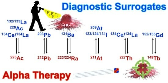
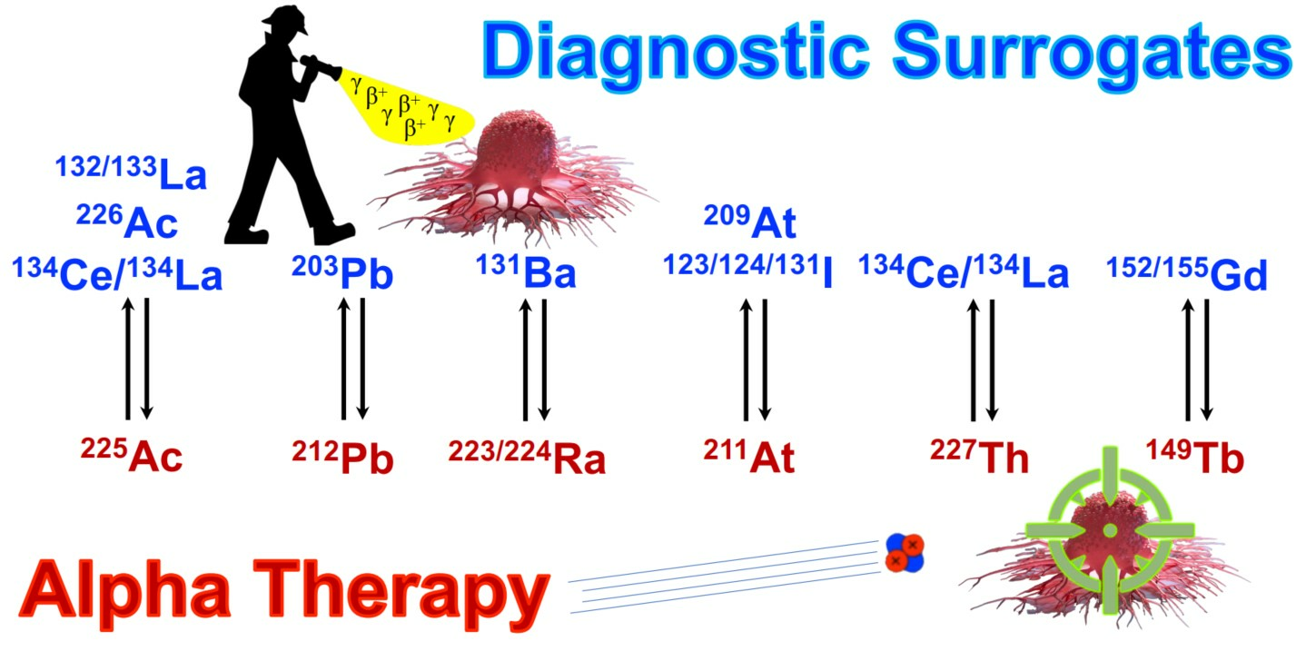
3. Theranostic Imaging Surrogates Proposed for Actinium-225

Actinium-225 (t1/2 = 9.9 d) has been explored extensively for TAT. Its long half-life permits extended dose delivery and decay via a cascade of six short-lived radionuclide progeny with four alpha particle emissions to near-stable 209Bi, making 225Ac particularly attractive for TAT. 225Ac studies have demonstrated efficacy in metastatic prostate cancer and neuroendocrine tumors, and additional radiopharmaceuticals are under development for other cancers [11,24,25,26,27,28,29,30] There are considerable efforts underway to significantly increase the 225Ac supply to meet the significant anticipated clinical demand [31,32,33,34].
However, 225Ac does not emit gamma rays of sufficient intensity for imaging. Although its 213Bi and 221Fr progeny possess gamma rays of suitable energy and intensity for SPECT imaging [9], the 225Ac activities injected into patients (~50–200 kBq/kg [11]) would be insufficient to resolve a high-quality image within a reasonable scan duration. Additionally, the supply of high-purity 225Ac from 225Ra/225Ac generators is limited, constraining AT development efforts [31]. While other sources of 225Ac from high-energy spallation reactions are available [32,35,36], these often contain a small activity of co-produced and inseparable 227Ac (t1/2 = 21 y), which complicates radioactive waste management. Therefore, the desire to enable 225Ac imaging and enhance research throughput motivates the development of imaging surrogates.
For SPECT imaging, 226Ac is an elementally matched surrogate for 225Ac. Radiolanthanum isotopes 133La, 132La, and 134La are particularly attractive for PET imaging of 225Ac due to the similar ionic radii of La3+ and Ac3+ (~1.03 and ~1.12 Å, respectively [37,38]) and their resulting similar chemistries. Both lanthanum and actinium possess similar chelation chemistry with chelators such as DOTA, macropa, and crown ethers, and exhibit similar in vivo biodistributions [39,40,41,42,43,44]. The subsequent sections will outline the properties, strengths, and limitations of 133La, 132La, 134Ce/134La, and 226Ac.

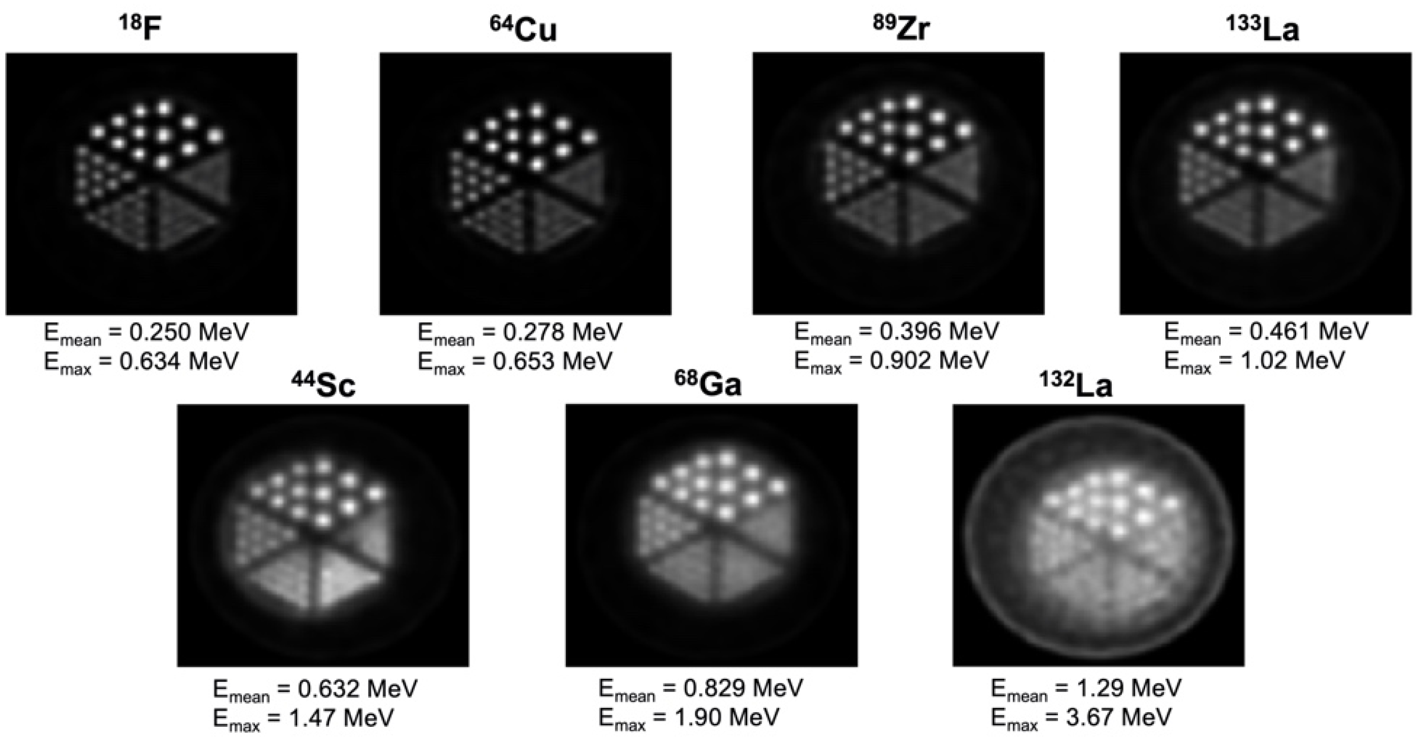
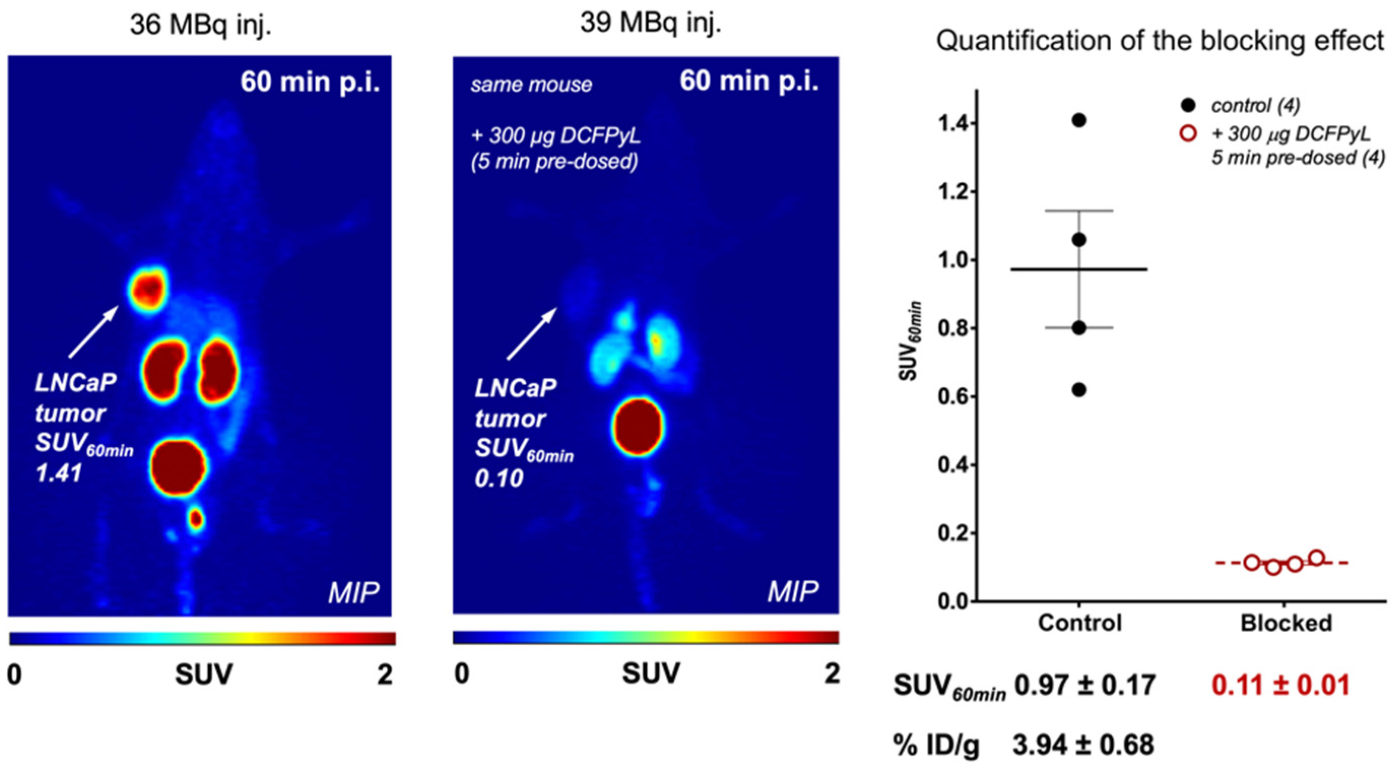
3.1. Lanthanum-133 (PET)

Lanthanum-133 (t1/2 = 3.9 h) has been synthesized via the 135Ba(p,3n)133La and 135Ba(p,2n)133La nuclear reactions on medical cyclotrons [45]. Natural Ba metal can be used as a target material, with one study producing 231 MBq 133La and 166 MBq 135La for 500 µA·min cyclotron irradiations at 22 MeV. Subsequent chemical processing using a diglycolamide (DGA) resin produced a highly pure [133La]LaCl3 product that, when used to radiolabel DOTA and macropa chelators, achieved molar activities sufficient for preclinical and clinical application [40]. Co-production of 135La (t1/2 = 18.9 h (44)) is unavoidable using natural barium target material. While 135La has potential applications for Auger-Meitner electron therapy, it would add additional patient radioactive dose and is undesirable for 133La PET imaging applications.
Alternatively, natural or isotopically enriched BaCO3 can be employed to simplify target preparation to boost 133La yields and selectivity from co-produced 135La. Another study irradiated [135Ba]BaCO3 at a 23.3 MeV proton energy, significantly improving 133La/135La selectivity relative to natural Ba target material, producing 214 MBq 133La with 28 MBq 135La using [135Ba]BaCO3, versus 59 MBq 133La with 35 MBq 135La using [natBa]BaCO3 [41]. Another approach involved irradiating isotopically enriched [134Ba]BaCO3 at a proton energy of 22 MeV, with subsequent purification yielding up to 1.2–1.8 GBq [133La]LaCl3 with 0.4% co-produced 135La and a radionuclidic purity of >99.5%. The decay of 133La into its long-lived daughter 133Ba (t1/2 = 10.6 y) resulted in 4 kBq 133Ba per 100 MBq 133La, which was deemed uncritical concerning dose and waste management [42].
As shown in Figure 1, 133La PET imaging analysis was performed in Derenzo phantoms and compared with other common PET radionuclides, with 133La found to have superior spatial resolution compared to 44Sc, 68Ga, and another radiolanthanum positron emitter, 132La [41].
As depicted in Figure 2, PET imaging was performed with [133La]La-PSMA I&T in a prostate cancer mouse model. The LNCaP prostate cancer tumors were delineated with high spatial resolution and minimal off-target uptake, demonstrating the potential for further 133La PET imaging applications [41].
Strengths of 133La include its 3.9 h half-life that allows sufficient time for separation and distribution to external clinics; a lower positron emission energy compared to 68Ga, 44Sc, and 132La that results in a higher PET imaging spatial resolution [47]; and low energy and intensity co-emitted gamma rays that reduce the radioactive dose. Limitations include the production requirement of medium-energy cyclotron facilities; its lower positron branching ratio of 7.2% that may require additional injected activity relative to other PET radionuclides such as 18F; and its decay into relatively long-lived 133Ba.

3.2. Lanthanum-132 (PET)

Lanthanum-132 (t1/2 = 4.6 h) can be produced via the 132Ba(p,n)132La nuclear reaction using natural Ba metal target material [48,49,50,51]. This beam energy co-produces significant activities of 135La and is just below the threshold of the 133La production. One study reported yields of 0.26 ± 0.05 MBq·µA−1·h−1 132La and 5.6 ± 1.1 MBq·µA−1·h−1 135La for irradiation with 11.9 MeV protons, with 132La activity approximately 5% relative to 135La activity at the end of bombardment [48,49]. Another study reported yields of 0.8 MBq 132La and 17.9 MBq 135La for 500 µA·min runs at 11.9 MeV [40]. 132La can be purified using DGA resin and complexed with chelators at molar activities suitable for radiopharmaceutical application [49]. A study using a tumor-targeting alkylphosphocholine, NM600, demonstrated significant tumor uptake of [132La]La-NM600 and a similar biodistribution to [225Ac]Ac-NM600 using PET/CT imaging and ex vivo analysis [48].
Strengths of 132La include its 4.6 h half-life, which allows ease of radiopharmaceutical preparation and distribution compared to shorter-lived PET emitters such as 68Ga; its stable 132Ba decay daughter; and its significant 41.2% positron branching ratio [9]. Limitations include severe cyclotron production constraints owing to the 0.1% natural isotopic abundance of 132Ba target material; high energy and intensity co-emitted gamma rays that contribute to excess radioactive dose; and the high maximum positron emission energy of 3.67 MeV, which leads to a low PET spatial resolution and image blurring as shown in Figure 1.

3.3. Lanthanum-134/Cerium-134 (PET)

Lanthanum-134 (t1/2 = 6.5 min) can be produced via irradiation of natural barium target material; however, its short half-life precludes its direct use for PET imaging. Cerium-134 (t1/2 = 3.2 d) decays into 134La, permitting an in vivo generator configuration where 134Ce can be labelled to a targeting vector, with 134La progeny used for PET imaging. Production involves irradiating natLa metal, with yields of 59 MBq·µA−1·h−1 at proton energies of 62.1–72.1 MeV [52]. A subsequent production route utilized 100 MeV protons to irradiate natLa metal, producing over 3 Ci of 134Ce with a 100 µA irradiation for 30 h. Chemical purification can be performed with Bio-Rad AGMP-1 resin, where 134Ce is eluted with 0.05 M HNO3. 134Ce can then be used to label DTPA in its 3+ oxidation state, allowing 134Ce to act as a 225Ac imaging surrogate, while 134Ce can label 3,4,3-LI(1,2-HOPO) in its 4+ oxidation state and act as a 227Th imaging surrogate [53,54]. A PET imaging phantom study investigating the spatial resolution and recovery coefficient of 134La was found to be inferior and similar to 18F, respectively [52].
Strengths of 134Ce/134La include the 3.2 d half-life of 134Ce, which permits PET imaging at extended time points after injection to track 225Ac and 227Th radiopharmaceuticals; the significant 63.6% positron branching ratio of 134La [9]; the stable 134Ba decay daughter of 134La; and the ability for 134Ce to act as a surrogate for both 225Ac and 227Th. Limitations include a scarcity of production facilities capable of achieving a ~100 MeV proton beam energy; the high positron emission energy of 134La, which would result in lower PET spatial resolution; unavoidable co-produced radionuclidic impurities (139Ce, t1/2 = 137.6); and the potential for in vivo 134La daughter redistribution following decay from 134Ce that could blur PET imaging [9,39].

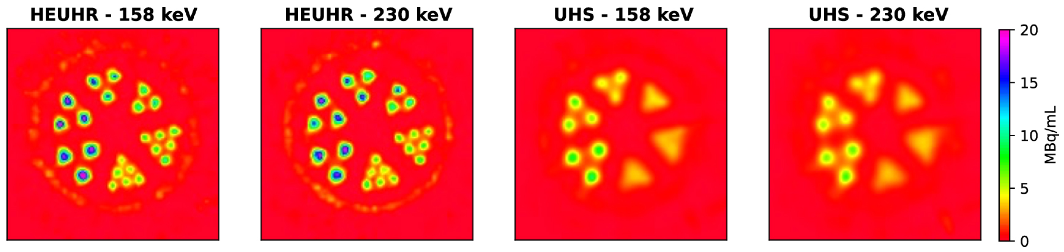
3.4. Actinium-226 (SPECT)

Actinium-226 (t1/2 = 29.4 h) can be produced via high-energy proton spallation of a uranium carbide target or lower-energy proton bombardment of 226Ra (t1/2 = 1600 y) target material. This involved bombarding a uranium carbide target with 480 MeV protons, with 226Ac separated using isotope separation online. This approach yielded 33.8 ± 2.7 MBq 226Ac for imaging purposes with high radionuclidic purity [55].
An alternative production route could employ 226Ra target material and the 226Ra(p,n)226Ac nuclear reaction on a lower energy proton cyclotron [9,55,56,57].
A phantom assembly with rods between 0.85 and 1.7 mm in diameter and a microSPECT/CT system was used to assess resolution using a high-energy ultra-high resolution (HEUHR) collimator and an extra ultra-high sensitivity (UHS) collimator. The primary 158 keV and 230 keV gamma photopeaks were reconstructed, with the 158 keV photopeak images demonstrating slightly better contrast recovery. For resolution, as depicted in Figure 3, the HEUHR collimator resolved all rods, while the UHS collimator could only resolve rods >1.3 mm and >1.5 mm for the 158 keV and 230 keV photopeaks, respectively [55]. This demonstrated the feasibility of using 226Ac as a SPECT imaging surrogate for 225Ac.
Advantages of 226Ac include its relatively long 29.4 h half-life compared to 132La and 133La, permitting imaging at extended time points, and its identical chemical properties to 225Ac. Limitations include challenges associated with routine irradiation of hazardous 226Ra target material, significant β co-emissions that would increase patient dose, and its decay to β emitting 226Th (t1/2 = 30 min), which further decays via multiple alpha and β emitting progeny before stabilizing at 206Pb [9].

4. Theranostic Imaging Surrogates Proposed for Lead-212

Lead-212 (t1/2 = 10.6 h) has cultivated a significant interest for TAT due to its payload of one alpha and two β particles in its decay chain and the rapid decay of its progeny to stable 208Pb. A recent study using a 212Pb somatostatin analogue demonstrated a significant antitumor effect in patients with metastatic neuroendocrine tumors, and additional radiopharmaceuticals are under development to treat other cancers [1,58,59,60,61,62]. Production of 212Pb involves synthesizing its parent radionuclide, 228Th (t1/2 = 1.9 y), via 226Ra irradiation in a nuclear reactor or high-energy proton spallation of 232Th target material. 212Pb can then be extracted in a convenient generator setup from 228Th or one of its intermediate progeny, 224Ra (t1/2 = 3.6 d) [12,63,64,65,66,67].
Previous clinical trials have employed imaging techniques with conventional radiometals such as 68Ga [58]. While direct SPECT imaging of 212Pb can be performed using its 239 keV (44%) gamma emissions [9], it is desirable to have an imaging surrogate that can be used for research owing to the limited supply of 212Pb and to provide the most accurate pre-therapy scans to assess patient eligibility for 212Pb TAT radiopharmaceuticals. While no positron-emitting Pb isotopes are suitable for use as 212Pb imaging surrogates, multiple gamma-ray emitters exist, with 203Pb being a prime candidate for SPECT imaging.

Lead-203 (SPECT)

Lead-203 (t1/2 = 51.9 h) emits X-rays and a primary 279 keV (81%) gamma photon that can be used for SPECT imaging. 203Pb has been synthesized via 203Tl(p,n)203Pb, 203Tl(d,2n)203Pb, and 205Tl(p,3n)203Pb nuclear reactions on cyclotrons [21,45,63,64,68,69,70,71]. Natural thallium metal can be used as a target material; however, significant precautions must be taken owing to the high toxicity of Tl, and its low thermal conductivity and melting point (304 °C) that makes it prone to melt or sublime under intense heat of a cyclotron beam. Natural Tl metal has been used as a target material, with one technique bombarding natTl at 25–26 MeV, producing up to 21 GBq 203Pb five days after end of bombardment [61]. However, irradiating natTl produces significant activities of 201Pb (t1/2 = 9.3 h), which must be permitted to decay significantly to achieve a 203Pb product with high radionuclidic impurity. 203Pb can be produced at lower proton energies using natural or isotopically enriched 203Tl and the 203Tl(p,n)203Pb nuclear reaction 63,71, with one process yielding up to 138.7 ± 5.1 MBq 203Pb [64]. However, yields are limited due to the low nuclear reaction cross-section in this energy window [45]. Alternatively, isotopically enriched 205Tl can be irradiated at 23–24 MeV proton energies to produce 203Pb via the 205Tl(p,3n)203Pb reaction. This produces significant activities of 203Pb (>12 GBq at the end of purification) with a high radionuclidic purity (>99.9%) made possible by the near absence of 203Tl and its resulting 201Pb co-production 21,63. Enriched 203Tl can also be bombarded with deuterons to produce 203Pb via the 203Tl(d,2n)203Pb reaction; however, this production route has a lower maximum cross-section compared to the 205Tl(p,3n)203Pb reaction, and 203Tl (29.5% natural isotopic abundance) is more expensive to enrich than 205Tl (70.5% natural isotopic abundance).
203Pb can be separated using ion exchange resins such as Pb resin, carboxymethyl resin, and Dowex-1X8 anion exchange resin. This can yield a concentrated 203Pb product in [203Pb]PbCl2 or [203Pb]Pb(OAc)2, with direct and rapid room temperature radiolabeling of [203Pb]Pb(OAc)2 using chelators such as DOTA, PSC, and TCMC. Radiolabeling achieves very high molar activities, and 203Pb chelate complexes have been shown to be highly stable in human serum up to 120 h [21,63,64,69,70].
Phantom imaging of 203Pb has been performed, with imaging spatial-resolution results comparable to 99mTc for 1.6–4.8 mm diameter fillable rod regions [72]. In vivo preclinical and clinical SPECT imaging of uncomplexed and chelated 203Pb has been performed [71,73]. Studies have included 203/212Pb-labeled PSMA and gastrin-releasing peptide receptor-targeting agents for imaging and radiotherapy of prostate-cancer-bearing mice [60,61,74,75], and 203/212Pb-labeled anti-melanin antibodies and melanocortin subtype 1 receptor targeting ligands for imaging and therapy of melanoma-bearing mice [59,72,73,76,77,78,79]. As shown in Figure 4, a PSMA-targeting 203Pb agent, [203Pb]Pb-CA012, exhibited a comparable biodistribution to [177Lu]Lu-PSMA 617 with high tumor uptake relative to other tissues [74].
Strengths of 203Pb include its relatively long 51.9 h half-life, which permits imaging at extended time points to inform 212Pb TAT dosimetry; its relatively clean X-ray and gamma photon emission spectrum that enables SPECT imaging using a low or high-energy collimator; its ability to rapidly and stably radiolabel targeting vectors under mild chemical conditions at room temperature (similar to 212Pb); and established production processes that provide 203Pb with high radionuclidic purity in yields suitable for multiple patients per production run. Limitations include risks associated with preparing and irradiating highly toxic thallium targets and potential uncertainties with using 203Pb pharmacokinetic data for 212Pb therapy planning due to the release of 212Bi progeny during 212Pb decay [80].

5. Theranostic Imaging Surrogates Proposed for Radium-223/224
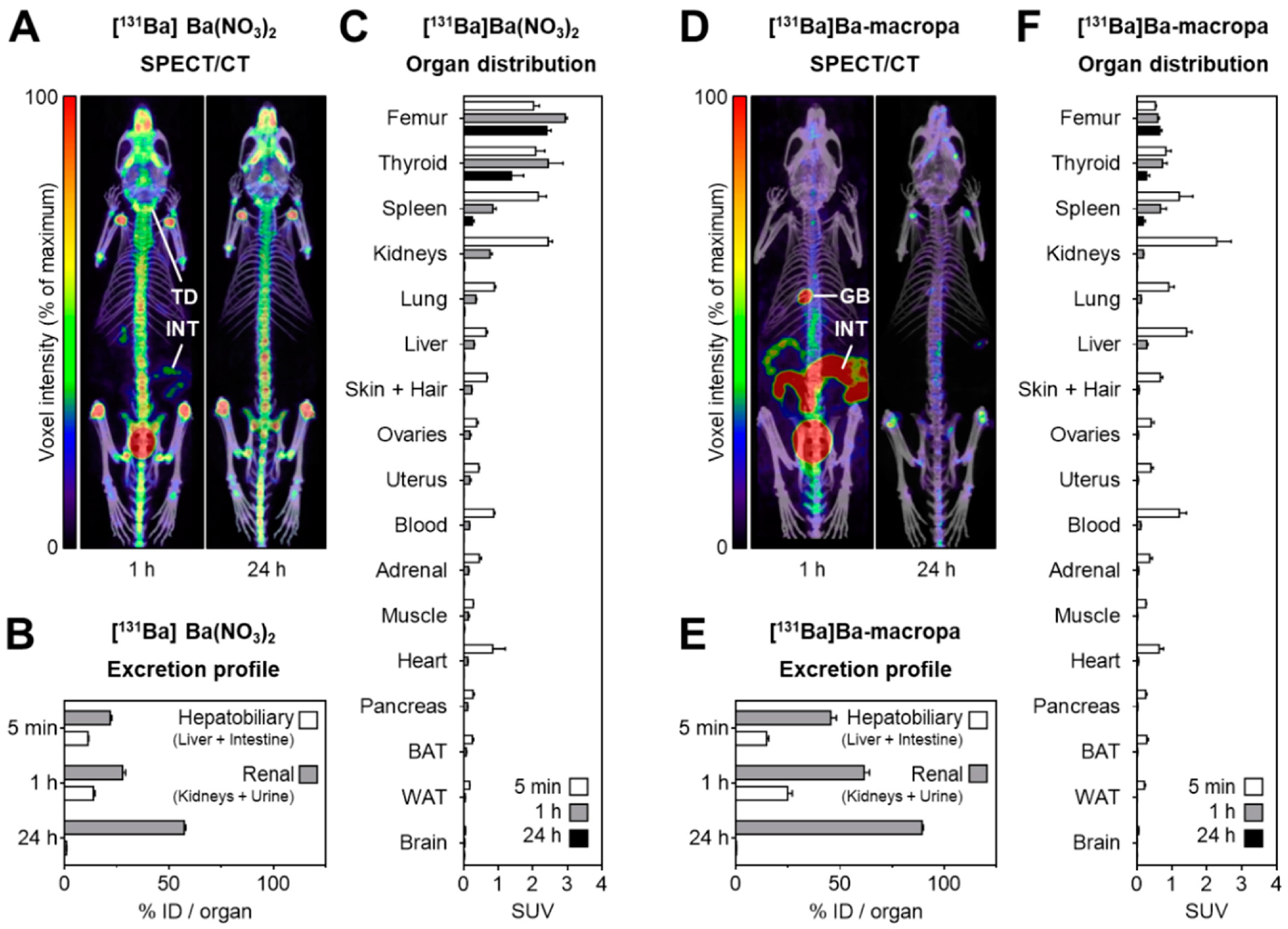
Radium-223 (t1/2 = 11.4 d) is used as an alpha therapy for men with bone-metastatic castration-resistant prostate cancer. It works as a calcium-mimetic by accumulating in and irradiating osteoblastic lesions, while sparing most surrounding healthy tissue [81]. It is the only FDA-proved alpha-particle-emitting radiopharmaceutical (Xofigo®) and has been used to treat over 18,000 patients since 2013 [82]. However, unlike targeted alpha therapy, 223Ra is currently administered as a [223Ra]RaCl2 salt in an aqueous buffer without a chelator or biological-targeting agent. Therefore, the established clinical efficacy and safety of 223Ra makes it an attractive TAT candidate [82]. Similarly, 224Ra (t1/2 = 3.6 d) has been employed in a dual targeting strategy with 212Pb, where 224Ra accumulates at primary bone cancer sites or bone metastases, while extra-skeletal metastases can be targeted with a 212Pb-labeled cancer-specific vector [83,84]. [224Ra]RaCl2 (marketed as 224SpondylAT® (Eckert & Ziegler, Berlin, Germany) has also been used to treat bone and joint disease, ankylosing spondylitis [85], while 224Ra is also under investigation for a novel brachytherapy called diffusing alpha-emitter radiation therapy (DaRT). In DaRT, 224Ra-infused seeds are inserted into solid tumors, which are then irradiated with alpha emissions released during the diffusion and subsequent decay cascade of its 220Rn progeny [86,87,88,89,90,91,92,93,94,95]. Both 223Ra and 224Ra are currently produced in significant activities as by-products and decay daughters of neutron irradiation of 226Ra in a nuclear reactor. With proven purification techniques, this positions these radionuclides well for TAT [67,96,97].
223Ra has recently been stably complexed with the chelator macropa, where a [223Ra]Ra–macropa complex exhibited rapid clearance and low 223Ra bone absorption, suggesting in vivo stability. This has opened the possibility of using 223Ra complexed using functionalized chelators to target metastases beyond the bone, similar to other radionuclides used in targeted alpha therapy [82,98].
While 223Ra possesses several gamma emissions within an energy window suitable for SPECT imaging (223Ra: 269 keV, (13%); 154 keV (6%); 224Ra: 241 keV (4.1%)), the low intensity of these gamma photons would likely be insufficient to generate a high-quality SPECT image when considering the relatively low injected therapeutic activity (~50 kBq/kg) injected [9,81]. Similarly, a relatively low injection activity of 224Ra due to its 3.6 d half-life could complicate direct SPECT imaging. Therefore, an imaging surrogate is desirable to assess the viability of 223/224Ra radiopharmaceuticals, with 131Ba emerging as a candidate.

Barium-131 (SPECT)

Barium-131 (t1/2 = 11.5 d) decays via electron capture to 131Cs (t1/2 = 9.7 d) and subsequently to stable 131Xe, emitting gamma rays suitable for SPECT imaging (496 keV (48%); 216 keV (20%); 124 keV (30%); 371 keV (14%)) [9]. Additionally, approaches designed to sequester Ra (nanoparticles, chelation via macropa or ligands based on the arene scaffold) [99,100] should be transferrable owing to the proven use of Ba as a non-radioactive surrogate for Ra [101]. Therefore, the favorable imaging emissions of 131Ba compared to other Ba radionuclides (135mBa, 133mBa), and the similar half-life and chemistry of 131Ba to 223/224Ra positions 131Ba as a promising surrogate to track in vivo 223/224Ra biodistribution.
131Ba can be produced via neutron irradiation of isotopically enriched 130Ba (natural abundance = 0.1%) in a nuclear reactor, which would co-produce significant activities of 133Ba [45,102]. Alternatively, 131Ba can be produced via proton irradiation of natural cesium target material in a cyclotron via the 133Cs(p,3n)133Ba nuclear reaction with a small 133Ba contamination (0.01%) at beam energies of 27.5 MeV [45,101]. A 4 h irradiation yielded 190 ± 26 MBq 131Ba, and an SR resin was used to separate 131Ba from the Cs target material. 131Ba was subsequently successfully radiolabeled to macropa, and exhibited stability in human serum [101].
SPECT imaging was performed in a cylindrical syringe, which enabled visualization of the radionuclide distribution. However, image quality was limited due to artifacts caused by the higher energy gamma photon emissions. As highlighted in Figure 5, small animal SPECT/CT was performed with [131Ba]Ba(NO3)2, showing 131Ba accumulation within the entire skeleton 1 h post-injection, which was still present 24 h after injection. Additional SPECT imaging was performed with [131Ba]Ba-macropa, with rapid clearance observed through the intestines and gallbladder [101]. This demonstrated the feasibility of using 131Ba as a SPECT imaging surrogate for 223/224Ra.
Advantages of 131Ba include its relatively long half-life, which is similar to 223Ra, permitting imaging at extended time points; the ability to sequester 131Ba in the macropa chelator similar to 223Ra; and established 131Ba production routes. Limitations include higher energy gamma photon emissions, which increase unintended patient dose and can cause image artifacts. The presence of co-produced 133Ba may also require additional dosimetric analysis. Additionally, the decay of 131Ba to 131Cs with X-ray emissions adds a suboptimal patient radioactive dose compared to an imaging radionuclide with direct decay to stable progeny. Finally, further improvements in the cyclotron production route would be required to synthesize enough activity for multiple patients in a single batch.

6. Theranostic Imaging Surrogates Proposed for Astatine-211

Astatine-211 (t1/2 = 7.2 h) has garnered interest for TAT owing to its decay to either 207Bi (t1/2 = 31.6 y) via alpha emission or to 211Pb via electron capture followed by alpha decay to stable 207Pb [9]. Therefore, each 211At decay yields one alpha particle. The 211At decay chain also emits few high-energy gamma photons, which avoids excess radiation dose [8]. 211At can be produced in medium-energy alpha cyclotrons using bismuth target material and the 209Bi(α,2n)211At nuclear reaction or via heavy ion irradiation and the 209Bi(7Li,5n)211Rn reaction, where 211At is obtained via decay of its longer-lived parent 211Rn (t1/2 = 14.6 h) in a generator configuration [8,103,104]. Production yields of up to 6.6 GBq have been reported, which would be sufficient for clinical radiopharmaceutical production for several patients and distribution several hours from the production site [8,105].
211At was initially investigated for treating thyroid disorders and is currently being evaluated in clinical trials for multiple myeloma, leukemia, myelodysplastic syndromes, thyroid cancer, and malignant pheochromocytoma [106]. While direct SPECT imaging of 211At is possible using the X-rays emitted during 211At decay to 211Po, it is desirable to have an imaging surrogate to perform pre-therapy assessment scans and research, owing to the limited supply and short half-life of 211At that generally precludes its use at facilities located more than several hours from a production site. Several candidates exist for use as 211At diagnostic imaging surrogates: chemically identical 209At, or chemically similar 123I, 124I and 131I.

6.1. Iodine-123 (SPECT)

Iodine-123 (t1/2 = 13.2 h) decays via electron capture to near-stable 123Te, and is commonly used in nuclear medicine and research of various malignancies and biological processes, including thyroid diseases and tumor imaging [107]. Its X-ray emissions and primary gamma photopeak of 159 keV (83.6%) are well suited for SPECT imaging [9].
123I is primarily produced via the 124Xe(p,2n)123I nuclear reaction using a highly enriched 124Xe gas target, which enables 123I production with a high yield and radionuclidic purity. The subsequent 123I product is commercially available in dilute NaOH solutions [108,109].
Strengths of 123I include its favorable emission spectrum for SPECT imaging, similar half-life relative to 211At, and commercial availability. Limitations include hazards associated with volatile radioactive products, the lower image quality of SPECT images to PET imaging, and the low natural abundance (0.095%) of 124Xe target material.

6.2. Iodine-124 (PET)

Iodine-124 (t1/2 = 4.2 d) undergoes positron decay to stable 124Te and is employed for PET imaging studies. Its relatively long half-life allows extended radiosynthesis, quantitative imaging over several days, and distribution to sites far from production facilities [9]. 124I is typically produced using isotopically enriched 124Te and the 124Te(d,2n)124I or 124Te(p,n)124I nuclear reactions [110,111]. Applications in nuclear medicine and research have been extensive, including thyroid and parathyroid imaging, studies of neurotransmitter receptors, and monoclonal antibody imaging in cancer [110].
Strengths of 124I include its long half-life that eases logistics and allows imaging at extended time points. Limitations include hazards associated with volatile radioactive products; a relatively low positron branching ratio (22.7%); relatively high average positron emission energy (Emean = 820 keV) that results in a lower spatial resolution compared to other PET radionuclides; and co-emitted gamma rays (603 keV (63%), 1691 keV (11%)) that increase dose and shielding requirements [9].

6.3. Iodine-131 (SPECT)

Iodine-131 (t1/2 = 8.0 d) undergoes β decay to stable 131Xe, and similar to 123I and 124I, it is primarily used for treating thyroid malignancies [107]. 131I can be produced in a nuclear reactor by irradiating either 130Te or uranium targets [112].
Strengths of 131I include its 8 d half-life that permits imaging at extended time points, commercial availability, and primary 364 keV (81.5%) gamma emission that is well suited for SPECT imaging. However, limitations include hazards associated with volatile radioactive products and significant β emissions that would increase patient dose [9].

6.4. Astatine-209 (SPECT)

Astatine-209 (t1/2 = 5.4 h) decays via alpha emissions (4%) to 205Bi (t1/2 = 14.9 d) followed by decay to stable 205Pb, or via electron capture (96%) to 209Po (t1/2 = 124 y). During decay to 209Po, X-rays and gamma emissions (545 keV (91.0%), 195 keV (22.6%), and 239 keV (12.4%) enable SPECT imaging. 209At can be produced via high-energy proton spallation of a uranium carbide target, followed by online surface ionization and A = 213 isobars separation. This can yield 209At in activities on the order of 102 MBq [113]. Subsequent chemical purification employs a Te column to obtain purified 209At [113,114]. As shown in Figure 6, subsequent studies using 209At for phantom imaging demonstrated that image reconstruction with 209At X-ray emissions was superior to using its gamma emissions [114]. Additionally, in vivo imaging measurements of 209At uptake in mice matched ex vivo measurements within 10%. This demonstrated the potential of using 209At to accurately determine astatine biodistributions [114].
Strengths include identical chemistry to 211At, which would give more certainty to 209At pharmacokinetic data. Limitations include alpha emissions in 209At decay that would require dosimetric evaluation; numerous high-energy gamma rays that complicate shielding and increase patient dose; the need to consider longer-lived 205Bi in dosimetry evaluations; and production/logistical challenges associated with distributing relatively short-lived 209At from a limited number of facilities capable of high-energy proton spallation and separation of 211At from actinide targets [8].

7. Theranostic Imaging Surrogates Proposed for Thorium-227

Thorium-227 (t1/2 = 18.7 d) decays via alpha emission to 223Ra and can be harvested from a generator containing 227Ac (t1/2 = 21.8 y) that is produced via nuclear reactor irradiation of 226Ra [115]. Thorium can be complexed with octadentate 3,2-hydroxypyridinone (3,2-HOPO) chelators attached to biological-targeting vectors 115. Ongoing clinical studies involving 227Th TAT include targeting tumors expressing human epidermal growth factor receptor 2 (HER2), PSMA, mesothelin (MSLN), and CD22 [116]. 227Th does emit a 236 keV (12.9%) gamma photon that would be suitable for SPECT imaging. However, the long half-life of 227Th relative to other TAT radionuclides would likely result in a low injected therapeutic activity, which could be insufficient for direct imaging 9. Therefore, an imaging surrogate to assess 227Th radiopharmaceutical pharmacokinetics is desirable, with the 134Ce/134La PET imaging pair showing promise (see Section 3.3). A significant uncertainty of using any theranostic imaging pair with 227Th involves its long-lived 223Ra progeny, which has the potential for substantial redistribution and alpha irradiation of healthy tissue after decay from 227Th. This would significantly complicate direct comparisons between imaging and inferred therapeutic dosimetry and require further study.

8. Theranostic Imaging Surrogates Proposed for Terbium-149

Terbium-149 (t1/2 = 4.1 h) is a unique radionuclide for TAT. It emits low-energy alpha particles with a short tissue range and decays via several daughter radionuclides to stable 145Nd and 141Pr, without any subsequent alpha emissions [9]. This absence of alpha-emitting progeny is regarded as a potential strength for 149Tb TAT. 149Tb is produced via high-energy proton spallation of a tantalum target followed by online isotope separation or 3He bombardment of a 151Eu target [19,20,117,118]. 100 MBq of 149Tb was obtained in a solution suitable for preclinical applications and successfully labeled to a DOTANOC targeting vector [118]. While PET images were successfully obtained using [149Tb]Tb-DOTANOC in a mouse model, 149Tb possesses a relatively low positron branching ratio (21%) and relatively high positron emission energy (Emean = 805 keV). These physical factors could present challenges to obtaining high-quality clinical PET images. Additionally, due to limited production and the resulting extreme scarcity of 149Tb, imaging surrogates would be helpful research tools to evaluate its potential for TAT. Two surrogate candidates are 155Tb and 152Tb.

8.1. Terbium-155 (SPECT)

Terbium-155 (t1/2 = 5.3 d) decays via electron capture to stable 155Gd, with X-ray and gamma-ray emissions including 87 keV (32%), 105 keV (25%), 180 keV (7.5%), and 262 keV (5%) [9]. 155Tb can be produced via the 156Gd(p,2n)155Tb reaction at 23 MeV, or the 155Gd(p,n)155Tb reaction at 10 MeV [119]. The 156Gd(p,2n)155Tb has higher demonstrated production yields (up to 1.7 GBq); however, it has a lower radionuclidic purity compared to the final product of the 155Gd(p,n)155Tb reaction (200 MBq yield). Subsequently, phantom and in vivo SPECT/CT studies were successfully performed with [155Tb]Tb-DOTATOC, demonstrating a similar image quality to 111In [119,120].
Advantages of 155Tb include its accessible production routes that can synthesize multi-patient activities per run, decay to stable 155Gd, and its long half-life that enables long-duration imaging. Limitations include relatively low imaging performance compared to other diagnostic radionuclides, such as PET emitters.

8.2. Terbium-152 (PET)

Terbium-152 (t1/2 = 17.5 h) decays via positron emission to near-stable 152Gd with a positron branching ratio of 20.3% and an average positron energy of 1140 keV [121]. Several primary co-emitted gamma rays include 344 keV (63.5%), 271 keV (9.5%), 586 keV (9.2%), and 779 keV (5.5%). 152Tb synthesis is extremely limited, with the existing production route involving high-energy proton spallation of a tantalum target at 1.4 GeV and online isotope separation [122]. Following chemical separation, phantom studies revealed increased image noise due to the smaller positron branching ratio of 152Tb, and subsequently [152Tb]Tb-DOTANOC was administered to a patient and used to acquire PET scans [121].
Advantages of 152Tb include a relatively long half-life permitting imaging at extended time points and its decay to near-stable 152Gd. Limitations include the scarcity of facilities capable of achieving proton energies for production, the higher average positron emission energy, and significant co-emitted gamma rays that increase the radioactive dose.

9. Summary and Outlook for Alpha-Emitter Imaging Surrogates

As highlighted in this article, multiple SPECT and PET imaging surrogates have demonstrated the potential to enhance clinical TAT applications and research. Table 1 presents a summary of proposed theranostic imaging surrogates for alpha emitters, along with their properties and production status.
Production capabilities must be augmented to enable more patients and research efforts to benefit from TAT imaging surrogates. Existing medium-energy cyclotron facilities are well positioned to improve the supply chain of imaging surrogates such as 133La, 203Pb, and 155Tb by adapting and optimizing established production techniques to the unique capabilities of each facility. A stable supply of isotopically enriched accelerator target material will be required to support growing production efforts for many of these radionuclides. Other imaging surrogates such as 226Ra, 152Tb, 209At, and 134Ce/134La require high-energy accelerators, bombarding hazardous target material, and techniques such as mass separation to enable their production. While these surrogates have demonstrated research potential, their widespread deployment for radiopharmaceutical development and clinical application may be limited owing to the scarcity of facilities capable of their production.
Except for 149Tb, which possesses a single alpha emission in its decay chain, most TAT radionuclides, including 225Ac, 212Pb, 223Ra, 224Ra, 227Th, and 211At, possess a cascade of decay progeny that are released from the original target site due to recoil energy and deposit additional alpha radiation in surrounding healthy tissues. While the highlighted imaging surrogates are well positioned to provide more accurate dosimetry data for the TAT parent radionuclide decay, there will be a degree of uncertainty regarding the dose from alpha-emitting decay progeny. This uncertainty will depend on the type of malignancy, internalization within targeted cells, and other factors within the disease microenvironment that influence the radiopharmaceutical pharmacokinetics. However, this limitation does not negate the improved accuracy of biodistribution dosimetry data conferred by using imaging surrogates matched to the TAT parent radionuclide, particularly when radionuclides are stably bound to their targeting vector. Therefore, TAT imaging surrogates have the potential to assist the preclinical development and clinical deployment of TAT radiopharmaceuticals and represent a significant improvement over conventional PET and SPECT imaging radionuclides currently paired with TAT.

10. Conclusions

Recent preclinical and clinical advances in targeted alpha therapy have spurred significant interest in utilizing alpha-emitting radiopharmaceuticals to treat metastatic cancers and other malignancies. Despite their strong potential, TAT radiopharmaceuticals suffer from an acute supply shortage of alpha-emitting radionuclides due to production constraints. This severely restricts the availability for patient therapy and slows the development of new TAT radiopharmaceuticals. Additionally, many alpha-emitting radionuclides do not possess radioactive emissions suitable for diagnostic imaging. This often leads to diagnostic radiopharmaceuticals being employed with suboptimally paired imaging radionuclides that possess different chemistries from their therapeutic counterpart, which can potentially result in different radiopharmaceutical biodistributions. Therefore, increasing the availability of SPECT and PET imaging TAT surrogates has strong potential to improve the accuracy of dosimetry and treatment tracking, and enhance TAT research output by using more economical and less potent diagnostic radionuclides for preclinical radiopharmaceutical development. Therefore, TAT imaging surrogates hold potential to improve the accuracy of diagnostic scans, equipping clinicians and researchers with more accurate biodistribution and dosimetry data that they can use to expedite the development and deployment of novel TAT radiopharmaceuticals.

Author Contributions

B.J.B.N. prepared the manuscript draft; B.J.B.N., J.W., J.D.A. and F.W. performed review and editing; and J.D.A. and F.W. provided supervision. All authors have read and agreed to the published version of the manuscript.

Funding

The authors would like to thank the Dianne and Irving Kipnes Foundation for supporting this work.

Data Availability Statement

Data sharing is not applicable.

Conflicts of Interest

The authors declare no conflict of interest.

References

  1. Schultz, M.K.; Pouget, J.P.; Wuest, F.; Nelson, B.; Andersson, J.; Cheal, S.; Li, M.; Ianzini, F.; Sangeeta, R.; Graves, S.; et al. Radiobiology of Targeted Alpha Therapy. In Nuclear Medicine and Molecular Imaging; Signore, A., Ed.; Elsevier: Amsterdam, The Netherlands, 2022; pp. 380–403. [Google Scholar] [CrossRef]
  2. Nelson, B.J.B.; Andersson, J.D.; Wuest, F. Targeted alpha therapy: Progress in radionuclide production, radiochemistry and applications. Pharmaceutics 2021, 13, 49. [Google Scholar] [CrossRef] [PubMed]
  3. Sartor, O.; de Bono, J.; Chi, K.N.; Fizazi, K.; Herrmann, K.; Rahbar, K.; Tagawa, S.T.; Nordquist, L.T.; Vaishampayan, N.; El-Haddad, G.; et al. Lutetium-177–PSMA-617 for Metastatic Castration-Resistant Prostate Cancer. NEJM 2021, 385, 1091–1103. [Google Scholar] [CrossRef]
  4. Strosberg, J.; El-Haddad, G.; Wolin, E.; Hendifar, A.; Yao, J.; Chasen, B.; Mittra, E.; Kunz, P.L.; Kulke, M.H.; Jacene, H.; et al. Phase 3 Trial of 177Lu-Dotatate for Midgut Neuroendocrine Tumors. NEJM 2017, 376, 125–135. [Google Scholar] [CrossRef] [PubMed]
  5. Garg, R.; Mills, K.; Allen, K.J.H.; Causey, P.; Perron, R.W.; Gendron, D.; Sanche, S.; Berman, J.W.; Gorny, M.K.; Dadachova, E. Comparison of various radioactive payloads for a human monoclonal antibody to glycoprotein 41 for elimination of HIV-infected cells. Nucl. Med. Biol. 2020, 82–83, 80–88. [Google Scholar] [CrossRef]
  6. Elgqvist, J.; Frost, S.; Pouget, J.P.; Albertsson, P. The Potential and Hurdles of Targeted Alpha Therapy–Clinical Trials and Beyond. Front. Oncol. 2014, 3, 324. Available online: https://www.frontiersin.org/articles/10.3389/fonc.2013.00324 (accessed on 29 September 2023). [CrossRef] [PubMed]
  7. Sollini, M.; Marzo, K.; Chiti, A.; Kirienko, M. The five “W”s and “How” of Targeted Alpha Therapy: Why? Who? What? Where? When? and How? Rend. Lincei 2020, 31, 231–247. [Google Scholar] [CrossRef]
  8. Lindegren, S.; Albertsson, P.; Bäck, T.; Jensen, H.; Palm, S.; Aneheim, E. Realizing Clinical Trials with Astatine-211: The Chemistry Infrastructure. Cancer Biother. Radiopharm. 2020, 35, 425–436. [Google Scholar] [CrossRef]
  9. Sonzogni, A.; Shu, B. Nudat 2.8 (Nuclear Structure and Decay Data). 2020. Available online: https://www.nndc.bnl.gov/nudat2/reCenter.jsp?z=56&n=77> (accessed on 20 September 2023).
  10. Bannik, K.; Madas, B.; Jarzombek, M.; Sutter, A.; Siemeister, G.; Mumberg, D.; Zitzmann-Kolbe, S. Radiobiological effects of the alpha emitter Ra-223 on tumor cells. Sci. Rep. 2019, 9, 18489. [Google Scholar] [CrossRef]
  11. Morgenstern, A.; Apostolidis, C.; Kratochwil, C.; Sathekge, M.; Krolicki, L.; Bruchertseifer, F. An Overview of Targeted Alpha Therapy with 225Actinium and 213Bismuth. Curr. Radiopharm. 2018, 11, 200–208. [Google Scholar] [CrossRef]
  12. Kokov, K.V.; Egorova, B.V.; German, M.N.; Klabukov, I.D.; Krasheninnikov, M.E.; Larkin-Kondrov, A.A.; Makoveeva, K.A.; Ovchinnikov, M.V.; Sidorova, M.V.; Chuvilin, D.Y. 212Pb: Production Approaches and Targeted Therapy Applications. Pharmaceutics 2022, 14, 189. [Google Scholar] [CrossRef]
  13. Abou, D.S.; Fears, A.; Summer, L.; Longtine, M.; Benabdallah, N.; Riddle, R.C.; Ulmert, D.; Michalski, J.; Wahl, R.L.; Chesner, D.; et al. Improved Radium-223 Therapy with Combination Epithelial Sodium Channel Blockade. J. Nucl. Med. 2021, 62, 1751–1758. [Google Scholar] [CrossRef] [PubMed]
  14. Hosono, M.; Ikebuchi, H.; Nakamura, Y.; Yanagida, S.; Kinuya, S. Introduction of the targeted alpha therapy (with Radium-223) into clinical practice in Japan: Learnings and implementation. Ann. Nucl. Med. 2019, 33, 211–221. [Google Scholar] [CrossRef] [PubMed]
  15. Mikalsen, L.T.G.; Kvassheim, M.; Stokke, C. Optimized SPECT Imaging of 224Ra α-Particle Therapy by 212Pb Photon Emissions. J. Nucl. Med. 2023, 64, 1131–1137. [Google Scholar] [CrossRef] [PubMed]
  16. Watabe, T.; Kaneda-Nakashima, K.; Shirakami, Y.; Kadonaga, Y.; Ooe, K.; Wang, Y.; Haba, H.; Toyoshima, A.; Cardinale, J.; Giesel, F.L.; et al. Targeted α-therapy using astatine (211At)-labeled PSMA1, 5, and 6: A preclinical evaluation as a novel compound. Eur. J. Nucl. Med. Mol. Imaging 2023, 50, 849–858. [Google Scholar] [CrossRef] [PubMed]
  17. Jang, A.; Kendi, A.T.; Johnson, G.B.; Halfdanarson, T.R.; Sartor, O. Targeted Alpha-Particle Therapy: A Review of Current Trials. Int. J. Mol. Sci. 2023, 24, 11626. [Google Scholar] [CrossRef] [PubMed]
  18. Beyer, G.J.; Miederer, M.; Vranjes-Durić, S.; Comor, J.J.; Künzi, G.; Hartley, O.; Senekowitsch-Schmidtke, R.; Soloviev, D.; Buchegger, F. Targeted alpha therapy in vivo: Direct evidence for single cancer cell kill using 149Tb-rituximab. Eur. J. Nucl. Med. Mol. Imaging 2004, 31, 547–554. [Google Scholar] [CrossRef]
  19. Umbricht, C.A.; Köster, U.; Bernhardt, P.; Gracheva, N.; Johnston, K.; Schibli, R.; van der Meulen, N.P.; Müller, C. Alpha-PET for Prostate Cancer: Preclinical investigation using 149Tb-PSMA-617. Sci. Rep. 2019, 9, 17800. [Google Scholar] [CrossRef]
  20. Aliev, R.A.; Zagryadskiy, V.A.; Latushkin, S.T.; Moiseeva, A.N.; Novikov, A.N.; Unezhev, V.N.; Kazakov, A.G. Production of a Short-Lived Therapeutic α-Emitter 149Tb by Irradiation of Europium by 63 MeV α-Particles. At. Energy 2021, 129, 337–340. [Google Scholar] [CrossRef]
  21. Saini, S.; Bartels, J.L.; Appiah, J.K.; Rider, J.H.; Baumhover, N.; Schultz, M.K.; Lapi, S.E. Optimized Methods for the Production of High-Purity 203Pb Using Electroplated Thallium Targets. J. Nucl. Med. 2023, 64, 1791–1797. [Google Scholar] [CrossRef]
  22. Fani, M.; Del Pozzo, L.; Abiraj, K.; Mansi, R.; Tamma, M.L.; Cescato, R.; Waser, B.; Weber, W.A.; Reubi, J.C.; Maecke, H.R. PET of Somatostatin Receptor–Positive Tumors Using 64Cu- and 68Ga-Somatostatin Antagonists: The Chelate Makes the Difference. J. Nucl. Med. 2011, 52, 1110. [Google Scholar] [CrossRef]
  23. Fani, M.; Braun, F.; Waser, B.; Beetschen, K.; Cescato, R.; Erchegyi, J.; Rivier, J.E.; Weber, W.A.; Maecke, H.R.; Reubi, J.C. Unexpected sensitivity of sst2 antagonists to N-Terminal radiometal modifications. J. Nucl. Med. 2012, 53, 1481. [Google Scholar] [CrossRef] [PubMed]
  24. Busslinger, S.D.; Tschan, V.J.; Richard, O.K.; Talip, Z.; Schibli, R.; Müller, C. [225Ac]Ac-SibuDAB for Targeted Alpha Therapy of Prostate Cancer: Preclinical Evaluation and Comparison with [225Ac]Ac-PSMA-617. Cancers 2022, 14, 5651. [Google Scholar] [CrossRef] [PubMed]
  25. King, A.P.; Gutsche, N.T.; Raju, N.; Fayn, S.; Baidoo, K.E.; Bell, M.M.; Olkowski, C.S.; Swenson, R.E.; Lin, F.I.; Sadowski, S.M. 225Ac-Macropatate: A Novel α-Particle Peptide Receptor Radionuclide Therapy for Neuroendocrine Tumors. J. Nucl. Med. 2023, 64, 549. [Google Scholar] [CrossRef]
  26. Yadav, M.P.; Ballal, S.; Sahoo, R.K.; Bal, C. Efficacy and safety of 225Ac-DOTATATE targeted alpha therapy in metastatic paragangliomas: A pilot study. Eur. J. Nucl. Med. Mol. Imaging 2022, 49, 1595–1606. [Google Scholar] [CrossRef] [PubMed]
  27. Kratochwil, C.; Bruchertseifer, F.; Giesel, F.L.; Weis, M.; Verburg, F.A.; Mottaghy, F.; Kopka, K.; Apostolidis, C.; Haberkorn, U.; Morgenstern, A. 225Ac-PSMA-617 for PSMA-targeted a-radiation therapy of metastatic castration-resistant prostate cancer. J. Nucl. Med. 2016, 57, 1941–1944. [Google Scholar] [CrossRef]
  28. Rathke, H.; Bruchertseifer, F.; Kratochwil, C.; Keller, H.; Giesel, F.L.; Apostolidis, C.; Haberkorn, U.; Morgenstern, A. First patient exceeding 5-year complete remission after 225Ac-PSMA-TAT. Eur. J. Nucl. Med. Mol. Imaging 2021, 48, 311–312. [Google Scholar] [CrossRef]
  29. Zacherl, M.J.; Gildehaus, F.J.; Mittlmeier, L.; Böning, G.; Gosewisch, A.; Wenter, V.; Unterrainer, M.; Schmidt-Hegemann, N.; Belka, C.; Kretschmer, A.; et al. First Clinical Results for PSMA-Targeted α-Therapy Using 225Ac-PSMA-I&T in Advanced-mCRPC Patients. J. Nucl. Med. 2021, 62, 669–674. [Google Scholar] [CrossRef] [PubMed]
  30. Sathekge, M.; Bruchertseifer, F.; Vorster, M.; Lawal, I.O.; Knoesen, O.; Mahapane, J.; Davis, C.; Reyneke, F.; Maes, A.; Kratochwil, C. Predictors of overall and disease-free survival in metastatic castration-resistant prostate cancer patients receiving 225Ac-PSMA-617 radioligand therapy. J. Nucl. Med. 2020, 61, 62–69. [Google Scholar] [CrossRef]
  31. Zimmermann, R. Is Actinium Really Happening? J. Nucl. Med. 2023, 64, 1516–1518. [Google Scholar] [CrossRef]
  32. Robertson, A.K.H.; Ramogida, C.F.; Schaffer, P.; Radchenko, V. Development of 225Ac Radiopharmaceuticals: TRIUMF Perspectives and Experiences. Curr. Radiopharm. 2018, 11, 156–172. [Google Scholar] [CrossRef]
  33. Engle, J. The Production of Ac-225. Curr. Radiopharm. 2018, 11, 173–179. [Google Scholar] [CrossRef] [PubMed]
  34. Grimm, T.; Grimm, A.; Peters, W.; Zamiara, M. High-Purity Actinium-225 Production from Radium-226 using a Superconducting Electron Linac. J. Med. Imaging Radiat. Sci. 2019, 50, S12–S13. [Google Scholar] [CrossRef]
  35. Augusto, R.S.; Smith, J.; Varah, S.; Paley, W.; Egoriti, L.; McEwen, S.; Day Goodacre, T.; Mildenberger, J.; Gottberg, A.; Trudel, A.; et al. Design and radiological study of the 225Ac medical target at the TRIUMF-ARIEL proton-target station. Radiat. Phys. Chem. 2022, 201, 110491. [Google Scholar] [CrossRef]
  36. Robertson, A.K.H.; Lobbezoo, A.; Moskven, L.; Schaffer, P.; Hoehr, C. Design of a thorium metal target for 225Ac production at TRIUMF. Instruments 2019, 3, 18. [Google Scholar] [CrossRef]
  37. Deblonde, G.J.P.; Zavarin, M.; Kersting, A.B. The coordination properties and ionic radius of actinium: A 120-year-old enigma. Coord. Chem. Rev. 2021, 446, 214130. [Google Scholar] [CrossRef]
  38. Shannon, R.D. Revised effective ionic radii and systematic studies of interatomic distances in halides and chalcogenides. Acta Crystallogr. 1976, 32, 751–767. [Google Scholar] [CrossRef]
  39. Nelson, B.J.B.; Andersson, J.D.; Wuest, F. Radiolanthanum: Promising theranostic radionuclides for PET, alpha, and Auger-Meitner therapy. Nucl. Med. Biol. 2022, 110–111, 59–66. [Google Scholar] [CrossRef]
  40. Nelson, B.J.B.; Wilson, J.; Andersson, J.D.; Wuest, F. High yield cyclotron production of a novel 133/135La theranostic pair for nuclear medicine. Sci. Rep. 2020, 10, 22203. [Google Scholar] [CrossRef]
  41. Nelson, B.J.B.; Ferguson, S.; Wuest, M.; Wilson, J.; Duke, M.J.M.; Richter, S.; Soenke-Jans, H.; Andersson, J.D.; Juengling, F.; Wuest, F. First in vivo and phantom imaging of cyclotron produced 133La as a theranostic radionuclide for 225Ac and 135La. J. Nucl. Med. 2022, 63, 584–590. [Google Scholar] [CrossRef]
  42. Brühlmann, S.A.; Kreller, M.; Pietzsch, H.J.; Kopka, K.; Mamat, C.; Walther, M.; Reissig, F. Efficient Production of the PET Radionuclide 133La for Theranostic Purposes in Targeted Alpha Therapy Using the 134Ba(p,2n)133La Reaction. Pharmaceuticals 2022, 15, 1167. [Google Scholar] [CrossRef]
  43. Yang, H.; Zhang, C.; Yuan, Z.; Rodriguez-Rodriguez, C.; Robertson, A.; Radchenko, V.; Perron, R.; Gendron, D.; Causey, P.; Gao, F.; et al. Synthesis and Evaluation of a Macrocyclic Actinium-225 Chelator, Quality Control and In Vivo Evaluation of 225Ac-crown-αMSH Peptide. Chem. Eur. J. 2020, 26, 11435–11440. [Google Scholar] [CrossRef]
  44. Thiele, N.A.; Brown, V.; Kelly, J.M.; Amor-Coarasa, A.; Jermilova, U.; MacMillan, S.N.; Nikolopoulou, A.; Ponnala, S.; Ramogida, C.F.; Robertson, A.K.H.; et al. An Eighteen-Membered Macrocyclic Ligand for Actinium-225 Targeted Alpha Therapy. Angew. Chem. Int. Ed. Engl. 2017, 129, 14904–14909. [Google Scholar] [CrossRef]
  45. Rochman, D.; Koning, A.J.; Sublet, J.C.; Fleming, M.; Bauge, E.; Hilaire, S.; Romain, P.; Morillon, B.; Duarte, H.; Goriely, S.; et al. The TENDL Library: Hope, Reality, and Future. In Proceedings of the International Conference on Nuclear Data for Science and Technology, Bruges, Belgium, 11–16 September 2016; EDP Sciences: Les Ulis, France, 2017. [Google Scholar]
  46. Ferguson, S.; Jans, H.S.; Wuest, M.; Riauka, T.; Wuest, F. Comparison of scandium-44 g with other PET radionuclides in preclinical PET phantom imaging. EJNMMI Phys. 2019, 6, 23. [Google Scholar] [CrossRef] [PubMed]
  47. Levin, C.S.; Hoffman, E.J. Calculation of positron range and its effect on the fundamental limit of positron emission tomography system spatial resolution. Phys. Med. Biol. 1999, 44, 781–799. [Google Scholar] [CrossRef] [PubMed]
  48. Aluicio-Sarduy, E.; Barnhart, T.E.; Weichert, J.; Hernandez, R.; Engle, J.W. Cyclotron-Produced 132La as a PET Imaging Surrogate for Therapeutic 225Ac. J. Nucl. Med. 2021, 62, 1012–1015. [Google Scholar] [CrossRef] [PubMed]
  49. Aluicio-Sarduy, E.; Hernandez, R.; Olson, A.P.; Barnhart, T.E.; Cai, W.; Ellison, P.A.; Engle, J.W. Production and in vivo PET/CT imaging of the theranostic pair 132/135La. Sci. Rep. 2019, 9, 10658. [Google Scholar] [CrossRef] [PubMed]
  50. Aluicio-Sarduy, E.; Thiele, N.A.; Martin, K.E.; Vaughn, B.A.; Devaraj, J.; Olson, A.P.; Barnhart, T.E.; Wilson, J.J.; Boros, E.; Engle, J.W. Establishing Radiolanthanum Chemistry for Targeted Nuclear Medicine Applications. Chem. Eur. J. 2020, 26, 1238–1242. [Google Scholar] [CrossRef] [PubMed]
  51. Abel, E.P.; Clause, H.K.; Fonslet, J.; Nickles, R.J.; Severin, G.W. Half-lives of 132La and 135La. Phys. Rev. C 2018, 97, 034312. [Google Scholar] [CrossRef]
  52. Lubberink, M.; Lundqvist, H.; Tolmachev, V. Production, PET performance and dosimetric considerations of 134Ce/134La, an Auger electron and positron-emitting generator for radionuclide therapy. Phys. Med. Biol. 2002, 47, 615–629. [Google Scholar] [CrossRef]
  53. Bailey, T.A.; Mocko, V.; Shield, K.M.; An, D.D.; Akin, A.C.; Birnbaum, E.R.; Brugh, M.; Cooley, J.C.; Engle, J.W.; Fassbender, M.E.; et al. Developing the 134Ce and 134La pair as companion positron emission tomography diagnostic isotopes for 225Ac and 227Th radiotherapeutics. Nat. Chem. 2021, 13, 284–289. [Google Scholar] [CrossRef]
  54. Bailey, T.A.; Lakes, A.; An, D.; Gauny, S.; Abergel, R.J. Biodistribution Studies of Chelated Ce-134/La-134 as Positron-Emitting Analogues of Alpha-Emitting Therapy Radionuclides. J. Nucl. Med. 2019, 60 (Suppl. S1), 130. Available online: https://jnm.snmjournals.org/content/60/supplement_1/130 (accessed on 29 September 2023).
  55. Koniar, H.; Rodríguez-Rodríguez, C.; Radchenko, V.; Yang, H.; Kunz, P.; Rahmim, A.; Uribe, C.; Schaffer, P. SPECT imaging of Ac-226 for radiopharmaceutical development: Performance evaluation as a theranostic isotope pair for Ac-225. J. Nucl. Med. 2022, 63 (Suppl. S2), 2341. Available online: http://jnm.snmjournals.org/content/63/supplement_2/2341.abstract (accessed on 29 September 2023).
  56. Nagatsu, K.; Suzuki, H.; Fukada, M.; Ito, T.; Ichinose, J.; Honda, Y.; Minegishi, K.; Higashi, T.; Zhang, M.R. Cyclotron production of 225Ac from an electroplated 226Ra target. Eur. J. Nucl. Med. Mol. Imaging 2021, 49, 279–289. [Google Scholar] [CrossRef] [PubMed]
  57. Apostolidis, C.; Molinet, R.; McGinley, J.; Abbas, K.; Möllenbeck, J.; Morgenstern, A. Cyclotron production of Ac-225 for targeted alpha therapy. Appl. Radiat. Isot. 2005, 62, 383–387. [Google Scholar] [CrossRef] [PubMed]
  58. Delpassand, E.S.; Tworowska, I.; Esfandiari, R.; Torgue, J.; Hurt, J.; Shafie, A.; Núñez, R. Targeted α-Emitter Therapy with 212Pb-DOTAMTATE for the Treatment of Metastatic SSTR-Expressing Neuroendocrine Tumors: First-in-Humans Dose-Escalation Clinical Trial. J. Nucl. Med. 2022, 63, 1326. [Google Scholar] [CrossRef] [PubMed]
  59. Li, M.; Zhang, X.; Quinn, T.P.; Lee, D.; Liu, D.; Kunkel, F.; Zimmerman, B.E.; McAlister, D.; Olewein, K.; Menda, Y.; et al. Automated cassette-based production of high specific activity [203/212Pb]peptide-based theranostic radiopharmaceuticals for image-guided radionuclide therapy for cancer. Appl. Radiat. Isot. 2017, 127, 52–60. [Google Scholar] [CrossRef]
  60. Banerjee, S.R.; Minn, I.; Kumar, V.; Josefsson, A.; Lisok, A.; Brummet, M.; Chen, J.; Kiess, A.P.; Baidoo, K.; Brayton, C.; et al. Preclinical evaluation of 203/212Pb-labeled low-molecular-weight compounds for targeted radiopharmaceutical therapy of prostate cancer. J. Nucl. Med. 2020, 61, 80–88. [Google Scholar] [CrossRef] [PubMed]
  61. Li, M.; Sagastume, E.A.; Lee, D.; McAlister, D.; DeGraffenreid, A.J.; Olewine, K.R.; Graves, S.; Copping, R.; Mirzadeh, S.; Zimmerman, B.E.; et al. 203/212Pb Theranostic Radiopharmaceuticals for Image-guided Radionuclide Therapy for Cancer. Curr. Med. Chem. 2020, 27, 7003–7031. [Google Scholar] [CrossRef]
  62. Yong, K.; Brechbiel, M. Application of 212Pb for Targeted α-particle Therapy (TAT): Preclinical and Mechanistic Understanding through to Clinical Translation. AIMS Med. Sci. 2015, 2, 228–245. [Google Scholar] [CrossRef]
  63. McNeil, B.L.; Robertson, A.K.H.; Fu, W.; Yang, H.; Hoehr, C.; Ramogida, C.F.; Schaffer, P. Production, purification, and radiolabeling of the 203Pb/212Pb theranostic pair. EJNMMI Radiopharm. Chem. 2021, 6, 6. [Google Scholar] [CrossRef]
  64. McNeil, B.L.; Mastroianni, S.A.; McNeil, S.W.; Zeisler, S.; Kumlin, J.; Borjian, S.; McDonagh, A.W.; Cross, M.; Schaffer, P.; Ramogida, C.F. Optimized production, purification, and radiolabeling of the 203Pb/212Pb theranostic pair for nuclear medicine. Sci. Rep. 2023, 13, 10623. [Google Scholar] [CrossRef]
  65. Li, R.G.; Stenberg, V.Y.; Larsen, R.H. An Experimental Generator for Production of High-Purity 212Pb for Use in Radiopharmaceuticals. J. Nucl. Med. 2023, 64, 173–176. [Google Scholar] [CrossRef] [PubMed]
  66. Artun, O. The investigation of the production of Ac-227, Ra-228, Th-228, and U-232 in thorium by particle accelerators for use in radioisotope power systems and nuclear batteries. Nucl. Instrum. Methods Phys. Res. B 2022, 512, 12–20. [Google Scholar] [CrossRef]
  67. Radchenko, V.; Morgenstern, A.; Jalilian, A.R.; Ramogida, C.F.; Cutler, C.; Duchemin, C.; Hoehr, C.; Haddad, F.; Bruchertseifer, F.; Gausemel, H.; et al. Production and supply of α-particle-emitting radionuclides for targeted α-therapy. J. Nucl. Med. 2021, 62, 1495–1503. [Google Scholar] [CrossRef] [PubMed]
  68. Horlock, P.L.; Thakur, M.L.; Watson, I.A. Cyclotron produced lead-203. Postgrad. Med. J. 1975, 51, 751–754. [Google Scholar] [CrossRef] [PubMed]
  69. Nelson, B.J.B.; Wilson, J.; Schultz, M.K.; Andersson, J.D.; Wuest, F. High-yield cyclotron production of 203Pb using a sealed 205Tl solid target. Nucl. Med. Biol. 2023, 116–117, 108314. [Google Scholar] [CrossRef]
  70. Nelson, B.; Wilson, J.; Andersson, J.; Wuest, F. O-18-High yield lead-203 cyclotron production using a thallium-205 sealed solid target for diagnostic SPECT imaging. Nucl. Med. Biol. 2022, 114–115, S12. [Google Scholar] [CrossRef]
  71. Máthé, D.; Szigeti, K.; Hegedűs, N.; Horváth, I.; Veres, D.S.; Kovács, B.; Szűcs, Z. Production and in vivo imaging of 203Pb as a surrogate isotope for in vivo 212Pb internal absorbed dose studies. Appl. Radiat. Isot. 2016, 114, 1–6. [Google Scholar] [CrossRef]
  72. Miao, Y.; Figueroa, S.D.; Fisher, D.R.; Moore, H.A.; Testa, R.F.; Hoffman, T.J.; Quinn, T.P. 203Pb-labeled α-melanocyte-stimulating hormone peptide as an imaging probe for melanoma detection. J. Nucl. Med. 2008, 49, 823–829. [Google Scholar] [CrossRef]
  73. Jiao, R.; Allen, K.J.H.; Malo, M.E.; Yilmaz, O.; Wilson, J.; Nelson, B.J.B.; Wuest, F.; Dadachova, E. A Theranostic Approach to Imaging and Treating Melanoma with 203Pb/212Pb-Labeled Antibody Targeting Melanin. Cancers 2023, 15, 3856. [Google Scholar] [CrossRef]
  74. Dos Santos, J.C.; Schäfer, M.; Bauder-Wüst, U.; Lehnert, W.; Leotta, K.; Morgenstern, A.; Kopka, K.; Haberkorn, U.; Mier, W.; Kratochwil, C. Development and dosimetry of 203Pb/212Pb-labelled PSMA ligands: Bringing “the lead” into PSMA-targeted alpha therapy? Eur. J. Nucl. Med. Mol. Imaging 2019, 46, 1081–1091. [Google Scholar] [CrossRef]
  75. Okoye, N.; Rold, T.; Berendzen, A.; Zhang, X.; White, R.; Schultz, M.; Li, M.; Dresser, T.; Jurisson, S.; Quinn, T.; et al. Targeting the BB2 receptor in prostate cancer using a Pb-203 labeled peptide. J. Nucl. Med. 2017, 58 (Suppl. S1), 321. Available online: http://jnm.snmjournals.org/content/58/supplement_1/321.abstract (accessed on 29 September 2023).
  76. Miao, Y.; Hylarides, M.; Fisher, D.R.; Shelton, T.; Moore, H.; Wester, D.W.; Fritzberg, A.R.; Winkelmann, C.T.; Hoffman, T.; Quinn, T.P. Melanoma therapy via peptide-targeted {alpha}-radiation. Clin. Cancer Res. 2005, 11, 5616–5621. [Google Scholar] [CrossRef]
  77. Yang, J.; Xu, J.; Cheuy, L.; Gonzalez, R.; Fisher, D.R.; Miao, Y. Evaluation of a Novel Pb-203-Labeled Lactam-Cyclized Alpha-Melanocyte-Stimulating Hormone Peptide for Melanoma Targeting. Mol. Pharm. 2019, 16, 1694–1702. [Google Scholar] [CrossRef] [PubMed]
  78. Li, M.; Liu, D.; Lee, D.; Kapoor, S.; Gibson-Corley, K.N.; Quinn, T.P.; Sagastume, E.A.; Mott, S.L.; Walsh, S.A.; Acevedo, M.R.; et al. Enhancing the Efficacy of Melanocortin 1 Receptor-Targeted Radiotherapy by Pharmacologically Upregulating the Receptor in Metastatic Melanoma. Mol. Pharm. 2019, 16, 3904–3915. [Google Scholar] [CrossRef] [PubMed]
  79. Allen, K.J.H.; Malo, M.E.; Jiao, R.; Dadachova, E. Targeting Melanin in Melanoma with Radionuclide Therapy. Int. J. Mol. Sci. 2022, 23, 9520. [Google Scholar] [CrossRef] [PubMed]
  80. Li, M.; Baumhover, N.J.; Liu, D.; Cagle, B.S.; Boschetti, F.; Paulin, G.; Lee, D.; Dai, Z.; Obot, E.R.; Marks, B.M.; et al. Preclinical Evaluation of a Lead Specific Chelator (PSC) Conjugated to Radiopeptides for 203Pb and 212Pb-Based Theranostics. Pharmaceutics 2023, 15, 414. [Google Scholar] [CrossRef] [PubMed]
  81. Parker, C.; Nilsson, S.; Heinrich, D.; Helle, S.I.; O’Sullivan, J.M.; Fosså, S.D.; Chodacki, A.; Wiechno, P.; Logue, J.; Seke, M.; et al. Alpha Emitter Radium-223 and Survival in Metastatic Prostate Cancer. NEJM 2013, 369, 213–223. [Google Scholar] [CrossRef] [PubMed]
  82. Abou, D.S.; Thiele, N.A.; Gutsche, N.T.; Villmer, A.; Zhang, H.; Woods, J.J.; Baidoo, K.E.; Escorcia, F.E.; Wilson, J.J.; Thorek, D.L.J. Towards the stable chelation of radium for biomedical applications with an 18-membered macrocyclic ligand. Chem. Sci. 2021, 12, 3733–3742. [Google Scholar] [CrossRef]
  83. Tornes, A.J.K.; Stenberg, V.Y.; Larsen, R.H.; Bruland, Ø.S.; Revheim, M.E.; Juzeniene, A. Targeted alpha therapy with the 224Ra/212Pb-TCMC-TP-3 dual alpha solution in a multicellular tumor spheroid model of osteosarcoma. Front. Med. 2022, 9, 1058863. [Google Scholar] [CrossRef]
  84. Juzeniene, A.; Stenberg, V.Y.; Bruland, Ø.S.; Revheim, M.E.; Larsen, R.H. Dual targeting with 224Ra/212Pb-conjugates for targeted alpha therapy of disseminated cancers: A conceptual approach. Front. Med 2023, 9, 1051825. [Google Scholar] [CrossRef]
  85. Braun, J.; Lemmel, E.M.; Manger, B.; Rau, R.; Sörensen, H.; Sieper, J. Therapie der ankylosierenden Spondylitis (AS) mit Radiumchlorid (224SpondylAT®). Z. Rheumatol. 2001, 60, 74–83. [Google Scholar] [CrossRef] [PubMed]
  86. Alpha Tau. AlphaDaRT Revolutionary Alpha-Emitters Radiotherapy. Alpha DaRT Technology Brochure. 2022. Available online: https://www.alphatau.com/_files/ugd/74925d_d8c28da928ba46bdab3f0272d356a8d9.pdf (accessed on 20 September 2023).
  87. Yang, G.Q.; Harrison, L.B. A Hard Target Needs a Sharper DaRT. Int. J. Radiat. Oncol. Biol. Phys. 2020, 107, 152–153. [Google Scholar] [CrossRef] [PubMed]
  88. Cooks, T.; Tal, M.; Raab, S.; Efrati, M.; Reitkopf, S.; Lazarov, E.; Etzyoni, R.; Schmidt, M.; Arazi, L.; Kelson, I.; et al. Intratumoral 224Ra-Loaded Wires Spread Alpha-Emitters Inside Solid Human Tumors in Athymic Mice Achieving Tumor Control. Anticancer Res. 2012, 32, 5315–5321. [Google Scholar] [PubMed]
  89. Reitkopf-Brodutch, S.; Confino, H.; Schmidt, M.; Cooks, T.; Efrati, M.; Arazi, L.; Rath-Wolfson, L.; Marshak, G.; Kelson, I.; Keisari, Y.; et al. Ablation of experimental colon cancer by intratumoral 224Radium-loaded wires is mediated by alpha particles released from atoms which spread in the tumor and can be augmented by chemotherapy. Int. J. Radiat. Biol. 2015, 91, 179–186. [Google Scholar] [CrossRef]
  90. Keisari, Y.; Popovtzer, A.; Kelson, I. Effective treatment of metastatic cancer by an innovative intratumoral alpha particle-mediated radiotherapy in combination with immunotherapy: A short review. J. Phys. Conf. Ser. 2020, 1662, 012016. [Google Scholar] [CrossRef]
  91. Confino, H.; Schmidt, M.; Efrati, M.; Hochman, I.; Umansky, V.; Kelson, I.; Keisari, Y. Inhibition of mouse breast adenocarcinoma growth by ablation with intratumoral alpha-irradiation combined with inhibitors of immunosuppression and CpG. Cancer Immunol. Immunother. 2016, 65, 1149–1158. [Google Scholar] [CrossRef]
  92. Confino, H.; Hochman, I.; Efrati, M.; Schmidt, M.; Umansky, V.; Kelson, I.; Keisari, Y. Tumor ablation by intratumoral Ra-224-loaded wires induces anti-tumor immunity against experimental metastatic tumors. Cancer Immunol. Immunother. 2015, 64, 191–199. [Google Scholar] [CrossRef]
  93. Feliciani, G.; Bellia, S.R.; Del Duca, M.; Mazzotti, G.; Monti, M.; Stanganelli, I.; Keisari, Y.; Kelson, I.; Popovtzer, A.; Romeo, A.; et al. A New Approach for a Safe and Reproducible Seeds Positioning for Diffusing Alpha-Emitters Radiation Therapy of Squamous Cell Skin Cancer: A Feasibility Study. Cancers 2022, 14, 240. [Google Scholar] [CrossRef]
  94. Domankevich, V.; Efrati, M.; Schmidt, M.; Glikson, E.; Mansour, F.; Shai, A.; Cohen, A.; Zilberstein, Y.; Flaisher, E.; Galalae, R.; et al. RIG-1-Like Receptor Activation Synergizes with Intratumoral Alpha Radiation to Induce Pancreatic Tumor Rejection, Triple-Negative Breast Metastases Clearance, and Antitumor Immune Memory in Mice. Front. Oncol. 2020, 10, 990. [Google Scholar] [CrossRef]
  95. Nishri, Y.; Vatarescu, M.; Luz, I.; Epstein, L.; Dumančić, M.; Del Mare, S.; Shai, A.; Schmidt, M.; Deutsch, L.; Den, R.B.; et al. Diffusing alpha-emitters radiation therapy in combination with temozolomide or bevacizumab in human glioblastoma multiforme xenografts. Front. Oncol. 2022, 12, 888100. [Google Scholar] [CrossRef]
  96. Bagheri, R.; Afarideh, H.; Ghannadi-Maragheh, M.; Bahrami-Samani, A.; Shirvani-Arani, S. Production of 223Ra from 226Ra in Tehran Research Reactor for treatment of bone metastases. J. Radioanal. Nucl. Chem. 2015, 304, 1185–1191. [Google Scholar] [CrossRef]
  97. Pruszyński, M.; Walczak, R.; Rodak, M.; Bruchertseifer, F.; Morgenstern, A.; Bilewicz, A. Radiochemical separation of 224Ra from 232U and 228Th sources for 224Ra/212Pb/212Bi generator. Appl. Radiat. Isot. 2021, 172, 109655. [Google Scholar] [CrossRef]
  98. Abou, D.; Thiele, N.; Villmer, A.; Gustche, N.; Escorcia, F.; Wilson, J.; Thorek, D. MACROPA highly stable chelator of Radium-223 and functionalization attempts for targeted treatment of cancer. J. Nucl. Med. 2020, 61 (Suppl. S1), 587. Available online: http://jnm.snmjournals.org/content/61/supplement_1/587.abstract (accessed on 29 September 2023).
  99. Bauer, D.; Blumberg, M.; Köckerling, M.; Mamat, C. A comparative evaluation of calix[4]arene-1,3-crown-6 as a ligand for selected divalent cations of radiopharmaceutical interest. RSC Adv. 2019, 9, 32357–32366. [Google Scholar] [CrossRef]
  100. Steinberg, J.; Bauer, D.; Reissig, F.; Köckerling, M.; Pietzsch, H.J.; Mamat, C. Modified Calix[4]crowns as Molecular Receptors for Barium. ChemistryOpen 2018, 7, 432–438. [Google Scholar] [CrossRef]
  101. Reissig, F.; Bauer, D.; Ullrich, M.; Kreller, M.; Pietzsch, J.; Mamat, C.; Kopka, K.; Pietzsch, H.J.; Walther, M. Recent insights in barium-131 as a diagnostic match for radium-223: Cyclotron production, separation, radiolabeling, and imaging. Pharmaceuticals 2020, 13, 272. [Google Scholar] [CrossRef]
  102. Kulage, Z.; Cantrell, T.; Griswold, J.; Denton, D.; Garland, M.; Copping, R.; Mirzadeh, S. Nuclear data for reactor production of 131Ba and 133Ba. Appl. Radiat. Isot. 2021, 172, 109645. [Google Scholar] [CrossRef]
  103. Feng, Y.; Zalutsky, M.R. Production, purification and availability of 211At: Near term steps towards global access. Nucl. Med. Biol. 2021, 100–101, 12–23. [Google Scholar] [CrossRef]
  104. Nolen, J.; Mustapha, B.; Gott, M.; Washiyama, K.; Sampathkumaran, U.; Winter, R. Development of 211At Production via Continuous Extraction of 211Rn. J. Med. Imaging Radiat. Sci. 2019, 50, S35–S36. [Google Scholar] [CrossRef]
  105. Zalutsky, M.R.; Zhao, X.G.; Alston, K.L.; Bigner, D. High-level production of alpha-particle-emitting (211)At and preparation of (211)At-labeled antibodies for clinical use. J. Nucl. Med. 2001, 42, 1508–1515. [Google Scholar]
  106. Albertsson, P.; Bäck, T.; Bergmark, K.; Hallqvist, A.; Johansson, M.; Aneheim, E.; Lindegren, S.; Timperanza, C.; Smerud, K.; Palm, S. Astatine-211 based radionuclide therapy: Current clinical trial landscape. Front. Med. 2023, 9, 1076210. [Google Scholar] [CrossRef] [PubMed]
  107. Urhan, M.; Dadparvar, S.; Mavi, A.; Houseni, M.; Chamroonrat, W.; Alavi, A.; Mandel, S.J. Iodine-123 as a diagnostic imaging agent in differentiated thyroid carcinoma: A comparison with iodine-131 post-treatment scanning and serum thyroglobulin measurement. Eur. J. Nucl. Med. Mol. Imaging 2007, 34, 1012–1017. [Google Scholar] [CrossRef] [PubMed]
  108. Ardisson, V.; Lepareur, N. Chapter 76-Labeling Techniques with 123I: Application to Clinical Settings. In Comprehensive Handbook of Iodine; Preedy, V.R., Burrow, G.N., Watson, R., Eds.; Academic Press: Cambridge, MA, USA, 2009; pp. 741–756. [Google Scholar] [CrossRef]
  109. Costa, O.D.; Barcellos, H.; Matsuda, H.; Sumiya, L.D.; Junqueira, F.C.; Matsuda, M.M.N.; Lapolli, A.L. A new 124Xe irradiation system for 123I production. Appl. Radiat. Isot. 2023, 200, 110926. [Google Scholar] [CrossRef]
  110. Lamparter, D.; Hallmann, B.; Hänscheid, H.; Boschi, F.; Malinconico, M.; Samnick, S. Improved small scale production of iodine-124 for radiolabeling and clinical applications. Appl. Radiat. Isot. 2018, 140, 24–28. [Google Scholar] [CrossRef] [PubMed]
  111. Cascini, G.L.; Niccoli Asabella, A.; Notaristefano, A.; Restuccia, A.; Ferrari, C.; Rubini, D.; Altini, C.; Rubini, G. 124 iodine: A longer-life positron emitter isotope-new opportunities in molecular imaging. Biomed. Res. Int. 2014, 2014, 672094. [Google Scholar] [CrossRef]
  112. Khalafi, H.; Nazari, K.; Ghannadi-Maragheh, M. Investigation of efficient 131I production from natural uranium at Tehran research reactor. Ann. Nucl. Energy 2005, 32, 729–740. [Google Scholar] [CrossRef]
  113. Crawford, J.R.; Kunz, P.; Yang, H.; Schaffer, P.; Ruth, T.J. 211Rn/211At and 209At production with intense mass separated Fr ion beams for preclinical 211At-based α-therapy research. Appl. Radiat. Isot. 2017, 122, 222–228. [Google Scholar] [CrossRef]
  114. Crawford, J.R.; Robertson, A.K.H.; Yang, H.; Rodríguez-Rodríguez, C.; Esquinas, P.L.; Kunz, P.; Blinder, S.; Sossi, V.; Schaffer, P.; Ruth, T.J. Evaluation of 209At as a theranostic isotope for 209At-radiopharmaceutical development using high-energy SPECT. Phys. Med. Biol. 2018, 63, 045025. [Google Scholar] [CrossRef]
  115. Hagemann, U.B.; Wickstroem, K.; Hammer, S.; Bjerke, R.M.; Zitzmann-Kolbe, S.; Ryan, O.B.; Karlsson, J.; Scholz, A.; Hennekes, H.; Mumberg, D.; et al. Advances in Precision Oncology: Targeted Thorium-227 Conjugates as a New Modality in Targeted Alpha Therapy. Cancer Biother. Radiopharm. 2020, 35, 497–510. [Google Scholar] [CrossRef]
  116. Karlsson, J.; Schatz, C.A.; Wengner, A.M.; Hammer, S.; Scholz, A.; Cuthbertson, A.; Wagner, V.; Hennekes, H.; Jardine, V.; Hagemann, U.B. Targeted thorium-227 conjugates as treatment options in oncology. Front. Med. 2023, 9, 1071086. [Google Scholar] [CrossRef]
  117. Moiseeva, A.N.; Aliev, R.A.; Unezhev, V.N.; Zagryadskiy, V.A.; Latushkin, S.T.; Aksenov, N.V.; Gustova, N.S.; Voronuk, M.G.; Starodub, G.Y.; Ogloblin, A.A. Cross section measurements of 151Eu(3He,5n) reaction: New opportunities for medical alpha emitter 149Tb production. Sci. Rep. 2020, 10, 508. [Google Scholar] [CrossRef]
  118. Müller, C.; Vermeulen, C.; Köster, U.; Johnston, K.; Türler, A.; Schibli, R.; van der Meulen, N.P. Alpha-PET with terbium-149: Evidence and perspectives for radiotheragnostics. EJNMMI Radiopharm. Chem. 2016, 1, 5. [Google Scholar] [CrossRef] [PubMed]
  119. Müller, C.; Fischer, E.; Behe, M.; Köster, U.; Dorrer, H.; Reber, J.; Haller, S.; Cohrs, S.; Blanc, A.; Grünberg, J.; et al. Future prospects for SPECT imaging using the radiolanthanide terbium-155—Production and preclinical evaluation in tumor-bearing mice. Nucl. Med. Biol. 2014, 41, e58–e65. [Google Scholar] [CrossRef]
  120. Favaretto, C.; Talip, Z.; Borgna, F.; Grundler, P.V.; Dellepiane, G.; Sommerhalder, A.; Zhang, H.; Schibli, R.; Braccini, S.; Müller, C.; et al. Cyclotron production and radiochemical purification of terbium-155 for SPECT imaging. EJNMMI Radiopharm. Chem. 2021, 6, 37. [Google Scholar] [CrossRef]
  121. Baum, R.P.; Singh, A.; Benešová, M.; Vermeulen, C.; Gnesin, S.; Köster, U.; Johnston, K.; Müller, D.; Senftleben, S.; Kulkarni, H.R.; et al. Clinical evaluation of the radiolanthanide terbium-152: First-in-human PET/CT with 152Tb-DOTATOC. Dalton Trans. 2017, 46, 14638–14646. [Google Scholar] [CrossRef] [PubMed]
  122. Naskar, N.; Lahiri, S. Theranostic Terbium Radioisotopes: Challenges in Production for Clinical Application. Front. Med. 2021, 8, 675014. [Google Scholar] [CrossRef]
Figure 1. Derenzo phantom PET images reconstructed with MAP for different PET radionuclides, listed in order of increasing positron emission energy. Figure from Nelson et al. [41], with 18F, 64Cu, 44Sc, and 68Ga data from Ferguson et al. [46].
Pharmaceuticals 16 01622 g001
Figure 2. Representative PET images (MIP—maximum intensity projection) at 60 min of [133La]La-PSMA-I&T with and without pre-dose of DCFPyL in LNCaP tumor-bearing mice. Figure from Nelson et al. [41].
Pharmaceuticals 16 01622 g002
Figure 3. Inter-rod contrast measurements were used to assess image resolution from 226Ac SPECT images acquired using two collimators. Figure from Koniar et al. [55].
Pharmaceuticals 16 01622 g003
Figure 4. Planar scans of a PSMA targeting ligand [203Pb]Pb-CA012 (a), versus a [177Lu]Lu-PSMA 617 treatment scan (b). Figure from dos Santos et al. [74].
Pharmaceuticals 16 01622 g004
Figure 5. (A) SPECT/CT images of [131Ba]Ba(NO3)2; (B,C) excretion profile and organ distribution of [131Ba]Ba(NO3)2; (D) SPECT/CT images of [131Ba]Ba-macropa; and (E,F) excretion profile and organ distribution of [131Ba]Ba-macropa [101].
Pharmaceuticals 16 01622 g005
Figure 6. SPECT images and inter-rod contrast data for a phantom containing 209At [114].
Pharmaceuticals 16 01622 g006
Table 1. Summary of prominent TAT radionuclides and their proposed theranostic SPECT and PET imaging surrogates.
Alpha EmitterProposed Imaging SurrogateHalf-LifeKey Decay ProgenyKey Imaging EmissionsPrimary Production RoutesProduction Status and References
225Ac 9.9 d211Fr, 217At, 213Bi, 213Po, 209Tl, 209Pb, 209Bi (stable)γ: 100 keV (1%), 218 keV (11.4%)229Th generator, 226Ra proton/photonuclear reactions, 232Th spallationRoutine production [31,32,33,34]
133La3.9 h133Baβ+: 460 keV (mean), 7.2%135Ba or 134Ba proton irradiationResearch [40,41,42]
132La4.8 h132Ba (stable)β+: 1290 keV (mean), 42.1%132Ba proton irradiationResearch [48,49,50]
134Ce/134La 3.2 d/6.5 min134Ba (stable)β+: 1217 keV (mean), 63.6%High-energy 139La proton irradiation Research [52,53,54]
226Ac29.4 h226Ra, 226Th, 222Ra, 218Rn, 214Po, 210Pb, 210Bi, 210Po, 206Pb (stable)γ: 230 keV (26.9%), 158 keV (17.5%)226Ra proton irradiationResearch [55]
212Pb 10.6 h212Bi, 212Po, 208Tl, 208Pb (stable)γ: 239 keV (44%)228Th generatorRoutine production [12,63,64,65,66,67]
203Pb51.9 h203Tl (stable)γ: 279 keV (81%)
X-ray: 73 keV (37%), 71 keV (22%)
205Tl proton irradiation, 203Tl proton or deutron irradiationRoutine production [21,63,64,68,69,70,71]
223Ra 11.4 d219Rn, 215Po, 215At, 211Pb, 211Bi, 211Po, 207Tl, 207Pb (stable)γ: 269 keV (13%), 154 keV (6%)226Ra nuclear reactor irradiationRoutine production [67,96,97]
224Ra 3.6 d220Rn, 216Po, 212Pb, 212Bi, 212Po, 208Tl, 208Pb (stable)γ: 241 keV (4%)228Th generatorRoutine production [67,96,97]
131Ba11.5 d131Csγ: 496 keV (48%), 124 keV (30%), 216 keV (20%), 371 keV (14%)133Cs proton irradiationResearch [101,102]
211At 7.2 h207Bi, 211Po, 207Pb (stable)X-ray: 79 keV (21%)209Bi alpha particle irradiationRoutine production [8,103,104,105]
123I13.2 h123Te (near stable)γ: 159 keV (83.6%)124Xe proton irradiationRoutine production [108,109]
124I4.2 d123Te (stable)β+: 820 keV (mean), 22.7%124Te proton or deutron irradiationRoutine production [110,111]
131I8.0 d131Xe (stable)γ: 364 keV (89.6%)130Te or uranium nuclear reactor irradiationRoutine production [112]
209At5.4 h209Po, 209Bi, 205Bi, 205Pb, 205Tlγ: 545 keV (91%), 239 keV (12.4%), 195 keV (22.6%)Proton spallation of uranium carbideResearch [113,114]
227Th 18.7 d223Ra, 219Rn, 215Po, 215At, 211Pb, 211Bi, 211Po, 207Tl, 207Pb (stable)γ: 235 keV (12.9%)226Ra nuclear reactor irradiationRoutine production [115]
134Ce/134La3.2 d/6.5 min134Ba (stable)β+: 1217 keV (mean), 63.6%High-energy 139La proton irradiation Research
[52,53,54]
149Tb 4.1 h149Gd, 149Eu, 149Sm (stable), 145Eu, 145Sm, 145Pm, 145Nd (stable)β+: 720 keV (mean), 7.1%
γ: 165 keV (26.4%)
151Eu helium-3 bombardment, proton spallation of TaResearch [19,20,117,118]
155Tb5.3 d155Gd (stable)γ: 87 keV (32%), 105 keV (25%), 180 keV (7.5%), and 262 keV (5%).155Gd proton irradiationResearch [119]
152Tb17.5 h152Gd (near stable)β+: 1140 keV (mean), 20.3%Proton spallation of TaResearch [122]
Disclaimer/Publisher’s Note: The statements, opinions and data contained in all publications are solely those of the individual author(s) and contributor(s) and not of MDPI and/or the editor(s). MDPI and/or the editor(s) disclaim responsibility for any injury to people or property resulting from any ideas, methods, instructions or products referred to in the content.

