


Phytochemistry and Anticancer Effects of Mangrove (Rhizophora mucronata Lam.) Leaves and Stems Extract against Different Cancer Cell Lines


Abstract
1. Introduction
2. Results
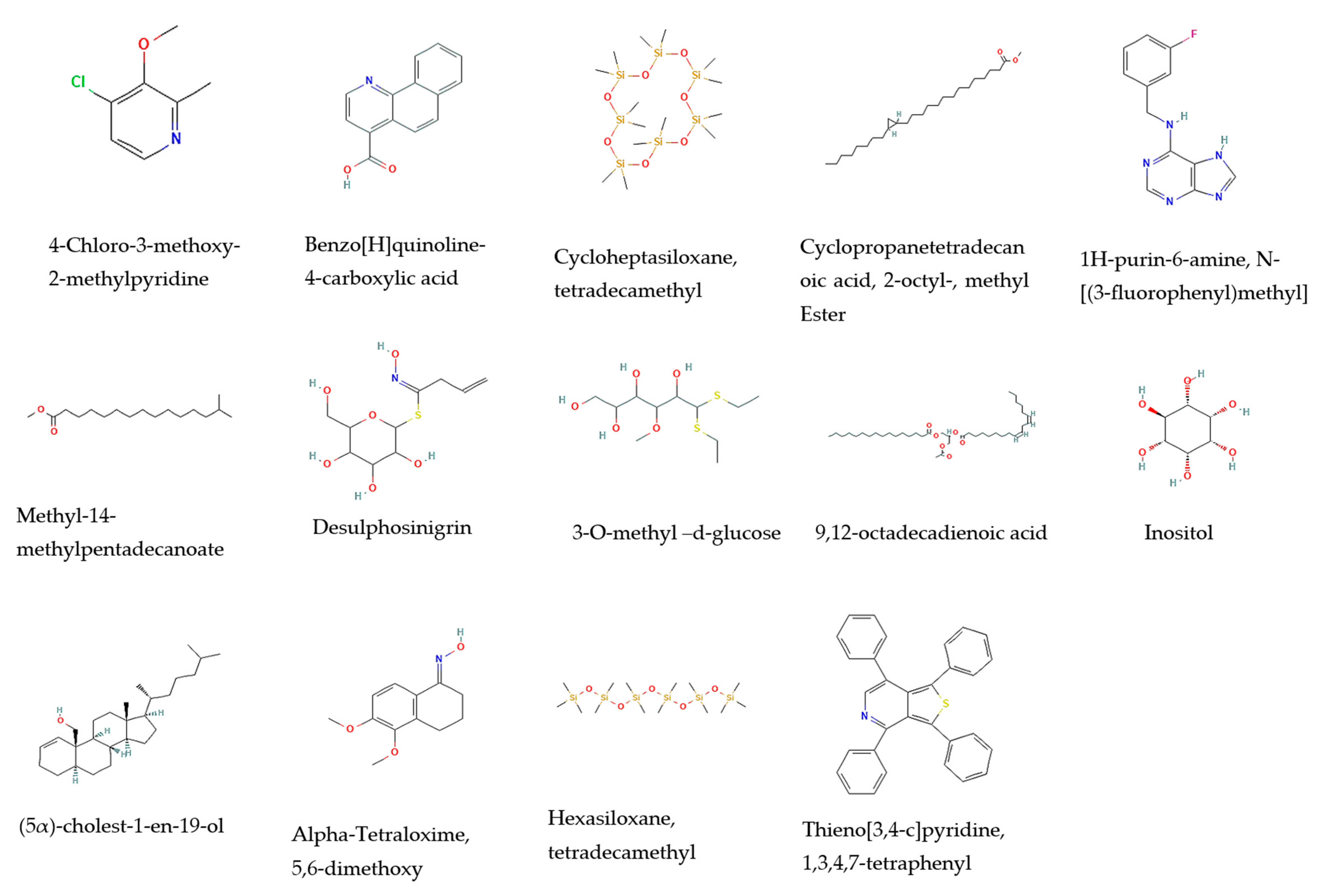
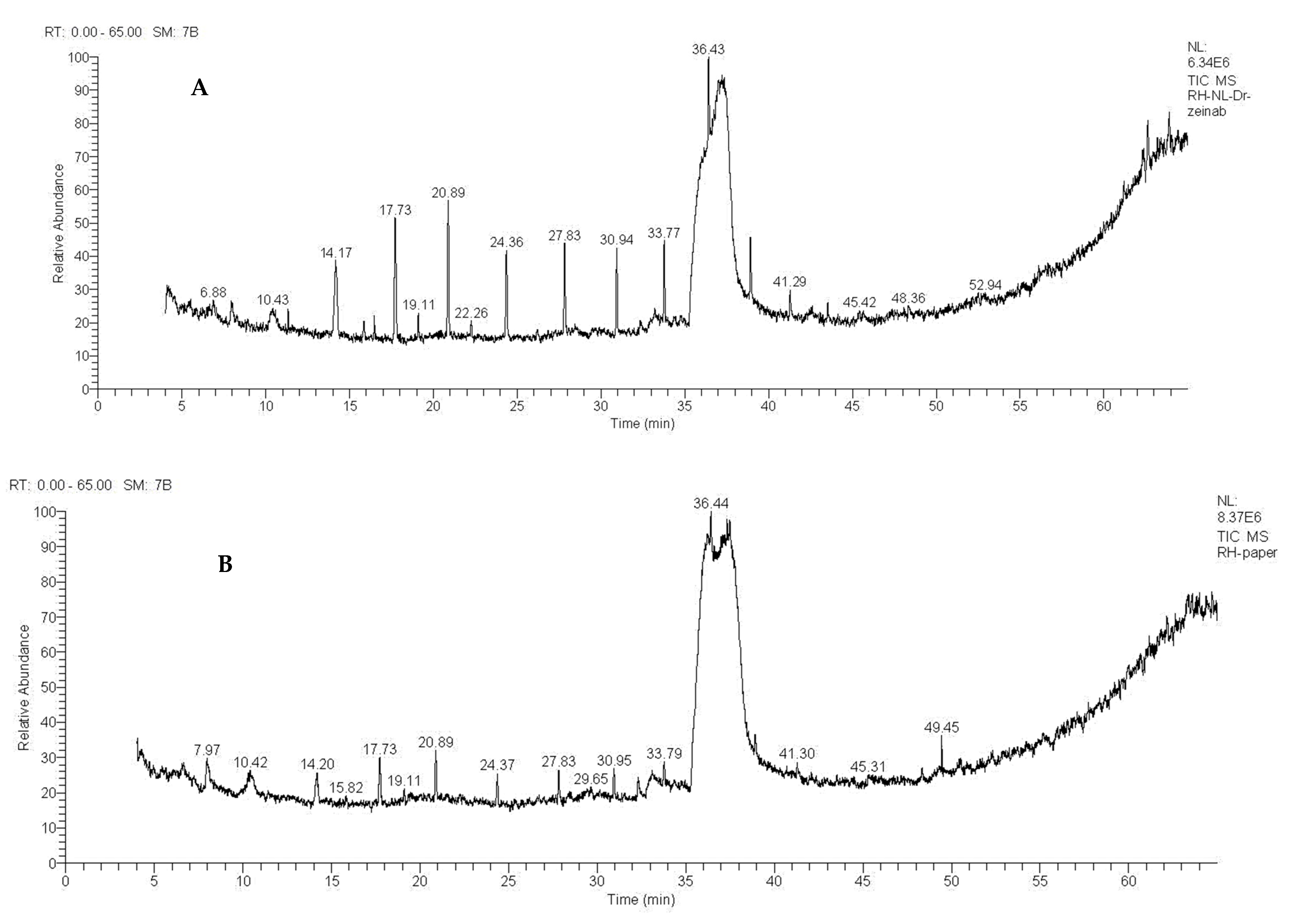
2.1. Phytochemistry

| No. | Leaves | Stems | Biological Activity | References | ||||||
|---|---|---|---|---|---|---|---|---|---|---|
| Compounds | MW | M.F. | Category | Rt | RA% | Rt | RA% | |||
| 1 | 4-Chloro-3-methoxy-2-methylpyridine | 157 | C7H8ClNO | Alkaloid | 7.98 | 1.94 | No data available | |||
| 2 | Benzo[H]quinoline-4-carboxylic acid | 223 | C14H9NO2 | Quinoline alkaloid | 10.42 | 0.67 | 10.42 | 0.9 | Anticancer | [26] |
| 3 | Cycloheptasiloxane, tetradecamethyl | 518 | C14H42O7Si7 | Organo-silicone compound | 14.19 | 2.37 | Anticancer and antimicrobial | [27,28] | ||
| 4 | Cyclopropanetetradecanoic acid, 2-octyl-, methyl Ester | 394 | C26H50O2 | Fatty acid | 19.11 | 0.61 | Antimicrobial | [29,30] | ||
| 5 | 1H-purin-6-amine, N- [(3-fluorophenyl)methyl] | 243 | C12H10FN5 | Fluorinated aromatic compound | 20.89 | 6.10 | 27.8 | 7.53 | Antioxidant | [31] |
| 6 | Methyl-14-methylpentadecanoate | 270 | C17H34O2 | Fatty acid | 32.31 | 4.34 | Antimicrobial | [29,30] | ||
| 7 | Desulphosinigrin | 279 | C10H17NO6S | Glucosinolate | 33.10 | 3.97 | 33.2 | 1.49 | Anticancer and Antimicrobial | [32,33,34] |
| 8 | 3-O-methyl-d-glucose | 194 | C7H14O6 | D-aldohexose | 35.40 | 6.45 | 35.79 | 2.63 | Anti-inflammatory and antioxidant | [35] |
| 9 | 9,12-octadecadienoic acid | 280 | C18H32O2 | Fatty acid | 33.78 | 1.14 | Anticancer and antibacterial | [36,37] | ||
| 10 | Inositol | 180 | C6H12O6 | A cyclic carbohydrate | 35.99 | 33.67 | 37.36 | 27.59 | Anticancer | [38] |
| 11 | (5α)-cholest-1-en-19-ol | 386 | C27H46O | Cholestane steroids | 49.45 | 2.30 | No data available | |||
| 12 | Alpha-Tetraloxime, 5,6-dimethoxy | 221 | C12H15NO3 | Aromatic-organic compound | 22.26 | 1.14 | No data available | |||
| 13 | Hexasiloxane, tetradecamethyl | 458 | C14H42O5Si6 | Linear siloxanes | 24.36 | 3.82 | Antimicrobial | [39] | ||
| 14 | Thieno[3,4-c]pyridine, 1,3,4,7-tetraphenyl | 439 | C31H21NS | Pyridine alkaloid | 45.42 | 0.95 | Anticancer | [40] | ||
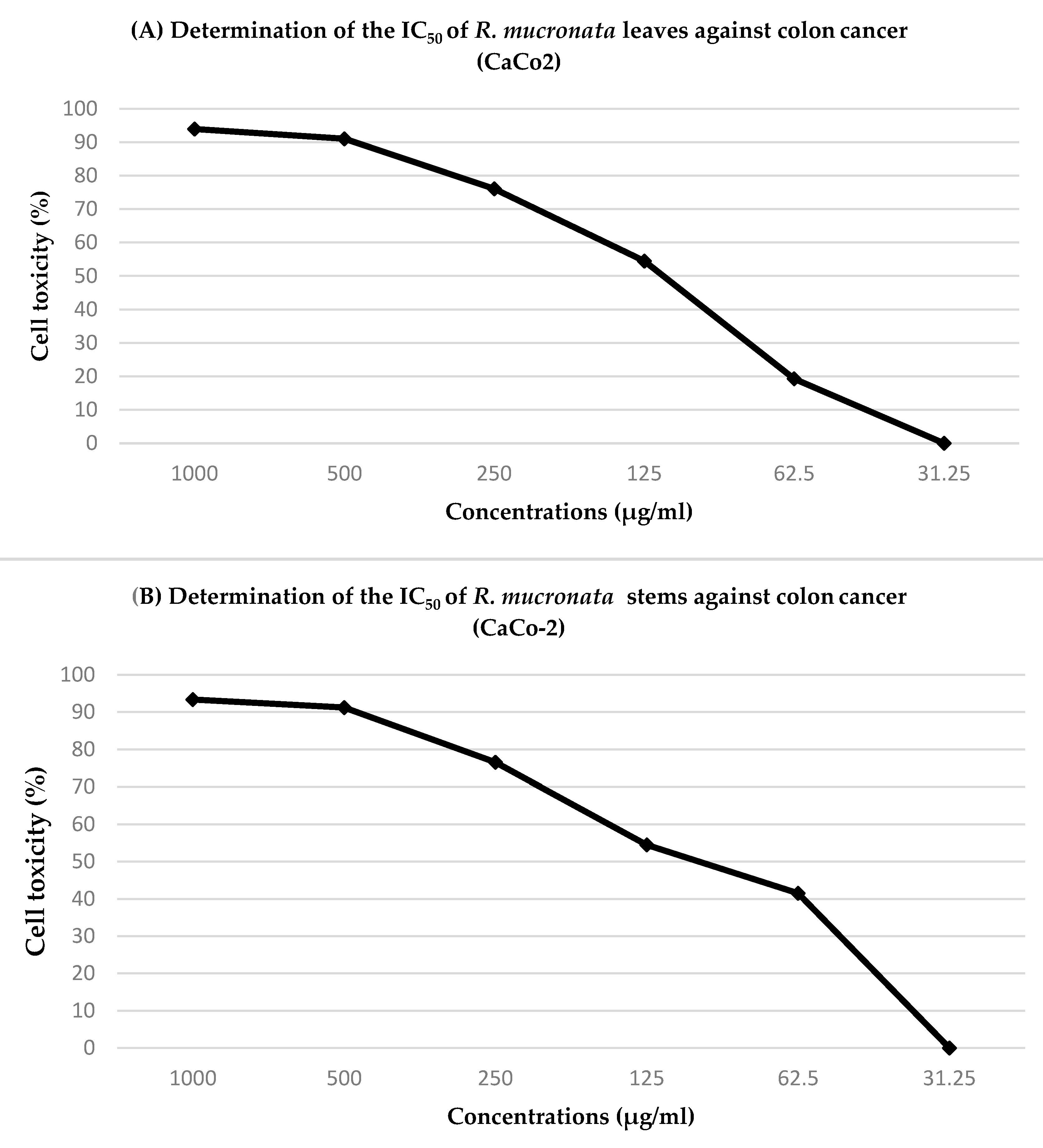
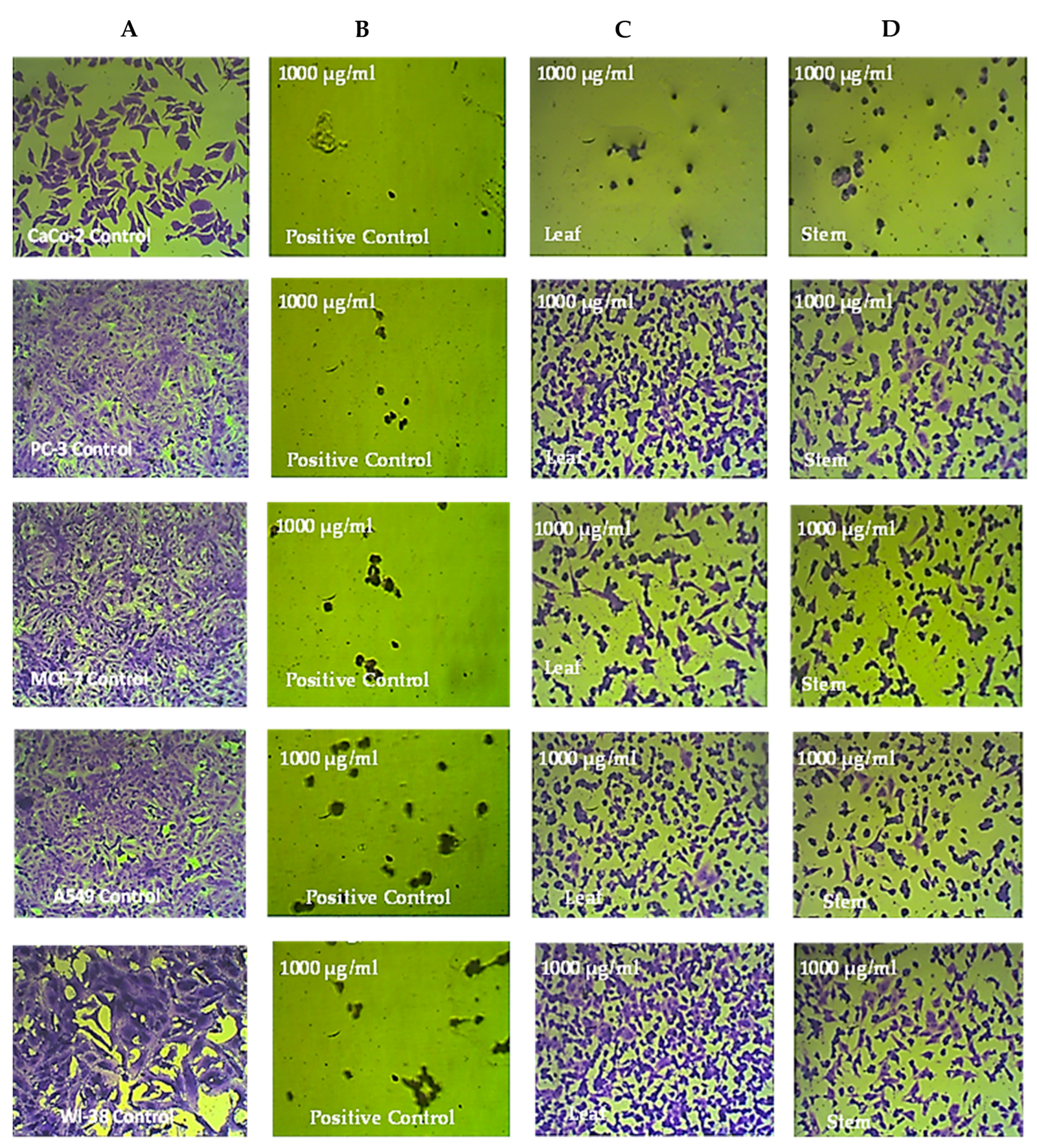
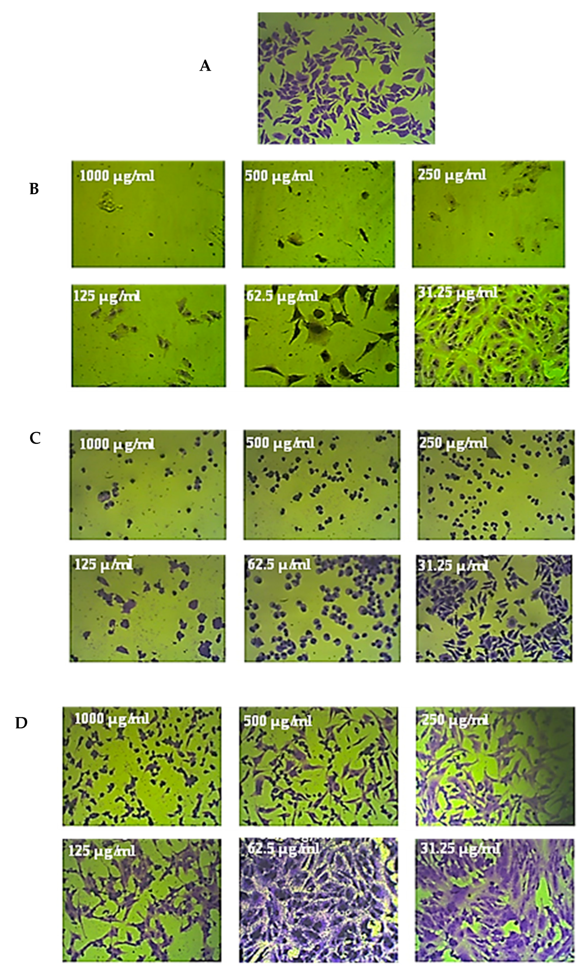
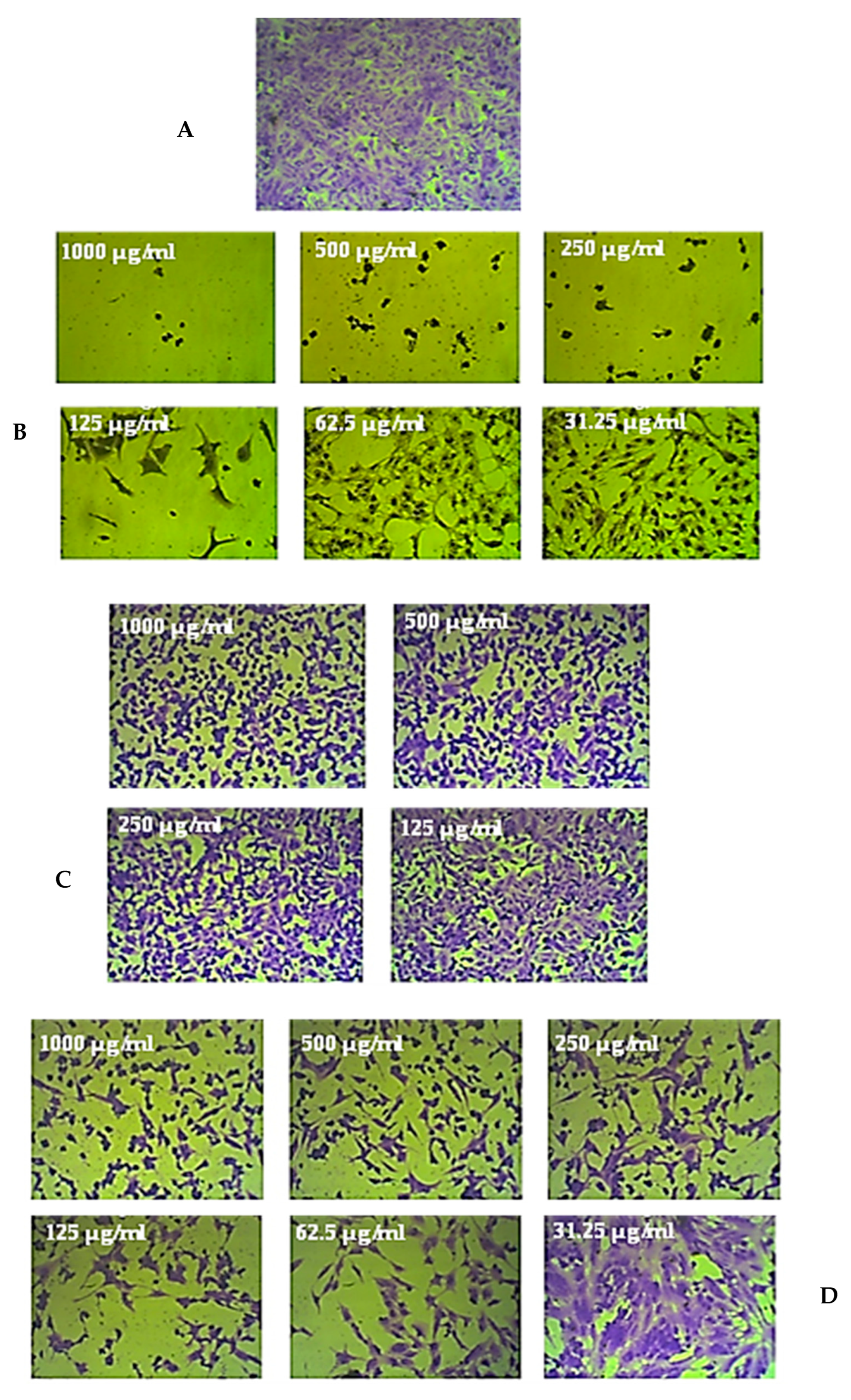
2.2. Cytotoxicity
3. Materials and Methods
3.1. Active Constituent Identification
3.1.1. Material of the Plant
3.1.2. Preparation of Plant Extract
3.1.3. Phytochemical Screening
3.2. Cytotoxic Evaluation
3.2.1. Culturing
3.2.2. MTT Assay
3.2.3. Determination of IC50 Values
3.2.4. Criteria for Anticancer Effect Levels
3.2.5. Selectivity Index
3.2.6. Microscope
4. Discussion
5. Conclusions
Author Contributions
Funding
Institutional Review Board Statement
Informed Consent Statement
Data Availability Statement
Acknowledgments
Conflicts of Interest
References
- Baskar, R.; Lee, K.A.; Yeo, R.; Yeoh, K.-W. Cancer and radiation therapy: Current advances and future directions. Int. J. Med. Sci. 2012, 9, 193–199. [Google Scholar] [CrossRef] [PubMed]
- Al-Saraireh, Y.M.; Alboaisa, N.S.; Alrawashdeh, H.M.; Hamdan, O.; Al-Sarayreh, S.; Al-Shuneigat, J.M.; Nofal, M.N. Screening of cytochrome 4Z1 expression in human non-neoplastic, pre-neoplastic and neoplastic tissues. Ecancermedicalscience 2020, 14, 1114. [Google Scholar] [CrossRef] [PubMed]
- Al-Saraireh, Y.M.; Alshammari, F.; Youssef, A.M.M.; Al-Sarayreh, S.; Almuhaisen, G.H.; Alnawaiseh, N.; Al Shuneigat, J.M.; Alrawashdeh, H.M. Profiling of CYP4Z1 and CYP1B1 expression in bladder cancers. Sci. Rep. 2021, 11, 5581. [Google Scholar] [CrossRef] [PubMed]
- Yang, C.; Mai, Z.; Liu, C.; Yin, S.; Cai, Y.; Xia, C. Natural Products in Preventing Tumor Drug Resistance and Related Signaling Pathways. Molecules 2022, 27, 3513. [Google Scholar] [CrossRef]
- Al-Saraireh, Y.M.; Alshammari, F.; Youssef, A.M.M.; Al-Sarayra, Y.M.; Al-Saraireh, R.A.; Al-Muhaisen, G.H.; Al-Mahdy, Y.S.; Al-Kharabsheh, A.M.; Abufraijeh, S.M.; Alrawashdeh, H.M. Cytochrome 4Z1 Expression Is Correlated with Poor Prognosis in Patients with Cervical Cancer. Curr. Oncol. 2021, 28, 3573–3584. [Google Scholar] [CrossRef]
- Al-Saraireh, Y.M.; Alshammari, F.; Youssef, A.M.M.; Al-Sarayreh, S.; Almuhaisen, G.H.; Alnawaiseh, N.; Al-Shuneigat, J.M.; Alrawashdeh, H.M. Cytochrome 4Z1 Expression is Associated with Poor Prognosis in Colon Cancer Patients. OncoTargets Ther. 2021, 14, 5249–5260. [Google Scholar] [CrossRef]
- Al-Saraireh, Y.; Alrawashdeh, F.; Al-Shuneigat, J.; Alsbou, M.; Alnawaiseh, N.; Al-Shagahin, H. Screening of Glypican-3 Expression in Human Normal versus Benign and Malignant Tissues: A Comparative Study Glypican-3 expression in cancers. Biosci. Biotechnol. Res. Asia 2016, 13, 687–692. [Google Scholar] [CrossRef]
- Youssef, A.; El-Swaify, Z.; Al-saraireh, Y.; Dalain, S. Cytotoxic activity of methanol extract of Cynanchumacutum L. seeds on human cancer cell lines. Latin Am. J. Pharm. 2018, 37, 1997–2003. [Google Scholar]
- Youssef, A.M.M.; El-Swaify, Z.A.S. Anti-Tumour Effect of two Persicaria species seeds on colon and prostate cancers. Biomed. Pharmacol. J. 2018, 11, 635–644. [Google Scholar] [CrossRef]
- Youssef, A.M.M.; El-Swaify, Z.A.S.; Al-Saraireh, Y.M.; Al-Dalain, S.M. Anticancer effect of different extracts of Cynanchum acutum L. seeds on cancer cell lines. Pharmacogn. Mag. 2019, 15, 261. [Google Scholar] [CrossRef]
- Boulos, L. Flora of Egypt; Al Hadara Publishing: Cairo, Egypt, 1999; Volume 1. [Google Scholar]
- Patra, J.K.; Thatoi, H.N. Metabolic diversity and bioactivity screening of mangrove plants: A review. Acta Physiol. Plant. 2011, 33, 1051–1061. [Google Scholar] [CrossRef]
- Bandaranayake, W.M. Bioactivities, bioactive compounds and chemical constituents of mangrove plants. Wetl. Ecol. Manag. 2002, 10, 421–452. [Google Scholar] [CrossRef]
- Ravikumar, S.; Nazar, S.; Nuralshiefa, A.; Abideen, S. Antibacterial activity of traditional therapeutic coastal medicinal plants against some pathogens. J. Environ. Biol. 2005, 26, 383–386. [Google Scholar] [PubMed]
- Padmakumar, K.; Ayyakkannu, K. Antiviral activity of marine plants. Indian J. Virol. 1997, 13, 33–36. [Google Scholar]
- Premanathan, M.; Kathiresan, K.; Yamamoto, N.; Nakashima, H. In vitro anti-human immunodeficiency virus activity of polysaccharide from Rhizophora mucronata Poir. Biosci. Biotechnol. Biochem. 1999, 63, 1187–1191. [Google Scholar] [CrossRef]
- Abeysinghe, P.D. Antibacterial activity of aqueous and ethanol extracts of mangrove species collected from Southern Sri Lanka. Asian J. Pharm. Biol Res. 2012, 2, 79–83. [Google Scholar]
- Sadeer, N.B.; Rocchetti, G.; Senizza, B.; Montesano, D.; Zengin, G.; Uysal, A.; Jeewon, R.; Lucini, L.; Mahomoodally, M.F. Untargeted metabolomic profiling, multivariate analysis and biological evaluation of the true mangrove (Rhizophora mucronata Lam.). Antioxidants 2019, 8, 489. [Google Scholar] [CrossRef]
- Manigandan, V.; Gurudeeban, S.; Satyavani, K.; Ramanathan, T. Molecular docking studies of Rhizophora mucronata alkaloids against neuroinflammatory marker cyclooxygenase 2. Int. J. Biol. Chem. 2014, 8, 91–99. [Google Scholar]
- Hardoko, E.S.; Puspitasari, Y.; Amalia, R. Study of ripe Rhizophora mucronata fruit flour as functional food for antidiabetic. Int. Food Res. J. 2015, 22, 953–959. [Google Scholar]
- Puspitasar, Y.E.; Hartiati, A.; Suprayitno, E. The potency of Rhizophora mucronata leaf extract as antidiarrhea. J. Appl. Sci. Res 2012, 8, 1180–1185. [Google Scholar]
- Faoziyah, A.R.; Kurniawan, W. Pemanfaatan ekstrak daun mangrove (Rhizophora mucronata sp.) dengan variasi pelarut sebagai bahan aktif sediaan farmasi terapi anti kanker. J. Health 2017, 4, 68–74. [Google Scholar] [CrossRef]
- Alsy, R. Effect Of Mangrove Leaf Extract Dosage Rhizophora Mucronata Lmk. On The Viability Of Hela Cells. J. Stem Cell Res. Tissue Eng. 2021, 5, 23–29. [Google Scholar] [CrossRef]
- Sachithanandam, V.; Lalitha, P.; Parthiban, A.; Muthukumaran, J.; Jain, M.; Misra, R.; Mageswaran, T.; Sridhar, R.; Purvaja, R.; Ramesh, R. A comprehensive in silico and in vitro studies on quinizarin: A promising phytochemical derived from Rhizophora mucronata Lam. J. Biomol. Struct. Dyn. 2021, 40, 1–12. [Google Scholar] [CrossRef] [PubMed]
- Sari, D.P.; Basyuni, M.; Hasibuan, P.A.Z.; Wati, R. Cytotoxic effect of polyisoprenoids from Rhizophora mucronata and Ceriops tagal leaves against WiDr colon cancer cell lines. Sains Malays. 2018, 47, 1953–1959. [Google Scholar] [CrossRef]
- Iqbal, J.; Ejaz, S.A.; Khan, I.; Ausekle, E.; Miliutina, M.; Langer, P. Exploration of quinolone and quinoline derivatives as potential anticancer agents. DARU J. Pharm. Sci. 2019, 27, 613–626. [Google Scholar] [CrossRef]
- El-Fayoumy, E.A.; Shanab, S.M.; Hassan, O.; Shalaby, E.A. Enhancement of active ingredients and biological activities of Nostoc linckia biomass cultivated under modified BG-110 medium composition. Biomass Convers. Biorefin. 2021, 2021, 1–18. [Google Scholar] [CrossRef]
- Phuong, T.; Lam, P.; Diep, C. Bioactive compounds from marine streptomyces Sp. by gas chromatography-mass spectrometry. Pharm. Chem. J. 2018, 5, 196–203. [Google Scholar]
- Srivastava, R.; Mukerjee, A.; Verma, A. GC-MS analysis of Phytocomponents in, pet ether fraction of wrightia tinctoria seed. Pharmacogn. J. 2015, 7, 249–253. [Google Scholar] [CrossRef]
- Egbung, G.E.; Anosike, C.; Utu-Baku, A.B.; Ogar, I.; Nna, V.U. Phytochemical evaluation and GC-MS analysis of Hyptis verticillata cultivated in Calabar Cross River State, Nigeria. Int. J. Biol. Chem. Sci. 2017, 11, 2548–2559. [Google Scholar] [CrossRef][Green Version]
- Budayatin, B.; Waluyo, J.; Wahyuni, D.; Dafik, D. Antibacterial effects of Pheretima javanica extract and bioactive chemical analysis using Gas Chromatography Mass Spectrum. J. Phys. Conf. Ser. 2021, 1751, 012055. [Google Scholar] [CrossRef]
- Krishnaveni, M. Docking, simulation studies of desulphosinigrin—Cyclin dependent kinase 2, an anticancer drug target. Int. J. Pharm. Sci. Rev. Res. 2015, 30, 115–118. [Google Scholar]
- Kamal, S.A.; Hamza, L.F.; Hameed, I.H. Antibacterial activity of secondary metabolites isolated from Alternaria alternata. Afr. J. Biotechnol. 2015, 14, 2972–2994. [Google Scholar]
- Azhar, A.S.; Suhaila, H.B.; Imad, H.H. Analysis of bioactive chemical compounds of Euphorbia lathyrus using gas chromatography-mass spectrometry and fourier-transform infrared spectroscopy. J. Pharmacogn. Phytother. 2016, 8, 109–126. [Google Scholar] [CrossRef]
- Guerrero, R.V.; Vargas, R.A.; Petricevich, V.L. Chemical compounds and biological activity of an extract from bougainvillea x buttiana (var. rose) holttum and standl. Int. J. Pharm. Pharm. Sci. 2017, 9, 42–46. [Google Scholar] [CrossRef]
- Jayaraman, L.; Shivaji, S.; Anandakumar, S. Phytochemical screening, cytotoxic activity and molecular docking studies of Eclipta alba leaves extract against oral cancer. Rasayan J. Chem. 2022, 15, 676–685. [Google Scholar] [CrossRef]
- Rossellia, S.; Maggio, A.; Formisano, C.; Napolitano, F.; Senatore, F.; Spadaro, V.; Bruno, M. Chemical composition and antibacterial activity of extracts of Helleborus bocconei Ten. subsp. intermedius. Nat. Prod. Commun. 2007, 2, 1934578X0700200611. [Google Scholar] [CrossRef]
- Vucenik, I. Anticancer properties of inositol hexaphosphate and inositol: An overview. J. Nutr. Sci. Vitaminol. 2019, 65, S18–S22. [Google Scholar] [CrossRef]
- Bele, A.A.; Khale, A. Comparison of Constituents in Aloe Vera Gel Collected in Different Seasons by Chromatography and Spectroscopy Techniques. World J. Pharm. Res. 2016, 5, 1028–1040. [Google Scholar]
- El-Naggar, M.; Almahli, H.; Ibrahim, H.S.; Eldehna, W.M.; Abdel-Aziz, H.A. Pyridine-Ureas as Potential Anticancer Agents: Synthesis and In Vitro Biological Evaluation. Molecules 2018, 23, 1459. [Google Scholar] [CrossRef]
- Youssef, A.; El-Swaify, Z.; Maaty, D.; Youssef, M. Comparative study of two Lotus species: Phytochemistry, cytotoxicity and antioxidant capacity. J. Pharm. Pharmacogn. Res. 2020, 8, 537–548. [Google Scholar]
- Al-saraireh, Y.M.; Youssef, A.M.; Alshammari, F.O.; Al-Sarayreh, S.A.; Al-Shuneigat, J.M.; Alrawashdeh, H.M.; Mahgoub, S.S. Phytochemical characterization and anti-cancer properties of extract of Ephedra foeminea (Ephedraceae) aerial parts. Trop. J. Pharm. Res. 2021, 20, 1675–1681. [Google Scholar] [CrossRef]
- Al-Saraireh, Y.M.; Youssef, A.M.M.; Alsarayreh, A.Z.; Hujran, T.A.A.; Al-Sarayreh, S.; Al-Shuneigat, J.M.; Alrawashdeh, H.M. Phytochemical and anti-cancer properties of Euphorbia hierosolymitana Boiss. crude extracts. J. Pharm. Pharmacogn. Res. 2021, 9, 13–23. [Google Scholar] [CrossRef] [PubMed]
- Watzka, M.; Medina, E. Mangroves in Contrasting Osmotic Environments: Photosynthetic Costs of High Salinity Tolerance. In Photosynthesis-From Its Evolution to Future Improvements in Photosynthetic Efficiency Using Nanomaterials; IntechOpen: London, UK, 2018. [Google Scholar]
- Yunos, N.M.; Ling, S.K.; Osman, A.; Abdullah, Z.; Sallehudin, N.J. Phytochemicals from Rhizophora mucronata Propagules, Its In Vitro Anti-Cancer and In Silico Drug-Likeness Potential. Chemistry 2021, 3, 979–990. [Google Scholar] [CrossRef]
- Taniguchi, K.; Funasaki, M.; Kishida, A.; Sadhu, S.K.; Ahmed, F.; Ishibashi, M.; Ohsaki, A. Two new coumarins and a new xanthone from the leaves of Rhizophora mucronata. Bioorg. Med. Chem. Lett. 2018, 28, 1063–1066. [Google Scholar] [CrossRef] [PubMed]


| a IC50 (µg/mL) | |||
|---|---|---|---|
| Cell Lines | R. mucronata Leaves | R. mucronata Stems | Doxorubicin (Positive Control) |
| b CaCo-2 | 127 ± 4 *** | 107 ± 6 ** | 83 ± 1 |
| c PC-3 | 480 ± 14 *** | 294 ± 3 *** | 79 ± 4 |
| d MCF-7 | 158 ± 10 *** | 138 ± 3 *** | 80 ± 3 |
| e A549 | 376 ± 9 *** | 155 ± 10 *** | 90 ± 5 |
| f WI-38 | 932 ± 30 *** | 629 ± 3 *** | 50 ± 5 |
| a SI | ||||
|---|---|---|---|---|
| Extract | b CaCo-2 | c PC-3 | d MCF-7 | e A549 |
| Leaves | 7.3 | 2 | 5.8 | 2.4 |
| Stems | 5.8 | 2.1 | 4.5 | 4 |
Disclaimer/Publisher’s Note: The statements, opinions and data contained in all publications are solely those of the individual author(s) and contributor(s) and not of MDPI and/or the editor(s). MDPI and/or the editor(s) disclaim responsibility for any injury to people or property resulting from any ideas, methods, instructions or products referred to in the content. |
© 2022 by the authors. Licensee MDPI, Basel, Switzerland. This article is an open access article distributed under the terms and conditions of the Creative Commons Attribution (CC BY) license (https://creativecommons.org/licenses/by/4.0/).
Share and Cite
Youssef, A.M.M.; Maaty, D.A.M.; Al-Saraireh, Y.M. Phytochemistry and Anticancer Effects of Mangrove (Rhizophora mucronata Lam.) Leaves and Stems Extract against Different Cancer Cell Lines. Pharmaceuticals 2023, 16, 4. https://doi.org/10.3390/ph16010004
Youssef AMM, Maaty DAM, Al-Saraireh YM. Phytochemistry and Anticancer Effects of Mangrove (Rhizophora mucronata Lam.) Leaves and Stems Extract against Different Cancer Cell Lines. Pharmaceuticals. 2023; 16(1):4. https://doi.org/10.3390/ph16010004
Chicago/Turabian StyleYoussef, Ahmed M. M., Doaa A. M. Maaty, and Yousef M. Al-Saraireh. 2023. "Phytochemistry and Anticancer Effects of Mangrove (Rhizophora mucronata Lam.) Leaves and Stems Extract against Different Cancer Cell Lines" Pharmaceuticals 16, no. 1: 4. https://doi.org/10.3390/ph16010004
APA StyleYoussef, A. M. M., Maaty, D. A. M., & Al-Saraireh, Y. M. (2023). Phytochemistry and Anticancer Effects of Mangrove (Rhizophora mucronata Lam.) Leaves and Stems Extract against Different Cancer Cell Lines. Pharmaceuticals, 16(1), 4. https://doi.org/10.3390/ph16010004