Article

Design and Synthesis of New Pyrimidine-Quinolone Hybrids as Novel hLDHA Inhibitors

Facultad de Ciencias Experimentales, Departamento de Química Inorgánica y Orgánica, Universidad de Jaén, E-23071 Jaén, Spain
*
Author to whom correspondence should be addressed.
Pharmaceuticals 2022, 15(7), 792; https://doi.org/10.3390/ph15070792
Submission received: 24 May 2022 / Revised: 17 June 2022 / Accepted: 18 June 2022 / Published: 24 June 2022
(This article belongs to the Special Issue Design of Enzyme Inhibitors as Potential Drugs 2022)

Abstract

:
A battery of novel pyrimidine-quinolone hybrids was designed by docking scaffold replacement as lactate dehydrogenase A (hLDHA) inhibitors. Structures with different linkers between the pyrimidine and quinolone scaffolds (10-21 and 2431) were studied in silico, and those with the 2-aminophenylsulfide (U-shaped) and 4-aminophenylsulfide linkers (2431) were finally selected. These new pyrimidine-quinolone hybrids (2431)(ac) were easily synthesized in good to excellent yields by a green catalyst-free microwave-assisted aromatic nucleophilic substitution reaction between 3-(((2/4-aminophenyl)thio)methyl)quinolin-2(1H)-ones 22/23(ac) and 4-aryl-2-chloropyrimidines (14). The inhibitory activity against hLDHA of the synthesized hybrids was evaluated, resulting IC50 values of the U-shaped hybrids 2427(ac) much better than the ones of the 1,4-linked hybrids 2831(ac). From these results, a preliminary structure–activity relationship (SAR) was established, which enabled the design of novel 1,3-linked pyrimidine-quinolone hybrids (3336)(ac). Compounds 35(ac), the most promising ones, were synthesized and evaluated, fitting the experimental results with the predictions from docking analysis. In this way, we obtained novel pyrimidine-quinolone hybrids (25a, 25b, and 35a) with good IC50 values (<20 μM) and developed a preliminary SAR.

Graphical Abstract

1. Introduction

One of the main diseases that cause death and, therefore, one of the main public health problems worldwide continues to be cancer [1,2]. In the last decades, most of the main hallmarks of many cancers have been established [3]. In the case of metabolism alteration, in normal cells, glucose is metabolized into pyruvate and afterwards into carbon dioxide and acetyl-CoA through an oxidative phosphorylation process. In tumor cells, this process is highly disordered, as anaerobic glycolysis is often preferred over oxidative phosphorylation. This metabolic switch is known as the Warburg effect and leads to the formation of lactate [4]. In this switch, several studies suggest that lactate dehydrogenase A (hLDHA) enzyme plays a key role in cancer proliferation, as it is responsible for catalyzing the conversion of pyruvate into lactate [5,6,7,8,9].
Recently, hLDHA has also been shown to be implicated in other diseases such as primary hyperoxaluria (PH), which converts glyoxylate into oxalate [10,11]. When oxalate is overproduced, calcium oxalate crystals appear in the kidney, leading to urolithiasis, nephroncalcinosis, renal failure [12], and, eventually, end-stage renal disease [13,14,15]. Consequently, the hLDHA enzyme is an ideal therapeutic target for cancer and PH treatment.
The development of new chemical entities (NCEs) based on small molecules wearing aza-heterocyclic nuclei still constitute one of the most important areas within the pharmaceutical industry [16]. Those systems can be found in a huge range of drugs and bioactive compounds due to the fact they are the main pharmacophoric residues responsible for their biological response and/or for being the key synthetic scaffold, which is the case of pyrimidines and quinolones.
In particular, pyrimidine derivatives have shown diverse activities, such as antimicrobial, antioxidant, antimalarial, and anti-inflammatory [17]. Furthermore, they have been used as potential agents in the treatment of neurodegenerative diseases such as Alzheimer’s [18] and in the treatment of cancer [17,19,20,21,22]. Thus, pyrimidine, as a biologically privileged scaffold, is commonly used in the development of new drugs towards different targets [23,24].
Quinolones are also considered to be biologically privileged, as they interact with a diverse biotargets and show a wide variety of bioactivities, such as antiviral, anti-parasitic [25], anti-malarial [26], or anti-inflammatory [27] activities, amongst many others. They are also used as biomarkers [28] in the treatment of different types of cancer, as only heteronucleus [29,30], or in combination with other different scaffolds [31], such as benzo[d]thiazolyl [32], cinnamic acid [33], or with hydantoin searching for antimicrobial activity [34].
The hybridization of both systems, in accordance with the Fragment-Based Drug Discovery (FBDD) strategy [35,36,37,38], has been demonstrated to be highly interesting regarding their antiproliferative action [39], such as anticancer [40], anti-HIV [41], and antimalarial/antiplasmodial [26,42,43,44,45,46,47,48], and for being inhibitors of human sphingomyelin synthase 2 [49].
Some hLDHA inhibitors wearing the pyrimidine (I–III) and quinolone (IV) nucleus have already been reported [50,51]. However, we have only found a few examples of structure-related hybrids that have been reported as hLDHA inhibitors (V and VI) [52,53,54] but without linkers between the pyrimidine and quinoline fragments (Figure 1).
Another structural feature observed after a thorough analysis of different hLDHA inhibitors is that most of them (VII–XII in Figure 2) had a hydrophilic scaffold and a hydrophobic one, with or without a linking moiety separating them [4,5].
In that sense, we have already reported the synthesis of diverse hybrids bearing the quinolone fragment as potential antimalarial and antitumoral agents [55,56,57] and, amongst them, some with pyrimidine residues, which contain both the hydrophobic and hydrophilic scaffolds, shown to be promising anticancer agents [58,59]. In particular, we have recently reported the synthesis of a series of pyrimidine-quinolone hybrids following a linear synthetic methodology starting from 2,4-dichloropyrimidine (Figure 3) and proved their bioactivity as sphingosine kinase (SphK) inhibitors, which are involved in cell proliferation [58] and P-glycoprotein (P-gp) inhibitors in the search for reversal agents of multidrug resistance [59].
Bearing all this in mind, and taking these hybrids as the starting point for the development of a novel family of hLDHA inhibitors, we here report their rational design, synthesis, and biological evaluation. These NCEs are based on pyrimidine-quinolone hybrids linked by an aminophenylsulfide fragment in a U- and non-U-shaped disposition, which are of potential interest regarding their behavior as hLDHA inhibitors according to what has been mentioned previously.

2. Results and Discussion

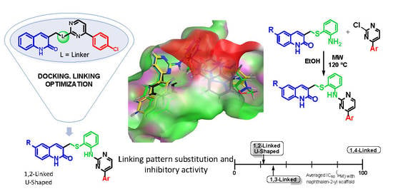
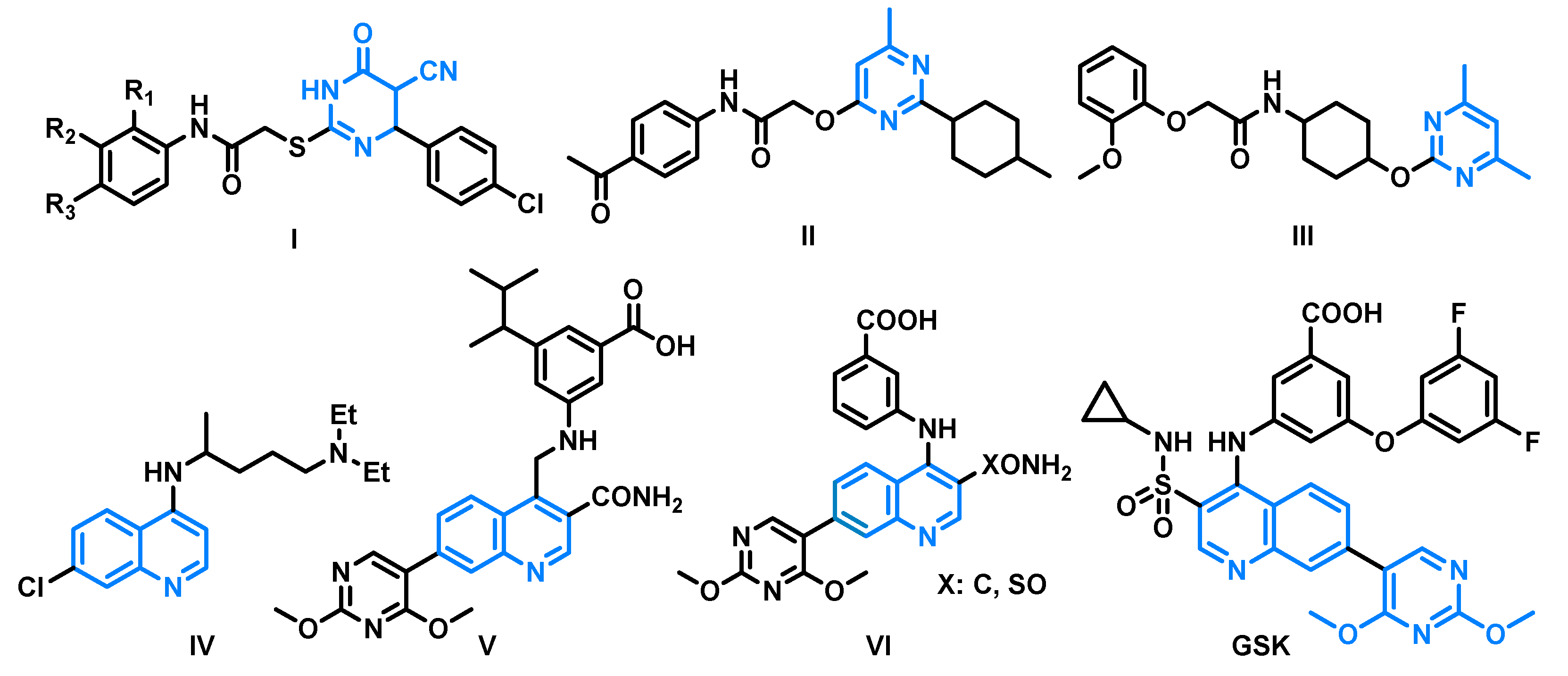
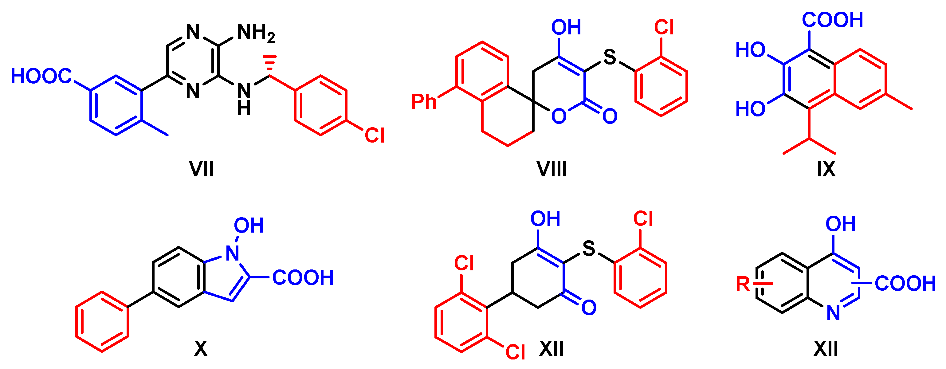
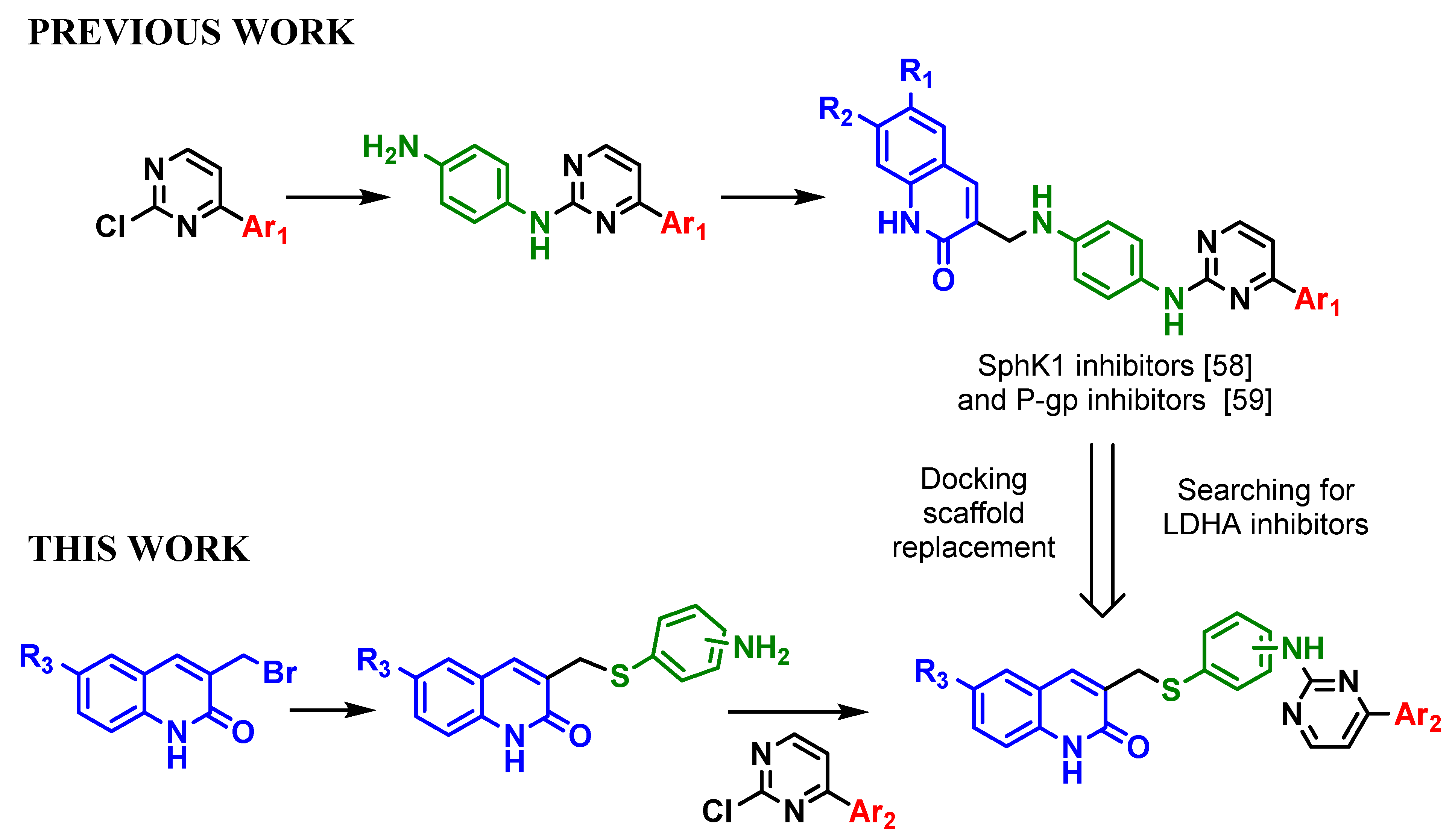
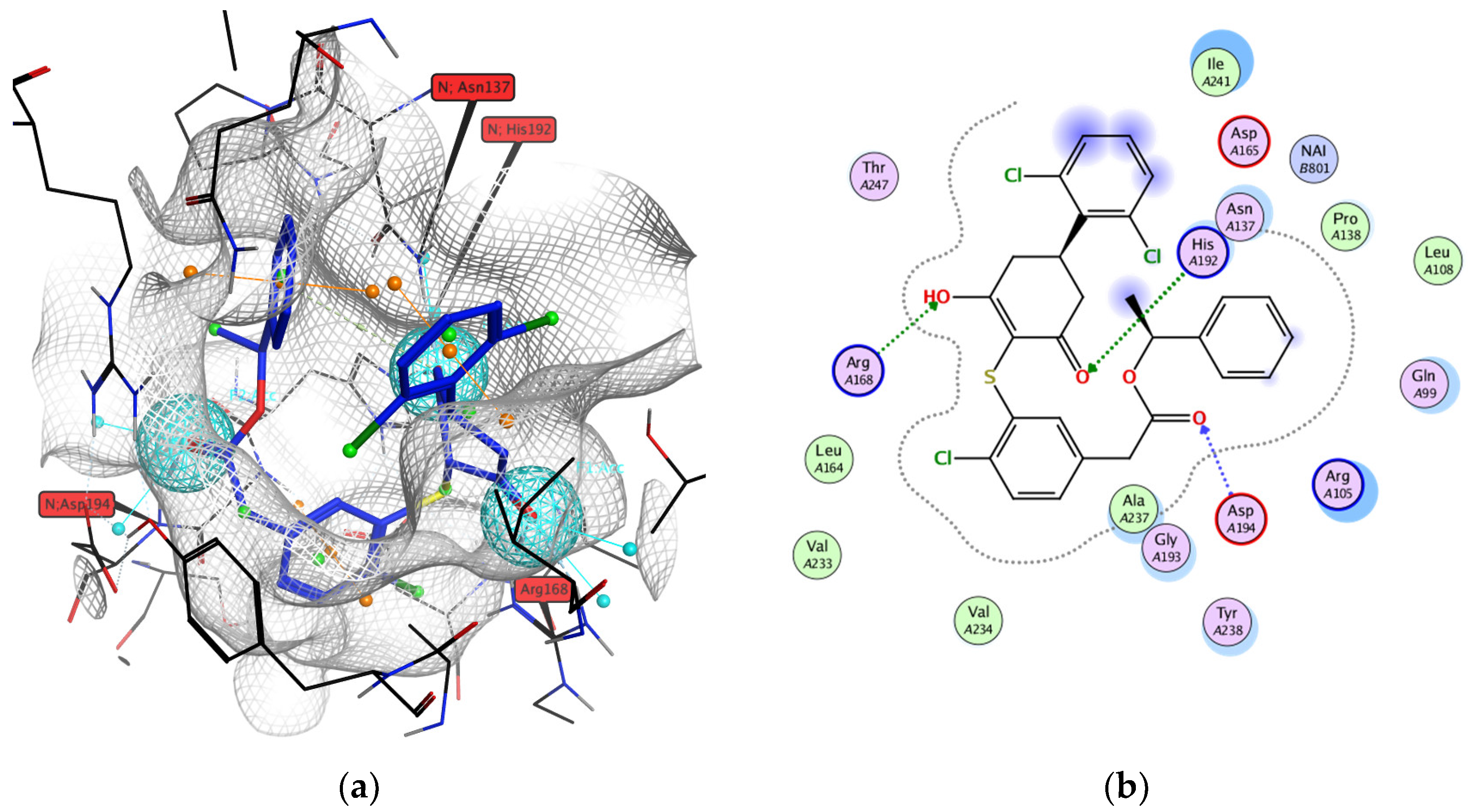
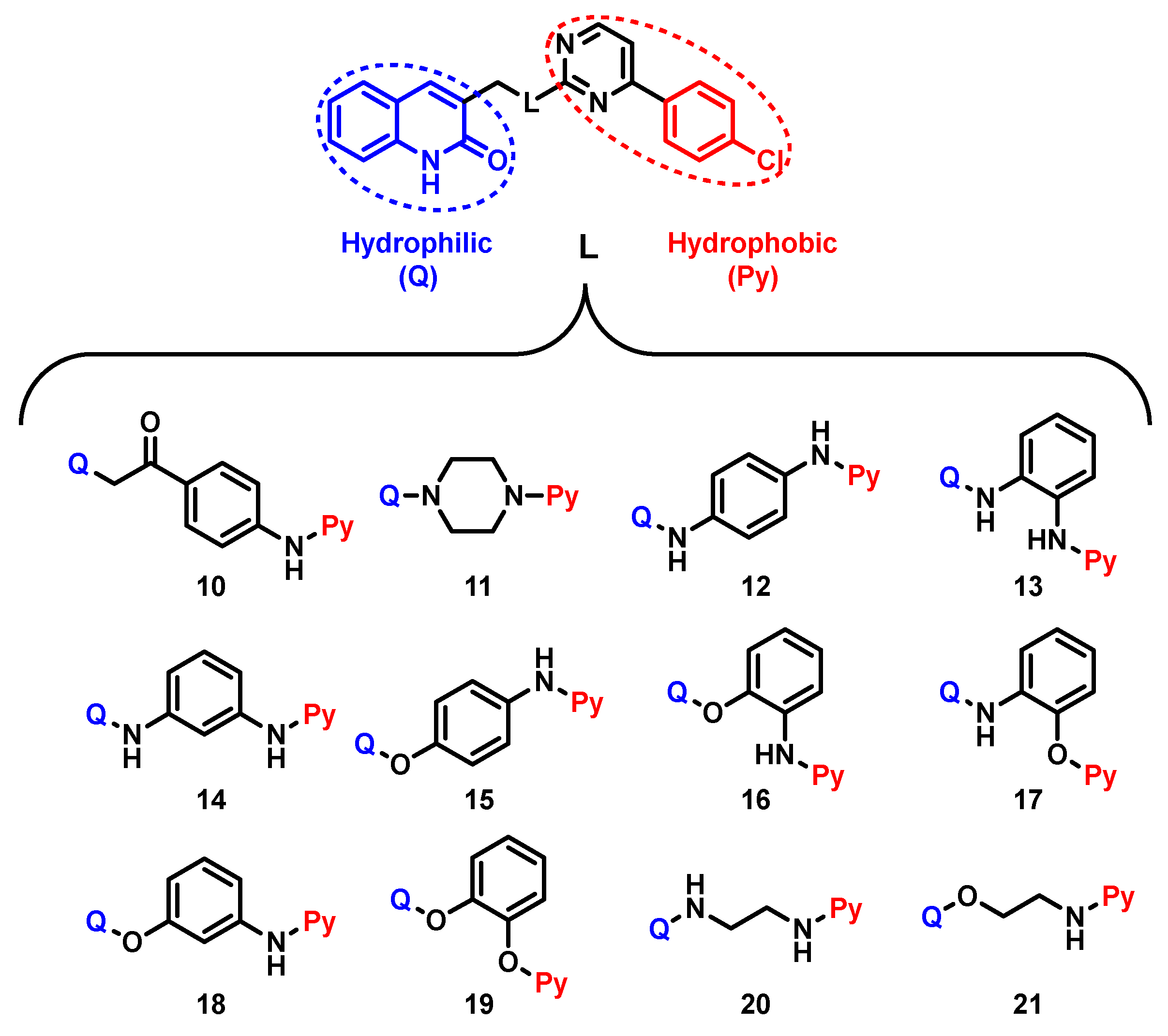
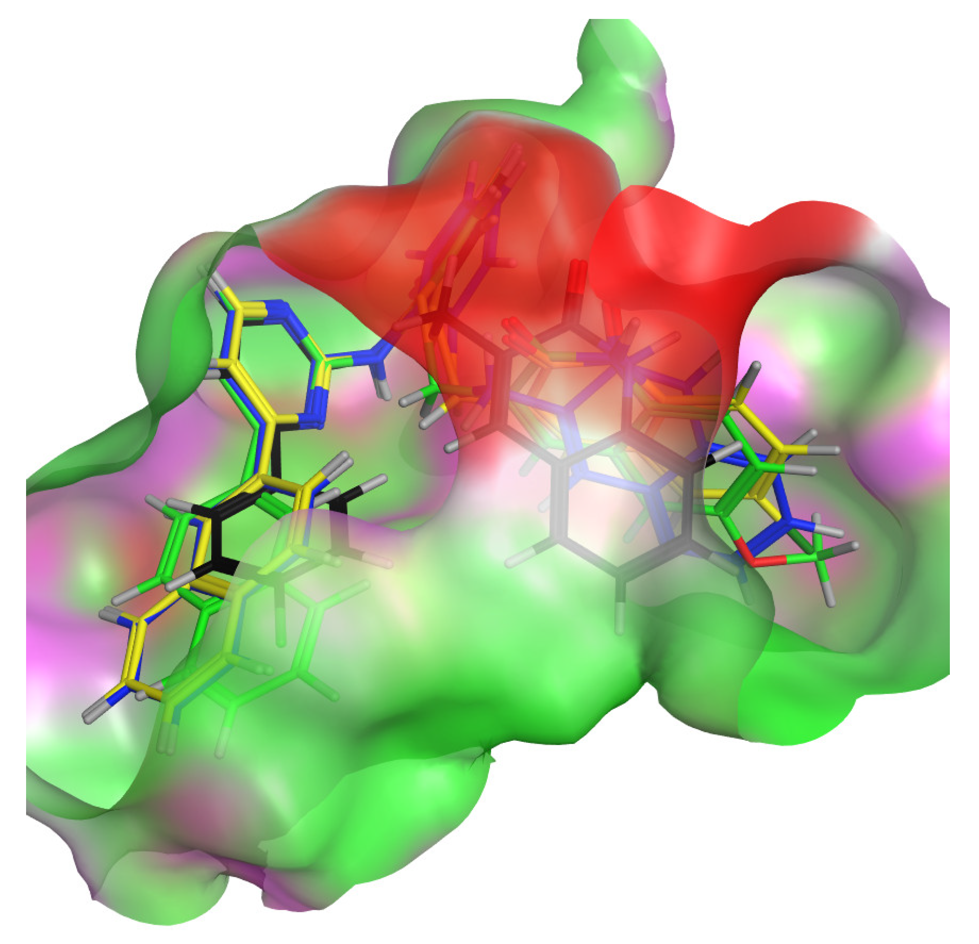
2.1. Virtual Screening Scaffold Replacement in the Optimization of Pyrimidine-Quinolone Hybrids as hLDHA Inhibitors
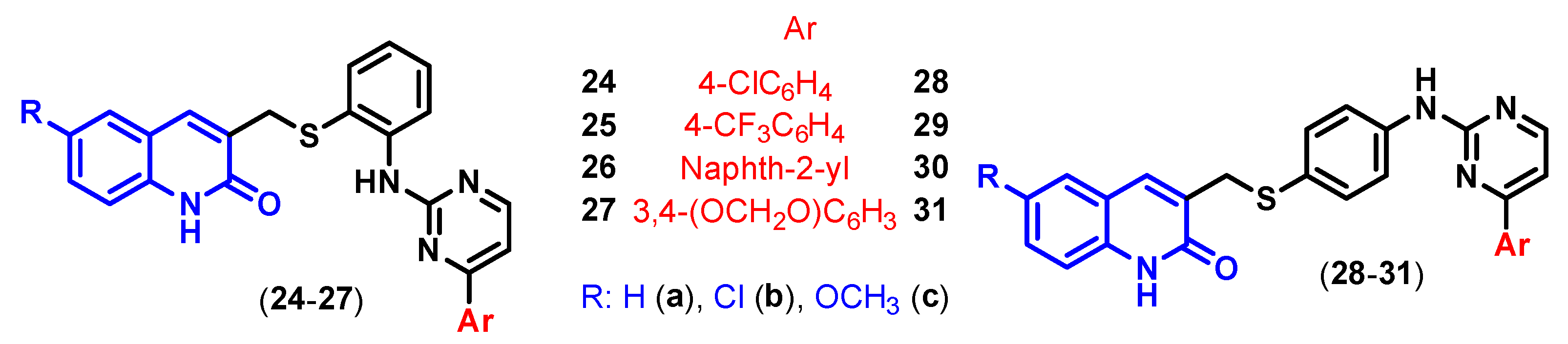
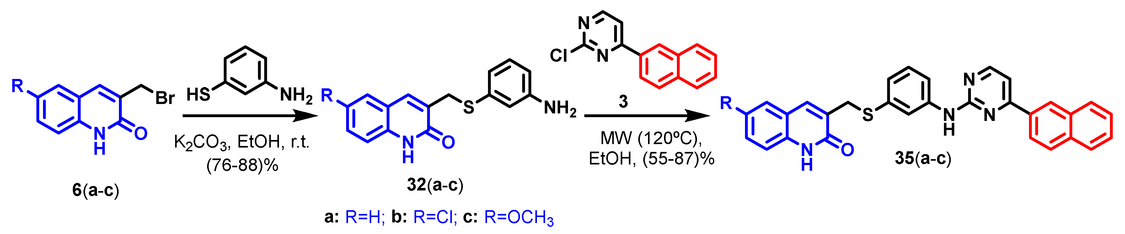
Complex hLDHA-W31 (code 4R68) was selected and downloaded from the Protein Data Bank (PDB) as reference for the docking studies due to the following reasons: (i) its ligand (W31) interacts with the main amino acid residues reported to be responsible for its activity (Arg168, Asn137, His192, and Asp194) [60], (ii) it occupies the whole substrate (pyruvate) pocket [61], and (iii) it has an IC50 = 6 nM [62].
The Figure 4a represents the W31 placement in the substrate pocket and Figure 4b its 2-D interaction diagram with the main amino acid residues in that active site Blue spheres in left image represent the pharmacophore descriptor by imposed features where a hydrogen donor/acceptor atom could be located to interact with such key residues.
Firstly, we proceeded by excluding through docking screening any possibility of NADH competitive inhibition. Thus, in order to discard any other possible interaction sites of the pyrimidine-quinolone hybrids deigned in this work besides the expected W31 site, we ran the docking process in triplicate with different docking areas and pharmacophoric descriptors [63] as described in Section 3.4: (i) in the hLDHA active site (W31 site), (ii) in the NADH site, and (iii) in the extension covering both sites.
In that regard, based on our previous experience in the synthesis of pyrimidine-quinolone compounds [58], a first set of compounds (1021) was designed, having the quinolone scaffold as the hydrophobic moiety and the 4-chlorophenyl scaffold as the hydrophobic one (Figure 5).
Compounds 1012 were already synthesized by us and evaluated as sphingosine kinase inhibitors [58]. Structures 1321, with new linking precursors (1,3-diaminobenzene, 1,2-diaminobenzene, aminophenol, catechol, ethylenediamine, and ethanolamine), were designed for their in silico study.
The docking results showed that the inhibition is unlikely to take place by displacement of the NADH cofactor in its site, as the affinity values are not close enough to compete against it. This is reinforced by the fact that they do not give any interaction with those amino acid residues that interact strongly with NADH in its site. This way, the affinity and energy values involved in the interactions with the mentioned key amino acids in the W31 site suggested that the inhibition may take place in such an hLDHA active site (see Supplementary Materials Tables S1–S3).
Once the docking analysis was focused in the W31 site, we proceeded with its deep analysis to determine the best poses for each ligand. We proceeded to filter them in the following order [64,65]: first, according to root mean square difference score (RMSD < 1.8 Å); second, after the refinement of the pose using molecular mechanics; and afterwards, according to affinity value (S < −9 kcal/mol) and then those showing interactions with key Arg168. Finally, the energy values involved in their interactions with the other key amino acid residues were compared.
After this filtering process, the docking results yielded a low affinity for structures 20 and 21 with an ethylene chain in the linker, and so, they did not overcome this filter criteria to pass the next level to check the interaction energies. Compounds 11, 12, and 1618 did not afford any interaction with the key Arg168, and thus, they were not considered for the last filtering step. Only compounds 10, 1315, and 19 succeeded this screening.
When synthesizing the suggested hybrids, some difficulties were faced (see Section 2.2), which forced us to accomplish tiny modifications in the linking fragment.
Considering that W31 ligand has a thio-substituted moiety, we postulated to exchange the oxygen atom for sulfur in such aminophenol linker. Therefore, the new structure (24a) redefined with the 2-aminothiophenol linker gives a slightly better affinity (−9.24 Kcal/mol) than some of those previously tested (10, 15, and 19) and similar to 13 and 14. Additionally, 24a shows interactions with two of the main amino acid residues (Arg168 and Asp194) as displayed in Figure 6. It is worth mentioning that this modification will also result in benefits during the synthetic stage.
At this point, we proceeded to extend the docking screening to a bigger battery of different pyrimidine-quinolone hybrids, regarding substitution in the designated hydrophilic and hydrophobic residues and also substitution at linker 1,2-linked (2427)(ac) and 1,4-linked (2831)(ac) (Figure 7).
After running the docking screening as above described, hybrids 2427(ac) showed promising in silico results, with the 1,2-substitution at linker having much better affinity and energy values than compounds 2831(ac) with the 1,4-substitution. Table 1 summarizes the docking results, reporting the mean energy and affinity data for each family regarding linker substitution. This way, hybrids 2831(ac) do not show any interaction with other amino acid residues apart from Arg168 and slightly with Asn137. On the contrary, compounds 2427(ac) do interact strongly not only with Arg168 but also with His192. They also show interactions with Asp194and Asn137.
To determine the effect of the aryl group attached to the pyrimidine nucleus, the so-called hydrophobic scaffold, within hybrids 2427, we proceeded similarly as described above, and the selected mean data are displayed in Table 2.
As it can be deduced from Table 2, hybrids 26(ac), with the naphthalen-2-yl moiety at pyrimidine, are expected to be the most interesting ones in order to inhibit the hLDHA enzyme, as they have the highest affinity value, and they show a very strong interaction with Arg168, which are the prime filtering criteria.
The higher affinity of derivatives 26(ac) is related to their better placement in the active site, as their hydrophobic naphthalen-2-yl moiety fits well in the lipophilic area of the active site (Figure 8).

2.2. Chemistry

In order to succeed in our first aim of synthesizing 1315 and 19, we first tried to benefit from our reported linear synthetic pathway based on the sequential introduction of fragments from 2,4-dichloropyrimidine [58].
Attempts to synthesize 13 resulted in extreme difficulties related to over-reactivity and, as a result, making the obtention of the mono-substituted intermediate almost impossible. This led us to discard that structure as well as its analogue, 14.
To prepare hybrid 15 by that methodology, intermediate 5, prepared from 4-aminophenol as linker precursor [58], was reacted with 3-bromomethylquinolin-2(1H)one 6a (Scheme 1), but this classic nucleophilic substitution did not work in any way tried. We proved a range of solvents from protic (EtOH) to polar aprotic (DMSO, ACN, DMF) or apolar (THF) and in combination with different bases (K2CO3, Et3N, NaH), but decomposition, or solvolysis in the case of EtOH, resulted. In turn, we made a detour and performed the nucleophilic substitution between 5 and the 3-bromomethyl-2-chloroquinoline 7 to give intermediate quinoline derivative 8 in 64%, which, after a further hydrolysis and heating in aqueous acetic acid solution, afforded the desired compound 15 in 61% (Scheme 1).
Both compounds 8 and 15 were completely characterized by the standard spectroscopic and analytical methods. Hence, all the characteristic NMR signals corresponding the different aryl residues are found in both structures as well as the proper masses found in both HRMS and MS, in which is clearly observed the difference in the isotopic pattern for the two chlorine atoms in 8 with respect to one in 15. The main difference in their 1H-NMR spectra is related to the change in quinoline residue because of the hydrolysis and loss of chlorine, resulting in the signal of the NH of the lactam-related structure at 11.99 ppm for 15, which is not observed in 8, and also the corresponding lactam C=O that now results for 15 both in 13C-RMN at 160.9 ppm and in its IR spectrum at 1661 cm−1.
Once compound 15 was synthesized, we found out that it was highly insoluble, which translated into a difficulty in measuring its inhibitory activity.
For the synthesis of 19, we started from 1,2-dihydroxybenzene (catechol) by following a similar linear synthetic pathway to the one shown in Scheme 1a for the obtention of 15, but we did not obtain any reaction. A considerable number of attempts were tried in the last reaction step by using different bases (K2CO3, DIPEA, t-BuOK, and NaH), different conditions (room temperature, conventional heating, and microwave irradiation), different solvents from polar protic (EtOH, t-BuOH) to polar aprotic (DMF, DMSO, ACN) or slightly polar (THF), as well as silver nitrate as a catalyst, but none of them afforded the expected 19.
To overcome the lack of reactivity of the free hydroxyl group when the catechol moiety is linked to the pyrimidine core, we first connected the catechol linker to the quinolone scaffold. Intermediate 9, formed by reaction of catechol with 6a, was reacted with 2-chloropyrimidine 1 to give the desired 19 (Scheme 2) in a reasonably good yield of 67% in hot DMSO, using potassium carbonate as base and silver nitrate as catalyst.
Both the intermediate 9 and final product 19 were completely characterized. The reaction monitoring was performed by following in 1H-NMR spectrum the disappearing of the signal at 9.21 ppm belonging to the free hydroxyl group in 9 and the change in the chemical shift concerning the methylene moiety (from 4.98 ppm in 9 to 5.08 ppm in 19).
After having had problems related with solubility (10 and 15) and reactivity (13 and 19), we decided to evaluate a slightly modified linker: 2-aminothiophenol. This way, after having studied in silico the benefits of this new linker as previously mentioned in Section 2.1 with structure 24a, we dealt with the synthesis of hybrids 2431 with 2/4-aminothiophenol as linker precursors.
Both methodologies (linear and convergent) were used to obtain 24a as the final product, and only the latter convergent one, shown in Scheme 2, succeeded. The synthesis of intermediate 22a was optimized, and 2-aminothiophenol was reacted with 3-bromomethylquinolone 6a at room temperature with a green solvent (ethanol) under the presence of potassium carbonate as a base.
For the last step, to afford the final hybrid 24a from 22a and 1, the optimization of the reaction was made by two different heating methodologies:
  • Under conventional heating (at reflux). Different polar solvents were tested, and after eight days, the reaction was not finished when ethanol was used. In order to increase reaction temperature, n-butanol was used, after which the reaction took more than eight days to complete but with a great deal of by-products;
  • Under microwave irradiation. Using ethanol, the reaction time was drastically reduced to 15 min, which allowed us to synthesize the desired hybrid 24a in 86% yield.
Following that convergent synthetic pathway under microwave irradiation, we managed to succeed in the synthesis of all the designed pyrimidine-quinolone hybrids 2431(ac) in a straightforward manner (Scheme 3), allowing us to corroborate the reliability of the previous in silico predictions. Reaction time and yields are indicated in Table 3.
Nonetheless, due to the drop in the reaction yield and higher reaction times in some cases, we tried to make some improvements in the methodology, but they were not achieved (see Supplementary Materials Table S7). From all the attempts carried, the vast majority of them ended in the same way: we did not find reaction, and if a reaction did happen, the result was an extremely high number of by-products and decomposition of intermediate 2223.
An explanation for the fact that compounds 2831(ac) showed higher yields than 2427(ac) might be found in the larger steric hindrance between fragments around the linker in the latter 1,2-linked, which is not found in the case of the former 1,4-linked.
All the pyrimidine-quinolone hybrids 2431(ac) shown in Scheme 3 as well as intermediates 22(ac) and 23(ac) were completely characterized using the standard spectroscopic and analytical methods. We found remarkable the disappearance in the 1H-NMR spectra of the signal corresponding to the hydrogens of the primary amine at 5.30–5.70 ppm (belonging to -NH2) of 22(ac) and 23(ac) and the appearance of a new one between 8–9 ppm (belonging to the hydrogen of the secondary amine linked to C2 at pyrimidine) of the hybrids 2427(ac), which is key to ensure the reaction has been produced.
IR spectra of intermediates 22(ac) and 23(ac) showed a double band at ≈3400 and ≈3300 cm−1 belonging to the asymmetric and symmetric stretching of the primary amine, respectively. Meanwhile, the final pyrimidine-quinolone hybrids 2431(ac) showed only one band at ≈3200 cm−1, belonging to the N-H stretching for the secondary amine. In addition to this, a wide signal between 3500 and 2100 cm−1 appeared for both intermediates 2223(ac) and hybrids 2431(ac), which is typical of the NH for the lactam-related of the quinolone scaffold.
For compound 24b, single crystals were obtained from DMSO, which allowed us to unambiguously corroborate its structure by single crystal X-ray diffraction (see Figure 9), which agrees with the spectroscopic characterization.

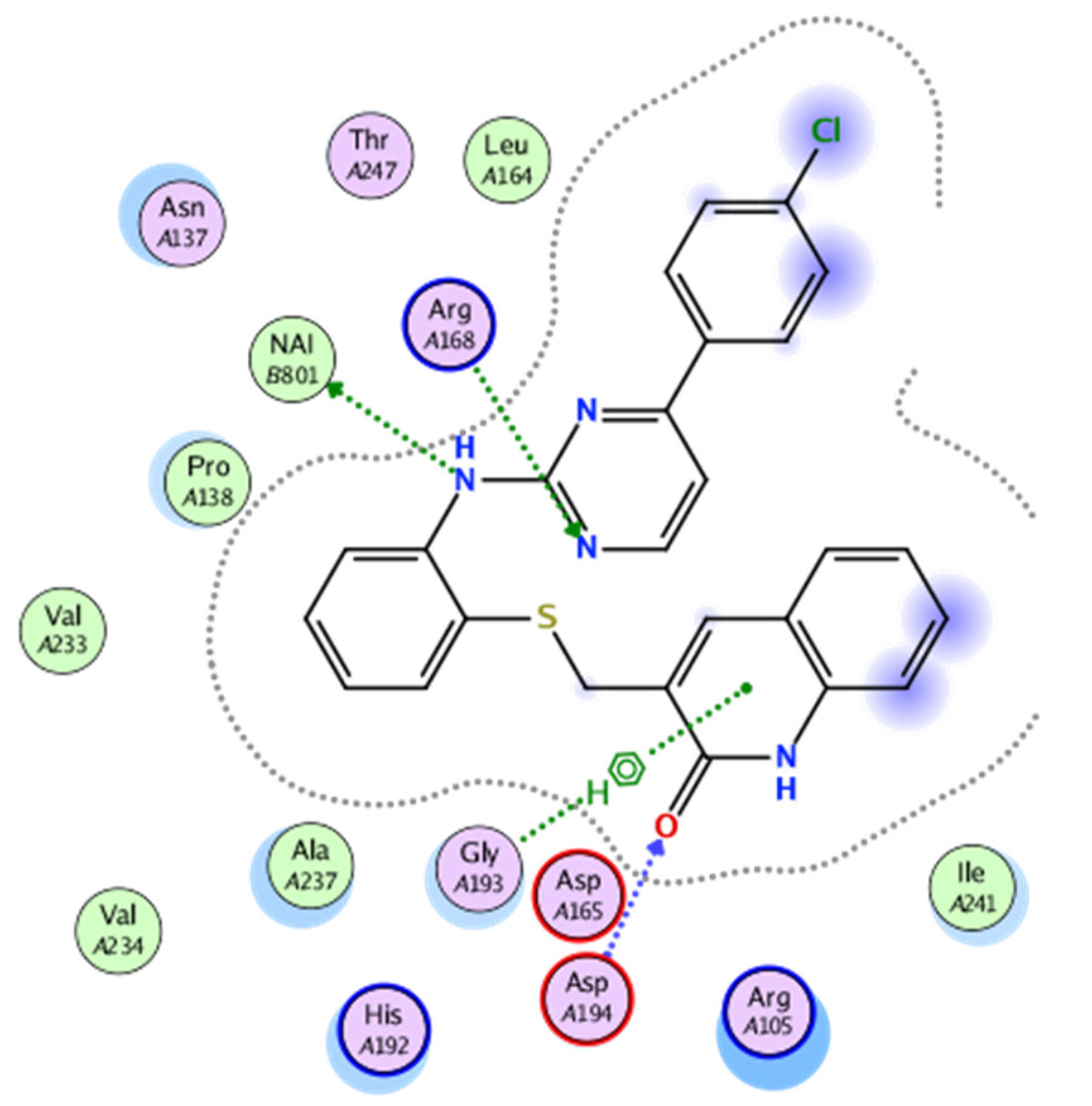
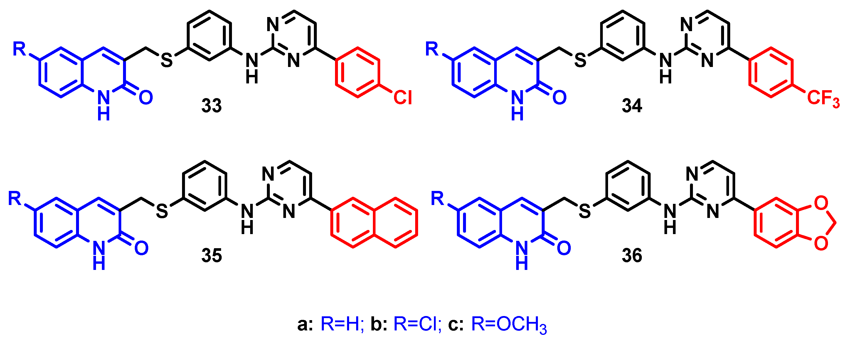
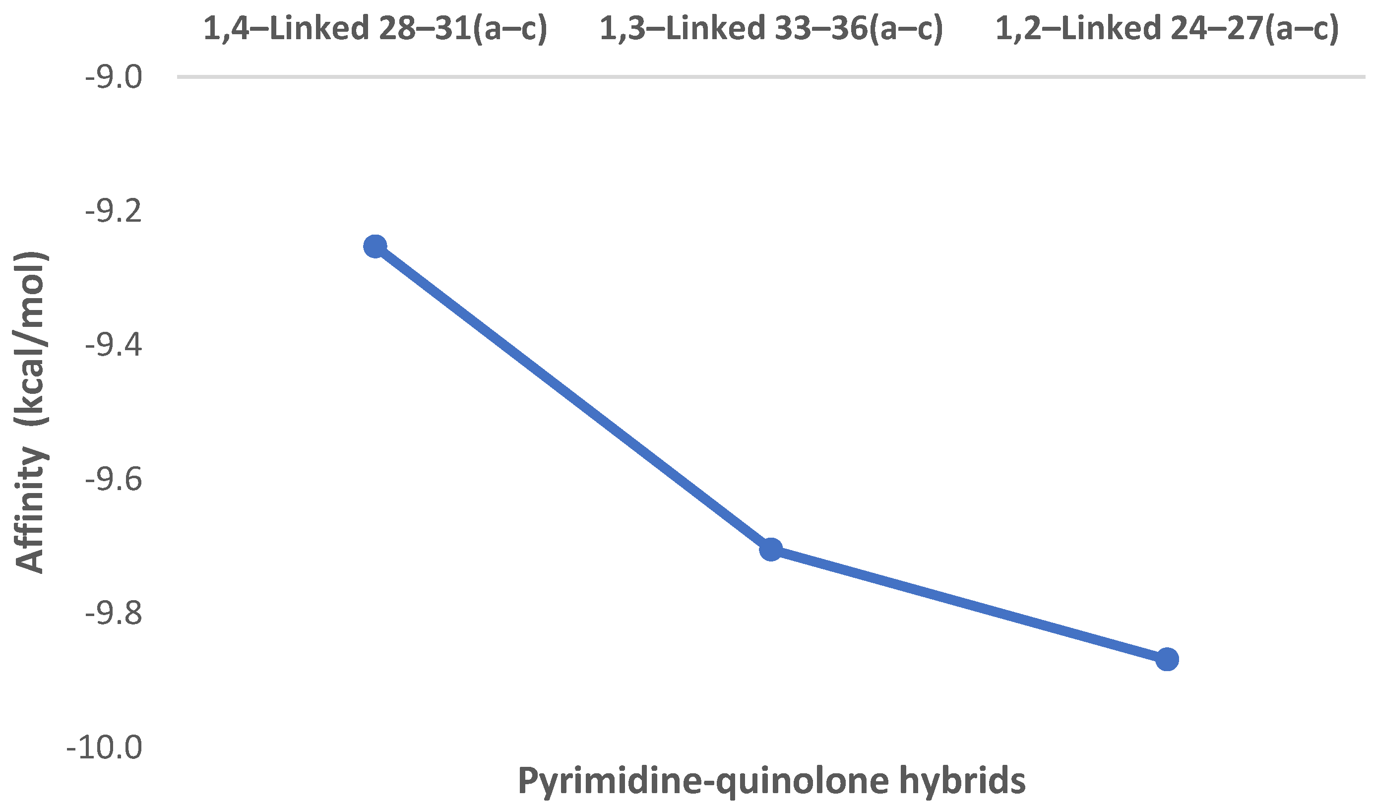
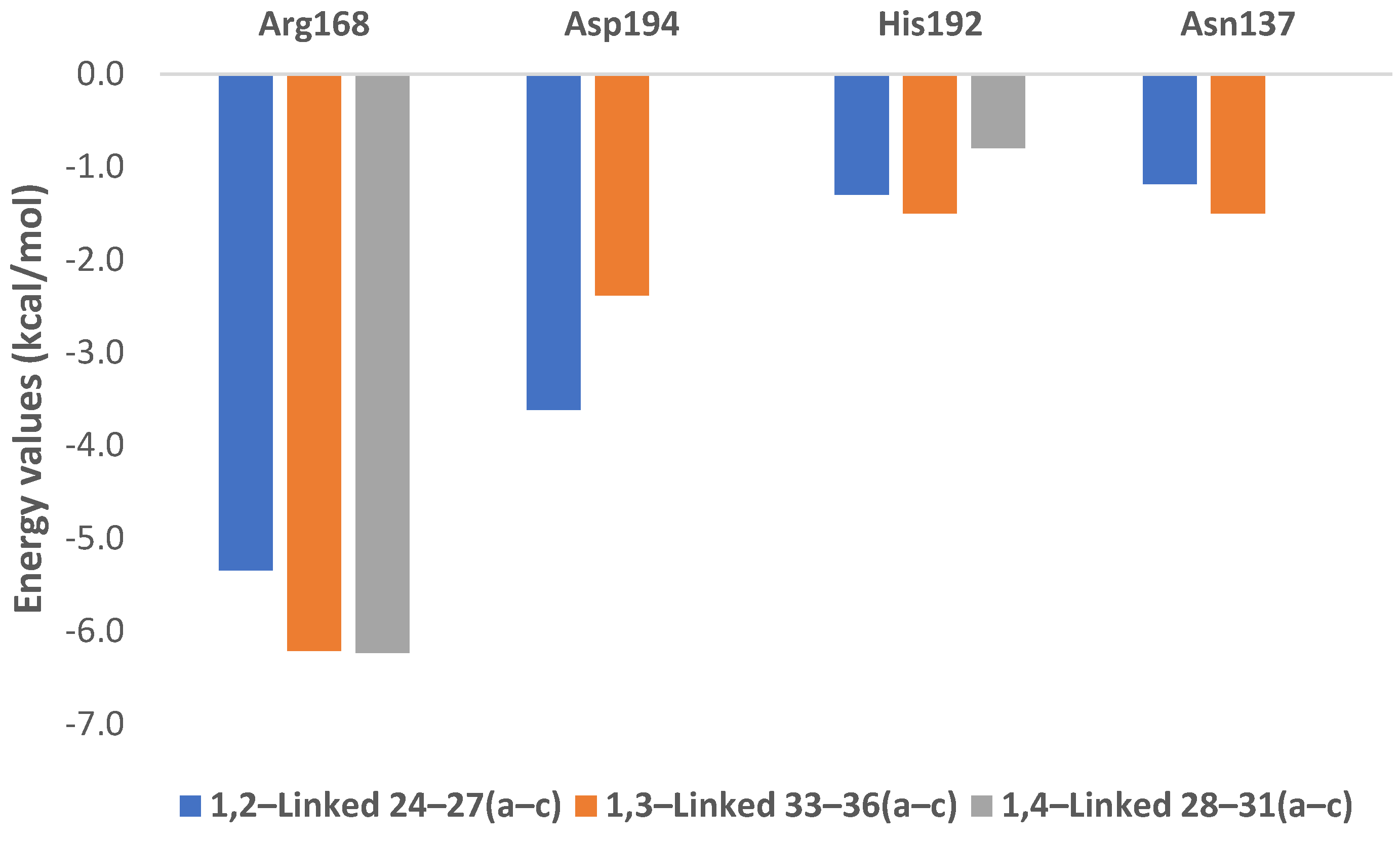
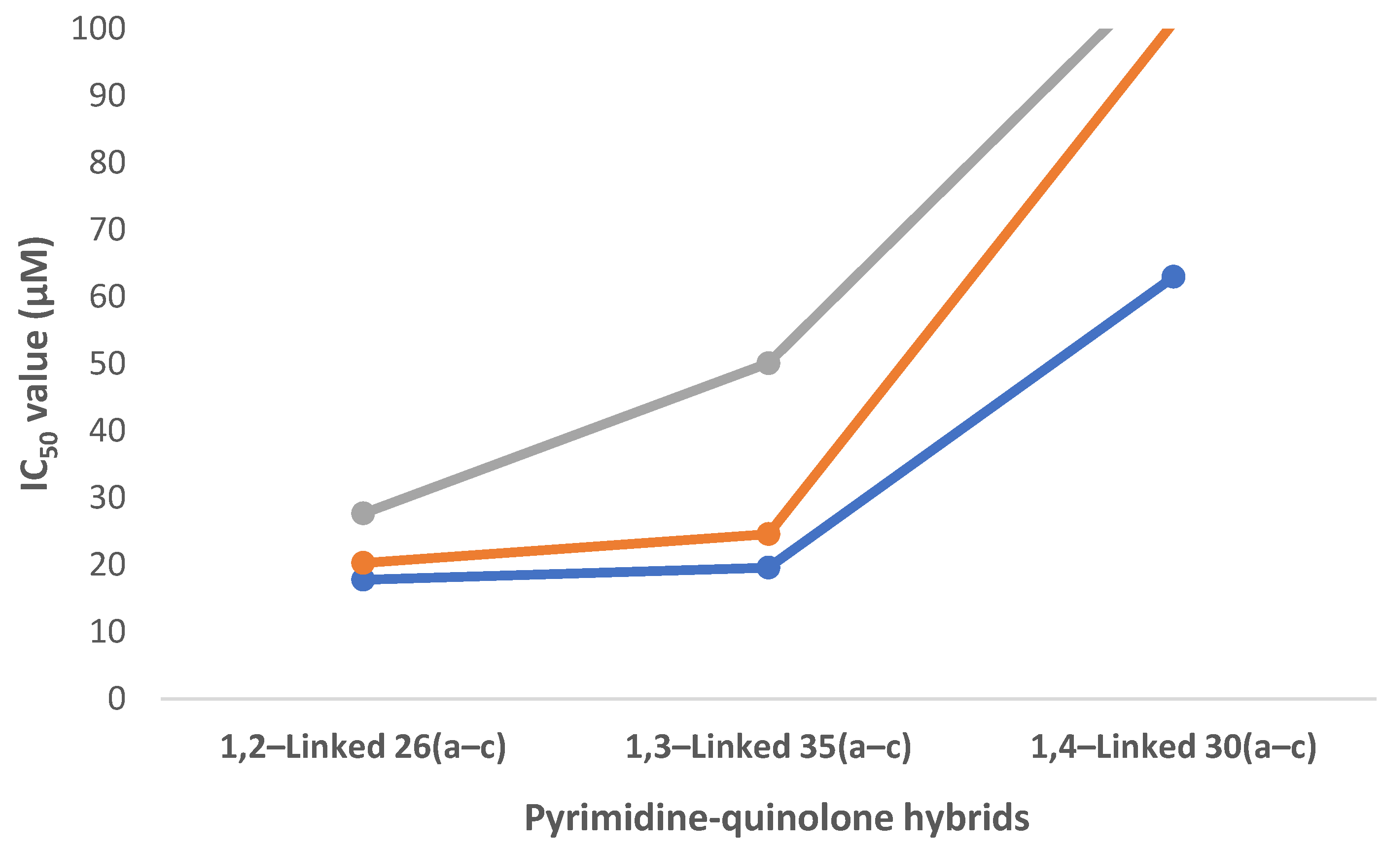
2.3. hLDHA Inhibitory Assays and Structure–Activity Relationship (SAR)

The inhibitory activity of the pyrimidine-quinolone hybrids 10, 15, 19, and 2431(ac) against the hLDHA enzyme was measured by a kinetic spectrofluorometric assay [66].
The first set of compounds (10, 15, and 19) did not show good inhibitory activity, as their IC50 was >100 μM. Thus, in concordance with the docking results for the second set of hybrids 2431(ac), the inhibitory activity of both 1,2-linked 2427(ac) and 1,4-linked 2831(ac) was measured.
1,2-Linked hybrids 2427(ac), with the exception of 27a, have IC50 values under 100 μM, from which seven have IC50 < 50 μM (24a, 24b, 25a, 25b, and 26(ac)). However, from those 1,4-linked hybrids, only 29b, 30a, and 31b have IC50 < 100 μM, with their values ranging between 50 and 83 μM (Table 4).
Amongst those 1,2-linked hybrids having IC50 < 50 μM, compounds 26(ac), having the napthalen2-yl moiety, are the ones with the best inhibitory results (as previously predicted), their IC50 values being 17.8, 20.3, and 27.7 μM, respectively.
1,4-Linked hybrids 2831(ac) were predicted to be inactive; however, despite 31a and 31c being inactive, compound 31b demonstrated an interesting IC50 = 49.9 μM as the only 1,4-linked hybrid with interesting inhibitory activity.
An explanation for that might be found in the placement of 31b in the active site. Thus, meanwhile, 31a did not even pass the filtering criteria, and 31c had a very different placement to that of W31, and 31b had a more similar one to W31, enabling some interactions with the different amino acid residues (Figure 10).
If we compare the inhibitory activity of all the 1,4-linked hybrids 2831(ac) with the inhibitory activity of those 1,2-linked 2427(ac), there is a correlation with the in silico studies, marking the importance of the U-shaped disposition to mimic the shaping of the reference W31.
The correlation found between the in silico studies and the experimental data encouraged us to design a preliminary structure–activity relationship. In this regard, we envisioned that perhaps the 1,3-linked pyrimidine-quinolone hybrids 3336(ac) (Figure 11) may also be of interest, and we decided to study them in silico following the process described for the 1,2- and 1,4-linked pyrimidine-quinolone hybrids 2431(ac) (Section 2.1).
Concerning to the affinity criteria, we found a tendency concerning the linker substitution where, when going from the 1,4-subtitution towards the 1,2-substituion, the affinity improved considerably as seen in Figure 12. In this figure, mean affinity values (kcal/mol) are represented grouped by linker families. Values were obtained from the minimization process made after obtaining the docking output file (see Supplementary Materials Tables S5 and S6).
Moreover, we found that there is also a clear relationship between the substitution pattern and the energy values involved in the interaction with the main amino acid residues (Figure 13). This way, all the structures evaluated show a strong interaction with key Arg168.
However, when considering the other amino acid residues (His192, Asn137, and especially with Asp194), there are some major differences. On the one hand, interactions with Asn137 and His192 do not enable a major difference in terms of defining whether the structure is a potential inhibitor or not, as their values are quite similar and around −1 kcal/mol.
On the other hand, and most importantly, interactions with Asp194 are an essential factor to discriminate, as those 1,4-linked hybrids (2831)(ac) do not interact with it, but those having the 1,2-substituition pattern (2427)(ac) demonstrate a strong interaction.
Now, after realizing the importance of the affinity and energy values involved in the interactions with the different amino acid residues, the fact that those compounds having the 4-chlorophenyl scaffold 24(ac) have similar IC50 values to those having the 4-trifluorophenyl 25(ac), respectively, is explained in silico by striking a balance between the better affinity values of 25(ac) and the better energy values involved in the interactions with Arg168 and Asp194 of 24(ac). Moreover, the absence of interactions in the 1,2-linked hybrids with the benzo[d][1,3]dioxol-5-yl moiety 27(ac) with Asp194 may explain the fact that they do not demonstrate high inhibitory activity despite having similar affinity values to the other 1,2-linked hybrids (see Table 2 in Section 2.1).
In light of the analysis of the data from the SAR, we realized that those 1,3-linked pyrimidine-quinolone hybrids (3336)(ac) might have interesting inhibitory activity, which is predicted to be between the 1,4-linked (2831)(ac) and 1,2-linked pyrimidine quinolone hybrids (2427)(ac).
The reason for this is that compounds 3336(ac) have better affinities than the 1,4-linked hybrids 2831(ac) but worse than the 1,2-linked 2427(ac); they have shown similar energy values when interacting with Arg168 to 2831(ac) and 2427(ac), and concerning the interaction with Asp194, they have similar energy values to 2427(ac) but much better than those of 2831(ac), which do not interact with this amino acid residue.
Therefore, at this point, we decided to synthesize, following the novel convergent pathway that we developed, those 1,3-linked pyrimidine-quinolone hybrids having the naphthalen-2-yl moiety as the hydrophobic tail 35(ac), which has already proven to be the most interesting one towards the inhibition of the hLDHA enzyme (Scheme 4).
The reason for doing so was to ensure that their biological activity was as predicted. This way, once 35(ac) were synthesized, they were subjected to the determination of their IC50 value. The reaction yields, time for the synthesis, and the IC50 value of the hybrids 35(ac) are shown in Table 5.
Lower reaction times were required for the synthesis of 35(ac), and yields were better than in the 1,2-linked 26(ac), with the exception of 35b, and similar to the 1,4-linked 30(ac) due to the meta disposition and thus were not affected by steric hindrance.
From these hybrids, it is important that the chemical shift of the proton located in position 2 of the 3-aminobenzenethiol moiety goes from 6.60 ppm in intermediates 32(ac) to 8.10 ppm in hybrids 35(ac) as a consequence of being linked to the pyrimidine moiety. That proton is coupled with those in positions 4 and 6, with its coupling constant (J) being 2.0 Hz in 35a. In hybrids 35b and 35c as well as in intermediates 32(ac), the spectrum is not clear enough in order to differentiate the coupling, being represented as a pseudo-singlet.
From the inhibition assays, it can be said that, as seen in Table 5, compound 35a has a slight worse inhibitory activity than 26a (19.6 and 17.8 μM, respectively) and 35b than 26b (20.3 and 24.6 μM, respectively), with the difference becoming even larger when comparing 35c (50.1 μM) to 26c (27.2 μM).
All of this is in concordance with what was previously predicted: the inhibitory activity of those hybrids having the U-shaped disposition 26(ac) is slightly better than in the case of 35(ac) and both of them drastically better than 30(ac). For an easier interpretation of the results, the different IC50 values for the differently linked hybrids are shown in Figure 14.
Results shown in Figure 14 demonstrate that, even though the inhibitory activity of the 1,3-linked pyrimidine-quinolone hybrids 35(ac) is close to those having the 1,2-linked disposition, this type of substitution it is not the best one, which was proven to be that of the 1,2-linked hybrids.
Additionally, it was shown that the effect of having a bulkier group, such as the methoxy one (8c), in the quinolone moiety is translated into a slightly lower inhibitory activity.

3. Materials and Methods

3.1. General

All chemicals and solvents were purchased from Sigma-Aldrich unless stated otherwise. Melting points were collected using a Brastead Electrothermal 9100 melting point apparatus, and the acquired data are uncorrected. IR spectra were recorded on a Fourier Bruker Tensor 27 Spectrophotometer using the ATR dura Sample IR accessory. NMR spectra were recorded in Bruker Avance NEO 400 spectrometer at 400 MHz (1H) and 100 MHz (13C) at 298 K and 393 K and Bruker Advance 500 spectrometer at 500 MHz (1H) and 125 MHz (13C) at 298 K and 393 K, using as solvent DMSO-d6 and as the internal reference tetramethylsilane (0 ppm) or the residual 1H/13C solvent signals, that is, 2.50/39.52. DEPT-135 and 2D-NMR (HSQC, HMBC, and COSY) experiments were used for the assignment of carbon and hydrogen signals. Chemical shifts (δ) are given in ppm, and coupling constants (J) are given in Hz. The following abbreviations are used for multiplicities: s, singlet; d, doublet; t, triplet; q, quartet; m, multiplet; ps, pseudo-singlet; pd, pseudo-doublet; and pt, pseudo-triplet. The mass spectra were recorded on a Thermo model DSQ II spectrometer equipped with a direct inlet probe and operating at 70 eV. HPLC-HRMS data were obtained on an Agilent Technologies Q-TOF 6530B coupled to an HPLC Agilent-1260 Infinity, equipped with a Kinetex C18 column (2.1 mm × 50 mm × 2.6 um) PN 00B-4462-AN using the following HPLC method: flow, 0.4 mL/min; elution gradient, 0–5 min from acetonitrile/water 10% (0.1% formic acid) to acetonitrile 100% (0.1% formic acid); plus 3 additional minutes at that concentration. Ionization method: electrospray ionization; (ESI+) acquisition software: MassHunter LC/MS Data Acquisition 6200 series TOD/6500 series Q-TOF, Version: B.06.01 (Build 6.01.6172 SP1). The single-crystal X-ray data were collected in a Diffractometer Bruker D8 Venture. All the equipment used in the spectroscopic and spectrometric analysis belong to “Centro de Instrumentación Científico y Técnico”, (CICT) in “Universidad de Jaén” (UJA). The reactions were monitored by TLC on a 0.2mm pre-coated aluminum plates of silica gel (Merck 60 F254), and spots were visualized by UV irradiation (254nm). All reagents were purchased from commercial sources and used without further purification unless otherwise noted. All starting materials were weighed and handled in air at room temperature. Precursor quinolone derivatives (8(ac)) [67] and 4-aryl-2-chloropyrimidines (14) [58] were prepared according to reported procedures.

3.2. Chemistry

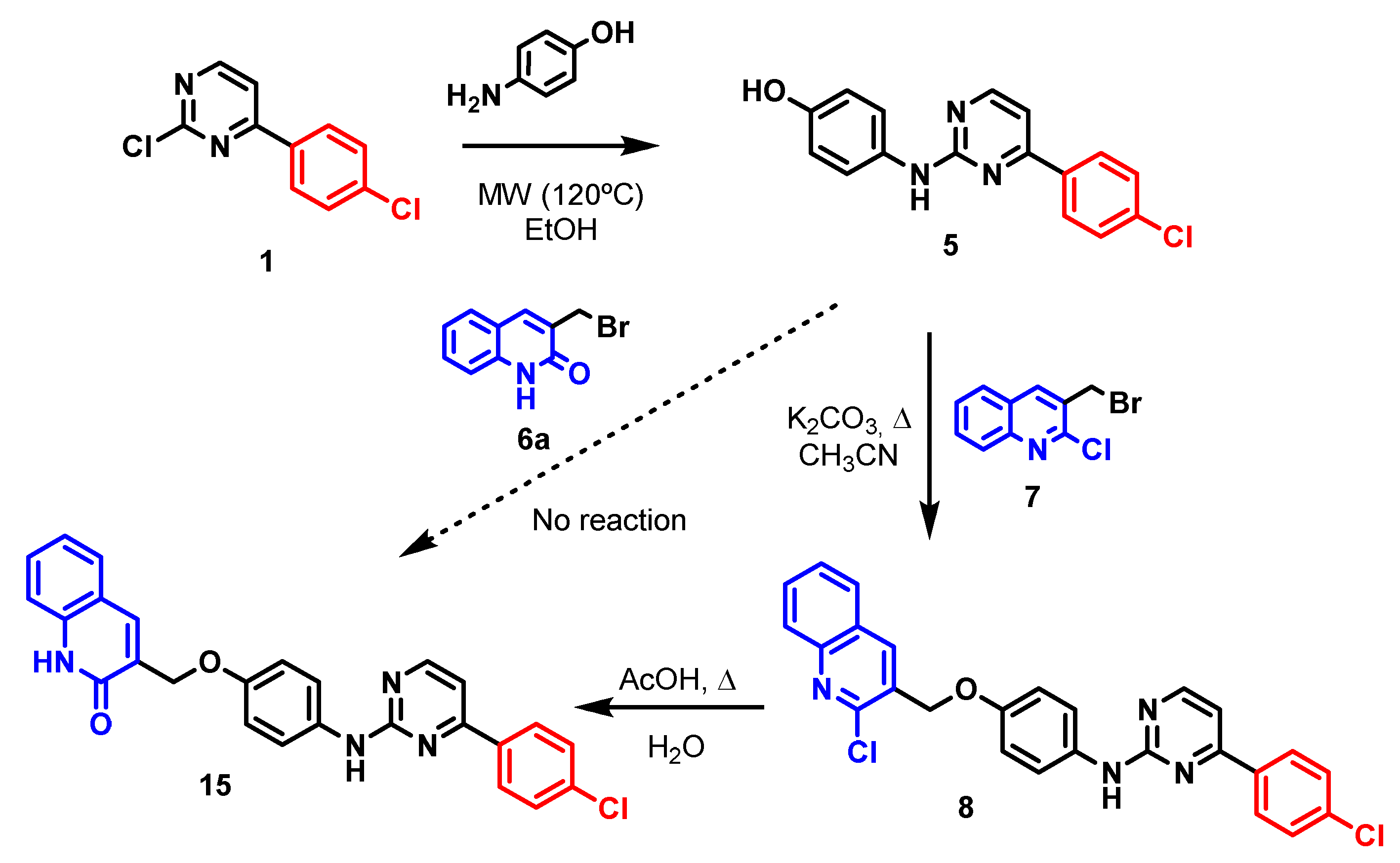
3.2.1. Synthesis of 4-(4-Chlorophenyl)-N-(4-((2-chloroquinolin-3-yl)methoxy)phenyl)pyrimidin-2-amine (8)

3-(Bromomethyl)-2-chloroquinoline (0.30 mmol) was added to a solution of 5 (0.30 mmol) and potassium carbonate (0.60 mmol) in acetonitrile (5mL). The mixture was heated up at reflux within 4 h and 25 min. Once the reaction was completed (TLC monitored), the mixture was cooled at room temperature, and the solid was filtered and washed with cold acetonitrile and water, respectively. No further purification was done. Yellow Solid (64%) M.p. 478–481 K. Rf Hex:AcOEt (6:4): 0.27. 1H NMR (400 MHz, DMSO-d6) δ 9.60 (s, 1H), 8.58 (d, J = 22.7 Hz, 1H), 8.52 (d, J = 5.2 Hz, 1H), 8.19–8.14 (m, 2H), 8.09 (d, J = 8.0 Hz, 1H), 8.00 (dd, J = 8.0, 6.9 Hz, 1H), 7.84 (ddd, J = 8.0, 6.9, 1.5 Hz, 1H), 7.74 (pd, J = 9.0 Hz, 2H), 7.71–7.67 (m, 1H), 7.63–7.58 (m, 2H), 7.36 (d, J = 5.2 Hz, 1H), 7.09 (dd, J = 9.0, 2.0 Hz, 2H), and 5.26 (d, J = 17.4 Hz, 2H). 13C NMR (100 MHz, DMSO-d6) δ 162.3, 160.2, 159.2, 152.9, 149.0, 147.2, 146.5, 142.1, 138.2, 137.7, 135.59, 135.57, 134.4, 130.9, 130.7, 128.99, 128.93, 128.6, 128.2, 128.1, 127.7, 127.6, 127.6, 126.9, 126.9, 120.7, 115.0, 107.4, 68.5, and 66.8. IR (ATR, cm−1): 3275 (NH), 3195, 3120, 1508, 1425, 1229, 804, and 748. EI MS (70eV): m/z (%): 472 (M+, 1), 298 (37), 196 (100), 140 (16), and 44 (69). HRMS (ESI-QTOF) M + H calc. for C26H18Cl2N4O: 473.0930 found: 473.0927.

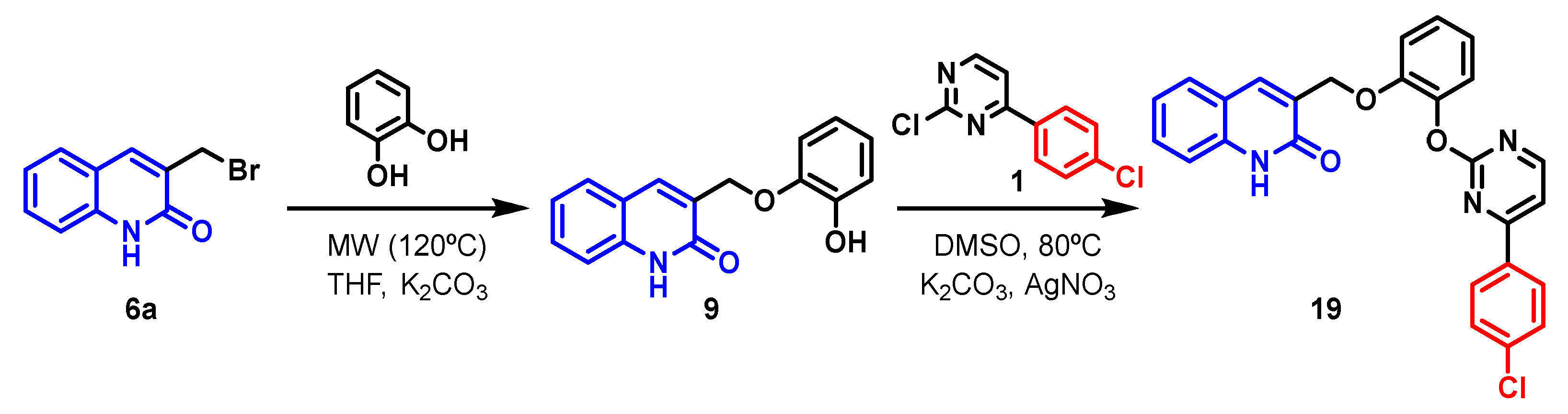
3.2.2. Synthesis of 3-((2-Hydroxyphenoxy)methyl)quinolin-2(1H)-one (9)

1,2-Dihydroxybenzene (4.30 mmol) was added to a solution of 6a (0.86 mmol) and potassium carbonate (1.72 mmol) in THF (3mL). The mixture was stirred at room temperature for 13h. After the reaction was completed (TLC monitored), the solvent was removed under vacuum, and water was added, introducing the mixture under ultrasound in order to enable the precipitation. After that, the solid was collected by filtration. The desired product was obtained by further purification with DCM:MeOH, 97:3. White Solid (57%) M.p. 496–499 K. Rf DCM:MeOH, (97:3): 0.28. 1H NMR (400 MHz, DMSO-d6) δ 12.06 (s, 1H), 9.21 (s, 1H), 8.14 (s, 1H), 7.70 (dd, J = 7.2, 1.5 Hz, 1H), 7.52 (ddd, J = 8.4, 7.2, 1.5 Hz, 1H), 7.36 (d, J = 8.4 Hz, 1H), 7.25–7.18 (m, 1H), 7.01 (dd, J = 7.9, 1.2 Hz, 1H), 6.89–6.80 (m, 2H), and 6.74 (ddd, J = 7.9, 6.6, 2.5 Hz, 1H), 4.95 (s, 2H). 13C NMR (100 MHz, DMSO-d6) δ 161.3, 147.5, 146.4, 138.1, 136.9, 130.3, 128.6, 127.9, 122.2, 122.1, 119.3, 119.0, 116.2, 115.4, 115.1, and 66.7. IR (ATR, cm−1): (3400–2400, wide NH amide and OH signals), 1654, 994, and 740. EI MS (70eV): m/z (%): 267 (M+, 7), 158 (100), and 130 (28). HRMS (ESI-QTOF) M + H calc. for C16H13NO3: 299′0582 found: 299′0582.

3.2.3. Synthesis of 3-((4-((4-(4-Chlorophenyl)pyrimidin-2-yl)amino)phenoxy)methyl)quinolin-2(1H)-one (15)

Acetic acid (10mL) was added to a solution of 8 (0.14 mmol) in water (4mL). The mixture was heated at reflux within 7 h and 10 min. Once the reaction was completed (TLC monitored), the mixture was cooled at room temperature and introduced overnight in the refrigerator in order to enable the precipitation. The desired product was obtained by filtration and washed with water. Yellow Solid (61%) M.p. 575–578 K. Rf Hex:AcOEt (6:4): 0.08. 1H NMR (400 MHz, DMSO-d6) δ 11.99 (s, 1H), 9.57 (s, 1H), 8.51 (d, J = 5.2 Hz, 1H), 8.16 (d, J = 8.3 Hz, 2H), 8.00 (s, 1H), 7.72–7.69 (m, 3H), 7.59 (d, J = 8.3 Hz, 2H), 7.49 (pt, J = 7.7 Hz, 1H), 7.36–7.32 (m, 2H), 7.18 (pt, J = 7.7 Hz, 1H), 7.03 (d, J = 9.0 Hz, 2H), and 4.98 (s, 2H). 13C NMR (100 MHz, DMSO-d6) δ 162.3, 160.9, 160.3, 159.2, 153.2, 138.1, 136.3, 135.6, 134.0, 130.1, 128.93, 128.89, 128.6, 127.9, 122.0, 120.7, 120.6, 118.9, 115.0, 114.7, 107.3, and 64.9. IR (ATR, cm−1): (3400–2400, wide NH amide signal), 3304 (NH), 3066, 2831, 1661 (C=O), 1554, 1508, 1424, 1231, and 802. EI MS (70eV): m/z (%): 454 (M+, 6), 298 (33), 296 (100), and 158 (15). HRMS (ESI-QTOF) M + H calc. for C26H19ClN4O2: 455.1269 found: 455.1266.

3.2.4. Synthesis of 3-((2-((4-(4-Chlorophenyl)pyrimidin-2-yl)oxy)phenoxy)methyl)quinolin-2(1H)-one (19)

Intermediate 1 (0.023 mmol) was added to a solution of 9 (0.023 mmol), potassium carbonate (0.045mmol), and silver nitrate (0.006 mmol) in dimethyl sulfoxide (0.3mL). The mixture was heated up to 80 °C for 8 h and 30 min. After the reaction was completed (TLC monitored), the mixture was neutralized with acetic acid, and the solid was filtered and washed with water. The desired product was obtained by further purification with Hex:AcOEt (4:6). White Solid (67%) M.p. 496–499 K. Rf Hex:AcOEt (4:6): 0.11. 1H NMR (400 MHz, DMSO-d6) δ 11.88 (s, 1H), 8.68 (d, J = 5.2 Hz, 1H), 8.09 (d, J = 8.3 Hz, 2H), 7.79 (d, J = 5.2 Hz, 1H), 7.58 (d, J = 8.3 Hz, 2H), 7.45–7.37 (m, 1H), 7.35–7.31 (m, 1H), 7.29–7.21 (m, 3H), 7.18 (s, 1H), 7.07 (td, J = 7.4, 2.1 Hz, 1H), 6.98–6.96 (m, 2H), and 4.93 (s, 2H). 13C NMR (100 MHz, DMSO-d6) δ 165.0, 164.4, 160.9, 160.4, 149.9, 142.0, 137.7, 136.4, 134.4, 134.3, 130.0, 129.1, 128.8, 128.5, 127.2, 126.4, 122.6, 121.7, 121.5, 118.4, 115.0, 114.3, 112.0, and 65.0. IR (ATR, cm−1): (3200–2600, wide NH amide signal), 3331, 3063, 2923, 2853, 1660 (C=O), 1577, 1501, 1434, 1379, 1262, 1087, 818, and 745. EI MS (70eV): m/z (%): 455 (M+, 8), 158 (100), and 130 (21). HRMS (ESI-QTOF) M + H calc. for C26H18ClN3O3: 456.1109 found: 456.1108.

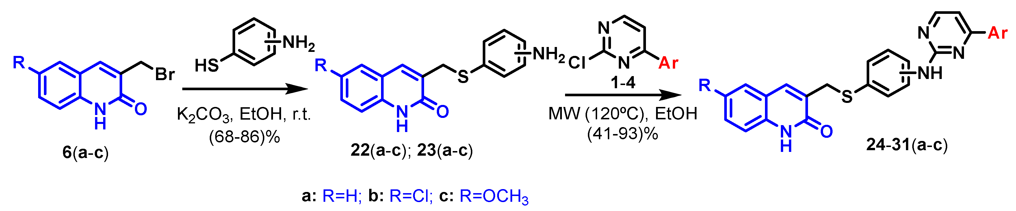
3.2.5. General Procedure for the Synthesis of 3-(((2′-Aminophenyl)thio)methyl)quinolin-2(1H)-ones (22(a–c)), 3-(((4′-aminophenyl)thio)methyl)quinolin-2(1H)-ones (23(a–c)), and 3-(((3′-aminophenyl)thio)methyl)quinolin-2(1H)-ones (32(a–c))

Potassium carbonate (1.5 mmol per mmol of (14) when using 4-aminothiphenol and 1 mmol per mmol when using 2-aminothiophenol) was added to a solution of the corresponding aminothiophenol (1.2 mmol per mmol of (14) for 4-aminothiphenol and 1 mmol per mmol for 2-aminothiophenol) in ethanol, and it was stirred 5 min at room temperature under argon atmosphere. After that, the different 3-(bromomethyl)quinolin-2(1H)-ones 6(ac), (1 mmol) were added, and the mixture was stirred until the reaction was completed; TLC monitored using Hex:AcOEt (6:4) as eluent. Once the reaction was completed, the solid was filtered and washed with ethanol and water to afford a pure solid.

3-(((2-Aminophenyl)thio)methyl)quinolin-2(1H)-one (22a)

White Solid (86%) M.p. 493–496 K. Rf Hex:AcOEt (6:4): 0.19. 1H NMR (400 MHz, DMSO-d6) δ 11.89 (s, 1H), 7.51–7.41 (m, 3H), 7.30 (d, J = 7.7 Hz, 1H), 7.14–7.12 (m, 2H), 7.04 (t, J = 7.9 Hz, 1H), 6.72 (d, J = 7.9 Hz, 1H), 6.44 (t, J = 7.9 Hz, 1H), 5.63 (s, 2H), and 3.82 (s, 2H). 13C NMR (100 MHz, DMSO-d6) δ 161.3, 149.6, 138.2, 137.2, 135.7, 129.84, 129.77, 129.0, 127.5, 121.9, 119.0, 116.3, 115.2, 114.9, 114.4, and 32.9. IR (ATR, cm−1): (3500–2400, wide NH amide signal), 3475 and 3373 (NH2), 3148, 3008, 2848, 1661 (C=O), 1599, and 744. EI MS (70eV): m/z (%): 282 (M+, 23), 158 (100), and 130 (33). HRMS (ESI-QTOF) M + H calc. for C16H14N2OS: 283.0900, found: 283.0895.

3-(((2-Aminophenyl)thio)methyl)-6-chloroquinolin-2(1H)-one (22b)

White Solid (68%) M.p. 509–512 K. Rf Hex:AcOEt (1:1): 0.24. 1H NMR (400 MHz, DMSO-d6) δ 12.01 (s, 1H), 7.58 (s, 1H), 7.48 (d, J = 8.5 Hz, 1H), 7.44 (s, 1H), 7.29 (d, J = 8.5 Hz, 1H), 7.11 (d, J = 7.5 Hz, 1H), 7.04 (t, J = 7.5 Hz, 1H), 6.70 (d, J = 7.5 Hz, 1H), 6.43 (t, J = 7.5 Hz, 1H), 5.52 (s, 2H), and 3.79 (s, 2H). 13C NMR (100 MHz, DMSO-d6) δ 161.1, 149.9, 136.9, 135.9, 135.9, 130.5, 129.9, 129.7, 126.4, 125.6, 120.2, 116.8, 116.1, 114.8, 114.3, and 33.0. IR (ATR, cm−1): (3500–2400, wide NH amide signal), 3430 and 3311 (NH2), 3156, 2986, 2819, 1661 (C=O), 818, 748, and 589. EI MS (70eV): m/z (%): 316 (M+, 15), 192 (100), 164 (39), and 80 (41). HRMS (ESI-QTOF) M + H calc. for C16H13ClN2OS: 317.0510, found: 317.0513.

3-(((2-Aminophenyl)thio)methyl)-6-methoxyquinolin-2(1H)-one (22c)

Yellowish Solid (76%) M.p. 469–472 K. Rf Hex:AcOEt (1:1): 0.11. 1H NMR (400 MHz, DMSO-d6) δ 11.79 (s, 1H), 7.50 (s, 1H), 7.24 (d, J = 9.0 Hz, 1H), 7.16–7.13 (m, 1H), 7.12–7.09 (m, 1H), 7.07–7.01 (m, 2H), 6.72 (d, J = 8.0 Hz, 1H), 6.45 (t, J = 8.0 Hz, 1H), 5.63 (s, 2H), 3.81 (s, 2H),and 3.74 (s, 3H). 13C NMR (100 MHz, DMSO-d6) δ 160.9, 154.2, 149.7, 136.9, 135.6, 132.7, 129.7, 129.5, 119.6, 119.0, 116.2, 116.2, 115.2, 114.4, 108.9, 55.4, and 33.1. IR (ATR, cm−1): (3400–2200, wide NH amide signal), 3443 and 3334 (NH2), 3144, 2928, 1604 (C=O), 756, 596, and 460. EI MS (70eV): m/z (%): 312 (M+, 10), 188 (100), 160 (15), and 117 (12). HRMS (ESI-QTOF) M + H calc. for C17H16N2O2S: 313.1005, found: 313.1008.

3-(((4-Aminophenyl)thio)methyl)quinolin-2(1H)-one (23a)

White Solid (82%) M.p. 488–491 K. Rf Hex:AcOEt (1:1): 0.11. 1H NMR (400 MHz, DMSO-d6) δ 11.83 (s, 1H), 7.47 (d, J = 8.0 Hz, 1H), 7.43 (pt, J = 7.7 Hz, 1H), 7.40 (s, 1H), 7.28 (d, J = 8.4 Hz, 1H), 7.11 (pt, J = 7.7 Hz, 1H), 7.03 (d, J = 8.5 Hz, 2H), 6.48 (d, J = 8.5 Hz, 2H), 5.35 (s, 2H), and 3.78 (s, 2H). 13C NMR (100 MHz, DMSO-d6) δ 161.2, 148.5, 138.1, 136.5, 134.5, 129.7, 129.7, 127.4, 121.8, 119.0, 118.8, 114.8, 114.4, and 35.8. IR (ATR, cm−1): (3500–2400, wide NH amide signal), 3470 and 3374 (NH2), 2991, 1658 (C=O), 758, and 495. EI MS (70eV): m/z (%): 282(M+, 15), 158 (100), and 130 (30). HRMS (ESI-QTOF) M + H calc. for C16H14N2OS: 283.0900, found: 283.0897.

3-(((4-Aminophenyl)thio)methyl)-6-chloroquinolin-2(1H)-one (23b)

White Solid (76%) M.p. 488–491 K. Rf Hex:AcOEt (4:6): 0.38. 1H NMR (400 MHz, DMSO-d6) δ 11.96 (s, 1H), 7.61 (ps, 1H), 7.46 (pd, J = 8.8 Hz, 1H), 7.39 (s, 1H), 7.28 (d, J = 8.8 Hz, 1H), 7.02 (d, J = 8.5 Hz, 2H), 6.47 (d, J = 8.5 Hz, 2H), 5.32 (s, 2H), and 3.76 (s, 2H). 13C NMR (101 MHz, DMSO) δ 160.9, 148.8, 136.8, 135.3, 134.7, 131.2, 129.5, 126.3, 125.6, 120.2, 118.3, 116.7, 114.3, and 35.9. IR (ATR, cm−1): (3500–2400, wide NH amide signal), 3444 and 3356 (NH2), 3224, 3146 (NH), 2992, 2909, 2829, 2732, 1648 (C=O), 1597, and 589. EI MS (70eV): m/z (%): 316(M+, 27), 192 (77), 124 (100), and 93 (34). HRMS (ESI-QTOF) M + H calc. for C16H13ClN2OS: 317.0510, found: 317.0508.

3-(((4-Aminophenyl)thio)methyl)-6-methoxyquinolin-2(1H)-one (23c)

White Solid (77%) M.p. 459–461 K. Rf Hex:AcOEt (4:6): 0.11. 1H NMR (400 MHz, DMSO-d6) δ 11.73 (s, 1H), 7.40 (s, 1H), 7.22 (d, J = 8.8 Hz, 1H), 7.08 (dd, J = 8.8, 2.9 Hz, 1H), 7.06–6.99 (m, 3H), 6.48 (d, J = 8.5 Hz, 2H), 5.31 (s, 2H), 3.78 (s, 2H), and 3.74 (s, 3H). 13C NMR (100 MHz, DMSO-d6) δ 160.7, 154.1, 148.7, 136.2, 134.4, 132.6, 130.2, 119.6, 118.8, 116.1, 114.3, 108.9, 55.4, and 35.9. IR (ATR, cm−1): (3500–2400, wide NH amide signal), 3469 and 3357 (NH2), 3147, 2928, 1611 (C=O), 810, and 599. EI MS (70eV): m/z (%): 312 (M+, 15), 188 (100), 160 (20), and 117 (20). HRMS (ESI-QTOF) M + H calc. for C17H16N2O2S: 313.1005, found: 313.1006.

3-(((3-Aminophenyl)thio)methyl)quinolin-2(1H)-one (32a)

White Solid (76%) M.p. 434–437 K. Rf Hex:AcOEt (1:1): 0.07. 1H NMR (400 MHz, DMSO-d6) δ 11.91 (s, 1H), 7.81 (s, 1H), 7.56 (pd, J = 8.0 Hz, 1H), 7.45 (pt, J = 7.7 Hz, 1H), 7.30 (pd, J = 8.4 Hz, 1H), 7.14 (pt, J = 7.7 Hz, 1H), 6.96 (pt, J = 8.0 Hz, 1H), 6.61 (ps, 1H), 6.53 (pd, J = 8.2 Hz, 1H), 6.42 (pd, J = 7.8 Hz, 1H), 5.56 (s, 2H), and 4.01 (s, 2H). 13C NMR (100 MHz, DMSO-d6) δ 161.3, 148.2, 138.1, 136.9, 136.3, 129.9, 129.5, 129.3, 127.6, 121.9, 119.0, 116.4, 114.9, 114.0, 112.3, and 31.7. IR (ATR, cm−1): (3400–2200, wide NH amide signal), 3435 and 3313 (NH2), 3157, 3023, 1641 (C=O), and 765. EI MS (70eV): m/z (%): 282 (M+, 9), 158 (100), 130 (52), and 80 (26). HRMS (ESI-QTOF) M + H calc. for C16H14N2OS: 283.0900 found: 283.0897.

3-(((3-Aminophenyl)thio)methyl)-6-chloroquinolin-2(1H)-one (32b)

White Solid (88%) M.p. 477–480 K. Rf Hex:AcOEt (6:4): 0.22. 1H NMR (400 MHz, DMSO-d6) δ 12.03 (s, 1H), 7.79 (s, 1H), 7.70 (s, 1H), 7.49 (d, J = 8.8 Hz, 1H), 7.29 (d, J = 8.8 Hz, 1H), 6.93 (pt, J = 8.0 Hz, 1H), 6.55 (ps, 1H), 6.47 (pd, J = 7.8 Hz, 1H), 6.38 (pd, J = 7.8 Hz, 1H), 5.18 (s, 2H), and 3.99 (s, 2H). 13C NMR (100 MHz, DMSO-d6) δ 161.0, 149.2, 136.8, 136.0, 135.7, 130.8, 129.8, 129.5, 126.5, 125.7, 120.2, 116.8, 115.9, 113.6, 112.0, and 31.9. IR (ATR, cm−1): (3200–2100, wide NH amide signal), 3468 and 3361 (NH2), 3147, 3055, 2979, 2818, 1654 (C=O), and 770. EI MS (70eV): m/z (%): 316 (M+, 12), 192 (100), 164 (42), and 80 (50). HRMS (ESI-QTOF) M + H calc. for C16H13ClN2OS: 317.0510 found: 317.0510.

3-(((3-Aminophenyl)thio)methyl)-6-methoxyquinolin-2(1H)-one (32c)

White Solid (79%) M.p. 448–451 K. Rf Hex:AcOEt (4:6): 0.09. 1H NMR (400 MHz, DMSO-d6) δ 11.81 (s, 1H), 7.79 (s, 1H), 7.24 (d, J = 8.8 Hz, 1H), 7.14–7.07 (m, 2H), 6.98 (pt, J = 8.0 Hz, 1H), 6.62 (ps, 1H), 6.56 (pd, J = 7.8 Hz, 1H), 6.45 (pd, J = 7.8 Hz, 1H), 5.76 (s, 2H), 4.02 (s, 2H), and 3.75 (s, 3H). 13C NMR (100 MHz, DMSO-d6) δ 160.8, 154.2, 147.7, 136.5, 132.6, 129.6, 129.5, 119.6, 119.2, 116.6, 116.2, 114.0, 112.6, 109.0, 55.4, and 31.7. IR (ATR, cm−1): (3300–2200, wide NH amide signal), 3443 and 3347 (NH2), 3141, 2931, 2830, 1617 (C=O), and 783. EI MS (70eV): m/z (%): 312 (M+, 8), 188 (100), 160 (23), 117 (26), and 80 (21). HRMS (ESI-QTOF) M + H calc. for C17H16N2O2S: 313.1005 found: 313.1005.

3.2.6. General Procedure for the Synthesis of 3-(((2/4-((4-(Aryl)pyrimidin-2-yl)amino)phenyl)thio)methyl)quinolin-2(1H)-ones (24–31)(a–c) and 3-(((3-((4-(naphthalen-2-yl)pyrimidin-2-yl)amino)phenyl)thio)methyl)quinolin-2(1H)-ones (35(a–c))

Intermediates (22, 23, and 31(ac), (1 mmol)) were added to a solution of 4-aryl-2-chloropyrimidine ((14), 1 mmol) in EtOH (3mL per mmol). The mixture was subjected to microwave irradiation at 120 °C until the reaction was completed (TLC monitored using Hex:AcOEt (4:6 or 1:1) as eluent), with a setting of 250 psi and 300 W for maximum pressure and power, respectively. After the reaction was completed, the desired product was obtained by filtration and washed with cold EtOH. No purification was needed, but to ensure maximum purity for biological assays, compounds 2831(ac) were recrystallized from DMF, leaving the recipient open to the air and, if necessary, introduced in the refrigerator. Compounds 2427(ac) were recrystallized (after having been filtrated) from ethanol under MW irradiation (1 min at 120 °C). Compounds 35(ac) were recrystallized from EtOH, leaving the recipient open to the air.

3-(((2-((4-(4-Chlorophenyl)pyrimidin-2-yl)amino)phenyl)thio)methyl)quinolin-2(1H)-one (24a)

White Solid (86%) M.p. 460–463 K. Rf Hex:AcOEt (6:4): 0.15. 1H NMR (400 MHz, DMSO-d6) δ 11.32 (s, 1H), 8.46–8.44 (m, 2H), 8.24 (dd, J = 8.2, 1.4 Hz, 1H), 8.11–8.02 (m, 2H), 7.57–7.55 (m, 3H), 7.44 (s, 1H), 7.37–7.35 (m, 2H), 7.31 (d, J = 8.0 Hz, 1H), 7.30 (d, J = 5.2 Hz, 1H), 7.20 (d, J = 8.4 Hz, 1H), 7.08–6.96 (m, 2H), and 3.97 (s, 2H). 13C NMR (100 MHz, DMSO-d6) δ 162.1, 160.3, 159.3, 158.3, 140.3, 137.7, 136.3, 135.1, 134.9, 134.0, 128.9, 128.5, 128.2, 128.1, 128.0, 126.6, 124.1, 122.2, 120.8, 120.2, 118.4, 114.2, 108.0, and 34.5. IR (ATR, cm−1): (3600–2400, wide NH amide signal), 3318 (NH), 3161, 1658 (C=O), 1526, 1436, and 753. EI MS (70eV): m/z (%): 470 (M+, 15), 312 (72), 280 (44), 158 (100), and 130 (50). HRMS (ESI-QTOF) M + H calc. for C26H19ClN4O2: 471.1041, found: 471.1042.

6-Chloro-3-(((2-((4-(4-chlorophenyl)pyrimidin-2-yl)amino)phenyl)thio)meth-yl)quinolin-2(1H)-one (24b)

Yellowish Solid (61%). M.p. 509–512 K. Rf Hex:AcOEt (1:1): 0.30. 1H NMR (400 MHz, DMSO-d6) δ 11.91 (s, 1H), 8.90 (s, 1H), 8.45 (d, J = 5.2 Hz, 1H), 8.05 (d, J = 8.3 Hz, 2H), 8.01 (pd, J = 8.2 Hz, 1H), 7.60–7.55 (m, 3H), 7.41–7.35 (m, 4H), 7.26 (dd, J = 8.8, 2.4 Hz, 1H), 7.12 (pt, J = 7.5 Hz, 1H), 7.09 (d, J = 8.8 Hz, 1H), and 3.89 (s, 2H). 13C NMR (100 MHz, DMSO-d6) δ 163.2, 160.7, 158.5, 157.5, 139.9, 136.7, 136.1, 135.7, 134.7, 134.2, 130.1, 129.5, 129.5, 129.0, 128.9, 126.2, 125.9, 125.5, 124.1, 122.2, 119.9, 116.6, 108.2, and 34.9. IR (ATR, cm−1): (3600–2000, wide NH amide signal), 3336 (NH), 3157, 3056, 2832, 1656 (C=O), 1568, 1513, 1433, and 745. EI MS (70eV): m/z (%): 504 (M+, 7), 312 (60), 280 (82), 192 (41), 164 (37), and 43 (100). HRMS (ESI-QTOF) M + H calc. for C26H18Cl2N4OS: 505.0651, found: 505.0650. Crystals suitable for X-ray single-crystal diffraction were obtained from DMSO solution, and the crystal data for 24b DMSO solvate were deposited at CCDC with reference CCDC 2159307: Chemical formula C26H18Cl2N4OS · C2H6OS, Mr 739,107; Monoclinic, C2/c; 116K, Cell dimensions a, b, c (Å)48.8146 (17), 5.1928 (1), 34.4150 (13) β (°) α, β, γ (º) 90, 127.875 (1), 90. V (Å3) 6886.0 (4), Z = 8, F (000) = 2416, Dx (Mg m−3) = 1.13, Mo Kα, μ (mm−1) = 0.47, Crystal size (mm) = 0.4 × 0.22 × 0.06. Data collection: Diffractometer Bruker D8 Venture (APEX 3), Monochromator multilayer mirror, CCD rotation images, thick slices φ and θ scans, Mo INCOATEC high-brilliance microfocus sealed tube (λ = 0.71073 Å), multiscan absorption correction (SADABS 2016/2), Tmin, Tmax 0.660, 0.746. No. of measured, independent and observed [I > 2σ(I)] reflections 116,172, 7911, 7618, Rint = 0. 056, (sin θ/λ)max (Å−1) 0.650, θ values (°): θmax = 28.3, θmin = 2.1; Range h = −62→62, k = −6→6, l = −44→44, Refinement on F2:R[F2 > 2σ(F2)] = 0. 096, wR(F2) = 0. 128, S=1.121. No. of reflections 8554, No. of parameters 345, No. of restraints 277. Weighting scheme: w = 1/σ2(Fo2) + (0.0432P)2 + 24.4231P where P = (Fo2 + 2Fc2)/3. (∆/σ) < 0.001, Δρmax, Δρmin (e Å−3) 0.783, −0.43. Several molecules of disorder DMSO were found in the difference map and the above data resulted from application of Squeeze (Version = 260918).

3-(((2-((4-(4-Chlorophenyl)pyrimidin-2-yl)amino)phenyl)thio)methyl)-6-methoxyquinolin-2(1H)-one (24c)

Yellowish Solid (71%) M.p. 401–404 K. Rf Hex:AcOEt (1:1): 0.14. 1H NMR (400 MHz, DMSO-d6) δ 11.71 (s, 1H), 8.82 (s, 1H), 8.44 (d, J = 5.2 Hz, 1H), 8.06 (d, J = 8.3 Hz, 2H), 8.04 (pd, J = 8.2 Hz, 1H), 7.60–7.54 (m, 3H), 7.39–7.34 (m, 3H), 7.11 (ptd, J = 7.5, 1.4 Hz, 1H), 7.07 (d, J = 8.8 Hz, 1H), 6.94 (dd, J = 8.8, 2.4 Hz, 1H), 6.82 (d, J = 2.4 Hz, 1H), 3.91 (s, 2H), and 3.62 (s, 3H). 13C NMR (100 MHz, DMSO-d6) δ 162.7, 160.6, 159.1, 158.2, 154.0, 139.9, 136.6, 135.9, 135.0, 133.7, 132.6, 129.2, 128.9, 128.8, 128.6, 126.3, 123.9, 122.1, 119.4, 118.8, 116.1, 108.7, 108.2, 55.2, and 34.8. IR (ATR, cm−1): (3600–2200, wide NH amide signal), 3331 (NH), 3060, 2826, 1659 (C=O), 1568, 1433, and 744. EI MS (70eV): m/z (%): 500 (M+, 5), 312 (43), 280 (100), and 188 (94). HRMS (ESI-QTOF) M + H calc. for C27H21ClN4O2S: 501.1147, found: 501.1138.

3-(((2-((4-(4-(Trifluoromethyl)phenyl)pyrimidin-2-yl)amino)phenyl)thio)methyl)quinolin-2(1H)-one (25a)

Pale-yellow Solid (41%) M.p. 478–482 K. Rf Hex:AcOEt (6:4): 0.12. 1H NMR (400 MHz, DMSO-d6) δ 11.81 (s, 1H), 8.75 (s, 1H), 8.48 (d, J = 5.2 Hz, 1H), 8.24 (d, J = 8.1 Hz, 2H), 8.05 (pd, J = 8.2 Hz, 1H), 7.87 (d, J = 8.1 Hz, 2H), 7.55 (pd, J = 7.8 Hz, 1H), 7.41 (s, 1H), 7.40 (d, J = 5.2 Hz, 1H), 7.35 (pt, J = 7.5 Hz, 1H), 7.31–7.29 (m, 2H), 7.13 (d, J = 8.4 Hz, 1H), 7.09 (pt, J = 7.5 Hz, 1H), 6.97 (pt, J = 7.7 Hz, 1H), and 3.93 (s, 2H). 13C NMR (100 MHz, DMSO-d6) δ 162.1, 161.1, 159.8, 159.2, 140.3, 140.1, 138.1, 137.0, 133.7, 130.8, 130.5, 129.7, 128.7, 128.6, 127.7, 127.3, 126.2, 125.7, 123.7, 122.0, 121.6, 118.8, 114.8, 108.9, and 34.6. IR (ATR, cm−1): (3700–2100, wide NH amide signal), 3333 (NH), 3060, 2998, 1658 (C=O), 1568, 1520, 1433, and 743. EI MS (70eV): m/z (%): 504 (M+, 6), 346 (40), 314 (25), 158 (100), and 130 (50). HRMS (ESI-QTOF) M + H calc. for C27H19F3N4OS: 505.1304, found: 505.1303.

6-Chloro-3-(((2-((4-(4-(trifluoromethyl)phenyl)pyrimidin-2-yl)amino)phenyl) thio)methyl)quinolin-2(1H)-one (25b)

Yellow Solid (50%) M.p. 509–513 K. Rf Hex:AcOEt (1:1): 0.17. 1H NMR (400 MHz, DMSO-d6) δ 11.87 (s, 1H), 8.63 (s, 1H), 8.46 (d, J = 5.2 Hz, 1H), 8.21 (d, J = 8.1 Hz, 2H), 8.07 (pd, J = 8.2 Hz, 1H), 7.86 (d, J = 8.1 Hz, 2H), 7.62–7.57 (m, 1H), 7.39–7.37 (m, 2H), 7.31 (d, J = 2.4 Hz, 1H), 7.27 (s, 1H), 7.22 (dd, J = 8.8, 2.4 Hz, 1H), 7.10 (pt, J = 7.5 Hz, 1H), 7.04 (d, J = 8.8 Hz, 1H), and 3.87 (s, 2H). 13C NMR (100 MHz, DMSO-d6) δ 161.8, 160.7, 159.6, 159.4, 140.7, 140.2, 136.7, 135.5, 134.5, 130.7, 130.4, 130.1, 129.4, 129.0, 127.6, 126.1, 125.7, 125.5, 125.2, 123.5, 121.6, 119.8, 116.6, 108.9, and 35.2. IR (ATR, cm−1): (3200–2400, wide NH amide signal), 3325 (NH), 3160, 2991, 2823, 1659 (C=O), 1523, and 807. EI MS (70eV): m/z (%): 538 (M+, 9), 346 (100), 314 (76), 192 (96), and 164 (73). HRMS (ESI-QTOF) M + H calc. for C27H18ClF3N4OS: 539.0915, found: 539.0906.

6-Methoxy-3-(((2-((4-(4-(trifluoromethyl)phenyl)pyrimidin-2-yl)amino)phenyl)thio)methyl)quinolin-2(1H)-one (25c)

White Solid (57%) M.p. 486–489 K. Rf Hex:AcOEt (1:1): 0.07. 1H NMR (400 MHz, DMSO-d6) δ 11.69 (s, 1H), 8.69 (s, 1H), 8.47 (d, J = 5.2 Hz, 1H), 8.23 (d, J = 8.1 Hz, 2H), 8.07 (pd, J = 8.2 Hz 1H), 7.86 (d, J = 8.1 Hz, 2H), 7.57 (pd, J = 7.8 Hz, 1H), 7.38 (d, J = 5.2 Hz, 1H), 7.35–7.33 (m, 2H), 7.10 (pt, J = 7.5 Hz, 1H), 7.04 (d, J = 8.8 Hz, 1H), 6.91 (dd, J = 8.8, 2.4 Hz, 1H), 6.80 (d, J = 2.4 Hz, 1H), 3.90 (s, 2H), and 3.58 (s, 3H). 13C NMR (100 MHz, DMSO-d6) δ 161.8, 160.6, 159.8, 159.4, 153.9, 140.3, 140.3, 136.5, 133.9, 132.6, 130.7, 130.4, 129.2, 128.7, 127.6, 125.9, 125.7, 123.6, 121.9, 119.3, 118.7, 116.0, 108.9, 108.7, 55.1, and 35.0. IR (ATR, cm−1): (3400–2400, wide NH amide signal), 3337 (NH), 3157, 3055, 3000, 2829, 1660 (C=O), 1514, and 808. EI MS (70eV): m/z (%): 534 (M+, 11), 346 (39), 314 (27), 188 (100), 160 (34), and 117 (32). HRMS (ESI-QTOF) M + H calc. for C28H21F3N4O2S: 535.1410, found: 535.1409.

3-(((2-((4-(Naphthalen-2-yl)pyrimidin-2-yl)amino)phenyl)thio)methyl)quinolin-2(1H)-one (26a)

White Solid (65%) M.p. 484–487 K. Rf Hex:AcOEt (1:1): 0.14. 1H NMR (400 MHz, DMSO-d6) δ 11.84 (s, 1H), 8.73 (s, 1H), 8.68 (s, 1H), 8.47 (d, J = 5.2 Hz, 1H), 8.21–8.13 (m, 2H), 8.09–8.02 (m, 2H), 7.98 (pd, J = 7.8 Hz 1H), 7.61–7.59 (m, 2H), 7.54 (pd, J = 7.8 Hz, 1H), 7.50 (d, J = 5.2, 1H), 7.44 (s, 1H), 7.39 (pt, J = 7.5 Hz, 1H), 7.34–7.29 (m, 2H), 7.16 (d, J = 8.4 Hz, 1H), 7.08 (pt, J = 7.5 Hz, 1H), 6.96 (pt, J = 7.7 Hz, 1H), and 3.95 (s, 2H). 13C NMR (100 MHz, DMSO-d6) δ 163.5, 161.1, 160.0, 158.9, 140.4, 138.1, 137.1, 134.1, 133.9, 133.8, 132.7, 129.7, 128.9, 128.7, 128.6, 128.4, 127.6, 127.5, 127.4, 127.1, 126.7, 125.8, 124.0, 123.4, 121.8, 121.7, 118.8, 114.8, 108.7, and 34.5. IR (ATR, cm−1): (3400–2200, wide NH amide signal), 3336 (NH), 3158, 3007, 2850, 1661 (C=O), 1570, 1523, 1427, and 744. EI MS (70eV): m/z (%): 486 (M+, 13), 328 (84), 296 (100), and 130 (40). HRMS (ESI-QTOF) M + H calc. for C30H22N4OS: 487.1587, found: 487.1589.

6-Chloro-3-(((2-((4-(naphthalen-2-yl)pyrimidin-2-yl)amino)phenyl)thio)me-thyl)quinolin-2(1H)-one (26b)

Yellow Solid (74%) M.p. 501–504 K. Rf Hex:AcOEt (1:1): 0.22. 1H NMR (400 MHz, DMSO-d6) δ 11.92 (s, 1H), 8.68 (s, 1H), 8.66 (s, 1H), 8.45 (d, J = 5.2 Hz, 1H), 8.20–8.14 (m, 2H), 8.06–8.04 (m, 2H), 8.01–7.96 (m, 1H), 7.63–7.55 (m, 3H), 7.49 (d, J = 5.2 Hz, 1H), 7.42 (pt, J = 7.5 Hz, 1H), 7.37–7.35 (m, 2H), 7.27 (dd, J = 8.8, 2.4 Hz, 1H), 7.11–7.09 (m, 2H), and 3.91 (s, 2H). 13C NMR (100 MHz, DMSO-d6) δ 163.5, 160.8, 159.6, 158.6, 140.6, 136.7, 135.7, 134.3, 134.1, 133.7, 132.7, 130.2, 129.5, 129.0, 128.4, 127.6, 127.5, 127.1, 126.6, 126.2, 125.5, 125.2, 123.9, 123.4, 121.7, 119.9, 116.6, 108.8, and 35.0. IR (ATR, cm−1): (3400–2400, wide NH amide signal), 3325 (NH), 3146, 2984, 2823, 1659 (C=O), 1518, and 809. EI MS (70eV): m/z (%): 520 (M+, 6), 328 (65), and 296 (100). HRMS (ESI-QTOF) M + H calc. for C30H21ClN4OS: 521.1197, found: 521.1179.

6-Methoxy-3-(((2-((4-(naphthalen-2-yl)pyrimidin-2-yl)amino)phenyl)thio)methyl)quinolin-2(1H)-one (26c)

Yellowish Solid (59%) M.p. 412–415 K. Rf Hex:AcOEt (1:1): 0.04. 1H NMR (400 MHz, DMSO-d6) δ 11.73 (s, 1H), 8.71 (s, 1H), 8.67 (s, 1H), 8.47 (d, J = 5.2 Hz, 1H), 8.21–8.15 (m, 2H), 8.08–8.01 (m, 2H), 7.98 (pd, J = 7.8 Hz, 1H), 7.64–7.58 (m, 2H), 7.56 (dd, J = 7.8, 1.4 Hz, 1H), 7.49 (d, J = 5.2 Hz, 1H), 7.43–7.38 (m, 2H), 7.13–7.06 (m, 2H), 6.94 (dd, J = 8.8, 2.4 Hz, 1H), 6.83 (d, J = 2.4 Hz, 1H), 3.94 (s, 2H), and 3.55 (s, 3H). 13C NMR (100 MHz, DMSO-d6) δ 163.5, 160.6, 159.8, 158.8, 154.0, 140.4, 136.6, 134.1, 133.8, 132.7, 132.6, 129.2, 128.9, 128.6, 128.4, 127.6, 127.5, 127.1, 126.7, 125.9, 123.9, 123.4, 121.8, 119.4, 118.8, 116.1, 108.7, 108.7, 55.1, and 34.9. IR (ATR, cm−1): (3600–2200, wide NH amide signal), 3333 (NH), 3155, 3054, 2996, 2830, 1657 (C=O), 1566, 1504, 1432, and 806. EI MS (70eV): m/z (%): 516 (M+, 23), 328 (86), 296 (100), 188 (92), and 117 (44). HRMS (ESI-QTOF) M + H calc. for C31H24N4O2S: 517.1693, found: 517.1692.

3-(((2-((4-(Benzo[d][1,3]dioxol-5-yl)pyrimidin-2-yl)amino)phenyl)thio)methyl)quinolin-2(1H)-one (27a)

Yellow Solid (68%) M.p. 508–511 K. Rf Hex:AcOEt (4:6): 0.36. 1H NMR (400 MHz, DMSO-d6) δ 11.83 (s, 1H), 9.11 (s, 1H), 8.40 (d, J = 5.2 Hz, 1H), 7.96 (dd, J = 8.2, 1.4 Hz, 1H), 7.70 (dd, J = 8.1, 1.8 Hz, 1H), 7.61 (d, J = 1.8 Hz, 1H), 7.54 (dd, J = 7.8, 1.4 Hz, 1H), 7.48 (s, 1H), 7.40–7.30 (m, 4H), 7.17 (d, J = 8.4 Hz, 1H), 7.13 (ptd, J = 7.5, 1.4 Hz, 1H), 7.06 (d, J = 8.1 Hz, 1H), 7.01 (pt, J = 7.7, 1H), 6.14 (s, 2H), and 3.95 (s, 2H). 13C NMR (101 MHz, DMSO) δ 164.4, 161.1, 157.9, 155.9, 150.3, 148.0, 139.1, 138.1, 137.1, 133.3, 129.9, 129.8, 128.7, 128.4, 127.4, 127.2, 124.4, 122.9, 122.6, 121.7, 118.8, 114.8, 108.6, 107.6, 107.0, 101.9, and 34.1. IR (ATR, cm−1): (3600–2200, wide NH amide signal), 3330 (NH), 3059, 2849, 1656 (C=O), 1569, 1518, 1436, and 748. EI MS (70eV): m/z (%): 480 (M+, 15), 322 894), 290 (77), 158 (100), and 130 (85). HRMS (ESI-QTOF) M + H calc. for C27H20N4O3S: 481.1329, found: 481.1327.

3-(((2-((4-(Benzo[d][1,3]dioxol-5-yl)pyrimidin-2-yl)amino)phenyl)thio)methyl)-6-chloroquinolin-2(1H)-one (27b)

Yellowish Solid (69%) M.p. 485–488 K. Rf Hex:AcOEt (1:1): 0.11. 1H NMR (400 MHz, DMSO-d6) δ 11.90 (s, 1H), 8.71 (s, 1H), 8.35 (d, J = 5.2 Hz, 1H), 8.03 (pd, J = 8.2 Hz, 1H), 7.64 (dd, J = 8.1, 1.8 Hz, 1H), 7.57 (d, J = 1.8 Hz, 1H), 7.56 (pd, J = 7.8 Hz, 1H), 7.37 (ptd, J = 7.5, 1.4 Hz, 1H), 7.34 (d, J = 2.4 Hz, 1H), 7.31 (s, 1H), 7.30–7.25 (m, 2H), 7.10–7.08 (m, 2H), 7.03 (d, J = 8.1 Hz, 1H), 6.12 (s, 2H), and 3.88 (s, 2H). 13C NMR (100 MHz, DMSO-d6) δ 163.4, 160.8, 158.7, 157.4, 149.9, 148.0, 140.3, 136.7, 135.7, 134.3, 130.2, 130.1, 129.5, 128.9, 126.2, 125.6, 125.5, 123.7, 122.1, 121.9, 119.9, 116.6, 108.5, 107.8, 106.8, 101.7, and 34.9. IR (ATR, cm−1): (3400–2400, wide NH amide signal), 3330 (NH), 3155, 2989, 2888, 1662 (C=O), 1503, and 801. EI MS (70eV): m/z (%): 514 (M+, 20), 322 (100), 290 (80), 192 (32), and 164 (32). HRMS (ESI-QTOF) M + H calc. for C27H19ClN4O3S: 515.0939, found: 515.0925.

3-(((2-((4-(Benzo[d][1,3]dioxol-5-yl)pyrimidin-2-yl)amino)phenyl)thio)methyl)-6-methoxyquinolin-2(1H)-one (27c)

Yellow Solid (58%) M.p. 515–518 K. Rf Hex:AcOEt (1:1): 0.13. 1H NMR (400 MHz, DMSO-d6) δ 11.72 (s, 1H), 9.14 (s, 1H), 8.40 (d, J = 5.2 Hz, 1H), 7.95 (pd, J = 8.2 Hz, 1H), 7.69 (pd, J = 8.1 Hz, 1H), 7.61–7.54 (m, 2H), 7.43–7.34 (m, 3H), 7.15 (pt, J = 7.5 Hz, 1H), 7.08–7.06 (m, 2H), 6.94 (dd, J = 8.8, 2.4 Hz, 1H), 6.85 (d, J = 2.4 Hz, 1H), 6.13 (s, 2H), 3.94 (s, 2H), and 3.63 (s, 3H). 13C NMR (100 MHz, DMSO-d6) δ 164.6, 160.6, 157.4, 155.3, 154.0, 150.4, 148.0, 139.0, 136.7, 133.4, 132.6, 129.7, 129.1, 128.4, 127.2, 124.6, 122.9, 122.7, 119.4, 118.9, 116.1, 108.7, 108.6, 107.6, 107.0, 101.9, 55.2, and 34.5. IR (ATR, cm−1): (3600–2200, wide NH amide signal), 3332 (NH), 3157, 3062, 2903, 1578 (C=O), 1501, 1441, and 793. EI MS (70eV): m/z (%): 510 (M+, 26), 322 (100), 290 (44), and 188 (92). HRMS (ESI-QTOF) M + H calc. for C28H22N4O4S: 511.1435, found: 511.1424.

3-(((4-((4-(4-Chlorophenyl)pyrimidin-2-yl)amino)phenyl)thio)methyl)quinolin-2(1H)-one (28a)

Pale-yellow Solid (93%) M.p. 546–549 K. Rf Hex:AcOEt (1:1): 0.22. 1H NMR (400 MHz, DMSO-d6) δ 11.88 (s, 1H), 9.81 (s, 1H), 8.56 (d, J = 5.2 Hz, 1H), 8.16 (d, J = 8.3 Hz, 2H), 7.78 (d, J = 8.5 Hz, 2H), 7.65 (s, 1H), 7.60 (d, J = 8.3 Hz, 2H), 7.52 (d, J = 8.0 Hz, 1H), 7.45 (pt, J = 7.7 Hz, 1H), 7.42 (d, J = 5.2 Hz, 2H), 7.34 (d, J = 8.5 Hz, 2H), 7.28 (d, J = 8.4 Hz, 1H), 7.10 (pt, J = 7.7 Hz, 1H), and 3.99 (s, 2H). 13C NMR (100 MHz, DMSO-d6) δ 162.4, 161.2, 159.9, 159.3, 139.6, 138.1, 136.8, 135.7, 135.4, 131.5, 129.8, 129.5, 129.0, 128.7, 127.5, 126.7, 121.8, 119.3, 119.0, 114.8, 108.0, and 33.9. IR (ATR, cm−1): (3400–2400, wide NH amide signal), 3260 (NH), 3172, 2999, 2855, 2361, 1664 (C=O), 1569, 1415, and 800. EI MS (70eV): m/z (%): 470 (M+, 10), 312 (35), 158 (100), and 130 (27). HRMS (ESI-QTOF) M + H calc. for C26H19ClN4OS: 471.1041, found: 471.1043.

6-Chloro-3-(((4-((4-(4-chlorophenyl)pyrimidin-2-yl)amino)phenyl)thio)meth-yl)quinolin-2(1H)-one (28b)

Yellow Solid (86%) M.p. 541–544 K. Rf Hex:AcOEt (4:6): 0.57. 1H NMR (400 MHz, DMSO-d6) δ 12.01 (s, 1H), 9.82 (s, 1H), 8.56 (d, J = 5.2 Hz, 1H), 8.16 (d, J = 8.3 Hz, 2H), 7.78 (d, J = 8.5 Hz, 2H), 7.66 (ps, 1H), 7.63 (s, 1H), 7.60 (d, J = 8.3 Hz, 2H), 7.47 (dd, J = 8.8, 2.4 Hz, 1H), 7.42 (d, J = 5.2 Hz, 1H), 7.33 (d, J = 8.5 Hz, 2H), 7.28 (d, J = 8.8 Hz, 1H), and 3.97 (s, 2H). 13C NMR (100 MHz, DMSO-d6) δ 162.4, 160.9, 159.9, 159.3, 139.7, 136.8, 135.7, 135.7, 135.4, 131.7, 130.9, 129.7, 129.0, 128.7, 126.5, 126.5, 125.7, 120.2, 119.3, 116.7, 108.1, and 34.0. IR (ATR, cm−1): (3300–2400, wide NH amide signal), 3259 (NH), 3174, 3001, 2882, 1669 (C=O), 1570, 1421, and 796. EI MS (70eV): m/z (%): 504 (M+, 11), 471 (16), 312 (100), 192 (39), 164 (23), and 101 (18). HRMS (ESI-QTOF) M + H calc. for C26H18Cl2N4OS: 505.0651, found: 505.0656.

3-(((4-((4-(4-Chlorophenyl)pyrimidin-2-yl)amino)phenyl)thio)methyl)-6-methoxyquinolin-2(1H)-one (28c)

Yellow Solid (90%) M.p. 524–527 K. Rf Hex:AcOEt (4:6): 0.14. 1H NMR (400 MHz, DMSO-d6) δ 11.79 (s, 1H), 9.81 (s, 1H), 8.55 (d, J = 5.2 Hz, 1H), 8.15 (d, J = 8.3 Hz, 2H), 7.78 (d, J = 8.5 Hz, 2H), 7.62 (s, 1H), 7.58 (d, J = 8.3 Hz, 2H), 7.41 (d, J = 5.2 Hz, 1H), 7.34 (d, J = 8.5 Hz, 2H), 7.23 (d, J = 8.8 Hz, 1H), 7.13–7.04 (m, 2H), 4.00 (s, 2H), and 3.70 (s, 3H). 13C NMR (100 MHz, DMSO-d6) δ 162.4, 160.8, 160.0, 159.2, 154.1, 139.5, 136.5, 135.7, 135.4, 132.6, 131.3, 129.9, 129.0, 128.7, 126.9, 119.6, 119.4, 119.0, 116.1, 108.9, 108.0, 55.3, and 33.9. IR (ATR, cm−1): (3400–2400, wide NH amide signal), 3266 (NH), 3169, 3000, 2362, 1666 (C=O), 1568, 1419, and 794. EI MS (70eV): m/z (%): 500 (M+, 5), 312 (100), 280 (34), 188 (92), 140 (23), and 117 (21). HRMS (ESI-QTOF) M + H calc. for C27H21ClN4O2S: 501.1147, found: 501.1149.

3-(((4-((4-(4-(Trifluoromethyl)phenyl)pyrimidin-2-yl)amino)phenyl)thio)methyl)quinolin-2(1H)-one (29a)

Yellow Solid (93%) M.p. 534–537 K. Rf Hex:AcOEt (1:1): 0.26. 1H NMR (400 MHz, DMSO-d6) δ 11.89 (s, 1H), 9.88 (s, 1H), 8.61 (d, J = 5.2 Hz, 1H), 8.33 (d, J = 8.1 Hz, 2H), 7.89 (d, J = 8.1 Hz, 2H), 7.79 (d, J = 8.5 Hz, 2H), 7.65 (s, 1H), 7.51 (d, J = 8.0 Hz, 1H), 7.48 (d, J = 5.2 Hz, 1H), 7.43 (pt, J = 7.7 Hz, 1H), 7.35 (d, J = 8.5 Hz, 2H), 7.28 (d, J = 8.4 Hz, 1H), 7.09 (pt, J = 7.7 Hz, 1H), and 4.00 (s, 2H). 13C NMR (100 MHz, DMSO-d6) δ 162.1, 161.2, 160.0, 159.5, 140.5, 139.5, 138.1, 136.9, 131.5, 130.8, 130.5, 129.8, 129.5, 127.7, 127.5, 126.9, 125.8, 125.8, 121.8, 119.4, 119.0, 114.9, 108.7, and 33.9. IR (ATR, cm−1): (3400–2400, wide NH amide signal), 3257 (NH), 3170, 3000, 2362, 1659 (C=O), 1570, 1417, and 799. EI MS (70eV): m/z (%): 504 (M+, 10), 346 (21), 158 (100), and 130 (22). HRMS (ESI-QTOF) M + H calc. for C27H19F3N4OS: 505.1304, found: 505.1305.

6-Chloro-3-(((4-((4-(4-(trifluoromethyl)phenyl)pyrimidin-2-yl)amino)phenyl) thio)methyl)quinolin-2(1H)-one (29b)

Yellow Solid (88%) M.p. 547–550 K. Rf Hex:AcOEt (1:1): 0.15. 1H NMR (400 MHz, DMSO-d6) δ 12.01 (s, 1H), 9.89 (s, 1H), 8.61 (d, J = 5.2 Hz, 1H), 8.32 (d, J = 8.1 Hz, 2H), 7.88 (d, J = 8.1 Hz, 2H), 7.79 (d, J = 8.5 Hz, 2H), 7.65–7.60 (m, 2H), 7.48 (d, J = 5.2 Hz, 1H), 7.45 (dd, J = 8.8, 2.4 Hz, 1H), 7.34 (d, J = 8.5 Hz, 2H), 7.28 (d, J = 8.8 Hz, 1H), and 3.98 (s, 2H). 13C NMR (100 MHz, DMSO-d6) δ 162.1, 161.0, 160.0, 159.5, 140.5, 139.6, 136.8, 135.7, 131.7, 130.9, 130.8, 130.5, 129.7, 127.7, 126.6, 126.4, 125.8, 125.7, 120.1, 119.4, 116.7, 108.7, and 34.0. IR (ATR, cm−1): (3300–2600, wide NH amide signal), 3258 (NH), 3170, 2999, 2919, 1667 (C=O), 1423, 1323, and 797. EI MS (70eV): m/z (%): 538 (M+, 19), 346 (100), 192 (61), 164 (25), and 151 (13). HRMS (ESI-QTOF) M + H calc. for C27H18ClF3N4OS: 539.0915, found: 539.0917.

6-Methoxy-3-(((4-((4-(4-(trifluoromethyl)phenyl)pyrimidin-2-yl)amino)phenyl)thio)methyl)quinolin-2(1H)-one (29c)

Yellow Solid (90%) M.p. 520–523 K. Rf Hex:AcOEt (4:6): 0.14. 1H NMR (400 MHz, DMSO-d6) δ 11.78 (s, 1H), 9.89 (s, 1H), 8.61 (d, J = 5.2 Hz, 1H), 8.33 (d, J = 8.1 Hz, 2H), 7.89 (d, J = 8.1 Hz, 2H), 7.79 (d, J = 8.5 Hz, 2H), 7.63 (s, 1H), 7.48 (d, J = 5.2 Hz, 1H), 7.34 (d, J = 8.5 Hz, 2H), 7.22 (d, J = 8.8 Hz, 1H), 7.10–7.06 (m, 2H), 4.00 (s, 2H), and 3.69 (s, 3H). 13C NMR (100 MHz, DMSO-d6) δ 162.1, 160.7, 160.0, 159.6, 154.1, 140.5, 139.4, 136.5, 132.6, 131.2, 130.8, 130.5, 129.8, 127.7, 127.0, 125.8, 119.6, 119.5, 119.0, 116.1, 108.9, 108.6, 55.3, and 33.9. IR (ATR, cm−1): (3400–2600, wide NH amide signal), 3274 (NH), 3172, 2998, 2833, 2362, 1664 (C=O), 1570, 1415, and 810. EI MS (70eV): m/z (%): 534 (M+, 9), 501 (12), 346 (14), 188 (100), 160 (15), and 117 (14). HRMS (ESI-QTOF) M + H calc. for C28H21F3N4O2S: 535.1410, found: 535.1416.

3-(((4-((4-(Naphthalen-2-yl)pyrimidin-2-yl)amino)phenyl)thio)methyl)quinolin-2(1H)-one (30a)

Yellow Solid (86%) M.p. 554–557 K. Rf Hex:AcOEt (6:4): 0.17. 1H NMR (400 MHz, DMSO-d6) δ 11.90 (s, 1H), 9.84 (s, 1H), 8.76 (s, 1H), 8.60 (d, J = 5.2 Hz, 1H), 8.27 (d, J = 8.5 Hz, 1H), 8.07 (d, J = 8.5 Hz, 2H), 7.99 (pd, J = 7.8 Hz, 1H), 7.85 (d, J = 8.5 Hz, 2H), 7.67 (s, 1H), 7.64–7.56 (m, 3H), 7.53 (d, J = 8.0 Hz, 1H), 7.44 (pt, J = 7.7 Hz, 1H), 7.38 (d, J = 8.5 Hz, 2H), 7.29 (d, J = 8.4 Hz, 1H), 7.10 (pt, J = 7.7 Hz, 1H), and 4.01 (s, 2H). 13C NMR (100 MHz, DMSO-d6) δ 163.6, 161.2, 160.0, 159.0, 139.7, 138.1, 136.8, 134.1, 134.0, 132.7, 131.5, 129.9, 129.5, 128.9, 128.5, 127.7, 127.5, 127.1, 126.8, 126.6, 123.9, 121.8, 119.3, 119.0, 114.9, 108.4, and 33.9. IR (ATR, cm−1): (3400–2400, wide NH amide signal), 3266 (NH), 3170, 3001, 2362, 1652 (C=O), 1570, 1415, and 805. EI MS (70eV): m/z (%): 486 (M+, 29), 453 (40), 328 (100), 158 (95), and 152 (38). HRMS (ESI-QTOF) M + H calc. for C30H22N4OS: 487.1587, found: 487.1591.

6-Chloro-3-(((4-((4-(naphthalen-2-yl)pyrimidin-2-yl)amino)phenyl)thio)methyl)quinolin-2(1H)-one (30b)

Yellow Solid (89%) M.p. 538–541 K. Rf Hex:AcOEt (4:6): 0.44. 1H NMR (500 MHz, DMSO-d6) δ 12.01 (s, 1H), 9.84 (s, 1H), 8.76 (s, 1H), 8.60 (d, J = 5.2 Hz, 1H), 8.27 (dd, J = 8.5, 1.8 Hz, 1H), 8.10–8.03 (m, 2H), 8.01–7.97 (m, 1H), 7.86 (d, J = 8.5 Hz, 2H), 7.67–7.64 (m, 2H), 7.63–7.59 (m, 2H), 7.57 (d, J = 5.2 Hz, 1H), 7.46 (dd, J = 8.8, 2.4 Hz, 1H), 7.38 (d, J = 8.5 Hz, 2H), 7.30 (d, J = 8.8 Hz, 1H), and 4.00 (s, 2H). 13C NMR (125 MHz, DMSO-d6) δ 163.6, 160.9, 159.9, 158.9, 139.8, 136.8, 135.6, 134.1, 133.9, 132.7, 131.7, 130.9, 129.6, 128.8, 128.4, 127.6, 127.4, 127.0, 126.7, 126.4, 126.4, 125.6, 123.8, 120.1, 119.3, 116.7, 108.4, and 34.0. IR (ATR, cm−1): (3300–2200, wide NH amide signal), 3268 (NH), 3171, 3050, 2888, 1669 (C=O), 1569, 1415, and 797. EI MS (70eV): m/z (%): 520 (M+, 7), 328 (100), 296 (20), 192 (12), 151 (17), and 148 (41). HRMS (ESI-QTOF) M + H calc. for C30H21ClN4OS: 521.1197, found: 521.1200.

6-Methoxy-3-(((4-((4-(naphthalen-2-yl)pyrimidin-2-yl)amino)phenyl)thio) methyl)quinolin-2(1H)-one (30c)

Yellowish Solid (93%) M.p. 533–536 K. Rf Hex:AcOEt (4:6): 0.13. 1H NMR (400 MHz, DMSO-d6) δ 11.79 (s, 1H), 9.84 (s, 1H), 8.75 (s, 1H), 8.59 (d, J = 5.2 Hz, 1H), 8.26 (dd, J = 8.5, 1.8 Hz, 1H), 8.09–8.01 (m, 2H), 7.99–7.96 (m, 1H), 7.85 (d, J = 8.5 Hz, 2H), 7.64 (s, 1H), 7.62–7.57 (m, 2H), 7.56 (d, J = 5.2 Hz, 1H), 7.37 (d, J = 8.5 Hz, 2H), 7.23 (d, J = 8.8 Hz, 1H), 7.10–7.06 (m, 2H), 4.01 (s, 2H), and 3.68 (s, 3H). 13C NMR (100 MHz, DMSO-d6) δ 163.5, 160.8, 160.0, 159.0, 154.1, 139.7, 136.5, 134.1, 134.0, 132.7, 132.6, 131.3, 129.9, 128.9, 128.5, 127.6, 127.5, 127.0, 126.8, 126.7, 123.9, 119.6, 119.4, 119.0, 116.1, 108.9, 108.4, 55.3, and 33.9. IR (ATR, cm−1): (3400–2400, wide NH amide signal), 3270 (NH), 3172, 3002, 2832, 2362, 1665 (C=O), 1569, 1414, and 800. EI MS (70eV): m/z (%): 516 (M+, <0.3), 328 (100), 296 (22), and 148 (86). HRMS (ESI-QTOF) M + H calc. for C31H24N4O2S: 517.1693, found: 517.1698.

3-(((4-((4-(Benzo[d][1,3]dioxol-5-yl)pyrimidin-2-yl)amino)phenyl)thio)methyl)quinolin-2(1H)-one (31a)

White Solid (74%) M.p. 567–570 K. Rf DCM:MeOH (9:1): 0.65. 1H NMR (400 MHz, DMSO-d6) δ 11.88 (s, 1H), 9.69 (s, 1H), 8.47 (d, J = 5.2 Hz, 1H), 7.78–7.74 (m, 3H), 7.70 (ps, 1H), 7.65 (s, 1H), 7.52 (d, J = 8.0 Hz, 1H), 7.43 (pt, J = 7.7 Hz, 1H), 7.37–7.31 (m, 3H), 7.28 (d, J = 8.4 Hz, 1H), 7.10 (pt, J = 7.7 Hz, 1H), 7.06 (d, J = 8.1 Hz, 1H), 6.12 (s, 2H), and 3.99 (s, 2H). 13C NMR (100 MHz, DMSO-d6) δ 163.0, 161.2, 159.8, 158.7, 149.7, 148.0, 139.7, 138.1, 136.8, 131.5, 130.7, 129.8, 129.5, 127.5, 126.5, 121.8, 121.7, 119.2, 119.0, 114.8, 108.5, 107.6, 106.7, 101.7, and 33.9. IR (ATR, cm−1): (3400–2400, wide NH amide signal), 3267 (NH), 3174, 3003, 2900, 2362, 1665 (C=O), 1571, 1413, and 799. EI MS (70eV): m/z (%): 480 (M+, 13), 322 (100), 158 (62), and 130 (35). HRMS (ESI-QTOF) M + H calc. for C27H20N4O3S: 481.1329, found: 481.1331.

3-(((4-((4-(Benzo[d][1,3]dioxol-5-yl)pyrimidin-2-yl)amino)phenyl)thio)methyl)-6-chloroquinolin-2(1H)-one (31b)

White Solid (91%) M.p. 526–529 K. Rf Hex:AcOEt (4:6): 0.31. 1H NMR (400 MHz, DMSO-d6) δ 12.01 (s, 1H), 9.70 (s, 1H), 8.47 (d, J = 5.2 Hz, 1H), 7.77 (d, J = 8.5 Hz, 2H), 7.74 (dd, J = 8.1, 1.8 Hz, 1H), 7.69 (d, J = 1.8 Hz, 1H), 7.64 (d, J = 2.4 Hz, 1H), 7.62 (s, 1H), 7.46 (dd, J = 8.8, 2.4 Hz, 1H), 7.35–7.30 (m, 3H), 7.28 (d, J = 8.8 Hz, 1H), 7.05 (d, J = 8.1 Hz, 1H), 6.12 (s, 2H), and 3.97 (s, 2H). 13C NMR (100 MHz, DMSO-d6) δ 163.0, 161.0, 159.8, 158.7, 149.7, 148.0, 139.9, 136.8, 135.6, 131.7, 130.9, 130.7, 129.7, 126.4, 126.3, 125.7, 121.7, 120.1, 119.3, 116.7, 108.5, 107.6, 106.7, 101.7, and 34.0. IR (ATR, cm−1): (3400–2400, wide NH amide signal), 3266 (NH), 3178, 3001, 2828, 2362, 1668 (C=O), 1572, 1414, and 794. EI MS (70eV): m/z (%): 514 (M+, 1), 322 (100), 193 (25), and 145 (45). HRMS (ESI-QTOF) M + H calc. for C27H19ClN4O3S: 515.0939, found: 515.0935.

3-(((4-((4-(Benzo[d][1,3]dioxol-5-yl)pyrimidin-2-yl)amino)phenyl)thio)methyl)-6-methoxyquinolin-2(1H)-one (31c)

Pale yellow Solid (94%) M.p. 515–518 K. Rf Hex:AcOEt (4:6): 0.14. 1H NMR (400 MHz, DMSO-d6) δ 11.78 (s, 1H), 9.69 (s, 1H), 8.47 (d, J = 5.2 Hz, 1H), 7.77 (d, J = 8.5 Hz, 2H), 7.74 (dd, J = 8.1, 1.8 Hz, 1H), 7.69 (d, J = 1.8 Hz, 1H), 7.62 (s, 1H), 7.34–7.32 (m, 3H), 7.22 (d, J = 8.8 Hz, 1H), 7.11–7.02 (m, 3H), 6.12 (s, 2H), 3.99 (s, 2H), and 3.70 (s, 3H). 13C NMR (100 MHz, DMSO-d6) δ 163.0, 160.8, 159.8, 158.7, 154.1, 149.7, 148.0, 139.7, 136.5, 132.6, 131.3, 131.3, 130.7, 129.9, 126.7, 121.7, 119.6, 119.3, 119.3, 119.0, 116.1, 108.9, 108.5, 107.6, 106.7, 101.7, 55.3, and 33.9. IR (ATR, cm−1): (3300–2400, wide NH amide signal), 3258 (NH), 3169, 2998, 2362, 1670 (C=O), 1572, 1420, and 796. EI MS (70eV): m/z (%): 510 (M+, 5), 322 (100), 188 (49), 145 (46), and 117 (22). HRMS (ESI-QTOF) M + H calc. for C28H22N4O4S: 511.1435, found: 511.1438.

3-(((3-((4-(Naphthalen-2-yl)pyrimidin-2-yl)amino)phenyl)thio)methyl)quinolin-2(1H)-one (35a)

White Solid (80%) M.p. 534–537 K. Rf Hex:AcOEt (1:1): 0.21. 1H NMR (400 MHz, DMSO-d6) δ 11.39 (s, 1H), 9.27 (s, 1H), 8.71 (d, J = 1.8 Hz, 1H), 8.55 (d, J = 5.2 Hz, 1H), 8.24 (dd, J = 8.5, 1.8 Hz, 1H), 8.10 (t, J = 2.0 Hz, 1H), 8.06–8.01 (m, 2H), 7.97–7.94 (m, 1H), 7.79 (s, 1H), 7.66 (ddd, J = 8.2, 2.0, 1.0 Hz, 1H), 7.61–7.54 (m, 2H), 7.51–7.46 (m, 2H), 7.41 (ddd, J = 8.2, 7.2, 1.4 Hz, 1H), 7.32 (pd, J = 8.2 Hz, 1H), 7.27 (pt, J = 8.0 Hz, 1H), 7.09 (ddd, J = 8.2, 7.2, 1.4 Hz, 1H), 7.04 (ddd, J = 7.8, 2.0, 1.0 Hz, 1H), and 4.16 (s, 2H). 13C NMR (100 MHz, DMSO-d6) δ 163.3, 160.5, 159.6, 158.0, 140.6, 137.7, 136.3, 135.8, 133.7, 133.6, 132.3, 129.0, 128.2, 128.1, 127.7, 126.9, 126.7, 126.6, 126.4, 125.9, 123.3, 121.9, 121.0, 119.5, 118.5, 116.8, 114.3, 107.9, and 32.2. IR (ATR, cm−1): (3400–2400, wide NH amide signal), 3057, 2947, 2886, 1638 (C=O), 1584, 1214, and 747. EI MS (70eV): m/z (%): 486 (M+, 87), 453 (60), 328 (43), 158 (100), and 130 (43). HRMS (ESI-QTOF) M + H calc. for C30H22N4OS: 487.1587 found: 487.1585.

6-Chloro-3-(((3-((4-(naphthalen-2-yl)pyrimidin-2-yl)amino)phenyl)thio)me-thyl)quinolin-2(1H)-one (35b)

White Solid (55%) M.p. 544–547 K. Rf DCM:MeOH (9:1): 0.22. 1H NMR (500 MHz, DMSO-d6) δ 12.03 (s, 1H), 9.81 (s, 1H), 8.76 (s, 1H), 8.57 (d, J = 5.2 Hz, 1H), 8.27 (dd, J = 8.5, 1.8 Hz, 1H), 8.10 (ps, 1H), 8.07–8.04 (m, 2H), 8.01–7.95 (m, 1H), 7.81 (s, 1H), 7.65–7.57 (m, 5H), 7.45 (dd, J = 8.8, 2.4 Hz, 1H), 7.30–7.25 (m, 2H), 6.99 (d, J = 7.8 Hz, 1H), and 4.11 (s, 2H). 13C NMR (125 MHz, DMSO-d6) δ 163.5, 161.0, 159.9, 158.9, 141.2, 136.8, 136.0, 135.9, 134.1, 133.9, 132.7, 130.6, 129.7, 129.2, 128.9, 128.4, 127.6, 127.5, 127.1, 126.7, 126.4, 125.7, 123.9, 121.7, 120.1, 118.9, 116.8, 116.7, 108.5, and 32.3. IR (ATR, cm−1): (3300–2400, wide NH amide signal), 3090, 2888, 1637 (C=O), 1584, 1290, and 772. EI MS (70eV): m/z (%): 520 (M+, 94), 487 (54), 328 (100), and 192 (56). HRMS (ESI-QTOF) M + H calc. for C30H21ClN4OS: 521.1197 found: 521.1195.

6-Methoxy-3-(((3-((4-(naphthalen-2-yl)pyrimidin-2-yl)amino)phenyl)thio) methyl)quinolin-2(1H)-one (35c)

White Solid (87%) M.p. 534–537 K. Rf Hex:AcOEt (1:1): 0.07. 1H NMR (500 MHz, DMSO-d6) δ 11.82 (s, 1H), 9.92 (s, 1H), 8.77 (s, 1H), 8.58 (d, J = 5.2 Hz, 1H), 8.26 (dd, J = 8.5, 1.8 Hz, 1H), 8.10 (ps, 1H), 8.07–8.03 (m, 2H), 7.97 (dd, J = 7.8, 1.8 Hz, 1H), 7.82 (s, 1H), 7.64–7.54 (m, 4H), 7.29 (pt, J = 8.0 Hz, 1H), 7.22 (d, J = 8.8 Hz, 1H), 7.09–7.04 (m, 2H), 7.00 (pd, J = 7.8 Hz, 1H), 4.13 (s, 2H), and 3.70 (s, 3H). 13C NMR (120 MHz, DMSO-d6) δ 163.9, 160.8, 159.5, 158.3, 154.2, 140.9, 136.7, 136.4, 134.2, 133.8, 132.7, 132.7, 129.5, 129.2, 128.9, 128.5, 127.7, 127.6, 127.3, 126.8, 123.9, 121.5, 119.6, 119.2, 118.7, 116.8, 116.2, 108.9, 108.4, 55.4, and 32.1. IR (ATR, cm−1): (3300–2200, wide NH amide signal), 3165, 3056, 1623 (C=O), 1584, 1231, and 769. EI MS (70eV): m/z (%): 516 (M+, 39), 483 (25), 188 (100), and 160 (18). HRMS (ESI-QTOF) M + H calc. for C31H24N4O2S: 517.1693 found: 517.1691.

3.3. Enzymatic Assay

hLDHA activity was determined throughout a fluorometric method with pyruvate as substrate and NADH co-factor, as previously reported [66,68,69] and modified as described here: in each well, the final volume was set to 200 µL, and the final concentrations were 100 mM potassium phosphate buffer, 0.041 units/mL hLDHA (95%, specific activity >300 units/mg and concentration of 0.5 mg/mL, Abcam, Cambridge, United Kingdom), 151 µM β-NADH, 1 mM pyruvate (saturated conditions), and DMSO solutions (2%, v/v) of pure compounds at concentrations in the range of 0.048–100 µM. The reaction was initiated by the addition of pyruvate, and the NADH concentration decrease was measured for 10 min in a TECAN Infinite 200 Pro M Plex fluorescence plate reader at 28 °C, with excitation at 340 nm and emission at 460 nm. The percentage of activity for each measure was calculated by comparison between the maximum slope of each compound concentration and the maximum slope when no inhibitor (DMSO only) was in the well (100% enzymatic activity). The compound 3-[[3[(cyclopropylamino)sulfonyl]-7-(2,4-dimethoxy-5-pyrimidinyl)-4-quinolinyl]amino]-5-(3,5-difluorophenoxy)benzoic acid (GSK 2837808 A, Tocris, Minneapolis, MN, USA) was used as a positive control [52].
The measurements were obtained thrice, and data were expressed as the mean ± SD of n = 3 replicates for IC50 values. As-obtained data were later plotted in GraphPad Prism version 5.00 for Windows (GraphPad Software, La Jolla, CA, USA). Nonlinear regression analysis was chosen for dose response curve, representing the logarithm of inhibitor concentration vs. normalized enzymatic activity in order to calculate IC50 values. For individual dose–response inhibition curve of hybrids having IC50 < 100 μM, see Supplementary Materials Figures S1–S16.

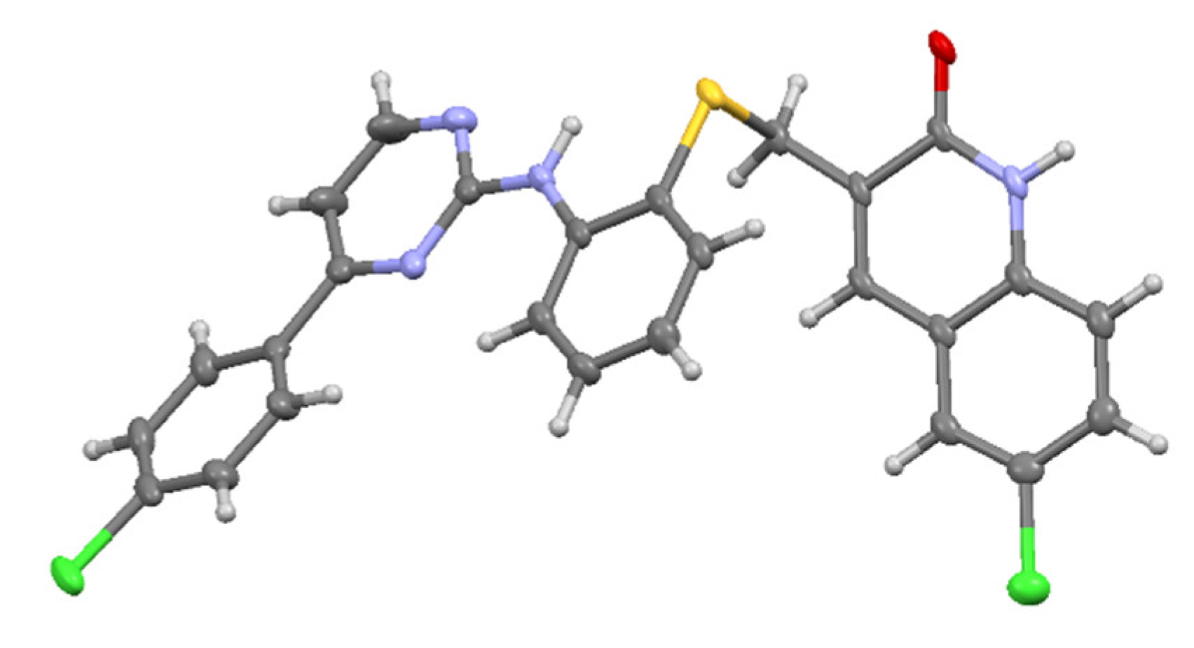
3.4. Molecular Modeling

The molecular modeling and Docking analysis were performed using the MOE 2020.09 suit from Chemical Computing Group’s Molecular Operating Environment, and the minimization of the energy of molecules and complexes were performed under molecular mechanics using the Amber14:EHT force field.
The complex of the hLDHA protein with the inhibitor W31, with PDB code 4R68, was downloaded from the Protein Data Bank (PDB) and prepared as follows: all the chains but one were deleted using the sequence editor (SEQ), the hydrogens were added to structure with the “Protonate 3D” tool and checked for the right charge in any heteroatom, and finally, the complex system was minimized using the force field Amber14. The energy minimization mode used is named “General”, in which force field minimization is performed with emphasis on tether layers. No restraints are applied. Constraints selected were to maintain rigid water molecules. The gradient was of 0.1 RMS, meaning that the energy minimization was finished when the root means square gradient fell below the specified value (0.1).
The input database of screened molecules were prepared from builder editor and imported in the corresponding database file (*.mdb), which was used as the input file in the docking process. To prepare the database input file, we followed a similar preparation process that included a first wash (set of cleaning rules to ensure that each structure is in a suitable form for subsequent modelling steps, such as conformational enumeration and protein-ligand docking), checking for the right partial charges, and finally, minimizing the energy of the molecules using the force field Amber14.
Three pharmacophoric models were created from the Pharmacophore Query Editor tool: (i) W31 site, (ii) NADH site, and (iii) extended site w31-NADH site. Three features were defined so as to interact with the main amino acid residues: Asn137, Arg168, His192, and Asp194. All three features were defined with a radius of 1.2 Å, and none of them was classified as essential nor ignored. When stablishing the search criteria, the partial match was clicked on and defined as at least 1 interaction with one of those features.
The docking screening was carried out with the following settings: Receptor: MOE (the previously prepared complex), receptor atoms; Site: Ligand atoms: Wall constraint: on; Pharmacophore: on; Ligand: MDB file (the input *.mdb database); Placement: Pharmacophore; Number of returned poses (poses returned by each ligand’s placement): 3000; Placement score: London dG; Placement poses: 100; Refinement method: rigid receptor; Refinement score: GBI/WSA dG; Refinement poses (number of poses retained to be written in the output file): 10.
Once the docking was complete, the best pose score for each ligand determined by a further minimization process (in the output file) was required using molecular mechanics and the specified forcefield. The best pose was determined by the following criteria: (i) RMSD [64] < 1.8 Å, (ii) affinity (S) [65] values < −9 kcal/mol, and (iii) energy values involved in the interactions with the main amino acid residues [65], selecting those interacting with Arg168 firstly and afterwards those with the higher number of interactions. In the case that they all interacted with the same amino acids, the ones with the highest energy values involved in the interactions with those amino acid residues were chosen.

4. Conclusions

After having synthesized and evaluated a first set of pyrimidine-quinolone hybrids, due to the different reasons explained, we designed, synthesized, and evaluated novel hLDHA inhibitors 1,2-linked (2427(ac)), 1,3-linked (35(ac)), and 1,4-linked (2831(ac)) pyrimidine-quinolone hybrids. Molecular modelling (docking) predicted that hybrids 1,2-linked were the most interesting ones to inhibit the hLDHA enzyme and that the 1,4-linked ones were inactive. Additionally, those hybrids having the naphthalene-2-yl moiety as the hydrophobic structure were predicted to be the most interesting ones.
Enzymatic assays confirmed the in silico predictions and a preliminary SAR was established, and 1,3-linked hybrids 3336(ac) were included for the study.
Data from SAR analysis enabled us to explain the difference in the experimental IC50 values between the different U-shaped pyrimidine-quinolone hybrids and predicted those 1,3-linked hybrids to have an intermediate inhibitory activity between those 1,2- and 1,4-linked, with a bias towards the U-shaped ones. In this way, hybrids 35(ac) with the naphthalene-2-yl moiety were synthesized and evaluated, confirming the predictions from SAR analysis.
In summary, we have been able to design and synthesize a new family of hLDHA inhibitors with good IC50 values and designed a preliminary SAR, which encourages us to design a promising next generation in order to improve their inhibitory potency.

Supplementary Materials

The following molecular docking, bioassays data and NMR, MS spectra can be downloaded at: https://www.mdpi.com/article/10.3390/ph15070792/s1.

Author Contributions

Conceptualization, J.C. and S.S.; methodology, J.C. and S.S.; software, J.C., S.S and I.D.; validation, I.D.; formal analysis, I.D., S.S. and J.C.; investigation, I.D., S.S, M.N. and J.C.; resources, M.N., S.S. and J.C.; data curation, I.D.; writing—original draft preparation, J.C. and I.D.; writing—review and editing, J.C., S.S., I.D.; visualization, M.N.; supervision, J.C.; project administration, S.S.; funding acquisition, S.S. and J.C. All authors have read and agreed to the published version of the manuscript.

Funding

Spanish “Ministerio de Ciencia, Innovación, y Universidades” (R+D projects RTI2018-098560-B-C22; co-financed by the FEDER funds of the European Union). Universidad de Jaén, Vicerrectorado de Investigación, PAIUJA “acción 1 plan 2019–2020” and 2021–2022. Consejería de Innovación, Ciencia y Empresa (Junta de Andalucía, Spain).

Institutional Review Board Statement

Not applicable.

Informed Consent Statement

Not applicable.

Data Availability Statement

Data is contained within the article and Supplementary Materials.

Acknowledgments

The authors thank the technical and human support provided by CICT of Universidad de Jaén (UJA, MINECO, Junta de Andalucía, FEDER), and Nikki Smith for the final language edition.

Conflicts of Interest

The authors declare no conflict of interest.

References

  1. Ferlay, J.; Colombet, M.; Soerjomataram, I.; Parkin, D.M.; Pineros, M.; Znaor, A.; Bray, F. Cancer statistics for the year 2020: An overview. Int. J. Cancer 2021, 194, 778–789. [Google Scholar] [CrossRef] [PubMed]
  2. Sung, H.; Ferlay, J.; Siegel, R.L.; Laversanne, M.; Soerjomataram, I.; Jemal, A.; Bray, F. Global Cancer Statistics 2020: GLOBOCAN Estimates of Incidence and Mortality Worldwide for 36 Cancers in 185 Countries. CA Cancer J. Clin. 2021, 71, 209–249. [Google Scholar] [CrossRef] [PubMed]
  3. Seth Nanda, C.; Vishaan Venkateswaran, S.; Patani, N.; Yuneva, M. Defining a metabolic landscape of tumours: Genome meets metabolism. Br. J. Cancer 2020, 122, 136–149. [Google Scholar] [CrossRef] [PubMed]
  4. Rani, R.; Kumar, V. Recent Update on Human Lactate Dehydrogenase Enzyme 5 (hLDH5) Inhibitors: A Promising Approach for Cancer Chemotherapy. J. Med. Chem. 2016, 59, 487–496. [Google Scholar] [CrossRef] [PubMed]
  5. Woodford, M.R.; Chen, V.Z.; Backe, S.J.; Bratslavsky, G.; Mollapour, M. Structural and functional regulation of lactate dehydrogenase-A in cancer. Future Med. Chem. 2020, 12, 439–455. [Google Scholar] [CrossRef] [PubMed]
  6. Yang, Y.; Chong, Y.; Chen, M.; Dai, W.; Zhou, X.; Ji, Y.; Qiu, G.; Du, X. Targeting lactate dehydrogenase a improves radiotherapy efficacy in non-small cell lung cancer: From bedside to bench. J. Transl. Med. 2021, 19, 170. [Google Scholar] [CrossRef]
  7. Pathria, G.; Scott, D.A.; Feng, Y.; Sang Lee, J.; Fujita, Y.; Zhang, G.; Sahu, A.D.; Ruppin, E.; Herlyn, M.; Osterman, A.L.; et al. Targeting the Warburg effect via LDHA inhibition engages ATF 4 signaling for cancer cell survival. EMBO J. 2018, 37, e99735. [Google Scholar] [CrossRef]
  8. Zhang, S.L.; He, Y.; Tam, K.Y. Targeting cancer metabolism to develop human lactate dehydrogenase (hLDH)5 inhibitors. Drug Discov. Today 2018, 23, 1407–1415. [Google Scholar] [CrossRef]
  9. Feng, Y.; Xiong, Y.; Qiao, T.; Li, X.; Jia, L.; Han, Y. Lactate dehydrogenase A: A key player in carcinogenesis and potential target in cancer therapy. Cancer Med. 2018, 7, 6124–6136. [Google Scholar] [CrossRef] [Green Version]
  10. Hoppe, B.; Koch, A.; Cochat, P.; Garrelfs, S.F.; Baum, M.A.; Groothoff, J.W.; Lipkin, G.; Coenen, M.; Schalk, G.; Amrite, A.; et al. Safety, pharmacodynamics, and exposure-response modeling results from a first in human phase 1 study of nedosiran in primary hyperoxaluria. Kidney Int. 2021, 101, 626–634. [Google Scholar] [CrossRef]
  11. Lai, C.; Pursell, N.; Gierut, J.; Saxena, U.; Zhou, W.; Dills, M.; Diwanji, R.; Dutta, C.; Koser, M.; Nazef, N.; et al. Specific Inhibition of Hepatic Lactate Dehydrogenase Reduces Oxalate Production in Mouse Models of Primary Hyperoxaluria. Mol. Ther. 2018, 26, 1983–1995. [Google Scholar] [CrossRef] [PubMed] [Green Version]
  12. Mahmoud, T.; Ghandour, E.C.; Jaar, B.G. A hidden cause of oxalate nephropathy: A case report. J. Med. Case Rep. 2021, 15, 106. [Google Scholar] [CrossRef] [PubMed]
  13. Dindo, M.; Conter, C.; Oppici, E.; Ceccarelli, V.; Marinucci, L.; Cellini, B. Molecular basis of primary hyperoxaluria: Clues to innovative treatments. Urolithiasis 2019, 47, 67–78. [Google Scholar] [CrossRef] [PubMed]
  14. Cochat, P.; Rumsby, G. Primary Hyperoxaluria. N. Engl. J. Med. 2013, 369, 649–658. [Google Scholar] [CrossRef]
  15. Salido, E.; Pey, A.L.; Rodriguez, R.; Lorenzo, V. Primary hyperoxalurias: Disorders of glyoxylate detoxification. Biochim. Biophys. Acta Mol. Basis Dis. 2012, 1822, 1453–1464. [Google Scholar] [CrossRef] [Green Version]
  16. Yuan, S.; Yu, B.; Liu, H.M. New drug approvals for 2019: Synthesis and clinical applications. Eur. J. Med. Chem. 2020, 205, 112667. [Google Scholar] [CrossRef]
  17. Kumar, S.; Narasimhan, B. Therapeutic potential of heterocyclic pyrimidine scaffolds. Chem. Cent. J. 2018, 12, 38. [Google Scholar] [CrossRef]
  18. Oukoloff, K.; Nzou, G.; Varricchio, C.; Lucero, B.; Alle, T.; Kovalevich, J.; Monti, L.; Cornec, A.S.; Yao, Y.; James, M.J.; et al. Evaluation of the Structure-Activity Relationship of Microtubule-Targeting 1,2,4-Triazolo[1,5- a]pyrimidines Identifies New Candidates for Neurodegenerative Tauopathies. J. Med. Chem. 2021, 64, 1073–1102. [Google Scholar] [CrossRef]
  19. Ayati, A.; Moghimi, S.; Toolabi, M.; Foroumadi, A. Pyrimidine-based EGFR TK inhibitors in targeted cancer therapy. Eur. J. Med. Chem. 2021, 221, 113523. [Google Scholar] [CrossRef]
  20. Faraji, A.; Oghabi Bakhshaiesh, T.; Hasanvand, Z.; Motahari, R.; Nazeri, E.; Boshagh, M.A.; Firoozpour, L.; Mehrabi, H.; Khalaj, A.; Esmaeili, R.; et al. Design, synthesis and evaluation of novel thienopyrimidine-based agents bearing diaryl urea functionality as potential inhibitors of angiogenesis. Eur. J. Med. Chem. 2021, 209, 112942. [Google Scholar] [CrossRef]
  21. Wang, S.; Yuan, X.H.; Wang, S.Q.; Zhao, W.; Chen, X.B.; Yu, B. FDA-approved pyrimidine-fused bicyclic heterocycles for cancer therapy: Synthesis and clinical application. Eur. J. Med. Chem. 2021, 214, 113218. [Google Scholar] [CrossRef] [PubMed]
  22. Madia, V.N.; Nicolai, A.; Messore, A.; De Leo, A.; Ialongo, D.; Tudino, V.; Saccoliti, F.; De Vita, D.; Scipione, L.; Artico, M.; et al. Design, synthesis and biological evaluation of new pyrimidine derivatives as anticancer agents. Molecules 2021, 26, 771. [Google Scholar] [CrossRef]
  23. Wagman, A.S.; Boyce, R.S.; Brown, S.P.; Fang, E.; Goff, D.; Jansen, J.M.; Le, V.P.; Levine, B.H.; Ng, S.C.; Ni, Z.J.; et al. Synthesis, Binding Mode, and Antihyperglycemic Activity of Potent and Selective (5-Imidazol-2-yl-4-phenylpyrimidin-2-yl)[2-(2-pyridylamino)ethyl]amine Inhibitors of Glycogen Synthase Kinase 3. J. Med. Chem. 2017, 60, 8482–8514. [Google Scholar] [CrossRef] [PubMed]
  24. Taglieri, L.; Saccoliti, F.; Nicolai, A.; Peruzzi, G.; Madia, V.N.; Tudino, V.; Messore, A.; Di Santo, R.; Artico, M.; Taurone, S.; et al. Discovery of a pyrimidine compound endowed with antitumor activity. Investig. New Drugs 2020, 38, 39–49. [Google Scholar] [CrossRef]
  25. Senerovic, L.; Opsenica, D.; Moric, I.; Aleksic, I.; Spasić, M.; Vasiljevic, B. Quinolines and quinolones as antibacterial, antifungal, anti-virulence, antiviral and anti-parasitic agents. Adv. Exp. Med. Biol. 2020, 1282, 37–69. [Google Scholar] [CrossRef] [Green Version]
  26. Vandekerckhove, S.; D’Hooghe, M. Quinoline-based antimalarial hybrid compounds. Bioorganic Med. Chem. 2015, 23, 5098–5119. [Google Scholar] [CrossRef] [PubMed]
  27. Saalim, M.; Villegas-Moreno, J.; Clark, B.R. Bacterial Alkyl-4-quinolones: Discovery, Structural Diversity and Biological Properties. Molecules 2020, 25, 5689. [Google Scholar] [CrossRef]
  28. Montagut, E.J.; Vilaplana, L.; Martin-Gomez, M.T.; Marco, M.P. High-Throughput Immunochemical Method to Assess the 2-Heptyl-4-quinolone Quorum Sensing Molecule as a Potential Biomarker of Pseudomonas aeruginosa Infections. ACS Infect. Dis. 2020, 6, 3237–3246. [Google Scholar] [CrossRef]
  29. El-Helw, E.A.E.; El-Badawy, A.A. Synthesis of chromenone, pyrimidinone, thiazoline, and quinolone derivatives as prospective antitumor agents. J. Heterocycl. Chem. 2020, 57, 2354–2364. [Google Scholar] [CrossRef]
  30. Relitti, N.; Saraswati, A.P.; Chemi, G.; Brindisi, M.; Brogi, S.; Herp, D.; Schmidtkunz, K.; Saccoccia, F.; Ruberti, G.; Ulivieri, C.; et al. Novel quinolone-based potent and selective HDAC6 inhibitors: Synthesis, molecular modeling studies and biological investigation. Eur. J. Med. Chem. 2021, 212, 112998. [Google Scholar] [CrossRef]
  31. Hu, Y.Q.; Gao, C.; Zhang, S.; Xu, L.; Xu, Z.; Feng, L.S.; Wu, X.; Zhao, F. Quinoline hybrids and their antiplasmodial and antimalarial activities. Eur. J. Med. Chem. 2017, 139, 22–47. [Google Scholar] [CrossRef] [PubMed]
  32. Bolakatti, G.; Palkar, M.; Katagi, M.; Hampannavar, G.; Karpoormath, R.V.; Ninganagouda, S.; Badiger, A. Novel series of benzo[d]thiazolyl substituted-2-quinolone hybrids: Design, synthesis, biological evaluation and in-silico insights. J. Mol. Struct. 2021, 1227, 129413. [Google Scholar] [CrossRef]
  33. Abu Almaaty, A.H.; Elgrahy, N.A.; Fayad, E.; Abu Ali, O.A.; Mahdy, A.R.E.; Barakat, L.A.A.; Behery, M. El Design, synthesis and anticancer evaluation of substituted cinnamic acid bearing 2-quinolone hybrid derivatives. Molecules 2021, 26, 4724. [Google Scholar] [CrossRef] [PubMed]
  34. Kania, A.; Tejchman, W.; Pawlak, A.M.; Mokrzyński, K.; Różanowski, B.; Musielak, B.M.; Greczek-Stachura, M. Preliminary Studies of Antimicrobial Activity of New Synthesized Hybrids of 2-Thiohydantoin and 2-Quinolone Derivatives Activated with Blue Light. Molecules 2022, 27, 1069. [Google Scholar] [CrossRef] [PubMed]
  35. Viegas-Junior, C.; Danuello, A.; Bolzani, V.S.; Barreiro, E.J.; Barreiro, E.; Fraga, C.A.M. Molecular Hybridization: A Useful Tool in the Design of New Drug Prototypes. Curr. Med. Chem. 2007, 14, 1829–1852. [Google Scholar] [CrossRef] [PubMed]
  36. Doak, B.C.; Norton, R.S.; Scanlon, M.J. The ways and means of fragment-based drug design. Pharmacol. Ther. 2016, 167, 28–37. [Google Scholar] [CrossRef]
  37. Kumar, A.; Voet, A.; Zhang, K.Y.J. Fragment Based Drug Design: From Experimental to Computational Approaches. Curr. Med. Chem. 2012, 19, 5128–5147. [Google Scholar] [CrossRef]
  38. Li, Q. Application of Fragment-Based Drug Discovery to Versatile Targets. Front. Mol. Biosci. 2020, 7, 180. [Google Scholar] [CrossRef] [PubMed]
  39. Zhang, X.; Li, D.; Fan, X.; Wang, X.; Li, X.; Qu, G.; Wang, J. Ionic liquid-promoted multi-component reaction: Novel and efficient preparation of pyrazolo[3,4-b]pyridinone, pyrazolo[3,4-b]-quinolinone and their hybrids with pyrimidine nucleoside. Mol. Divers. 2010, 14, 159–167. [Google Scholar] [CrossRef]
  40. Song, R.; Wang, Y.; Wang, M.; Gao, R.; Yang, T.; Yang, S.; Yang, C.G.; Jin, Y.; Zou, S.; Cai, J.; et al. Design and synthesis of novel desfluoroquinolone-aminopyrimidine hybrids as potent anti-MRSA agents with low hERG activity. Bioorg. Chem. 2020, 103, 104176. [Google Scholar] [CrossRef]
  41. Mao, T.Q.; He, Q.Q.; Wan, Z.Y.; Chen, W.X.; Chen, F.E.; Tang, G.F.; De Clercq, E.; Daelemans, D.; Pannecouque, C. Anti-HIV diarylpyrimidine-quinolone hybrids and their mode of action. Bioorganic Med. Chem. 2015, 23, 3860–3868. [Google Scholar] [CrossRef] [PubMed]
  42. Pretorius, S.I.; Breytenbach, W.J.; De Kock, C.; Smith, P.J.; N’Da, D.D. Synthesis, characterization and antimalarial activity of quinoline-pyrimidine hybrids. Bioorganic Med. Chem. 2013, 21, 269–277. [Google Scholar] [CrossRef]
  43. Singh, K.; Kaur, H.; Chibale, K.; Balzarini, J. Synthesis of 4-aminoquinoline—Pyrimidine hybrids as potent antimalarials and their mode of action studies. Eur. J. Med. Chem. 2013, 66, 314–323. [Google Scholar] [CrossRef] [PubMed]
  44. Singh, K.; Kaur, H.; Smith, P.; De Kock, C.; Chibale, K.; Balzarini, J. Quinoline-pyrimidine hybrids: Synthesis, antiplasmodial activity, SAR, and mode of action studies. J. Med. Chem. 2014, 57, 435–448. [Google Scholar] [CrossRef] [PubMed]
  45. Kaur, H.; Balzarini, J.; De Kock, C.; Smith, P.J.; Chibale, K.; Singh, K. Synthesis, antiplasmodial activity and mechanistic studies of pyrimidine-5-carbonitrile and quinoline hybrids. Eur. J. Med. Chem. 2015, 101, 52–62. [Google Scholar] [CrossRef]
  46. Kumar, D.; Khan, S.I.; Tekwani, B.L.; Ponnan, P.; Rawat, D.S. 4-Aminoquinoline-Pyrimidine hybrids: Synthesis, antimalarial activity, heme binding and docking studies. Eur. J. Med. Chem. 2015, 89, 490–502. [Google Scholar] [CrossRef]
  47. Maurya, S.S.; Bahuguna, A.; Khan, S.I.; Kumar, D.; Kholiya, R.; Rawat, D.S. N-Substituted aminoquinoline-pyrimidine hybrids: Synthesis, in vitro antimalarial activity evaluation and docking studies. Eur. J. Med. Chem. 2019, 162, 277–289. [Google Scholar] [CrossRef]
  48. Kayamba, F.; Malimabe, T.; Ademola, I.K.; Pooe, O.J.; Kushwaha, N.D.; Mahlalela, M.; van Zyl, R.L.; Gordon, M.; Mudau, P.T.; Zininga, T.; et al. Design and synthesis of quinoline-pyrimidine inspired hybrids as potential plasmodial inhibitors. Eur. J. Med. Chem. 2021, 217, 113330. [Google Scholar] [CrossRef]
  49. Adachi, R.; Ogawa, K.; Matsumoto, S.-i.; Satou, T.; Tanaka, Y.; Sakamoto, J.; Nakahata, T.; Okamoto, R.; Kamaura, M.; Kawamoto, T. Discovery and characterization of selective human sphingomyelin synthase 2 inhibitors. Eur. J. Med. Chem. 2017, 136, 283–293. [Google Scholar] [CrossRef]
  50. Dragovich, P.S.; Fauber, B.P.; Corson, L.B.; Ding, C.Z.; Eigenbrot, C.; Ge, H.; Giannetti, A.M.; Hunsaker, T.; Labadie, S.; Liu, Y.; et al. Identification of substituted 2-thio-6-oxo-1,6-dihydropyrimidines as inhibitors of human lactate dehydrogenase. Bioorganic Med. Chem. Lett. 2013, 23, 3186–3194. [Google Scholar] [CrossRef]
  51. Zhou, Y.; Tao, P.; Wang, M.; Xu, P.; Lu, W.; Lei, P.; You, Q. Development of novel human lactate dehydrogenase A inhibitors: High-throughput screening, synthesis, and biological evaluations. Eur. J. Med. Chem. 2019, 177, 105–115. [Google Scholar] [CrossRef]
  52. Billiard, J.; Dennison, J.B.; Briand, J.; Annan, R.S.; Chai, D.; Colón, M.; Dodson, C.S.; Gilbert, S.A.; Greshock, J.; Jing, J.; et al. Quinoline 3-sulfonamides inhibit lactate dehydrogenase A and reverse aerobic glycolysis in cancer cells. Cancer Metab. 2013, 1, 19. [Google Scholar] [CrossRef] [PubMed] [Green Version]
  53. Granchi, C.; Paterni, I.; Rani, R.; Minutolo, F. Small-molecule inhibitors of human LDH5. Future Med. Chem. 2013, 5, 1967–1991. [Google Scholar] [CrossRef] [PubMed] [Green Version]
  54. Kolappan, S.; Shen, D.L.; Mosi, R.; Sun, J.; McEachern, E.J.; Vocadlo, D.J.; Craig, L. Structures of lactate dehydrogenase A (LDHA) in apo, ternary and inhibitor-bound forms. Acta Crystallogr. Sect. D Biol. Crystallogr. 2015, 71, 185–195. [Google Scholar] [CrossRef] [PubMed]
  55. Insuasty, B.; Montoya, A.; Becerra, D.; Quiroga, J.; Abonia, R.; Robledo, S.; Vélez, I.D.; Upegui, Y.; Nogueras, M.; Cobo, J. Synthesis of novel analogs of 2-pyrazoline obtained from [(7-chloroquinolin-4-yl)amino]chalcones and hydrazine as potential antitumor and antimalarial agents. Eur. J. Med. Chem. 2013, 67, 252–262. [Google Scholar] [CrossRef]
  56. Laali, K.K.; Insuasty, D.; Abonia, R.; Insuasty, B.; Bunge, S.D. Novel quinoline-imidazolium adducts via the reaction of 2-oxoquinoline-3-carbaldehyde and quinoline-3-carbaldehydes with 1-butyl-3-methylimidazolium chloride [BMIM][Cl]. Tetrahedron Lett. 2014, 55, 4395–4399. [Google Scholar] [CrossRef]
  57. Abonia, R.; Insuasty, D.; Castillo, J.; Insuasty, B.; Quiroga, J.; Nogueras, M.; Cobo, J. Synthesis of novel quinoline-2-one based chalcones of potential anti-tumor activity. Eur. J. Med. Chem. 2012, 57, 29–40. [Google Scholar] [CrossRef]
  58. Vettorazzi, M.; Insuasty, D.; Lima, S.; Gutiérrez, L.; Nogueras, M.; Marchal, A.; Abonia, R.; Andújar, S.; Spiegel, S.; Cobo, J.; et al. Bioorganic Chemistry Design of new quinolin-2-one-pyrimidine hybrids as sphingosine kinases inhibitors. Bioorg. Chem. 2020, 94, 103414. [Google Scholar] [CrossRef]
  59. Laiolo, J.; Lanza, P.A.; Parravicini, O.; Barbieri, C.; Insuasty, D.; Cobo, J.; Vera, D.M.A.; Enriz, R.D.; Carpinella, M.C. Structure Activity Relationships and the Binding Mode of Quinolinone—Pyrimidine Hybrids as Reversal Agents of Multidrug Resistance Mediated by P—Gp. Sci. Rep. 2021, 11, 16856. [Google Scholar] [CrossRef]
  60. Pineda, J.R.E.T.; Callender, R.; Schwartz, S.D. Ligand binding and protein dynamics in lactate dehydrogenase. Biophys. J. 2007, 93, 1474–1483. [Google Scholar] [CrossRef] [Green Version]
  61. Eigenbrot, C.; Ultsch, M. 4R68 PDB. Available online: https://www.rcsb.org/structure/4R68 (accessed on 17 April 2022).
  62. Labadie, S.; Dragovich, P.S.; Chen, J.; Fauber, B.P.; Boggs, J.; Corson, L.B.; Ding, C.Z.; Eigenbrot, C.; Ge, H.; Ho, Q.; et al. Optimization of 5-(2,6-dichlorophenyl)-3-hydroxy-2-mercaptocyclohex-2-enones as potent inhibitors of human lactate dehydrogenase. Bioorganic Med. Chem. Lett. 2015, 25, 75–82. [Google Scholar] [CrossRef] [PubMed] [Green Version]
  63. Jiang, S.; Feher, M.; Williams, C.; Cole, B.; Shaw, D.E. AutoPH4: An Automated Method for Generating Pharmacophore Models from Protein Binding Pockets. J. Chem. Inf. Model. 2020, 60, 4326–4338. [Google Scholar] [CrossRef] [PubMed]
  64. Goto, J.; Kataoka, R.; Hirayama, N. Ph4Dock: Pharmacophore-Based Protein−Ligand Docking. J. Med. Chem. 2004, 47, 6804–6811. [Google Scholar] [CrossRef] [PubMed]
  65. Binzet, G. QSAR and molecular docking studies on 4-quinoline carboxylic acid derivatives as inhibition of vesicular stomatitis virus replication. Eur. J. Chem. 2018, 4, 360–368. [Google Scholar] [CrossRef]
  66. Rupiani, S.; Buonfiglio, R.; Manerba, M.; Di Ianni, L.; Vettraino, M.; Giacomini, E.; Masetti, M.; Falchi, F.; Di Stefano, G.; Roberti, M.; et al. Identification of N-acylhydrazone derivatives as novel lactate dehydrogenase A inhibitors. Eur. J. Med. Chem. 2015, 101, 63–70. [Google Scholar] [CrossRef]
  67. Somagond, S.M.; Kamble, R.R.; Kattimani, P.P.; Shaikh, S.K.J.; Dixit, S.R.; Joshi, S.D.; Devarajegowda, H.C. Design, Docking, and Synthesis of Quinoline-2H-1,2,4-triazol-3(4H)-ones as Potent Anticancer and Antitubercular Agents. ChemistrySelect 2018, 3, 2004–2016. [Google Scholar] [CrossRef]
  68. Dragovich, P.S.; Fauber, B.P.; Boggs, J.; Chen, J.; Corson, L.B.; Ding, C.Z.; Eigenbrot, C.; Ge, H.; Giannetti, A.M.; Hunsaker, T.; et al. Identification of substituted 3-hydroxy-2-mercaptocyclohex-2-enones as potent inhibitors of human lactate dehydrogenase. Bioorganic Med. Chem. Lett. 2014, 24, 3764–3771. [Google Scholar] [CrossRef]
  69. Li, X.M.; Xiao, W.H.; Zhao, H.X. Discovery of potent human lactate dehydrogenase A (LDHA) inhibitors with antiproliferative activity against lung cancer cells: Virtual screening and biological evaluation. Medchemcomm 2017, 8, 599–605. [Google Scholar] [CrossRef]
Figure 1. Reported pyrimidine and quinoline derivatives and their joint hybrids as hLDHA inhibitors.
Pharmaceuticals 15 00792 g001
Figure 2. Examples of selected hLDHA inhibitors bearing hydrophilic (blue) and hydrophobic (red) fragments.
Pharmaceuticals 15 00792 g002
Figure 3. Overview of our previous and current work with pyrimidine-quinoline hybrids.
Pharmaceuticals 15 00792 g003
Figure 4. (a) W31 (blue) placement in the prepared x-ray structure of 4R68 with interactions with the main labelled amino acid residues in W31 active site; (b) W31 2-D interaction diagram.
Pharmaceuticals 15 00792 g004
Figure 5. First set of pyrimidine-quinolone hybrids evaluated in silico.
Pharmaceuticals 15 00792 g005
Figure 6. 2-D representation of ligand–protein interaction of 24a in the hLDHA active site.
Pharmaceuticals 15 00792 g006
Figure 7. Second set of pyrimidine-quinolone hybrids with the 2-aminothiophenol (2427)(ac) and 4-aminothiophenol (2831)(ac) linker.
Pharmaceuticals 15 00792 g007
Figure 8. Selected poses of compounds 26a (blue), 26b (yellow), and 26c (green) in the hLDHA active site cavity along with W31 (black). Color code in surface: purple, polar features; green, apolar features; red, solvent-exposed ligand atoms.
Pharmaceuticals 15 00792 g008
Scheme 1. Linear synthetic pathway to obtain pyrimidine-quinolone hybrid 15.
Pharmaceuticals 15 00792 sch001
Scheme 2. Convergent synthetic pathway to obtain hybrid 19 from intermediate 9.
Pharmaceuticals 15 00792 sch002
Scheme 3. Convergent synthetic pathway to novel pyrimidine-quinolone hybrids 2431(ac).
Pharmaceuticals 15 00792 sch003
Figure 9. Molecular structure of compound 24b; the asymmetric unit was obtained as DMSO solvate.
Pharmaceuticals 15 00792 g009
Figure 10. Placement comparison of compounds 31b (blue) and 31c (red) in the hLDHA active site with reference W31 (black).
Pharmaceuticals 15 00792 g010
Figure 11. Novel 1,3-linked pyrimidine-quinolone hybrids 3336(ac).
Pharmaceuticals 15 00792 g011
Figure 12. Mean affinity values (kcal/mol) after minimization refinement of the 1,2-linked 2427(ac), 1,4-linked 2831(ac) and 1,3-linked 3336(ac) pyrimidine-quinolone hybrids in the hLDHA active site.
Pharmaceuticals 15 00792 g012
Figure 13. Mean energy values (kcal/mol) involved in the interaction of the 1,2-linked 2427(ac), 1,4-linked 2831(ac), and 1,3-linked 3336(ac) pyrimidine-quinolone hybrids with the main amino acid residues in the hLDHA active site.
Pharmaceuticals 15 00792 g013
Scheme 4. Convergent pathway to obtain pyrimidine-quinolone hybrids 35(ac).
Pharmaceuticals 15 00792 sch004
Figure 14. Variation of the inhibitory activity (IC50) of the different pyrimidine-quinolone hybrids. Key colors depend on the quinolone substitution: blue R, H (26a, 30a, and 35a); orange R, Cl (26b, 30b, and 35b); and grey R, OMe (26c, 30c, and 35c).
Pharmaceuticals 15 00792 g014
Table 1. Mean energy values (kcal/mol) involved in the interactions of hybrids (2431)(ac) with the main amino acid residues and mean affinity (S) values (kcal/mol) grouped by linker substitution.
HybridsArg168His192Asn137Asp194Affinity (S)
(24–27)a–c−5.3−3.6−1.3−1.2−9.869
(28–31)a–c−6.20.0−0.80.0−9.253
Table 2. Mean energy values (kcal/mol) involved in the interaction of U-shaped pyrimidine-quinolone hybrids (2427)(ac) with the main amino acid residues and mean affinity (S) values (kcal/mol) grouped by aryl moieties at pyrimidine.
Hybrids *ArylArg168His192Asn137Asp194S
24a–c4-Chlorophenyl−6.9−0.6−0.6−5.0−9.48
25a–c4-Trifluoromethylphenyl−3.70−0.6−3.0−9.78
26a–cNaphthalen-2-yl−5.90−0.7−3.1−10.46
27a–cBenzo[d][1,3]dioxol-5-yl−4.8−2.0−4.20−9.71
* For detailed information, see Supplementary Materials Table S4.
Table 3. Reaction time and yields for the synthesis of pyrimidine-quinolone hybrids 2431(ac).
1,2-Linked Hybrids1,4-Linked Hybrids
RArHybridReaction Time (min)Yield (%)HybridReaction Time (min)Yield (%)
H4-ClC6H424a158628a16093
Cl4-ClC6H424b1206128b2086
OCH34-ClC6H424c307128c9090
H4-CF3C6H425a804129a8093
Cl4-CF3C6H425b3605029b4588
OCH34-CF3C6H425c905729c5090
HNaphth-2-yl26a1206530a14086
ClNaphth-2-yl26b1807430b18089
OCH3Naphth-2-yl26c3005930c6093
H3,4-(OCH2O) C6H327a1506831a8074
Cl3,4-(OCH2O) C6H327b1206931b12091
OCH33,4-(OCH2O) C6H327c905831c4094
Table 4. IC50 values (μM) of pyrimidine-quinolone hybrids 2431 (ac).
1,2-Linked hybrids1,4-Linked hybrids
RArHybrida IC50 (μM)R2Hybrida IC50 (μM)R2
H4-ClC6H424a31.50.976228a>100-
Cl4-ClC6H424b34.80.853028b>100-
OCH34-ClC6H424c79.10.851928c>100-
H4-CF3C6H425a26.90.929629a>100-
Cl4-CF3C6H425b42.30.914329b83.20.9487
OCH34-CF3C6H425c71.80.927729c>100-
HNaphth-2-yl26a17.80.941430a62.90.9049
ClNaphth-2-yl26b20.30.953830b>100-
OCH3Naphth-2-yl26c27.70.986430c>100-
H3,4-(OCH2O) C6H327a>100-31a>100-
Cl3,4-(OCH2O) C6H327b89.00.936131b49.90.8468
OCH33,4-(OCH2O) C6H327c60.00.901431c>100-
a Data are presented as the mean ± SD of n = 3 replicates for IC50 values.
Table 5. Reaction time, yield, and IC50 (μM) values of 1,3-linked pyrimidine-quinolone hybrids 37(ac).
RArHybridReaction Time (min)Yield (%)a IC50 (μM)R2
HNaphthalen2-yl35a508019.60.9382
ClNaphthalen2-yl35b1405524.60.9523
OCH3Naphthalen2-yl35c408750.10.9551
a Data are presented as the mean ± SD of n = 3 replicates for IC50 values
Publisher’s Note: MDPI stays neutral with regard to jurisdictional claims in published maps and institutional affiliations.

