Review

Norbornene and Related Structures as Scaffolds in the Search for New Cancer Treatments

by
Gorka Calvo-Martín
1,2,†,
Daniel Plano
1,2,3,*,†,
Nuria Martínez-Sáez
1,2,
Carlos Aydillo
1,2,3,*,
Esther Moreno
1,2,3,
Socorro Espuelas
1,2,3 and
Carmen Sanmartín
1,2,3
1
Departamento de Tecnología y Química Farmacéuticas, Universidad de Navarra, Irunlarrea 1, E-31008 Pamplona, Spain
2
Instituto de Investigación Sanitaria de Navarra (IdiSNA), Irunlarrea, 3, E-31008 Pamplona, Spain
3
Instituto de Salud Tropical, Universidad de Navarra, Irunlarrea 1, E-31008 Pamplona, Spain
*
Authors to whom correspondence should be addressed.
These authors contributed equally to this work.
Pharmaceuticals 2022, 15(12), 1465; https://doi.org/10.3390/ph15121465
Submission received: 2 November 2022 / Revised: 20 November 2022 / Accepted: 22 November 2022 / Published: 25 November 2022
(This article belongs to the Topic Advances in Anti-Cancer Drugs)

Abstract

:
The norbornene scaffold has arisen as a promising structure in medicinal chemistry due to its possible therapeutic application in cancer treatment. The development of norbornene-based derivatives as potential chemotherapeutic agents is attracting significant attention. Here, we report an unprecedented review on the recent advances of investigations into the antitumoral efficacy of different compounds, including the abovementioned bicyclic scaffold in their structure, in combination with chemotherapeutic agents or forming metal complexes. The impact that structural modifications to these bicyclic compounds have on the antitumoral properties and the mechanisms by which these norbornene derivatives act are discussed in this review. In addition, the use of norbornene, and its related compounds, encapsulation in nanosystems for its use in cancer therapies is here detailed.

1. Introduction

At present, cancer is the second leading cause of premature death worldwide. The International Agency for Research on Cancer (IARC) estimated that 19.3 million new cancer patients were reported in 2020, and the global cancer burden is expected to be 28.4 million cases in 2040 [1]. Despite the development of several cytotoxic agents, their clinical use is still very limited due to the appearance of undesirable side effects, such as toxicity and the development of resistance to treatment [2]. Even though targeted therapies have gained interest as cancer treatments [3], chemotherapeutic agents continue to play an important role in cancer pharmacotherapy. Currently, norbornene derivatives have attracted great attention from the scientific community due to their possible therapeutic applications in cancer treatment. Due to norbornene’s fast reactivity, it forms polymers that can be used as chemotherapeutic agent carriers [4] or in theranostic applications [5]. In addition, as will be discussed later in this review, a wide range of recent works have revealed interesting antitumoral properties concerning this molecule and its derivatives.
Norbornene (bicyclo[2.2.1]hept-2-ene) (1a) derivatives are active compounds derived from the Diels–Alder (DA) cycloaddition between cyclopentadiene and different dienophiles (Figure 1A). Due to the spontaneous dimerization of cyclopentadiene, it must be produced in situ after cracking the dicyclopentadiene by distillation, and should be used readily [6,7]. Thus, it reacts quickly and efficiently with activated dienophiles such as maleic anhydride and maleimides [8,9]. This unique reactivity provides norbornene and its derivatives with vast possibilities of chemical modification. As it has been previously mentioned, one of the main uses of norbornene is in polymer chemistry, where its polymerization is the most versatile among the cycloolefin addition polymerization mechanisms [10]. Norbornene can also be part of copolymers such as ethene–norbornene [11] or styrene–norbornene [12] copolymers. In addition, the double bond of this bicyclic structure produces greater ring strain, which can be released, turning it into a highly reactive alkene [13]. This feature makes the norbornene double bond especially reactive with sulfur species [14,15], which is a very useful tool for labeling cysteine [16]. Moreover, this bicyclic structure has been widely employed in biochemistry for the biorthogonal labeling of proteins and other biologically relevant molecules, taking advantage of its reactivity with tetrazines through an inverse electron demand DA reaction [16,17].
In addition, norbornene and related bicyclo[2.2.1] analogues, along with adamantane, have become the most exploited polycyclic structures as non-aromatic and non-planar motifs in drug design, in attempts to introduce variations in the so-called “flatland drugs” [18]. The preponderance of bicyclo[2.2.1] among all polycyclic motifs is mainly due to its abovementioned chemical accessibility through the Diels–Alder reaction.
Norbornene’s particular reactivity, together with the biological properties that this entity confers on other molecules, has drawn the attention of researchers during the last few decades, especially in the field of medicinal chemistry and cancer research. In this sense, this review provides an overview of the state of the art research on norbornene and its derivatives, with special focus on the antitumoral properties and therapeutic applications of these molecules.

2. Therapeutic Potential of Norbornene Derivatives in Cancer

A variety of norbornene derivatives with different substituents around its scaffold have been investigated as potential anticancer agents. Figure 1B shows the structures of norbornene and some norbornene derivatives that could potentially be employed for cancer treatment. The first study on the use norbornene derivatives was performed in 1994 using norbornyl isothiocyanates (1b), whose antitumoral properties where tested in mammary tumors being able to significantly reduce their size and delay tumor progression [19]. In 1997, Porn-Ares et al. [20] described the compound D609 (tricyclodecane-9-yl-xanthogenate, 1c) as an apoptosis inducer in human monocytic leukemia cells (U937) through sphingomyelin synthase. The apoptosis induced by 1c has also been associated with the level of cyclic adenosine monophosphate (cAMP) and p53 expression [21]. Additionally, it was able to increase ceramide and enhance rapidly induced apoptosis [22]. Moreover, further studies on D609 performed by Kalluri et al. [23] showed that this compound can induce cell death, with a concomitant decrease in GADD45β expression and an increase in the phosphorylation of p38 MAP kinase in glioma stem-like cells. Later, the same authors performed a study involving a chronic treatment with D609, reporting that not only the expression of GADD45β was decreased, but also cyclin-D1 and Olig2, two proteins that impede the progression of the cell cycle and curtail the growth of glioma stem-like cells [24]. However, despite its promising antitumor properties, D609 displays moderate antitumor activity in vivo. Another limitation of 1c is that it can readily oxidize to form a disulfide bond, with the subsequent loss of its biological activities. In this sense, Bai et al. [25] proposed the inclusion of S-(alkoxyacyl) groups to form a prodrug with increased antitumor activity in U937 leukemic cells (1d).
Continuing with the focus on the norbornene framework, biperiden (1e), an anticholinergic drug used for the treatment of Parkinson’s disease [26] that contains norbornene in its structure, was studied as a new anticancer drug for the treatment of pancreatic cancer. This molecule was able to target the protein mucosa-associated lymphoid tissue 1 (MALT1), which is frequently overexpressed in pancreatic ductal adenocarcinoma (PDAC). Biperiden reduced the cell proliferation of Panc-1 and Panc-2 cells in a time- and dose-dependent manner. In an in vivo study, the average tumor size after treatment with biperiden was reduced by 83% relative to the control in immunodeficient mice injected with Panc-1 cells. Furthermore, biperiden had tolerable side effects with minor motor side effects on mice. This norbornene-containing compound has the capacity to antagonize muscarinergic receptors, which are frequently expressed in cancer cells, responsible for cancer progression and the promotion of the proapoptotic factor BAX [27].
Among the different tumors, prostate cancer is one of the most common cancers observed in men. Androgen deprivation therapy (ADT) has long been the treatment of choice as the backbone of all other therapies, and functions by reducing circulating androgens to castration levels and slowing the progression of the disease [28]. Figure 2 shows a range of androgen receptor (AR) agonists that include norbornene in their structure. In this context, Salvati et al. [29] presented bicyclic 1H-isoindole-1,3(2H)-dione as a potent AR antagonist (2ab). These compounds (activities are shown in Table 1) presented similar cytotoxic profiles to bicalutamide, a clinically used antiandrogen drug for the treatment of prostate cancer, with the ability to block the AR. These analogues also demonstrated potent binding affinity (Ki), functional antagonism (IC50) of the wild type (WT) AR, found in the MDA-453 cell line, and functional antagonism (IC50) of the mutant type (MT) T877A AR, found in the human prostate cancer cell line LNCaP. From a structural point of view, the reduction of the double bond in the norbornene moiety (2c) led to a significant increase in binding to the AR in MDA-MB-453 cell lines, but not significantly in LNCaP and PCa2b. In addition, considering the stereochemistry of the bicyclic structure, the endo-isomer showed significantly better binding and functional antagonist activity toward the WT AR compared to the exo-isomer [30]. Based on these findings, new compounds with the expansion of the bicyclic portion were synthesized, showing that there is not a clear correlation between the bridge expansion and the AR binding activity. Additionally, substitutions around the bicyclic portion did not generate a clear pattern in the activity of these molecules, and the activity against the AR isoforms found in the LNCaP and Pca2b cell lines was dictated by the aniline portion of the molecule, not by the bicyclic one [29]. Another study, performed on similar structures, showed that the bicyclic imides (2d–f) presented low IC50 (14, 13 and 10 µM, respectively) values in African green monkey kidney cells (Vero). This research determined that replacing the bicyclic structure with other planar structures, such as phthalimides, may provoke a decrease in the cytotoxic activity, proving that the norbornene structure is the key for the activity of these molecules. Moreover, the substituent at the N-position seems to be a determinant factor. These bicyclic imides exhibited marked affinity for DNA binding and significant apoptosis levels, as indicated by their percentages for inducing apoptosis in blood neutrophils after 72 h, these being 68.1, 68.0, and 70.0 % for 2d, 2f, and 2g, respectively [31].
As a next step, Shan et al. [32] synthesized structurally related analogues, i.e., [2.2.1]bicyclic sultams (2g and 2h), by inserting the sulfone group into the bicyclic core. These compounds displayed improved chemical stability compared to bicyclic imides, and were tested as AR antagonists. The lead compound (2g) was evaluated in vivo in immature rats with a daily oral dosage of 150 mg/kg for 20 days. The bicyclic sultam 2g presented tumor stasis during the treatment, whereas bicalutamide showed only a 39% growth inhibition, and toxicity was not observed. Moreover, the structural alteration of the norbornene scaffold of compound 2g through the hydroxylation of the C7 in the bicyclic bridge reduced the activity of the compound (2h) [32].
The use of similar structures to the abovementioned to treat other tumors and explore new mechanisms of action has been performed by several research groups. Figure 3 displays several structures of inhibitors of carbonic anhydrase (CA) and the Wnt response that contain norbornene in their structure. The role of Wnt signaling in carcinogenesis has most prominently been described for colorectal cancer, but aberrant Wnt signaling is also observed in other types of cancers [33]. In this sense, compounds 3a (endo-IWR-1) and 3b (endo-IWR-2) have arisen as antagonists of the Wnt/β catenin pathway. They were selected from about 200,000 compounds from a synthetic chemical library through a cell-based screening strategy. Endo-IWR-1 and endo-IWR-2 suppressed Wnt signaling by stabilizing the axon destruction complex, a negative regulator of canonical Wnt signaling. The addition of endo-IWR-1 (3a) to the aquarium water of zebrafish suppressed fin regeneration after mechanical resection, but not when exo-IWR-1 was added. This simple and rapid assay of Wnt/β catenin pathway activity showed notable differences between endo and exo forms, one example being that exo-IWR-1 was 25-fold less active than endo-IWR-1. Accordingly, endo-IWR-1, but not exo-IWR-1, inhibited the expression of FGF20a, a gene whose expression is induced by Wnt/β catenin pathway activity [34]. Further studies on the structure–activity relationship (SAR) of these molecules showed that the saturation of the olefin on the norbornene moiety did not affect the activity of the compounds, indicating that norbornene can tolerate subtle steric perturbations [34,35].
Additional studies performed on IWR-1 confirmed that this norbornene derivative is a potent agent for colon cancer cell destruction. It inhibited epithelial mesenchymal transmission (EMT) of the colon carcinoma cell lines as well as HCT116 and HT29 cell invasion and migration. IWR-1 has also demonstrated potential to suppress tumor metastasis by inhibiting the Wnt/β-catenin pathway and survivin expression [36]. In addition, compound 3a is also specifically cytotoxic for osteosarcoma cancer stem-like cells, inducing apoptosis of osteosarcoma spheres. In a study performed by Martins-Neves et al., it was observed that the combination of this norbornene derivative with doxorubicin provides synergistic effects by increasing cytotoxicity and substantially decreasing tumor progression [37].
Other CA inhibitors are norbornene bicyclic imides (3c–e). This enzyme plays a pivotal role in the treatment of diverse diseases as glaucoma, obesity, and cancer. Methanoisoindole-1,3(2H)-diones (3c) showed very high anticancer activity, with the inhibition range between 80.5–97.0%. The presence of different phenyl rings with substituents in the ortho-position as well as furan or pyridine resulted in the most promising derivative against the C6 cell line derived from gliocarcinoma [39]. These same authors described tetrabromo chalcone derivatives containing 4,7-methanoisoindol-1,3-dione (3d) that exhibited excellent inhibitory effects in the low nanomolar range, resulting in better inhibitory effects than acetazolamide (AZA), which is used in clinic as CA inhibitor [40]. Following the abovementioned investigation, other compounds structurally related to 3e that showed antiproliferative activity, with IC50 values in the range of 33.4–95.0 µM, on C6 cells were reported [41].
Apart from the abovementioned use of norbornene bicyclic imide derivatives as agonists of AR, CA, or Wnt responses, this structure has also demonstrated antiproliferative properties against different tumor cell lines (Figure 4). In this sense, new trifluoromethyl-containing succinimides (4a) have been reported. The introduction of fluorine and fluorinated groups as CF3 in prodrugs is an important tool in medicinal chemistry [42], and the incorporation of this group into the bicyclic structure to form the exo-CF3 cycloadducts developed potent cytotoxic activity on several cancer cell lines, such as leukemia cell lines (RPMI-8226—myeloma cell line), non-small cell lung cancer cell lines (A549/ATCC—lung carcinoma epithelial cells), and SN12C renal cancer cell lines [43]. Moreover, the conjugation of the norbornene–succinimide motif with a derivative of the natural product shikonin (4b) displayed the strongest antiproliferative effects against HeLa, HepG2, and MCF-7 cancer cells from a library of 50 diverse compounds [44].
In the last few years, numerous triterpenoids have been studied due to their cytotoxic effects against several types of cancer. Oleanolic acid, a common triterpenoid found in olive oil, is a potent inhibitor of cellular inflammatory processes [45]. It inhibits tumor progression, invasion, and metastasis [46,47]. The antitumor activity of oleanolic acid can be improved by the addition of norbornene-2,3-dicarboximide moieties. The best example is compound 4c. The incorporation of the norbornene-imide moiety to oleanolic acid increased the cytotoxicity of the molecule about 2.8 times in HeLa cells (4.27 µM), four times more active against KB, HepG2, and HDF cells (3.7, 4.0, and 4.4 µM, respectively), and five times more active against breast cancer cells MCF-7 (2.7 µM) in comparison with oleanolic acid alone [48].
Other types of norbornene derivatives with antitumoral properties, which present different chemical structures from the bicyclic imides above described, are represented in Figure 5, displaying different mechanisms for tumor regression. Anaplastic lymphoma kinase (ALK) is a kinase implicated in tumoral processes, mainly lymphomas and neuroblastomas. Compound 5a displayed in vivo antitumor activity with the activation of the proapoptotic caspases 3/7 and selective cytotoxicity against ALK-positive ALCL cells. The antitumor activity was dose-dependent, with complete tumor regression in mice at 55 mg/kg [49]. Compound 5a was optimized through modifications of the diaminopyrimidine moiety of the molecule, resulting in a potent orally bioavailable ALK inhibitor [50]. Following these works, these authors described a next generation of ALK inhibitors that improved these results. The preclinical candidate CEP-28122 (5b) presented 600-fold higher selectivity compared to the insulin receptor (InR) toward previously described ALK inhibitors, along with better absorption than 5a. In a panel of 255 kinases, CEP-28122 achieved ≥ 90% inhibition at 1 µM for 15 kinases [51].
In addition, it is worth mentioning a research study performed to explore whether norbornene was suitable for reinforcing/modulating the antitumoral properties of well-known chemotherapeutic agents such as podophyllotoxin. Norbornene-derived podophyllotoxin carboxylate isomers 7α, 8β (5c) presented IC50 values of 5 nM for P-388, and 4 nM for A-549, HT-29, and MEL-28 cancer cell lines. These values were threefold lower than those produced by etoposide (12–24 nM), a podophyllotoxin precursor [52]. Further investigation showed an EC50 of 0.19 µM in human lung carcinoma A549 cell line. This compound, and similar derivatives with a ring extension in the norbornene moiety, also show a total depolymerization of the cell microtubules, such as podophyllotoxin. Ring expansion of the norbornene moiety also causes a decrease in the cytotoxic activity [53].

3. Camphor and Borneol Derivatives

Camphor (1,7,7-trimethylbicyclo[2.2.1]heptan-2-one, 6a) (Figure 6) is another interesting norbornene-related structure. It is a natural product used since antiquity in a wide range of biological applications, anticancer properties being one of the most representative. Camphor is present in Salvia libanotica, and its essential oil possess antitumor properties against two isogenic human colon cancer cell lines HCT-116 (p53+/+ and p53−/−), and has no effect on the growth of normal human intestinal cell line FHs74Int [54]. These properties have been corroborated for other camphor-containing plant extracts, such as Cinnamomum camphora [55] in a mouse model of keratinocyte-derived skin cancer; Chiliadenus antiatlanticus, which showed significant toxicity towards HepG2 and melanoma cells [56]; and Thymus algeriensis activity against the colon and hepatic cancer cell line HCT116 [57].
In this context, 4-methylbenzylidenecamphor (6b) was able to activate the estrogen receptors (ER), ERα and ERβ, in an animal model [58]. Other modifications to this structure have implicated the formation of hydrazones, imines, nitroimines (6c) [59], and sulfonylimines (6d) [60]. Silver complexes with this sulfonylimine displayed IC50 values by, at least, one order of magnitude lower than cisplatin, a well-known chemotherapeutic drug. The combination of camphorsulfonamides with ferrocene (6e) showed potent cytotoxicity values in breast cancer cells (MCF-7 and MDA-MB-231), with cell cycle arrest in G1 phase [61], and in lung cancer cells (A549 and H1299) [62].
An important derivative of camphor is camphorquinone (6f), which inhibited the cell growth in L5178/TK+/− in a concentration-dependent manner, and is able to trigger high intracellular reactive oxygen species/reactive nitrogen species (ROS/RNS) generation [63]. In addition, Nakano et al. [64] synthesized a variety of β-diketones, some of which derived from camphor. In this regard, the analogue BD13 (6g) showed cytotoxicity towards HSC-2, HSC-3, HSG, and HL-60 cells, presenting great tumor-specific cytotoxicity (SI = 4.3) without apoptosis induction.
Figure 7. Borneol derivatives [65,66,67,68,69,70,71,72,73,74,75,76,77].
Pharmaceuticals 15 01465 g007
Borneol (7a) is a natural compound that can be easily derived from camphor, and it is present in the essential oil of several plants, such as Tanacetum gracile. Borneol-containing essential oils have been proposed as mitochondrial-dependent apoptosis inductors in human leukemia HL-60 cells [65]. Natural borneol has been identified as a potential chemosensitizer in treating human cancers. In melanoma cells (A375), borneol sensitized these cells to apoptosis induced by curcumin through the activation of ROS-mediated DNA damage, upregulation of phosphorylated JNK, and downregulation of phosphorylated ERK and Akt [66]. Similar behavior was observed with bisdemethoxycurcumin in HepG2 cells [68,69]. The co-treatment of borneol with temozolomide, a chemotherapeutic agent used for treating human glioma, provoked DNA damage through ROS induction and mitochondrial dysfunction [69]. It also synergized with paclitaxel, another chemotherapy drug, consequently inhibiting the survival of human esophageal squamous cell carcinoma through apoptosis induction [70], and increased the anticancer efficacy of doxorubicin against human glioma in an in vivo model [71]. In addition, borneol also augmented the therapeutic effects of doxorubicin in vitro and in vivo through TRPM8-regulated calcium mobilization in A549 human lung adenocarcinoma, probably by the suppression of the biological function of multidrug resistance protein P-gp, and enhancement of cellular uptake of doxorubicin [72]. Another interesting property of borneol is its ability to selectively open the blood–tumor barrier (BTB), and consequently increasing the permeability [73], possibly through the activation of phosphorylation of ERK, and further downregulating reversibly the expression of associated proteins [74]. This favors the decrease in some limitations of the chemotherapeutic agents, such as cisplatin, due to difficulties in penetrating the BTB [75].
Some structural modifications have been introduced into the borneol structure (Figure 7), such as the esterification of the hydroxyl group (7b). The new compounds generated can act as inhibitors of microtubule depolymerization with higher potency than paclitaxel [76]. Moreover, the derivatization of the borneol isomer isoborneol with sulfonamide moiety led to the MT103 (7c) compound, which demonstrated the capacity to inhibit tumor growth of a great variety of the NCI-60 cancer cell line panel via the induction of apoptosis [77].

4. Metal Complexes with Norbornene and Norbornene-Related Compounds

Metal complexes (Figure 8), mainly platinum, have been used in chemotherapy for cancer patients with multiple types of solid tumors. Since the Food Drug Administration (FDA) approved the use of cisplatin for cancer treatment, the search for new platinum complexes as antitumor drugs has attracted extensive research [78]. Currently, cisplatin, carboplatin, and oxaliplatin are the most common platinum complexes employed in clinical practice [79,80,81]. Considering norbornene as a privileged scaffold and the importance of platinum in cancer, De Mier-Vinué et al. [81] reported a chemical library of six platinum (II) complexes possessing 1,2-bis(aminomethyl)carbobicyclic ligands that exhibited cytotoxic activity when tested against cisplatin resistant cell lines. The increase in steric hindrance produced an increase in the biological activity. Compounds with an ethylene bridge (8a) afforded better results than those with a methylene bridge (8b) [81].
With the aim of improving the pharmacological properties of platinum complexes, different bicyclo[2.2.2]octane ligands have been incorporated to these metal complexes (8ce) and tested by Liu et al. [82,83]. Metal complex 8e showed potent cytotoxicity against HCT-116, HepG2, and A549 cancer cell lines (see Table 2). Its cytotoxic activity against HCT-116 colorectal cancer cells was comparable to the observed values for oxaliplatin and higher that values derived from cisplatin and carboplatin data. Furthermore, 8c also showed lower toxicity (IC50 = 44.3 µM) than cisplatin and oxaliplatin (IC50 = 25.3 and 31.8 µM, respectively) in HUVECs (human umbilical vein endothelial cells), indicating a greater selectivity. The introduction of the bicyclic framework increased the antitumor activity of the platinum complexes due to its steric hindrance. Docking and agarose gel electrophoresis studies revealed that the interaction of 8c with the DNA was similar to that of oxaliplatin. Western blot indicated mitochondrial-dependent apoptosis pathway, stronger than oxaliplatin [83].
Derived from camphorquinone, new camphor carboxylate (8f) and carboxamide silver complexes have been reported that are more active than cisplatin, with high selectivity towards A2780 ovarian cancer cell line [84].
In recent years, organometallic iridium complexes have arisen as an alternative to platinum-based drugs owing to their higher antineoplastic activity, lower side effects, photochemical activity, and redox properties. The norbornene-containing ligand 2-bicyclo[2.2.1]hept-5-en-yl-1H-1,3,7,8-tetraazacyclopenta[l]phenanthrene (BTCP) was prepared by reaction of 1,10-phenanthroline-5,6-dione with 5-norbornene-2-carboxaldehyde [85]. BTCP presented no antitumor effect in isolation, but the reaction with [Ir(ppy)2Cl2]2 to form 8g improved the cytotoxic effects of [Ir(ppy)2Cl2]2. Derivative 8g showed IC50 values of 3.9, 5.4 and 4.1 µM in SGC-7901, MG-63 and SiHa cells, respectively. Additionally, 8g increased the ROS levels, induced a decrease in the mitochondrial membrane potential, and inhibited cell growth at G0/G1 phase of the cell cycle in human gastric cancer cell line (SGC-7901).
The use of Zn(II) instead of Pt(II) is another potential approach for cancer therapy considering that it is essential for all forms of life and it is critical for numerous cell processes. In this sense, Zn(II) complexes with endo-norbornene-cis-5,6-dicarboxylic acid (8h) have been demonstrated to affect viability of HeLa and KBr cell lines and to induce apoptosis via DNA-cleavage activity [86].

5. 7 Oxanorbornene Derivatives

Heteroatom introduction on methylene bridge in norbornene structure is a clear modification strategy due to the readiness of the DA reaction with the corresponding heterocycle. However, this modification is not as easy in practice because of the high aromaticity of heterocycles such as furan, thiophene, and pyrrole, reducing the scope of this strategy [87]. Furan is the most reactive heterocyclic diene, and its DA adducts have shown biological activities against cancer; thus, these derivatives are the main object of discussion in this section.
Norcantharidin (9a), the simplest synthetically available 7-oxanorbornene derived from the DA reaction of maleic anhydride and furan, is the demethylated analogue of naturally occurring cantharidin (9g). Norcantharidin inhibited the proliferation of many tumor cells both in vitro and in vivo, including cervical, hepatoma, ovarian, laryngocarcinoma, colon, breast, prostate, osteocarcinoma, and leukemia [88,89,90,91,92,93]. Different mechanisms of action have been described for its antitumoral activity from apoptosis to autophagy or cell proliferation inhibition [94]. In general, norcantharidin induced mitotic arrest of the G2/M phase in several cancer cell lines [95,96,97,98,99]. In HT-29 and HCT-116 colon cancer cell lines, norcantharidin induced a G2/M phase cell population accumulation and cell death, affecting apoptosis related to signaling proteins in a dose-dependent manner, and resulting in an effect more potent than gefitinib. Norcantharidin also inhibited HT-29 colorectal cancer cells growth through a decrease in the ανβ6 expression and inhibited ERK phosphorylation. In addition, this derivative has been studied in other tumoral cell lines, and different mechanisms of action have been proposed. In hepatocellular carcinoma cells, results revealed an increase in the expression of FAM46C with a decrease in Ras, MEK1/2, and ERK1/2 phosphorylation [100,101]. In nasopharyngeal carcinoma, 9a inhibited the proliferation and induced apoptosis with activation of caspases 3, 8, and 9, and a significant reduction in B-cell lymphoma-extra large (Bcl-XL) expression [102]. In osteosarcoma, norcantharidin provoked G2/M phase arrest and cell apoptosis with a decrease in the phosphorylation of Akt [103]. In triple-negative breast cancer, it induced cell senescence and cell cycle arrest, which was accompanied by a decline in phosphorylated Akt and ERK1/2 [104]. In non-small cell lung cancer, 9a restrained cell progression via the regulation of the AMPK/mTOR-mediated autophagy pathway [105]. In human oral squamous cell carcinoma, norcantharidin 9a activated the p38 MAPK pathway [106]. In renal cell carcinoma it induced apoptosis, G2/M phase cell cycle arrest accomplished with decrease in the expression of procaspases 3 and 9, cyclin B1, and phosphorylation of ERK and JNK, but not p-38 MAPK [107].
These results have encouraged researchers to construct new structures combining the norcantharidin scaffold with other fragments. An interesting structural modification is the preparation of N-substituted norcantharimide compounds such as the N-farnesyloxy and N-farnesyl norcantharimides (9b and 9c, respectively). These derivatives exhibited the highest cytotoxicity, and antiproliferative and apoptotic effects against HepG2 human hepatoma cell lines without cytotoxic effects on murine embryonic liver BNL CL.2 cells [108]. In addition, they proved to be safer anticancer drugs than norcantharidin [109]. Another study based on norcantharidin derivatives was developed by Cheng et al. [110]. They reported new unsaturated norcantharimide dimers (9d), and concluded that the in vitro antitumor properties against Hep3B HCC and the KG1a AML cell lines depended mainly on the length of link chains. In this sense, norcantharidin inspired tetrahydroepoxyindole carboxamides (9e) displayed superior activity against breast cancer line MCF-7 than cisplatin [111]. Wang et al. [112] reported new conjugates constructed by assembling different monoacid monoesters of norcantharidin derivatives and camptothecin using hydroxyl acetic acid as linker (9f). The new molecules preserved similar activities against HepG2, BGC803, SW480, and PANC-1 cell lines in vitro compared with positive control cantharidin [113]. Another simpler approach for norcantharidin modification is salt formation. Zhao et al. [114] described a new series of norcantharidic acid mixed salts (Na, K, Mg, and Ba) with promising antitumor activity against both liver and colon cancer cell lines in vitro.
It is remarkable that norcantharidin has been combined with classic chemotherapeutic agents. For example, the combination of norcantharidin and crizotinib, a mesenchymal epithelial transition factor (c-Met) inhibitor, was more effective than norcantharidin alone in restraining both proliferation in vitro and tumor growth in vivo in hepatocellular carcinoma [115]. On the other hand, co-administration with diamine dichloroplatinum had an additive effect with significant suppressions of tumor growth and metastasis [116]. In addition, this complex provoked an increase in nuclear condensation and mitochondrial membrane depolarization, along with apoptosis induction in hepatocellular carcinoma when combined with a glycolytic inhibitor, such as 2-deoxy-D-glucose [117]. In a study carried out on prostate cancer, the combination of norcantharidin and paclitaxel inhibited cell proliferation, enhanced G2/M phase arrest, induced cell death, ER stress, and decreased the mRNA expression of SIRT7 [118].
As mentioned above, the natural precursor of norcantharidin is cantharidin (9g). This 7-oxanorbornene scaffold-containing compound also presents anticancer properties, and is the major bioactive component that can be extracted from dried Chinese blister beetle Mylabris phalerata [119]. Numerous reports can be found in the literature regarding the cytotoxic activity of cantharidin and its analogues against different types of cancer, such as breast [120], pancreas [121], colorectal [122], and lung [123]. From all the mechanisms of action proposed for cantharidin derivatives, apoptosis, and DNA damage through the inhibition of protein phosphatases are the most commonly accepted. The growing attention of scientific community toward cantharidin has led to the proposal of several modifications. Among all the derivatizations, salt formation has been one of the most interesting due its simplicity. For example, sodium cantharidinate (Figure 9, 9h) induced apoptosis and DNA fragmentation of pancreatic cancer cells (PANC-1) with perturbation in p53 signaling pathway [124]. Kian’s group tested this salt against breast cancer and detected apoptosis and cell cycle arrest with possible implication of p53 [125]. Moreover, Chen et al. [126] reported a study related to the synergistic effect between cisplatin and sodium cantharidinate for cervical cancer treatment. On the other hand, the acid form (cantharidic acid) was able to induce apoptosis through p-38 upregulation [127].
Another subtle derivatization proposed for cantharidin is the synthesis of imides. The new designed analogue methyl cantharidimide (Figure 9, 9i) was effective against endocervical adenocarcinoma cell line KB-C2 overexpressing ABCB1 and against human SDHB knockout HEK-293 cell line, which overexpresses ABCG2, as well as cisplatin-resistant cells, such as KCB-4 [128]. Other relevant derivatives are the cantharidin–sulfanilamides (Figure 9, 9j) that exhibited anti-HL-60 and anti-Hep3B cell activities [129].
On occasion, the type of heteroatoms in the bridge substitution is a key point in the cytotoxicity of the molecule. The drawbacks of cantharidin and norcantharidin are their cytotoxicity against normal cells [130]. Compound 9k, a norbornene analogue of dehydro-norcantharidine in which the oxygen in the bridge has been replaced by a methylene, showed IC50 values of 62 µM (HepG2) and 152 µM (SK-Hep1), similar to norcantharidin (74 and 126 µM, respectively). Nevertheless, the toxicity in rat hepatocytes is clearly lower for 9k (IC50 = 253 µM) than for norcantharidin (IC50 = 75 µM). Additionally, hepatocellular carcinoma cells HepG2 and SK-Hep1 treated with cantharidin (9g), norcantharidin (9a), and 9k showed chromatin and nucleus condensation, a signal of apoptosis. Rat hepatocytes treated with cantharidin and norcantharidin presented the same results, but not for 10 h, where chromatin was homogenously distributed within the nuclei [131].
Other functionalization in the parent diene derived from furan produced sulfonamide and sulfonate derivatives (compounds of Figure 10). These molecules are ligands for the ERα studied for the treatment of breast cancer. Most of these compounds are synthesized by a DA reaction of substituted furans with a variety of dienophiles in a simple manner [132]. OBHS (10a) is an ERα ligand, which is effective for the prevention and treatment of estrogen-dependent endometriosis in vivo [133]. OBHS can arrest the cell cycle of MCF-7 breast cancer cells at S phase, and induce apoptosis along with the decreased protein expression of antiapoptotic proteins BCL-2 and MCL-1. In combination with olaparib and doxorubicin, OBHS delays the tumor progression in vivo and avoids the lethal effect of these anticancer drugs. OBHS also decrease the expression of ERα and knockdown of Erα-sensitive breast cancer cells to olaparib and doxorubicin [134].
Taking into account the importance of the molecular hybridization strategy in the design of drugs, new conjugated hybrids of OBHS have been reported. Among the new analogues, hybrids with histone deacetylase inhibitors (HDAC) [135], such as vorinostat, (compound 10b) have emerged for breast cancer treatment. The conjugation of these two complementary bioactive units demonstrated higher beneficial effects and fewer side effects than single-target agents via simultaneously modulating multiple targets and circumventing differences in pharmacokinetic profiles. In order to extend the therapeutic effectivity of OBHS–HDAC conjugates above for both ER(+) and ER(−) breast cancer cells, Li et al. [136] reported a new family of ferrocene complexes (termed FcOBHS–HDACi conjugate) based on the OBHS–HDAC core scaffold (10c). These compounds presented significant antiproliferative effects on ER(−) MDA-MB-231 and ER(+) MCF-7 breast cancer cells and potent HDAC inhibition [136]. Another interesting example of new conjugates is compound 10d, that results from the attachment of resveratrol moiety into the OBHS scaffold [137]. This derivative acted as a good ER ligand with excellent ERα antagonistic activity, potent anti-inflammatory activity, and exhibited higher activity than tamoxifen in vivo. In addition, when the sulfonate group on OBHS was replaced with sulfonamide, the resulting OBH-sulfonamide 10e (OBHSA) exerted full antagonist activity with efficacy in ERα-positive MCF-7 comparable to that of fulvestrant [138].
Another attractive modification for molecular hybridization in cancer therapy is the inclusion of selenium functional groups [139]. Based on the DA reaction, our research team designed and synthesized a novel series of oxabicycle[2.2.1]selenocyanates and diselenides. Among them, derivatives 11a and 11b (Figure 11) showed potent and broad antitumor activity against different cancer cell lines. The breast carcinoma cell line MCF-7 was the most sensitive, showing TGI values of 9.2 µM and 11.6 µM for 11a and 11b, respectively. Despite selenocyanates [140] and diselenides [141] having demonstrated their potential antitumor activity, the incorporation of this bicycle core via DA cycloaddition enhanced the antitumor effect in comparison to other analogues containing different scaffolds. Compound 11b arrested the cell cycle at phase S and triggers caspase-independent programed cell death. The increased levels of beclin1 and LC3-IIB and reduced levels of SQSTM1/p62 in MCF-7 treated cells suggest autophagy as the cell death mechanism [142].

6. Drug Delivery Systems Containing Norbornene and Related Scaffolds in the Treatment of Cancer

Drug delivery systems (DDS) have gained attention because they can modify the chemical absorption, distribution, metabolism, excretion, and toxicity (ADMET) profiles of pharmaceutical entities. DDS not only can increase the water solubility of drugs and protect them within the bloodstream, which improves the pharmacokinetic and pharmacological properties of the drugs, they can also deliver them to a target tissue or specific cell, limiting drug accumulation in non-targeted organs, which reduces their side effects and leads to an improvement in the therapeutic efficacy of antitumoral drugs [143]. Due to their beneficial properties, the development of DDS loading norbornene and other derivatives has also been explored in the treatment of cancer either using passive or active targeting. In this context, studies suggest that the norbornene derivative borneol enhances drug delivery across different physiological barriers such as the blood–brain barrier (BBB) [144], as mentioned before. Because of its property, different DDS have been designed to treat glioma or glioblastoma. In detail, carmustine-loaded micelles functionalized with borneol and a peptide (pep-1) evaluated in a Luc-BT325 glioma tumor-bearing nude mice model exhibited a strong inhibition of tumor growth, long survival period, and low systemic toxicity, with a long retention time in brain tissues [145]. In the same line, doxorubicin-loaded nanomicelles combined with borneol inhibited the tumor growth and metastasis of glioblastoma in vivo [146]. On the other hand, norcantharidin-loaded multifunctional metal–organic framework incorporated in a thermosensitive gel was efficiently delivered to Hepa1-6 liver cancer cells, decreasing the toxicity and increasing the bioavailability of the drug in vitro, which represents an effective method for passive targeting therapy of liver tumors [147]. Studies carried out with norcantharidin conjugated to carboxymethyl chitosan showed inhibition of migration of lung carcinoma tumor cells both in vitro and in vivo in a dose-dependent manner. The enhanced antitumor effects were confirmed by inhibiting the growth of solid tumors and extending the survival in a metastasis mice model [148]. Norcantharidin was also loaded in a poly(D,L-lactide)-β-poly(ethyleneglycol)-β-poly(D,L-lactide) thermosensitive hydrogel (13% w/w) for the treatment of primary hepatocellular carcinoma. After being intratumorally injected, the hydrogel was able to prolong the drug retention time in tumor compared to the free drug solution. The in vivo efficacy also demonstrated its capability to improve the curative effect of the drug and reduce its toxicity [149]. Furthermore, in vivo studies using nanoparticles of Zn(II) coordination polymers containing norcantharidin as ligands confirmed high efficacy in the inhibition of the growth of Hep3B tumors and relatively few side effects compared with the free drug [150]. On the other hand, a diacid metabolite of norcantharidin combined with ABT-737 and incorporated in silica nanoparticles coated with folate acid (FA-LB-CHMSN), with the aim of targeting the folate receptor, presented a synergistic effect, which resulted in tumor cell apoptosis and, remarkably, tumor inhibition in the H22 tumor-bearing mice model [151]. Moreover, the antitumoral effect of different DDS containing the derivative cantharidin have been assayed either in vitro or in vivo [152,153,154]. In fact, cantharidin encapsulated in nanostructured lipid carriers decorated with hyaluronic acid coupled with mPEG-NH2 reduced drug toxicity and increased in vivo efficacy with a 65.96% of tumor inhibition rate in a mouse model of human hepatoma using SMMC-7721 cells [155]. Finally, nanomedicines based on ring-opening metathesis polymerization under the norbornene backbone containing antitumoral drugs have appeared as a versatile platform for drug delivery and image-guided diagnostics, playing a crucial role in the field of cancer therapy [156,157].

7. Conclusions

Norbornene derivatives are undoubtedly among the DA compounds with outstanding potential in cancer due to diverse therapeutic benefits, as demonstrated by several research groups in recent years. Indeed, since the first observation of the anticancer effect of this structure in 1994, growing attention has been given to the use of norbornene-derived compounds as a mono- or adjuvant therapy with anticancer drugs such as paclitaxel or doxorubicin.
Additionally, there is a remarkably broad spectrum of anticancer properties of these structures towards several types of cancer, including renal, sarcoma, pancreatic, kidney, melanoma, liver, colon, leukemia, lung, glioma, and breast cancers. The targets analyzed for diverse norbornene based analogues included ER and AR, CA inhibition, apoptosis modulation, Beclin1, LC3-IIB, CDK2, BAX, Bcl2, EGFR, and VEGFR-2.
This review also summarizes how structural modifications of the bicyclic ring using different functional groups can increase their efficacy. The decoration at various positions of norbornene scaffold with different groups such as alkyl, aryl, or heteroaryl rings, sulfone, imines, and esters played a key role in governing their ability to interact with various biological targets, making them an attractive scaffold in the field of drug discovery. In order to develop novel, safer, and more effective therapeutic candidates, molecular hybridization with other pharmacophores, the use of prodrugs, the formation of metal complexes, and development of new drug delivery systems is suggested.
In summary, norbornene is a versatile nucleus in the field of medicinal chemistry, and could serve as a future therapeutic scaffold in the development of different biological active agents by modulation of its structure. Forthcoming years should bring progress in the elucidation of the activities, mechanisms of action, and efficacy of these derivatives that will provide a solid support for further in vitro and in vivo research.

Author Contributions

Conceptualization, G.C.-M., D.P. and C.S.; literature review, G.C.-M., D.P.; writing—original draft preparation, G.C.-M.; writing—review and editing, D.P., C.A., N.M.-S., E.M. and S.E.; supervision, D.P., C.S., and C.A.; funding acquisition, D.P. The authors have contributed substantially to the work. All authors have read and agreed to the published version of the manuscript.

Funding

This work was financially supported by Universidad de Navarra under PIUNA (2018–19) project.

Institutional Review Board Statement

Not applicable.

Informed Consent Statement

Not applicable.

Data Availability Statement

Not applicable.

Acknowledgments

G.C.-M. wishes to express his gratitude to Fundación La Caixa and the Asociación de Amigos de la Universidad de Navarra for the award of his grant.

Conflicts of Interest

The authors declare no conflict of interest.

Abbreviations

ABCB1ABC transporters multidrug resistance 1
ABCG2ABC transporters
ADMETChemical absorption, distribution, metabolism, excretion, and toxicity
ADTAndrogen deprivation therapy
AgAgonist
AktSerine/threonine kinase
ALKAnaplastic lymphoma kinase
AMPK/mTORAMP-activated protein kinase/mechanistic target of rapamycin kinase
APCAdenomatous polyposis coli gen
ARAndrogen receptor
AZAAcetazolamide
BAXBCL2 associated X protein
BBBBlood–brain barrier
Bcl-XLB-cell lymphoma-extra large
BTBBlood–tumor barrier
BCL-2B-cell lymphoma 2
BTCP2-bicyclo[2.2.1]hept-5-enyl-1H-1,3,7,8-tetraazacyclopenta[l]phenanthrene
CACarbonic anhydrase
cAMPCyclic adenosine monophosphate
CDK2Cyclin dependent kinase 2
c-MetMesenchymal epithelial transition factor
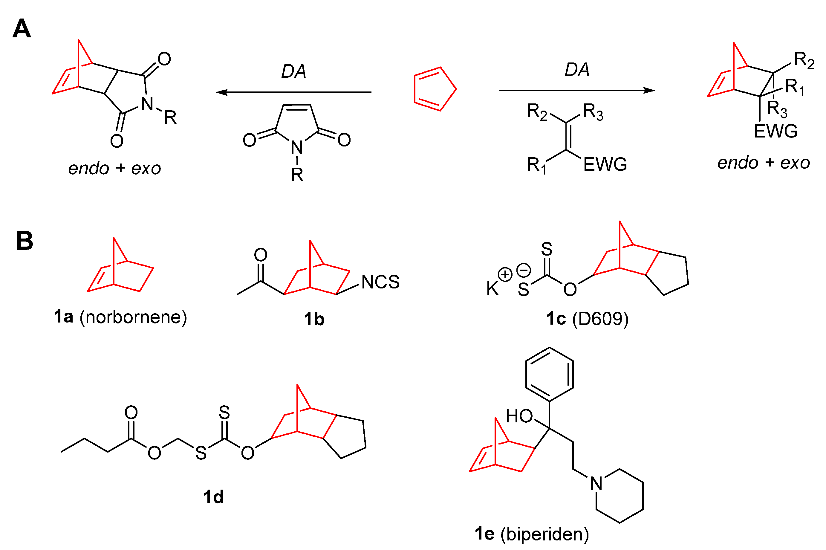
DADiels–Alder
DDSDrug delivery systems
DNADeoxyribonucleic acid
EC50Half maximal effective concentration
EGFREpidermal growth factor receptor
EMTEpithelial mesenchymal transmission
EREstrogen receptors
ERKEstrogen receptor kinase
FAM46CNon-canonical poly(A) polymerase protein
FDAFood drug administration
HDACHistone deacetylase
HUVECHuman Umbilical Vein Endothelial Cell
IARC International Agency for Research on Cancer
IC50Half maximal inhibitory concentration
InRInsulin receptor
JNKc-Jun N-terminal kinase
KiInhibitory constant
LC3-IIBMicrotubule-associated protein 1A/1B-light chain 3-phosphatidylethanolamine conjugate B
MALT1Mucosa-associated lymphoid tissue lymphoma translocation protein 1
MAPMitogen-activated protein
MCL-1Myeloid cell leukemia-1
MEK1/2Mitogen-activated protein kinase kinases 1/2
MOFMetal–organic framework
MTMutant type
PDACPancreatic ductal adenocarcinoma
P-gpMDR1/P-glycoprotein
RasRat sarcoma virus
RNSReactive nitrogen species
ROSReactive oxygen species
SARStructure–activity relationship
SDHBSuccinate dehydrogenase complex subunit b
SISelectivity Index
SIRT7Sirtuin protein 7
SQSTM1/p62Sequestosome 1
TGITotal growth inhibition
TNFTumor necrosis factor
TRPM8Transient receptor potential cation channel subfamily M member 8
VEGFR-2Vascular endothelial growth factor receptor 2
WTWild type

References

  1. World Health Organization Cancer. Available online: https://www.who.int/news-room/fact-sheets/detail/cancer (accessed on 22 July 2022).
  2. Vasan, N.; Baselga, J.; Hyman, D.M. A View on Drug Resistance in Cancer. Nature 2019, 575, 299–309. [Google Scholar] [CrossRef] [PubMed] [Green Version]
  3. Hou, J.; He, Z.; Liu, T.; Chen, D.; Wang, B.; Wen, Q.; Zheng, X. Evolution of Molecular Targeted Cancer Therapy: Mechanisms of Drug Resistance and Novel Opportunities Identified by CRISPR-Cas9 Screening. Front. Oncol. 2022, 12, 755053. [Google Scholar] [CrossRef] [PubMed]
  4. N, V.R.; Dinda, H.; Ganivada, M.N.; Das Sarma, J.; Shunmugam, R. Efficient Approach to Prepare Multiple Chemotherapeutic Agent Conjugated Nanocarrier. Chem. Commun. 2014, 50, 13540–13543. [Google Scholar] [CrossRef] [PubMed]
  5. Mukherjee, S.; Dinda, H.; Chakraborty, I.; Bhattacharyya, R.; Das Sarma, J.; Shunmugam, R. Engineering Camptothecin-Derived Norbornene Polymers for Theranostic Application. ACS Omega 2017, 2, 2848–2857. [Google Scholar] [CrossRef] [PubMed] [Green Version]
  6. Magnusson, G. A Convenient Preparation of Cyclopentadiene from Its Dimer. J. Org. Chem. 1985, 50, 1998. [Google Scholar] [CrossRef]
  7. Monson, R.S. The Diels-Alder Reaction. In Advanced Organic Synthesis; Elsevier: Amsterdam, The Netherlands, 1971; Volume 3, pp. 71–79. ISBN 047180343X. [Google Scholar]
  8. Huertas, D.; Florscher, M.; Dragojlovic, V. Solvent-Free Diels–Alder Reactions of in Situ Generated Cyclopentadiene. Green Chem. 2009, 11, 91–95. [Google Scholar] [CrossRef]
  9. Glassner, M.; Blinco, J.P.; Barner-Kowollik, C. Diels–Alder Reactions as an Efficient Route to High Purity Cyclic Polymers. Macromol. Rapid Commun. 2011, 32, 724–728. [Google Scholar] [CrossRef]
  10. Kaminsky, W.; Boggioni, L.; Tritto, I.; Aqida, S.N. Cycloolefin Polymerization. In Reference Module in Materials Science and Materials Engineering; Elsevier: Amsterdam, The Netherlands, 2019. [Google Scholar]
  11. Hasan, T.; Ikeda, T.; Shiono, T. Ethene−Norbornene Copolymer with High Norbornene Content Produced by a Nsa -Fluorenylamidodimethyltitanium Complex Using a Suitable Activator. Macromolecules 2004, 37, 8503–8509. [Google Scholar] [CrossRef]
  12. Chowdhury, S.I.; Tanaka, R.; Nakayama, Y.; Shiono, T. Copolymerization of Norbornene and Styrene with Anilinonaphthoquinone-Ligated Nickel Complexes. Polymers 2019, 11, 1100. [Google Scholar] [CrossRef] [Green Version]
  13. Spanget-Larsen, J.; Gleiter, R. On the Exceptional Reactivity of the Norbornene Double Bond. Tetrahedron Lett. 1982, 23, 2435–2438. [Google Scholar] [CrossRef]
  14. Bartlett, P.D.; Ghosh, T. Sulfuration of the Norbornene Double Bond. J. Org. Chem. 1987, 52, 4937–4943. [Google Scholar] [CrossRef]
  15. Balaban, T.S.; Balaban, A.T. Five-Membered Rings with Three Oxygen or Sulfur Atoms in 1,2,4-Positions. In Comprehensive Heterocyclic Chemistry III; Elsevier: Amsterdam, The Netherlands, 2008; Volume 6, pp. 191–256. ISBN 9780080449920. [Google Scholar]
  16. Lang, K.; Davis, L.; Torres-Kolbus, J.; Chou, C.; Deiters, A.; Chin, J.W. Genetically Encoded Norbornene Directs Site-Specific Cellular Protein Labelling via a Rapid Bioorthogonal Reaction. Nat. Chem. 2012, 4, 298–304. [Google Scholar] [CrossRef] [PubMed] [Green Version]
  17. Oliveira, B.L.; Guo, Z.; Bernardes, G.J.L. Inverse Electron Demand Diels–Alder Reactions in Chemical Biology. Chem. Soc. Rev. 2017, 46, 4895–4950. [Google Scholar] [CrossRef] [PubMed] [Green Version]
  18. Stockdale, T.P.; Williams, C.M. Pharmaceuticals That Contain Polycyclic Hydrocarbon Scaffolds. Chem. Soc. Rev. 2015, 44, 7737–7763. [Google Scholar] [CrossRef] [PubMed] [Green Version]
  19. Zhang, Y.; Kensler, T.W.; Cho, C.G.; Posner, G.H.; Talalay, P. Anticarcinogenic Activities of Sulforaphane and Structurally Related Synthetic Norbornyl Isothiocyanates. Proc. Natl. Acad. Sci. USA 1994, 91, 3147–3150. [Google Scholar] [CrossRef] [Green Version]
  20. Pörn-Ares, M.I.; Chow, S.C.; Slotte, J.P.; Orrenius, S. Induction of Apoptosis and Potentiation of TNF- and Fas-Mediated Apoptosis in U937 Cells by the Xanthogenate Compound D609. Exp. Cell Res. 1997, 235, 48–54. [Google Scholar] [CrossRef]
  21. Du, C.; Zhao, Q.; Araki, S.; Zhang, S.; Miao, J. Apoptosis Mediated by Phosphatidylcholine-Specific Phospholipase C Is Associated with CAMP, P53 Level, and Cell-Cycle Distribution in Vascular Endothelial Cells. Endothelium 2003, 10, 141–147. [Google Scholar] [CrossRef]
  22. Milhas, D.; Andrieu-Abadie, N.; Levade, T.; Benoist, H.; Ségui, B. The Tricyclodecan-9-Yl-Xanthogenate D609 Triggers Ceramide Increase and Enhances FasL-Induced Caspase-Dependent and -Independent Cell Death in T Lymphocytes. Int. J. Mol. Sci. 2012, 13, 8834–8852. [Google Scholar] [CrossRef]
  23. Kalluri, H.S.G.; Kuo, J.S.; Dempsey, R.J. Effect of D609 on the Expression of GADD45β Protein: Potential Inhibitory Role in the Growth of Glioblastoma Cancer Stem like Cells. Eur. J. Pharmacol. 2016, 791, 510–517. [Google Scholar] [CrossRef]
  24. Kalluri, H.S.G.; Kuo, J.S.; Dempsey, R.J. Chronic D609 Treatment Interferes with Cell Cycle and Targets the Expression of Olig2 in Glioma Stem like Cells. Eur. J. Pharmacol. 2017, 814, 81–86. [Google Scholar] [CrossRef]
  25. Bai, A.; Meier, G.P.; Wang, Y.; Luberto, C.; Hannun, Y.A.; Zhou, D. Prodrug Modification Increases Potassium Tricyclo[5.2.1.0 2,6 ]-Decan-8-Yl Dithiocarbonate (D609) Chemical Stability and Cytotoxicity against U937 Leukemia Cells. J. Pharmacol. Exp. Ther. 2004, 309, 1051–1059. [Google Scholar] [CrossRef] [PubMed] [Green Version]
  26. Kostelnik, A.; Cegan, A.; Pohanka, M. Anti-Parkinson Drug Biperiden Inhibits Enzyme Acetylcholinesterase. Biomed Res. Int. 2017, 2017, 2532764. [Google Scholar] [CrossRef] [PubMed] [Green Version]
  27. Konczalla, L.; Perez, D.R.; Wenzel, N.; Wolters-Eisfeld, G.; Klemp, C.; Lüddeke, J.; Wolski, A.; Landschulze, D.; Meier, C.; Buchholz, A.; et al. Biperiden and Mepazine Effectively Inhibit MALT1 Activity and Tumor Growth in Pancreatic Cancer. Int. J. Cancer 2020, 146, 1618–1630. [Google Scholar] [CrossRef] [PubMed] [Green Version]
  28. Heinlein, C.A.; Chang, C. Androgen Receptor in Prostate Cancer. Endocr. Rev. 2004, 25, 276–308. [Google Scholar] [CrossRef] [Green Version]
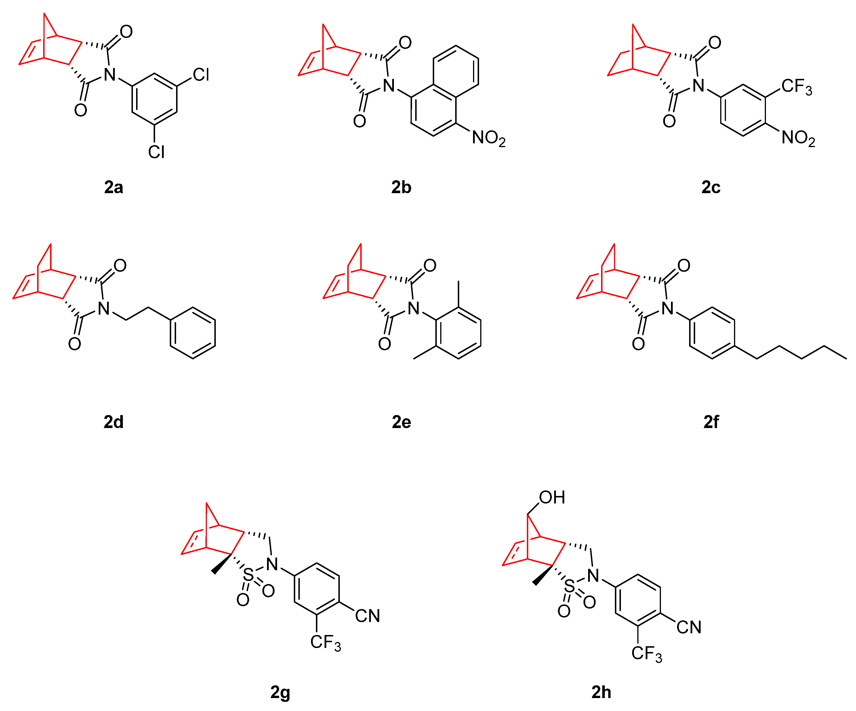
  29. Salvati, M.E.; Balog, A.; Shan, W.; Wei, D.D.; Pickering, D.; Attar, R.M.; Geng, J.; Rizzo, C.A.; Gottardis, M.M.; Weinmann, R.; et al. Structure Based Approach to the Design of Bicyclic-1H-Isoindole-1,3(2H)-Dione Based Androgen Receptor Antagonists. Bioorg. Med. Chem. Lett. 2005, 15, 271–276. [Google Scholar] [CrossRef]
  30. Salvati, M.E.; Balog, A.; Wei, D.D.; Pickering, D.; Attar, R.M.; Geng, J.; Rizzo, C.A.; Hunt, J.T.; Gottardis, M.M.; Weinmann, R.; et al. Identification of a Novel Class of Androgen Receptor Antagonists Based on the Bicyclic-1H-Isoindole-1,3(2H)-Dione Nucleus. Bioorg. Med. Chem. Lett. 2005, 15, 389–393. [Google Scholar] [CrossRef]
  31. Abdel-Aziz, A.A.M. Novel and Versatile Methodology for Synthesis of Cyclic Imides and Evaluation of Their Cytotoxic, DNA Binding, Apoptotic Inducing Activities and Molecular Modeling Study. Eur. J. Med. Chem. 2007, 42, 614–626. [Google Scholar] [CrossRef]
  32. Shan, W.; Balog, A.; Nation, A.; Zhu, X.; Chen, J.; Cvijic, M.E.; Geng, J.; Rizzo, C.A.; Spires, T.; Attar, R.M.; et al. [2.2.1]-Bicyclic Sultams as Potent Androgen Receptor Antagonists. Bioorg. Med. Chem. Lett. 2016, 26, 5707–5711. [Google Scholar] [CrossRef]
  33. Zhan, T.; Rindtorff, N.; Boutros, M. Wnt Signaling in Cancer. Oncogene 2017, 36, 1461–1473. [Google Scholar] [CrossRef]
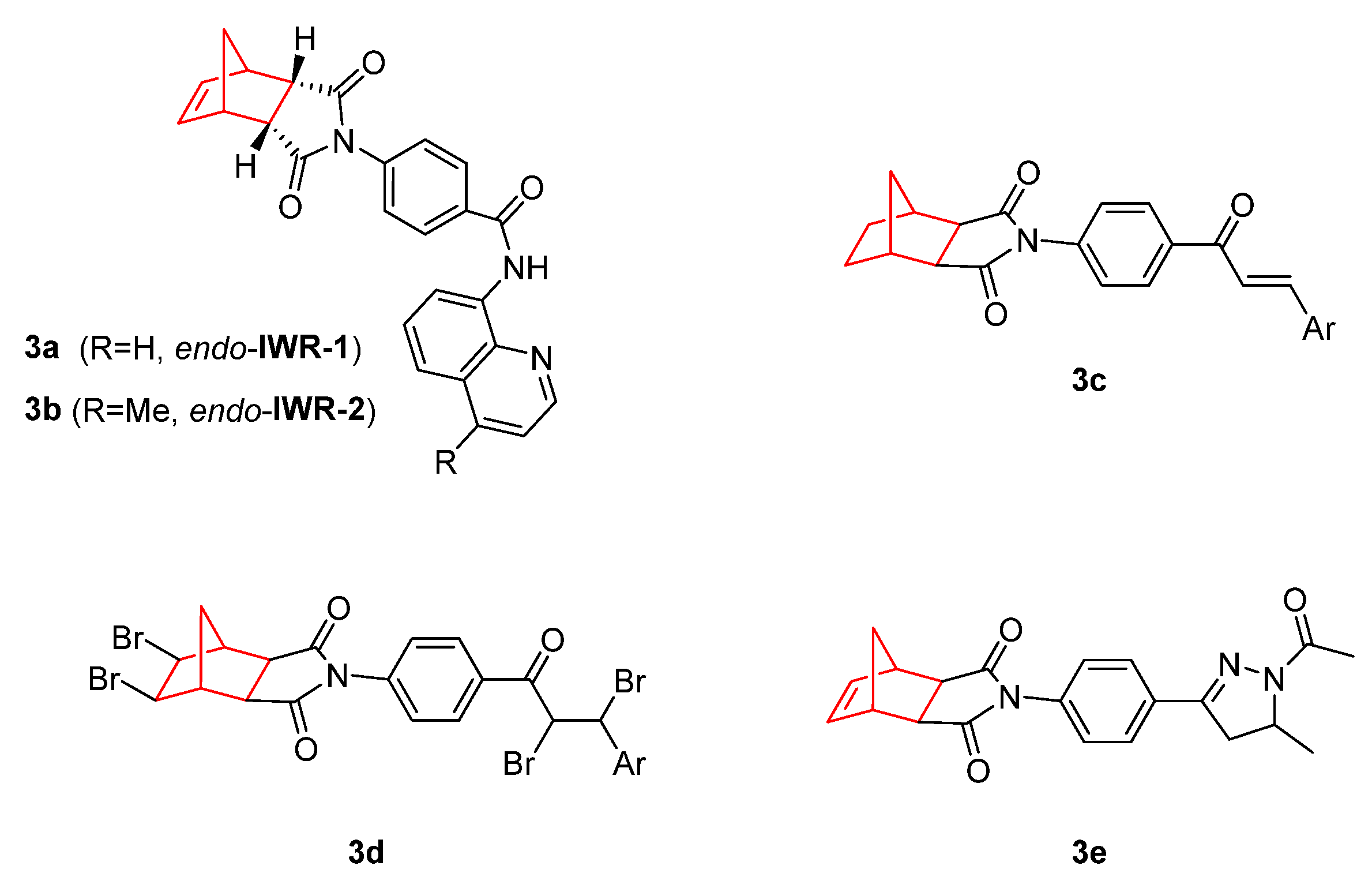
  34. Chen, B.; Dodge, M.E.; Tang, W.; Lu, J.; Ma, Z.; Fan, C.-W.; Wei, S.; Hao, W.; Kilgore, J.; Williams, N.S.; et al. Small Molecule–Mediated Disruption of Wnt-Dependent Signaling in Tissue Regeneration and Cancer. Nat. Chem. Biol. 2009, 5, 100–107. [Google Scholar] [CrossRef]
  35. Lu, J.; Ma, Z.; Hsieh, J.-C.; Fan, C.-W.; Chen, B.; Longgood, J.C.; Williams, N.S.; Amatruda, J.F.; Lum, L.; Chen, C. Structure–Activity Relationship Studies of Small-Molecule Inhibitors of Wnt Response. Bioorg. Med. Chem. Lett. 2009, 19, 3825–3827. [Google Scholar] [CrossRef] [Green Version]
  36. Lee, S.C.; Kim, O.-H.; Lee, S.K.; Kim, S.-J. IWR-1 Inhibits Epithelial-Mesenchymal Transition of Colorectal Cancer Cells through Suppressing Wnt/β-Catenin Signaling as Well as Survivin Expression. Oncotarget 2015, 6, 27146–27159. [Google Scholar] [CrossRef] [Green Version]
  37. Martins-Neves, S.R.; Paiva-Oliveira, D.I.; Fontes-Ribeiro, C.; Bovée, J.V.M.G.; Cleton-Jansen, A.-M.; Gomes, C.M.F. IWR-1, a Tankyrase Inhibitor, Attenuates Wnt/β-Catenin Signaling in Cancer Stem-like Cells and Inhibits in Vivo the Growth of a Subcutaneous Human Osteosarcoma Xenograft. Cancer Lett. 2018, 414, 1–15. [Google Scholar] [CrossRef]
  38. Narwal, M.; Venkannagari, H.; Lehtiö, L. Structural Basis of Selective Inhibition of Human Tankyrases. J. Med. Chem. 2012, 55, 1360–1367. [Google Scholar] [CrossRef]
  39. Kocyigit, U.M.; Budak, Y.; Gürdere, M.B.; Tekin, Ş.; Köprülü, T.K.; Ertürk, F.; Özcan, K.; Gülçin, İ.; Ceylan, M. Synthesis, Characterization, Anticancer, Antimicrobial and Carbonic Anhydrase Inhibition Profiles of Novel (3aR,4S,7R,7aS)-2-(4-((E)-3-(3-Aryl)Acryloyl)Phenyl)-3a,4,7,7a-Tetrahydro-1H-4,7-Methanoisoindole-1,3(2H)-Dione Derivatives. Bioorg. Chem. 2017, 70, 118–125. [Google Scholar] [CrossRef]
  40. Kocyigit, U.M.; Budak, Y.; Eligüzel, F.; Taslimi, P.; Kılıç, D.; Gulçin, İ.; Ceylan, M. Synthesis and Carbonic Anhydrase Inhibition of Tetrabromo Chalcone Derivatives. Arch. Pharm. 2017, 350, 1700198. [Google Scholar] [CrossRef]
  41. Kocyigit, U.M.; Budak, Y.; Gürdere, M.B.; Dürü, N.; Taslimi, P.; Gülçin, İ.; Ceylan, M. Synthesis and Investigation of Anticancer, Antibacterial Activities and Carbonic Anhydrase, Acetylcholinesterase Inhibition Profiles of Novel (3aR,4S,7R,7aS)-2-[4-[1-Acetyl-5-(Aryl/Heteroaryl)-4,5-Dihydro-1H-Pyrazol-3-Yl]Phenyl]-3a,4,7,7a-Tetrahydro-1H-4. Mon. Chem.-Chem. Mon. 2019, 150, 721–731. [Google Scholar] [CrossRef]
  42. Meanwell, N.A. Fluorine and Fluorinated Motifs in the Design and Application of Bioisosteres for Drug Design. J. Med. Chem. 2018, 61, 5822–5880. [Google Scholar] [CrossRef]
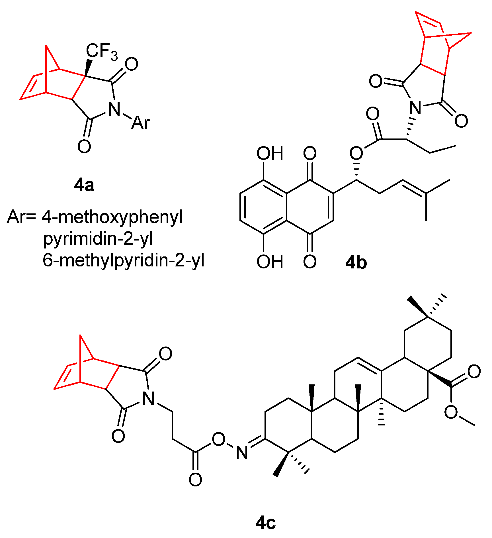
  43. Luzina, E.L.; Popov, A.V. Synthesis and Anticancer Activity Evaluation of 3,4-Mono- and Bicyclosubstituted N-(Het)Aryl Trifluoromethyl Succinimides. J. Fluor. Chem. 2014, 168, 121–127. [Google Scholar] [CrossRef] [Green Version]
  44. Baloch, S.K.; Ma, L.; Wang, X.-L.; Shi, J.; Zhu, Y.; Wu, F.-Y.; Pang, Y.-J.; Lu, G.-H.; Qi, J.-L.; Wang, X.-M.; et al. Design, Synthesis and Mechanism of Novel Shikonin Derivatives as Potent Anticancer Agents. RSC Adv. 2015, 5, 31759–31767. [Google Scholar] [CrossRef]
  45. Žiberna, L.; Šamec, D.; Mocan, A.; Nabavi, S.; Bishayee, A.; Farooqi, A.; Sureda, A.; Nabavi, S. Oleanolic Acid Alters Multiple Cell Signaling Pathways: Implication in Cancer Prevention and Therapy. Int. J. Mol. Sci. 2017, 18, 643. [Google Scholar] [CrossRef] [Green Version]
  46. Castrejón-Jiménez, N.S.; Leyva-Paredes, K.; Baltierra-Uribe, S.L.; Castillo-Cruz, J.; Campillo-Navarro, M.; Hernández-Pérez, A.D.; Luna-Angulo, A.B.; Chacón-Salinas, R.; Coral-Vázquez, R.M.; Estrada-García, I.; et al. Ursolic and Oleanolic Acids Induce Mitophagy in A549 Human Lung Cancer Cells. Molecules 2019, 24, 3444. [Google Scholar] [CrossRef] [Green Version]
  47. Ayeleso, T.; Matumba, M.; Mukwevho, E. Oleanolic Acid and Its Derivatives: Biological Activities and Therapeutic Potential in Chronic Diseases. Molecules 2017, 22, 1915. [Google Scholar] [CrossRef] [Green Version]
  48. Bednarczyk-Cwynar, B.; Ruszkowskp, P.; Atamanyuk, D.; Lesyk, R.; Zaprutko, L. Hybrids of Oleanolic Acid with Norbornene-2,3-Dicarboximide-N-Carboxylic Acids as Potential Anticancer Agents. Acta Pol. Pharm. 2017, 74, 827–835. [Google Scholar]
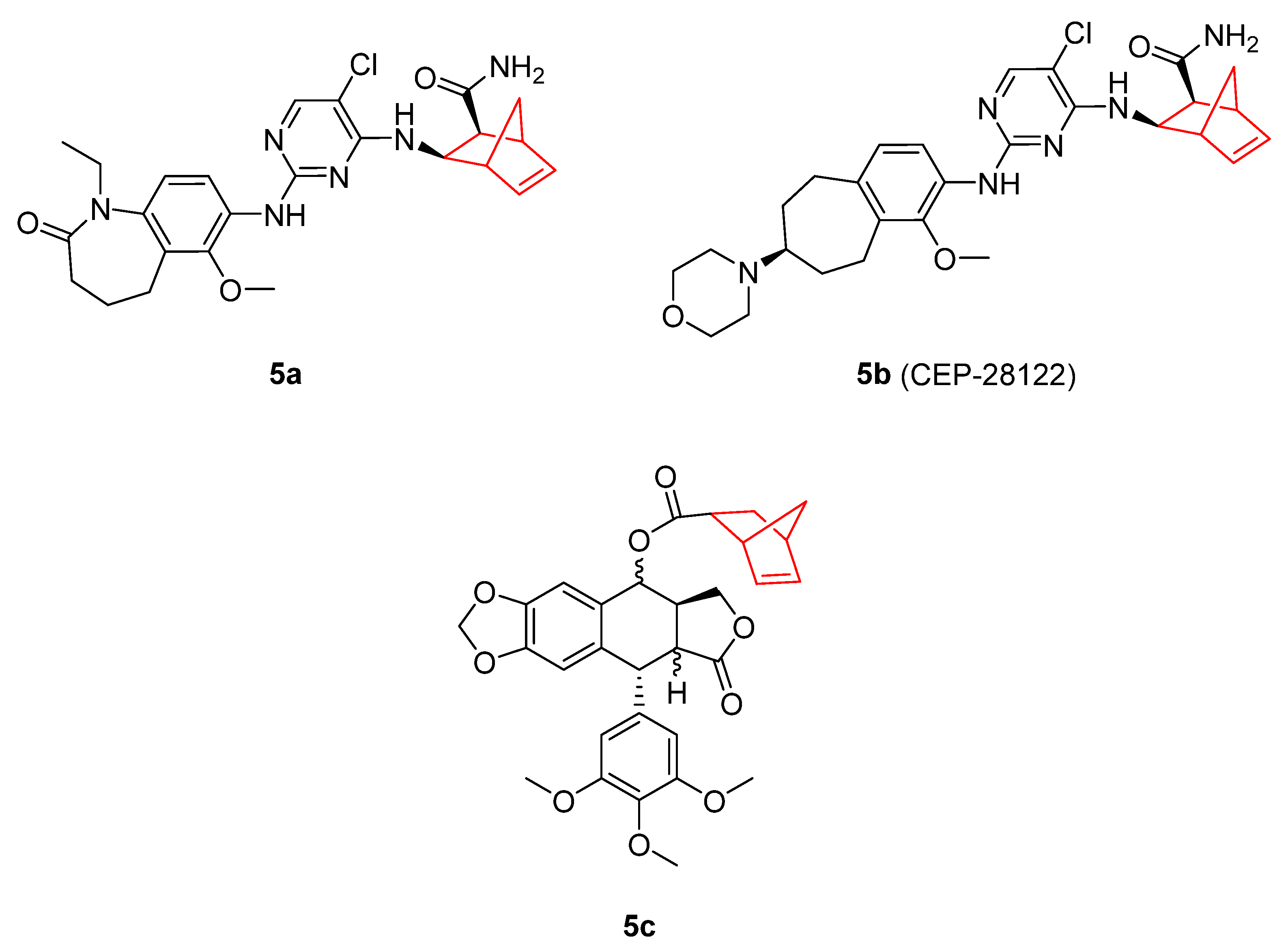
  49. Ott, G.R.; Tripathy, R.; Cheng, M.; McHugh, R.; Anzalone, A.V.; Underiner, T.L.; Curry, M.A.; Quail, M.R.; Lu, L.; Wan, W.; et al. Discovery of a Potent Inhibitor of Anaplastic Lymphoma Kinase with in Vivo Antitumor Activity. ACS Med. Chem. Lett. 2010, 1, 493–498. [Google Scholar] [CrossRef] [Green Version]
  50. Gingrich, D.E.; Lisko, J.G.; Curry, M.A.; Cheng, M.; Quail, M.; Lu, L.; Wan, W.; Albom, M.S.; Angeles, T.S.; Aimone, L.D.; et al. Discovery of an Orally Efficacious Inhibitor of Anaplastic Lymphoma Kinase. J. Med. Chem. 2012, 55, 4580–4593. [Google Scholar] [CrossRef]
  51. Cheng, M.; Quail, M.R.; Gingrich, D.E.; Ott, G.R.; Lu, L.; Wan, W.; Albom, M.S.; Angeles, T.S.; Aimone, L.D.; Cristofani, F.; et al. CEP-28122, a Highly Potent and Selective Orally Active Inhibitor of Anaplastic Lymphoma Kinase with Antitumor Activity in Experimental Models of Human Cancers. Mol. Cancer Ther. 2012, 11, 670–679. [Google Scholar] [CrossRef] [Green Version]
  52. López-Pérez, J.L.; del Olmo, E.; de Pascual-Teresa, B.; Abad, A.; San Feliciano, A. Synthesis and Cytotoxicity of Hydrophobic Esters of Podophyllotoxins. Bioorg. Med. Chem. Lett. 2004, 14, 1283–1286. [Google Scholar] [CrossRef]
  53. Zefirov, N.A.; Kruth, A.; Wobith, B.; Nurieva, E.V.; Riyaz, S.; Reddy, C.V.R.; Kuznetsov, S.A.; Zefirova, O.N. Novel Bridged and Caged C4-Podophyllotoxin Derivatives as Microtubule Disruptors: Synthesis, Cytotoxic Evaluation and Structure–Activity Relationship. Mendeleev Commun. 2018, 28, 475–478. [Google Scholar] [CrossRef]
  54. Itani, W.S.; El-Banna, S.H.; Hassan, S.B.; Larsson, R.L.; Bazarbachi, A.; Gali-Muhtasib, H.U. Anti Colon Cancer Components from Lebanese Sage (Salvia Libanotica) Essential Oil: Mechanistic Basis. Cancer Biol. Ther. 2008, 7, 1765–1773. [Google Scholar] [CrossRef] [Green Version]
  55. Moayedi, Y.; Greenberg, S.A.; Jenkins, B.A.; Marshall, K.L.; Dimitrov, L.V.; Nelson, A.M.; Owens, D.M.; Lumpkin, E.A. Camphor White Oil Induces Tumor Regression through Cytotoxic T Cell-dependent Mechanisms. Mol. Carcinog. 2019, 58, 722–734. [Google Scholar] [CrossRef] [PubMed]
  56. El Yaagoubi, M.; Ortiz, S.; Mechqoq, H.; Cavaleiro, C.; Lecsö-Bornet, M.; Rodrigues, M.J.; Custódio, L.; El Mousadik, A.; Grougnet, R.; El Aouad, N.; et al. Chemical Composition, Antibacterial Screening and Cytotoxic Activity of Chiliadenus Antiatlanticus (Asteraceae) Essential Oil. Chem. Biodivers. 2021, 18, e2100115. [Google Scholar] [CrossRef] [PubMed]
  57. Ouakouak, H.; Benarfa, A.; Messaoudi, M.; Begaa, S.; Sawicka, B.; Benchikha, N.; Simal-Gandara, J. Biological Properties of Essential Oils from Thymus Algeriensis Boiss. Plants 2021, 10, 786. [Google Scholar] [CrossRef]
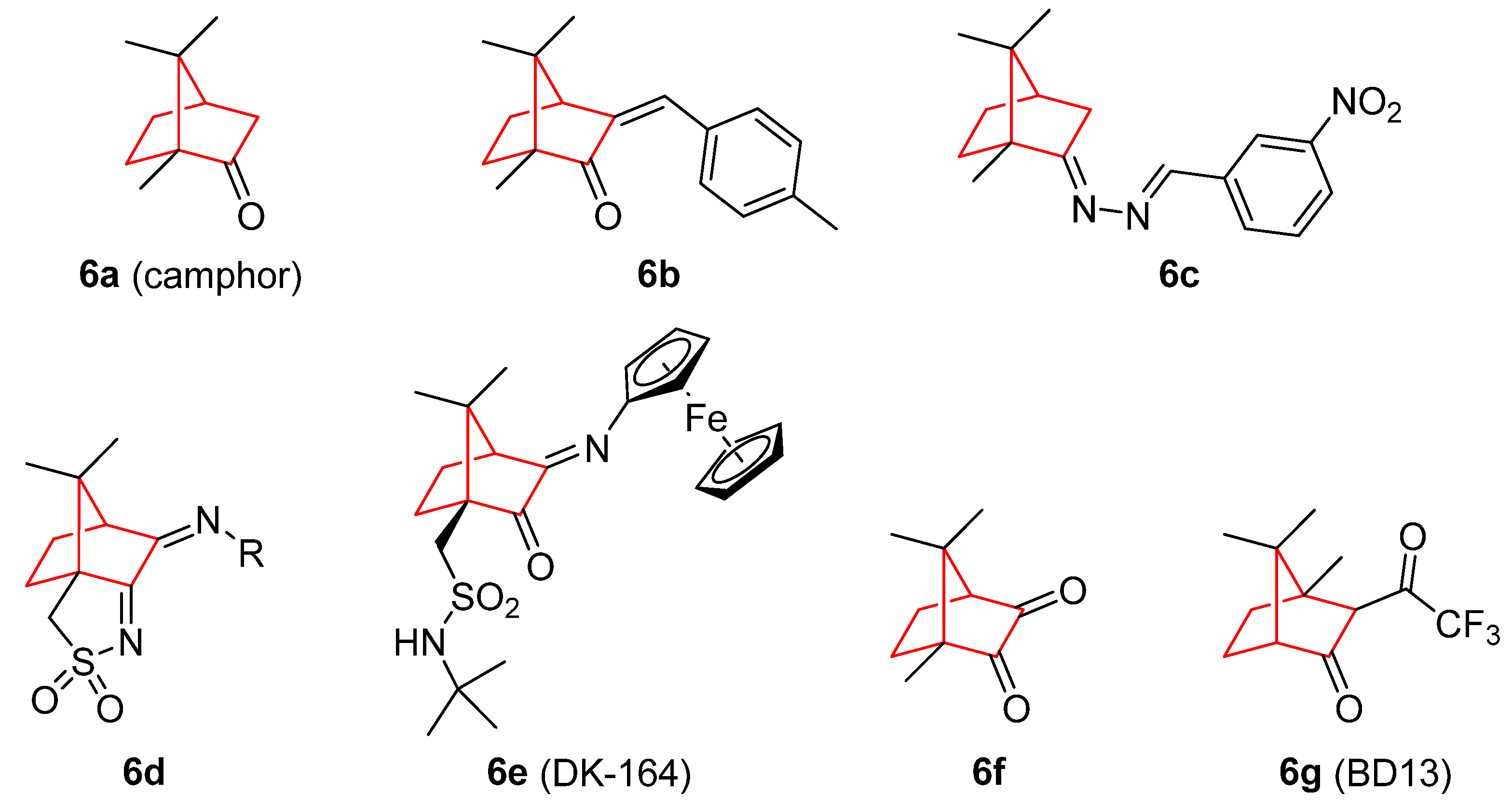
  58. Mueller, S.O.; Kling, M.; Arifin Firzani, P.; Mecky, A.; Duranti, E.; Shields-Botella, J.; Delansorne, R.; Broschard, T.; Kramer, P.-J. Activation of Estrogen Receptor α and ERβ by 4-Methylbenzylidene-Camphor in Human and Rat Cells: Comparison with Phyto- and Xenoestrogens. Toxicol. Lett. 2003, 142, 89–101. [Google Scholar] [CrossRef]
  59. da Silva, E.; da Silva Araújo, A.; Moraes, A.; de Souza, L.; Silva Lourenço, M.; de Souza, M.; Wardell, J.; Wardell, S. Synthesis and Biological Activities of Camphor Hydrazone and Imine Derivatives. Sci. Pharm. 2015, 84, 467–483. [Google Scholar] [CrossRef] [Green Version]
  60. Cardoso, J.M.S.; Correia, I.; Galvão, A.M.; Marques, F.; Carvalho, M.F.N.N. Synthesis of Ag(I) Camphor Sulphonylimine Complexes and Assessment of Their Cytotoxic Properties against Cisplatin -Resistant A2780cisR and A2780 Cell Lines. J. Inorg. Biochem. 2017, 166, 55–63. [Google Scholar] [CrossRef]
  61. Schröder, M.; Yusein-Myashkova, S.; Petrova, M.; Dobrikov, G.; Kamenova-Nacheva, M.; Todorova, J.; Pasheva, E.; Ugrinova, I. The Effect of a Ferrocene Containing Camphor Sulfonamide DK-164 on Breast Cancer Cell Lines. Anticancer. Agents Med. Chem. 2019, 19, 1874–1886. [Google Scholar] [CrossRef]
  62. Schröder, M.; Petrova, M.; Vlahova, Z.; Dobrikov, G.M.; Slavchev, I.; Pasheva, E.; Ugrinova, I. In Vitro Anticancer Activity of Two Ferrocene-Containing Camphor Sulfonamides as Promising Agents against Lung Cancer Cells. Biomedicines 2022, 10, 1353. [Google Scholar] [CrossRef]
  63. Volk, J.; Ziemann, C.; Leyhausen, G.; Geurtsen, W. Genotoxic and Mutagenic Potential of Camphorquinone in L5178/TK+/− Mouse Lymphoma Cells. Dent. Mater. 2018, 34, 519–530. [Google Scholar] [CrossRef]
  64. Nakano, K.; Nakayachi, T.; Yasumoto, E.; Morshed, S.R.M.D.; Hashimoto, K.; Kikuchi, H.; Nishikawa, H.; Sugiyama, K.; Amano, O.; Kawase, M.; et al. Induction of Apoptosis by Beta-Diketones in Human Tumor Cells. Anticancer Res. 2004, 24, 711–717. [Google Scholar]
  65. Verma, M.; Singh, S.; Bhushan, S.; Pal, H.; Kitchlu, S.; Koul, M.; Thappa, R.; Saxena, A. Induction of Mitochondrial-Dependent Apoptosis by an Essential Oil from Tanacetum Gracile. Planta Med. 2008, 74, 515–520. [Google Scholar] [CrossRef] [PubMed] [Green Version]
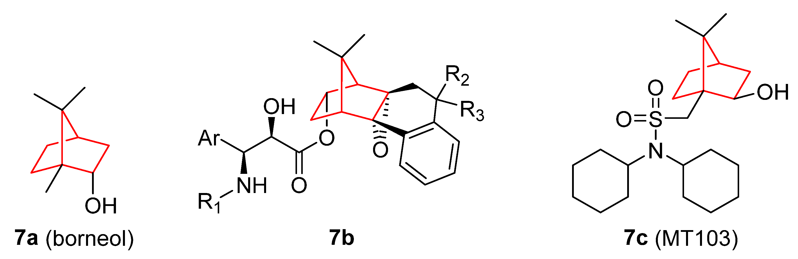
  66. Chen, J.; Li, L.; Su, J.; Li, B.; Chen, T.; Wong, Y.-S. Synergistic Apoptosis-Inducing Effects on A375 Human Melanoma Cells of Natural Borneol and Curcumin. PLoS ONE 2014, 9, e101277. [Google Scholar] [CrossRef] [PubMed]
  67. Chen, J.; Li, L.; Su, J.; Chen, T. Natural Borneol Enhances Bisdemethoxycurcumin-Induced Cell Cycle Arrest in the G2/M Phase through up-Regulation of Intracellular ROS in HepG2 Cells. Food Funct. 2015, 6, 740–748. [Google Scholar] [CrossRef] [PubMed]
  68. Chen, J.; Li, L.; Su, J.; Li, B.; Zhang, X.; Chen, T. Proteomic Analysis of G2/M Arrest Triggered by Natural Borneol/Curcumin in HepG2 Cells, the Importance of the Reactive Oxygen Species-P53 Pathway. J. Agric. Food Chem. 2015, 63, 6440–6449. [Google Scholar] [CrossRef] [PubMed]
  69. Liu, W.; Yin, Y.; Sun, J.; Feng, S.; Ma, J.; Fu, X.; Hou, Y.; Yang, M.; Sun, B.; Fan, C. Natural Borneol Is a Novel Chemosensitizer That Enhances Temozolomide-Induced Anticancer Efficiency against Human Glioma by Triggering Mitochondrial Dysfunction and Reactive Oxide Species-Mediated Oxidative Damage. Onco. Targets. Ther. 2018, 11, 5429–5439. [Google Scholar] [CrossRef] [Green Version]
  70. Meng, X.; Dong, X.; Wang, W.; Yang, L.; Zhang, X.; Li, Y.; Chen, T.; Ma, H.; Qi, D.; Su, J. Natural Borneol Enhances Paclitaxel-Induced Apoptosis of ESCC Cells by Inactivation of the PI3K/AKT. J. Food Sci. 2018, 83, 1436–1443. [Google Scholar] [CrossRef]
  71. Cao, W.; Li, Y.; Hou, Y.; Yang, M.; Fu, X.; Zhao, B.; Jiang, H.; Fu, X. Enhanced Anticancer Efficiency of Doxorubicin against Human Glioma by Natural Borneol through Triggering ROS-Mediated Signal. Biomed. Pharmacother. 2019, 118, 109261. [Google Scholar] [CrossRef]
  72. Lai, H.; Liu, C.; Hou, L.; Lin, W.; Chen, T.; Hong, A. TRPM8-Regulated Calcium Mobilization Plays a Critical Role in Synergistic Chemosensitization of Borneol on Doxorubicin. Theranostics 2020, 10, 10154–10170. [Google Scholar] [CrossRef]
  73. Duan, M.; Xing, Y.; Guo, J.; Chen, H.; Zhang, R. Borneol Increases Blood–Tumour Barrier Permeability by Regulating the Expression Levels of Tight Junction-Associated Proteins. Pharm. Biol. 2016, 54, 3009–3018. [Google Scholar] [CrossRef] [Green Version]
  74. Chen, Z.; Xu, Q.; Shan, C.; Shi, Y.; Wang, Y.; Chang, R.C.-C.; Zheng, G. Borneol for Regulating the Permeability of the Blood-Brain Barrier in Experimental Ischemic Stroke: Preclinical Evidence and Possible Mechanism. Oxid. Med. Cell. Longev. 2019, 2019, 2936737. [Google Scholar] [CrossRef] [Green Version]
  75. Yin, Y.; Cao, L.; Ge, H.; Duanmu, W.; Tan, L.; Yuan, J.; Tunan, C.; Li, F.; Hu, R.; Gao, F.; et al. L-Borneol Induces Transient Opening of the Blood–Brain Barrier and Enhances the Therapeutic Effect of Cisplatin. Neuroreport 2017, 28, 506–513. [Google Scholar] [CrossRef] [PubMed]
  76. Klar, U.; Graf, H.; Schenk, O.; Röhr, B.; Schulz, H. New Synthetic Inhibitors of Microtubule Depolymerization. Bioorg. Med. Chem. Lett. 1998, 8, 1397–1402. [Google Scholar] [CrossRef] [PubMed]
  77. Jasinski, P.; Zwolak, P.; Isaksson Vogel, R.; Bodempudi, V.; Terai, K.; Galvez, J.; Land, D.; Dudek, A.Z. MT103 Inhibits Tumor Growth with Minimal Toxicity in Murine Model of Lung Carcinoma via Induction of Apoptosis. Investig. New Drugs 2011, 29, 846–852. [Google Scholar] [CrossRef]
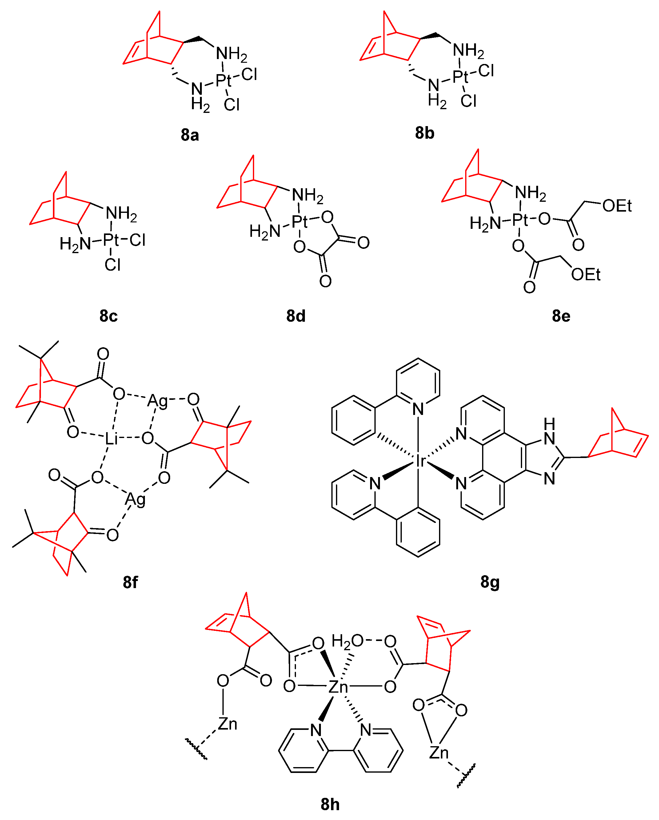
  78. Dilruba, S.; Kalayda, G.V. Platinum-Based Drugs: Past, Present and Future. Cancer Chemother. Pharmacol. 2016, 77, 1103–1124. [Google Scholar] [CrossRef]
  79. Wang, G.; Yang, B.; Fu, Z.; Wang, X.; Zhang, Z. Efficacy and Safety of Oxaliplatin-Based Regimen versus Cisplatin-Based Regimen in the Treatment of Gastric Cancer: A Meta-Analysis of Randomized Controlled Trials. Int. J. Clin. Oncol. 2019, 24, 614–623. [Google Scholar] [CrossRef] [PubMed]
  80. Ho, G.Y.; Woodward, N.; Coward, J.I.G. Cisplatin versus Carboplatin: Comparative Review of Therapeutic Management in Solid Malignancies. Crit. Rev. Oncol. Hematol. 2016, 102, 37–46. [Google Scholar] [CrossRef] [PubMed] [Green Version]
  81. de Mier-Vinué, J.; Gay, M.; Montaña, Á.M.; Sáez, R.-I.; Moreno, V.; Kasparkova, J.; Vrana, O.; Heringova, P.; Brabec, V.; Boccarelli, A.; et al. Synthesis, Biophysical Studies, and Antiproliferative Activity of Platinum(II) Complexes Having 1,2-Bis(Aminomethyl)Carbobicyclic Ligands. J. Med. Chem. 2008, 51, 424–431. [Google Scholar] [CrossRef]
  82. Liu, F.; Hu, W.; Fang, L.; Gou, S. Synthesis and Biological Evaluation of Water-Soluble Trans-[Bicyclo[2.2.2]Octane-7 R,8 R-Diamine]Platinum(II) Complexes with Linear or Branched Alkoxyacetates as Leaving Groups. J. Coord. Chem. 2016, 69, 1284–1292. [Google Scholar] [CrossRef]
  83. Liu, F.; Gou, S.; Chen, F.; Fang, L.; Zhao, J. Study on Antitumor Platinum(II) Complexes of Chiral Diamines with Dicyclic Species as Steric Hindrance. J. Med. Chem. 2015, 58, 6368–6377. [Google Scholar] [CrossRef]
  84. Carvalho, M.F.N.N.; Botelho do Rego, A.M.; Galvão, A.M.; Herrmann, R.; Marques, F. Search for Cytotoxic Compounds against Ovarian Cancer Cells: Synthesis, Characterization and Assessment of the Activity of New Camphor Carboxylate and Camphor Carboxamide Silver Complexes. J. Inorg. Biochem. 2018, 188, 88–95. [Google Scholar] [CrossRef]
  85. Zhang, C.; Lai, S.-H.; Zeng, C.-C.; Tang, B.; Wan, D.; Xing, D.-G.; Liu, Y.-J. The Induction of Apoptosis in SGC-7901 Cells through the ROS-Mediated Mitochondrial Dysfunction Pathway by a Ir(III) Complex. JBIC J. Biol. Inorg. Chem. 2016, 21, 1047–1060. [Google Scholar] [CrossRef] [PubMed]
  86. Gao, E.; Sun, N.; Zhang, S.; Ding, Y.; Qiu, X.; Zhan, Y.; Zhu, M. Synthesis, Structures, Molecular Docking, Cytotoxicity and Bioimaging Studies of Two Novel Zn(II) Complexes. Eur. J. Med. Chem. 2016, 121, 1–11. [Google Scholar] [CrossRef] [PubMed]
  87. Jursic, B.S. Suitability of Furan, Pyrrole and Thiophene as Dienes for Diels–Alder Reactions Viewed through Their Stability and Reaction Barriers for Reactions with Acetylene, Ethylene and Cyclopropene. An AM1 Semiempirical and B3LYP Hybrid Density Functional Theory Study. J. Mol. Struct. THEOCHEM 1998, 454, 105–116. [Google Scholar] [CrossRef]
  88. McCluskey, A.; Ackland, S.P.; Bowyer, M.C.; Baldwin, M.L.; Garner, J.; Walkom, C.C.; Sakoff, J.A. Cantharidin Analogues: Synthesis and Evaluation of Growth Inhibition in a Panel of Selected Tumour Cell Lines. Bioorg. Chem. 2003, 31, 68–79. [Google Scholar] [CrossRef]
  89. Mo, L.; Zhang, X.; Shi, X.; Wei, L.; Zheng, D.; Li, H.; Gao, J.; Li, J.; Hu, Z. Norcantharidin Enhances Antitumor Immunity of GM-CSF Prostate Cancer Cells Vaccine by Inducing Apoptosis of Regulatory T Cells. Cancer Sci. 2018, 109, 2109–2118. [Google Scholar] [CrossRef] [Green Version]
  90. Yang, P.-Y.; Hu, D.-N.; Kao, Y.-H.; Lin, I.-C.; Chou, C.-Y.; Wu, Y.-C. Norcantharidin Induces Apoptosis in Human Prostate Cancer Cells through Both Intrinsic and Extrinsic Pathways. Pharmacol. Rep. 2016, 68, 874–880. [Google Scholar] [CrossRef]
  91. Peng, C.; Li, Z.; Niu, Z.; Niu, W.; Xu, Z.; Gao, H.; Niu, W.; Wang, J.; He, Z.; Gao, C.; et al. Norcantharidin Suppresses Colon Cancer Cell Epithelial-Mesenchymal Transition by Inhibiting the Avβ6-ERK-Ets1 Signaling Pathway. Sci. Rep. 2016, 6, 20500. [Google Scholar] [CrossRef] [Green Version]
  92. Pan, M.-S.; Cao, J.; Fan, Y.-Z. Insight into Norcantharidin, a Small-Molecule Synthetic Compound with Potential Multi-Target Anticancer Activities. Chin. Med. 2020, 15, 55. [Google Scholar] [CrossRef]
  93. Lin, C.-L.; Chen, C.-M.; Lin, C.-L.; Cheng, C.-W.; Lee, C.-H.; Hsieh, Y.-H. Norcantharidin Induces Mitochondrial-Dependent Apoptosis through Mcl-1 Inhibition in Human Prostate Cancer Cells. Biochim. Biophys. Acta-Mol. Cell Res. 2017, 1864, 1867–1876. [Google Scholar] [CrossRef]
  94. Zhou, J.; Ren, Y.; Tan, L.; Song, X.; Wang, M.; Li, Y.; Cao, Z.; Guo, C. Norcantharidin: Research Advances in Pharmaceutical Activities and Derivatives in Recent Years. Biomed. Pharmacother. 2020, 131, 110755. [Google Scholar] [CrossRef]
  95. Qiu, P.; Wang, S.; Liu, M.; Ma, H.; Zeng, X.; Zhang, M.; Xu, L.; Cui, Y.; Xu, H.; Tang, Y.; et al. Norcantharidin Inhibits Cell Growth by Suppressing the Expression and Phosphorylation of Both EGFR and C-Met in Human Colon Cancer Cells. BMC Cancer 2017, 17, 55. [Google Scholar] [CrossRef] [Green Version]
  96. Peng, C.; Liu, X.; Liu, E.; Xu, K.; Niu, W.; Chen, R.; Wang, J.; Zhang, Z.; Lin, P.; Wang, J.; et al. Norcantharidin Induces HT-29 Colon Cancer Cell Apoptosis through the Avβ6-Extracellular Signal-Related Kinase Signaling Pathway. Cancer Sci. 2009, 100, 2302–2308. [Google Scholar] [CrossRef] [PubMed]
  97. Li, J.-L.; Cai, Y.-C.; Liu, X.-H.; Xian, L.-J. Norcantharidin Inhibits DNA Replication and Induces Apoptosis with the Cleavage of Initiation Protein Cdc6 in HL-60 Cells. Anticancer. Drugs 2006, 17, 307–314. [Google Scholar] [CrossRef] [PubMed]
  98. Yang, P.-Y.; Chen, M.-F.; Kao, Y.-H.; Hu, D.-N.; Chang, F.-R.; Wu, Y.-C. Norcantharidin Induces Apoptosis of Breast Cancer Cells: Involvement of Activities of Mitogen Activated Protein Kinases and Signal Transducers and Activators of Transcription. Toxicol. Vitr. 2011, 25, 699–707. [Google Scholar] [CrossRef] [PubMed]
  99. An, W.; Gong, X.; Wang, M.; Tashiro, S.; Onodera, S.; Ikejima, T. Norcantharidin Induces Apoptosis in HeLa Cells through Caspase, MAPK, and Mitochondrial Pathways. Acta Pharmacol. Sin. 2004, 25, 1502–1508. [Google Scholar]
  100. Zhang, Q.-Y.; Yue, X.-Q.; Jiang, Y.-P.; Han, T.; Xin, H.-L. FAM46C Is Critical for the Anti-Proliferation and pro-Apoptotic Effects of Norcantharidin in Hepatocellular Carcinoma Cells. Sci. Rep. 2017, 7, 396. [Google Scholar] [CrossRef] [Green Version]
  101. Zhang, S.; Yang, Y.; Hua, Y.; Hu, C.; Zhong, Y. NCTD Elicits Proapoptotic and Antiglycolytic Effects on Colorectal Cancer Cells via Modulation of Fam46c Expression and Inhibition of ERK1/2 Signaling. Mol. Med. Rep. 2020, 22, 774–782. [Google Scholar] [CrossRef]
  102. Chen, A.W.-G.; Tseng, Y.-S.; Lin, C.-C.; Hsi, Y.-T.; Lo, Y.-S.; Chuang, Y.-C.; Lin, S.-H.; Yu, C.-Y.; Hsieh, M.-J.; Chen, M.-K. Norcantharidin Induce Apoptosis in Human Nasopharyngeal Carcinoma through Caspase and Mitochondrial Pathway. Environ. Toxicol. 2018, 33, 343–350. [Google Scholar] [CrossRef]
  103. Zhu, Y.; Mi, Y.; Wang, Z.; Jia, X.; Jin, Z. Norcantharidin Inhibits Viability and Induces Cell Cycle Arrest and Apoptosis in Osteosarcoma. Oncol. Lett. 2018, 17, 456–461. [Google Scholar] [CrossRef] [Green Version]
  104. He, Q.; Xue, S.; Tan, Y.; Zhang, L.; Shao, Q.; Xing, L.; Li, Y.; Xiang, T.; Luo, X.; Ren, G. Dual Inhibition of Akt and ERK Signaling Induces Cell Senescence in Triple-Negative Breast Cancer. Cancer Lett. 2019, 448, 94–104. [Google Scholar] [CrossRef]
  105. Liu, Z.; Li, B.; Cao, M.; Jiang, J. Norcantharidin Triggers Apoptotic Cell Death in Non-Small Cell Lung Cancer via a Mitophagy-Mediated Autophagy Pathway. Ann. Transl. Med. 2021, 9, 971. [Google Scholar] [CrossRef]
  106. Ahn, C.-H.; Hong, K.-O.; Jin, B.; Lee, W.; Jung, Y.C.; Lee, H.; Shin, J.-A.; Cho, S.-D.; Hong, S.D. Contribution of P38 MAPK Pathway to Norcantharidin-Induced Programmed Cell Death in Human Oral Squamous Cell Carcinoma. Int. J. Mol. Sci. 2019, 20, 3487. [Google Scholar] [CrossRef] [PubMed] [Green Version]
  107. Huang, S.; Tuergong, G.; Zhu, H.; Wang, X.; Weng, G.; Ren, Y. Norcantharidin Induces G2/M Arrest and Apoptosis via Activation of ERK and JNK, but Not P38 Signaling in Human Renal Cell Carcinoma ACHN Cells. Acta Pharm. 2021, 71, 267–278. [Google Scholar] [CrossRef]
  108. Wu, J.-Y.; Kuo, C.-D.; Chu, C.-Y.; Chen, M.-S.; Lin, J.-H.; Chen, Y.-J.; Liao, H.-F. Synthesis of Novel Lipophilic N-Substituted Norcantharimide Derivatives and Evaluation of Their Anticancer Activities. Molecules 2014, 19, 6911–6928. [Google Scholar] [CrossRef] [PubMed] [Green Version]
  109. Chang, M.-C.; Wu, J.-Y.; Liao, H.-F.; Chen, Y.-J.; Kuo, C.-D. Comparative Assessment of Therapeutic Safety of Norcantharidin, N-Farnesyloxy-Norcantharimide, and N-Farnesyl-Norcantharimide against Jurkat T Cells Relative to Human Normal Lymphoblast. Medicine 2016, 95, e4467. [Google Scholar] [CrossRef] [PubMed]
  110. Cheng, S.-S.; Shi, Y.; Ma, X.-N.; Xing, D.-X.; Liu, L.-D.; Liu, Y.; Zhao, Y.-X.; Sui, Q.-C.; Tan, X.-J. Synthesis, Crystal Structure, Spectroscopic Properties and Potential Anti-Cancerous Activities of Four Unsaturated Bis-Norcantharimides. J. Mol. Struct. 2016, 1115, 228–240. [Google Scholar] [CrossRef]
  111. Spare, L.K.; Falsetta, P.; Gilbert, J.; Harman, D.G.; Baker, M.A.; Li, F.; McCluskey, A.; Clegg, J.K.; Sakoff, J.A.; Aldrich-Wright, J.R.; et al. Cytotoxicity of a Series of Norcantharidin-Inspired Tetrahydroepoxyisoindole Carboxamides. ChemMedChem 2017, 12, 130–145. [Google Scholar] [CrossRef] [PubMed]
  112. Wang, X.H.; Huang, M.; Zhao, C.K.; Li, C.; Xu, L. Design, Synthesis, and Biological Activity Evaluation of Campthothecin-HAA-Norcantharidin Conjugates as Antitumor Agents in Vitro. Chem. Biol. Drug Des. 2019, 93, 986–992. [Google Scholar] [CrossRef]
  113. Zhao, C.K.; Li, C.; Wang, X.H.; Bao, Y.J.; Yang, F.H.; Huang, M. The Regio-Selective Synthesis of 10-Hydroxy Camptothecin Norcantharidin Conjugates and Their Biological Activity Evaluation in Vitro. R. Soc. Open Sci. 2018, 5, 172317. [Google Scholar] [CrossRef] [Green Version]
  114. Zhao, C.; Jia, J.; Wang, X.; Luo, C.; Wang, Y. Synthesis of Norcantharidin Complex Salts. J. Heterocycl. Chem. 2019, 56, 1567–1570. [Google Scholar] [CrossRef]
  115. Sun, C.-Y.; Zhu, Y.; Li, X.-F.; Tang, L.-P.; Su, Z.-Q.; Wang, X.-Q.; Li, C.-Y.; Yang, H.-M.; Zheng, G.-J.; Feng, B. Norcantharidin Alone or in Combination with Crizotinib Induces Autophagic Cell Death in Hepatocellular Carcinoma by Repressing C-Met-MTOR Signaling. Oncotarget 2017, 8, 114945–114955. [Google Scholar] [CrossRef] [PubMed] [Green Version]
  116. Zhang, X.-P.; Luo, L.-L.; Liu, Y.-Q.; Liu, X.-S.; An, F.-Y.; Sun, S.-B.; Xie, X.-R.; Geng, G.-Q.; Chen, X.-J.; Li, Z.-D. Norcantharidin Combined with Diamminedichloroplatinum Inhibits Tumor Growth and Cancerometastasis of Hepatic Carcinoma in Murine. J. Cancer Res. Ther. 2018, 14, 1035. [Google Scholar] [CrossRef]
  117. Singh, R.; Cheng, S.; Li, J.; Kumar, S.; Zeng, Q.; Zeng, Q. Norcantharidin Combined with 2-Deoxy-d-Glucose Suppresses the Hepatocellular Carcinoma Cells Proliferation and Migration. 3 Biotech 2021, 11, 142. [Google Scholar] [CrossRef]
  118. Wu, M.; Hui, S.; Chen, Y.; Chiou, H.; Lin, C.; Lee, C.; Hsieh, Y. Norcantharidin Combined with Paclitaxel Induces Endoplasmic Reticulum Stress Mediated Apoptotic Effect in Prostate Cancer Cells by Targeting SIRT7 Expression. Environ. Toxicol. 2021, 36, 2206–2216. [Google Scholar] [CrossRef]
  119. Wang, G.-S. Medical Uses of Mylabris in Ancient China and Recent Studies. J. Ethnopharmacol. 1989, 26, 147–162. [Google Scholar] [CrossRef] [PubMed]
  120. Li, H.; Xia, Z.; Chen, Y.; Yang, F.; Feng, W.; Cai, H.; Mei, Y.; Jiang, Y.; Xu, K.; Feng, D. Cantharidin Inhibits the Growth of Triple-Negative Breast Cancer Cells by Suppressing Autophagy and Inducing Apoptosis in Vitro and in Vivo. Cell. Physiol. Biochem. 2017, 43, 1829–1840. [Google Scholar] [CrossRef]
  121. Wang, W.-J.; Wu, M.-Y.; Shen, M.; Zhi, Q.; Liu, Z.-Y.; Gong, F.-R.; Tao, M.; Li, W. Cantharidin and Norcantharidin Impair Stemness of Pancreatic Cancer Cells by Repressing the β-Catenin Pathway and Strengthen the Cytotoxicity of Gemcitabine and Erlotinib. Int. J. Oncol. 2015, 47, 1912–1922. [Google Scholar] [CrossRef] [Green Version]
  122. Sakoff, J.A.; Ackland, S.P.; Baldwin, M.L.; Keane, M.A.; McCluskey, A. Anticancer Activity and Protein Phosphatase 1 and 2A Inhibition of a New Generation of Cantharidin Analogues. Investig. New Drugs 2002, 20, 1–11. [Google Scholar] [CrossRef]
  123. Zhang, Y.; Yang, S.; Zhang, H.; Gao, L.; Gao, X.; Liu, P.; Yi, Z.; Li, N.; Xu, Z. Combination Radiotherapy and Cantharidin Inhibits Lung Cancer Growth through Altering Tumor Infiltrating Lymphocytes. Futur. Oncol. 2017, 13, 1173–1180. [Google Scholar] [CrossRef]
  124. Liu, X.; Zhang, L.; Thu, P.M.; Min, W.; Yang, P.; Li, J.; Li, P.; Xu, X. Sodium Cantharidinate, a Novel Anti-Pancreatic Cancer Agent That Activates Functional P53. Sci. China Life Sci. 2021, 64, 1295–1310. [Google Scholar] [CrossRef]
  125. Pang, J.; Huang, F.; Zhang, Y.; Wu, Y.; Ge, X.; Li, S.; Li, X. Sodium Cantharidate Induces Apoptosis in Breast Cancer Cells by Regulating Energy Metabolism via the Protein Phosphatase 5-P53 Axis. Toxicol. Appl. Pharmacol. 2021, 430, 115726. [Google Scholar] [CrossRef] [PubMed]
  126. Chen, X.; Zhou, M.; Fan, W.; Yang, M.; Yang, L. Combination of Sodium Cantharidinate with Cisplatin Synergistically Hampers Growth of Cervical Cancer. Drug Des. Devel. Ther. 2021, 15, 171–183. [Google Scholar] [CrossRef]
  127. Chen, Y.; Chen, P.; Lin, C.; Yang, W.; Ho, Y.; Yang, S.; Chuang, C. Cantharidic Acid Induces Apoptosis in Human Nasopharyngeal Carcinoma Cells through P38-mediated Upregulation of Caspase Activation. Environ. Toxicol. 2020, 35, 619–627. [Google Scholar] [CrossRef] [PubMed]
  128. Li, Y.D.; Mao, Y.; Dong, X.D.; Lei, Z.N.; Yang, Y.; Lin, L.; Ashby, C.R.; Yang, D.H.; Fan, Y.F.; Chen, Z.S. Methyl-Cantharidimide (MCA) Has Anticancer Efficacy in ABCB1- and ABCG2-Overexpressing and Cisplatin Resistant Cancer Cells. Front. Oncol. 2020, 10, 932. [Google Scholar] [CrossRef]
  129. Chiang, L.-L.; Tseng, I.-J.; Lin, P.-Y.; Sheu, S.-Y.; Lin, C.-T.; Hsieh, Y.-H.; Lin, Y.-J.; Chen, H.-L.; Lin, M.-H. Synthesis of Canthardin Sulfanilamides and Their Acid Anhydride Analogues via a Ring-Opening Reaction of Activated Aziridines and Their Associated Pharmacological Effects. Molecules 2016, 21, 100. [Google Scholar] [CrossRef] [PubMed] [Green Version]
  130. Wang, G.; Dong, J.; Deng, L. Overview of Cantharidin and Its Analogues. Curr. Med. Chem. 2018, 25, 2034–2044. [Google Scholar] [CrossRef] [PubMed]
  131. Yeh, C.-B.; Su, C.-J.; Hwang, J.-M.; Chou, M.-C. Therapeutic Effects of Cantharidin Analogues without Bridging Ether Oxygen on Human Hepatocellular Carcinoma Cells. Eur. J. Med. Chem. 2010, 45, 3981–3985. [Google Scholar] [CrossRef] [PubMed]
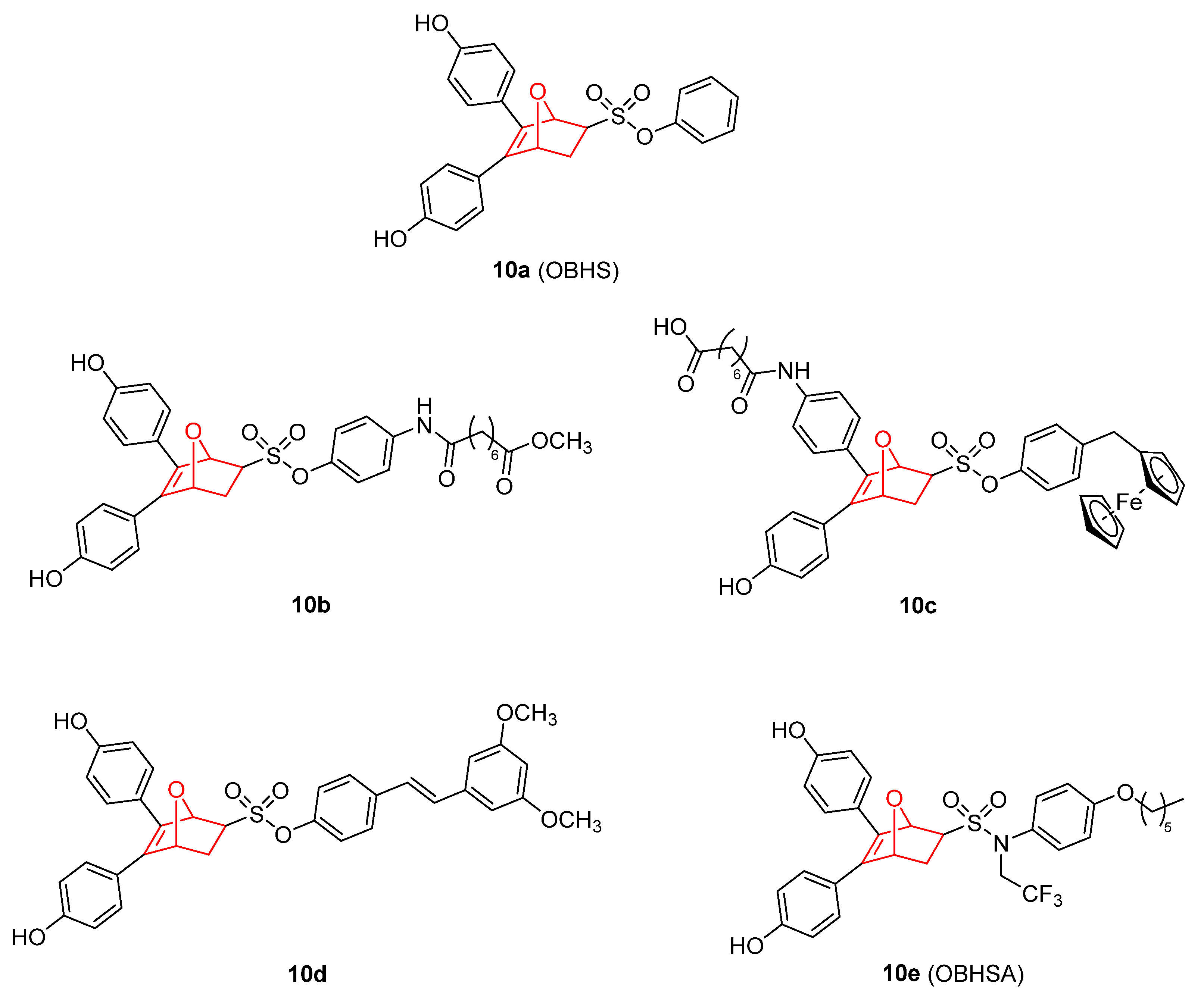
  132. Zhou, H.-B.; Comninos, J.S.; Stossi, F.; Katzenellenbogen, B.S.; Katzenellenbogen, J.A. Synthesis and Evaluation of Estrogen Receptor Ligands with Bridged Oxabicyclic Cores Containing a Diarylethylene Motif: Estrogen Antagonists of Unusual Structure. J. Med. Chem. 2005, 48, 7261–7274. [Google Scholar] [CrossRef]
  133. Zhao, Y.; Gong, P.; Chen, Y.; Nwachukwu, J.C.; Srinivasan, S.; Ko, C.; Bagchi, M.K.; Taylor, R.N.; Korach, K.S.; Nettles, K.W.; et al. Dual Suppression of Estrogenic and Inflammatory Activities for Targeting of Endometriosis. Sci. Transl. Med. 2015, 7, 271ra9. [Google Scholar] [CrossRef] [Green Version]
  134. Wu, J.; Yan, J.; Fang, P.; Zhou, H.; Liang, K.; Huang, J. Three-Dimensional Oxabicycloheptene Sulfonate Targets the Homologous Recombination and Repair Programmes through Estrogen Receptor α Antagonism. Cancer Lett. 2020, 469, 78–88. [Google Scholar] [CrossRef]
  135. Tang, C.; Li, C.; Zhang, S.; Hu, Z.; Wu, J.; Dong, C.; Huang, J.; Zhou, H.-B. Novel Bioactive Hybrid Compound Dual Targeting Estrogen Receptor and Histone Deacetylase for the Treatment of Breast Cancer. J. Med. Chem. 2015, 58, 4550–4572. [Google Scholar] [CrossRef] [PubMed]
  136. Li, C.; Tang, C.; Hu, Z.; Zhao, C.; Li, C.; Zhang, S.; Dong, C.; Zhou, H.-B.; Huang, J. Synthesis and Structure–Activity Relationships of Novel Hybrid Ferrocenyl Compounds Based on a Bicyclic Core Skeleton for Breast Cancer Therapy. Bioorg. Med. Chem. 2016, 24, 3062–3074. [Google Scholar] [CrossRef] [PubMed]
  137. Ning, W.; Hu, Z.; Tang, C.; Yang, L.; Zhang, S.; Dong, C.; Huang, J.; Zhou, H.-B. Novel Hybrid Conjugates with Dual Suppression of Estrogenic and Inflammatory Activities Display Significantly Improved Potency against Breast Cancer. J. Med. Chem. 2018, 61, 8155–8173. [Google Scholar] [CrossRef]
  138. Hu, Z.; Li, Y.; Xie, B.; Ning, W.; Xiao, Y.; Huang, Y.; Zhao, C.; Huang, J.; Dong, C.; Zhou, H.-B. Novel Class of 7-Oxabicyclo[2.2.1]Heptene Sulfonamides with Long Alkyl Chains Displaying Improved Estrogen Receptor α Degradation Activity. Eur. J. Med. Chem. 2019, 182, 111605. [Google Scholar] [CrossRef] [PubMed]
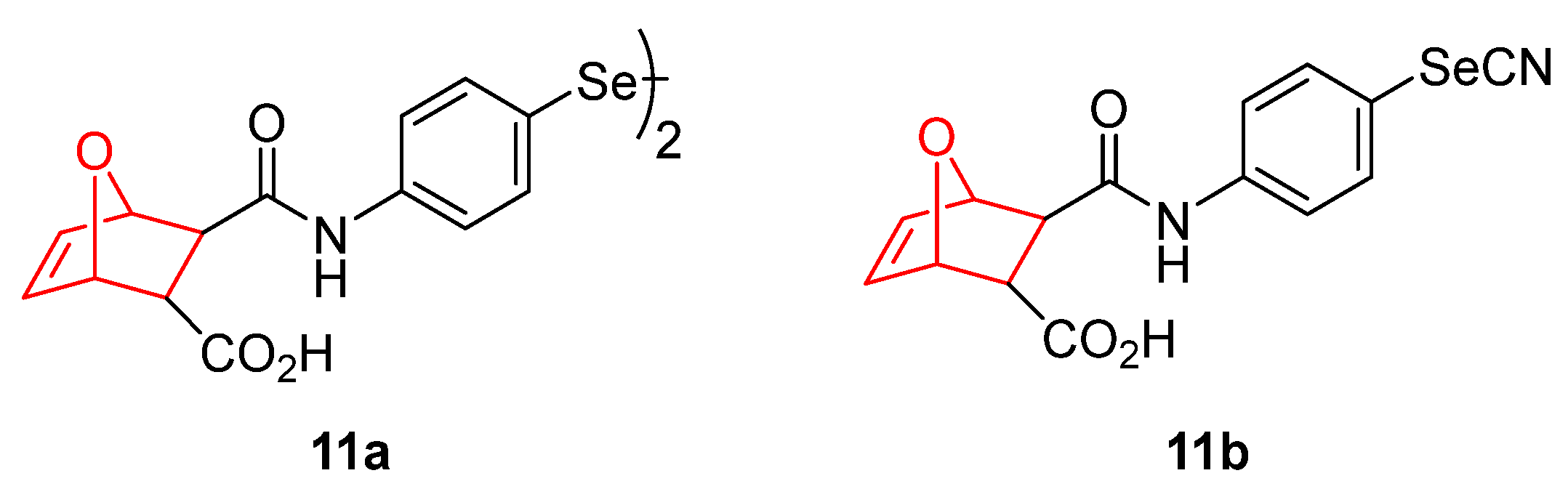
  139. Bartolini, D.; Sancineto, L.; Fabro de Bem, A.; Tew, K.D.; Santi, C.; Radi, R.; Toquato, P.; Galli, F. Selenocompounds in Cancer Therapy: An Overview. Adv. Cancer Res. 2017, 136, 259–302. [Google Scholar] [PubMed]
  140. Ali, W.; Álvarez-Pérez, M.; Marć, M.A.; Salardón-Jiménez, N.; Handzlik, J.; Domínguez-Álvarez, E. The Anticancer and Chemopreventive Activity of Selenocyanate-Containing Compounds. Curr. Pharmacol. Rep. 2018, 4, 468–481. [Google Scholar] [CrossRef]
  141. Gandin, V.; Khalkar, P.; Braude, J.; Fernandes, A.P. Organic Selenium Compounds as Potential Chemotherapeutic Agents for Improved Cancer Treatment. Free Radic. Biol. Med. 2018, 127, 80–97. [Google Scholar] [CrossRef]
  142. Garnica, P.; Encío, I.; Plano, D.; Palop, J.A.; Sanmartín, C. Organoseleno Cytostatic Derivatives: Autophagic Cell Death with AMPK and JNK Activation. Eur. J. Med. Chem. 2019, 175, 234–246. [Google Scholar] [CrossRef]
  143. Moradi Kashkooli, F.; Soltani, M.; Souri, M. Controlled Anti-Cancer Drug Release through Advanced Nano-Drug Delivery Systems: Static and Dynamic Targeting Strategies. J. Control. Release 2020, 327, 316–349. [Google Scholar] [CrossRef]
  144. Kulkarni, M.; Sawant, N.; Kolapkar, A.; Huprikar, A.; Desai, N. Borneol: A Promising Monoterpenoid in Enhancing Drug Delivery Across Various Physiological Barriers. AAPS PharmSciTech 2021, 22, 145. [Google Scholar] [CrossRef]
  145. Guo, X.; Wu, G.; Wang, H.; Chen, L. Pep-1&borneol–Bifunctionalized Carmustine-Loaded Micelles Enhance Anti-Glioma Efficacy Through Tumor-Targeting and BBB-Penetrating. J. Pharm. Sci. 2019, 108, 1726–1735. [Google Scholar] [CrossRef] [PubMed]
  146. Meng, L.; Chu, X.; Xing, H.; Liu, X.; Xin, X.; Chen, L.; Jin, M.; Guan, Y.; Huang, W.; Gao, Z. Improving Glioblastoma Therapeutic Outcomes via Doxorubicin-Loaded Nanomicelles Modified with Borneol. Int. J. Pharm. 2019, 567, 118485. [Google Scholar] [CrossRef] [PubMed]
  147. Li, X.-Y.; Guan, Q.-X.; Shang, Y.-Z.; Wang, Y.-H.; Lv, S.-W.; Yang, Z.-X.; Wang, R.; Feng, Y.-F.; Li, W.-N.; Li, Y.-J. Metal-Organic Framework IRMOFs Coated with a Temperature-Sensitive Gel Delivering Norcantharidin to Treat Liver Cancer. World J. Gastroenterol. 2021, 27, 4208–4220. [Google Scholar] [CrossRef] [PubMed]
  148. Chi, J.; Jiang, Z.; Qiao, J.; Peng, Y.; Liu, W.; Han, B. Synthesis and Anti-Metastasis Activities of Norcantharidin-Conjugated Carboxymethyl Chitosan as a Novel Drug Delivery System. Carbohydr. Polym. 2019, 214, 80–89. [Google Scholar] [CrossRef] [PubMed]
  149. Xue, B.; Lei, M.; Shi, K.; Wang, M.; Hao, Y.; Xiao, Y.; Yuan, L.; Peng, J.; Qian, Z. Intratumoral Injection of Norcantharidin-Loaded Poly(D,L-Lactide)-b-Poly(Ethylene Glycol)-b-Poly(D,L-Lactide) Thermosensitive Hydrogel for the Treatment of Primary Hepatocellular Carcinoma. J. Biomed. Nanotechnol. 2019, 15, 2025–2044. [Google Scholar] [CrossRef] [PubMed]
  150. Wang, J.; Huang, X.; Li, H.; Yan, D.; Huang, W. Two Zn(II) Coordination Polymers with Anticancer Drug Norcantharidin as Ligands for Cancer Chemotherapy. Dalt. Trans. 2022, 51, 5624–5634. [Google Scholar] [CrossRef] [PubMed]
  151. Liu, M.; Tu, J.; Feng, Y.; Zhang, J.; Wu, J. Synergistic Co-Delivery of Diacid Metabolite of Norcantharidin and ABT-737 Based on Folate-Modified Lipid Bilayer-Coated Mesoporous Silica Nanoparticle against Hepatic Carcinoma. J. Nanobiotechnology 2020, 18, 114. [Google Scholar] [CrossRef]
  152. Zhou, L.; Zou, M.; Zhu, K.; Ning, S.; Xia, X. Development of 11-DGA-3-O-Gal-Modified Cantharidin Liposomes for Treatment of Hepatocellular Carcinoma. Molecules 2019, 24, 3080. [Google Scholar] [CrossRef] [Green Version]
  153. Yang, X.; Tong, J.; Guo, L.; Qian, Z.; Chen, Q.; Qi, R.; Qiu, Y. Bundling Potent Natural Toxin Cantharidin within Platinum (IV) Prodrugs for Liposome Drug Delivery and Effective Malignant Neuroblastoma Treatment. Nanomed. Nanotechnol. Biol. Med. 2017, 13, 287–296. [Google Scholar] [CrossRef]
  154. Zhang, X.; Lin, C.; Chan, W.; Liu, K.; Lu, A.; Lin, G.; Hu, R.; Shi, H.; Zhang, H.; Yang, Z. Dual-Functional Liposomes with Carbonic Anhydrase IX Antibody and BR2 Peptide Modification Effectively Improve Intracellular Delivery of Cantharidin to Treat Orthotopic Hepatocellular Carcinoma Mice. Molecules 2019, 24, 3332. [Google Scholar] [CrossRef] [Green Version]
  155. Sun, S.; Shang, E.; Ju, A.; Li, Y.; Wu, Q.; Li, Q.; Yang, Y.; Guo, Y.; Yang, D.; Lv, S. Tumor-Targeted Hyaluronic Acid-MPEG Modified Nanostructured Lipid Carriers for Cantharidin Delivery: An in Vivo and in Vitro Study. Fitoterapia 2021, 155, 105033. [Google Scholar] [CrossRef] [PubMed]
  156. Braga, C.B.; Pilli, R.A.; Ornelas, C.; Weck, M. Near-Infrared Fluorescent Micelles from Poly(Norbornene) Brush Triblock Copolymers for Nanotheranostics. Biomacromolecules 2021, 22, 5290–5306. [Google Scholar] [CrossRef] [PubMed]
  157. Patra, D.; Mukherjee, S.; Chakraborty, I.; Dash, T.K.; Senapati, S.; Bhattacharyya, R.; Shunmugam, R. Iron(III) Coordinated Polymeric Nanomaterial: A Next-Generation Theranostic Agent for High-Resolution T 1 -Weighted Magnetic Resonance Imaging and Anticancer Drug Delivery. ACS Biomater. Sci. Eng. 2018, 4, 1738–1749. [Google Scholar] [CrossRef] [PubMed]
Figure 1. (A) Main synthetic routes for norbornene derivatives (EWG, electron-withdrawing group). (B) Norbornene and several other derivatives with antitumoral properties [19,20,21,22,23,24,25,26,27].
Pharmaceuticals 15 01465 g001
Figure 2. Androgen receptor (AR) antagonist with antitumor activity based on bicyclic imides containing the norbornene structure (compounds 2a–2f) [29,30,31] and bicyclic sultams (compounds 2g–h) [32].
Pharmaceuticals 15 01465 g002
Figure 3. Wnt response inhibitors containing norbornene [34,35,36,37,38] (compounds 3a, 3b). Carbonic anhydrase (CA) inhibitors containing norbornene scaffolds [39,40,41] (compounds 3c–e).
Pharmaceuticals 15 01465 g003
Figure 4. Different norbornene-containing molecules with antitumor effects [42,43,44,45,46,47,48].
Pharmaceuticals 15 01465 g004
Figure 5. Anaplastic lymphoma kinase inhibitors containing norbornene (compounds 5a–b) [49,50,51]. Podophyllotoxin–norbornene derivative (compound 5c) [52,53].
Pharmaceuticals 15 01465 g005
Figure 6. Camphor [54,55,56,57] and several other derivatives with antitumoral activity [58,59,60,61,62,63,64].
Pharmaceuticals 15 01465 g006
Figure 8. Metal complexes containing a norbornene scaffold and its bicyclo[2.2.2]octane analogue [81,82,83,84,85,86].
Pharmaceuticals 15 01465 g008
Figure 9. Norcantharidin [88,89,90,91,92,93,94,95,96,97,98,99,100,101,102,103,104,105,106,107,108,109,110,111,112,113,114], cantharidin [119,120,121,122,123,124,125,126,127,128,129,130,131], and related molecules.
Pharmaceuticals 15 01465 g009
Figure 10. OBHS derivatives with estrogen receptor agonist activity [133,134,135,136,137,138].
Pharmaceuticals 15 01465 g010
Figure 11. Selenium containing oxabicyclic derivatives [142].
Pharmaceuticals 15 01465 g011
Table 1. Binding (Ki) and IC50 values for different cancer cell lines produced by androgen receptor (AR) antagonist containing norbornene [29,30,32].
MDA-MB-453
Ki, nM
MDA-MB-453
IC50, nM
LNCaP
Ki, nM
LNCaP
IC50, nM
PCa2b
IC50, nM
Bicalutamide6417335400725
2a158307641180
2b1>50.5Ag *Ag *
2c5122Ag *Ag *
2g230NDNDND
* Ag (agonist): a compound with the ability to activate AR in the absence of dihydrotestosterone (DHT).
Table 2. IC50 of different platinum containing bicycles [82,83].
IC50 (µM)
HCT-116HepG2A549
8c4.45 ± 0.33 [83]10.74 ± 1.05 [83]7.72 ± 0.64 [83]
8d9.69 ± 0.91 [83]7.86 ± 0.61 [83]46.43 ± 3.86 [83]
8e9.14 ± 0.27 [82]16.70 ± 0.48 [82]57.82 ± 2.17 [82]
Cisplatin6.50 ± 0.473.65 ± 0.313.86 ± 0.28
Carboplatin44.53 ± 4.6439.08 ± 2.9555.20 ± 4.76
Oxaliplatin3.28 ± 0.3412.80 ± 1.288.95 ± 0.78
Publisher’s Note: MDPI stays neutral with regard to jurisdictional claims in published maps and institutional affiliations.

