Article

In Vitro and In Vivo Gastrointestinal Survival of Non-Encapsulated and Microencapsulated Salmonella Bacteriophages: Implications for Bacteriophage Therapy in Poultry

by
Laura Lorenzo-Rebenaque
1,
Danish J. Malik
2,
Pablo Catalá-Gregori
1,3,
Clara Marin
1,* and
Sandra Sevilla-Navarro
1,3
1
Departamento de Producción y Sanidad Animal, Salud Pública Veterinaria y Ciencia y Tecnología de los Alimentos, Instituto de Ciencias Biomédicas, Facultad de Veterinaria, Universidad Cardenal Herrera-CEU, CEU Universities, Calle Tirant lo Blanc, 7, 46115 Alfara del Patriarca, Spain
2
Chemical Engineering Department, Loughborough University, Loughborough LE11 3TU, UK
3
Centro de Calidad Avícola y Alimentacion Animal de la Comunidad Valenciana (CECAV), 12539 Castellón, Spain
*
Author to whom correspondence should be addressed.
Pharmaceuticals 2021, 14(5), 434; https://doi.org/10.3390/ph14050434
Submission received: 30 March 2021 / Revised: 24 April 2021 / Accepted: 30 April 2021 / Published: 6 May 2021
(This article belongs to the Special Issue Bacteriophages as Therapeutic Delivery Vehicles)

Abstract

:
The therapeutic use of bacteriophages is recognized as a viable method to control Salmonella. Microencapsulation of phages in oral dosage forms may protect phages from inherent challenges of the gastrointestinal tract in chickens. Therefore, the main objective of this study was to assess the survival of Salmonella BP FGS011 (non-encapsulated and microencapsulated) through the gastrointestinal tract under in vitro as well as in vivo conditions after oral administration to 1-day-old chicks. To this end, the phage FGS011 was encapsulated in two different pH-responsive formulations with polymers Eudragit® L100, and Eudragit® S100 using the process of spray drying. Phages encapsulated in either of the two formulations were able to survive exposure to the proventriculus-gizzard in vitro conditions whereas free phages did not. Moreover, phages formulated in polymer Eudragit® S100 would be better suited to deliver phage to the caeca in chickens. In the in vivo assay, no statistically significant differences were observed in the phage concentrations across the gastrointestinal tract for either the free phage or the encapsulated phage given to chicks. This suggested that the pH of the proventriculus/gizzard in young chicks is not sufficiently acidic to cause differential phage titre reductions, thereby allowing free phage survival in vivo.

1. Introduction

Non-typhoidal Salmonella spp. has been recognized as one of the most important zoonotic pathogens worldwide [1,2]. Latest data reported by the World Health Organization estimated 78 million annual cases of foodborne illness worldwide, of which 59,000 resulted in death [1]. By the same token, in the European Union (EU), the latest data published in 2019, revealed more than 90,000 cases of human salmonellosis [3]. Salmonella sources in human infection are relatively diverse, however poultry is considered a major reservoir [3,4,5,6]. Due to this situation, since 2008, National Salmonella Control Programs (NSCP) in accordance with Regulation (EC) No 2160/2003 have been implemented in the EU [7,8]. These measures have resulted in significant Salmonella prevalence reduction in poultry flocks throughout Europe [9]. However, some Salmonella serovars related to food-borne outbreaks are still present in some poultry farms, due to their ability to survive and grow in the gastrointestinal tract (GIT) of chickens and/or farm environment [4,10,11,12]. When Salmonella comes into contact with the bird and reaches the GIT, its ability to rapidly colonize and multiply results in long-term bacterial excretion in faeces especially in 1-day-old chicks [13,14,15,16]. Salmonella, almost colonizes every part of the chicken GIT, nevertheless, the pH of the GIT could affect the bacteria colonization [17]. Salmonella encounters the acidic environment of the crop, with a pH of 5, that is maintained and controlled by the bacteria of the genus Lactobacillus [17]. Subsequently, Salmonella survives the low pH of the proventriculus and gizzard (pH 2.5), and colonize the gut (pH 5.5–8), with highest predilection potential noticed in the ceca [18,19,20]. Thus, innovative and cost-effective techniques are needed suitable for deployment under field conditions to control the bacteria in the poultry sector [15,21,22].
In this sense, the use of bacteriophages (BP) has garnered increasing interest as a possible method to achieve Salmonella control in poultry farms in recent years, due to its high degree of specificity against the bacteria [22,23,24,25,26]. Among the different routes of BP therapy administration, the oral route is likely to be the most applicable in humans and animals [27,28,29]. The lack of BP stability upon exposure to acidic conditions (e.g., in the proventriculus/gizzard) and the relatively short residence times in the intestinal tract may limit the efficacy of orally delivered BP [30]. These limitations can however be overcome, such as through the co-administration of antacids [30,31] or through encapsulation of the BP [32,33,34]. Encapsulation provides protection from gastric acidity and could release high doses of the BP at the ceca, the predilected Salmonella site, incorporating approaches for burst release and/or sustained release; improving the efficacy of the orally administered BP [34,35,36]. Nevertheless, there is a need for better understanding of the stability and viability of the non-encapsulated and microencapsulated BP after being orally administered [37,38,39]. Therefore, the main objective of this study was to assess the release of the encapsulated BPs and the survival of Salmonella BP FGS011 (non-encapsulated and microencapsulated) through the chicken’s GIT under in vitro and in vivo conditions after oral administration to 1-day-old chicks.

2. Results

2.1. BP Characterization Assay

2.1.1. BP Lytic Spectrum

The lytic host range against a panel of 13 strains indicated that BP FGS011 was able to lyse seven strains of Salmonella and one Citrobacter strain (Table 1).

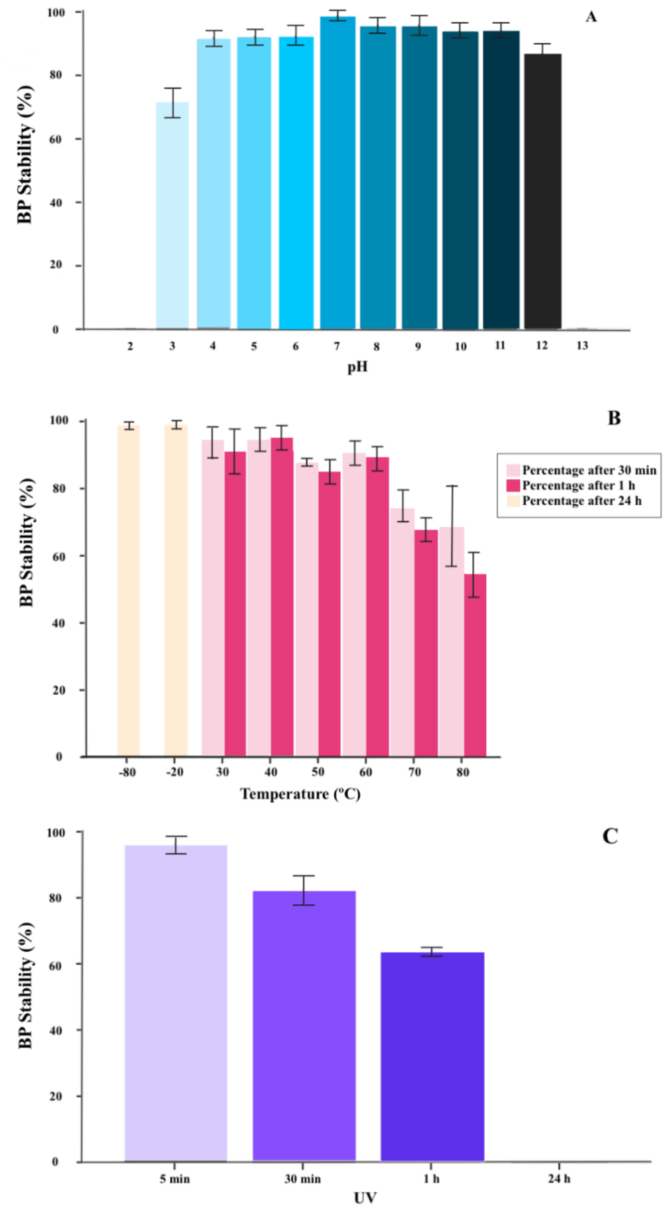
2.1.2. BP Thermal and pH Stability

pH stability tests showed that FGS011 survived exposure to acidic pH as low as pH 4 and alkalinity as high as pH 11 after 2 h of incubation (Figure 1A). However, FGS011 titers declined when exposed to acidic pH less than pH 3 or alkaline pH greater than 12. The BP was rapidly inactivated at low pH 2 and high pH 13 (Figure 1A). Thermal stability tests indicated that BP FGS011 was relatively heat stable for up to 60 min at temperatures between 30 to 60 °C. However, the viable BP concentration decreased upon exposure to temperatures between 70 to 80 °C (Figure 1B). BP FGS011 also survived upon undergoing a freeze-thaw cycle at −80 °C.

2.1.3. BP Inactivation with UV Radiation

The viability of BP FGS011 was maintained for up to 1 h of exposure to UV irradiation, nevertheless, no viable BP was measurable after 24 h of exposure (Figure 1C).

2.2. In Vitro Evaluation of the Release of BP under Different GIT Conditions (Experiment 1)

Protection of encapsulation from GIT pH stress and BP release under different GIT pH conditions was evaluated for FGS011 encapsulated in formulations L100 and S100 simulating in vitro GIT conditions. All results represented in these experiments are with regard to the starting concentration of BP used in the experiment (1010 PFU). BP encapsulated in L100 was released in the crop due to the pH 5.5 (p < 0.05) (Figure 2A). However, BPs encapsulated in S100 did not release in the crop (pH 5.5), proventriculus-gizzard (pH 2.5), and the duodenum (pH 5) (Figure 2B). BP release was noted in the jejunum (pH 6.5), ileum (pH 7) and any residual encapsulated BP in the caeca (pH 8), indicating a statistically significant decrease along the lower GIT (p < 0.05) (Figure 2).

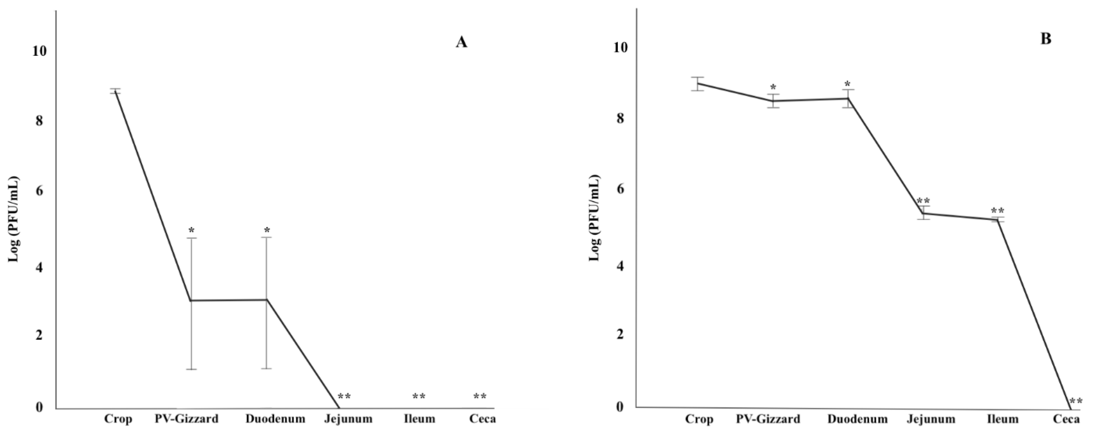
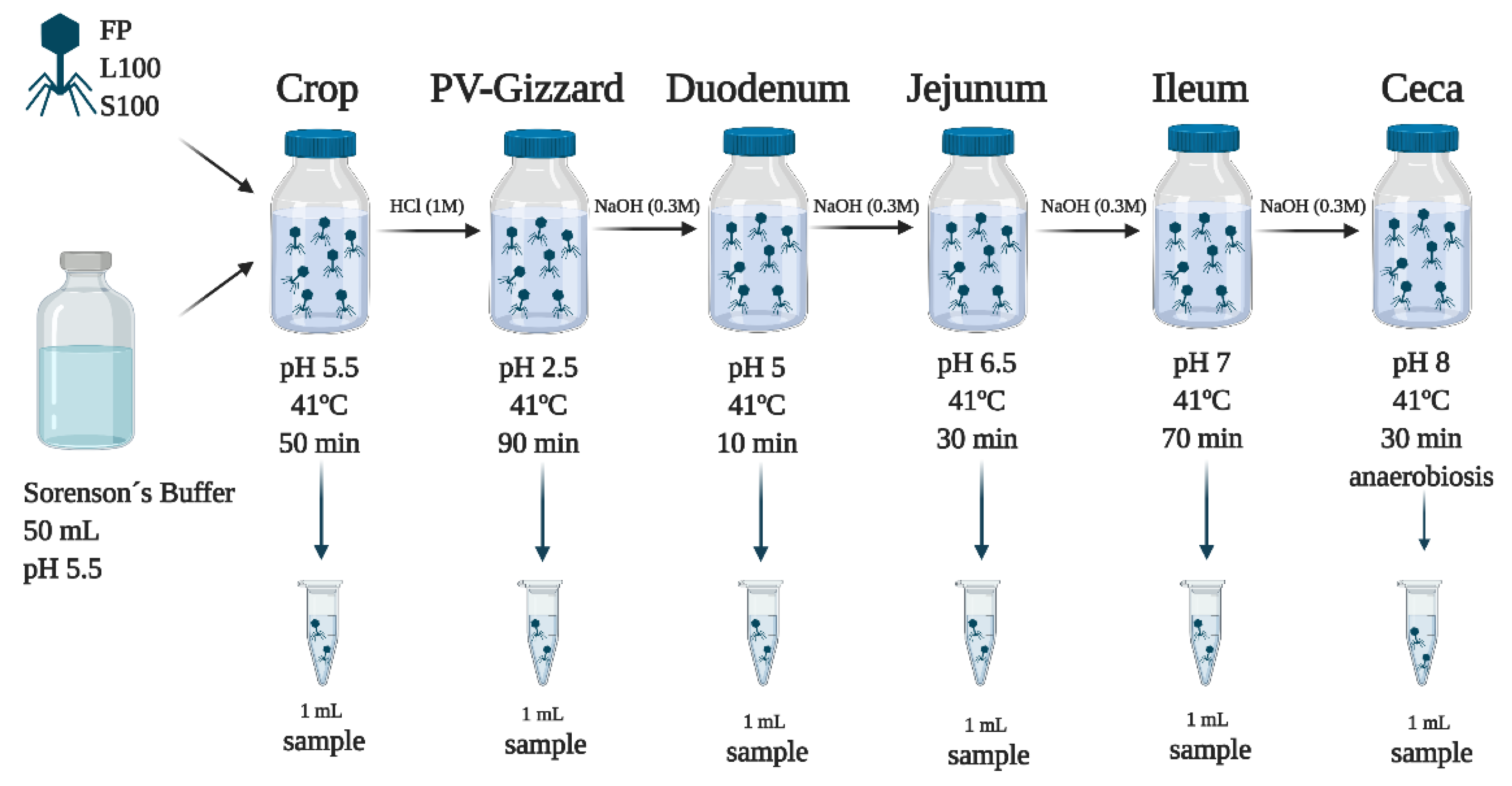
2.3. In Vitro Evaluation of BP Titres along the GIT (Experiment 2)

The survival of the BP (FP, L100 and S100) across the GIT was evaluated in vitro by passing the BP sequentially through each simulated GIT section (Figure 3). All results represented in these experiments are with regard to the starting concentration of BP used in the experiment (1010 PFU). The results showed that FP were inactivated upon exposure to proventriculus-gizzard conditions, and viable BP were not detected again in the following stages downstream of the proventriculus-gizzard. For L100, significant differences were observed along the simulated GIT sections (p < 0.05). BP released from L100 were detected in the crop, but no BP were detected in the neutralized supernatant of the simulated proventriculus-gizzard section. Thereafter, BP detection in the supernatant increased in the duodenum and BP titres were maintained in the jejunum through to the caeca (p > 0.05). For S100, the highest concentration of BP was detected in the crop (p < 0.05). No viable BP was detected in the proventriculus-gizzard. The concentration of BP in the supernatant increased in the duodenum and jejunum and was highest in the ileum with a 0.5 log decrease observed in the ceca (p < 0.05).
Moreover, statistically significant differences were observed among FP, L100, and S100 across the different controlled GIT conditions (pH, transit-time, and temperature) (Figure 3).

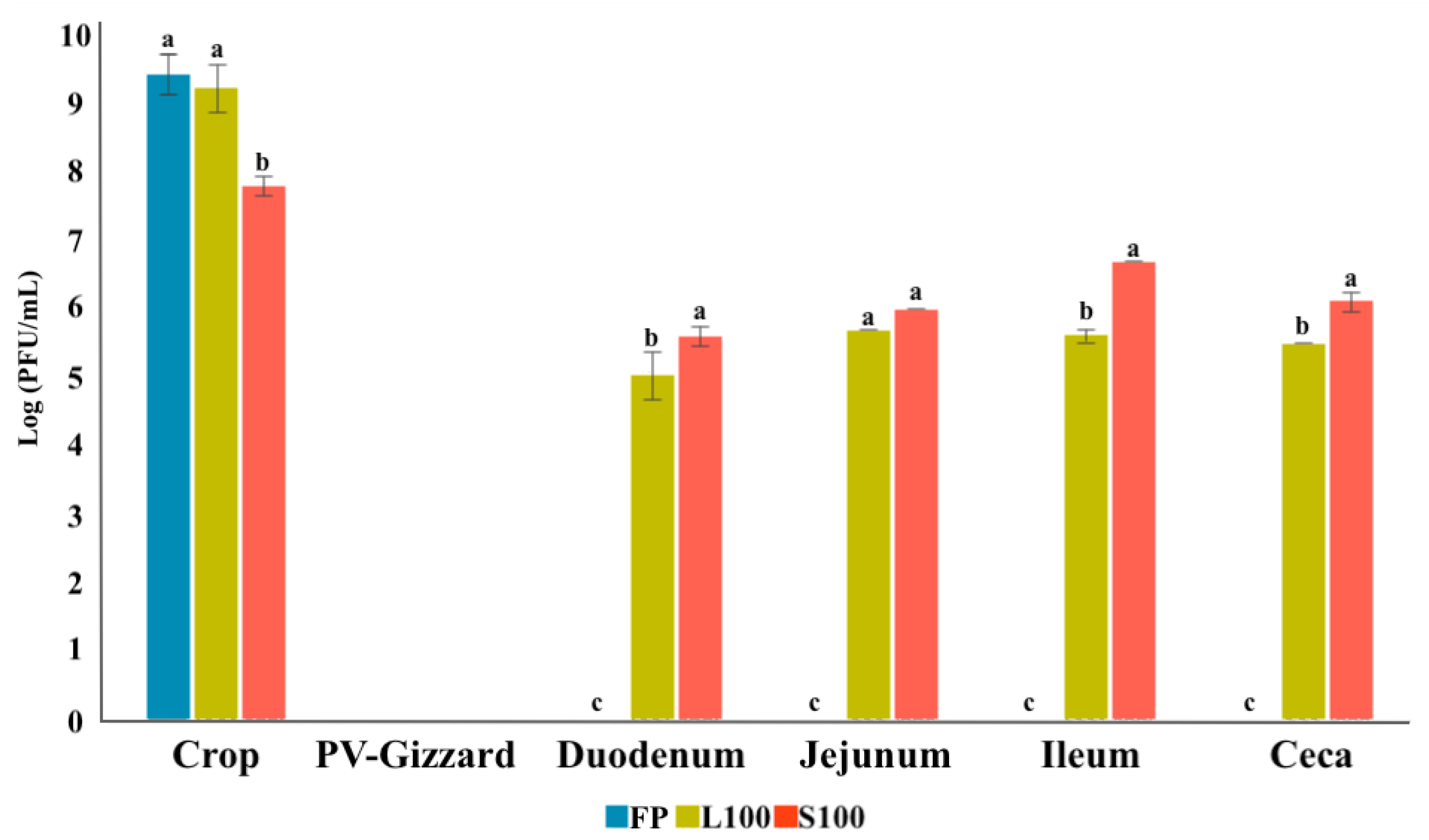
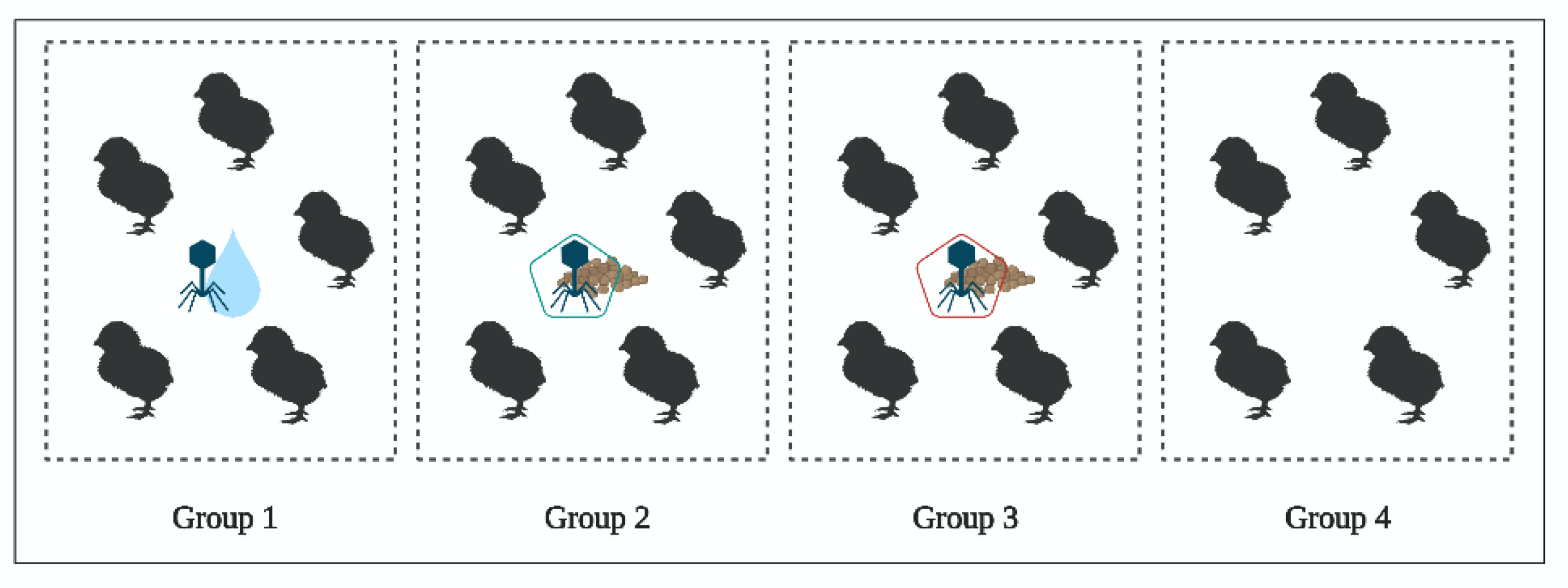
2.4. In Vivo Study of BP Survival along GIT (Experiment 3)

The distribution of FP, L100 and S100 within the GIT of 1-day-old chicks administered BP either in drinking water or in feed was determined after 24 h in the absence of Salmonella host. After 24 h, all the BP were present in all of the GIT sections (crop, proventriculus, gizzard, gut and ceca) of the treated groups. The control group was negative for the presence of BP in any of the samples analyzed.
From each free/released FGS011 (FP, L100 and S100), no statistically significant differences of BP concentration were observed through the different GIT sections (p > 0.05). For the different GIT sections, BP counts are summarized in Table 2.
On the other hand, statistically significant differences were shown between BP in the gut, with L100 being the BP to show the highest concentration (p < 0.05). However, no statistically significant differences were observed in the crop, proventriculus, gizzard, and ceca (p > 0.05) (Table 2).
For faecal samples, statistical differences were found in BP counts among the different groups (p < 0.05). The highest BP counts were in faeces of chicks fed L100 (6.4 Log10 PFU/g), followed by S100 (6.1 Log10 PFU/g) and finally FP (5.8 Log10 PFU/g) (p = 0.000).

3. Discussion

The survival of Salmonella BP FGS011 (non-encapsulated and microencapsulated) through the GIT under in vitro and in vivo conditions after oral administration to 1-day-old chicks was assessed. This is the first study to report the dynamics of the Salmonella BP encapsulated with the anionic polymers Eudragit® L100 and Eudragit® S100 through the chicken GIT.
Despite the NSCP, Salmonella continues to be a threat pathogen to the poultry sector and a potential risk from farm to fork [40]. Therefore, prevention and control are required throughout the poultry chain [12]. The negative economic impact of Salmonella for the poultry sector, the effects on human health, increasing antimicrobial resistance and the absence of effective alternatives for controlling Salmonella are drivers for exploration of the potential of BP therapy as a more effective biocontrol tool, reducing economic loses in the poultry industry and reducing the risk of food-borne diseases [15]. Oral BP therapy has been used as a tool for Salmonella control in the poultry industry [41,42,43]. Nevertheless, inherent challenges, such as the GIT environmental conditions, have led to divergent BP therapy results [32,37]. Although is this study both BP forms reached the ceca, previous studies reported a reduction in BP titres in vivo and suggested that this may be due to BP inactivation attributed to the acidic environment of the chicken’s gizzard. This may especially be the case for sensitive BP that do not survive low acidic pH exposure [32,37]. Previous studies have therefore highlighted the need for encapsulated BP that allow high titres of BP to be delivered to the cecum which is the most likely site of Salmonella colonization [37,44]. The encapsulation of BP and their controlled release may help in ensuring that the in-situ BP concentration remains at a therapeutically effective level, thereby allowing BP to amplify once the bacteria concentration increases to levels sufficient for in situ BP amplification [32]. The in vitro results of this study demonstrated that FP are likely to be inactivated upon exposure to proventriculus-gizzard pH (Figure 3). Whereas encapsulation of BPs significantly improved BP survival with S100 remaining encapsulated until the end of gut (Figure 2 and Figure 3). Encapsulation would therefore ensure that high BP concentration would be delivered to the caeca (Figure 3). Therapeutically effective levels reaching the intended Salmonella infection site, such as the lower intestine including caeca may improve BP therapy outcomes [32,38]. The current results indeed demonstrated that BP, both non-encapsulated and microencapsulated, appeared in the faeces, showing its high capacity of dissemination in the environment [22]. This matter will allow not only the control of the bacteria in the environment but also will limit the reinfection of the animals [22]. The reduction of faecal Salmonella shedding controlled by the NSCP, will entail an important economic reduction for the poultry sector [7,22].
Several complex external factors could influence the treatment success in vivo, such as the rapid clearance of the BP by active or passive host immunity, interactions of BP with the intestinal mucosa and with other gut microbiota [45,46,47]. The in vivo results obtained after BP administration in 1-day-old chicks demonstrated that encapsulated and non-encapsulated BP could survive through the GIT and were excreted in the faeces (Table 2). This fact may be explained due to the higher pH of the gizzard in the young animals (4.3 pH), which would allow not only the survival of the encapsulated BP, but also the survival of the FP (unpublished data) [48]. These results are in agreement with those obtained by Ma et al. (2016), who also observed that although FP did not survive gastric fluid conditions in vitro, in 1-day-old chicks BP were able to survive throughout the GIT, without no notable differences between free BP titres compared with the encapsulated form. Other authors have highlighted the possibility that for mature chicken GIT where the pH is lower, FP could not survive the gastric passage, and were not found to effectively reduce Salmonella numbers in the chicken intestine [49,50], compromising BP therapy outcomes in older animals [30,48].
Regarding the survival of BP formulated in different encapsulated forms administered to 1-day-old chicks, slightly higher concentrations of BP encapsulated in L100 were found to be delivered to the intestine compared with the S100 formulation (5.9 Log10 and 4.6 Log10 PFU, respectively). This may be due to lower amounts of BP released in the caeca of 1-day-old chicks from S100, which require higher pH levels to release BP [51,52]. BP therapy has previously been used to control Salmonella in poultry farms [22,23,24,25,26]. There is, however, a lack of available data at the field level [36]. Further studies are therefore needed to study the BP dynamics in the GIT throughout the six-week rearing period of broilers to assess the best way to incorporate phage in animal feed as well as the best intervention moment to prevent the spread of Salmonella in chicken farms.

4. Materials and Methods

All the animals were handled according to the principles of animal care published by Spanish Royal Decree 53/2013 [53].

4.1. BP Characterization Assay

BP FGS011 used in this study was isolated by Sevilla-Navarro et al. [40]. It was isolated from faeces samples from commercial poultry farms (broilers and layers) in Eastern Spain in the Poultry Quality and Animal Nutrition Centre of the Valencia Region (CECAV). The BP was stored at 4 °C until use. This BP was selected for this study due to its high lytic activity against its propagating strain and the wide lytic host range against Salmonella strains isolated from poultry farms. The bacterial strain used for BP isolation and amplification was Salmonella Senftenberg (S. Senftenberg), a field strain isolated from poultry farms during the NSCP [54] and selected from the collection repository of CECAV.

4.1.1. BP Lytic Spectrum

In order to assess the lytic spectrum of the BP, the sensitivity of 13 field and reference bacterial strains including ten Salmonella serovars, one Escherichia coli strain, one Citrobacter freundi strain and one Pseudomonas aeruginosa strain were determined by spot test using the double agar method [40]. Salmonella serovars selected for BP lytic spectrum were those more prevalent in European poultry farms (S. Enteritidis, S. Typhimurium, monophasic S. Typhimurium, S. Infantis, S. Virchow, S. Kentucky, S. Ohio, S. Senftemberg, S. Indiana and S. Havana). Then, 200 µL of a log-phase culture of the bacterial suspensions in LB (Luria Bertani, VWR Chemicals, Barcelona, Spain) at an optical density (OD) 600 nm of 0.2 (~108 CFU/mL) was added to 5 mL of LB agar (LB with 0.6% agar) tempered to 45 °C and poured onto previously prepared and dried LB basal agar (with 1.6% agar). The plates were dried in a laminar flow hood for 15 min. Subsequently, 10 µL of the BP FGS011 (1010 PFU/mL) suspension was spotted onto the surface of the double layer agar. The resulting plates were incubated overnight at 37 °C, and subsequently checked for the BP plaque formation on the bacterial lawns [40].

4.1.2. BP Thermal and pH Stability

For thermal-stability testing, tubes with 100 μL of FGS011 (1010 PFU/mL) were mixed with 900 μL pre-heated sterile LB broth and were kept in a water bath ranging from 30 to 80 °C for 30 min and 60 min [41]. In addition, BP stability at −20 °C in a standard refrigerator, and at −80 °C in an ultra-low temperature freezer, was also evaluated for 24 h [55]. For pH stability testing, 100 μL of FGS011 (1010 PFU/mL) aliquots were mixed in a series of tubes containing 900 μL of sterile BPW (Buffered Peptone Water, VWR Chemicals, Barcelona, Spain) with varying pH values ranging from pH 2 to pH 13 (adjusted using NaOH or HCl) and incubated for 2 h at 37 °C [41,56]. After the incubation, ten-fold serial dilutions of the samples were carried out with LB and were plated by the spot test method and incubated 24 h at 37 °C. BP titration was performed per triplicate. The rates of BP pH/thermal stability were determined calculated with the formula: BP stability rate (%) = BP concentration (PFU/mL) under certain condition/initial BP concentration added (PFU/mL) × 100% [56]. These experiments were performed three times.

4.1.3. BP Inactivation with UV Radiation

To quantify BP inactivation by UV radiation, 1 mL of FGS011 (1010 PFU/mL) was exposed to UV irradiation in glass petri dishes at room temperature for 24 h [57]. A 15-watt, low-pressure mercury germicidal lamp (U.V ESTERIL, J.P. SELECTA s.a.) with a sharp emission maximum at 230 nm was used as the UV source for irradiation of the BP. Samples were taken at different time intervals, at 5, 30, 60 min, and 24 h. Then, ten-fold serial dilutions of the samples were plated by the spot test method described above and incubated 24 h at 37 °C. BP titration was performed per triplicate. The rate of BP UV radiation inactivation was calculated with the stability rate (%) formula described above. This experiment was performed three times.

4.1.4. BP Encapsulation

Encapsulation was performed according to Malik et al. [33]. For this study, two anionic polymers Eudragit® L100, and Eudragit® S100 were used for the BP encapsulation. Eudragit® L100 dissolves at pH 6 and greater, while Eudragit® Sl00 is less soluble than Eudragit® Ll00 and dissolves at a pH of 7 or greater [58]. During this study, the BP FGS011 was evaluated without encapsulation as free BP (FP), and encapsulated with the polymers Eudragit® L100 (L100) and Eudragit® S100 (S100).
Eudragit L100 and S100 were kindly supplied by Evonik Germany. d-(+)-Trehalose dihydrate was purchased from Fisher Scientific (Loughborough, UK). Solutions containing different excipient (Eudragit S100, L100 with added trehalose) amounts were dissolved in 500 mL of deionised distilled water (dH2O). The ratio of S100 and L100 to trehalose was 2:1 and total solids content 12 g per 100 mL of solution.
In order to dissolve Eudragit, the pH of the water was changed to alkaline (pH 12) via addition of 4 M NaOH (Fisher Scientific, Hampshire, UK) to allow polymer dissolution, followed by pH adjustment to pH 7 using 0.1 M HCl prior to addition of trehalose powder, its dissolution, and further addition of bacteriophages to the solution. For each formulation, typically 10% (v/v) high-titre phage (~1010 PFU/mL) was added to the solution, yielding phage titres of ~109 PFU/mL in the final formulations. The phage-containing solutions were spray-dried using a commercially available Labplant spray-dryer SD-06 (Labplant, UK Limited), which is a co-current dryer with a pneumatic atomiser and a cylindrical drying chamber of dimensions 215 mm outer diameter and 420 mm height. The air exit stream was passed through a high-efficiency particulate air (HEPA) filter prior to discharge. The diameter of the atomization nozzle used throughout the work was 0.5 mm with the measured feed liquid flow rate at 280 mL∙h−1 and a drying gas air flow rate of ~20 L∙s−1. The air inlet temperatures were set at 100 °C resulting in corresponding air outlet temperatures of 60 ± 2 °C respectively. The outlet temperature is only indicative of the highest temperature the phages could be exposed to as dry powders in the collection bottle. The temperature in the collection bottle varied between 40 and 60 °C.

4.2. In Vitro Evaluation of the Release of BP under Different GIT Conditions (Experiment 1)

To assess the release of the encapsulated BP (L100 and S100) along the GIT section, an in vitro assay was performed. For this, the amount of BP that remained encapsulated throughout the simulated GIT was analysed. The GIT conditions of the crop, proventriculus-gizzard, duodenum, ileum, jejunum, and ceca were simulated in vitro according to Ravindran [20] (Figure 4).
Firstly, to mimic the crop conditions, 10 mL aliquots of Sorenson’s buffer pH 5.5 was added to six falcon tubes for each of the encapsulated BP (L100 and S100). After that, an initial inoculum of 1010 PFU of each encapsulated BP (L100 and S100) was added into each respective tube. Samples were then incubated, with shaking, at 41 °C for 50 min. At the end of the incubation, all the tubes were centrifuged to sediment the phage containing capsules for 5 min at 8000× g and the supernatant was removed. In one tube for each BP, the capsule pellet was resuspended with 10 mL of Sorenson’s buffer at pH 8, and was vortexed and left for 10 min to allow the capsules to dissolve. After that, 100 µL of aliquot were taken for BP enumeration. From the remaining tubes, the pellets were resuspended in 10 mL of Sorenson’s buffer with adjusted to pH 2.5 with 1M HCl to mimic the proventriculus and gizzard conditions. The samples were incubated for another 90 min. After the incubation period, all tubes were centrifuged, and the procedure was repeated as described above. Hereafter, the process was repeated with 10 mL of Sorenson´s buffer at pH 5 for 10 min, followed by pH 6.5 for 30 min, and pH 7 for 70 min, to mimic the duodenum, ileum, and jejunum conditions, respectively. Finally, ceca conditions were mimicked using 10 mL of Sorenson´s buffer at pH 8 for 30 min under anaerobic conditions obtained with AnaeroGen Gas pack (Oxoid, Hampshire, UK, AN0035A) in anaerobic jars. Ten-fold serial dilutions of all the aliquots were carried out with LB and were plated by the spot test method and incubated for 24 h at 37 °C. BP titration was performed per triplicate.

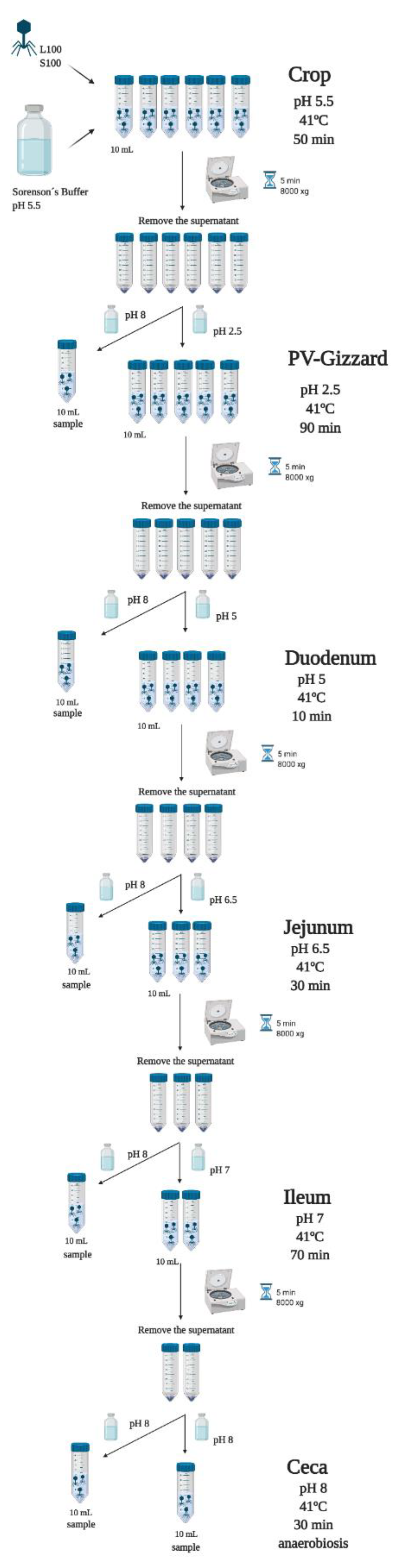
4.3. In Vitro Evaluation of BP Titres along the GIT (Experiment 2)

To assess the survival of the BP (FP, L100 and S100) across the GIT an in vitro assay was performed [37]. For this, the amount of released BP that survived throughout the simulated GIT was analysed. The BP (FP, L100 and S100) were passed sequentially through each simulated GIT section according to Ravindran [20]. To this end, the following stages were carried out (Figure 5).
Firstly, to mimic the crop conditions, 50 mL of Sorenson’s Buffer (pH 5.5) was inoculated with 1010 PFU of FP or 1010 PFU of encapsulated L100 and S100. Samples were then incubated, with shaking, at 41 °C for 50 min. At the end of the incubation period, 1 mL of aliquot was taken for BP enumeration. Then, to mimic the proventriculus and gizzard conditions, HCl (1 M) was added to each tube to adjust solution pH to pH 2.5 and the incubation continued for another 90 min. After this additional incubation period, 1 mL of aliquot was taken for BP enumeration and the procedure was repeated by adjusting the solution pH to pH 5 (using 0.3 M NaOH) for 10 min, followed by pH 6.5 for 30 min, and pH 7 for 70 min, to mimic the duodenum, ileum, and jejunum conditions, respectively. Finally, the ceca conditions were mimicked with pH 8 for 30 min under anaerobic conditions obtained with AnaeroGen Gas pack (Oxoid, Hampshire, UK, AN0035A) in anaerobic jars. At the end of each incubation, 1 mL of aliquot was taken for BP enumeration. The samples were centrifugated for 5 min at 8000 × g and the supernatant was taken. Ten-fold serial dilutions of the supernatants were carried out with LB and were plated by the spot test method and incubated 24 h at 37 °C. BP titration was performed per triplicate.

4.4. In Vivo Study of BP Survival along GIT (Experiment 3)

An in vivo study of BP (FP, L100 and S100) survival along the GIT after oral administration of phages to 1-day-old chicks was carried out. Faecal shedding of BP was also measured. Twenty 1-day-old Salmonella free chicks (Ross 308) were randomly divided into four groups of five birds (Figure 6).
To assess the survival of the BP through the GIT, group 1 received 100 mL of 106 PFU/mL of FP via drinking water, group 2 received 100 g of 106 PFU/g of L100 via feed, group 3 received 100 g of 106 PFU/g of S100 via feed, and group 4 did not receive any BP (control group). Then, 24 h after BP administration, animals from each experimental group (n = 5/group) were taken and the GIT was removed and processed for BP enumeration. The following sections were processed from each animal: crop, proventriculus, gizzard, gut, and ceca. Samples of each sections were collected under sterile conditions. The material used was sterile and it was changed between groups. Between samples, the material was sterilised with alcohol and fire [59].
Moreover, to assess fecal shedding, at least 10 g of faeces were taken from each experimental group. A total of 104 samples were weighed and homogenized in 1:10 mL of LB broth medium, centrifuged for 5 min at 16,000 × g, and the supernatant was filtered through a 0.45 µm membrane. Afterwards, 100 μL of each dilution was transferred to an empty well and ten-fold serial dilutions were performed using sterile dilution buffer (LB). Then, 10 μL of each dilution with 200 μL of the bacterial host suspension was mixed with 5 mL of LB 0.6% top agar layer and placed over a 1.6% LB agar bottom layer. Plates were incubated overnight at 37 °C. BP titration was performed per triplicate.

4.5. Statistical Analysis

Concentrations (PFU/mL) of BP were converted to Log10 (PFU/mL) and then averaged. A general linear model (GLM) was used to evaluate the release of encapsulated phage BP (L100 and S100) along the in vitro GIT conditions. GLM was used to evaluate the survival of the released BP (L100 and S100) and non-encapsulated BP along the in vitro GIT conditions. As a fixed effect, we included the gastrointestinal localization tract simulated (crop, proventriculus-gizzard, duodenum, ileum, jejunum and ceca), the initial BP concentration, and the BP (FP, L100 and S100). GLM was used to compare the activity of the BP (FP, L100 and S100) in vivo, including as a fixed effect, the gastrointestinal localization (crop, proventriculus, gizzard, gut, ceca, and faeces), and the BP (FP, L100, and S100). A p-value of p < 0.05 was considered to indicate a statistically significant difference. All statistical analyses were carried out using SPSS 16.0 software.

5. Conclusions

Significant differences were observed between phage delivery results of in vitro studies compared with in vivo results. In 1-day-old chicks there were no statistically significant differences between phage delivered along the GIT for the encapsulated and non-encapsulated phage (the gut being the exception, but differences were small here too). Encapsulation of the BP using the polymers Eudragit® L100 and Eudragit® S100 resulted in the delivery of phage in 1-day-old chicks with no adverse reactions observed in the animals. Further studies are needed to better understand the dynamics of the encapsulated phage released during transit through the GIT of the chickens during the entire production cycle.

Author Contributions

Conceptualization, D.J.M., P.C.-G., C.M., and S.S.-N.; methodology, D.J.M. and S.S.-N.; formal analysis, L.L.-R., D.J.M., P.C.-G., C.M., and S.S.-N.; investigation, L.L.-R., D.J.M., C.M., and S.S.-N.; resources, L.L.-R., D.J.M., P.C.-G., and S.S.-N.; data curation, L.L.-R., D.J.M., P.C.-G., C.M., and S.S.-N.; writing—original draft preparation, L.L.-R., D.J.M., C.M., and S.S.-N. writing—review and editing, L.L.-R., D.J.M., P.C.-G., C.M., and S.S.-N.; supervision, D.J.M., P.C.-G., C.M., and S.S.-N.; project administration, D.J.M., P.C.-G., and S.S.-N.; funding acquisition, L.L.-R., D.J.M., P.C.-G., and S.S.-N. All authors have read and agreed to the published version of the manuscript.

Funding

L.L.-R. was supported by a research grant from the Generalitat Valenciana-Fondo Social Europeo (ACIF/2020/376). D.J.M. acknowledges funding from BBSRC (Grant No. BB/R012822/1), the development of a phage food additive with the aim to control Salmonella in swine and poultry.

Institutional Review Board Statement

The study was conducted according to the principles of animal care published by Spanish Royal Decree 53/2013.

Informed Consent Statement

Not applicable.

Data Availability Statement

Not applicable.

Acknowledgments

We would like to thank the Centre for Poultry Quality and Animal Feed of the Valencian Community (CECAV) for their financial support and to Montoro-Dasí for the technical support.

Conflicts of Interest

The authors declare no conflict of interest.

References

  1. WHO. WHO Estimates of the Global Burden of Foodborne Diseases; WHO: Geneva, Switzerland, 2015; pp. 1–16. Available online: https://apps.who.int/iris/bitstream/handle/10665/199350/9789241565165_eng.pdf;jsessionid=F5D0B289A1506C32255D8921C9119A5B?sequence=1 (accessed on 25 February 2021).
  2. WHO. 23 Million People Falling Ill from Unsafe Food Each Year in Europe is just the Tip of the Iceberg. Available online: https://www.who.int/news-room/detail/05-06-2019-23-million-people-falling-ill-from-unsafe-food-each-year-in-europe-is-just-the-tip-of-the-iceberg (accessed on 22 June 2020).
  3. EFSA. ECDC The European Union One Health 2018 Zoonoses Report. EFSA J. 2019, 17. [Google Scholar] [CrossRef] [Green Version]
  4. EFSA; Koutsoumanis, K.; Allende, A.; Alvarez-Ordóñez, A.; Bolton, D.; Bover-Cid, S.; Chemaly, M.; De Cesare, A.; Herman, L.; Hilbert, F.; et al. Salmonella control in poultry flocks and its public health impact. EFSA J. 2019, 17. [Google Scholar] [CrossRef]
  5. Andino, A.; Hanning, I. Salmonella enterica: Survival, colonization, and virulence differences among serovars. Sci. World J. 2015, 2015. [Google Scholar] [CrossRef] [Green Version]
  6. Shah, D.H.; Paul, N.C.; Sischo, W.C.; Crespo, R.; Guard, J. Microbiology and food safety: Population dynamics and antimicrobial resistance of the most prevalent poultry-associated Salmonella serotypes. Poult. Sci. 2017, 96, 687–702. [Google Scholar] [CrossRef] [PubMed]
  7. EC. Regulation (EC) No 2160/2003 of the European Parlament and of the council of 17 November 2003 on the control of salmonella and other specified food-borne zoonotic agents. Off. J. 2003, L325, 1–15. [Google Scholar]
  8. Vandeplas, S.; Dubois Dauphin, R.; Beckers, Y.; Thonart, P.; Théwis, A. Salmonella in chicken: Current and developing strategies to reduce contamination at farm level. J. Food Prot. 2010, 73, 774–785. [Google Scholar] [CrossRef]
  9. EFSA. ECDC The European Union summary report on antimicrobial resistance in zoonotic and indicator bacteria from humans, animals and food in 2015. EFSA J. 2017, 15, 4694. [Google Scholar] [CrossRef] [Green Version]
  10. Davies, R.; Wales, A. Antimicrobial resistance on farms: A review including biosecurity and the potential role of disinfectants in resistance selection. Compr. Rev. Food Sci. Food Saf. 2019, 18, 753–774. [Google Scholar] [CrossRef] [Green Version]
  11. Carrique-Mas, J.J.; Marín, C.; Breslin, M.; McLaren, I.; Davies, R. A comparison of the efficacy of cleaning and disinfection methods in eliminating Salmonella spp. from commercial egg laying houses. Avian. Pathol. 2009, 38, 419–424. [Google Scholar] [CrossRef] [Green Version]
  12. Walker, G.K.; Jalukar, S.; Brake, J. The effect of refined functional carbohydrates from enzymatically hydrolyzed yeast on the transmission of environmental Salmonella Senftenberg among broilers and proliferation in broiler housing. Poult. Sci. 2018, 97, 1412–1419. [Google Scholar] [CrossRef] [PubMed]
  13. Marin, C.; Lainez, M. Salmonella detection in feces during broiler rearing and after live transport to the slaughterhouse. Poult. Sci. 2009, 88, 1999–2005. [Google Scholar] [CrossRef]
  14. Berry, E.; Wells, J. Reducing Foodborne pathogen persistence and transmission in animal production environments: Challenges and opportunities. Microbiol Spectr. 2016, 4, 1–18. [Google Scholar] [CrossRef] [Green Version]
  15. Barrow, P.A.; Bumstead, N.; Marston, K.; Lovell, M.A.; Wigley, P. Faecal shedding and intestinal colonization of Salmonella enterica in in-bred chickens: The effect of host-genetic background. Epidemiol. Infect. 2004, 132, 117–126. [Google Scholar] [CrossRef]
  16. Foley, S.L.; Johnson, T.J.; Ricke, S.C.; Nayak, R.; Danzeisen, J. Salmonella pathogenicity and host adaptation in chicken-associated serovars. Microbiol. Mol. Biol. Rev. 2013, 77, 582–607. [Google Scholar] [CrossRef] [PubMed] [Green Version]
  17. Cosby, D.E.; Cox, N.A.; Harrison, M.A.; Wilson, J.L.; Jeff Buhr, R.; Fedorka-Cray, P.J. Salmonella and antimicrobial resistance in broilers: A review. J. Appl. Poult. Res. 2015, 24, 408–426. [Google Scholar] [CrossRef]
  18. Shah, D.H.; Elder, J.R.; Chiok, K.L.; Paul, N.C. Genetic Basis of Salmonella Enteritidis Pathogenesis in Chickens. In Producing Safe Eggs: Microbial Ecology of Salmonella; Ricke, S.C., Gast, R.K., Eds.; Elsevier Inc.: London, UK, 2017; pp. 187–208. ISBN 9780128025826. [Google Scholar]
  19. Nair, D.V.T.; Johny, A.K. Salmonella in poultry meat production. In Food Safety in Poultry Meat Production; Venkitanarayanan, K., Thakur, S., Ricke, S.C., Eds.; Springer Nature: Gewerbestrasse, Switzerland, 2019; pp. 1–25. ISBN 978-3-030-05010-8. [Google Scholar]
  20. Ravindran, V. Feed enzymes: The science, practice, and metabolic realities. J. Appl. Poult. Res. 2013, 22, 628–636. [Google Scholar] [CrossRef]
  21. Clavijo, V.; Baquero, D.; Hernandez, S.; Farfan, J.C.; Arias, J.; Arévalo, A.; Donado-Godoy, P.; Vives-Flores, M. Phage cocktail SalmoFREE® reduces Salmonella on a commercial broiler farm. Poult. Sci. 2019, 98, 5054–5063. [Google Scholar] [CrossRef] [PubMed]
  22. Sevilla-Navarro, S.; Marín, C.; Cortés, V.; García, C.; Vega, S.; Catalá-Gregori, P. Autophage as a control measure for Salmonella in laying hens. Poult. Sci. 2018, 97, 4367–4373. [Google Scholar] [CrossRef]
  23. Toro, H.; Price, S.B.; McKee, S.; Hoerr, F.J.; Krehling, J.; Perdue, M.; Bauermeister, L. Use of bacteriophages in combination with competitive exclusion to reduce salmonella from infected chickens. Avian Dis. 2005, 49, 118–124. [Google Scholar] [CrossRef]
  24. Atterbury, R.J.; Van Bergen, M.A.P.; Ortiz, F.; Lovell, M.A.; Harris, J.A.; De Boer, A.; Wagenaar, J.A.; Allen, V.M.; Barrow, P.A. Bacteriophage therapy to reduce Salmonella colonization of broiler chickens. Appl. Environ. Microbiol. 2007, 73, 4543–4549. [Google Scholar] [CrossRef] [Green Version]
  25. Wernicki, A.; Nowaczek, A.; Urban-Chmiel, R. Bacteriophage therapy to combat bacterial infections in poultry. Virol. J. 2017. [Google Scholar] [CrossRef]
  26. Sevilla-Navarro, S.; Catalá-Gregori, P.; García, C.; Cortés, V.; Marin, C. Salmonella infantis and salmonella enteritidis specific bacteriophages isolated form poultry faeces as a complementary tool for cleaning and disinfection against salmonella. Comp. Immunol. Microbiol. Infect. Dis. 2020, 68, 101405. [Google Scholar] [CrossRef] [PubMed]
  27. Zelasko, S.; Gorski, A.; Dabrowska, K. Delivering phage therapy per os: Benefits and barriers. Expert Rev. Anti. Infect. Ther. 2017, 15, 167–179. [Google Scholar] [CrossRef] [PubMed]
  28. Otero, J.; García-Rodríguez, A.; Cano-Sarabia, M.; Maspoch, D.; Marcos, R.; Cortés, P.; Llagostera, M. Biodistribution of liposome-encapsulated bacteriophages and their transcytosis during oral phage therapy. Front. Microbiol. 2019, 10, 689. [Google Scholar] [CrossRef] [Green Version]
  29. Romero-Calle, D.; Benevides, R.G.; Góes-Neto, A.; Billington, C. Bacteriophages as alternatives to antibiotics in clinical care. Antibiotics 2019, 8, 138. [Google Scholar] [CrossRef] [PubMed] [Green Version]
  30. Sabouri, S.; Sepehrizadeh, Z.; Amirpour-Rostami, S.; Skurnik, M. A minireview on the in vitro and in vivo experiments with anti-Escherichia coli O157:H7 phages as potential biocontrol and phage therapy agents. Int. J. Food Microbiol. 2016. [Google Scholar] [CrossRef] [Green Version]
  31. Koo, J.; Marshall, D.L.; DePaola, A. Antacid increases survival of vibrio vulnificus and vibrio vulnificus phage in a gastrointestinal model. Appl. Environ. Microbiol. 2001, 67, 2895–2902. [Google Scholar] [CrossRef] [Green Version]
  32. Malik, D.J.; Sokolov, I.J.; Vinner, G.K.; Mancuso, F.; Cinquerrui, S.; Vladisavljevic, G.T.; Clokie, M.R.J.; Garton, N.J.; Stapley, A.G.F.; Kirpichnikova, A. Formulation, stabilisation and encapsulation of bacteriophage for phage therapy. Adv. Colloid Interface Sci. 2017, 249, 100–133. [Google Scholar] [CrossRef] [Green Version]
  33. Malik, D.J. Bacteriophage encapsulation using spray drying for phage therapy. Curr. Issues Mol. Biol. 2021, 40, 303–316. [Google Scholar] [CrossRef] [PubMed]
  34. Rastogi, V.; Pragya; Verma, N.; Mishra, A.K.; Nath, G.; Gaur, P.K.; Verma, A. An overview on bacteriophages: A natural nanostructured antibacterial agent. Curr. Drug Deliv. 2016, 15, 3–20. [Google Scholar] [CrossRef]
  35. Ly-Chatain, M.H. The factors affecting effectiveness of treatment in phages therapy. Front. Microbiol. 2014, 5, 51. [Google Scholar] [CrossRef] [PubMed] [Green Version]
  36. Hussain, M.A.; Liu, H.; Wang, Q.; Zhong, F.; Guo, Q.; Balamurugan, S. Use of encapsulated bacteriophages to enhance farm to fork food safety. Crit. Rev. Food Sci. Nutr. 2015, 57, 2801–2810. [Google Scholar] [CrossRef]
  37. Ma, Y.H.; Islam, G.S.; Wu, Y.; Sabour, P.M.; Chambers, J.R.; Wang, Q.; Wu, S.X.Y.; Griffiths, M.W. Temporal distribution of encapsulated bacteriophages during passage through the chick gastrointestinal tract. Poult. Sci. 2016, 95, 2911–2920. [Google Scholar] [CrossRef]
  38. Vinner, G.K.; Richards, K.; Leppanen, M.; Sagona, A.P.; Malik, D.J. Microencapsulation of enteric bacteriophages in a pH-Responsive solid oral dosage formulation using a scalable membrane emulsification process. Pharmaceutics 2019, 11, 475. [Google Scholar] [CrossRef] [PubMed] [Green Version]
  39. Dąbrowska, K. Interaction of bacteriophages with the immune system: Induction of bacteriophage-specific antibodies. In Methods in Molecular Biology; Humana Press Inc.: New York, NY, USA, 2018; Volume 1693, pp. 139–150. [Google Scholar]
  40. Sevilla-Navarro, S.; Catalá-Gregori, P.; Marin, C. Salmonella bacteriophage diversity according to most prevalent salmonella serovars in layer and broiler poultry farms from eastern spain. Animals 2020, 10, 1456. [Google Scholar] [CrossRef] [PubMed]
  41. Ahmadi, M.; Amir Karimi Torshizi, M.; Rahimi, S.; Dennehy, J.J. Prophylactic bacteriophage administration more effective than post-infection administration in reducing Salmonella enterica serovar enteritidis shedding in Quail. Front. Microbiol. 2016, 7, 1–10. [Google Scholar] [CrossRef] [PubMed] [Green Version]
  42. Adhikari, P.A.; Cosby, D.E.; Cox, N.A.; Lee, J.H.; Kim, W.K. Effect of dietary bacteriophage supplementation on internal organs, fecal excretion, and ileal immune response in laying hens challenged by Salmonella Enteritidis. Poult. Sci. 2017, 96, 3264–3271. [Google Scholar] [CrossRef]
  43. Nabil, N.M.; Tawakol, M.M.; Hassan, H.M. Assessing the impact of bacteriophages in the treatment of Salmonella in broiler chickens. Infect. Ecol. Epidemiol. 2018, 8. [Google Scholar] [CrossRef]
  44. Colom, J.; Cano-Sarabia, M.; Otero, J.; Cortés, P.; Maspoch, D.; Llagostera, M. Liposome-encapsulated bacteriophages for enhanced oral phage therapy against Salmonella spp. Appl. Environ. Microbiol. 2015, 81, 4841–4849. [Google Scholar] [CrossRef] [Green Version]
  45. Bull, J.J.; Gill, J.J. The habits of highly effective phages: Population dynamics as a framework for identifying therapeutic phages. Front. Microbiol. 2014, 5, 618. [Google Scholar] [CrossRef] [Green Version]
  46. Colom, J.; Cano-Sarabia, M.; Otero, J.; Aríñez-Soriano, J.; Cortés, P.; Maspoch, D.; Llagostera, M. Microencapsulation with alginate/CaCO3: A strategy for improved phage therapy. Sci. Rep. 2017, 7, 1–10. [Google Scholar] [CrossRef]
  47. Dąbrowska, K. Phage therapy: What factors shape phage pharmacokinetics and bioavailability? Systematic and critical review. Med. Res. Rev. 2019, 39, 2000–2025. [Google Scholar] [CrossRef] [PubMed] [Green Version]
  48. Smith, H.W.; Shaw, N.M. Factors Influencing the Survival and Mu1 Tiplica tion of Bacteriophages in Calves and in Their Environment. Microbiology 1987, 133, 1127–1135. [Google Scholar] [CrossRef] [Green Version]
  49. Chibani-Chennoufi, S.; Sidoti, J.; Bruttin, A.; Kutter, E.; Sarker, S.; Brüssow, H. In vitro and in vivo bacteriolytic activities of Escherichia coli phages: Implications for phage therapy. Antimicrob. Agents Chemother. 2004, 48, 2558–2569. [Google Scholar] [CrossRef] [PubMed] [Green Version]
  50. Ma, Y.; Pacan, J.C.; Wang, Q.; Xu, Y.; Huang, X.; Korenevsky, A.; Sabour, P.M. Microencapsulation of bacteriophage felix o1 into chitosan-alginate microspheres for oral delivery. Appl. Environ. Microbiol. 2008, 74, 4799–4805. [Google Scholar] [CrossRef] [PubMed] [Green Version]
  51. Cheng, G.; An, F.; Zou, M.-J.; Sun, J.; Hao, X.-H.; He, Y.-X. Time-and pH-dependent colon-specific drug delivery for orally administered diclofenac sodium and 5-aminosalicylic acid. World J. Gastroenterol. World J. Gastroenterol. 2004, 10, 1769–1774. [Google Scholar] [CrossRef]
  52. Asghar, L.F.A.; Chure, C.B.; Chandran, S. Colon specific delivery of indomethacin: Effect of incorporating pH sensitive polymers in xanthan gum matrix bases. AAPS Pharm. Sci. Tech. 2009, 10, 418–429. [Google Scholar] [CrossRef] [PubMed] [Green Version]
  53. Royal Degree 53/2013, 1st of Febrary, por el que se Establecen las Normas Básicas Aplicables Para la Protección de los Animales Utilizados en Experimentación y Otros Fines Científicos, Incluyendo la Docencia. Available online: https://www.boe.es/diario_boe/txt.php?id=BOE-A-2013-1337 (accessed on 1 May 2021).
  54. NSCP. Programa Nacional de Control de Determinados Serotipos de Salmonella en la Especie Gallus Gallus; Ministerio de Agricultura, Pesca y Alimentación: Madrid, Spain, 2020.
  55. Krasowska, A.; Biegalska, A.; Augustyniak, D.; Łoś, M.; Richert, M.; Łukaszewicz, M. Isolation and characterization of phages infecting bacillus subtilis. Biomed Res. Int. 2015, 2015. [Google Scholar] [CrossRef] [Green Version]
  56. Huang, C.; Virk, S.M.; Shi, J.; Zhou, Y.; Willias, S.P.; Morsy, M.K.; Abdelnabby, H.E.; Liu, J.; Wang, X.; Li, J. Isolation, characterization, and application of Bacteriophage LPSE1 against Salmonella enterica in Ready to Eat (RTE) Foods. Front. Microbiol. 2018, 9, 1046. [Google Scholar] [CrossRef]
  57. Kim, S.; Kim, S.H.; Rahman, M.; Kim, J. Characterization of a Salmonella Enteritidis bacteriophage showing broad lytic activity against Gram-negative enteric bacteria. J. Microbiol. 2018, 56, 917–925. [Google Scholar] [CrossRef]
  58. Kislalioglu, M.S.; Khan, M.A.; Blount, C.; Goettsch, R.W.; Bolton, S. Physical characterization and dissolution properties of ibuprofen: Eudragit coprecipitates. J. Pharm. Sci. 1991, 80, 799–804. [Google Scholar] [CrossRef] [PubMed]
  59. Marcó, M.B.; Suárez, V.B.; Quiberoni, A.; Pujato, S.A. Inactivation of dairy bacteriophages by thermal and chemical treatments. Viruses 2019, 11, 480. [Google Scholar] [CrossRef] [PubMed] [Green Version]
Figure 1. Stability of FGS011 exposed to different stress conditions. (A) pH stability of FGS011; (B) thermal stability of FGS011; and (C) stability upon exposure to UV radiation of FGS011. Data reported are means ± standard error of three independent trials. Error bars show standard error.
Pharmaceuticals 14 00434 g001
Figure 2. Amount of BP that remains encapsulated with the polymer Eudragit® L100 (A) and Eudragit® S100 (B) in vitro under simulated transit times and pH conditions of the crop (50 min at pH 5.5), proventriculus-gizzard (90 min at pH 2.5), duodenum (10 min at pH 5), jejunum (30 min at pH 6.5), ileum (70 min at pH 7), and ceca under anaerobic conditions (30 min at pH 8). Values are presented in Log10 (PFU/mL). L100: encapsulated BP with the polymer Eudragit® L100; S100: encapsulated BP with the polymer Eudragit® S100; PV: Proventriculus. Values shown are means ± standard deviations. Error bars show one standard deviation. The statistically significant differences in the count of BP that remain encapsulated throughout the simulated GIT with respect to the initial BP administered was represented as *, p < 0.001; and **, p = 0.0001.
Pharmaceuticals 14 00434 g002
Figure 3. Amount of BP that remains in the supernatant whilst transiting across the simulated chicken GIT. Values are presented in Log10 (PFU/mL). FP: free BP; L100: encapsulated BP with the polymer Eudragit® L100; S100: encapsulated BP with the polymer Eudragit® S100; PV: Proventriculus. Error bars show one standard deviation. a,b,c: different letters indicate statistically significant differences between groups in the count of BPs that released and survives throughout the simulated GIT at p < 0.05.
Pharmaceuticals 14 00434 g003
Figure 4. Scheme of the in vitro assay simulating the broiler GIT to assess the encapsulation maintenance (L100 and S100) along the GIT. The transit-time, temperature and pH of the crop (pH 5.5), proventriculus-gizzard (pH 2.5), duodenum (pH 5), ileum (pH 6.5), jejunum (pH 7), and ceca (pH 8 in anaerobiosis) were simulated. L100: encapsulated BP with the polymer Eudragit® L100; S100: encapsulated BP with the polymer Eudragit® S100. Created with BioRender.com.
Pharmaceuticals 14 00434 g004
Figure 5. Scheme of the in vitro assay simulating the broiler to assess the release and survival of microencapsulated BP and the survival of non-encapsulated BP through the different pH, times and temperature conditions. The pH, transit-time and temperature of the crop (pH 5.5), proventriculus-gizzard (pH 2.5), duodenum (pH 5), ileum (pH 6.5), jejunum (pH 7), and ceca (pH 8 in anaerobiosis), were simulated. FP: free BP, L100: encapsulated BP with the polymer Eudragit® L100; S100: encapsulated BP with the polymer Eudragit® S100. Created with BioRender.com.
Pharmaceuticals 14 00434 g005
Figure 6. Experimental design of BP application in 1-day-old chicks. Group 1 received the FP via drinking water; group 2 received L100 via feed; group 3 received S100 via feed; and group 4 did not receive BP (control group). Created with BioRender.com.
Pharmaceuticals 14 00434 g006
Table 1. The lytic spectrum of FGS011 against 13 bacterial strains from 4 genera.
StrainSource/ReferenceLysis by BP FGS011
Salmonella EnteritidisCCM160+
Salmonella TyphimuriumCCM157+
mSTCCM188+
Salmonella VirchowCECAV-
Salmonella OhioCECAV-
Salmonella KentuckyCECAV-
Salmonella InfantisCECAV+
Salmonella SenftembergCECAV+
Salmonella IndianaCECAV+
Salmonella HavanaCECAV+
Escherichia coliCCM099-
Citrobacter freundiCCM091+
Pseudomonas aeruginosaCCM054-
mST: monophasic Salmonella Typhimurium.
Table 2. In vivo comparation of BP treatments (FP, L100 and S100) within each GIT section.
FP Log10 (PFU/g Content)L100 Log10 (PFU/g Content)S100 Log10 (PFU/g Content)
Crop4.9 ± 0.46.3 ± 0.25.6 ± 0.3
Proventriculus3.8 ± 0.45.7 ± 0.94.7 ± 0.7
Gizzard4.5 ± 0.24.9 ± 0.54.6 ± 0.4
Gut4.7 ± 0.2 b5.9 ± 0.5 a4.6 ± 0.5 b
Ceca5.2 ± 0.65.9 ± 0.24.7 ± 0.5
FP: free BP; L100: encapsulated BP with the polymer Eudragit® L100; S100: encapsulated BP with the polymer Eudragit® S100. Values shown are means ± standard error. a,b Different superscripts within each row indicates significant differences between means at p < 0.05.
Publisher’s Note: MDPI stays neutral with regard to jurisdictional claims in published maps and institutional affiliations.

