


Clinical Factors Associated with Low-Contrast Visual Acuity after Reduced-Fluence Photodynamic Therapy in Patients with Resolved Central Serous Chorioretinopathy and Good Baseline Visual Acuity
 , , and
Abstract
1. Introduction
2. Results
3. Discussion
4. Materials and Methods
5. Conclusions
Author Contributions
Funding
Institutional Review Board Statement
Informed Consent Statement
Data Availability Statement
Conflicts of Interest
References
- Gemenetzi, M.; De Salvo, G.; Lotery, A.J. Central serous chorioretinopathy: An update on pathogenesis and treatment. Eye 2010, 24, 1743–1756. [Google Scholar] [CrossRef] [Scilit] [PubMed]
- Klais, C.M.; Ober, M.D.; Ciardella, A.P.; Yanuzzi, L.A. Central serous chorioretinopathy. In Retina, 4th ed.; Ryan, S.J., Hinton, D.R., Schachat, A.P., Wilkinson, P., Eds.; Elsevier-Mosby: Philadelphia, PA, USA, 2006; pp. 1135–1161. [Google Scholar]
- Mmatuskova, V.; Vyslouzilova, D.; Uther, M. Half-fluence photodynamic therapy for chronic central serous chorioretinopathy: Predisposing factors for visual acuity outcomes. Semin. Ophthalmol. 2018, 33, 690–699. [Google Scholar] [CrossRef] [Scilit]
- Stewart, J.M. Half-dose verteporfin PDT for central serous-chorioretinopathy. Br. J. Ophthalmol. 2006, 90, 805–806. [Google Scholar] [CrossRef] [Scilit]
- Tsai, M.J.; Hsieh, Y.T. Half-time photodynamic therapy for central serous chorioretinopathy. Optom. Vis. Sci. 2014, 91, 1140–1145. [Google Scholar] [CrossRef] [Scilit] [PubMed]
- Hayashida, M.; Miki, A.; Honda, S.; Nakamura, M. Comparison between the outcomes of fluorescein angiography-guided and indocyanine green angiography-guided half-time photodynamic therapy for central serous chorioretinopathy. Photodiagnosis Photodyn. 2020, 31, 101955. [Google Scholar] [CrossRef] [Scilit]
- Hayashida, M.; Miki, A.; Nakai, S.; Matsumiya, W.; Imai, H.; Kusuhara, S.; Nakamura, M. Predictive factors of metamorphopsia after reduced-fluence photodynamic therapy in patients with central serous chorioretinopathy with good baseline visual acuity. PLoS ONE 2020, 15, e0240557. [Google Scholar] [CrossRef] [Scilit]
- Fujita, K.; Imamura, Y.; Shinoda, K.; Matsumoto, C.S.; Mizutani, Y.; Mizota, A.; Yuzawa, M. Quantification of metamorphopsia in chronic central serous chorioretinopathy after half-dose verteporfin photodynamic therapy. Retina 2014, 34, 964–970. [Google Scholar] [CrossRef] [Scilit]
- Orzalesi, N.; Bottoni, F.; Staurenghi, G. Is there a role for cone interphotoreceptor matrix in the pathogenesis of central serous chorioretinopathy? Ophthalmologica 1990, 210, 49–51. [Google Scholar] [CrossRef] [Scilit] [PubMed]
- Baran, N.V.; Gürlü, V.P.; Esgin, H. Long-term macular function in eyes with central serous chorioretinopathy. Clin. Exp. Ophthalmol. 2005, 33, 369–372. [Google Scholar] [CrossRef] [Scilit] [PubMed]
- Plainis, S.; Anastasakis, A.G.; Tsilimbaris, M.K. The value of contrast sensitivity in diagnosing central serous chorioretinopathy. Clin. Exp. Optom. 2007, 90, 296–298. [Google Scholar] [CrossRef] [Scilit]
- Koskela, P.; Laatikainen, L.; von Dickhoff, K. Contrast sensitivity after resolution of central serous retinopathy. Graefe’s Arch. Clin. Exp. Ophthalmol. 1994, 232, 473–476. [Google Scholar] [CrossRef] [Scilit]
- Regan, D.; Neima, D. Low-contrast letter charts in early diabetic retinopathy, ocular hypertension, glaucoma, and Parkinson’s disease. Br. J. Ophthalmol. 1984, 68, 885–889. [Google Scholar] [CrossRef] [Scilit]
- Brown, B.; Lovie-Kitchin, J.E. High and low contrast acuity and clinical contrast sensitivity tested in a normal population. Optom. Vis. Sci. 1989, 66, 467–473. [Google Scholar] [CrossRef] [Scilit]
- Lourthai, P.; Bhurayanontachai, P. Pattern of contrast sensitivity changes in acute central serous chorioretinopathy. J. Ophthalmol. 2017, 2017, 9053932. [Google Scholar] [CrossRef] [Scilit] [PubMed]
- Maaranen, T.; Mäntyjärvi, M. Contrast sensitivity in patients recovered from central serous chorioretinopathy. Int. Ophthalmol. 1999, 23, 31–35. [Google Scholar] [CrossRef] [Scilit]
- Prasad, G.V.; Divya, P. Prospective study of role of contrast sensitivity and colour vision in central serous chorioretinopathy. Int. J. Adv. Res. 2018, 1665–1670. [Google Scholar] [CrossRef] [Scilit]
- Ozdemir, I.; Eren, A.; Ersöz, G. Outer nuclear layer thickness at the central fovea relation with symptom duration in central serous chorioretinopathy. Int. Ophthalmol. 2019, 39, 1323–1328. [Google Scholar] [CrossRef] [Scilit] [PubMed]
- Fujita, K.; Shinoda, K.; Matsumoto, C.S.; Imamura, Y.; Mizutani, Y.; Tanaka, E.; Mizota, A.; Oda, K.; Yuzawa, M. Low luminance visual acuity in patients with central serous chorioretinopathy. Clin. Exp. Optom. 2013, 96, 100–105. [Google Scholar] [CrossRef] [Scilit]
- Wood, L.J.; Jolly, J.K.; Andrews, C.D.; Wilson, I.R.; Hickey, D.; Cehajic-Kapetanovic, J.; MacLaren, R.E. Low-contrast visual acuity versus low-luminance visual acuity in choroideremia. Clin. Exp. Optom. 2021, 104, 90–94. [Google Scholar] [CrossRef] [Scilit]
- Maruko, I.; Iida, T.; Sugano, Y.; Ojima, A.; Sekiryu, T. Subfoveal choroidal thickness in fellow eyes of patients with central serous chorioretinopathy. Retina 2011, 31, 1603–1608. [Google Scholar] [CrossRef] [Scilit]
- Iidam, T.; Kishi, S.; Hagimura, N.; Shimizu, K. Persistent and bilateral choroidal vascular abnormalities in central serous chorioretinopathy. Retina 1999, 19, 508–512. [Google Scholar] [CrossRef] [Scilit]
- Isola, V.; Pece, A.; Parodi, M.B. Choroidal ischemia after photodynamic therapy with verteporfin for choroidal neovascularization. Am. J. Ophthalmol. 2006, 142, 680–683. [Google Scholar] [CrossRef] [Scilit] [PubMed]
- Karakus, S.; Basarir, B.; Pinarci, E.; Kirandi, E.U.; Demirok, A. Long-term results of half-dose photodynamic therapy for chronic central serous chorioretinopathy with contrast sensitivity changes. Eye 2013, 27, 612–620. [Google Scholar] [CrossRef] [Scilit] [PubMed]

| Clinical Charecteristics | Results |
|---|---|
| Case number, n | 45 |
| Age, years | 47.9 ± 8.3 |
| Sex (male/female) | 32/13 |
| Duration of symptoms, months | 8.6 ± 12.5 |
| Baseline VA, logMAR | −0.08 ± 0.10 |
| Spot size, μm | 3978.0 ± 863.1 |
| ONL thickness, μm | 95.2 ± 19.9 |
| CFT, μm | 369.0 ± 130.5 |
| Height of SRD, μm | 173.8 ± 128.8 |
| Disruption of ELM layer (+/−) | 3/42 |
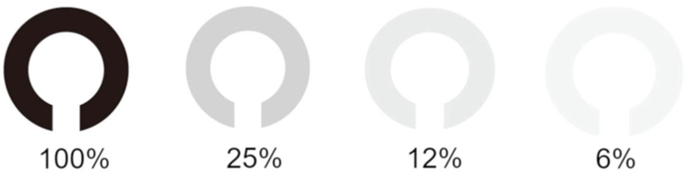
| Index Concentration, % | VA | p-Value * vs. 100% | VA | p-Value ** vs. 100% | p-Value *** Affected vs. Fellow |
|---|---|---|---|---|---|
| Affected Eye | Fellow Eye | ||||
| 100 | −0.13 ± 0.08 | −0.18 ± 0.08 | 0.01 | ||
| 25 | −0.01 ± 0.11 | <0.001 | −0.08 ± 0.09 | <0.001 | <0.01 |
| 12 | 0.10 ± 0.19 | <0.001 | 0.03 ± 0.09 | <0.001 | 0.024 |
| 6 | 0.15 ± 0.14 | <0.001 | 0.18 ± 0.07 | <0.001 | <0.01 |
| Index Concentration | Affected Eye | Fellow Eye | p-Value * |
|---|---|---|---|
| 100–25% | 0.12 ± 0.071 | 0.098 ± 0.062 | 0.279 |
| 100–12% | 0.24 ± 0.13 | 0.21 ± 0.069 | 0.420 |
| 100–6% | 0.37 ± 0.13 | 0.32 ± 0.11 | 0.038 |
| Clinical Parameters | r | p-Value * |
|---|---|---|
| Age, years | 0.147 | 0.334 |
| Symptom duration, months | 0.299 | 0.048 |
| PDT spot size, μm | 0.148 | 0.331 |
| VA before PDT, logMAR | −0.068 | 0.656 |
| ONL thickness before PDT, μm | −0.292 | 0.052 |
| ONL thickness after PDT, μm | −0.207 | 0.173 |
| CFT before PDT, μm | −0.097 | 0.520 |
| CFT after PDT, μm | −0.132 | 0.389 |
| Height of SRD before PDT, μm2 | 0.003 | 0.985 |
| Disruption of ELM layer, before PDT (+/−) | −0.035 | 0.821 |
| Disruption of ELM layer, after PDT (+/−) | 0.131 | 0.390 |
| Disruption of EZ layer, after PDT (+/−) | 0.209 | 0.168 |
Publisher’s Note: MDPI stays neutral with regard to jurisdictional claims in published maps and institutional affiliations. |
© 2021 by the authors. Licensee MDPI, Basel, Switzerland. This article is an open access article distributed under the terms and conditions of the Creative Commons Attribution (CC BY) license (http://creativecommons.org/licenses/by/4.0/).
Share and Cite
Chubachi, A.; Miki, A.; Hayashida, M.; Sakamoto, M.; Imai, H.; Kusuhara, S.; Nakamura, M. Clinical Factors Associated with Low-Contrast Visual Acuity after Reduced-Fluence Photodynamic Therapy in Patients with Resolved Central Serous Chorioretinopathy and Good Baseline Visual Acuity. Pharmaceuticals 2021, 14, 303. https://doi.org/10.3390/ph14040303
Chubachi A, Miki A, Hayashida M, Sakamoto M, Imai H, Kusuhara S, Nakamura M. Clinical Factors Associated with Low-Contrast Visual Acuity after Reduced-Fluence Photodynamic Therapy in Patients with Resolved Central Serous Chorioretinopathy and Good Baseline Visual Acuity. Pharmaceuticals. 2021; 14(4):303. https://doi.org/10.3390/ph14040303
Chicago/Turabian StyleChubachi, Aya, Akiko Miki, Mayuka Hayashida, Mari Sakamoto, Hisanori Imai, Sentaro Kusuhara, and Makoto Nakamura. 2021. "Clinical Factors Associated with Low-Contrast Visual Acuity after Reduced-Fluence Photodynamic Therapy in Patients with Resolved Central Serous Chorioretinopathy and Good Baseline Visual Acuity" Pharmaceuticals 14, no. 4: 303. https://doi.org/10.3390/ph14040303
APA StyleChubachi, A., Miki, A., Hayashida, M., Sakamoto, M., Imai, H., Kusuhara, S., & Nakamura, M. (2021). Clinical Factors Associated with Low-Contrast Visual Acuity after Reduced-Fluence Photodynamic Therapy in Patients with Resolved Central Serous Chorioretinopathy and Good Baseline Visual Acuity. Pharmaceuticals, 14(4), 303. https://doi.org/10.3390/ph14040303

