Article

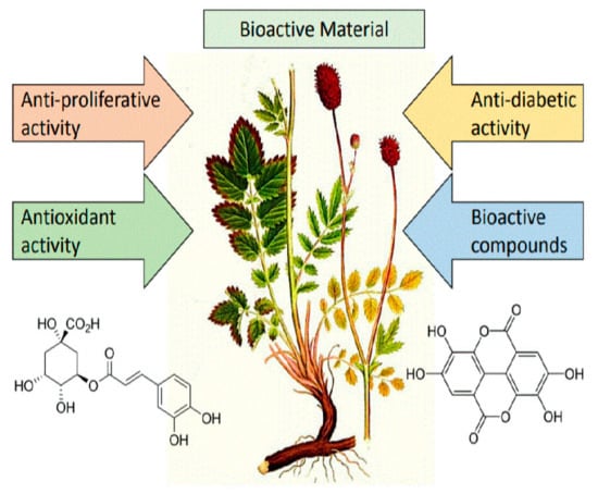
Profile and Content of Phenolic Compounds in Leaves, Flowers, Roots, and Stalks of Sanguisorba officinalis L. Determined with the LC-DAD-ESI-QTOF-MS/MS Analysis and Their In Vitro Antioxidant, Antidiabetic, Antiproliferative Potency

1
Department of Fermentation and Cereals Technology, Wrocław University of Environmental and Life Science, 51-630 Wrocław, Poland
2
Department of Fruit, Vegetables and Nutraceutical Technology, Wrocław University of Environmental and Life Science, 51-630 Wroclaw, Poland
3
Laboratory of Tumor Molecular Immunobiology, Ludwik Hirszfeld Institute of Immunology and Experimental Therapy, Polish Academy of Sciences, 53-114 Wrocław, Poland
4
Department of Horticulture, West Pomeranian University of Technology in Szczecin, 71-434 Szczecin, Poland
*
Author to whom correspondence should be addressed.
Pharmaceuticals 2020, 13(8), 191; https://doi.org/10.3390/ph13080191
Submission received: 8 July 2020 / Revised: 30 July 2020 / Accepted: 3 August 2020 / Published: 12 August 2020

Abstract

:
The aim of this study was to accurately determine the profile of polyphenols using the highly sensitive LC-DAD-ESI-QTOF-MS/MS technique and to determine in vitro antioxidant activity, the ability of inhibition of α-amylase, α-glucoamylase, and pancreatic lipase activity, and antiproliferative activity in leaves, flowers, roots, and stalks of medical plant Sanguisorba officinalis L. The results of the analysis of the morphological parts indicated the presence of 130 polyphenols, including 62 that were detected in S. officinalis L. for the first time. The prevailing group was tannins, with contents ranging from 66.4% of total polyphenols in the flowers to 43.3% in the stalks. The highest content of polyphenols was identified in the flowers and reached 14,444.97 mg/100 g d.b., while the lowest was noted in the stalks and reached 4606.33 mg/100 g d.b. In turn, the highest values of the antiradical and reducing capacities were determined in the leaves and reached 6.63 and 0.30 mmol TE/g d.b, respectively. In turn, a high ability to inhibit activities of α-amylase and α-glucoamylase was noted in the flowers, while a high ability to inhibit the activity of pancreatic lipase was demonstrated in the leaves of S. officinalis L. In addition, the leaves and the flowers showed the most effective antiproliferative properties in pancreatic ductal adenocarcinoma, colorectal adenocarcinoma, bladder cancer, and T-cell leukemia cells, whereas the weakest activity was noted in the stalks. Thus, the best dietetic material to be used when composing functional foods were the leaves and the flowers of S. officinalis L., while the roots and the stalks were equally valuable plant materials.

Graphical Abstract

1. Introduction

The interest in alternative plants with a health-promoting potential has been growing in recent years not only in the pharmaceutical and cosmetic industries but also in the food industry where they are expected to contribute to the design of novel functional food. Therefore, it is believed that various morphological parts of Sanguisorba officinalis L. represent a good source of compounds exhibiting the aforementioned properties [1].
S. officinalis L. (great burnet or burnet bloodwort) is a species belonging to the Rosaceae family. It grows wild in Asia and Europe (except for the northern regions [1,2]. This melliferous, perennial plant usually occurs on arid and semi-arid grasslands and blooms from June till September. Its shoots can grow up to ca. 1.2–1.5 m. S. officinalis L. is resistant to frost as well as to diseases. It has been used for culinary purposes as an additive to salads and in animal feeding as an additive to feed mixtures due to its high nutritional value [3]. However, in folk medicine of both the Far East and Europe, S. officinalis L. was used as an herbal medicine in relieving inflammation, controlling external and internal bleeding, in the treatment of ulcers, burns, eczema, acne, as well as diarrhea [4,5]. In turn, the available experimental data prove a number of its biological properties, e.g., anti-inflammatory [3], anticancer [6], antiviral [7], antioxidant [1], prevention of the Alzheimer’s disease [3], and anti-wrinkle effects. [8]. In addition, the above studies have shown that all the biological properties exhibited by this perennial plant are due to a broad range of its bioactive compounds such as phenolic acids, tannins, flavonoids, triterpenes, and polysaccharides [1,3,4,5,6,7,8]. The richness of these compounds is sought in alternative plant sources that could be used in the treatment and prevention of many diseases and even as a dietary component [9].
Considering a number of biological properties of S. officinalis L., this plant has a high nutraceutical potential. However, there are a few reports on the profile and content of secondary metabolites in all of its morphological parts, which may differ and therefore exhibit various properties. Thus, research was undertaken into the accurate characterization of flowers, leaves, stalks, and roots in terms of the profile and content of polyphenols using the highly sensitive LC-DAD-ESI-QTOF-MS/MS technique. Analyses were also conducted to determine the in vitro antioxidant, antiproliferative, and antidiabetic activity for the individual morphological parts of S. officinalis L. This study aims to provide valuable information about differences in contents of bioactive compounds and their biological properties in the flowers, leaves, stalks, and roots of S. officinalis L., which will be used to compose not only functional foods but also nutraceuticals in the future.

2. Results and Discussion

2.1. Identification of Polyphenolic Compounds

The present study involved a thorough identification of the profile of bioactive compounds in extracts from leaves, flowers, stalks, and roots of Sanguisorba officinalis L. plant with the use of an ultrasensitive LC-DAD-ESI-QTOF-MS/MS method in the negative and positive ion mode. In total, 130 compounds were identified in extracts from the selected morphological parts of S. officinalis L., including 77 hydrolyzable tannins, 9 sanguiins, 3 sanguisorbic acids, 13 phenolic acids, 6 anthocyanins, 12 catechins and proanthocyanidins, and 9 flavonols, as well as 1 triterpenoid saponins (Table 1; Figures S1–S4). In turn, 62 compounds were identified in S. officinalis L. for the first time ever, including 42 hydrolyzable tannins, 5 sanguiins, 8 phenolic acids, 2 anthocyanins, 1 proanthocyanidins, and 3 flavonols as well as 1 triterpenoid saponins. Peaks were identified based on the determined exact molecular weights, peak retention times, primary ions from MS fragmentation, and comparison of data obtained with commercial standards and literature findings (Table 1). However, the profile of the compounds examined was strongly dependent on the morphological part of the plant, since 70, 76, 66, and 62 compounds were identified in the flowers, leaves, roots, and stalks, respectively.
The prevailing group of polyphenolic compounds were hydrolyzable tannins belonging to the family of tannins and being hydrolyzed conjugates that contain one or more hexahydroxydiphenoyl (HHDP) groups, thus leading to the esterification of sugars, glucose in particular. During fragmentation of the primary ions, losses observed were typical of these compounds and involved losses of galloyl, hexahydroxydiphenoyl, gallic acid, HHDP glucose, galloyl-glucose, and galloyl-HHDP-glucose residues with 152, 302, 170, 482, 332, 634 Da, respectively. Additionally, fragments were noted at m/z 169 and at m/z 301 formed through lactonization of the characteristic hexahydroxydiphenoyl group to ellagic acid. These compounds comprise typical galloyl and HHDP groups, respectively, which have earlier been described in the available literature [1,2,3,9,10,11]. Furthermore, if ellagitannin or galloyl derivates are composed of one or a few galloyl groups taking part in sugar synthesis, the fragmentary ion first discards a molecule of gallic acid and then a galloyl group or groups during fragmentation [10]. Among the 77 compounds, only 36 had previously been identified in S. officinalis L., and they all were methyl-6-O-galloyl-β-d-glucopyranoside (peak 17, 64; m/z 345), pedunculagin1 (18, 23, 29; m/z 785), galloyl-HHDP-glucose otherwise called corilagin isomer (25, 44, 55; m/z 633), di-galloyl-glucoside (37; m/z 483), methyl-4,6-digalloyl-β-d-glucopyranoside (50, 62, 71, 88; m/z 497), HHDP-galloyl-glucose (53; m/z 633), ellagic acid pentoside (60, 99; m/z 433), ellagic acid hexoside (67, 68, 102; m/z 463), di-galloyl hexoside (72, 118; m/z 483), galloyl-bis-HHDP-glucose otherwise called potentilin/casuarictin isomer (84, 85, 95, 97, 104, 106; m/z 935), lambertianin C (86; m/z 1401), ellagic acid (108; m/z 300.99), trigalloyl-HHDP-glucose (92, 114; m/z 937), trigalloyl-β-D-methyl glucoside (115; m/z 649), 3,3′,4′-O-trimethyl ellagic acid (127, 128; m/z 343), and 3,4′-O-dimethyl ellagic acid (129, 130; m/z 329) [2,3,12]. In turn, 16 compounds had earlier been detected and identified in flowers and fruits of Punica granatum but in this study were for the first time detected in the morphological parts of S. officinalis L. These compounds were referred to as: 2,3-HHDP-(α/β)-glucose (1; m/z 481), HHDP-hexoside(2,3-(S)-Hexahydroxydiphenoyl-d-glucose) (2, 4; m/z 481), HHDP-hexoside(1-galloyl-2,3-hexahydroxydiphenoyl-α-glucose) (3; m/z 481), galloyl-hexoside(β-glucogallin) (5; m/z 331), galloyl-hexoside (7–10, 13; m/z 331), di-HHDP-glucoside (34; m/z 783), di-galloyl-HHDP-glucose (14, 56, 66; m/z 785), galloyl-HHDP-hexoside (77; m/z 633), and pentagalloyl-glucoside (111; m/z 939) [10,13]. Another 6 compounds belonging to the group of hydrolyzable tannins were detected during identification of Duchesnea indica and they were: di-HHDP-glucose also known as pedunculalagin isomer (15, 20, 24, 26, 27, 30; m/z 783) [14]. However, 12 subsequent compounds were identified and determined based on their main ion and MS/MS fragmentation as β-1-O-galloyl-2,3-(S)-HHDP-d-glucose (28; m/z 633), methyl ellagic acid-pentoside (35; m/z 477), HHDP-NHTP-glucose (47, 51; m/z 933), castalagin/vescalagin isomer (58, 70, 79, 81, 98, 110; m/z 933), HHDP-NHTP-glucose-galloyl-di-HHDP-glucose (cocciferind2) (82; m/z 933), and tetragalloyl-glucose (100; m/z 787). They had earlier been detected in various plant materials like Castanea sativa Miller, Quercus suber L., Betula pubescens, raspberry fruits, and oak [15,16,17,18]. However, 8 compounds were identified for the first time ever. Compound No. 6 was tentatively identified as galloyl-pentoside based on the primary ion at m/z 301 and the loss of the pentose group (132 Da) giving a peak at m/z 169. Compound No. 49 was tentatively identified as HHDP-glucose based the primary ion at m/z 481 and MS/MS fragment at m/z 301. In the case of compound No. 54, the primary peak was at m/z at 345 due to the loss of a 176 Da residue that resulted in a peak formed at m/z 169, which was tentatively identified as galloyl-glucoronide. Compounds No. 73 and 74 were tentatively identified as eucaglobulin based on the primary ion at m/z 497 and MS/MS fragmentary ions revealing peaks at m/z 345, 327, 313, 183, and 169. In turn, compounds No. 93 and 94 were tentatively described as ellagic acid-hexoside-pentoside based the primary ion at m/z 595 and its fragmentation ions at m/z 433 and 301 due to the loss of a hexose residue (162 Da) and a pentose residue (132 Da). Finally, compound No. 113 was tentatively identified as methyl galloyl-glucoside based on the primary peak at m/z 345 and the loss of a glucosyl residue (162 Da), yielding a base peak at m/z 183.
Another described class of polyphenolic compounds belonging to hydrolyzed tannins were sanguiins. Among the 9 identified compounds, only 4 had earlier been detected in S. officinalis L. as sanguiin H-6 (11, 89; m/z 1870), sanguiin H-4 (41; m/z 633), and sanguiin H-10 isomer (48; m/z 783) by Karkanis et al. [3] and Zhu et al. [2], whereas the other 5 were never identified, as shown by literature data. Therefore, based on the primary peak at m/z 785 and MS/MS fragmentation peaks at m/z 633 and 301, and due to the loss of 152 and 332 Da groups, compounds No. 65, 69, and 96 were tentatively identified as sanguiin H-1. In turn, compounds No. 119 and 122 were tentatively identified as sanguiin H-7 and sanguiin H-7 isomers considering their primary ion at m/z 801 and fragmentation peaks at m/z 649 and 301 resulting from the loss of 152, 332, and 16 Da.
In contrast, sanguisorbic acids, belonging to the hydrolyzed tannins, also have been previously defined for these plants by Zhu et al. [2] as sanguisorbic acid dilactone (9, 12; m/z 469) and sanguisorbic acid glucoside (52; m/z 667). These compounds were determined only in the leaves, stalks, and roots of S. officinalis L. Moreover, 1 sanguisorbigenin, belonging to the triterpenoid saponins, was detected during identification P. granatum [12].
UV detection at the characteristic absorption maximum between 310 and 330 nm [19] showed the presence of 13 hydroxycinnamic acids in flowers, leaves, and stalks in the case of which the esterification of their quinic acid residue occurs at positions 3, 4, and 5, but not at position 1 [19]. Of these, 5 were identified early in S. officinalis as caffeoylquinic acid (16, m/z 353), 3-O-caffeoylquinic acid (19; m/z 353), 3-O-p-coumaroylquinic acid (32; m/z 337), 5-O-caffeoylquinic acid (42; m/z 353), and 3-O-feruloylquinic acid (78; m/z 367) [12]. However, 4 more were previously identified in other botanical sources like Eryngium alpinum L. and Chrysanthemum as rosmarinic acid (33; m/z 359), disuccinoyl-caffeoylquinic acids (116; m/z 553), and 3,5-dicaffeoylquinic (120, 121; m/z 515), however, for the first time in S. officinalis L., compounds No. 123–125 were tentatively identified as caffeoyl dihexoside based on the highest peak at m/z 505 and its fragmentation yielding peaks at m/z 341 and 179 due to the loss of 2 hexose residues (162 + 162 Da). What is more, these compounds were also described for the first time ever in morphological parts of S. officinalis L.
Anthocyanins are natural plant pigments occurring in the plant kingdom. They were identified in the positive ion mode because they bear a positive charge and easily donate protons to free radicals under ESI conditions. In turn, their detection was carried out at the typical absorption maximum between 440 and 540 nm [10,20]. Among the tentatively identified 6 anthocyanins, that were detected only in the flowers, only 4 were earlier determined in S. officinalis L. as cyanidin 3,5-diglucoside (21; m/z 611), cyanidin 3-O-glucoside (46; m/z 449), and cyanidin 3-malonylglucoside (76, 90; m/z 535) [12]. The other 2 compounds were described based on previous information about fragmentation of pomegranate and grape berry skin [13,21] as cyanidin 3-O-rutinoside (87; m/z 595) and cyanidin 3-(6-O-acetyl)-glucoside (91; m/z 491).
Flavan-3-ols occur as monomers, oligomers, and polymers formed by linking to (epi)catechin monomers via interflavonoid bonds (C–C) [22]. Their fragmentation proceeds through the loss of a (epi)catechin unit with a molecular weight of 289 Da. The identified proanthocyanins occurred as catechin dimers, trimers, and tetramers and were identified as A and B procyanidins [22]. These 11 compounds were characterized based on available standards and the latest research works addressing S. officinalis L as (+)-catechin and (−)-epicatechin (31, 40; m/z 289), B-type (epi)catechin dimmer (36, 38, 39, 63, 83; m/z 577), B-type (epi)catechin trimmer (43; m/z 865), and B-type (epi)catechin tetramer (57, 59, 80; m/z 1153) [2,3]. In turn, compound No. 74 was tentatively identified as a A-type (epi)catechin tetramer at m/z 1153 and the base ion at m/z 289. Although it was earlier detected in black soybean [23], it was described in S. officinalis L. for the first time ever.
Flavonols were identified as derivatives of taxifolin, kaempferol, and quercetin based on the base fragments at m/z 300, 285, and 301. UV detection of flavonols revealed characteristic absorption maximum between 315 and 359 nm, and some of the identified compounds had additional peaks between 207 and 280 nm [24]. Besides, derivatives of these compounds are usually detected at positions C-7 and/or C-3. Fragmentation of the primary ions resulted in losses of hexose (162 Da), pentose (146 Da), and deoxyhexose (308 Da) [24]. Of the 9 flavonols initially suggested for S. officinalis L., only 6 have previously been described for this species as quercetin-3-O-glucoside (45; 463), quercetin-3-O-(6″-galloylglucose) (101; m/z 615), taxifolin-7-O-β-d-glucopyranoside (103; m/z 465), quercetin-3-O-glucuronide (109; m/z 477), quercetin-3-O-acetyl glucoside (112; m/z 505), and kaempferol-3-O-glucuronide (117; m/z 461) [2,3,12]. In turn, 3 compounds have not been previously described according to the available literature. Compound No. 61 was tentatively identified as kaempferol-di-O-rhamnoside based on the primary peak at m/z 577 and fragmentation peaks at m/z 431 and 285 due to the loss of two rhamnoside residues (146 + 146 Da). Another compound (103) was tentatively described as quercetin-glucoside-dirhamnoside based on the primary peak at m/z 755 and fragmentation peaks at m/z 609, 463, and 301 due to the loss of two rhamnose residues and one glycosyl residue. Finally, compound No. 107 was tentatively presented as quercetin rhamnosyl-rutinoside based on the primary peak at m/z 755 and fragmentation peaks at m/z 609 and 301.

2.2. Quantification of Polyphenolic Compounds

The content of polyphenols in the analyzed morphological parts of S. officinalis L. is shown in Table 2. The highest content of bioactive compounds was determined in the flowers, it reached 14,444.97 mg/100 g d.b. and was 1.5, 1.7, and 3.2 times higher than in the leaves, roots, and stalks, respectively. In turn, the content of polyphenols in the leaves + stalks of Sanguisorba minor Scop. was comparable to the content of these compounds in S. officinalis L., while the roots of S. minor Scop. were 4 times more abundant in the studied compounds than the roots of S. officinalis L. [3]. In turn, the sum of polyphenols analysed in the roots of the same species from Korea was 2 times lower than in the roots of plants grown in Poland. However, the extract from S. officinalis L. cultivated in China contained 3150 mg GAE/100 g dry weight polyphenols, which was 4.9, 3.2, 2.8, and 1.5 times lower compared to the flowers, leaves, roots, and stalks of the same species growing in Poland. The content of polyphenols in the leaves of green and white tea was 67.21 and 40.94 mg/g d.b. and was 1.5 and 2.4 times lower than in the leaves of the studied species, respectively [25]. Total content of polyphenols analyzed in the flowers, leaves, roots, and stalks of S. officinalis L. was 8.2, 8.4, 7.8, and 8.4 times higher, respectively, compared to edible flowers of Allium schoenoprasum (Liliaceae), Salvia pratensis (Lamiaceae), Sambucus nigra (Caprifoliaceae), Taraxacum officinale [26]. However, according to Zeng et al. [27] the contents of bioactive compounds in the flowers of green and black tea of Camellia sinensis were 2.4 and 5.4 times lower, respectively, compared to the flowers of S. officinalis L. Moreover, the content of bioactive compounds in the flowers and the leaves of Punica granatum L. was 2.2 and 6.7 times lower, respectively, than in the same morphological parts of S. officinalis L. [28]. In addition, the content of compounds tested in the leaves and the stalks of Fallopia japonica was 1.7 and 2.3 times lower, respectively, while their content in the roots of F. japonica was similar to S. officinalis L. [9]. The differences in the contents of polyphenolic compounds among individual species can be affected by various factors, such as the place of cultivation, climate, environmental conditions, and also the method of extraction and analysis [29]. Thus, the tested material is characterized by a high content of compounds exhibiting a number of biological properties and can be used to compose not only nutraceuticals in the pharmaceutical industry but also to produce functional food.
The profile and content of phenols present in various morphological parts of S. officinalis L. were quite diverse and strongly dependent on the morphological part tested. The flowers were dominated by hydrolyzed tannins (66.4% in all phenols) > flavan-3-ols (13.1%) > phenolic acids (9.9%) > flavonols (5%) > anthocyanins (3.8%) > triterpenoids (1.8%). In turn, in the leaves were dominated by hydrolyzed tannins (49.3%) > phenolic acids (20.5%) > flavonols (19.8%) > flavan-3-ols (7.4%) > triterpenoids (3%). However, in the roots, hydrolyzed tannins were also the dominant class (62.1%) > flavan-3-ols (37.3%) > phenolic acids and flavonols (<0.5%), whereas the stalks were dominated by hydrolyzed tannins (43.3%) > flavan-3-ols (26.2%) > flavonols (17.1%) > phenolic acids (7.8%) > triterpenes (5.5%). The analysis of phenols profile revealed flavonols to be the major group in leaf + stalks, whereas hydrolyzed tannins to be the major group in the roots of S. minor [3], similarly to the roots of S. officinalis L. and to the results presented in the work of Kim et al. [1].
Tannins are compounds that occur naturally in plants and also play a defensive role in them. They exhibit anti-inflammatory properties against inflammation of the mucous membranes and skin, as well as antiastringent, antioxidative, free radical-scavenging, and antiproliferative properties. In addition, they are also an important component of food because they affect its storage stability, taste, and color [30]. The highest content of these compounds was recorded in the flowers (9594.27 mg/100 g d.b.) and the lowest one in the stalks (1996.51 mg/100 g d.b.). According to Karkanis et al. [3], their content in S. minor was comparable in the leaves and stalks while 4 times higher in the roots compared to the morphological parts of S. officinalis L., respectively. In turn, the major compound in all morphological parts tested was Lambertian C, with its content ranging from 62% in the roots to 17% in the stalks, and similar observations were made in S. minor [3].
Phenolic acids are another naturally occurring class of polyphenolic compounds that have a number of biological properties, including antioxidative ones, or are used in the prevention of cardiovascular diseases. They also affect the sour and bitter taste of food of plant origin, imparting them astringent flavones [31]. They dominated in the leaves of S. officinalis L. and their content amounted to 2044.37 mg/100 g d.b., while their poorest presence was in the roots (only 6.64 mg/100 g d.b.). Their content in the leaves was 5.3 times higher compared to their total content in leaves and stalks of S. minor, but similar while comparing to the stalks of S. officinalis L. and S. minor [3]. In turn, chlorogenic acid turned out to be the major compound in the flowers, neochlorogenic acid prevailed in the stalks and leaves, while ellagic acid was found in the leaves and stalks of S. minor [3].
Anthocyanins occurred only in flowers, giving them an intense red color. They belong to the group of polyphenols which show a number of health-promoting properties [9,32]. Their content was 549.57 mg/100 g d.b., and the dominant compounds were cyanidin 3-O-glucoside and cyanidin 3-O-malonylglucoside and they constituted of 62% and 28% of all anthocyanins, respectively.
Catechins and proanthocyanidins are compounds that also play an important role in the prevention of many diseases [9,32]. Their content ranged from 739.47 to 3239.19 mg/100 g d.b. in the leaves and roots of S. officinalis L, respectively, and was 5.6 and 20 times higher compared to the leaves and roots of Fallopia japonica, respectively [9]. The dominant compounds were: B-type (epi)catechin dimmer constituting 41% in the leaves to 18% in the stalks of all flavan-3-ols, and (−)-epicatechin constituting from 37% in the stalks to 19% in the leaves. Although in F. japonica, the major compound was procyanidin dimer B [9].
Flavonols are also a valuable class of natural secondary metabolites due to their anti-inflammatory and antioxidative properties [9]. The highest content of these compounds was noted in the leaves and reached 1969.85 mg/100 g d.b. It was 2.7, 2.5, and 41 times higher compared to the flowers, stalks, and roots, respectively. This difference results from the fact that these compounds are mainly located in the top layer of plants, protecting them from harmful UV radiation [32]. In turn, quercetin-O-glucuronide was the dominant compound in the flowers, leaves, and stalks, constituting 69%, 83%, and 85% of all flavonols, respectively, whereas taxifolin 7-O-β-d-glucopyranoside prevailed in the roots, constituting 91%. These observations have also been confirmed by Kim et al. [1].

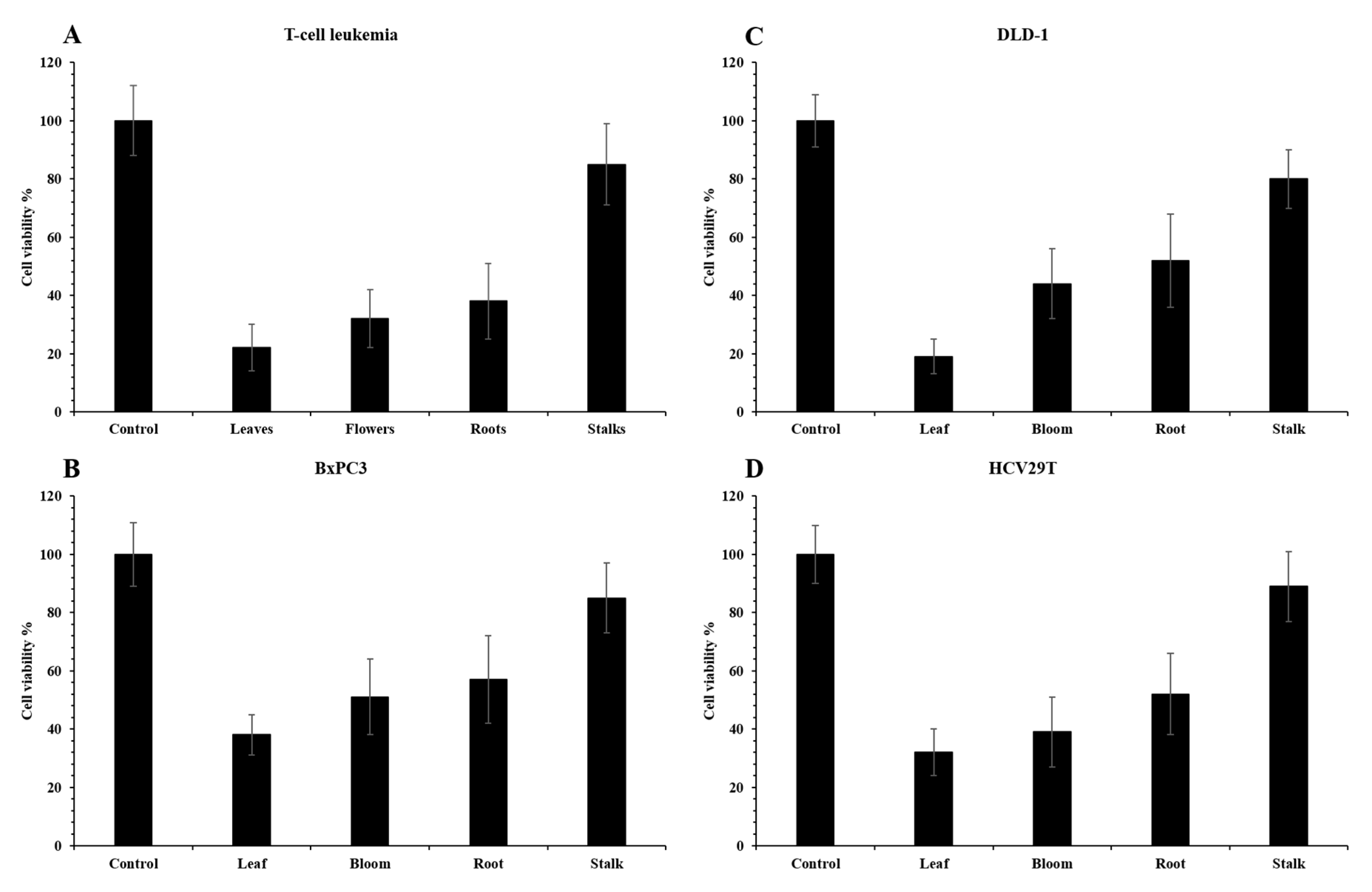
2.3. Pro-Health Properties

The average antioxidative activity determined for S. officinalis L. was 4.45 mmol Troloxu (TE)/g dry basis (d.b.) in the ABTS test and 0.18 mmol TE/g d.b. in the FRAP assay (Table 3). The highest activity was determined in the leaves and was 6.63 and 0.30 mmol TE/g d.b. in the ABTS and FRAP tests, respectively. It was 1.2 and 1.6 times higher than in the stalks, 12.0 and 2.1 times higher than in the roots, and comparable to that found in the flowers for the ABTS radicals and for Fe3+ reduction to Fe2+, respectively (Table 3). Similar results of the antioxidative activity assays were obtained for the roots of S. officinalis gathered in China [5]. In turn, previous research shows that the antiradical activity of the leaves, stalks, and roots of S. officinalis L. was 6.2, 1.7, and 10.6 times higher compared to the same parts of F. japonica as well as 7.9, 1.8, and 9.3 times higher compared to the same parts of F. sachalinensis, respectively [9]. Antiradical activity for the roots was comparable to that obtained for the medical plant—Ruta montana [33]. Moreover, the average reducing activity of the tested parts of S. officinalis L. was comparable to the antioxidant potential determined for Melissae folium and about 6 times higher than for Spiraea herba, Uvae ursi folium, Rubi fructose folium, or Fragariae herba folium [34]. Thus, the results obtained indicate that the roots, flowers, and leaves of S. officinalis L. have a high ability to scavenge free radicals, which may be due to the high content of bioactive compounds determined for these morphological parts of the plant. What’s more, the results presented a strong Pearson’s correlation with the sum content of phenolic acids and anthocyanins and with the antioxidative activity as r2 = 0.734 and 0.539 for ABTS assay and r2 = 0.746 and 0.869 for FRAP, whereas the correlation between the reducing activity and sum of hydrolysable tannins and polyphenols was also strong r2 = 0.769 and 0.823.
The leaves, flowers, stalks, and roots of S. officinalis L. were also tested for their ability of inhibition of α-amylase (αA) and α-glucosidase (αG) activity, and their ability of inhibition of pancreatic lipase (LP) activity (Table 3). αA and αG are carbohydrate-degrading enzymes, but the mechanisms of their action differ; αA accelerates the hydrolysis of bonds inside a compound, whereas αG hydrolyzes α-1,4-glucosidic bonds, leading to the release of glucose absorbed by the body [35]. In turn, LP is an enzyme responsible for the degradation of triglycerides to simple lipids and fatty acids absorbable by the human body. However, it has been proved that excess fatty acids can lead to the formation of free radicals and insulin resistance [36]. Therefore, the inhibition of the above enzymes may be used in the treatment of diabetes type II or obesity [35]. The obtained results show that the highest ability to inhibit αA and αG activity was recorded for flowers of S. officinalis L. and reached EC50 6.03 and 9.60 mg/mL, respectively. Therefore, the flowers were 1.6 and 1.3 times more active than the leaves, 4.0 and 3.3 times more active than the stalks, and 1.7 and 2.0 times more active than the roots, respectively. In turn, the highest ability to inhibit pancreatic lipase was found for the leaves of S. officinalis L. (EC50 = 18.75 mg/mL) which were 1.2, 3.0, and 3.9 times more active compared to the flowers, stalks, and roots of the tested plant, respectively. As far as the results showed that the ability to inhibit αA, αG, and LP strongly depended on the sum of flavan-3-ols and the correlations were r2 = 0.944, 0.836, and 0.593, respectively. However, in the case of phenolic acids and flavonols, the correlations were strongly negative: r2 = 0.813, 0.921, and 0.872 and r2 = 0.842, 0.825, and 0.857, respectively.
The antiproliferative potency of the flowers, leaves, roots, and stems of S. officinalis L. were tested against four different cancer cell lines as BxPC3 (pancreatic ductal adenocarcinoma), DLD-1 (colorectal adenocarcinoma), HCV29T (bladder cancer), and Jurkat (T-cell leukemia). This is the first report on these cancer cell lines. The effect against the used cell lines was clearly noted (Figure 1). The extract from S. officinalis L. leaves significantly reduces the viability of all tested cell lines, especially DLD-1 colon cancer cells (to 19%) and Jurkat leukemia cells (to 22%). The flower extract reduced the viability of Jurkat cells to 32% and the remaining cells by 39–50%. Extract from the root showed similar results. In contrast, the extract from the stem acted the weakest on all cell lines, reducing cell viability to 85–97%. What’s more, the results presented a strong Pearson’s correlation between the sum of flavan-3-ols and with the viability of Jurkat leukemia cells and DLD-1 colon cancer cells—r2 = 0.731 and 0.545, while lower the viability of HCV29T cells strongly depended on anthocyanins and the correlation was r2 = 0.705. Liu et al. [37] noted that aqueous root extracts of S. officinalis L. showed synergic effect on inhibition of activity against HCT-116 and CPR cell lines (colon cancer) with 5-fluorouracil. Shin et al. [38] observed that the extract of S. officinalis L. inhibited cell growth against HSC4 and HN22 cell line (oral cancer) and induced death. According to Liu et al. [39], aqueous plant extracts of S. officinalis L. decreased the target Wnt and β-catenin genes by inhibiting the signal pathway of Wnt/β-catenin in cells of colorectal cancer. Moreover, Karkanis et al. [3] noted that the highest ability to inhibit of cervical carcinoma (HeLa), breast adenocarcinoma (MCF-7), and nonsmall cell lung cancer (NCl-H460) cell line was recorded for extract of roots of S. minor, whereas the extract of leaves + stalks of S. minor showed high ability to inhibit of hepatocellular carcinoma (HepG2) cell line. Thus, our own results and other authors presented that the highest cytotoxicity for the examined tumor cell lines covered depends on the analyzed morphological parts of S. officinalis L. and their bioactive substances. Moreover, the leaves, flowers, and roots showed high and differed antiproliferative potency to inhibit activity of various tumor cell lines.

3. Materials and Methods

3.1. Material, Reagents, and Instruments

Materials: Sanguisorba officinalis L. flowers, stalks, roots, and leaves (~5 kg) were obtained from a private garden in Szczytna (53°33′46″ N 20°59′07″ E), Lower Silesia, Poland. The plant was collected randomly in August 2019 from different parts of field (total area of cultivation is 1 ha). Then, material was washed and dried in a freeze dryer Alpha 1-4 LSC (Christ, Osterode, Germany).
Reagents: acetonitrile, formic acid, methanol, ABTS (2,2′-azinobis(3-ethylbenzothiazoline-6-sulfonic acid), 6-hydroxy-2,5,7,8-tetramethylchroman-2-carboxylic acid (Trolox), 2,4,6-tri(2-pyridyl)-s-triazine (TPTZ), methanol, acetic acid, α-amylase from porcine pancreas, α-glucoamylase from Rhizopus sp., lipase from porcine pancreas, Antibiotic-Antimycotic Solution, and RPMI 1640 culture medium were purchased from Sigma-Aldrich (Steinheim, Germany). (−)-Epicatechin, (+)-catechin, procyanidin B2, p-coumaric acid, ferulic acid, 5-caffeoylquinic acid, procyanidin A2, caffeic acid, quercetin 3-O-rutinoside, quercetin-3-O-galactoside, quercetin-3-O-glucoside, kaempferol 3-O-galactoside, ellagic acid, and cyanidin-3-O-glucoside were purchased from Extrasynthese (Lyon, France). DMEM culture medium with 10% FBS were purchased from Gibco (Thermo Fisher Scientific, Waltham, MA, USA), and MTS solution was purchased from Promega (Madison, WI, USA).
Instruments: UV-2401 PC spectrophotometer (Shimadzu, Kyoto, Japan) for antioxidant activity; Sonic 6D, Polsonic, Warsaw, Poland, for extraction; LC-DAD-ESI-QTOF-MS/MS (ultraperformance liquid chromatography equipped with a binary solvent manager and a Q-Tof Micro Mass Spectrometer (Waters, Manchester, UK) with an ESI source operating in negative and positive modes (Waters Corporation, Milford, MA, USA) for polyphenolic compounds; and Wallac 1420 VICTOR2 Plate Reader (PerkinElmer, Waltham, MA, USA) for antiproliferative activity.

3.2. Determination of Polyphenols

For the extraction and determination of phenolic compounds, a protocol described before by Lachowicz et al. [9] was followed. Briefly, samples (0.1 g) were mixed with 5 mL of 30% of UPLC-grade methanol. The extracts were sonicated for 20 min and centrifuged (at 19,000× g/10 min). Finally, the extracts were filtered by hydrophilic PTFE 0.20 μm membrane (Millex Samplicity Filter, Darmstadt, Germany) and used for testing.
The runs were monitored at the following wavelengths: phenolic acids at 320 nm, flavonols at 360 nm, anthocyanins at 520 nm, flavan-3-ols at 280 nm, and hydrolysable tannins at 240 nm. Separations of individual polyphenols were carried out using a UPLC BEH C18 column (1.7 μm, 2.1 mm × 100 mm) at 30 °C. The samples (10 μL) were injected, and the elution was completed in 15 min with a sequence of linear gradients and isocratic flow rates of 0.45 mL/min. The mobile phase consisted of solvent A (0.1% formic acid, v/v) and solvent B (100% acetonitrile). The program began with isocratic elution with 99% solvent A (0–1 min), and then, a linear gradient was used until 12 min, lowering solvent A to 0%; from 12.5 to 13.5 min, the gradient returned to the initial composition (99% A), and then, it was held constant to re-equilibrate the column. The analysis was carried out using full-scan, data-dependent MS scanning from m/z 100 to 1500. Leucine enkephalin was used as the reference compound at a concentration of 500 pg/μL, at a flow rate of 2 μL/min, and the [M − H] ion at 554.2615 Da was detected. The [M − H] ion was detected during 15 min analysis performed within ESI–MS accurate mass experiments, which were permanently introduced via the LockSpray channel using a Hamilton pump. The lock mass correction was ±1.000 for the mass window. The mass spectrometer was operated in negative- and positive-ion mode, set to the base peak intensity (BPI) chromatograms, and scaled to 12,400 counts per second (cps) (100%). The optimized MS conditions were as follows: capillary voltage of 2500 V, cone voltage of 30 V, source temperature of 100 °C, desolvation temperature of 300 °C, and desolvation gas (nitrogen) flow rate of 300 L/h. Collision-induced fragmentation experiments were performed using argon as the collision gas, with voltage ramping cycles from 0.3 to 2 V. Characterization of the single components was carried out via the retention time and the accurate molecular masses. Each compound was optimized to its estimated molecular mass [M − H]/[M + H]+ in the negative and positive mode before and after fragmentation. The data obtained from UPLC-MS were subsequently entered into the MassLynx 4.0ChromaLynx Application Manager software. On the basis of these data, the software is able to scan different samples for the characterized substances. The PDA spectra were measured over the wavelength range of 200–800 nm in steps of 2 nm. The calibration curves were prepared for the standard: gallic acid (y = 1222.5x − 1972.7; r2 = 0.9999), procyanidin B2 (y = 6566.2x − 15,957; r2 = 0.9999), (+)-catechin (y = 1565.9x + 2243; r2 = 0.9999), p-coumaric acid (y = 68.109x + 49.224; r2 = 0.9996), ferulic acid (y = 50,215x + 36,206; r2 = 0.9997), 5-caffeoylquinic acid (y = 14,332x + 1315.1; r2 = 0.9999), procyanidin A2 (y = 9484.1x − 6770.5; r2 = 0.9997), caffeic acid (y = 17,431x + 40,114; r2 = 0.9999), quercetin 3-O-rutinoside (y = 13,362x − 1795; r2 = 0.9997), qercetin-3-O-galactoside (y = 20,926x − 18,309; r2 = 0.9991), qercetin-3-O-glucoside (y = 11,923x + 8188; r2 = 0.9999), kaempferol 3-O-galactoside (y = 12,057x − 1922.4; r2 = 0.9997), ellagic acid (y = 26754x + 172359; r2 = 0.9995), cyanidin-3-O-glucoside (y = 30,726x + 190,297; r2 = 0.9976), and (−)-epicatechin (y = 39,233x − 360,853; r2 = 0.9994) at concentrations ranging between 0.05 and 0.5 mg/mL. All data were obtained in triplicate. The results were expressed as mg/100 g of dry basis (d.b.).

3.3. Pro-Health Properties

3.3.1. Antiradical Capacity

Samples (1 g) were mixed with methanol (80%; 10 mL) and then with hydrochloric acid (1%). This process was performed twice by incubating the above slurry for 20 min under sonication. Next, the slurry was centrifuged at 19,000× g for 10 min, and the supernatant was filtered through a hydrophilic PTFE 0.20 μm membrane (Merck, Darmstadt, Germany) and used for analysis.
The ABTS method was carried out with the method described by Re et al. [40]. For this, 0.03 mL of sample was mixed with 3 mL of ABTS + solution, and after 6 min of reaction, the absorbance was measured at 734 nm using the spectrophotometer. All data were obtained in triplicate. The activity was expressed in mmol Trolox/g d.b.

3.3.2. Reducing Potential

The FRAP method was carried out with the method described by Benzie et al. [41]. The reagent was prepared by mixing 10 mmol 2,4,6-Tris(2-pyridyl)-s-triazine (TPTZ)/L reagent with 20 mmol/L ferric chloride in acetate buffer (pH 3.6). Precisely, 0.1 mL of sample was mixed with 0.9 mL of distilled water and 3 mL of ferric complex. After 10 min of reaction, the absorbance was measured at 593 nm using the spectrophotometer. All data were obtained in triplicate. The activity was expressed in mmol Trolox/g d.b.

3.3.3. Determination of Enzyme Inhibition Potency

Anti-diabetic activity, α-amylase, α-glucosidase inhibitory, and lipase activity effect of the materials were described previously by Nakai et al. [42], Podsędek et al. [43], and Nickavar et al. [44]. The extraction of mixed material was done with 70% acetone (or water) at room temperature for 60 min with constant stirring. After centrifuging at 4000 rpm for 10 min, and filtration, the supernatants were concentrated at 40 °C (vacuum evaporator) to remove the acetone and the aqueous phase was diluted with water. For further analytical and biological activity assays, a gradient of concentrations was prepared via serial dilution of the fruit extracts in pure water. The amount of the inhibitor (expressed as mg of fruit per 1 mL of reaction mixture under assay conditions) required to inhibit 50% of the enzyme activity was defined as the IC50 value. The IC50 of the fruits tested was obtained from the line of the plot of the fruit concentration in 1 mL of reaction mixture versus the % inhibition. All samples were assayed in triplicate.

3.3.4. Antiproliferative Potency

Cell Lines and Cell Culture

The human cancer cell lines BxPC3 (pancreatic ductal adenocarcinoma), DLD-1 (colorectal adenocarcinoma), and HCV29T (bladder cancer) were cultured in DMEM culture medium with 10% FBS and Antibiotic-Antimycotic Solution. Jurkat cell line (T-cell leukemia) was maintained in RPMI 1640 culture medium supplemented with 2 mM L-glutamine, 100 U/mL penicillin, 100 µg/mL streptomycin, and 10% fetal bovine serum (FBS). All cell lines were cultured at 37 °C in a humidified atmosphere of 5% CO2. The cells were seeded at densities of 5 × 10³ cells/0.1 mL (0.32 cm2) for cell viability assay. All cell lines were obtained from the collection of the Institute of Immunology and Experimental Therapy, Polish Academy of Sciences, Wroclaw, Poland.

Determination of Cell Viability

For determination of cell viability, cells were seeded in 96-well-plate (NUNC, Roskilde, Denmark). The plant extract was prepared by suspending 100 mg of dry plant material in 1 mL of 30% ethanol. The suspension was heated at 50 °C for 30 min and then centrifuged at 10,000× g for 15 min. The clear supernatant was diluted 30-fold in cell culture medium. As a control, 1% ethanol in the cell medium was used. The cells were incubated in 200 µL of the above culture medium for 48 h. Following the incubation, 20 µL of MTS solution was added to each well for 4 h; next, absorbance at 490 nm was recorded by a plate reader. Each treatment within a single experiment was performed in triplicate. Data were normalized to control medium containing 1% ethanol.

3.4. Statistical Analysis

Statistical analysis such as one-way ANOVA (p < 0.05) was analyzed using Statistica 12.5 (StatSoft, Kraków, Poland).

4. Conclusions

It needs to be noted that the flowers and leaves of S. officinalis L. are a good source of polyphenols, including hydrolyzable tannins, phenolic acids, flavonols, and anthocyanins, and exhibit a significant antiradical and reducing potential. In turn, the roots and stalks are a valuable source of flavan-3-ols. The most effective the inhibition of α-amylase, α-glucosidase, and pancreatic lipase and antiproliferative activities, reflected in the inhibition of viability of pancreatic ductal adenocarcinoma, colorectal adenocarcinoma, and bladder cancer as well as T-cell leukemia cell, were shown by the flowers and leaves of S. officinalis L. Thus, the data provided in this work indicate the possibility of using its individual morphological parts in the prevention of selected disease entities. In addition, this plant material can be used not only in the food industry as a functional additive to food, increasing its health value, but also in the cosmetic and pharmaceutical industries as a nutraceutical. The data obtained justify the need for further research on the morphological parts of S. officinalis L. with special emphasis put on leaves and flowers, to identify mechanisms potentially responsible for the antiproliferative activity.

Supplementary Materials

The following are available online at https://www.mdpi.com/1424-8247/13/8/191/s1. Figure S1: LC-DAD-ESI-QTOF-MS/MS chromatogram fragile of the Sanguisorba officinalis L. flowers extract at 320 and 360 nm; Figure S2: LC-DAD-ESI-QTOF-MS/MS chromatogram fragile of the Sanguisorba officinalis L. leaves extract at 320 and 360 nm; Figure S3: LC-DAD-ESI-QTOF-MS/MS chromatogram fragile of the Sanguisorba officinalis L. roots extract at 320 and 360 nm; Figure S4: LC-DAD-ESI-QTOF-MS/MS chromatogram fragile of the Sanguisorba officinalis L. stalks extract at 320 and 360 nm.

Author Contributions

Conceptualization, S.L. and J.O.; methodology, S.L., J.O., A.R. and I.O.; validation, S.L. and J.O.; formal analysis, S.L. and J.O.; investigation, S.L. and J.O.; resources, S.L. and J.O.; data curation, S.L. and J.O.; writing—original draft preparation, S.L. and J.O.; writing—review and editing, S.L. and J.O.; visualization, S.L. and J.O.; project administration, S.L. and J.O.; and funding acquisition, S.L. and J.O. All authors have read and agreed to the published version of the manuscript.

Funding

This research received no external funding.

Acknowledgments

The work was created in a leading research team “Food&Health”.

Conflicts of Interest

The authors declare no conflicts of interest.

References

  1. Kim, S.; Oh, S.; Noh, H.B.; Ji, S.; Lee, S.H.; Koo, J.M.; Choi, C.W.; Jhun, H.P. In vitro antioxidant and anti-propionibacterium acnes activities of cold water, hot water, and methanol extracts, and their respective ethyl acetate fractions, from Sanguisorba officinalis L. Roots. Molecules 2018, 23, 3001. [Google Scholar] [CrossRef] [Green Version]
  2. Zhu, H.-L.; Chen, G.; Chen, S.-N.; Wang, Q.-R.; Wan, L.; Jian, S.-P. Characterization of polyphenolic constituents from Sanguisorba officinalis L. and its antibacterial activity. Eur. Food Res. Technol. 2019, 245, 1487–1498. [Google Scholar] [CrossRef]
  3. Karkanis, A.C.; Fernandes, A.; Vaz, J.; Petropoulos, S.A.; Georgiou, E.; Ćirić, A.; Sokovic, M.D.; Oludemi, T.; Barros, L.; Ferreira, I.C. Chemical composition and bioactive properties of Sanguisorba minor Scop. under Mediterranean growing conditions. Food Funct. 2019, 10, 1340–1351. [Google Scholar] [CrossRef] [Green Version]
  4. Nguyen, T.T.H.; Cho, S.O.; Ban, J.Y.; Kim, J.Y.; Ju, H.S.; Koh, S.B.; Song, K.-S.; Seong, Y.H. Neuroprotective effect of Sanguisorbae radix against oxidative stress-induced brain damage: In vitro and in vivo. Boil. Pharm. Bull. 2008, 31, 2028–2035. [Google Scholar] [CrossRef] [Green Version]
  5. Zhang, L.; Koyyalamudi, S.R.; Jeong, S.C.; Reddy, N.; Smith, P.T.; Rajendran, A.; Longvah, T. Antioxidant and immunomodulatory activities of polysaccharides from the roots of Sanguisorba officinalis. Int. J. Boil. Macromol. 2012, 51, 1057–1062. [Google Scholar] [CrossRef]
  6. Wang, Z.; Loo, W.T.; Wang, N.; Chow, L.W.; Wang, N.; Han, F.; Zheng, X.; Chen, J.-P. Effect of Sanguisorba officinalis L on breast cancer growth and angiogenesis. Expert Opin. Ther. Targets 2012, 16, S79–S89. [Google Scholar] [CrossRef] [PubMed]
  7. Liang, J.; Chen, J.; Tan, Z.; Peng, J.; Zheng, X.; Nishiura, K.; Ng, J.; Wang, Z.; Wang, D.; Chen, Z.; et al. Extracts of the medicinal herb Sanguisorba officinalis inhibit the entry of human immunodeficiency virus-1. J. Food Drug Anal. 2013, 21, S52–S58. [Google Scholar] [CrossRef] [PubMed] [Green Version]
  8. Kim, Y.H.; Chung, C.B.; Kim, J.G.; Ko, K.I.; Park, S.H.; Kim, J.-H.; Eom, S.-Y.; Kim, Y.S.; Hwang, Y.-I.; Kim, K.-H. Anti-Wrinkle Activity of Ziyuglycoside I Isolated from a Sanguisorba officinalis Root Extract and Its Application as a Cosmeceutical Ingredient. Biosci. Biotechnol. Biochem. 2008, 72, 303–311. [Google Scholar] [CrossRef] [Green Version]
  9. Lachowicz, S.; Oszmiański, J.; Wojdyło, A.; Cebulak, T.; Hirnle, L.; Siewiński, M. UPLC-PDA-Q/TOF-MS identification of bioactive compounds and on-line UPLC-ABTS assay in Fallopia japonica Houtt and Fallopia sachalinensis (F.Schmidt) leaves and rhizomes grown in Poland. Eur. Food Res. Technol. 2018, 245, 691–706. [Google Scholar] [CrossRef] [Green Version]
  10. Yisimayili, Z.; Abdulla, R.; Tian, Q.; Wang, Y.; Chen, M.; Sun, Z.; Li, Z.; Liu, F.; Aisa, H.A.; Huang, C. A comprehensive study of pomegranate flowers polyphenols and metabolites in rat biological samples by high-performance liquid chromatography quadrupole time-of-flight mass spectrometry. J. Chromatogr. A 2019, 1604, 460472. [Google Scholar] [CrossRef] [PubMed]
  11. Nawrot-Hadzik, I.; Ślusarczyk, S.; Granica, S.; Hadzik, J.; Matkowski, A. Phytochemical Diversity in Rhizomes of Three Reynoutria Species and their Antioxidant Activity Correlations Elucidated by LC-ESI-MS/MS Analysis. Molecules 2019, 24, 1136. [Google Scholar] [CrossRef] [PubMed] [Green Version]
  12. Bunse, M.; Lorenz, P.; Stintzing, F.C.; Kammerer, D.R. Characterization of Secondary Metabolites in Flowers of Sanguisorba officinalis L. by HPLC-DAD-MS n and GC/MS. Chem. Biodivers. 2020, 17, 1900724. [Google Scholar] [CrossRef] [PubMed]
  13. Sentandreu, E.; Cerdán-Calero, M.; Sendra, J.M. Phenolic profile characterization of pomegranate (Punica granatum) juice by high-performance liquid chromatography with diode array detection coupled to an electrospray ion trap mass analyzer. J. Food Compos. Anal. 2013, 30, 32–40. [Google Scholar] [CrossRef]
  14. Zhu, M.-Z.; Dong, X.; Guo, M. Phenolic Profiling of Duchesnea indica Combining Macroporous Resin Chromatography (MRC) with HPLC-ESI-MS/MS and ESI-IT-MS. Molecules 2015, 20, 22463–22475. [Google Scholar] [CrossRef] [PubMed] [Green Version]
  15. Esposito, T.; Celano, R.; Pane, C.; Piccinelli, A.L.; Sansone, F.; Picerno, P.; Zaccardelli, M.; Aquino, R.P.; Mencherini, T. Chestnut (Castanea sativa Miller.) burs extracts and functional compounds: UHPLC-UV-HRMS profiling, antioxidant activity, and inhibitory effects on Phytopathogenic Fungi. Molecules 2019, 24, 302. [Google Scholar] [CrossRef] [PubMed] [Green Version]
  16. Fernandes, A.; Sousa, A.; Mateus, N.; Cabral, M.; De Freitas, V. Analysis of phenolic compounds in cork from Quercus suber L. by HPLC-DAD/ESI-MS. Food Chem. 2011, 125, 1398–1405. [Google Scholar] [CrossRef]
  17. Mämmelä, P.; Savolainen, H.; Lindroos, L.; Kangas, J.; Vartiainen, T. Analysis of oak tannins by liquid chromatography-electrospray ionisation mass spectrometry. J. Chromatogr. A 2000, 891, 75–83. [Google Scholar] [CrossRef]
  18. Mullen, W.; Yokota, T.; Lean, M.E.; Crozier, A. Analysis of ellagitannins and conjugates of ellagic acid and quercetin in raspberry fruits by LC-MSn. Phytochemistry 2003, 64, 617–624. [Google Scholar] [CrossRef]
  19. Clifford, M.N.; Johnston, K.L.; Knight, S.; Kuhnert, N. Hierarchical Scheme for LC-MSnIdentification of Chlorogenic Acids. J. Agric. Food Chem. 2003, 51, 2900–2911. [Google Scholar] [CrossRef]
  20. Sun, J.; Lin, L.-Z.; Chen, P. Study of the mass spectrometric behaviors of anthocyanins in negative ionization mode and its applications for characterization of anthocyanins and non-anthocyanin polyphenols. Rapid Commun. Mass Spectrom. 2012, 26, 1123–1133. [Google Scholar] [CrossRef]
  21. Brar, H.S.; Singh, Z.; Swinny, E. Dynamics of anthocyanin and flavonol profiles in the ‘Crimson Seedless’ grape berry skin during development and ripening. Sci. Hortic. 2008, 117, 349–356. [Google Scholar] [CrossRef] [Green Version]
  22. Rockenbach, I.I.; Jungfer, E.; Ritter, C.; Santiago-Schübel, B.; Thiele, B.; Fett, R.; Galensa, R. Characterization of flavan-3-ols in seeds of grape pomace by CE, HPLC-DAD-MSn and LC-ESI-FTICR-MS. Food Res. Int. 2012, 48, 848–855. [Google Scholar] [CrossRef] [Green Version]
  23. Ito, C.; Oki, T.; Yoshida, T.; Nanba, F.; Yamada, K.; Toda, T. Characterisation of proanthocyanidins from black soybeans: Isolation and characterisation of proanthocyanidin oligomers from black soybean seed coats. Food Chem. 2013, 141, 2507–2512. [Google Scholar] [CrossRef] [PubMed]
  24. Abad-García, B.; Garmón-Lobato, S.; Berrueta, L.; Gallo, B.; Vicente, F. Practical guidelines for characterization ofO-diglycosyl flavonoid isomers by triple quadrupole MS and their applications for identification of some fruit juices flavonoids. J. Mass Spectrom. 2009, 44, 1017–1025. [Google Scholar] [CrossRef] [PubMed]
  25. Lin, Y.-S.; Tsai, Y.-J.; Tsay, J.-S.; Lin, J.-K. Factors Affecting the Levels of Tea Polyphenols and Caffeine in Tea Leaves. J. Agric. Food Chem. 2003, 51, 1864–1873. [Google Scholar] [CrossRef]
  26. López-García, J.; Kucekova, Z.; Humpolíček, P.; Mlček, J.; Saha, P. Polyphenolic Extracts of Edible Flowers Incorporated onto Atelocollagen Matrices and Their Effect on Cell Viability. Molecules 2013, 18, 13435–13445. [Google Scholar] [CrossRef]
  27. Zeng, Y.; Deng, M.; Lv, Z.; Peng, Y. Evaluation of antioxidant activities of extracts from 19 Chinese edible flowers. SpringerPlus 2014, 3, 315. [Google Scholar] [CrossRef] [Green Version]
  28. Elfalleh, W. Total phenolic contents and antioxidant activities of pomegranate peel, seed, leaf and flower. J. Med. Plants Res. 2012, 6, 4724–4730. [Google Scholar] [CrossRef]
  29. Loza-Mejía, M.A.; Salazar, J.R. Sterols and triterpenoids as potential anti-inflammatories: Molecular docking studies for binding to some enzymes involved in inflammatory pathways. J. Mol. Graph. Model. 2015, 62, 18–25. [Google Scholar] [CrossRef]
  30. Arapitsas, P. Hydrolyzable tannin analysis in food. Food Chem. 2012, 135, 1708–1717. [Google Scholar] [CrossRef]
  31. Parus, A. Antioxidant and pharmacological properties of phenolic acids. Postępy Fitoter. 2012, 1, 48–53. [Google Scholar]
  32. Lachowicz, S.; Oszmiański, J. Profile of Bioactive Compounds in the Morphological Parts of Wild Fallopia japonica (Houtt) and Fallopia sachalinensis (F. Schmidt) and Their Antioxidative Activity. Molecules 2019, 24, 1436. [Google Scholar] [CrossRef] [PubMed] [Green Version]
  33. Djeridane, A.; Yousfi, M.; Nadjemi, B.; Boutassouna, D.; Stocker, P.; Vidal, N. Antioxidant activity of some algerian medicinal plants extracts containing phenolic compounds. Food Chem. 2006, 97, 654–660. [Google Scholar] [CrossRef]
  34. Katalinic, V.; Milos, M.; Kulisic, T.; Jukić, M. Screening of 70 medicinal plant extracts for antioxidant capacity and total phenols. Food Chem. 2006, 94, 550–557. [Google Scholar] [CrossRef]
  35. Kunyanga, C.N.; Imungi, J.K.; Okoth, M.W.; Biesalski, H.K.; Vadivel, V. Total phenolic content, antioxidant and antidiabetic properties of methanolic extract of raw and traditionally processed Kenyan indigenous food ingredients. LWT 2012, 45, 269–276. [Google Scholar] [CrossRef]
  36. Koska, J.; Yassine, H.; Trenchevska, O.; Sinari, S.; Schwenke, D.C.; Yen, F.T.; Billheimer, D.; Nelson, R.W.; Nedelkov, B.; Reaven, P.D. Disialylated apolipoprotein C-III proteoform is associated with improved lipids in prediabetes and type 2 diabetes1[S]. J. Lipid Res. 2016, 57, 894–905. [Google Scholar] [CrossRef] [Green Version]
  37. Liu, M.-P.; Liao, M.; Dai, C.; Chen, J.-F.; Yang, C.-J.; Liu, M.; Chen, Z.-G.; Yao, M.-C. Sanguisorba officinalis L synergistically enhanced 5-fluorouracil cytotoxicity in colorectal cancer cells by promoting a reactive oxygen species-mediated, mitochondria-caspase-dependent apoptotic pathway. Sci. Rep. 2016, 6, 34245. [Google Scholar] [CrossRef] [Green Version]
  38. Shin, J.-A.; Kim, J.-S.; Kwon, K.-H.; Nam, J.-S.; Jung, J.-Y.; Cho, N.-P.; Cho, S.-D. Apoptotic effect of hot water extract of Sanguisorba officinalis L. in human oral cancer cells. Oncol. Lett. 2012, 4, 489–494. [Google Scholar] [CrossRef] [Green Version]
  39. Liu, M.-P.; Li, W.; Dai, C.; Lam, C.W.K.; Li, Z.; Chen, J.-F.; Chen, Z.-G.; Zhang, W.; Yao, M. Aqueous extract of Sanguisorba officinalis blocks the Wnt/β-catenin signaling pathway in colorectal cancer cells. RSC Adv. 2018, 8, 10197–10206. [Google Scholar] [CrossRef] [Green Version]
  40. Re, R.; Pellegrini, N.; Proteggente, A.; Pannala, A.; Yang, M.; Rice-Evans, C. Antioxidant activity applying an improved ABTS radical cation decolorization assay. Free. Radic. Boil. Med. 1999, 26, 1231–1237. [Google Scholar] [CrossRef]
  41. Benzie, I.F.; Strain, J. The Ferric Reducing Ability of Plasma (FRAP) as a Measure of “Antioxidant Power”: The FRAP Assay. Anal. Biochem. 1996, 239, 70–76. [Google Scholar] [CrossRef] [PubMed] [Green Version]
  42. Nakai, M.; Fukui, Y.; Asami, S.; Toyoda-Ono, Y.; Iwashita, T.; Shibata, H.; Mitsunaga, T.; Hashimoto, A.F.; Kiso, Y. Inhibitory effects of oolong tea polyphenols on pancreatic lipase in vitro. J. Agric. Food Chem. 2005, 53, 4593–4598. [Google Scholar] [CrossRef] [PubMed]
  43. Podsędek, A.; Majewska, I.; Redzynia, M.; Sosnowska, D.; Koziolkiewicz, M. In vitro inhibitory effect on digestive enzymes and antioxidant potential of commonly consumed fruits. J. Agric. Food Chem. 2014, 62, 4610–4617. [Google Scholar] [CrossRef] [PubMed]
  44. Nickavar, B.; Yousefian, N. Evaluation of α-amylase inhibitory activities of selected antidiabetic medicinal plants. J. Consum. Prot. Food Saf. 2010, 6, 191–195. [Google Scholar] [CrossRef]
Figure 1. Cell viability of Jurkat (A), BxPC3 (B), DLD-1 (C), and HCV29T (D) cell lines after treatment with plant extracts for 48 h. Data are presented as means SD normalized to untreated control (1% ethanol).
Pharmaceuticals 13 00191 g001
Table 1. Characterization of polyphenolic compounds in Sanguisorba officinalis L. by LC-DAD-ESI-QTOF-MS/MS.
NoCompoundsRt [min]Δ [nm]MS/MSF LRS
Hydrolyzable Tannins
12,3-HHDP-(α/β)-glucose1.31272481/463/301 x
2HHDP-hex(2,3-(S)-Hexahydroxydiphenoyl-d-glucose)1.34314481/332/301/182xxxx
3HHDP-hexoside(1-galloyl-2,3-hexahydroxydiphenoyl-α-glucose)1.41218481/301/275/257/229 x
4HHDP-hex(2,3-(S)-Hexahydroxydiphenoyl-d-glucose)1.50314481/330/306/301/203/182xxxx
5Galloyl-hexoside(β-glucogallin)1.86278331/169 x
6Galloyl-pentoside1.99274301/169 x
7Galloyl-hexoside2.08272331/169 x
8Galloyl-hexoside2.09268331/169 x
10Galloyl-hexoside2.52278331/169 x x
13Galloyl-hexoside3.08273331/169 x
14Di-galloyl-HHDP-glucose (tellimagrandin I)3.16236/322785/633/615/483/301xx x
15Di-HHDP-glucose (pedunculagin isomer)3.34230, 275 sh783/481/301/257xxxx
17Methyl-6-O-galloyl-β-D-glucopyranoside3.54274345/169/124.99 xx
18Pedunculagin13.67279783/481/301x
20Di-HHDP-glucose (pedunculagin isomer)3.90230, 275 sh783/481/301/257x
23Pedunculagin14.05324783/481/301x
24Di-HHDP-glucose (pedunculagin isomer)4.15230, 275 sh783/481/301/257x
25Galloyl-HHDP-glucose (corilagin isomer)4.18235, 280 sh633/300.99 x
26Di-HHDP-glucose (pedunculagin isomer)4.24326783/481/301/257 x
27Di-HHDP-glucose (pedunculagin isomer)4.24230, 275 sh783/481/301/257x x
28β-1-O-galloyl-2,3-(S)-HHDP-d-glucose4.30326633/617/595/515/454/432/
319/297/179
xx x
29Pedunculagin14.30279783/481/301 x
30Di-HHDP-glucose (pedunculagin isomer)4.40313783/613/447/423/274/211/
196/169
xx x
34Di-HHDP-glucoside4.54273783/481/301 x
35Methylellagic acid-pentose4.55324447/315/301xx x
37Di-galloyl-glucoside4.59273483/313/169 x
44Galloyl-HHDP-glucose4.98219/276633/463/301xxxx
47HHDP-NHTP-glucose (castalagin/vescalagin)5.08219933/915/889/871/631/613/587/569xxxx
49HHDP-glucose5.30222481/301xxxx
50Methyl-4,6-digalloyl-β-d-glucopyranoside5.39212497/345/169xxxx
51HHDP-NHTP-glucose (castalagin/vescalagin)5.44282/343933/915/889/871/631/613/587/569 x
53HHDP-galloyl-glucose5.50318633/463/301/273/257/229/201/185x
54Galloylglucoronide5.52276345/169 x
55Galloyl-HHDP-glucose (corilagin isomer)5.55218633/463/301 x
56Di-galloyl-HHDP-glucose (tellimagrandin I)5.63230, 280 sh785/633/615/483/301xx
58Castalagin/vescalagin isomer5.69230, 285 sh933/915/889/871/631/613/587/569xx
60Ellagic acid-pentoside5.73330433/300.99xx x
62Methyl-4,6-digalloyl-β-d-glucopyranoside5.90216497/345/169xxxx
64Methyl-6-O-galloyl-β-d-glucopyranoside 5.97374345/169/124.99xx x
66Di-galloyl-HHDP-glucose (tellimagrandin I)6.01203/279785/633/615/483/301 x
67Ellagic acid hexoside16.05251/362463/301xxxx
68Ellagic acid hexoside 6.09329463/301 x
70Castalagin/vescalagin isomer6.15230, 285 sh933/915/889/871/631/613/587/569 x
71Methyl-4,6-digalloyl-β-D-glucopyranoside6.19213497/345/169 xxx
72Di-galloyl hexoside6.22203483/301/169 x
73Eucaglobulin6.23276497/345/327/313/183/169xx x
75Eucaglobulin6.25270497/345/327/313/183/169xx x
77Galloyl-HHDP-hexoside6.30215633/301 x
79Castalagin/vescalagin isomer6.37230, 285 sh933/915/889/871/631/613/587/569xxxx
81Castalagin/vescalagin isomer6.41222933/915/889/871/631/613/587/569 xxx
82HHDP-NHTP-glucose-galloyl-di-HHDP-glucose (cocciferind2)6.46224933/915/633/631/301xxxx
84Galloyl-bis-HHDP-glucose (potentilin/casuarictin isomer)6.51221935/917/873//783/633/301xxxx
85Galloyl-bis-HHDP-glucose (potentilin/casuarictin isomer)6.55225, 280 sh935/917/873//783/633/301 x
86Lambertianin C6.582501401/1237/935/633303xxxx
88Methyl-4,6-digalloyl-β-D-glucopyranoside6.66212497/345/169 x
92Trigalloyl-HHDP-glucose6.93251 nm937/767/635/465/301 x
93Ellagic acid-hexoside-pentoside6.99253/361595/433/301xxxx
94Ellagic acid-hexoside-pentoside7.04247/361595/433/301 x
95Galloyl-bis-HHDP-glucose (potentilin/casuarictin isomer)7.06253/357935/917/873//783/633/301 x
97Galloyl-bis-HHDP-glucose (potentilin/casuarictin isomer)7.13221935/917/873//783/633/301x
98Castalagin/vescalagin isomer7.14230, 285 sh933/915/889/871/631/613/587/569 x
99Ellagic acid pentoside7.23254/361433/301xxxx
100Tetragalloyl-glucose7.27227787/635/617/573/465/403 x
102Ellagic acid hexoside7.34254/362463/301xxxx
104Galloyl-bis-HHDP-glucose (potentilin/casuarictin isomer)7.41218935/917/873//783/633/301 x
106Galloyl-bis-HHDP-glucose (potentilin/casuarictin isomer)7.43219935/917/873//783/633/301x x
108Ellagic acid a7.50255/365300.99xxxx
110Castalagin/vescalagin isomer7.81250/373933/915/889/871/631/613/587/569 x
111Pentagalloylglucoside8.04280939/769/617/465/313/169 x
113Methyl galloyl-glucoside8.24297/325345/183 x
114Trigalloyl-HHDP- glucose8.26259/360937/7767/301 x
115Trigalloyl-β-D-methyl glucoside8.35263/356649/497/479/345 x
118Di-galloyl hexoside8.54261/374483/301 x
1273,3′,4′-O-trimethyl ellagic acid9.66352343/328 x
1283,3′,4′-O-trimethyl ellagic acid9.79353343/328 x
1293,4′-O-dimethyl ellagic acid10.55249/359329/314/298/285 x
1303,4′-O-dimethyl ellagic acid11.11247/362329/314/298/285 x
Sanguiin
11Sanguiin H-62.74234/3201870/1567/1265/933/631/301xx x
41Sanguiin H-44.84235/280 sh633/300.99x
48Sanguiin H-10 isomer5.233131567/1265/1103/933/301xx x
65Sanguiin H-15.99230/280 sh785/633/465/301x
69Sanguiin H-16.13254/371785/633/465/301 xxx
89Sanguiin H-66.752361870/1567/1265/933/631/301xxxx
96Sanguiin H-17.12221785/633/465/301 xx
119Sanguiin H-78.59261/361801/649/301 x
122Sanguiin H-7 isomer9.05334801/649/301xx x
Sanguisorbic acids
9Sanguisorbic acid dilactone2.13272469/314/301/286 xx
12Sanguisorbic acid dilactone2.89275469/314/301/286 x
52Sanguisorbic acid glucoside5.47325667/285 x x
Phenolic acids
16Caffeoylquinic acid a3.50322353/191/179/161xx
193-O-caffeoylquinic acid a3.72323353/191/179/135xx x
323-O-p-coumaroylquinic acid a4.50311337163xx
33Rosmarinic acid4.54325359/191/179/173/163/152 x x
425-O-caffeoylquinic acid a4.87324353/191/179xx x
783-O-feruloylquinic acid a6.36324367/193/191xx x
116Disuccinoyl-caffeoylquinic acids8.41326553/537/515/375/353/191/
179/173
xx x
1203,5-dicaffeoylquinic acid8.83326515/353/191/179/173xx x
1213,5-dicaffeoylquinic acid8.91326515/353/191/179/173xx x
123Caffeoyl dihexoside9.27325503/341/179xx x
124Caffeoyl dihexoside9.36313503/341/179xx x
125Caffeoyl dihexoside9.50326503/341/179xx x
126Caffeoyl dihexoside9.64326503/341/179 x
Anthocyanins
21Cyanidin 3,5-O-diglucoside3.91520611/449/287x
46Cyanidin 3-O-glucoside a5.05516449/287x
76Cyanidin 3-O-malonylglucoside6.28517535/287x
87Cyanidin 3-O-rutinoside6.60518595/449/287x
90Cyanidin 3-O-malonylglucoside6.77517535/287x
91Cyanidin 3-(6-O-acetyl)-glucoside6.91518491/317/303/287x
Catechins and Proanthocyanidins
31(+)-Catechin a4.43281289xxxx
36B-type (epi)catechin dimmer a4.58276577/289xx x
38B-type (epi)catechin dimmer a4.67279577/289 xx
39B-type (epi)catechin dimmer a4.69279577/289xx x
40(−)-Epicatechin a4.83279289xxxx
43B-type (epi)catechin trimmer4.94280865/577/289 x
57B-type (epi)catechin tetramer5.63278 1153/863/577/289xxxx
59B-type (epi)catechin tetramer5.702781153/863/577/290xxxx
63B-type (epi)catechin dimmer a5.90274577/289xxxx
74A-type procyanidins tetramer6.23221/2731153/865/575/ x
80B-type (epi)catechin tetramer6.412781153/863/577/289 x
83B-type (epi)catechin dimmer a6.46276577/289 x
Flavonols
45Quercetin 3-O-glucoside a5.03358463/301 x x
61Kaempferol-di-O-rhamnoside5.80350577/431/285xx x
101Quercetin 3-O-(6″-galloylglucose)7.30224615/463/300.027 x
103Taxifolin 7-O-β-D-glucopyranoside7.35229465/285 x
105Quercetin-glucoside-rhamnoside-rhamnoside7.41254/337755/609/463/300.027xx x
107Quercetin rhamnosyl-rutinoside7.47368755/609/301xx x
109Quercetin 3-O-glucuronide7.68255/353477/300.027xxxx
112Quercetin 3-O-acetyl glucoside8.15355505/300.027xx x
117Kaempferol 3-O-glucuronide8.49347461/285 x x
Triterpenoid saponins
22Sanguisorbigenin3.98223/271453/345/183/169xx x
F, flowers; L, leaves; R, roots; S, stalks; a identification confirmed by commercial standards.
Table 2. Content of polyphenolic compounds in Sanguisorba officinalis [mg/100 g d.w.].
CompoundsFlowerLeavesRootsStalk
Hydrolyzable tannins
12,3-HHDP-(α/β)-glucosend nd12.33 ± 0.25a nd
2HHDP-hex(2,3-(S)-Hexahydroxydiphenoyl-d-glucose)141.89 ± 2.84a102.71 ± 2.05b13.28 ± 0.27c11.49 ± 0.23c
3HHDP-hexoside(1-galloyl-2,3-hexahydroxydiphenoyl-α-glucose)nd14.36 ± 0.29andnd
4HHDP-hex(2,3-(S)-Hexahydroxydiphenoyl-d-glucose)161.00 ± 3.22a63.35 ± 1.27b40.73 ± 0.81c12.49 ± 0.25d
5Galloyl-hexoside(β-glucogallin)ndnd92.13±1.84and
6Galloyl-pentosidendnd38.51±0.77and
7Galloyl-hexosidendnd20.66±0.41and
8Galloyl-hexosidend13.89 ± 0.28andnd
10Galloyl-hexosidend5.18 ± 0.10bnd9.52 ± 0.19a
13Galloyl-hexosidend4.41 ± 0.09andnd
14Di-galloyl-HHDP-glucose (tellimagrandin I)5.57 ± 0.11a6.34 ± 0.13and1.35 ± 0.03b
15Di-HHDP-glucose (pedunculagin isomer)100.66 ± 2.01b24.25 ± 0.49c136.03 ± 2.72a15.78 ± 0.32d
17Methyl-6-O-galloyl-β-D-glucopyranosidendnd234.27 ± 4.69a7.20 ± 0.14b
18Pedunculagin12.55 ± 0.05andndnd
20Di-HHDP-glucose (pedunculagin isomer)2.23 ± 0.04andndnd
23Pedunculagin19.08 ± 0.18andndnd
24Di-HHDP-glucose (pedunculagin isomer)20.00 ± 0.40andndnd
25Galloyl-HHDP-glucose (corilagin isomer)ndnd29.73 ± 0.59and
26Di-HHDP-glucose (pedunculagin isomer)nd17.21 ± 0.34andnd
27Di-HHDP-glucose (pedunculagin isomer)97.32 ± 1.95and42.58 ± 0.85bnd
28β-1-O-galloyl-2,3-(S)-HHDP-d-glucose513.20 ± 10.26a433.89±8.68bnd83.52 ± 1.67c
29Pedunculagin1ndnd24.37 ± 0.49and
30Di-HHDP-glucose (pedunculagin isomer)9.66 ± 0.19b11.96 ± 0.24and2.01 ± 0.04c
34Di-HHDP-glucosidendnd19.51 ± 0.39a0
35Methylellagic acid-pentose26.83 ± 0.54a5.45 ± 0.11cnd8.17 ± 0.16b
37Di-galloyl-glucosidendnd53.85 ± 1.08and
44Galloyl-HHDP-glucose165.31 ± 3.31a8.65 ± 0.17c145.15 ± 2.90b5.25 ± 0.11d
47HHDP-NHTP-glucose (castalagin/vescalagin)87.29 ± 1.75b100.59 ± 2.01a41.30 ± 0.83c23.36 ± 0.47d
49HHDP-glucose97.26 ± 1.95a45.3 ± 0.91b11.32 ± 0.23c11.44 ± 0.23c
50Methyl-4,6-digalloyl-β-d-glucopyranoside7.94 ± 0.16b1.06 ± 0.02c17.12 ± 0.34a0.58 ± 0.01d
51HHDP-NHTP-glucose (castalagin/vescalagin)ndnd24.08 ± 0.48and
53HHDP-galloyl-glucose43.97 ± 0.88andndnd
54Galloylglucoronidendnd93.44 ± 1.87and
55Galloyl-HHDP-glucose (corilagin isomer)nd22.90 ± 0.46andnd
56Di-galloyl-HHDP-glucose (tellimagrandin I)85.77 ± 1.72a35.62 ± 0.71bndnd
58Castalagin/vescalagin isomer37.38 ± 0.75a70.71 ± 1.41bndnd
60Ellagic acid-pentoside9.31 ± 0.19b13.70 ± 0.27and3.96 ± 0.08c
62Methyl-4,6-digalloyl-β-d-glucopyranoside256.75 ± 5.14a104.29 ± 2.09b254.04 ± 5.08a71.93 ± 1.44c
64Methyl-6-O-galloyl-β-d-glucopyranoside 6.75 ± 0.14b10.71 ± 0.21and3.47 ± 0.07c
66Di-galloyl-HHDP-glucose (tellimagrandin I)ndnd13.52 ± 0.27and
67Ellagic acid hexoside5.76 ± 0.12b7.16 ± 0.14a4.05 ± 0.08b2.61 ± 0.05c
68Ellagic acid hexoside ndndnd4.53 ± 0.09a
70Castalagin/vescalagin isomerndnd68.46 ± 1.37and
71Methyl-4,6-digalloyl-β-D-glucopyranosidend1.80 ± 0.04a1.70 ± 0.03a0.58 ± 0.01b
72Di-galloyl hexosidendnd43.6±0.87and
73Eucaglobulin51.84 ± 1.04b102.83 ± 2.06and16.79 ± 0.34c
75Eucaglobulin71.19 ± 1.42a71.72 ± 1.43and22.59 ± 0.45b
77Galloyl-HHDP-hexosidendnd106.23 ± 2.12and
79Castalagin/vescalagin isomer26.13 ± 0.52c62.30 ± 1.25a52.75 ± 1.06b14.52 ± 0.29d
81Castalagin/vescalagin isomernd92.82 ± 1.86a67.43 ± 1.35b13.19 ± 0.26c
82HHDP-NHTP-glucose-galloyl-di-HHDP-glucose (cocciferind2)87.01 ± 1.74b41.02 ± 0.82c155.76 ± 3.12a13.57 ± 0.27d
84Galloyl-bis-HHDP-glucose (potentilin/casuarictin isomer)38.45 ± 0.77b132.33 ± 2.65a32.87 ± 0.66c30.56 ± 0.61c
85Galloyl-bis-HHDP-glucose (potentilin/casuarictin isomer)ndnd52.26 ± 1.05and
86Lambertianin C3029.28 ± 60.59a2232.84 ± 44.66b898.98 ± 17.98d1236.77 ± 24.74c
88Methyl-4,6-digalloyl-β-D-glucopyranosidendnd4.82 ± 0.1and
92Trigalloyl-HHDP-glucosendnd86.34 ± 1.73and
93Ellagic acid-hexoside-pentoside33.54 ± 0.67a32.53 ± 0.65a32.80 ± 0.66a7.09 ± 0.14b
94Ellagic acid-hexoside-pentosidend51.34 ± 1.03andnd
95Galloyl-bis-HHDP-glucose (potentilin/casuarictin isomer)ndnd12.48 ± 0.25and
97Galloyl-bis-HHDP-glucose (potentilin/casuarictin isomer)30.53 ± 0.61andndnd
98Castalagin/vescalagin isomerndndnd43.38 ± 0.87a
99Ellagic acid pentoside14.50 ± 0.29b15.22 ± 0.3b18.07 ± 0.36a3.47 ± 0.07c
100Tetragalloyl-glucosendnd328.94 ± 6.58and
102Ellagic acid hexoside11.14 ± 0.02a0.33 ± 0.01c0.61 ± 0.01b0.36 ± 0.01c
104Galloyl-bis-HHDP-glucose (potentilin/casuarictin isomer)nd56.41 ± 1.13andnd
106Galloyl-bis-HHDP-glucose (potentilin/casuarictin isomer)202.46 ± 4.05and147.72 ± 2.95bnd
108Ellagic acid 17.69 ± 0.35c26.90 ± 0.54a13.49 ± 0.27b5.20 ± 0.10d
110Castalagin/vescalagin isomerndnd1.91 ± 0.04and
111Pentagalloylglucosidendnd36.57 ± 0.73and
113Methyl galloyl-glucosidend13.75 ± 0.28andnd
114Trigalloyl-HHDP- glucosendnd0.71 ± 0.01and
115Trigalloyl-β-D-methyl glucosidendnd35.65 ± 0.71and
118Di-galloyl hexosidendnd3.61 ± 0.07and
1273,3′,4′-O-trimethyl ellagic acidnd31.41 ± 0.63andnd
1283,3′,4′-O-trimethyl ellagic acidnd1.47 ± 0.03andnd
1293,4′-O-dimethyl ellagic acidndnd49.05 ± 0.98and
1303,4′-O-dimethyl ellagic acidndnd251.11 ± 5.02and
SUM5497.24 ± 109.94a4090.71 ± 81.81b3865.92 ± 77.32c1686.73 ± 33.73d
Sanguiin
11Sanguiin H-62.57 ± 0.05b10.13 ± 0.20and1.22 ± 0.02c
41Sanguiin H-4352.14 ± 7.04andndnd
48Sanguiin H-10 isomer130.92 ± 2.62a5.33 ± 0.11bnd4.14 ± 0.08b
65Sanguiin H-143.36 ± 0.87ndndnd
69Sanguiin H-1nd1.01 ± 0.02b2.95 ± 0.06a0.15 ± 0.01c
89Sanguiin H-63566.15 ± 71.32a621.04 ± 12.42d763.91 ± 15.28c289.86 ± 5.80b
96Sanguiin H-1nd61.95 ± 1.24b730.22 ± 14.60and
119Sanguiin H-7ndnd4.42 ± 0.09and
122Sanguiin H-7 isomer1.89 ± 0.04a2.24 ± 0.04and0.98 ± 0.02b
SUM4097.03 ± 81.94a701.7 ± 14.03c1501.5 ± 30.03b296.35 ± 5.93d
Sanguisorbic acids
9Sanguisorbic acid dilactonend6.61 ± 0.13d10.95 ± 0.22and
12Sanguisorbic acid dilactonendnd15.44 ± 0.31and
52Sanguisorbic acid glucosidend109.18 ± 2.18and13.43 ± 0.27b
SUMnd115.79 ± 2.32a26.39 ± 0.53b13.43 ± 0.27c
Phenolic acids
16Caffeoylquinic acid23.07 ± 0.46b47.52 ± 0.95andnd
19Caffeoylquinic acid539.00 ± 10.78b1363.67 ± 27.27and182.92 ± 3.66c
323-p-Coumaroylquinic acid87.17 ± 1.74a42.55 ± 0.85bndnd
33Rosmarinic acidnd8.39 ± 0.17and2.98 ± 0.06b
425-Caffeoylquinic acid673.42 ± 13.47a436.44 ± 8.73bnd129.09 ± 2.58c
783-Feruloylquinic acid11.46 ± 0.23a4.95 ± 0.10bnd3.17 ± 0.06c
116Disuccinoyl-caffeoylquinic acids69.02 ± 1.38b89.00 ± 1.78and31.51 ± 0.63c
120Di-caffeoylquinic4.81 ± 0.10b17.66 ± 0.35and2.79 ± 0.06c
121Dicaffeoylquinic4.12 ± 0.08c12.78 ± 0.26and1.33 ± 0.03c
123Caffeoyl dihexoside2.72 ± 0.05b6.68 ± 0.13and3.10 ± 0.06b
124Caffeoyl dihexoside13.38 ± 0.27a8.47 ± 0.17bnd2.04 ± 0.04c
125Caffeoyl dihexoside3.51 ± 0.07b6.26 ± 0.13and2.23 ± 0.04c
126Caffeoyl dihexosidendnd6.64 ± 0.13and
SUM1431.68 ± 28.63b2044.37 ± 40.89a6.64 ± 0.13d361.16 ± 7.22c
Anthocyanins
21Cyanidin 3,5-O-diglucoside 19.56 ± 0.39andndnd
46Cyanidin 3-O-glucoside 339.87 ± 6.80andndnd
76Cyanidin 3-O-malonylglucoside 154.35 ± 3.09andndnd
87Cyanidin 3-O-rutinoside 4.83 ± 0.10andndnd
90Cyanidin 3-O-malonylglucoside 14.40 ± 0.29andndnd
91Cyanidin 3-(6-O-acetyl)glucoside16.56 ± 0.33andndnd
SUM549.57 ± 10.99andndnd
Catechins and Proanthocyanins
31(+)-Catechin46.77 ± 0.94d160.08 ± 3.20b374.41 ± 7.49a133.37 ± 2.67c
36B-type (epi)catechin dimmer111.05 ± 2.22a33.03 ± 0.66bnd28.85 ± 0.58c
38B-type (epi)catechin dimmernd19.88 ± 0.40b383.49 ± 7.67and
39B-type (epi)catechin dimmer136.33 ± 2.73a15.04 ± 0.30cnd125.77 ± 2.52b
40(−)-Epicatechin656.57 ± 13.13b138.19 ± 2.76d700.12 ± 14.00a457.66 ± 9.15c
43B-type (epi)catechin trimmerndndnd86.20 ± 1.72a
57B-type (epi)catechin tetramer120.62 ± 2.41c45.32 ± 0.91d448.56 ± 8.97a142.85 ± 2.86b
59B-type (epi)catechin tetramer57.12 ± 1.14a22.38 ± 0.45b21.69 ± 0.43b18.43 ± 0.37c
63B-type (epi)catechin dimmer760.26 ± 15.21b305.55 ± 6.11c796.86 ± 15.94a214.39 ± 4.29d
74A-type procyanidin tetramerndnd51.53 ± 1.03and
80B-type (epi)catechin tetramerndnd105.67 ± 2.11and
83B-type (epi)catechin dimmerndnd356.86 ± 7.14and
SUM1888.72 ± 37.77b739.47 ± 14.79d3239.19 ± 64.78a1207.52 ± 24.15c
Flavonols
45Quercetin 3-O-glucoside nd15.00 ± 0.30and4.15 ± 0.08b
61Kaempferol-di-O-rhamnoside5.23±0.10a0.59 ± 0.01bnd0.31 ± 0.01b
101Quercetin 3-O-(6″-galloylglucose)nd77.72 ± 1.55andnd
103Taxifolin 7-O-β-D-glucopyranosidendnd43.41 ± 0.87and
105Quercetin-glucoside-rhamnoside-rhamnoside26.29 ± 0.53a9.93 ± 0.20cnd13.33 ± 0.27b
107Quercetin rhamnosyl-rutinoside5.93 ± 0.12a3.11 ± 0.06bnd2.54 ± 0.05b
109Quercetin 3-O-glucuronide494.97 ± 9.90c1645.76 ± 32.92a4.13 ± 0.08d675.15 ± 13.50b
112Quercetin 3-O-acetyl glucoside47.89 ± 0.96b54.56 ± 1.09and26.73 ± 0.53c
117Kaempferol 3-O-glucuronide137.89 ± 2.76b163.18 ± 3.26and65.65 ± 1.31c
SUM718.2 ± 14.36c1969.85 ± 39.40a47.54 ± 0.95d787.86 ± 15.76b
Sanguisorbigenin262.53 ± 5.25b300.60 ± 6.01and253.28 ± 5.07c
Total mg/100 g d.w.14444.97 ± 288.90a9962.55 ± 199.25b8687.16 ± 173.74c4606.33 ± 92.13d
Values are expressed as the mean (n = 3) ± standard deviation and different letters (between morphological parts) within the same row indicates statistically significant differences (p < 0.05); nd, not identified.
Table 3. The antioxidant activity and the biological activity in vitro.
Componentsα-Amylase [EC50 MG/ML]α-Glucosidase [EC50 MG/ML]Pancreatic Lipase [EC50 MG/ML]ABTS [mmol/g d.b.]FRAP [mmol/g d.b.]
Leaves9.48 ± 0.24b 11.86 ± 0.24b18.75 ± 0.38a6.63 ± 0.1a30.30 ± 0.01a
Flowers6.03 ± 0.19a9.60 ± 0.19a21.40 ± 0.43b5.56 ± 0.11b0.20 ± 0.01b
Stalks23.91 ± 0.63c31.74 ± 0.63d56.47 ± 1.13c0.52 ± 0.01d0.09 ± 0.01d
Roots10.44 ± 0.39b19.54 ± 0.39c72.68 ± 1.45d5.08 ± 0.10c0.13 ± 0.01c
Values are expressed as the mean (n = 3) ± standard deviation and different letters (between morphological parts) within the same row indicates statistically significant differences (p < 0.05).

