A Novel Method for Motion Blur Detection and Quantification Using Signal Analysis on a Controlled Empirical Image Dataset

Abstract
1. Introduction
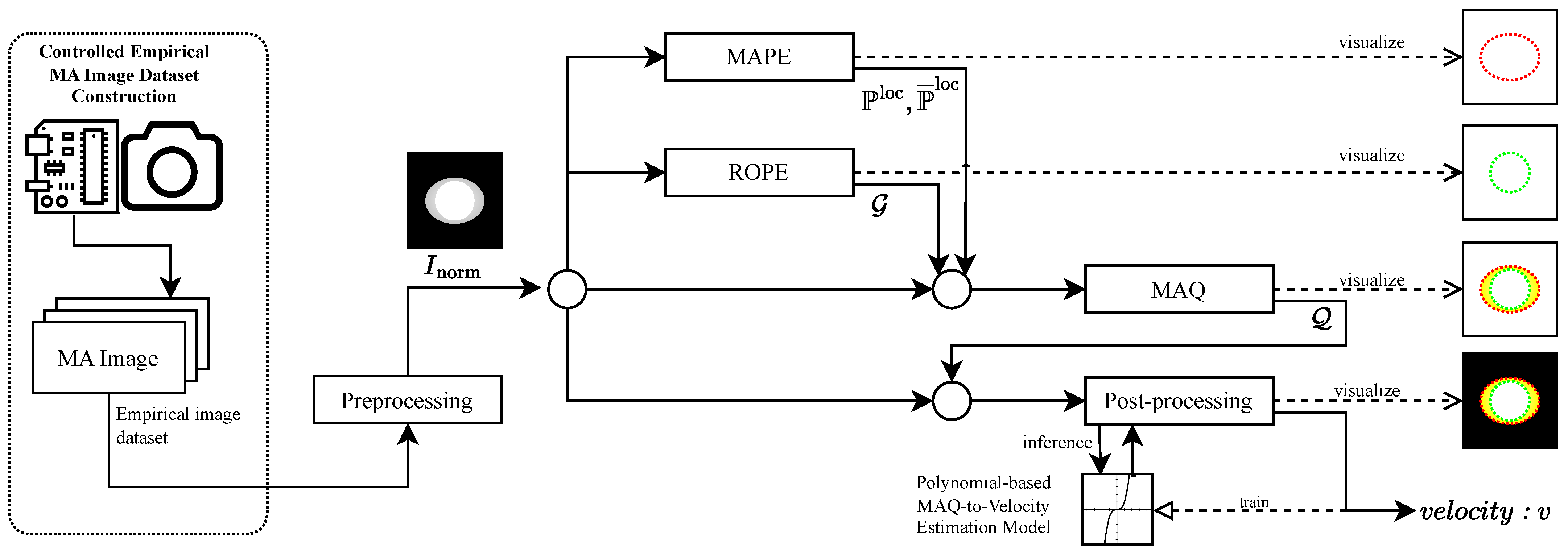
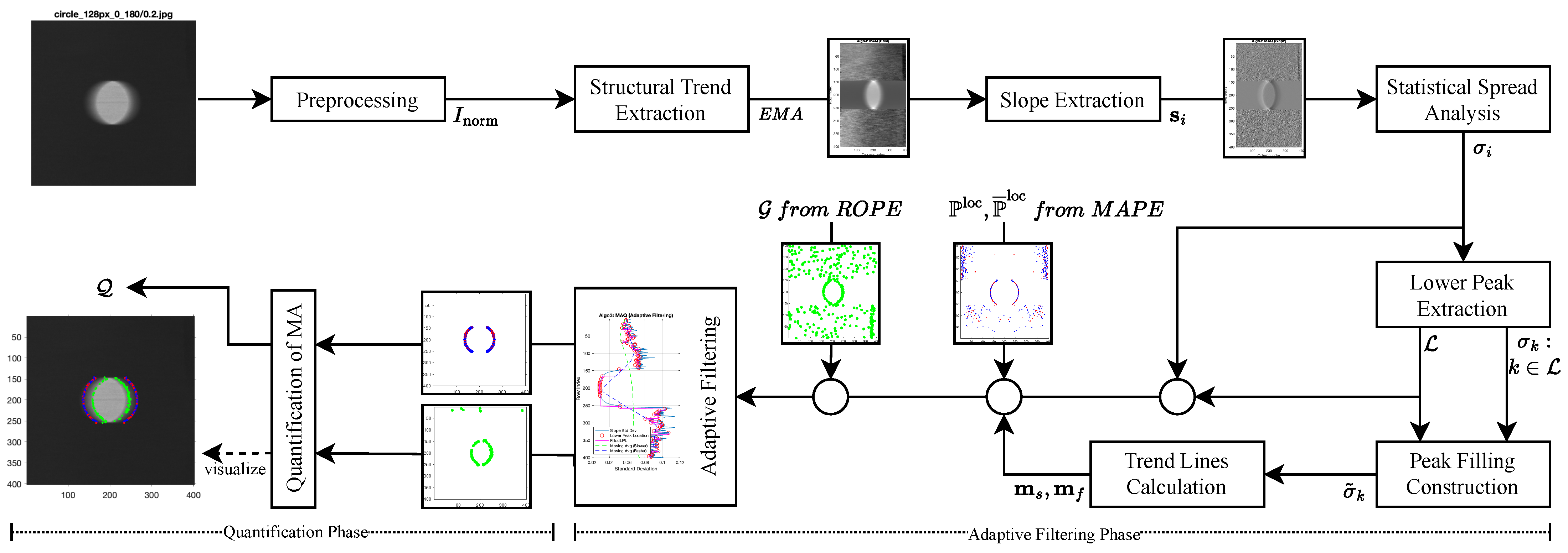
- A measure-first signal-analysis pipeline (MAPE–ROPE–MAQ) that detects likely blur-affected scanlines, estimates reference origin points, and quantifies blur magnitude as an interpretable spatial displacement.
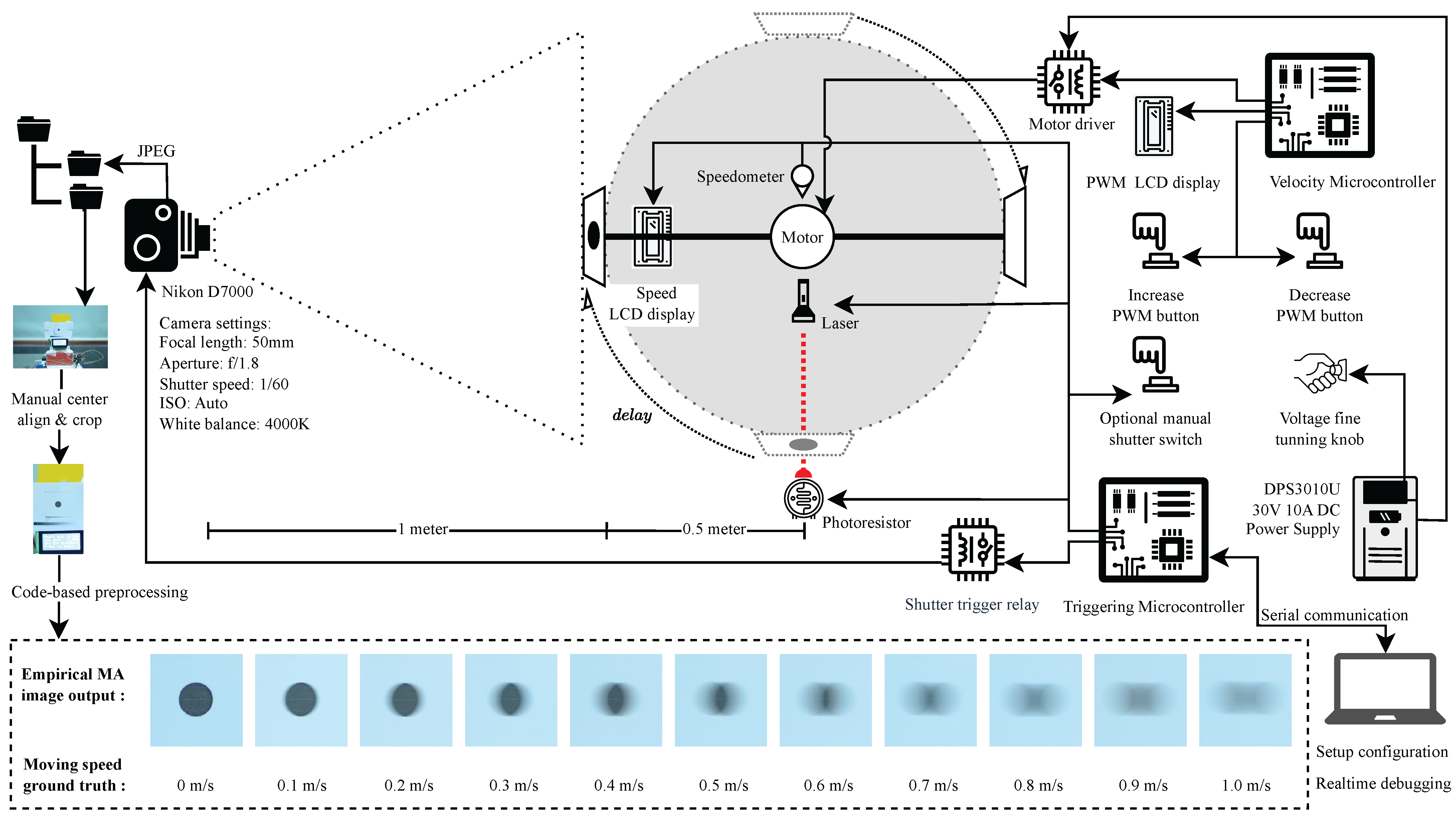
- A controlled empirical single-frame image dataset with known tangential velocities at capture time, enabling quantitative, ground-truth validation of motion-blur quantification.
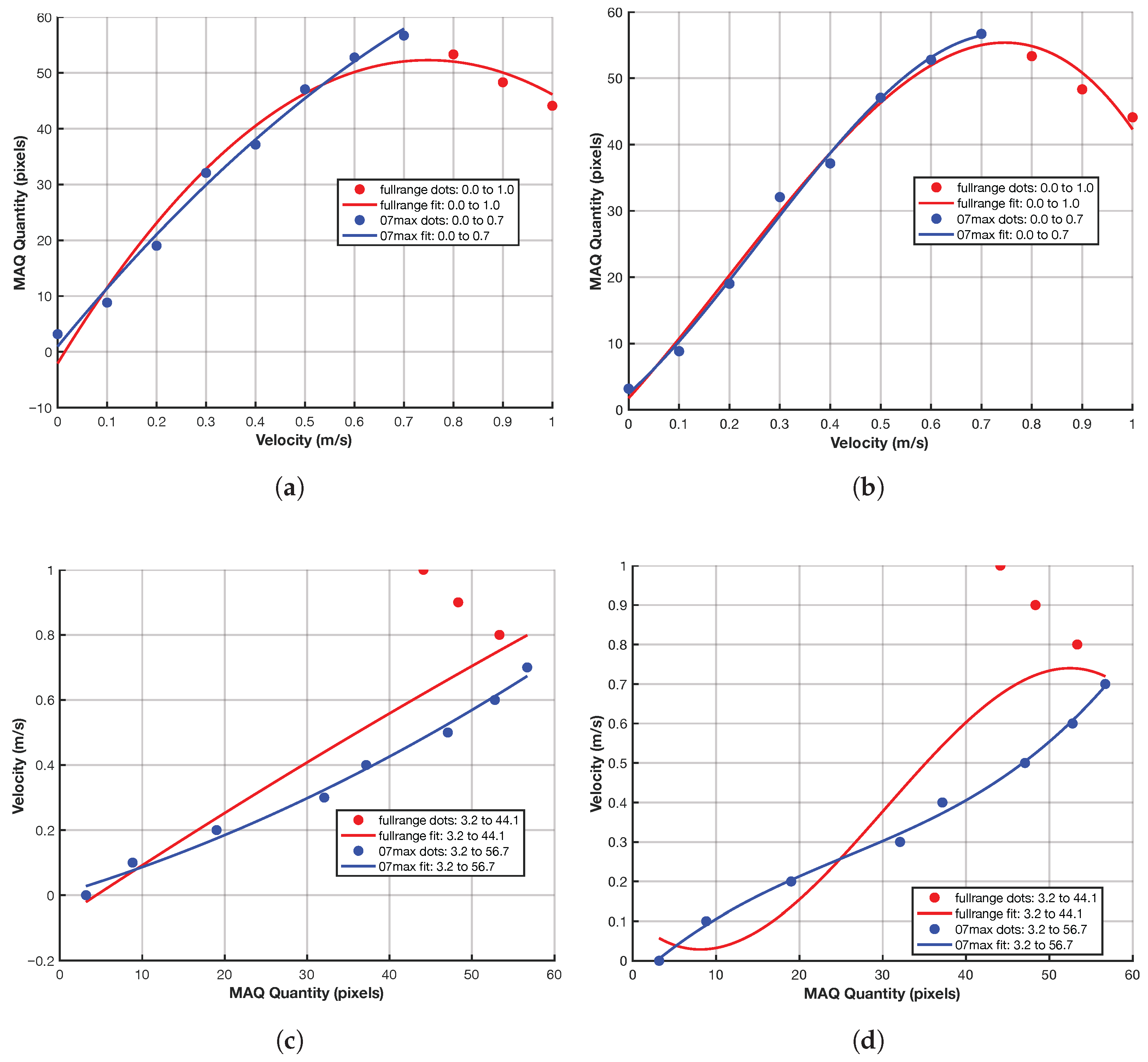
- An empirical mapping from MAQ displacement to velocity under the validated assumption of one-dimensional horizontal uniform motion blur.
2. Proposed Motion Blur Detection and Quantification Methodology
2.1. Mathematical Signal Representation of Motion-Blur Properties (Illustrative)
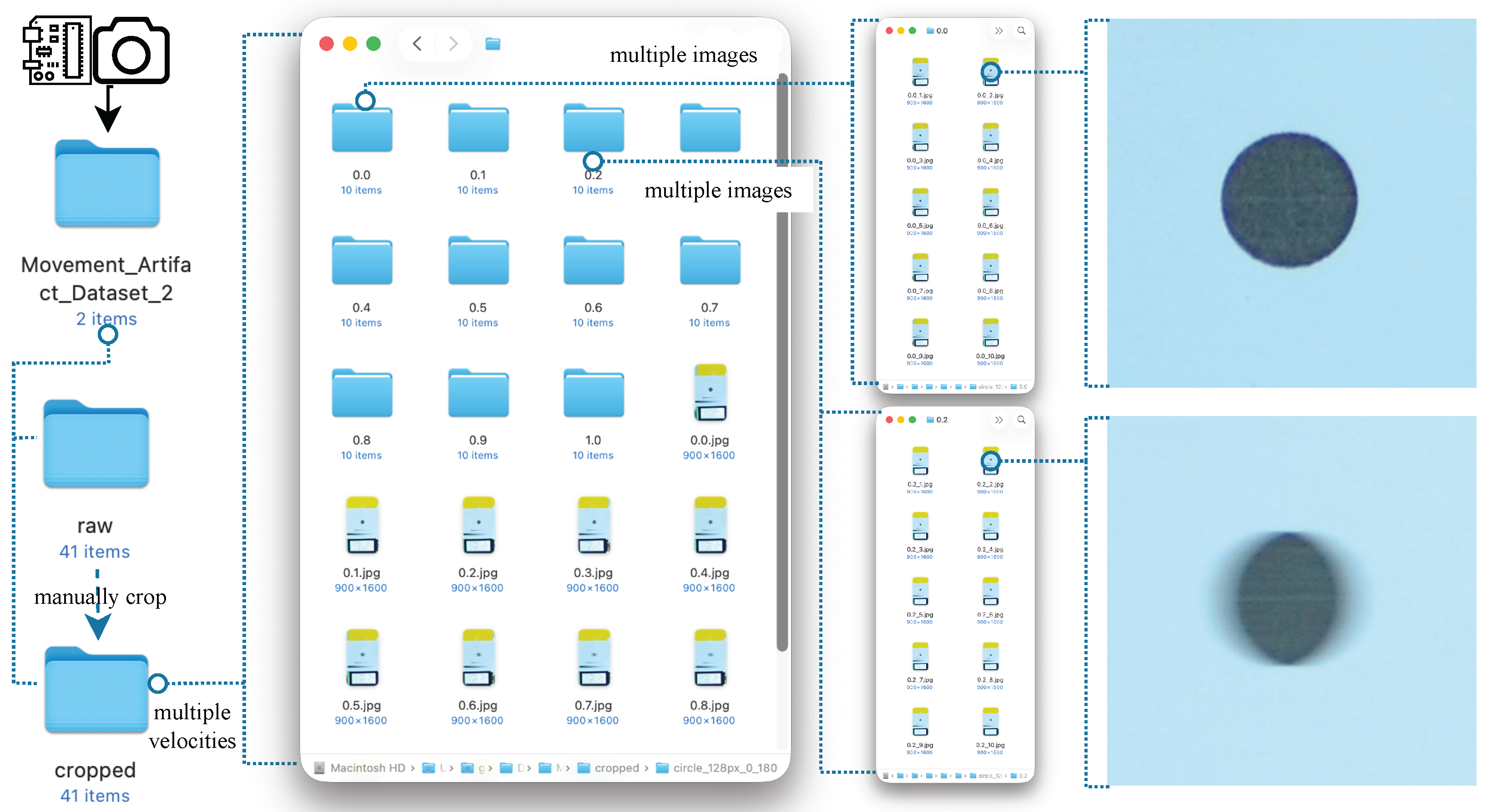
2.2. Empirical Dataset Construction
2.2.1. Objective and Ground-Truth Determination
2.2.2. Velocity Control Subsystem
2.2.3. Shutter Triggering Subsystem
2.3. Preprocessing
2.4. Movement Artifact Position Estimation (MAPE)
2.4.1. Self-Similarity Analysis
2.4.2. Peak Detection via Prominence Criterion
2.4.3. Identification of the Significant Second Peak
2.4.4. Symmetry-Based Lag Complementation
2.5. Reference Origin Point Estimation (ROPE)
2.5.1. Structural Trend Extraction
2.5.2. Gradient Computation
2.5.3. Dominant Transition Localization
2.5.4. Threshold-Based Filtering
2.6. Movement Artifact Quantification (MAQ)
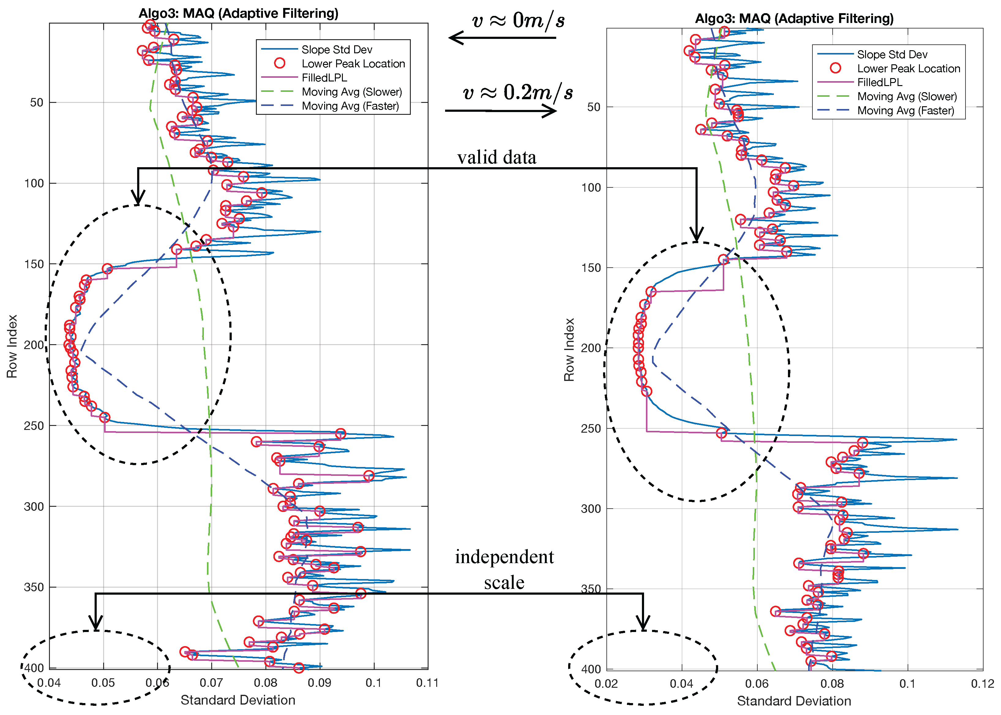
2.6.1. Adaptive Filtering Phase
2.6.2. Quantification Phase
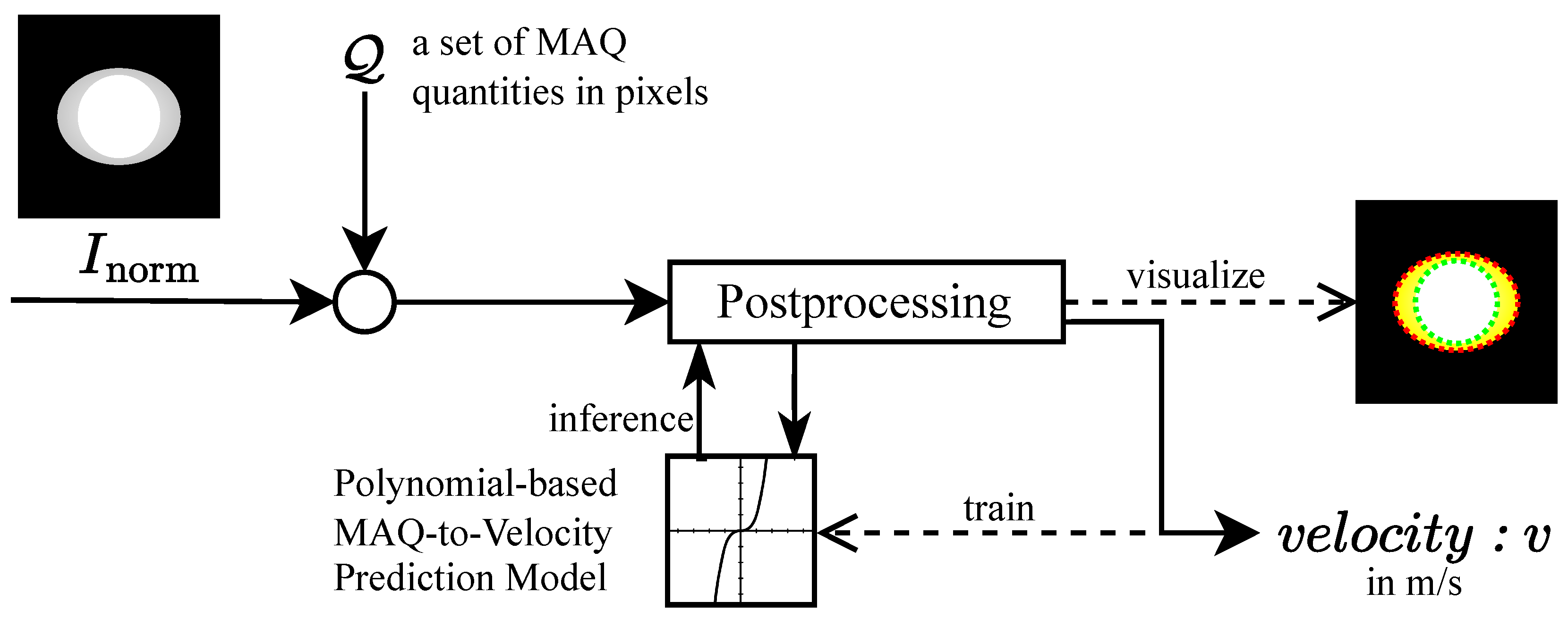
2.7. Post-Processing
2.7.1. Inference Phase: Velocity Estimation from MAQ
2.7.2. Training Phase: Polynomial Model Fitting
3. Experimental Setup
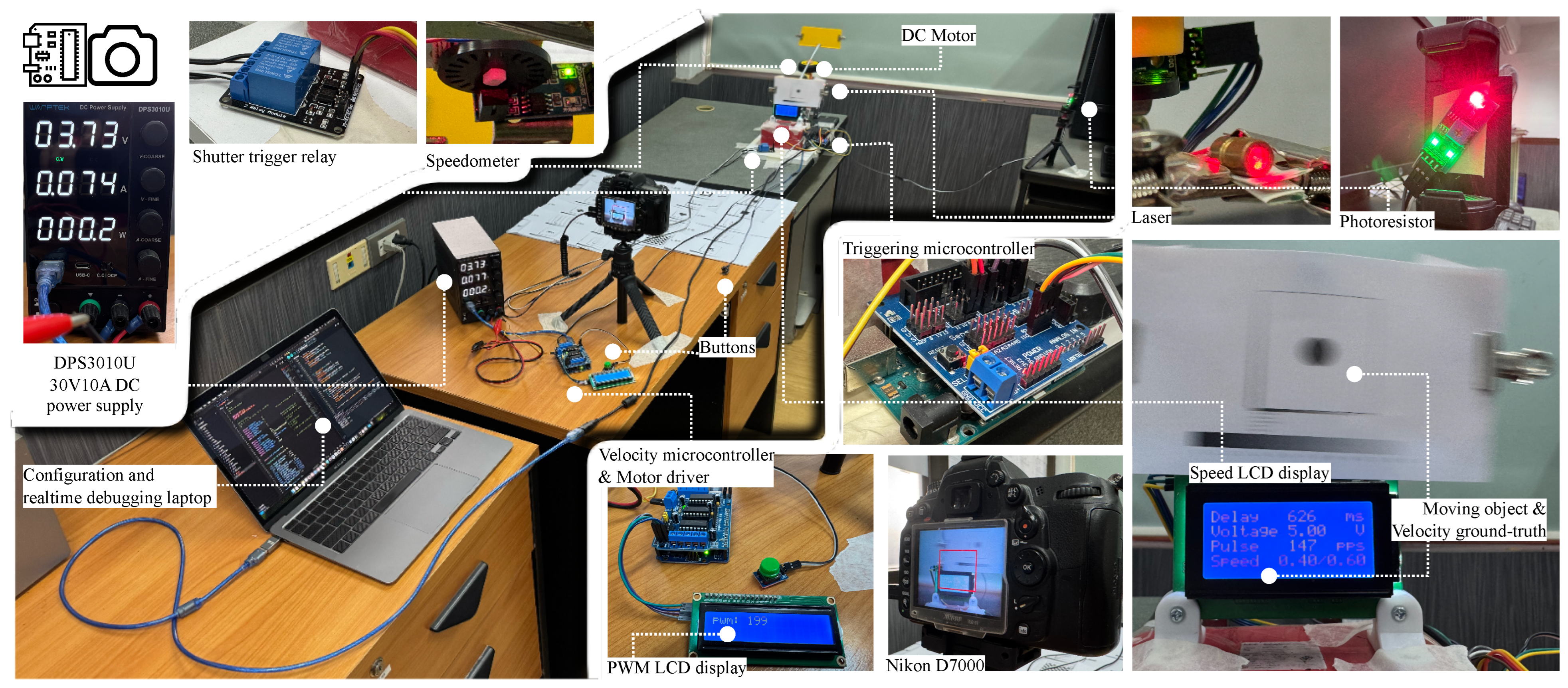
3.1. Dataset Construction and Hardware Configuration
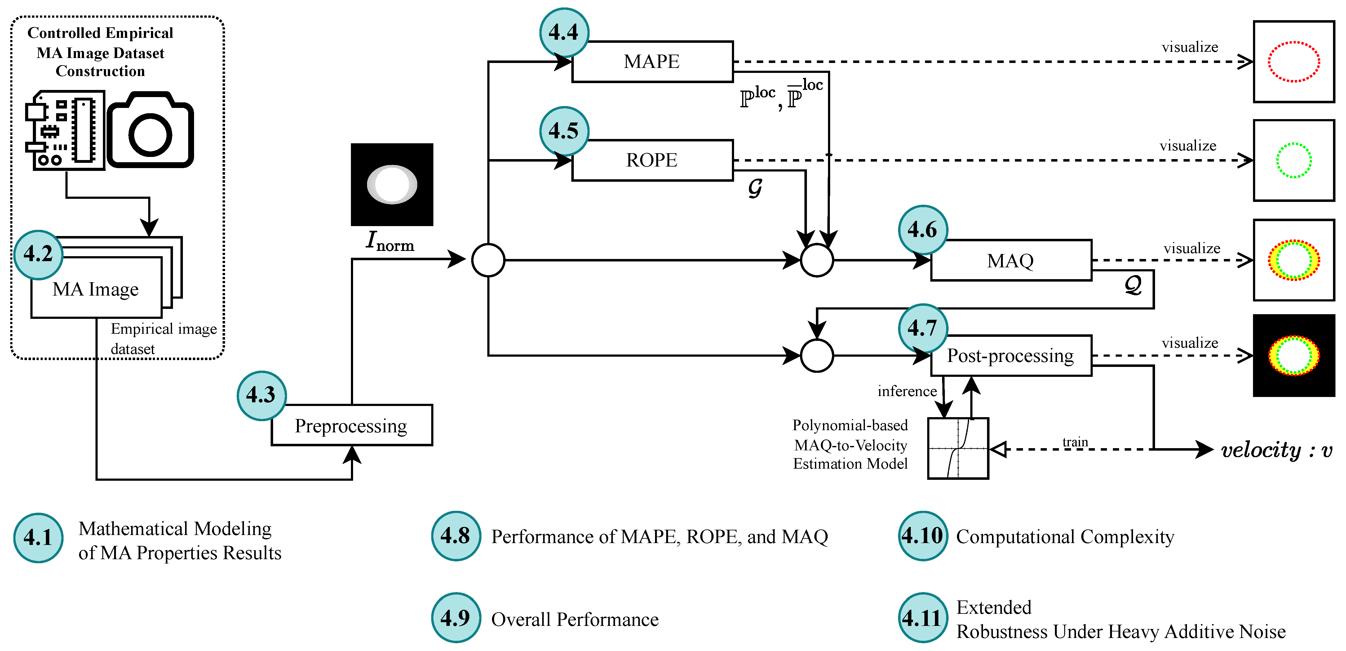
3.2. Software and Pipeline Configuration
3.3. Evaluation Protocol
4. Experimental Results and Analysis
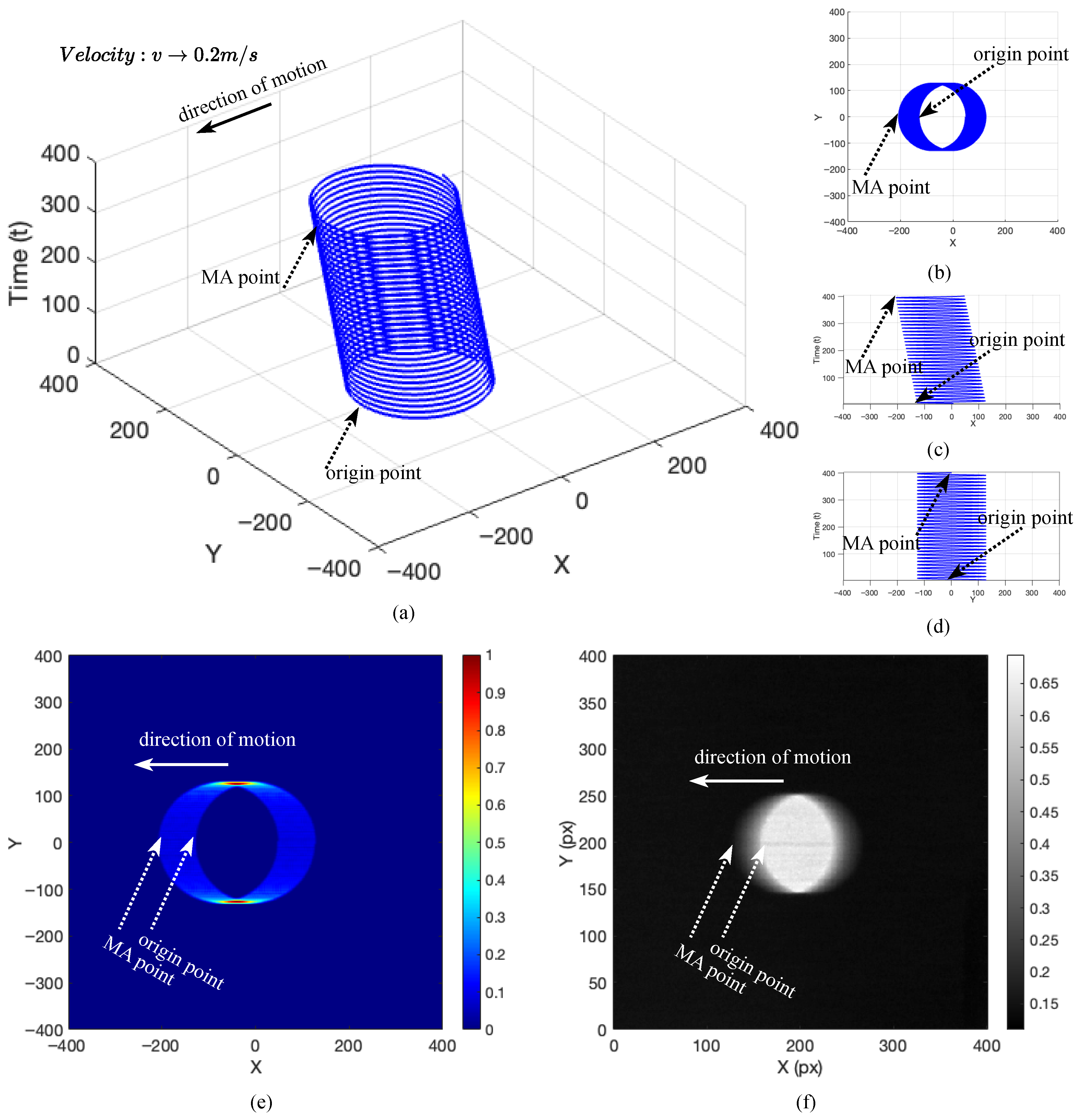
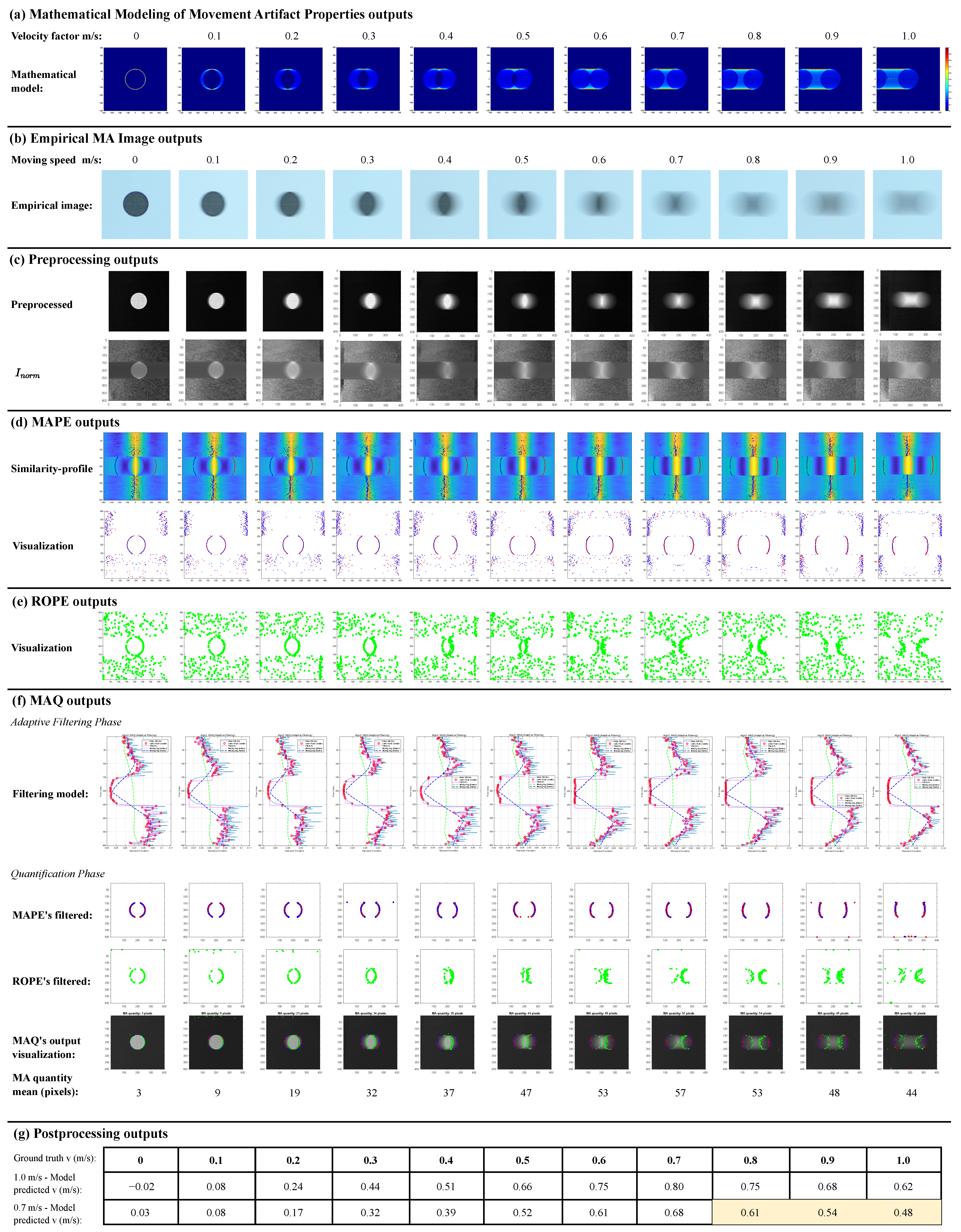
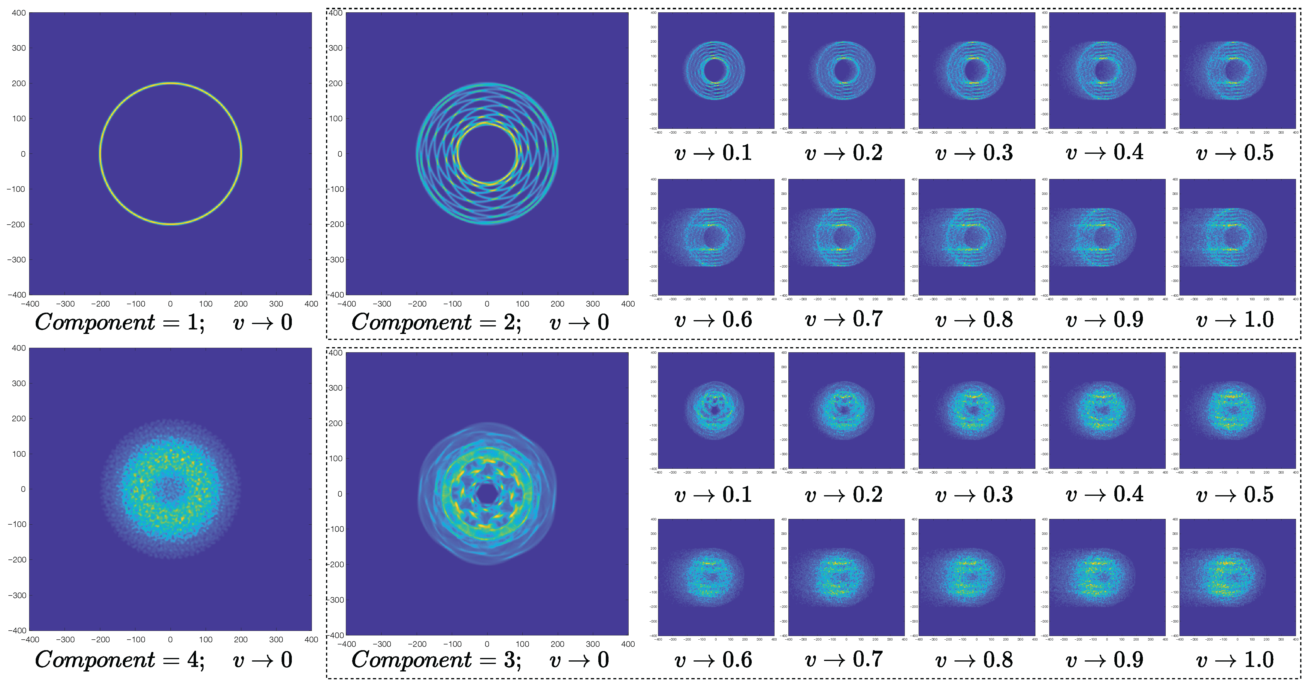
4.1. Results of Mathematical Modeling of MA Properties
4.2. Empirical Dataset Results
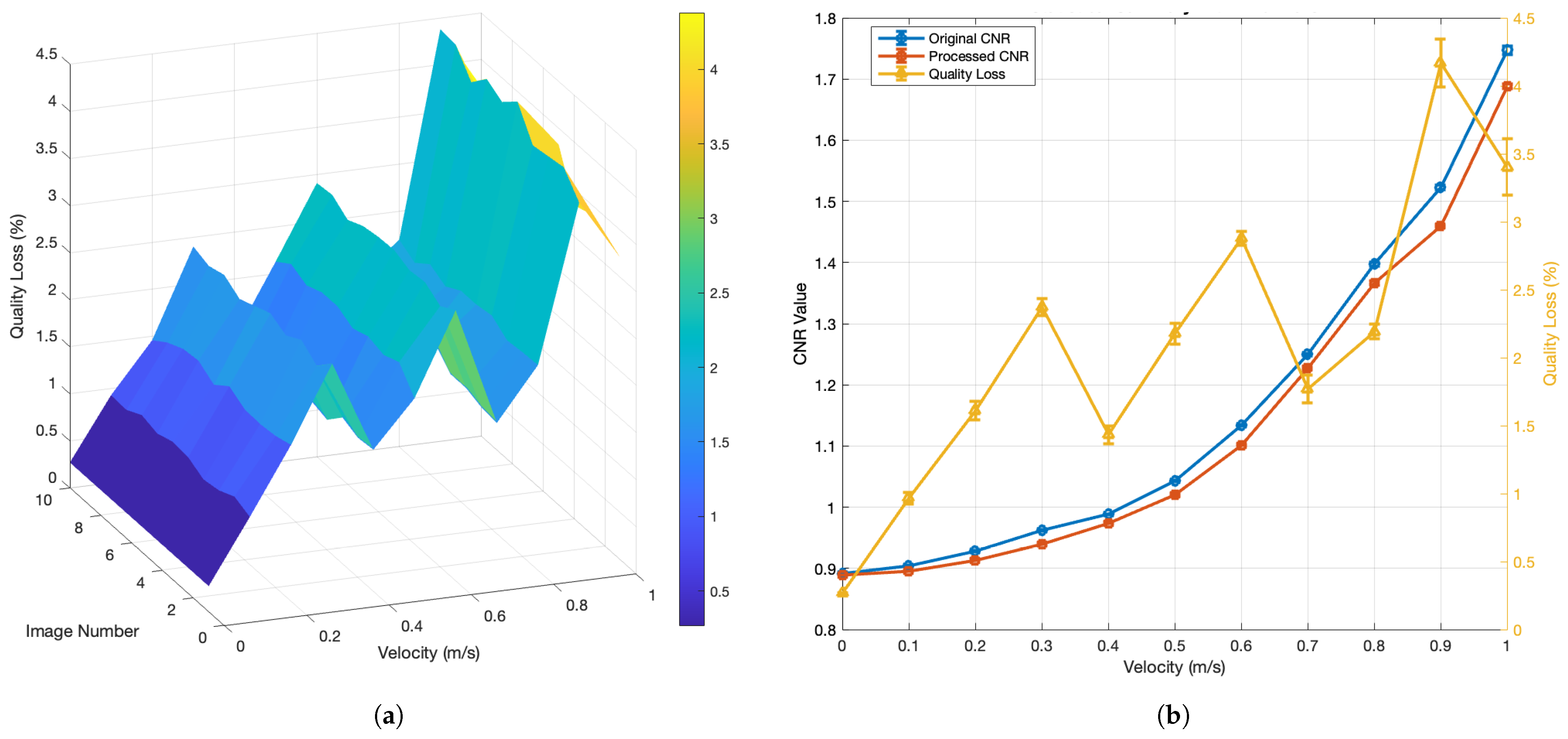
4.3. Preprocessing Results
4.4. MAPE Results
4.5. ROPE Results
4.6. MAQ Results
4.7. Post-Processing Results
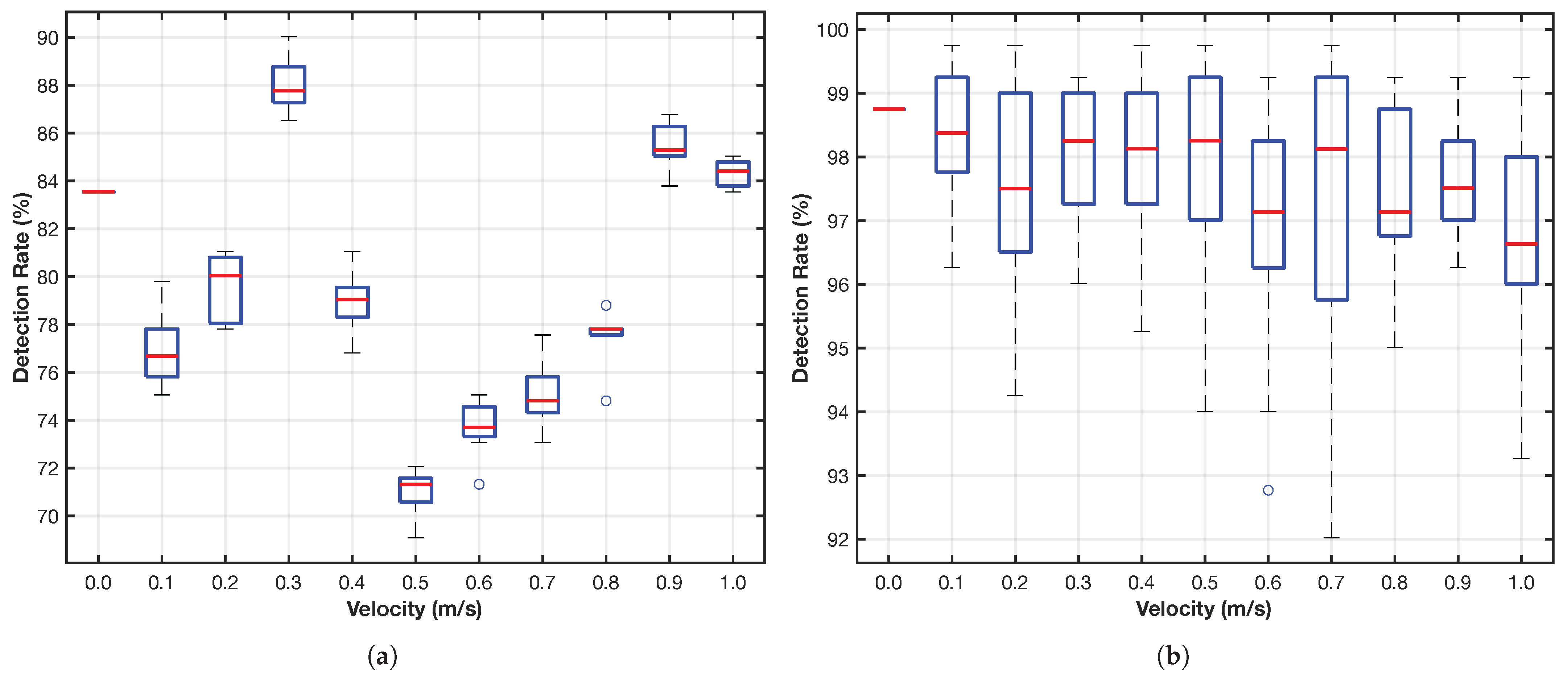
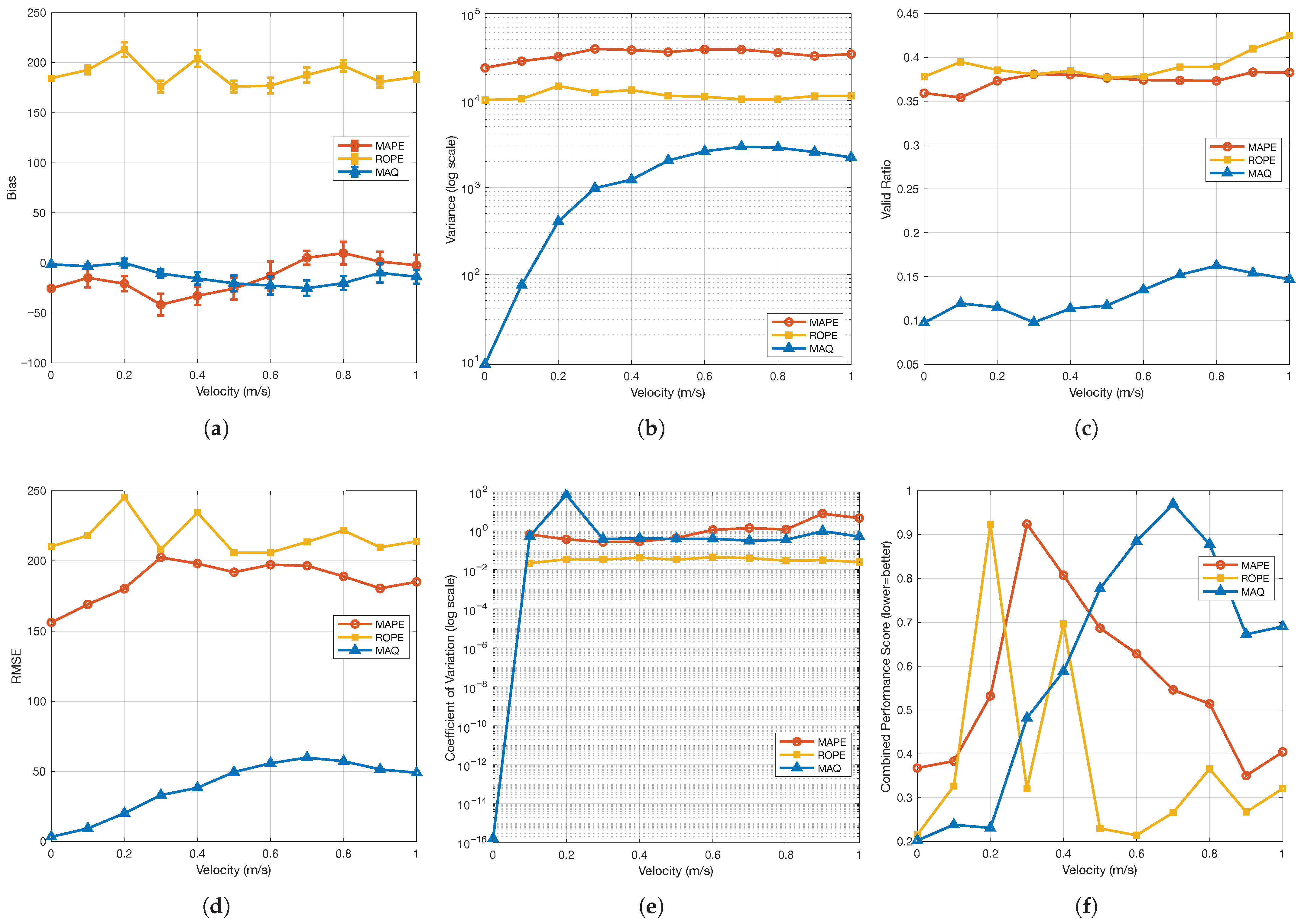
4.8. Performance of MAPE, ROPE, and MAQ
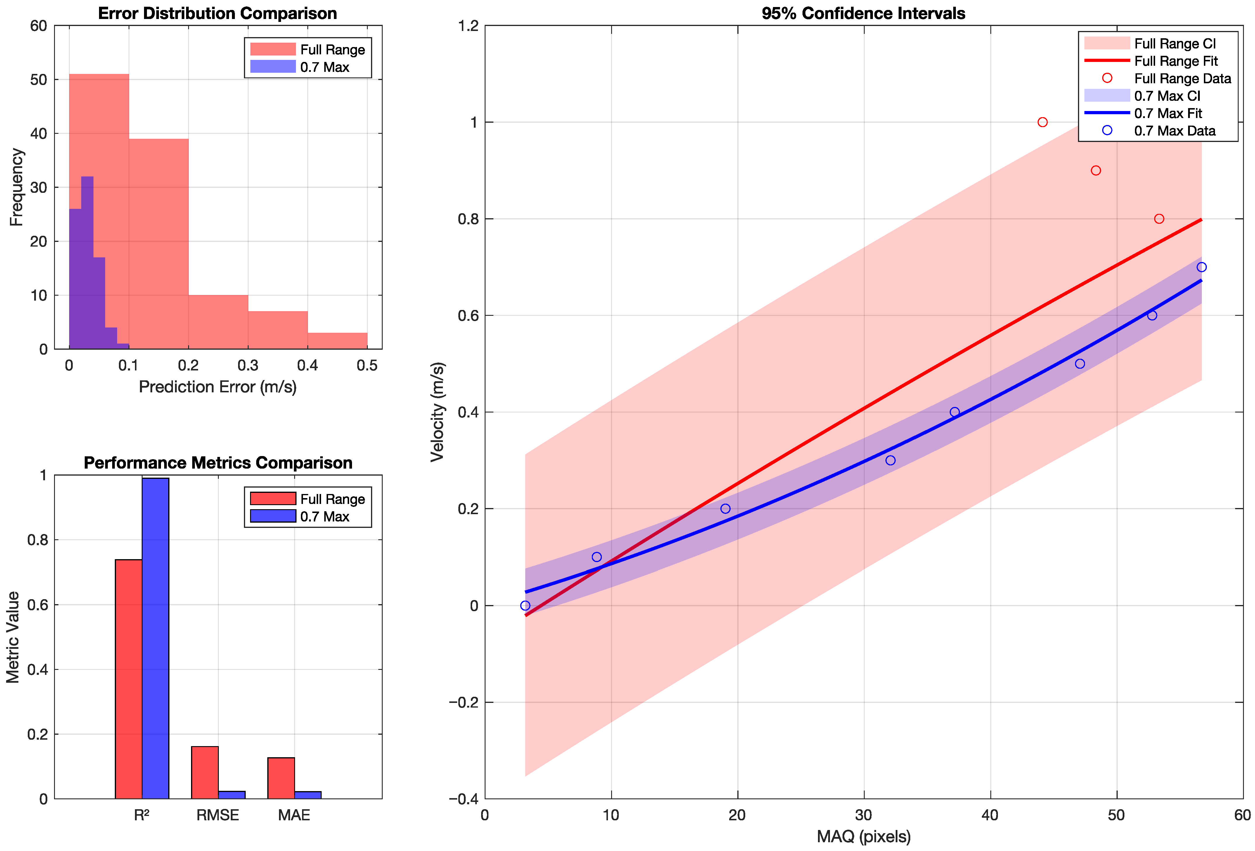
4.9. Overall MA Detection and Quantification Performance
4.10. Computational Complexity
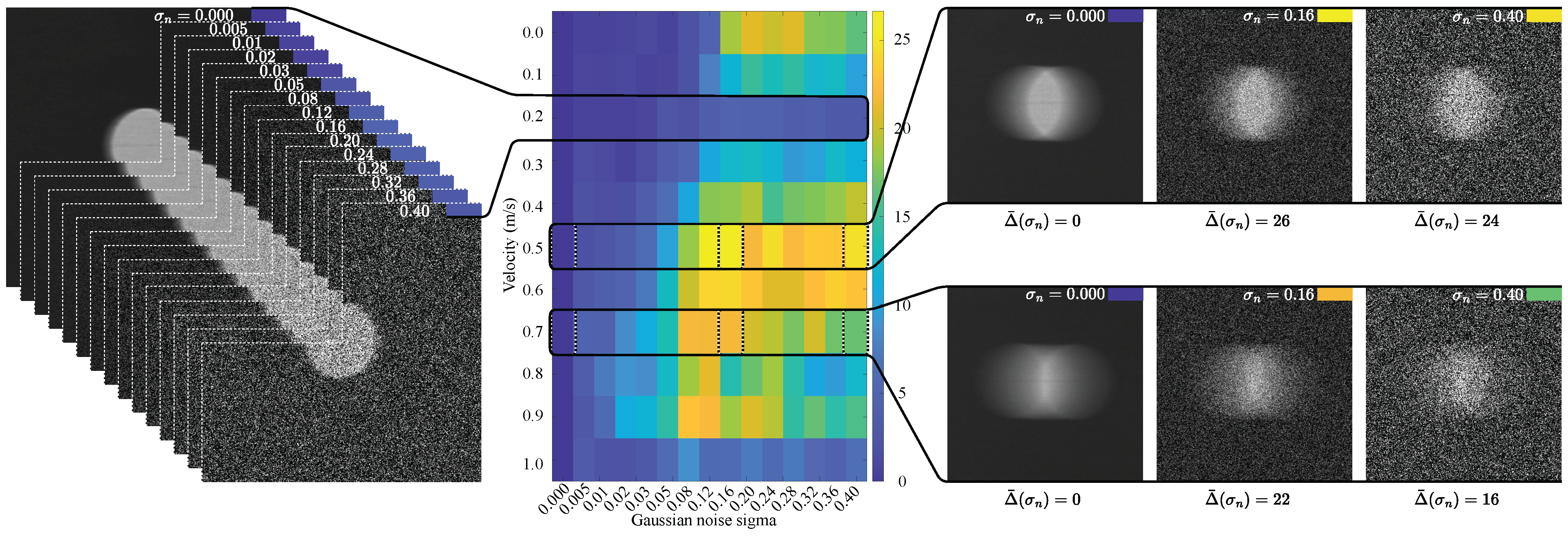
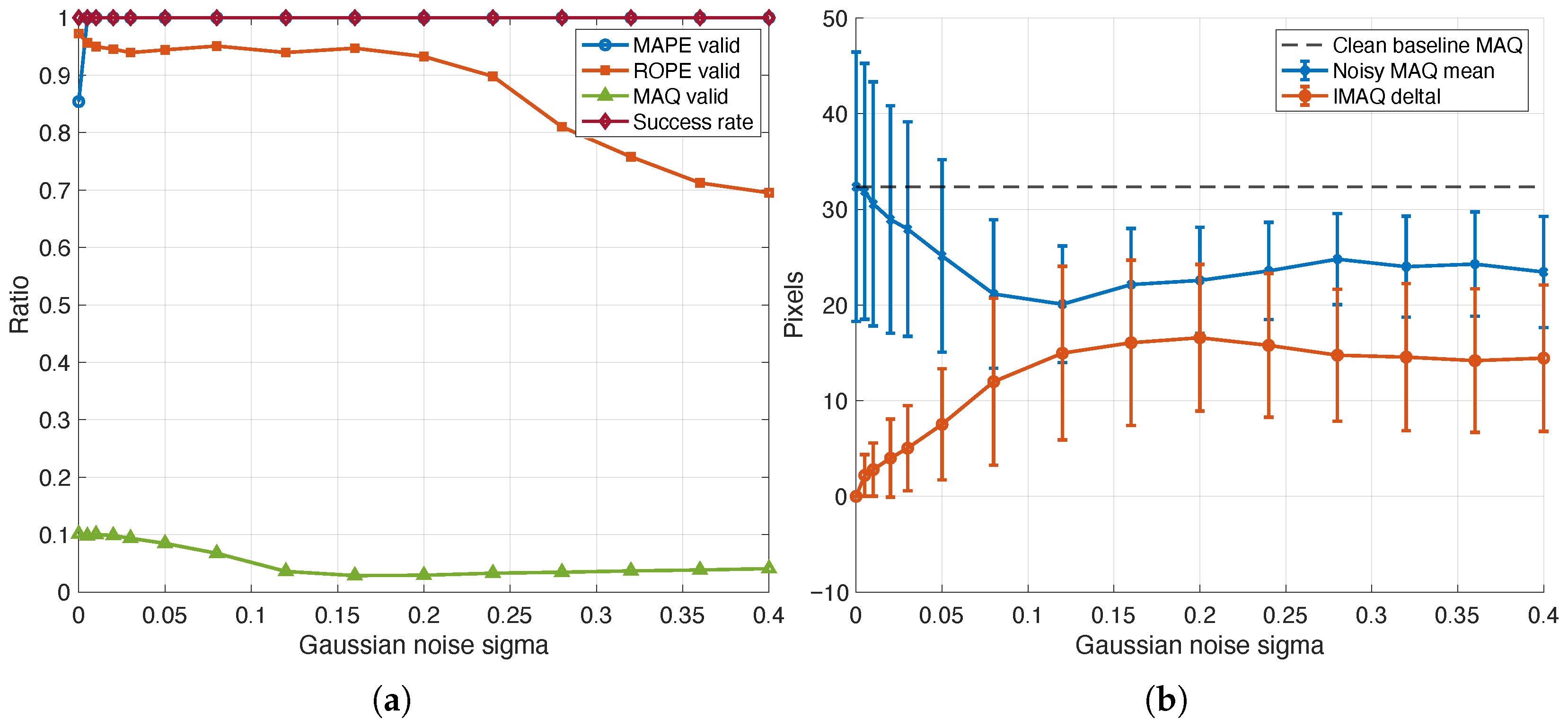
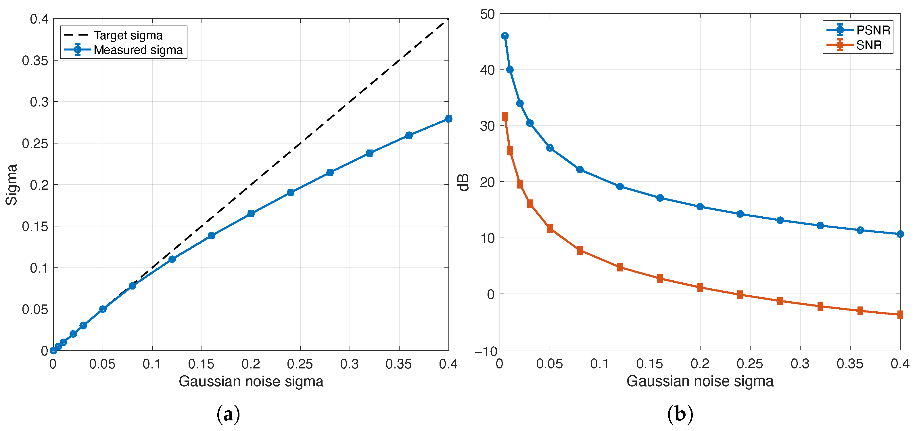
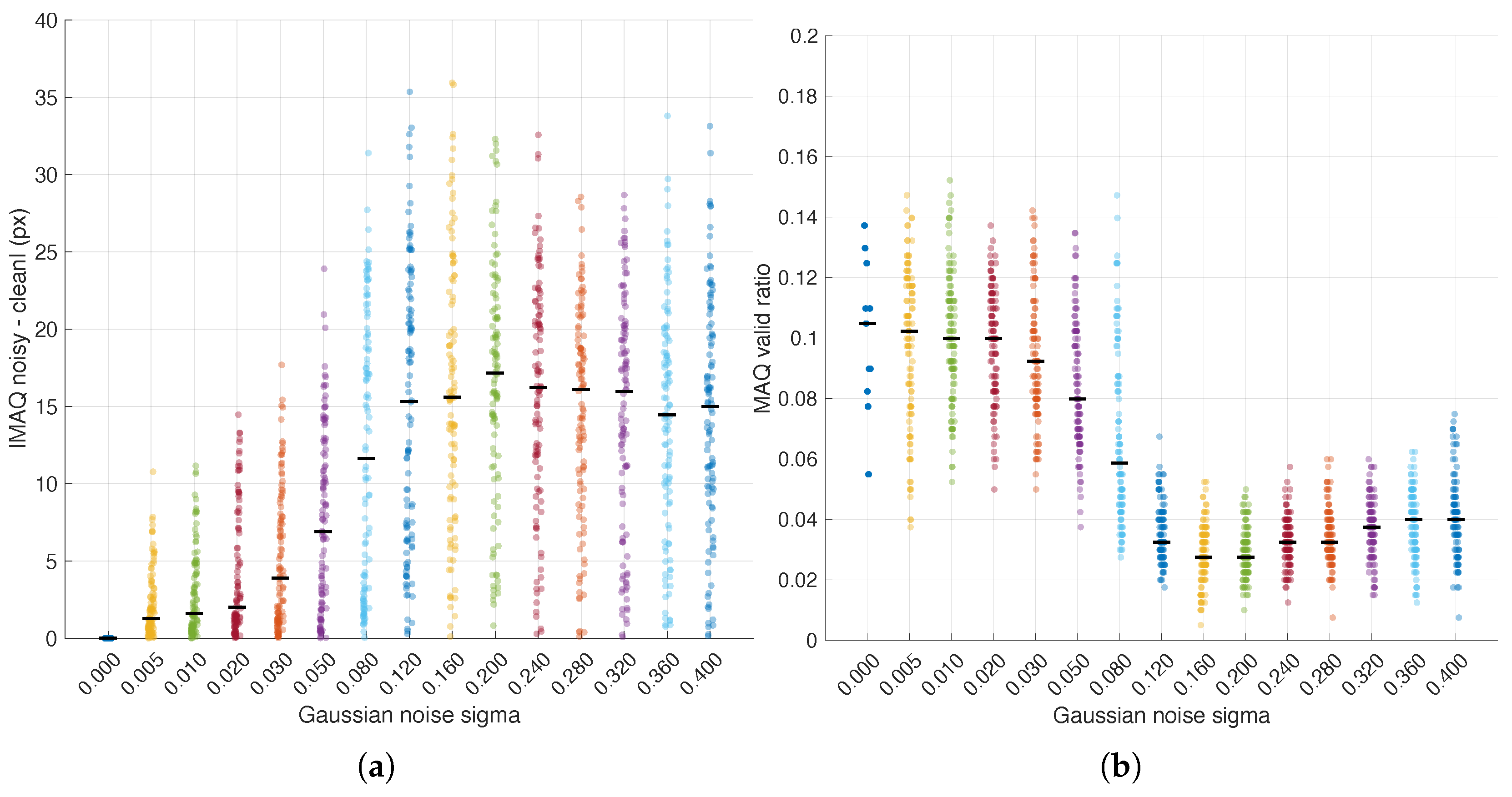
4.11. Extended Robustness Under Heavy Additive Noise
5. Discussion
5.1. Performance Analysis
5.2. Methodological Contributions
5.3. Practical Implications
5.4. Limitations and Considerations
5.5. Future Works and Research Directions
6. Conclusions
Author Contributions
Funding
Institutional Review Board Statement
Informed Consent Statement
Data Availability Statement
Acknowledgments
Conflicts of Interest
Abbreviations
| CASCAP | Cholangiocarcinoma Screening and Care Program |
| CCA | Cholangiocarcinoma |
| CNR | Contrast-to-Noise Ratio |
| CUDA | Compute Unified Device Architecture |
| DC | Direct Current |
| DFT | Discrete Fourier Transform |
| EMA | Exponential Moving Average |
| I2C | Inter-Integrated Circuit |
| ISO | ISO speed (camera sensitivity setting) |
| JPEG | Joint Photographic Experts Group |
| LCD | Liquid Crystal Display |
| LDR | Light Dependent Resistor |
| LUIAS | Liver Ultrasound Image Analysis System |
| MA | Movement Artifact (used here as a shorthand for motion blur) |
| MAE | Mean Absolute Error |
| MAPE | Movement Artifact Position Estimation |
| MAQ | Movement Artifact Quantification |
| MSE | Mean Squared Error |
| PSNR | Peak Signal-to-Noise Ratio |
| PWM | Pulse-Width Modulation |
| Coefficient of Determination | |
| RGB | Red, Green, Blue |
| RMSE | Root Mean Square Error |
| ROPE | Reference Origin Point Estimation |
| SNR | Signal-to-Noise Ratio |
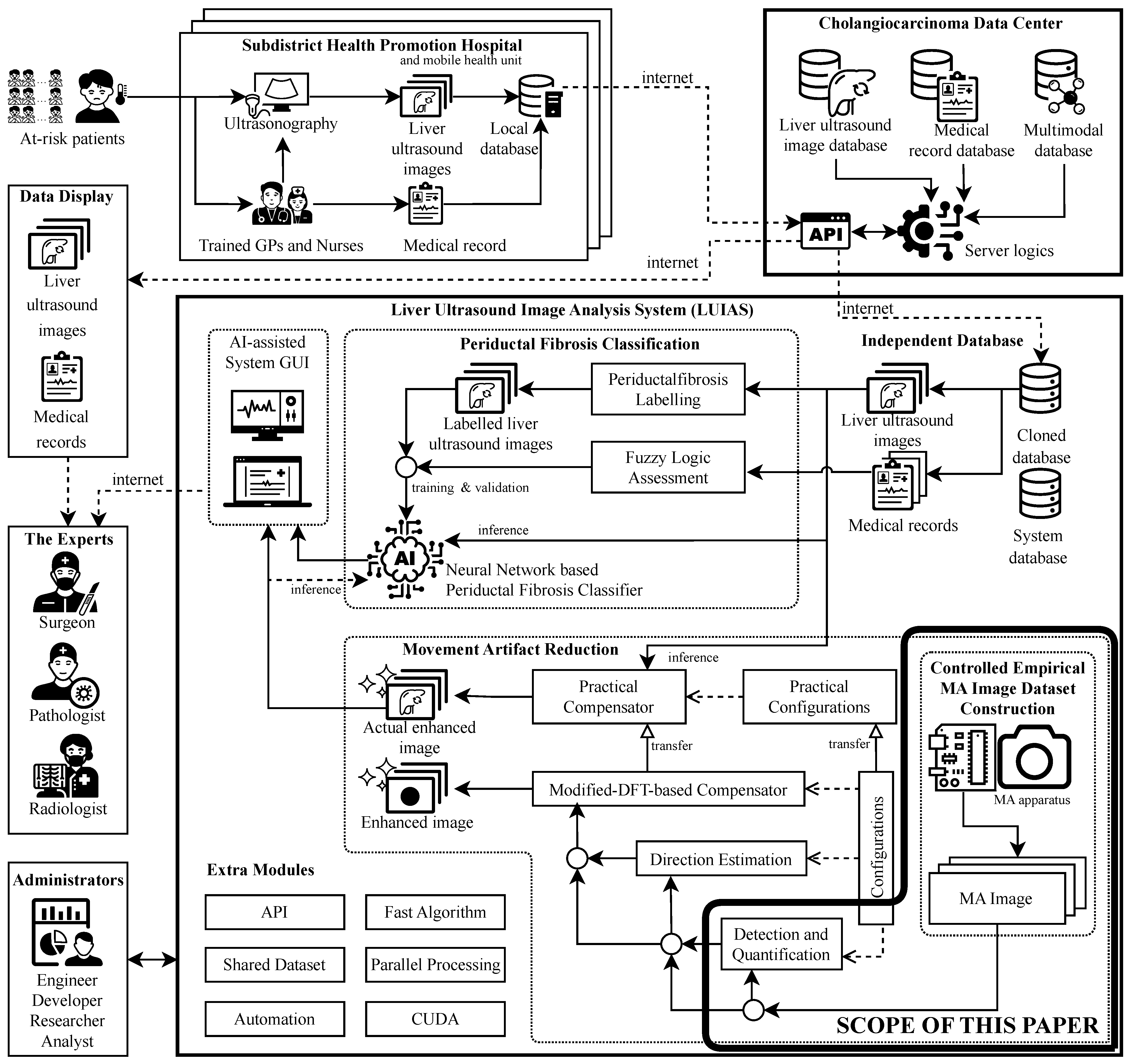
Appendix A. Prior Medical Imaging Motivation and System Context

Appendix B. Equivalence Between Real-Valued and Complex Signal Representations
Appendix C. 3D Trajectory to 2D Projection
Appendix D. Multiple-Component Trajectory Simulation

Appendix E. Standard Error for Normalized Impulse Response
Appendix F. Angular Velocity Calculation
Appendix G. Contrast to Noise Ratio (CNR) Calculation
Appendix H. Camera Settings and the Effects of Each Variable
Appendix H.1. Aperture (A)
Appendix H.2. Shutter Speed (S)
Appendix H.3. ISO Sensitivity (ISO)
Appendix I. Movement Artifact Generator System Architecture
Appendix I.1. Hardware Configuration
Appendix I.2. Speed Measurement Algorithm
Appendix I.3. Control Logic Implementation
Appendix I.4. Camera Synchronization Protocol
Appendix I.5. Serial Communication Interface
Appendix J. Supplementary Details for the Additive-Noise Robustness Analysis


References
- Santamarina, J.C.; Fratta, D. Discrete Signals and Inverse Problems: An Introduction for Engineers and Scientists; Wiley: Hoboken, NJ, USA, 2005. [Google Scholar] [CrossRef] [Scilit]
- Levin, A.; Weiss, Y.; Durand, F.; Freeman, W.T. Understanding and evaluating blind deconvolution algorithms. In Proceedings of the 2009 IEEE Conference on Computer Vision and Pattern Recognition (CVPR), Miami, FL, USA, 20–25 June 2009; pp. 1964–1971. [Google Scholar] [CrossRef] [Scilit]
- Wayalun, P.; Saiyod, S.; Chamadol, N. A hybrid technique for enhancement of Periductal fibrosis ultrasound images for Cholangiocacinoma surveillance. In Proceedings of the 2014 6th International Conference on Information Technology and Electrical Engineering (ICITEE), Yogyakarta, Indonesia, 7–8 October 2014; pp. 1–5. [Google Scholar] [CrossRef] [Scilit]
- Nonsakhoo, W.; Saiyod, S.; Sirisawat, P.; Suwanwerakamtorn, R.; Chamadol, N.; Khuntikeo, N. Liver Ultrasound Image Classification of Periductal Fibrosis Based on Transfer Learning and FCNet for Liver Ultrasound Images Analysis System. In Proceedings of the 2021 International Conference on Computing, Communication, and Intelligent Systems (ICCCIS), Greater Noida, India, 19–20 February 2021; pp. 569–575. [Google Scholar] [CrossRef] [Scilit]
- Rhyou, S.-Y.; Yoo, J.-C. Cascaded Deep Learning Neural Network for Automated Liver Steatosis Diagnosis Using Ultrasound Images. Sensors 2021, 21, 5304. [Google Scholar] [CrossRef] [Scilit]
- Wiacek, A.; González, E.; Bell, M.A.L. CohereNet: A Deep Learning Architecture for Ultrasound Spatial Correlation Estimation and Coherence-Based Beamforming. IEEE Trans. Ultrason. Ferroelectr. Freq. Control 2020, 67, 2574–2583. [Google Scholar] [CrossRef] [Scilit]
- Lee, J.H.; Hwang, Y.N.; Park, S.Y.; Jeong, J.S.; Kim, S.M. An Analysis of Contrast Agent Flow Patterns from Sequential Ultrasound Images Using a Motion Estimation Algorithm Based on Optical Flow Patterns. IEEE Trans. Biomed. Eng. 2015, 62, 49–59. [Google Scholar] [CrossRef] [Scilit] [PubMed]
- Lei, S.; Zhang, G.; Zhu, B.; Long, X.; Jiang, Z.; Liu, Y.; Hu, D.; Sheng, Z.; Zhang, Q.; Wang, C.; et al. In Vivo Ultrasound Localization Microscopy Imaging of the Kidney’s Microvasculature with Block-Matching 3-D Denoising. IEEE Trans. Ultrason. Ferroelectr. Freq. Control 2022, 69, 523–533. [Google Scholar] [CrossRef] [Scilit] [PubMed]
- Song, Z.; Zou, S.; Zhou, W.; Huang, Y.; Shao, L.; Yuan, J.; Gou, X.; Jin, W.; Wang, Z.; Chen, X.; et al. Clinically applicable histopathological diagnosis system for gastric cancer detection using deep learning. Nat. Commun. 2020, 11, 4294. [Google Scholar] [CrossRef] [Scilit] [PubMed]
- Tounsi, M.; Aram, E.; Azar, A.T.; Al-Khayyat, A.; Ibraheem, I.K. A Comprehensive Review on Biomedical Image Classification using Deep Learning Models. Eng. Technol. Appl. Sci. Res. 2025, 15, 19538–19545. [Google Scholar] [CrossRef] [Scilit]
- Cui, L.; Song, Y.; Wang, Y.; Wang, R.; Wu, D.; Xie, H.; Li, J.; Yang, G. Motion artifact reduction for magnetic resonance imaging with deep learning and k-space analysis. PLoS ONE 2023, 18, e0278668. [Google Scholar] [CrossRef] [Scilit]
- Yan, Z.; Chen, Z.; Li, L.; Zhang, L.; Wu, D. An end-to-end neural network for 4D cardiac CT reconstruction using single-beat scans. Phys. Med. Biol. 2025, 70, 095003. [Google Scholar] [CrossRef] [Scilit]
- Gardner, M.; Dillon, O.; Reynolds, T.; Kipritidis, J.; Bazalova-Carter, M.; Byrne, H.; Stewart, M.; Booth, J.; Keall, P.J. Evaluation of 4D cone-beam CT reconstruction methods for lung images acquired using rapid cone-beam CT acquisition: A phantom study. Phys. Med. Biol. 2025, 70, 135004. [Google Scholar] [CrossRef] [Scilit]
- Jurß, P.; Knopp, T.; Busse, B.; von Brackel, F.N.; Boberg, M. Optimization-Based Motion Estimation in HR-PQCT. In Proceedings of the 2025 IEEE 22nd International Symposium on Biomedical Imaging (ISBI), Houston, TX, USA, 14–17 April 2025; pp. 1–4. [Google Scholar] [CrossRef] [Scilit]
- Loupas, T.; Powers, J.T.; Gill, R.W. An axial velocity estimator for ultrasound blood flow imaging, based on a full evaluation of the Doppler equation by means of a two-dimensional autocorrelation approach. IEEE Trans. Ultrason. Ferroelectr. Freq. Control 1995, 42, 672–688. [Google Scholar] [CrossRef] [Scilit]
- Ye, C.-Q.; Luo, L.; Gao, X.-R.; Jiang, L.-R.; Yue, H.-L. Cross-correlation phase correction method on ultrasonic synthetic focused signal distortion. In Proceedings of the 2015 IEEE Far East NDT New Technology and Application Forum (FENDT), Zhuhai, China, 28–31 May 2015; pp. 223–228. [Google Scholar] [CrossRef] [Scilit]
- Nonsakhoo, W.; Sirisawat, P.; Saiyod, S.; Benjamas, N. Angle of arrival estimation by using stereo ultrasonic technique for local positioning system. In Proceedings of the 2017 IEEE 13th International Colloquium on Signal Processing & its Applications (CSPA), Penang, Malaysia, 10–12 March 2017; pp. 112–117. [Google Scholar] [CrossRef] [Scilit]
- Gonzalez, R.C.; Woods, R.E. Digital Image Processing, 4th ed.; Pearson: New York, NY, USA, 2018. [Google Scholar]
- Jain, A.K. Fundamentals of Digital Image Processing; Prentice-Hall: Hoboken, NJ, USA, 1989. [Google Scholar]
- Selesnick, I.W.; Baraniuk, R.G.; Kingsbury, N.C. The dual-tree complex wavelet transform. IEEE Signal Process. Mag. 2005, 22, 123–151. [Google Scholar] [CrossRef] [Scilit]
- Buades, A.; Coll, B.; Morel, J.-M. A non-local algorithm for image denoising. In Proceedings of the 2005 IEEE Computer Society Conference on Computer Vision and Pattern Recognition (CVPR’05), San Diego, CA, USA, 20–26 June 2005; Volume 2, pp. 60–65. [Google Scholar] [CrossRef] [Scilit]
- Wang, F.; Zhao, X.-L.; Ng, M.K. Multiplicative Noise and Blur Removal by Framelet Decomposition and l1-Based L-Curve Method. IEEE Trans. Image Process. 2016, 25, 4222–4232. [Google Scholar] [CrossRef] [Scilit]
- Damodaran, N.; Ramamurthy, S.; Velusamy, S.; Gayathri, K.M. Speckle Noise Reduction in Ultrasound Biomedical B-Scan Images Using Discrete Topological Derivative. Ultrasound Med. Biol. 2012, 38, 276–286. [Google Scholar] [CrossRef] [Scilit]
- Freund, I. Joseph W. Goodman: Speckle Phenomena in Optics: Theory and Applications. J. Stat. Phys. 2008, 130, 413–414. [Google Scholar] [CrossRef] [Scilit]
- Zhao, C.; Liu, J.; Zhang, J. A Dual Model for Restoring Images Corrupted by Mixture of Additive and Multiplicative Noise. IEEE Access 2021, 9, 168869–168888. [Google Scholar] [CrossRef] [Scilit]
- Che, H.; Tang, Y. A Simplified Convex Optimization Model for Image Restoration with Multiplicative Noise. J. Imaging 2023, 9, 229. [Google Scholar] [CrossRef] [Scilit] [PubMed]
- Izzetoglu, M.; Devaraj, A.; Bunce, S.; Onaral, B. Motion artifact cancellation in NIR spectroscopy using Wiener filtering. IEEE Trans. Biomed. Eng. 2005, 52, 934–938. [Google Scholar] [CrossRef] [Scilit] [PubMed]
- Kadah, Y.M.; El-Sharkawy, A.E.-M.; Youssef, A.-B.M. Navigator echo motion artifact suppression in synthetic aperture ultrasound imaging. IEEE Trans. Biomed. Eng. 2005, 52, 127–131. [Google Scholar] [CrossRef]
- Yolgunlu, P.S.; Martiartu, N.K.; Gerber, U.R.; Frenz, M.; Jaeger, M. Excluding Echo Shift Noise in Real-Time Pulse-Echo Speed-of-Sound Imaging. Sensors 2023, 23, 5598. [Google Scholar] [CrossRef] [Scilit]
- Haloubi, T.; Thomas, S.; Hines, C.; Dhaliwal, K.; Hopgood, J.R. Motion Compensation in Pulmonary Fluorescence Lifetime Imaging: An Image Processing Pipeline for Artefact Reduction and Clinical Precision. IEEE Open J. Eng. Med. Biol. 2025, 6, 432–441. [Google Scholar] [CrossRef] [Scilit]
- Harput, S.; Christensen-Jeffries, K.; Brown, J.; Li, Y.; Williams, K.J.; Davies, A.H.; Eckersley, R.J.; Dunsby, C.; Tang, M.-X. Two-Stage Motion Correction for Super-Resolution Ultrasound Imaging in Human Lower Limb. IEEE Trans. Ultrason. Ferroelectr. Freq. Control. 2018, 65, 803–814. [Google Scholar] [CrossRef] [Scilit]
- Mohan, K.A.; Panas, R.M.; Cuadra, J.A. SABER: A Systems Approach to Blur Estimation and Reduction in X-Ray Imaging. IEEE Trans. Image Process. 2020, 29, 7751–7764. [Google Scholar] [CrossRef] [Scilit]
- Iwabuchi, K.; Kameda, Y.; Hamamoto, T. Image Quality Improvements Based on Motion-Based Deblurring for Single-Photon Imaging. IEEE Access 2021, 9, 30080–30094. [Google Scholar] [CrossRef] [Scilit]
- Tretbar, S.H.; Hewener, H.J.; Speicher, D.; Barthscherer, T.; Bongers, A.; Jenne, J.W.; Günther, M. MR-compatible ultrasound research platform for motion tracking to reduce motion induced artifacts in MR imaging. In Proceedings of the 2013 IEEE International Ultrasonics Symposium (IUS), Prague, Czech Republic, 21–25 July 2013; pp. 553–556. [Google Scholar] [CrossRef] [Scilit]
- Seo, J.; Kim, S.K.; Kim, Y.-S.; Choi, K.; Kong, D.G.; Bang, W.-C. Motion Compensation for Ultrasound Thermal Imaging Using Motion-Mapped Reference Model: An in vivo Mouse Study. IEEE Trans. Biomed. Eng. 2014, 61, 2669–2678. [Google Scholar] [CrossRef] [Scilit]
- Yang, C.; Tavassolian, N. Motion Artifact Cancellation of Seismocardiographic Recording from Moving Subjects. IEEE Sens. J. 2016, 16, 5702–5708. [Google Scholar] [CrossRef] [Scilit]
- Fergus, R.; Singh, B.; Hertzmann, A.; Roweis, S.T.; Freeman, W.T. Removing camera shake from a single photograph. ACM Trans. Graph. 2006, 25, 787–794. [Google Scholar] [CrossRef] [Scilit]
- Levin, A.; Fergus, R.; Durand, F.; Freeman, W.T. Image and depth from a conventional camera with a coded aperture. ACM Trans. Graph. 2007, 26, 70. [Google Scholar] [CrossRef]
- Oliveira, J.P.; Figueiredo, M.A.T.; Bioucas-Dias, J.M. Parametric blur estimation for blind restoration of natural images: Linear motion and out-of-focus. IEEE Trans. Image Process. 2014, 23, 466–477. [Google Scholar] [CrossRef] [Scilit]
- Zhu, X.; Cohen, S.; Schiller, S.; Milanfar, P. Estimating spatially varying defocus blur from a single image. IEEE Trans. Image Process. 2013, 22, 4879–4891. [Google Scholar] [CrossRef] [Scilit]
- Whyte, O.; Sivic, J.; Zisserman, A.; Ponce, J. Non-uniform deblurring for shaken images. In Proceedings of the 2010 IEEE Conference on Computer Vision and Pattern Recognition (CVPR), San Francisco, CA, USA, 13–18 June 2010; pp. 491–498. [Google Scholar] [CrossRef] [Scilit]
- Yue, T.; Suo, J.; Cao, X.; Dai, Q. Efficient method for high-quality removal of nonuniform blur in the wavelet domain. IEEE Trans. Circuits Syst. Video Technol. 2017, 27, 1869–1881. [Google Scholar] [CrossRef] [Scilit]
- Ding, D.; Shi, F.; Li, Y.; Zhao, P.; Wang, L.; Yan, L.; Yang, Y. Single blind image deblurring: Advances and prospects. Displays 2026, 93, 103423. [Google Scholar] [CrossRef] [Scilit]
- Zhang, T.; Lu, J.; Jin, Q.; Zeng, T. A Survey of Single Image Blind Motion Deblurring from Traditional to Deep Learning. ACM Comput. Surv. 2026, 58, 228:1–228:38. [Google Scholar] [CrossRef] [Scilit]
- Frigo, M.; Johnson, S.G. The Design and Implementation of FFTW3. Proc. IEEE 2005, 93, 216–231. [Google Scholar] [CrossRef] [Scilit]
- Pan, L.; Hartley, R.; Liu, M.; Dai, Y. Phase-Only Image Based Kernel Estimation for Single Image Blind Deblurring. In Proceedings of the 2019 IEEE/CVF Conference on Computer Vision and Pattern Recognition (CVPR), Long Beach, CA, USA, 15–20 June 2019; pp. 6027–6036. [Google Scholar] [CrossRef] [Scilit]
- Saiyod, S.; Wayalun, P.; Khorinphan, C.; Chaichawananit, J.; Boonkwang, S. Motion blur parameter estimation based on autocorrelation for liver ultrasound image. In Proceedings of the 2016 International Computer Science and Engineering Conference (ICSEC), Chiang Mai, Thailand, 14–17 December 2016; pp. 1–6. [Google Scholar] [CrossRef] [Scilit]
- Saiyod, S.; Wayalun, P.; Khorinphan, C.; Chaichawananit, J.; Boonkwang, S. A novel technique of liver ultrasound image enhancement by modified Fourier transform for Bile Duct Cancer surveillance. In Proceedings of the 2016 International Computer Science and Engineering Conference (ICSEC), Chiang Mai, Thailand, 14–17 December 2016; pp. 1–6. [Google Scholar] [CrossRef] [Scilit]
- Golemati, S.; Stoitsis, J.S.; Gastounioti, A.; Dimopoulos, A.C.; Koropouli, V.; Nikita, K.S. Comparison of Block Matching and Differential Methods for Motion Analysis of the Carotid Artery Wall from Ultrasound Images. IEEE Trans. Inf. Technol. Biomed. 2012, 16, 852–858. [Google Scholar] [CrossRef]
- Stanziola, A.; Toulemonde, M.; Li, Y.; Papadopoulou, V.; Corbett, R.; Duncan, N.; Eckersley, R.J.; Tang, M.X. Motion Artifacts and Correction in Multipulse High-Frame Rate Contrast-Enhanced Ultrasound. IEEE Trans. Ultrason. Ferroelectr. Freq. Control 2019, 66, 417–420. [Google Scholar] [CrossRef] [Scilit]
- Yu, J.; Cai, Y.; Zeng, Z.; Xu, K. VoxelMorph-Based Deep Learning Motion Correction for Ultrasound Localization Microscopy of Spinal Cord. IEEE Trans. Ultrason. Ferroelectr. Freq. Control 2024, 71, 1752–1764. [Google Scholar] [CrossRef] [Scilit] [PubMed]
- Qin, J.; Ma, Y.; Huang, J.; Cai, Z.; Fan, F.; Du, Y. An End-to-End Network for Rotary Motion Deblurring in the Polar Coordinate System. IEEE Trans. Circuits Syst. Video Technol. 2025, 35, 2422–2435. [Google Scholar] [CrossRef] [Scilit]
- Lyu, Q.; Shan, H.; Xie, Y.; Kwan, A.C.; Otaki, Y.; Kuronuma, K.; Li, D.; Wang, G. Cine Cardiac MRI Motion Artifact Reduction Using a Recurrent Neural Network. IEEE Trans. Med. Imaging 2021, 40, 2170–2181. [Google Scholar] [CrossRef] [Scilit] [PubMed]
- Mondal, S.; Das, S.; Ghosh, P. Non-Blind and Blind Deconvolution Methodologies in Restoration of Motion-Blurred images. In Proceedings of the 2024 International Conference on Big Data Analytics in Bioinformatics (DABCon), Kolkata, India, 21–23 November 2024; pp. 1–6. [Google Scholar] [CrossRef] [Scilit]
- Sun, J.; Cao, W.; Xu, Z.; Ponce, J. Learning a convolutional neural network for non-uniform motion blur removal. In Proceedings of the 2015 IEEE Conference on Computer Vision and Pattern Recognition (CVPR), Boston, MA, USA, 7–12 June 2015; pp. 769–777. [Google Scholar] [CrossRef] [Scilit]
- Adke, D.; Karnik, A.; Berman, H.; Mathi, S. Detection and Blur-Removal of Single Motion Blurred Image using Deep Convolutional Neural Network. In Proceedings of the 2021 International Conference on Artificial Intelligence and Computer Science Technology (ICAICST), Yogyakarta, Indonesia, 29–30 June 2021; pp. 79–83. [Google Scholar] [CrossRef] [Scilit]
- Zhang, Y.; Wang, C.; Tao, D. Neural Maximum a Posteriori Estimation on Unpaired Data for Motion Deblurring. IEEE Trans. Pattern Anal. Mach. Intell. 2023, 45, 15203–15218. [Google Scholar] [CrossRef] [Scilit]
- Jin, K.H.; McCann, M.T.; Froustey, E.; Unser, M. Deep Convolutional Neural Network for Inverse Problems in Imaging. IEEE Trans. Image Process. 2017, 26, 4509–4522. [Google Scholar] [CrossRef] [Scilit]
- Zhou, S.K.; Greenspan, H.; Davatzikos, C.; Duncan, J.S.; Ginneken, B.V.; Madabhushi, A.; Prince, J.L.; Rueckert, D.; Summers, R.M. A Review of Deep Learning in Medical Imaging: Imaging Traits, Technology Trends, Case Studies with Progress Highlights, and Future Promises. Proc. IEEE 2021, 109, 820–838. [Google Scholar] [CrossRef] [Scilit]
- Ahishakiye, E.; Gijzen, M.B.V.; Tumwiine, J.; Wario, R.; Obungoloch, J. A survey on deep learning in medical image reconstruction. Intell. Med. 2021, 1, 118–127. [Google Scholar] [CrossRef] [Scilit]
- Litjens, G.; Kooi, T.; Bejnordi, B.E.; Setio, A.A.A.; Ciompi, F.; Ghafoorian, M.; Van Der Laak, J.A.; Van Ginneken, B.; Sánchez, C.I. A survey on deep learning in medical image analysis. Med. Image Anal. 2017, 42, 60–88. [Google Scholar] [CrossRef] [Scilit] [PubMed]
- Wang, Z.; Bovik, A.C.; Sheikh, H.R.; Simoncelli, E.P. Image quality assessment: From error visibility to structural similarity. IEEE Trans. Image Process. 2004, 13, 600–612. [Google Scholar] [CrossRef] [Scilit]
- Abdullah-Al-Mamun, M.; Tyagi, V.; Zhao, H. A New Full-Reference Image Quality Metric for Motion Blur Profile Characterization. IEEE Access 2021, 9, 156361–156371. [Google Scholar] [CrossRef] [Scilit]
- Rodriguez-Molares, A.; Rindal, O.M.H.; D’hooge, J.; Måsøy, S.-E.; Austeng, A.; Bell, M.A.L.; Torp, H. The Generalized Contrast-to-Noise Ratio: A Formal Definition for Lesion Detectability. IEEE Trans. Ultrason. Ferroelectr. Freq. Control 2020, 67, 745–759. [Google Scholar] [CrossRef] [Scilit]
- Schlunk, S.; Byram, B. Expanding generalized contrast-to-noise ratio into a clinically relevant measure of lesion detectability by considering size and spatial resolution. J. Med. Imaging 2024, 11, 057001. [Google Scholar] [CrossRef] [Scilit] [PubMed]
- Liebgott, H.; Rodriguez-Molares, A.; Cervenansky, F.; Jensen, J.A.; Bernard, O. Plane-Wave Imaging Challenge in Medical Ultrasound. In Proceedings of the 2016 IEEE International Ultrasonics Symposium (IUS), Tours, France, 18–21 September 2016; pp. 1–4. [Google Scholar] [CrossRef] [Scilit]
- Nonsakhoo, W.; Saiyod, S. Movement Artifact Direction Estimation Based on Signal Processing Analysis of Single-Frame Images. Sensors 2025, 25, 7487. [Google Scholar] [CrossRef] [Scilit]
- Nahin, P.J. Dr. Euler’s Fabulous Formula: Cures Many Mathematical Ills; Princeton University Press: Princeton, NJ, USA, 2011. [Google Scholar]
- Ilyas, S.I.; Khan, S.A.; Hallemeier, C.L.; Kelley, R.K.; Gores, G.J. Cholangiocarcinoma—Evolving concepts and therapeutic strategies. Nat. Rev. Clin. Oncol. 2018, 15, 95–111. [Google Scholar] [CrossRef] [Scilit]
- Sripa, B.; Brindley, P.J.; Mulvenna, J.; Laha, T.; Smout, M.J.; Mairiang, E.; Bethony, J.M.; Loukas, A. The tumorigenic liver fluke Opisthorchis viverrini–multiple pathways to cancer. Trends Parasitol. 2012, 28, 395–407. [Google Scholar] [CrossRef] [Scilit]
- Pattanathien, P.; Khuntikeo, N.; Promthet, S.; Kamsa-Ard, S. Survival rate of extrahepatic cholangiocarcinoma patients after surgical treatment in Thailand. Asian Pac. J. Cancer Prev. 2013, 14, 321–324. [Google Scholar] [CrossRef] [Scilit] [PubMed]
- Abernethy, A.; Adams, L.; Barrett, M.; Bechtel, C.; Brennan, P.; Butte, A.; Faulkner, J.; Fontaine, E.; Friedhoff, S.; Halamka, J.; et al. The Promise of Digital Health: Then, Now, and the Future. NAM Perspect. 2022, 2022, 10-31478. [Google Scholar] [CrossRef] [Scilit] [PubMed]
- Khuntikeo, N.; Chamadol, N.; Yongvanit, P.; Loilome, W.; Namwat, N.; Sithithaworn, P.; Andrews, R.H.; Petney, T.N.; Promthet, S.; Thinkhamrop, K.; et al. Cohort profile: Cholangiocarcinoma screening and care program (CASCAP). BMC Cancer 2015, 15, 459. [Google Scholar] [CrossRef] [Scilit] [PubMed]



| Stage | Symbol | Value | Type | Role and Rationale |
|---|---|---|---|---|
| Preprocessing | 400, 400 | Fixed ROI | Keeps a centered, size-controlled region of interest across all samples. | |
| Preprocessing | vertical offset | 720 px | Fixed ROI | Preserves a consistent vertical placement after manual alignment. |
| MAPE | 0.1 | Empirical | Minimum prominence chosen to suppress weak secondary peaks while preserving stable detections on the controlled dataset. | |
| ROPE | 0.15 | Empirical | Moderate EMA smoothing used to attenuate fine-scale fluctuations without erasing the dominant structural transition. | |
| ROPE | 0.8 | Empirical | Retains only strong slope extrema relative to the global peak magnitude in each image. | |
| MAQ | 30 px | Empirical | Removes implausible origin-artifact pairings with unusually large displacement. | |
| MAQ | 100, 25 | Empirical | Implements a two-scale filter on the row-variability profile for the fixed image height . |
| Attribute | Value |
|---|---|
| Velocity range | to m/s |
| Velocity step size | m/s |
| Velocity levels | 11 |
| Images per velocity level | 10 |
| Total images | 110 |
| Resolution (cropped) | pixels |
| Organization | Speed-specific directories (one folder per velocity level) |
| Algorithm Stage | Time Complexity | Space Complexity | Dominant Operation |
|---|---|---|---|
| MAPE | Self-similarity computation | ||
| ROPE | EMA smoothing gradient | ||
| MAQ | Adaptive filtering | ||
| Post-processing | |||
| Inference | Polynomial evaluation | ||
| Training | Regression fitting | ||
| Overall | MAPE-limited |
| Aspect | Analysis |
|---|---|
| Scalability | Quadratic dependency on image width in MAPE limits scalability. For typical dimensions : ∼64 M operations per image. |
| Parallelization | Row-independent processing enables straightforward parallelization across scanlines. Effective complexity reduces to with M parallel processors. |
| Memory | Consistent footprint across all stages ensures reasonable memory requirements for practical implementations. |
| Optimization | Spatial downsampling or region-of-interest processing can maintain acceptable performance while preserving detection accuracy. |
| Bottleneck | MAPE self-similarity computation dominates overall system complexity, suggesting parallel acceleration for larger inputs. |
Disclaimer/Publisher’s Note: The statements, opinions and data contained in all publications are solely those of the individual author(s) and contributor(s) and not of MDPI and/or the editor(s). MDPI and/or the editor(s) disclaim responsibility for any injury to people or property resulting from any ideas, methods, instructions or products referred to in the content. |
© 2026 by the authors. Licensee MDPI, Basel, Switzerland. This article is an open access article distributed under the terms and conditions of the Creative Commons Attribution (CC BY) license.
Share and Cite
Nonsakhoo, W.; Saiyod, S. A Novel Method for Motion Blur Detection and Quantification Using Signal Analysis on a Controlled Empirical Image Dataset. Sensors 2026, 26, 2360. https://doi.org/10.3390/s26082360
Nonsakhoo W, Saiyod S. A Novel Method for Motion Blur Detection and Quantification Using Signal Analysis on a Controlled Empirical Image Dataset. Sensors. 2026; 26(8):2360. https://doi.org/10.3390/s26082360
Chicago/Turabian StyleNonsakhoo, Woottichai, and Saiyan Saiyod. 2026. "A Novel Method for Motion Blur Detection and Quantification Using Signal Analysis on a Controlled Empirical Image Dataset" Sensors 26, no. 8: 2360. https://doi.org/10.3390/s26082360
APA StyleNonsakhoo, W., & Saiyod, S. (2026). A Novel Method for Motion Blur Detection and Quantification Using Signal Analysis on a Controlled Empirical Image Dataset. Sensors, 26(8), 2360. https://doi.org/10.3390/s26082360