
Development of an Electrochemical Sensor Using a Modified Carbon Paste Electrode with Silver Nanoparticles Capped with Saffron for Monitoring Mephedrone


Abstract
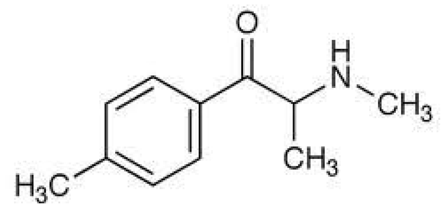
:1. Introduction
2. Material and Methods
2.1. Reagents
2.2. Instrumentation
2.3. Synthesis of Silver Nanoparticles Capped with Saffron (AgNPs@Sa)
2.4. Fabrication of the Electrochemical Sensor and Detection of Mephedrone
2.5. Determination of Mephedrone in Real Samples
3. Results and Discussion
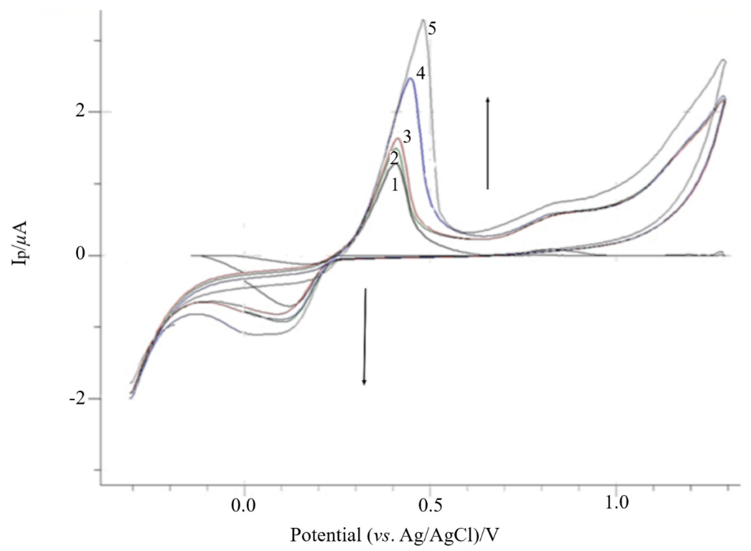
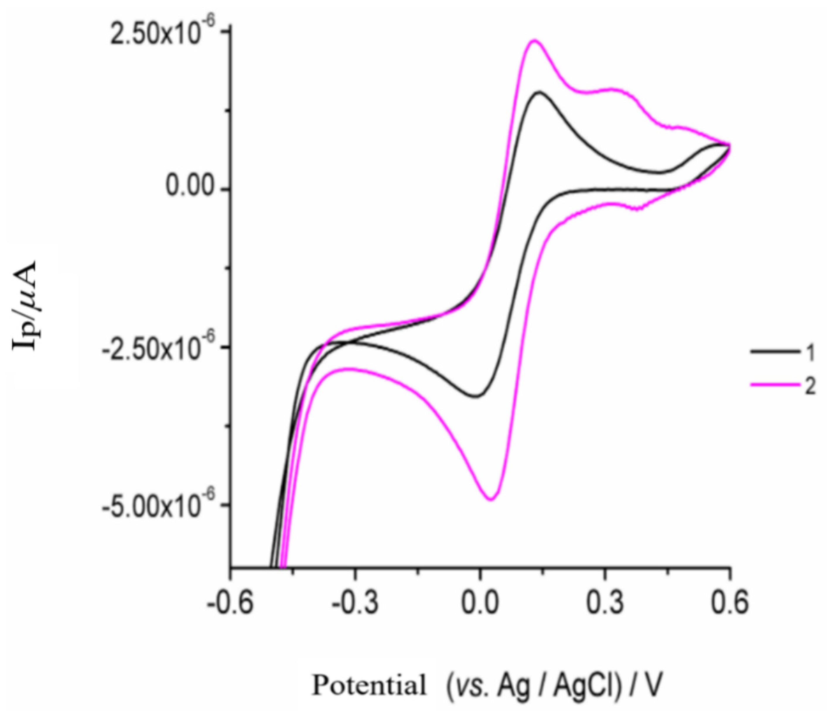
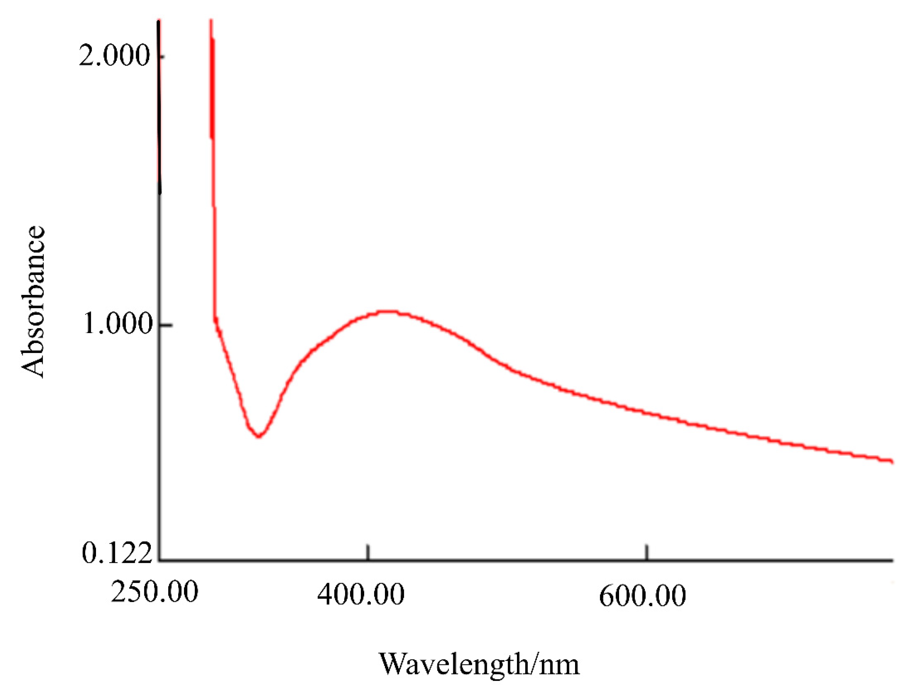
3.1. Electropolymerization of Silver Nanoparticles Capped with Saffron and Characterization of the Modified CPE
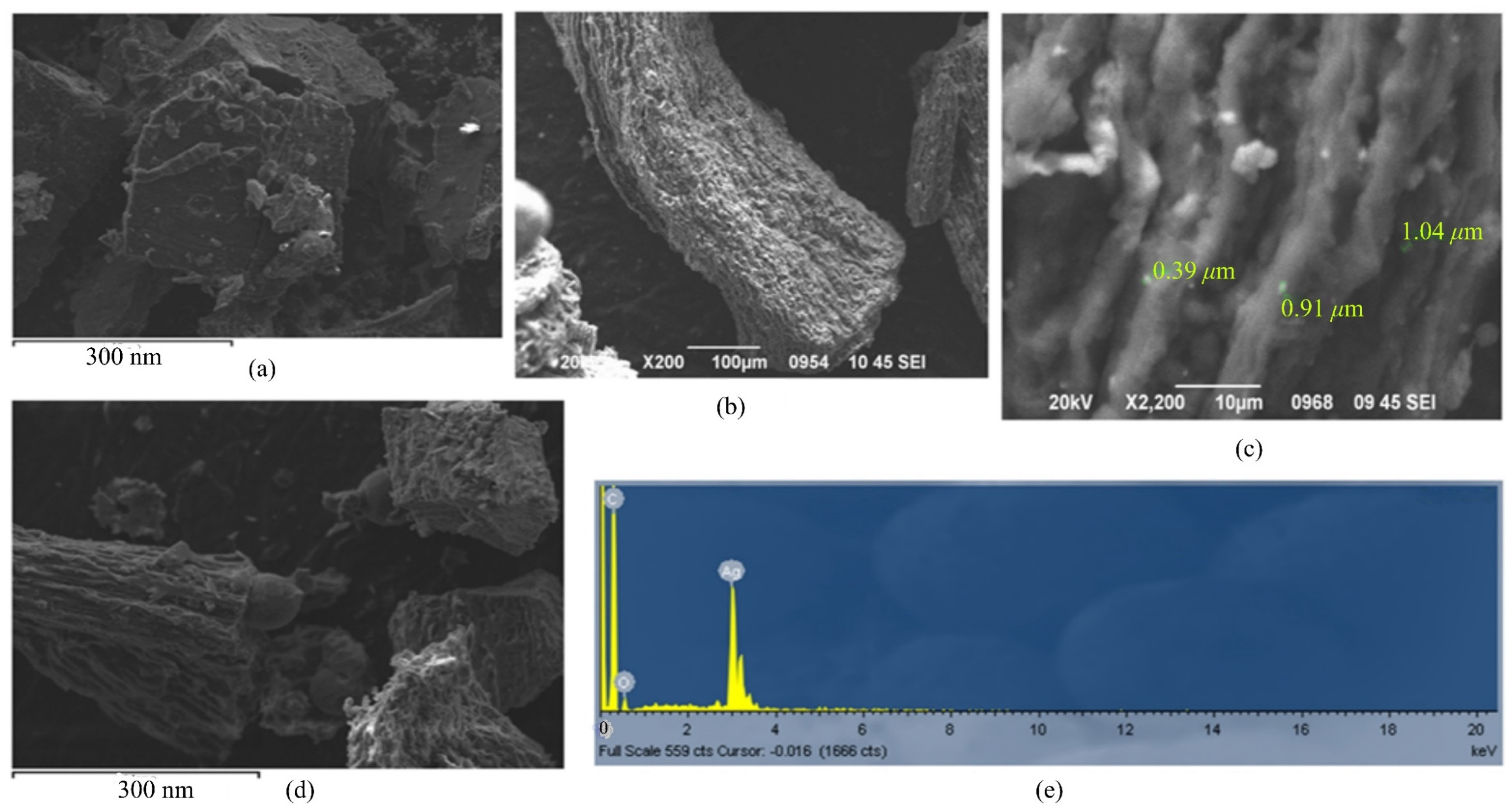
3.2. Morphology of Sa@AgNPs and Poly-AgNPs@Sa-CPE
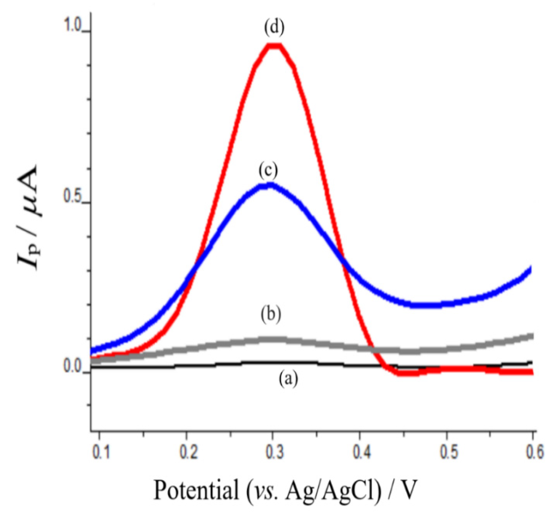
3.3. Electrochemical Behavior of Mephedrone on Modified AgNPs@Sa-CPE
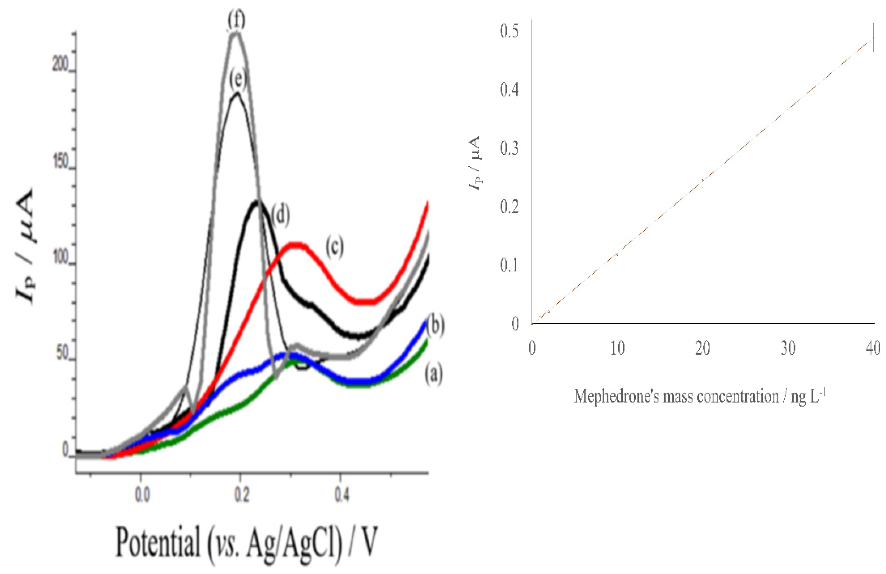
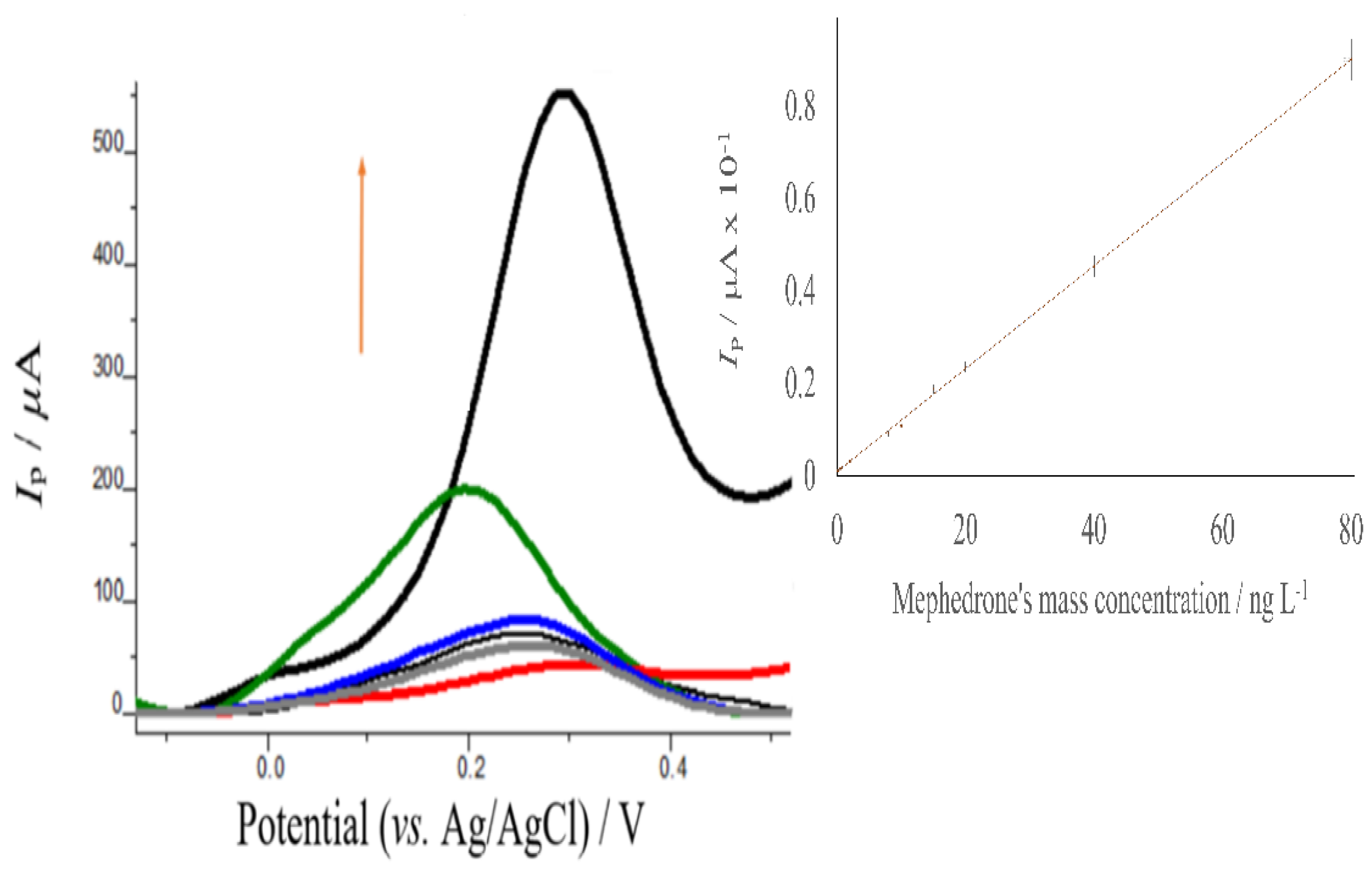
4. Analytical Performance of the Proposed Assay
5. Determination of Mephedrone in Real Samples
6. Recovery Studies
7. Conclusions
Supplementary Materials
Author Contributions
Funding
Institutional Review Board Statement
Informed Consent Statement
Acknowledgments
Conflicts of Interest
References
- Palazzoli, F.; Santunione, A.L.; Verri, P.; Vandelli, D.; Silingardi, E. Post-mortem distribution of mephedrone and its metabolites in body fluids and organ tissues of an intoxication case. J. Pharm. Biomed. Anal. 2021, 201, 114093. [Google Scholar] [CrossRef] [PubMed]
- Jamey, C.; Kintz, P.; Martrille, L.; Raul, J.S. Fatal Combination with 3-Methylmethcathinone (3-MMC) and Gamma-Hydroxybutyric Acid (GHB). J. Anal. Toxicol. 2016, 40, 546–552. [Google Scholar] [CrossRef] [PubMed] [Green Version]
- Schifano, F.; Albanese, A.; Fergus, S.; Stair, J.L.; Deluca, P.; Corazza, O.; Davey, Z.; Corkery, J.; Siemann, H.; Scherbaum, N.; et al. Psychonaut Web Mapping & ReDNet Research Groups. Mephedrone (4-methylmethcathinone; ‘meow meow’): Chemical, pharmacological and clinical issues. Psychopharmacology 2010, 214, 593–602. [Google Scholar] [CrossRef] [Green Version]
- Advisory Council on the Misuse of Drugs/ACMD Consideration of the Cathinones. 2010. Available online: http://www.homeoffice.gov.uk/publications/drugs/acmd1/acmd-cathinodes-report-2010?view=Binary (accessed on 16 January 2022).
- Measham, F.; Moore, K.; Newcombe, R.; Welch, Z. Tweaking, bombing, dabbing and stockpiling: The emergence of mephedrone and the perversity of prohibition. Drugs Alcohol Today 2010, 10, 14–21. [Google Scholar] [CrossRef] [Green Version]
- Schifano, F.; Corkery, J.; Ghodse, J.M.; Hamid, A. Suspected and Confirmed Fatalities Associated with Mephedrone (4-Methylmethcathinone, “Meow Meow”) in the United Kingdom. J. Clin. Psychopharmacol. 2012, 32, 710–714. [Google Scholar] [CrossRef]
- Wood, D.M.; Davis, S.; Puchnarewicz, M.; Button, J.; Archer, R.; Ramsey, J.; Lee, T.; Holt, D.W.; Darganet, P.I. Recreational use of 4-methylmethcathinone (4-MMC) presenting with sympathomimetic toxicity and confirmed by toxicological screening. J. Med. Toxicol. 2010, 6, 327–330. [Google Scholar] [CrossRef]
- Dar, R.A.; Brahman, P.K.; Khurana, N.; Wagay, J.A.; Lone, Z.A.; Ganaie, M.A.; Pitre, K.S. Evaluation of antioxidant activity of crocin, podophyloxotin and kaempferol by chemical, biochemical, and electrochemical assays. Arab. J. Chem. 2017, 10, S1119–S1128. [Google Scholar] [CrossRef] [Green Version]
- Jorgensen, L.; Henrik, J.; Andersen, L.; Skibsted, H. Kinetics of Reduction of Hypervalent Iron in Myoglobin by Crocin in Aqueous Solution. Free Radic. Res. 1997, 27, 73–87. [Google Scholar] [CrossRef]
- Kispert, L.D.; Konovalova, T.; Gao, Y. Carotenoid radical cations and dications: EPR, optical, and electrochemical studies. Arch. Biochem. Biophys. 2004, 430, 49–60. [Google Scholar] [CrossRef]
- Bone, A.J.; Colman, B.J.; Gondikas, A.P.; Newton, K.M.; Harrold, K.H.; Cory, R.M.; Unrine, J.M.; Klaine, S.J.; Matson, C.W.; Di Giulio, R.T. Biotic and Abiotic Interactions in Aquatic Microcosms Determine Fate and Toxicity of Ag Nanoparticles: Part 2–Toxicity and Ag Speciation. Environ. Sci. Technol. 2012, 46, 6915–6924. [Google Scholar] [CrossRef]
- Florea, A.; Cowen, T.; Piletsky, S.; De Wael, K. Polymer platforms for selective detection of cocaine in street samples adulterated with levamisole. Talanta 2018, 186, 362–367. [Google Scholar] [CrossRef] [PubMed]
- Ahmad, N.; Al-Fatesh, A.S.; Wahab, R.; Alam, M.; Fakeeha, A.H. Synthesis of silver nanoparticles decorated on reduced graphene oxide nanosheets and their electrochemical sensing towards hazardous 4-nitrophenol. J. Mater. Sci. 2020, 31, 11927–11937. [Google Scholar] [CrossRef]
- Eissa, S.; Almthen, R.A.; Mikrochim, Z.M. Disposable electrochemical immunosensor array for the multiplexed detection of the drug metabolites morphine, tetrahydrocannabinol and benzoylecgonine. Microchim. Acta 2019, 186, 523. [Google Scholar] [CrossRef]
- De Rycke, E.; Stove, C.; Dubruel, P.; De Saeger, S.; Beloglazova, N. Recent developments in electrochemical detection of illicit drugs in diverse matrices. Biosens. Bioelectron. 2020, 169, 112579. [Google Scholar] [CrossRef] [PubMed]
- Karastogianni, S.; Girousi, S. Electrochemical (Bio)Sensing of Maple Syrup Urine Disease Biomarkers Pointing to Early Diagnosis: A Review. Appl. Sci. 2020, 10, 7023. [Google Scholar] [CrossRef]
- Maskell, P.D.; De Paoli, G.; Seneviratne, C.; Pounder, D.J. Mephedrone (4-Methylmethcathinone)-Related Deaths. J. Anal. Toxicol. 2011, 35, 188–191. [Google Scholar] [CrossRef] [Green Version]
- Czerwinska, J.; Jang, M.; Costa, C.; Parkin, M.C.; George, C.; Kicman, A.T.; Bailey, M.J.; Darganf, P.I.; Abbate, V. Detection of mephedrone and its metabolites in fingerprints from a controlled human administration study by liquid chromatography-tandem mass spectrometry and paper spray-mass spectrometry. Analyst 2020, 145, 3038–3048. [Google Scholar] [CrossRef] [PubMed]
- Cheng, S.-Y.; Ng-A-Qui, T.; Eng, B.; Cheng, J.H. Detection of cathinone and mephedrone in plasma by LC-MS/MS using standard addition quantification technique. J. Anal. Sci. Technol. 2017, 8, 19. [Google Scholar] [CrossRef]
- Pozo, O.J.; Ibáñez, M.; Sancho, J.V.; Lahoz-Beneytez, J.; Farré, M.; Papaseit, E.; de la Torre, R.; Hernández, F. Mass spectrometric evaluation of mephedrone in vivo human metabolism: Identification of phase I and phase II metabolites, including a novel succinyl conjugate. Drug Metab. Dispos. 2015, 43, 248–257. [Google Scholar] [CrossRef] [Green Version]
- Mercolini, L.; Protti, M.; Catapano, M.C.; Rudge, J.; Sberna, A.E. LC-MS/MS and volumetric absorptive microsampling for quantitative bioanalysis of cathinone analogues in dried urine, plasma and oral fluid samples. J. Pharm. Biomed. Anal. 2016, 123, 186–194. [Google Scholar] [CrossRef] [PubMed]
- Li, X.; Uboh, C.E.; Soma, L.R.; Liu, Y.; Guan, F.; Aurand, C.R.; Bell, D.S.; You, Y.; Chen, J.; Maylin, G.E. Sensitive hydrophilic interaction liquid chromatography/tandem mass spectrometry method for rapid detection, quantification and confirmation of cathinone-derived designer drugs for doping control in equine plasma. Rapid Commun. Mass Spectrom. 2014, 28, 217–229. [Google Scholar] [CrossRef] [PubMed]
- Adamowicz, P.; Tokarczyk, B.; Stanaszek, R.; Slopianka, M. Fatal Mephedrone Intoxication—A case report. J. Anal. Toxicol. 2012, 37, 37–42. [Google Scholar] [CrossRef] [PubMed] [Green Version]
- Johnson, R.D.; Botch-Jones, S.R. The stability of four designer drugs: MDPV, mephedrone, BZP and TFMPP in three biological matrices under various storage conditions. J. Anal. Toxicol. 2013, 37, 51–55. [Google Scholar] [CrossRef] [PubMed] [Green Version]
- Mohamed, K.M. GC–MS Method for Quantification of Mephedrone in Human Blood Sample. J. Chromatogr. Sci. 2017, 55, 784–789. [Google Scholar] [CrossRef]
- Meyer, M.R.; Wilhelm, J.; Peters, F.T.; Maurer, H.H. Beta-keto amphetamines: Studies on the metabolism of the designer drug mephedrone and toxicological detection of mephedrone, butylone, and methylone in urine using gas chromatography–mass spectrometry. Anal. Bioanal. Chem. 2010, 397, 1225–1233. [Google Scholar] [CrossRef]
- Řezanka, P.; Macková, D.; Jurok, R.; Himl, M.; Kuchař, M. Enantioseparation and Determination of Mephedrone and Its Metabolites by Capillary Electrophoresis Using Cyclodextrins as Chiral Selectors. Molecules 2020, 25, 2879. [Google Scholar] [CrossRef]
- Muhamadali, H.; Watt, A.; Xu, Y.; Chisanga, M.; Subaihi, A.; Jones, C.; Ellis, D.I.; Sutcliffe, O.B.; Goodacre, R. Rapid Detection and Quantification of Novel Psychoactive Substances (NPS) Using Raman Spectroscopy and Surface-Enhanced Raman Scattering. Front. Chem. 2019, 7, 412. [Google Scholar] [CrossRef]
- Shen, B.; Li, J.; Cheng, W.; Yan, Y.; Tang, R.; Li, Y. Electrochemical aptasensor for highly sensitive determination of cocaine using a supramolecular aptamer and rolling circle amplification. Microchim. Acta 2015, 182, 361–367. [Google Scholar] [CrossRef]
- Florea, A.; de Jong, M.; De Wael, K. Electrochemical strategies for the detection of forensic drugs. Curr. Opin. Electrochem. 2018, 11, 34–40. [Google Scholar] [CrossRef]
- Greco, P.G.; Garris, P.A. In vivo interaction of cocaine with the dopamine transporter as measured by voltammetry. Eur. J. Pharmacol. 2003, 479, 117–125. [Google Scholar] [CrossRef]
- Broderick, P.A. In vivo voltammetric studies on release mechanisms for cocaine with γ-butyrolactone. Pharmacol. Biochem. Behav. 1991, 40, 969–975. [Google Scholar] [CrossRef]
- Kan, X.; Zhou, H.; Li, C.; Zhu, A.; Xing, Z.; Zhao, Z. Imprinted electrochemical sensor for dopamine recognition and determination based on a carbon nanotube/polypyrrole film. Electrochim. Acta 2012, 63, 69–75. [Google Scholar] [CrossRef]
- Oliva, P.H.B.; Katayama, J.M.T.; Oiye, E.N.; Ferreira, B.; Ribeiro, M.F.; Ipólito, A.J.; de Andrade, J.F.; de Oliveira, M.F. Determination of Cocaine by Square Wave Voltammetry with Carbon Paste Electrodes. Braz. J. Forensic Sci. Med. Law Bioeth. 2019, 8, 149–164. [Google Scholar] [CrossRef] [Green Version]
- Naseri, A.M.K.; Mehrpour, O.; Shoeib, I.S. Electrochemical determination of atypical antipsychotic drug quetiapine using nano-molecularly imprinted polymer modified carbon paste electrode. Anal. Chim. Acta 2020, 1097, 214–221. [Google Scholar] [CrossRef]
- Gupta, V.K.; Khosravi, S.; Karimi-Maleh, H.; Alizadeh, M.; Sharafi, S. A Voltammetric Sensor for Determination of Methyldopa in the Presence of Hydrochlorothiazide Using Fe: Co Nano alloy Modified Carbon Paste Electrode. Int. J. Electrochem. Sci. 2015, 10, 3269–3281. [Google Scholar]
- Waddell, S.A.; Fernandez, C.; Inverarity, C.C.; Prabhu, R. Extending the capability of forensic electrochemistry to the novel psychoactive substance benzylpiperazine. Sens. Bio-Sens. Res. 2017, 13, 28–39. [Google Scholar] [CrossRef]
- Azab, S.M.; Shehata, M.; Fekry, A.M. A novel electrochemical analysis of the legal psychoactive drug caffeine using a zeolite/MWCNT modified carbon paste sensor. New J. Chem. 2019, 43, 15359–15367. [Google Scholar] [CrossRef]
- Renaud-Young, M.; Mayall, R.M.; Salehi, V.; Goledzinowski, M.; Comeau, F.J.E.; Mac Callum, J.L.; Birss, V.I. Development of an ultra-sensitive electrochemical sensor for D9-tetrahydrocannabinol (THC) and its metabolites using carbon paper electrodes. Electrochim. Acta 2019, 307, 351–359. [Google Scholar] [CrossRef]
- Chebotarev, A.; Pliuta, K.; Koicheva, A.; Bevziuk, K.; Snigur, D. Determination of Levodopa in Pharmaceuticals Using a Disposable Electrochemically Activated Carbon-Paste Electrode by Linear Sweep Voltammetry. Anal. Lett. 2018, 51, 1520–1528. [Google Scholar] [CrossRef]
- Panahi, Y.; Motaharian, A.; Hosseini, M.R.M.; Mehrpour, O. High sensitive and selective nano-molecularly imprinted polymer-based electrochemical sensor for midazolam drug detection in pharmaceutical formulation and human urine samples. Sens. Actuators B Chem. 2018, 273, 1579–1586. [Google Scholar] [CrossRef]
- González-Hernández, J.; Alvarado-Gámez, A.L.; Arroyo-Mora, L.E.; Barquero-Quirós, M. Electrochemical determination of novel psychoactive substances by differential pulse voltammetry using a microcell for boron-doped diamond electrode and screen-printed electrodes based on carbon and platinum. J. Electroanal. Chem. 2021, 882, 114994. [Google Scholar] [CrossRef]
- Elbardisy, H.M.; Ferrari, A.G.-M.; Foster, C.W.; Sutcliffe, O.B.; Brownson, D.A.C.; Belal, T.S.; Talaat, W.; Daabees, H.G.; Banks, C.E. Forensic Electrochemistry: The Electroanalytical Sensing of Mephedrone Metabolites. ACS Omega 2019, 4, 1947–1954. [Google Scholar] [CrossRef]
- Ferrari, A.G.M.; Rowley-Neale, S.J.; Banks, C.E. Screen-printed electrodes: Transitioning the laboratory in-to-the field. Talanta Open 2021, 3, 100032. [Google Scholar] [CrossRef]
- Razavipanaha, I.; Alipour, E.; Deiminiata, B.; Rounagh, G.H. A novel electrochemical imprinted sensor for ultrasensitive detection of the new psychoactive substance “Mephedrone”. Biosens. Bioelectron. 2018, 119, 163–169. [Google Scholar] [CrossRef] [PubMed]
- Lima, C.D.; Couto, R.A.S.; Arantes, L.C.; Marinho, P.A.; Pimentel, D.M.; Quinaz, M.B.; da Silva, R.A.B.; Richter, E.M.; Barbosa, S.L.; dos Santos, W.T.P. Electrochemical detection of the synthetic cathinone 3,4-methylenedioxypyrovalerone using carbon screen-printed electrodes: A fast, simple and sensitive screening method for forensic samples. Electrochim. Acta 2020, 354, 136728. [Google Scholar] [CrossRef]
- Smith, J.P.; Metters, J.P.; Irving, C.; Sutcliffe, O.B.; Banks, C.E. Forensic electrochemistry: The electroanalytical sensing of synthetic cathinone-derivatives and their accompanying adulterants in “legal high” products. Analyst 2014, 139, 389–400. [Google Scholar] [CrossRef] [Green Version]
- Tan, F.; Smith, J.P.; Sutcliffe, O.B.; Banks, C.E. Regal electrochemistry: Sensing of the synthetic cathinone class of new psychoactive substances (NPSs). Anal. Methods 2015, 7, 6470–6474. [Google Scholar] [CrossRef]
- Mc Neill, L.; Pearson, C.; Megson, D.; Norrey, J.; Watson, D.; Ashworth, D.; Linton, P.E.; Sutcliffe, O.B.; Shaw, K.J. Origami chips: Development and validation of a paper-based Lab-on-a-Chip device for the rapid and cost-effective detection of 4-methylmethcathinone (mephedrone) and its metabolite 4-methylephedrine in urine. Forensic Chem. 2021, 22, 100293. [Google Scholar] [CrossRef]
- Karastogianni, S.; Girousi, S. A novel electrochemical bioimprinted sensor of butylparaben on a modified carbon paste electrode with safranineo capped to silver nanoparticles. Int. J. Curr. Res. 2017, 9, 61118–61124. [Google Scholar]
- Thakare, R.; Chhonker, Y.S.; Gautam, N.; Alamoudi, J.A.; Alnouti, Y. Quantitative analysis of endogenous compounds. J. Pharm. Biomed. Anal. 2016, 128, 426–437. [Google Scholar] [CrossRef]
- Kang, J.F.; Perry, J.D.; Tian, P.; Kilbey, S.M. Growth and Morphology of Polythiophene on Thiophene-Capped Monolayers: 1. Single Component. Monolayers Langmuir 2002, 18, 10196–10201. [Google Scholar] [CrossRef]
- Sheberla, D.; Patra, S.; Wijsboom, Y.H.; Sharma, S.; Sheynin, Y.; Haj-Yahia, A.-E.; Barak, A.H.; Gidron, O.; Bendikov, M. Conducting polyfurans by electropolymerization of oligofurans. Chem. Sci. 2015, 6, 360–371. [Google Scholar] [CrossRef] [Green Version]
- Armellini, R.; Compagnone, D.; Scampicchio, M.; Pittia, P. Hydrogen and Atom Transfer Activity of Saffron Extracts by Square Wave Voltammetry. Electroanalysis 2017, 29, 521–528. [Google Scholar] [CrossRef]
- Bard, A.J.; Faulkner, L.R. Electrochemical Methods: Fundamentals and Applications; Wiley & Sons: New York, NY, USA, 2001. [Google Scholar]
- Wopschall, R.H.; Shain, I. Effects of adsorption of electroactive species in stationary electrode polarography. Anal. Chem. 1967, 39, 1514–1527. [Google Scholar] [CrossRef]
- Poriel, C.; Ferrand, Y.; Le Maux, P.; Berthelot, J.R.; Simonneaux, G. Organic crosslinked electrolysers as supported oxidation catalysts: Poly((tetrakis(9,9′-spirobifluorenyl)porphyrin)manganese) films. Inorg. Chem. 2004, 43, 5086–5095. [Google Scholar] [CrossRef]
- Schram, J.; Parrilla, M.; Sleegers, N.; Van Durme, F.; van den Berg, J.; van Nuijs, A.L.N.; De Wae, K. Electrochemical profiling and liquid chromatography–massspectrometry characterization of synthetic cathinones: Frommethodology to detection in forensic samples. Drug Test. Anal. 2021, 13, 1282–1294. [Google Scholar] [CrossRef] [PubMed]
- Paul, M.; Ippisch, J.; Herrmann, C.; Guber, S.; Schultis, W. Analysis of new designer drugs and common drugs of abuse in urine by a combined targeted and untargeted LC-HR-QTOFMS approach. Anal. Bioanal. Chem. 2014, 406, 4425–4441. [Google Scholar] [CrossRef] [PubMed]
- Available online: https://www.fda.gov/media/128343/download (accessed on 16 January 2022).


| Detection Method | Electrode | Analytes | Limit of Detection (LOD) | Linear Range | Sample | Reference |
|---|---|---|---|---|---|---|
| LC-MS-MS | - | Mephedrone | 3 ng mL−1 | 10–500 ng mL−1 | Urine, plasma and oral fluid | [21] |
| GC-MS | - | Mephedrone | 2 ng mL−1 | 5–2000 ng mL−1 | Plasma sample | [24] |
| GC-MS | - | Mephedrone | 5 ng mL−1 | 10–2000 ng mL−1 | Human blood sample | [25] |
| Cyclic voltammetry and differential pulse voltammetry | Screen printed graphite electrode | Two metabolites of mephedrone: nor-mephedrone (4-methylcathinone, 4-MC) and dihydromephedrone (4-methylephedrine, 4-MMC-R) | 3.97 μg mL−1 for 4-MC and 3.64 μg mL−1 for 4-MMC-R (PBS, pH 7.0 and 3.0, respectively, differential pulse voltammetry) | 40–300 μg mL−1 for 4-MC (PBS, pH 7.0) and 15–300 μg mL−1 (PBS, pH 3.0) (cyclic voltammetry) | Urine samples | [43] |
| Square wave voltammetry | sol-gel molecular imprinted polymer, polytyramine, and functionalized multi-walled carbon nanotube@ gold nanoparticles (f-MWCNT@AuNPs) | 0.8 nM (142 pg L−1) | 1 to 10 n mol L−1 and 10 to 100 n mol L−1 | Urine and plasma | [45] | |
| - | Screen-printed graphite electrode | 39.8 μg mL−1 (pH 2) | 16–350 μg mL-1 (pH 2) | Real street samples | [47] | |
| - | Pence British coin | 0.56 μg mL−1 | 0.01-0.1 μg mL−1 | Street samples | [48] | |
| Colorimetric | Portable paper-based Lab-on-a-Chip (LOC) device | Mephedrone | 2.51 ng mL−1 | 0.078 to 10.0 mg mL−1 | Urine | [49] |
| Adsorptive stripping square wave voltammetry | Silver nanoparticles capped with saffron modified carbon paste electrode | Mephedrone | 0.608 pg mL−1 | 1.841–80.000 pg mL−1 | Urine samples | This work |
| Mephedrone Added/ng L−1 | Expected Value/ng L−1 | Measured Value/ng L−1 | Recovery | sr |
|---|---|---|---|---|
| 2.00 | 2.00 | 1.89 ng mL−1 | 95 | 1.0 |
| 10.0 | 10.0 | 9.81 ng mL−1 | 98 | 1.0 |
| 20.0 | 20.0 | 19.9 ng mL−1 | 100 | 0.6 |
Publisher’s Note: MDPI stays neutral with regard to jurisdictional claims in published maps and institutional affiliations. |
© 2022 by the authors. Licensee MDPI, Basel, Switzerland. This article is an open access article distributed under the terms and conditions of the Creative Commons Attribution (CC BY) license (https://creativecommons.org/licenses/by/4.0/).
Share and Cite
Papaioannou, G.C.; Karastogianni, S.; Girousi, S. Development of an Electrochemical Sensor Using a Modified Carbon Paste Electrode with Silver Nanoparticles Capped with Saffron for Monitoring Mephedrone. Sensors 2022, 22, 1625. https://doi.org/10.3390/s22041625
Papaioannou GC, Karastogianni S, Girousi S. Development of an Electrochemical Sensor Using a Modified Carbon Paste Electrode with Silver Nanoparticles Capped with Saffron for Monitoring Mephedrone. Sensors. 2022; 22(4):1625. https://doi.org/10.3390/s22041625
Chicago/Turabian StylePapaioannou, Georgios Christos, Sophia Karastogianni, and Stella Girousi. 2022. "Development of an Electrochemical Sensor Using a Modified Carbon Paste Electrode with Silver Nanoparticles Capped with Saffron for Monitoring Mephedrone" Sensors 22, no. 4: 1625. https://doi.org/10.3390/s22041625
APA StylePapaioannou, G. C., Karastogianni, S., & Girousi, S. (2022). Development of an Electrochemical Sensor Using a Modified Carbon Paste Electrode with Silver Nanoparticles Capped with Saffron for Monitoring Mephedrone. Sensors, 22(4), 1625. https://doi.org/10.3390/s22041625