Article

Using Sparse Patch Annotation for Tumor Segmentation in Histopathological Images
1
Institute of Biopharmaceutical and Health Engineering, Tsinghua Shenzhen International Graduate School, Shenzhen 518055, China
2
Department of Pathology, The First Affiliated Hospital, Sun Yat-sen University, Guangzhou 510080, China
*
Authors to whom correspondence should be addressed.
These authors contributed equally to this work.
Sensors 2022, 22(16), 6053; https://doi.org/10.3390/s22166053
Submission received: 19 July 2022 / Revised: 5 August 2022 / Accepted: 10 August 2022 / Published: 13 August 2022

Abstract

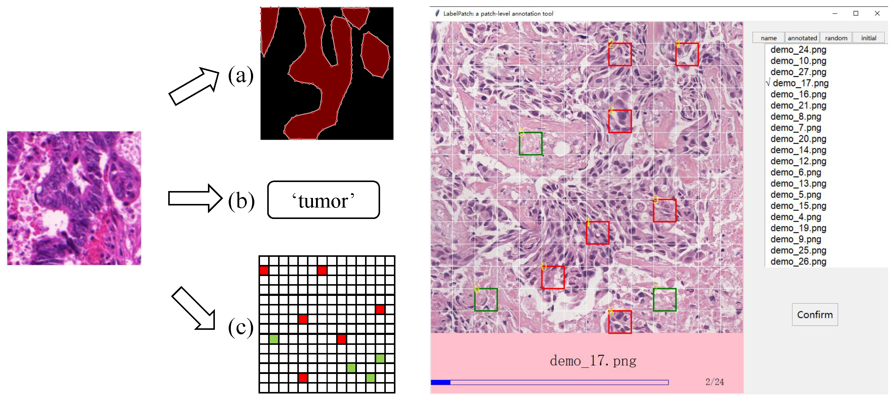
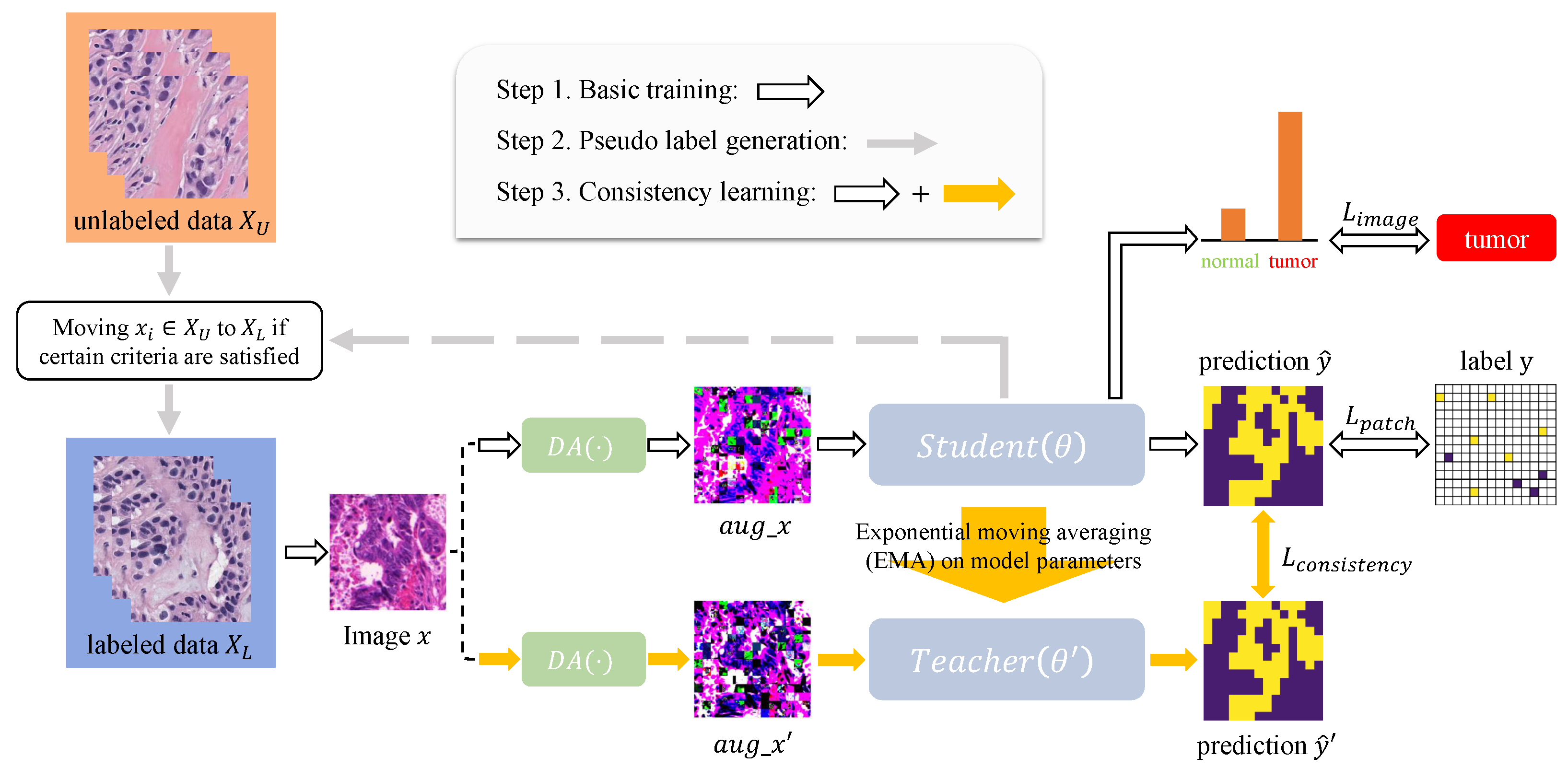
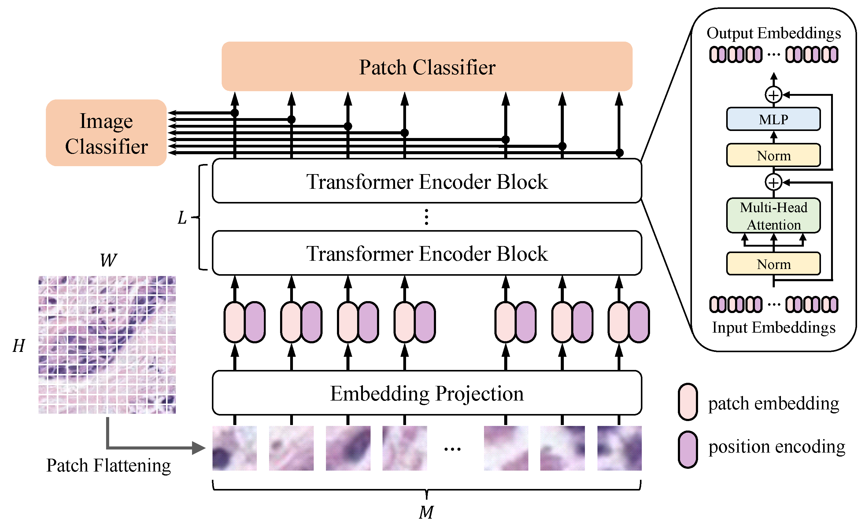
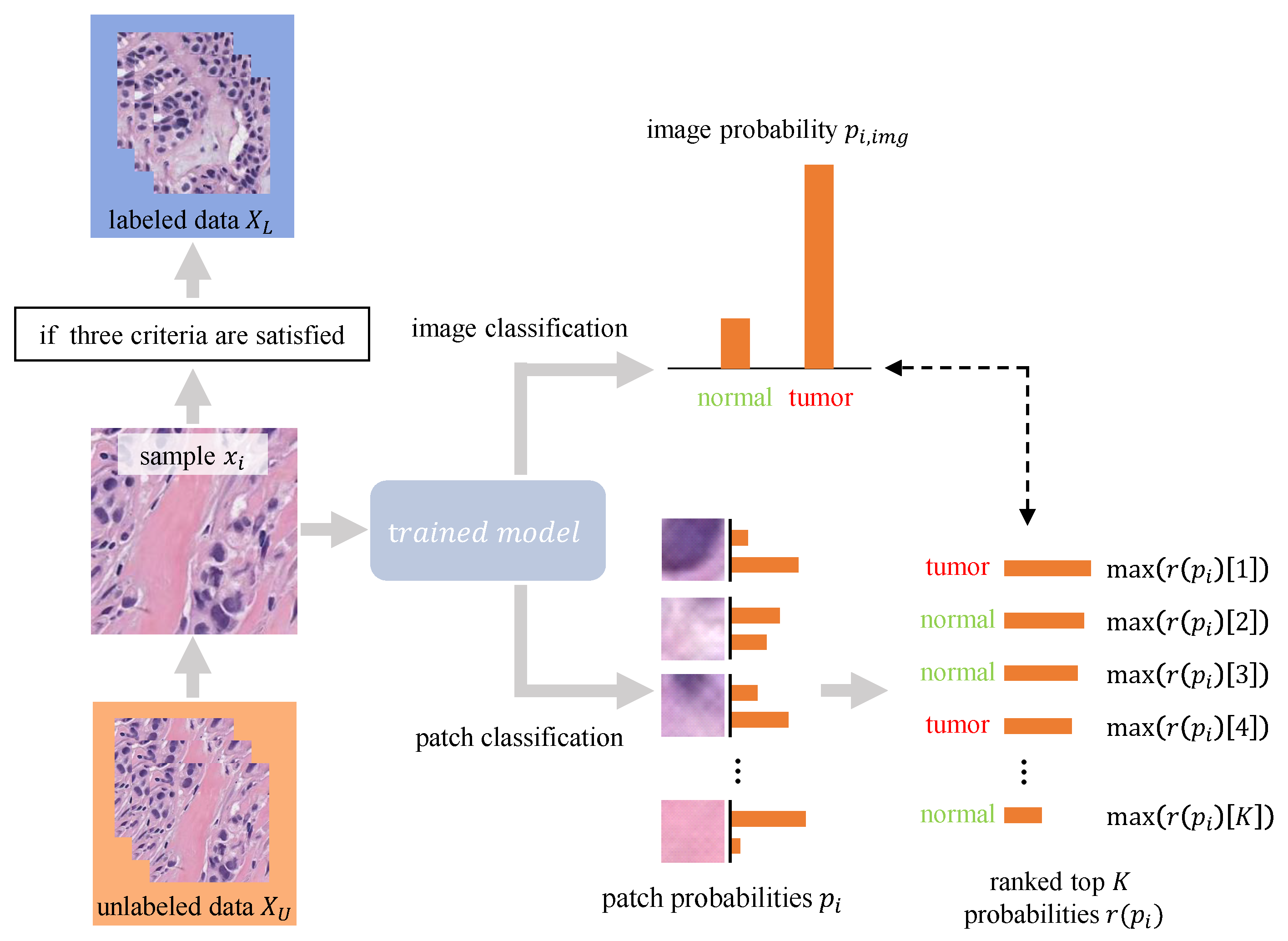
Tumor segmentation is a fundamental task in histopathological image analysis. Creating accurate pixel-wise annotations for such segmentation tasks in a fully-supervised training framework requires significant effort. To reduce the burden of manual annotation, we propose a novel weakly supervised segmentation framework based on sparse patch annotation, i.e., only small portions of patches in an image are labeled as ‘tumor’ or ‘normal’. The framework consists of a patch-wise segmentation model called PSeger, and an innovative semi-supervised algorithm. PSeger has two branches for patch classification and image classification, respectively. This two-branch structure enables the model to learn more general features and thus reduce the risk of overfitting when learning sparsely annotated data. We incorporate the idea of consistency learning and self-training into the semi-supervised training strategy to take advantage of the unlabeled images. Trained on the BCSS dataset with only 25% of the images labeled (five patches for each labeled image), our proposed method achieved competitive performance compared to the fully supervised pixel-wise segmentation models. Experiments demonstrate that the proposed solution has the potential to reduce the burden of labeling histopathological images.
Keywords: histology images; tumor segmentation; sparse annotation; weakly-supervised learning; semi-supervised learning histology images; tumor segmentation; sparse annotation; weakly-supervised learning; semi-supervised learning