Optical Polarimetric Detection for Dental Hard Tissue Diseases Characterization
Abstract
1. Introduction
2. Materials and Methods
2.1. Experimental Setup
2.2. Samples
2.3. Lu-Chipman Decomposition Method
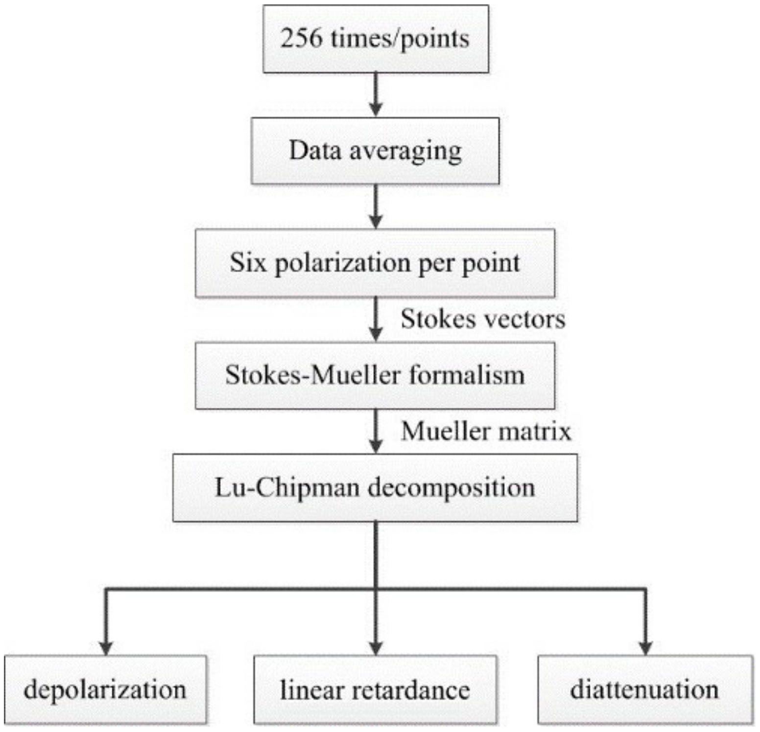
2.4. Data Processing
3. Results
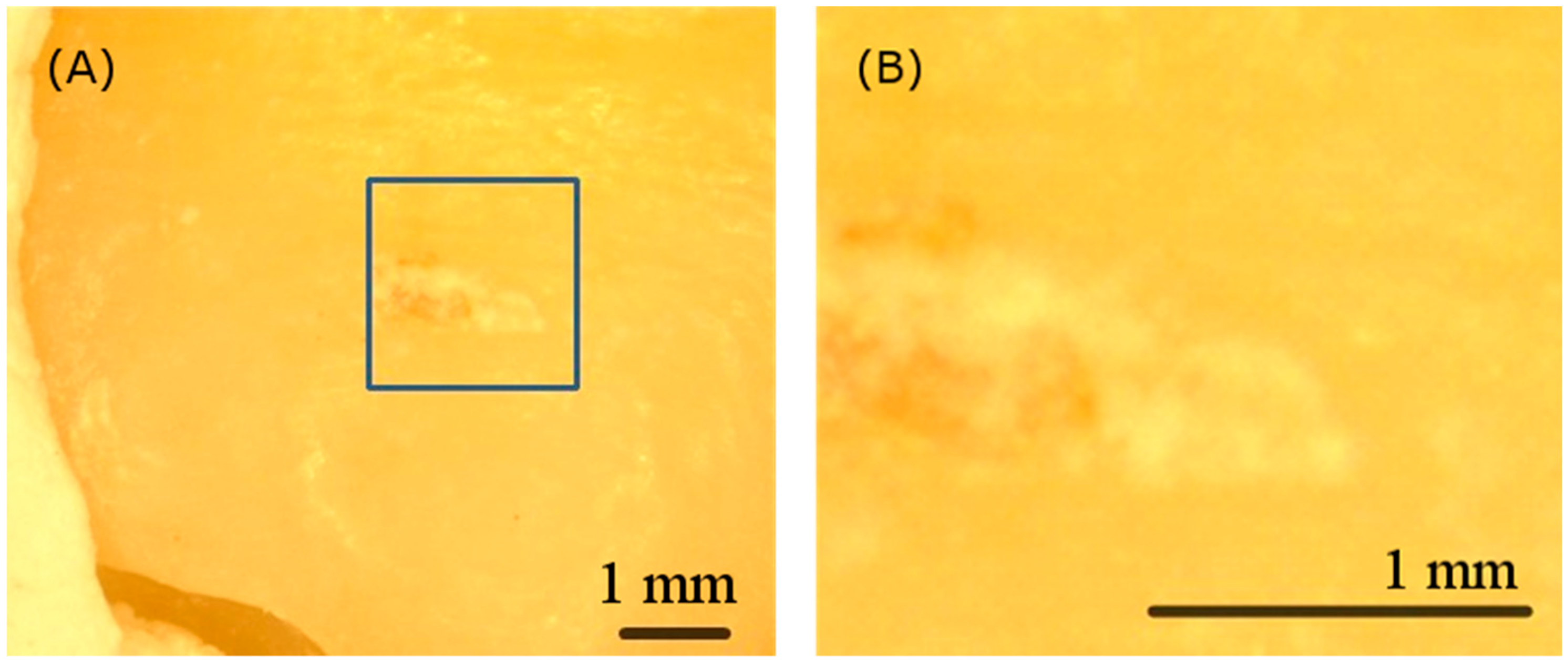
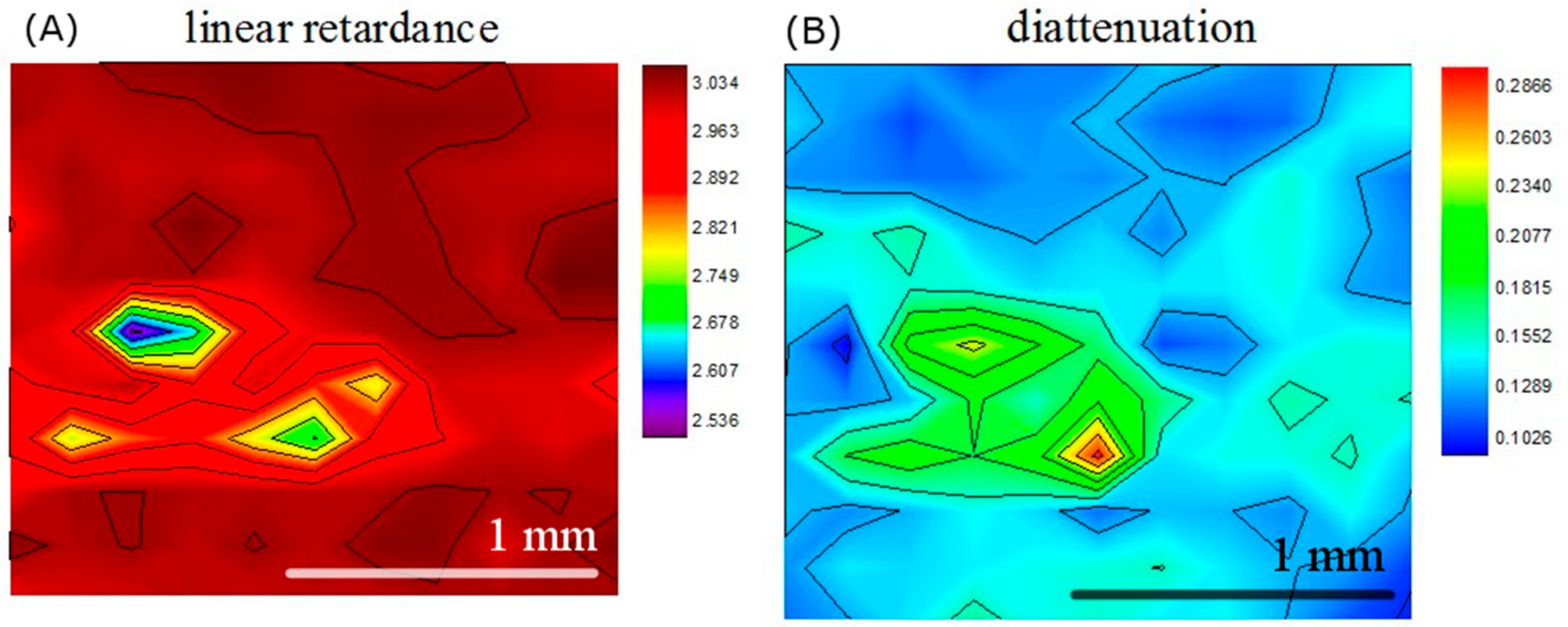
3.1. Dental Calculus
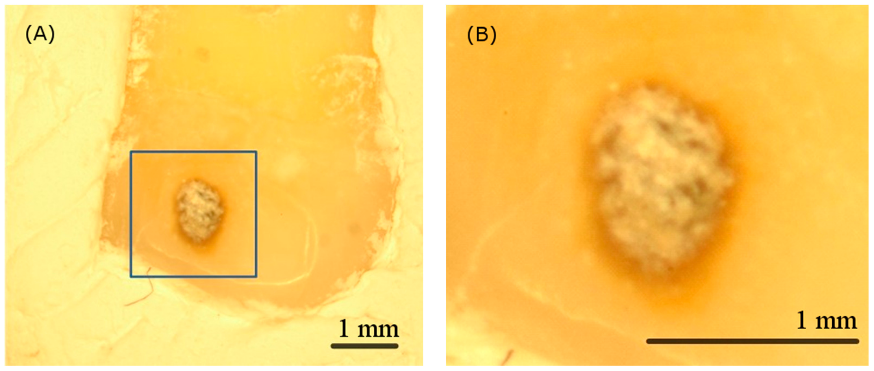
3.2. Dental Caries
3.3. Cracked Tooth Syndrome
4. Discussion
5. Conclusions
Author Contributions
Acknowledgments
Conflicts of Interest
References
- Lacruz, R.S.; Habelitz, S.; Wright, J.T.; Paine, M.L. Dental enamel formation and implications for oral health and disease. Physiol. Rev. 2017, 97, 939–993. [Google Scholar] [CrossRef] [PubMed]
- Engel, J. Enamel is the Hardest Biomaterial Known. In A Critical Survey of Biomineralization; Springer: Berlin/Heidelberg, Germany, 2017; pp. 17–27. [Google Scholar]
- Wang, C.; Ou, Y.; Zhang, L.; Zhou, Z.; Li, M.; Xu, J.; Fan, J.; Fu, B.; Hannig, M. Effects of regional enamel and prism orientations on bovine enamel bond strength and cohesive strength. Eur. J. Oral Sci. 2018, 126, 334–342. [Google Scholar] [CrossRef] [PubMed]
- Boyde, A. Microstructure of enamel. Ciba Found. Symp. 1997, 205, 18–31. [Google Scholar] [PubMed]
- Amar, S.; Han, X. The impact of periodontal infection on systemic diseases. Med. Sci. Monit. 2003, 9, 291–299. [Google Scholar]
- Li, X.; Kolltveit, K.M.; Tronstad, L.; Olsen, I. Systemic Diseases Caused by Oral Infection. Clin. Microbiol. Rev. 2000, 13, 547–558. [Google Scholar] [CrossRef] [PubMed]
- Petersen, P.E.; Bourgeois, D.; Ogawa, H.; Estupinan-Day, S.; Ndiaye, C. The global burden of oral diseases and risks to oral health. Bull. World Health Organ. 2005, 83, 661–669. [Google Scholar]
- Eke, P.I.; Dye, B.A.; Wei, L.; Slade, G.D.; Thornton-Evans, G.O.; Borgnakke, W.S.; Taylor, G.W.; Page, R.C.; Beck, J.D.; Genco, R.J. Update on Prevalence of Periodontitis in Adults in the United States: NHANES 2009 to 2012. J. Periodontol. 2015, 86, 611–622. [Google Scholar] [CrossRef]
- Assaf, A.V.; Meneghim, M.D.C.; Zanin, L.; Mialhe, F.L.; Pereira, A.C.; Ambrosano, G.M.B. Assessment of different methods for diagnosing dental caries in epidemiological surveys. Community Dent. Oral Epidemiol. 2004, 32, 418–425. [Google Scholar] [CrossRef]
- Ekstrand, K.; Ricketts, D.; Longbottom, C.; Pitts, N. Visual and Tactile Assessment of Arrested Initial Enamel Carious Lesions: An in vivo Pilot Study. Caries Res. 2005, 39, 173–177. [Google Scholar] [CrossRef]
- Douglass, C.W.; Valachovic, R.W.; Wijesinha, A.; Chauncey, H.H.; Kapur, K.K.; McNeil, B.J. Clinical efficacy of dental radiography in the detection of dental caries and periodontal diseases. Oral Surg. Oral Med. Oral Pathol. 1986, 62, 330–339. [Google Scholar] [CrossRef]
- El-Housseiny, A.; Jamjoum, H. The effect of caries detector dyes and a cavity cleansing agent on composite resin bonding to enamel and dentin. J. Clin. Pediatr. Dent. 2001, 25, 57–63. [Google Scholar] [CrossRef] [PubMed][Green Version]
- Zakian, C.; Pretty, I.; Ellwood, R. Near-infared hyperspectral imaging of teeth for dental caries detection. J. Biomed. Opt. 2009, 14, 064047. [Google Scholar] [CrossRef] [PubMed]
- Jones, R.S.; Huynh, G.D.; Jones, G.C.; Fried, D. Near-infrared transillumination at 1310-nm for the imaging of early dental decay. Opt. Express 2003, 11, 2259–2265. [Google Scholar] [CrossRef] [PubMed]
- Darling, C.L.; Jiao, J.J.; Lee, C.; Kang, H.; Fried, D. Near-IR Polarization Imaging of Sound and Carious Dental Enamel. Proc. SPIE Int. Soc. Opt. Eng. 2010, 7549, 75490L. [Google Scholar]
- Chifor, R.; Badea, M.E.; Hedeşiu, M.; Chifor, I. Identification of the anatomical elements used in periodontal diagnosis on 40 MHz periodontal ultrasonography. Rom. J. Morphol. Embryol. Rev. Roum. Morphol. Embryol. 2015, 56, 149–153. [Google Scholar]
- Krause, F.; Braun, A.; Jepsen, S.; Frentzen, M. Detection of Subgingival Calculus with a Novel LED-Based Optical Probe. J. Periodontol. 2005, 76, 1202–1206. [Google Scholar] [CrossRef]
- Kurihara, E.; Koseki, T.; Gohara, K.; Nishihara, T.; Ansai, T.; Takehara, T. Detection of subgingival calculus and dentine caries by laser fluorescence. J. Periodontal Res. 2004, 39, 59–65. [Google Scholar] [CrossRef]
- Lee, Y.-K. Fluorescence properties of human teeth and dental calculus for clinical applications. J. Biomed. Opt. 2015, 20, 40901. [Google Scholar] [CrossRef]
- Xiang, X.; Sowa, M.G.; Iacopino, A.M.; Maev, R.G.; Hewko, M.D.; Man, A.; Liu, K.-Z. An Update on Novel Non-Invasive Approaches for Periodontal Diagnosis. J. Periodontol. 2010, 81, 186–198. [Google Scholar] [CrossRef]
- Lynch, C.D.; McConnell, R.J. The cracked tooth syndrome. J. Can. Dent. Assoc. 2002, 68, 470–475. [Google Scholar]
- Mathew, S. Diagnosis of cracked tooth syndrome. J. Pharm. Bioallied Sci. 2012, 4, S242–S244. [Google Scholar] [CrossRef] [PubMed]
- Carlstrom, D.; Glas, J.E. Studies on the ultrastructure of dental enamel. III. The birefringence of human enamel. J. Ultrastruct. Res. 1963, 8, 1–11. [Google Scholar] [CrossRef]
- Sousa, F.B.; Vianna, S.S.; Santos-Magalhães, N.S.; Santos--Magalhães, N.S. A new approach for improving the birefringence analysis of dental enamel mineral content using polarizing microscopy. J. Microsc. 2006, 221, 79–83. [Google Scholar] [CrossRef] [PubMed]
- De Medeiros, R.; Soares, J.; De Sousa, F. Natural enamel caries in polarized light microscopy: Differences in histopathological features derived from a qualitative versus a quantitative approach to interpret enamel birefringence. J. Microsc. 2012, 246, 177–189. [Google Scholar] [CrossRef] [PubMed]
- Wang, X.-J.; Milner, T.E.; De Boer, J.F.; Zhang, Y.; Pashley, D.H.; Nelson, J.S. Characterization of dentin and enamel by use of optical coherence tomography. Appl. Opt. 1999, 38, 2092–2096. [Google Scholar] [CrossRef]
- Chen, Y.; Otis, L.; Piao, D.; Zhu, Q. Characterization of dentin, enamel, and carious lesions by a polarization-sensitive optical coherence tomography system. Appl. Opt. 2005, 44, 2041–2048. [Google Scholar] [CrossRef]
- Fried, D.; Xie, J.; Shafi, S.; Featherstone, J.D.B.; Breunig, T.M.; Le, C.Q. Imaging caries lesions and lesion progression with polarization sensitive optical coherence tomography. J. Biomed. Opt. 2002, 7, 618–628. [Google Scholar] [CrossRef]
- Holmen, L.; Thylstrup, A.; Ogaard, B.; Kragh, F. A polarized light microscopic study of progressive stages of enamel caries in vivo. Caries Res. 1985, 19, 348–354. [Google Scholar] [CrossRef]
- Manesh, S.K.; Darling, C.L.; Fried, D. Polarization-sensitive optical coherence tomography for the nondestructive assessment of the remineralization of dentin. J. Biomed. 2019, 14, 044002. [Google Scholar] [CrossRef]
- Jones, R.S.; Darling, C.L.; Featherstone, J.D.B.; Fried, D. Remineralization of in vitro dental caries assessed with polarization-sensitive optical coherence tomography. J. Biomed. Opt. 2006, 11, 14016. [Google Scholar] [CrossRef]
- Louie, T.; Lee, C.; Hsu, D.; Hirasuna, K.; Manesh, S.; Staninec, M.; Darling, C.L.; Fried, D. Clinical assessment of early tooth demineralization using polarization sensitive optical coherence tomography. Lasers Surg. Med. 2010, 42, 738–745. [Google Scholar] [CrossRef] [PubMed]
- Sun, C.-W.; Hsieh, Y.-S.; Ho, Y.-C.; Jiang, C.-P.; Chuang, C.-C.; Lee, S.-Y. Characterization of tooth structure and the dentin-enamel zone based on the Stokes–Mueller calculation. J. Biomed. Opt. 2012, 17, 116026. [Google Scholar] [CrossRef] [PubMed]
- Lu, S.-Y.; Chipman, R.A. Interpretation of Mueller matrices based on polar decomposition. J. Opt. Soc. Am. A 1996, 13, 1106. [Google Scholar] [CrossRef]
- Alali, S.; Vitkin, A. Polarized light imaging in biomedicine: Emerging Mueller matrix methodologies for bulk tissue assessment. J. Biomed. Opt. 2015, 20, 61104. [Google Scholar] [CrossRef]
- Kunnen, B. Application of circularly polarized light for non-invasive diagnosis of cancerous tissues and turbid tissue-like scattering media. J. Biophotonics 2015, 8, 317–323. [Google Scholar] [CrossRef]
- Ghassemi, P.; Lemaillet, P.; Germer, T.A.; Shupp, J.W.; Venna, S.S.; Boisvert, M.E.; Flanagan, K.E.; Jordan, M.H.; Ramella-Roman, J.C. Out-of-plane Stokes imaging polarimeter for early skin cancer diagnosis. J. Biomed. Opt. 2012, 17, 0760141. [Google Scholar] [CrossRef]
- Deyhle, H.; White, S.N.; Bunk, O.; Beckmann, F.; Müller, B. Nanostructure of carious tooth enamel lesion. Acta Biomater. 2014, 10, 355–364. [Google Scholar] [CrossRef]
- Schroeder, H.E.; Shanley, D. Formation and Inhibition of Dental Calculus. J. Periodontol. 1969, 40, 643–646. [Google Scholar] [CrossRef]
- Pellillo, S. Effects of Sodium Hypochlorite on Enamel Composition. Master’s Thesis, Nova Southeastern University, Fort Lauderdale, FL, USA, 2015. [Google Scholar]
- Sim, T.P.C.; Knowles, J.C.; Ng, Y.-L.; Shelton, J.; Gulabivala, K. Effect of sodium hypochlorite on mechanical properties of dentine and tooth surface strain. Int. Endod. J. 2001, 34, 120–132. [Google Scholar] [CrossRef]


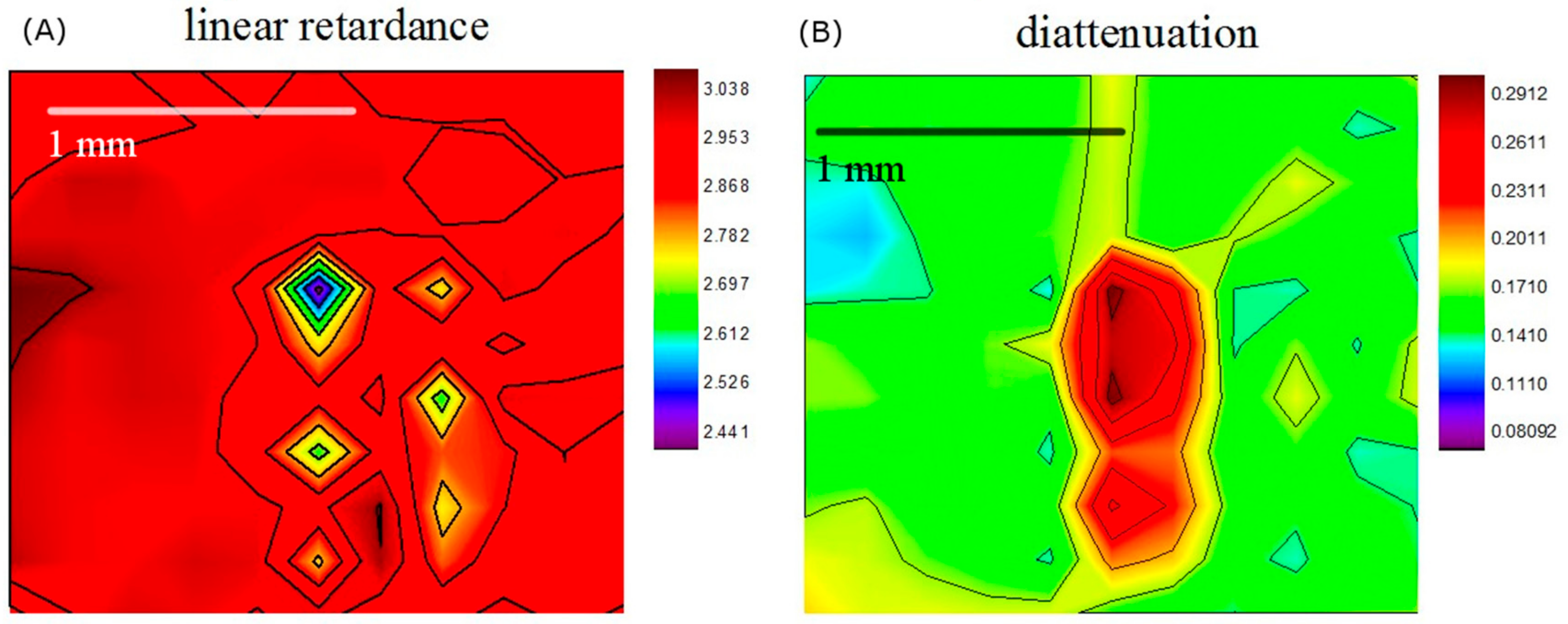
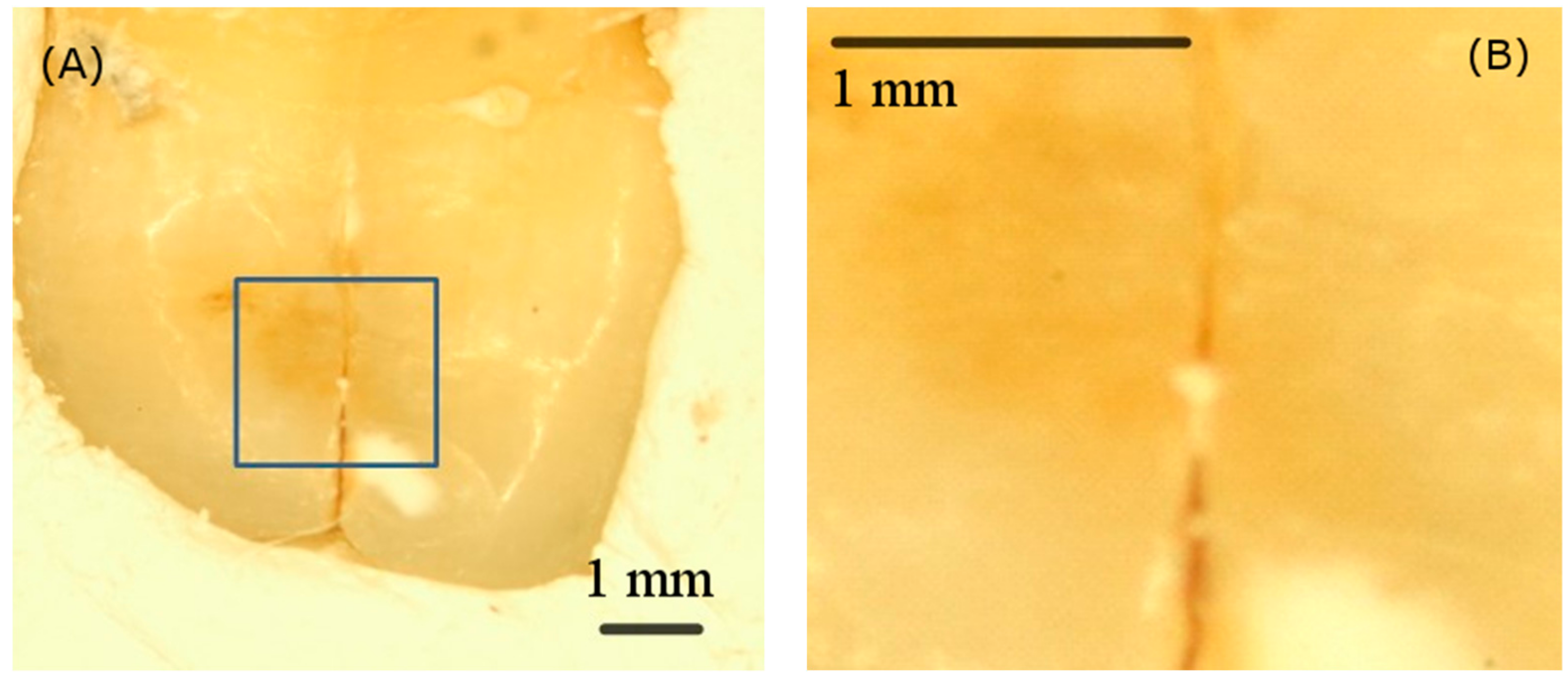

© 2019 by the authors. Licensee MDPI, Basel, Switzerland. This article is an open access article distributed under the terms and conditions of the Creative Commons Attribution (CC BY) license (http://creativecommons.org/licenses/by/4.0/).
Share and Cite
Hsiao, T.-Y.; Lee, S.-Y.; Sun, C.-W. Optical Polarimetric Detection for Dental Hard Tissue Diseases Characterization. Sensors 2019, 19, 4971. https://doi.org/10.3390/s19224971
Hsiao T-Y, Lee S-Y, Sun C-W. Optical Polarimetric Detection for Dental Hard Tissue Diseases Characterization. Sensors. 2019; 19(22):4971. https://doi.org/10.3390/s19224971
Chicago/Turabian StyleHsiao, Tien-Yu, Shyh-Yuan Lee, and Chia-Wei Sun. 2019. "Optical Polarimetric Detection for Dental Hard Tissue Diseases Characterization" Sensors 19, no. 22: 4971. https://doi.org/10.3390/s19224971
APA StyleHsiao, T.-Y., Lee, S.-Y., & Sun, C.-W. (2019). Optical Polarimetric Detection for Dental Hard Tissue Diseases Characterization. Sensors, 19(22), 4971. https://doi.org/10.3390/s19224971


