Application of 2D Non-Graphene Materials and 2D Oxide Nanostructures for Biosensing Technology

 , , ,
, , , 
Abstract
:1. Introduction

2. Principle of Biosensors Operation and Current Trends in Biosensing Technology
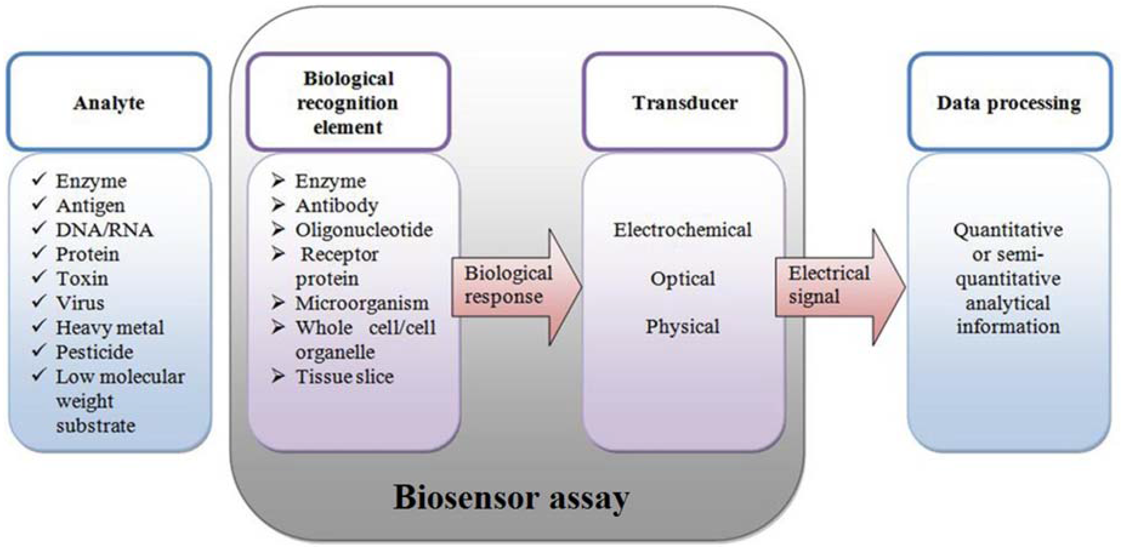
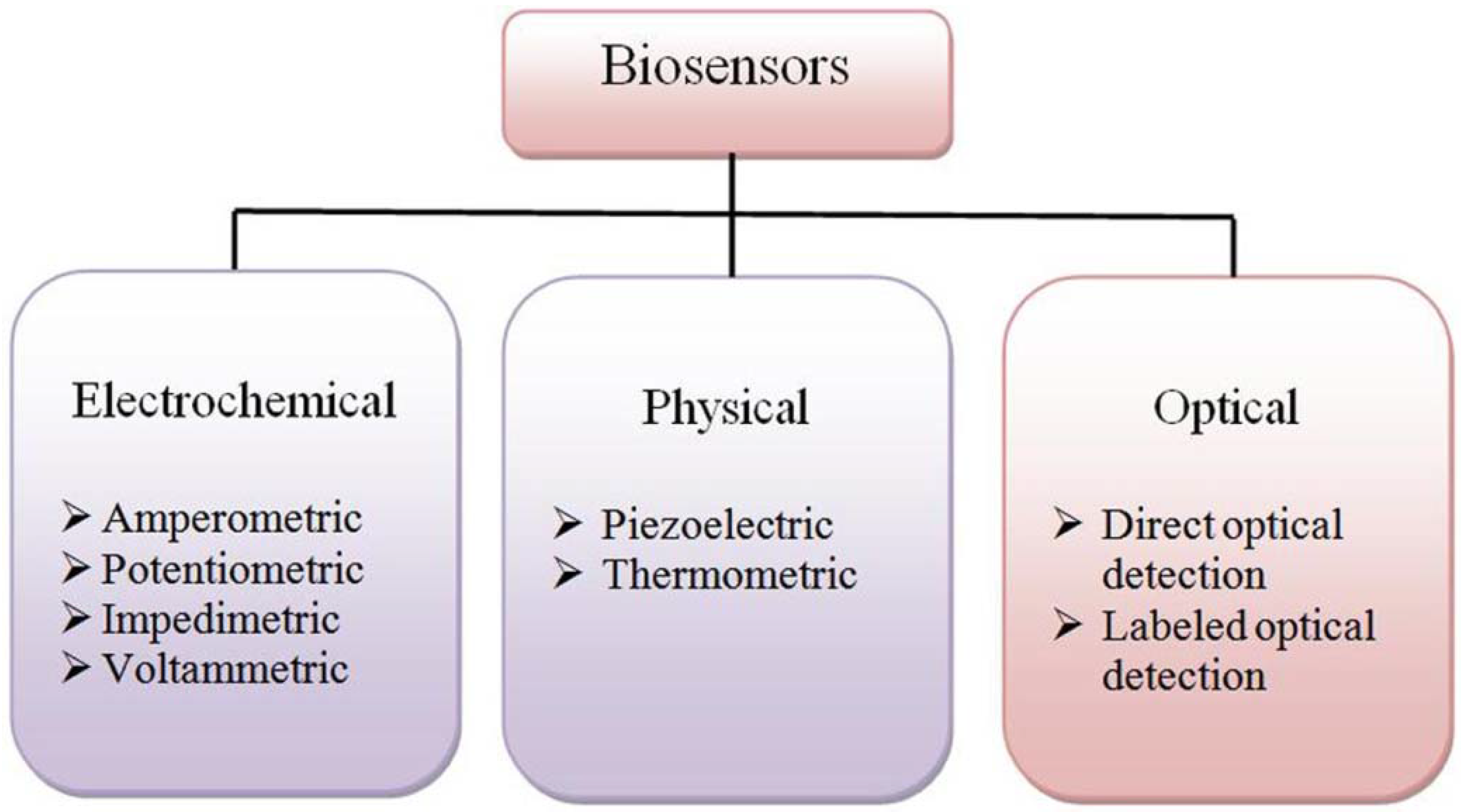
2.1. Biosensors Design and Principles of Operation

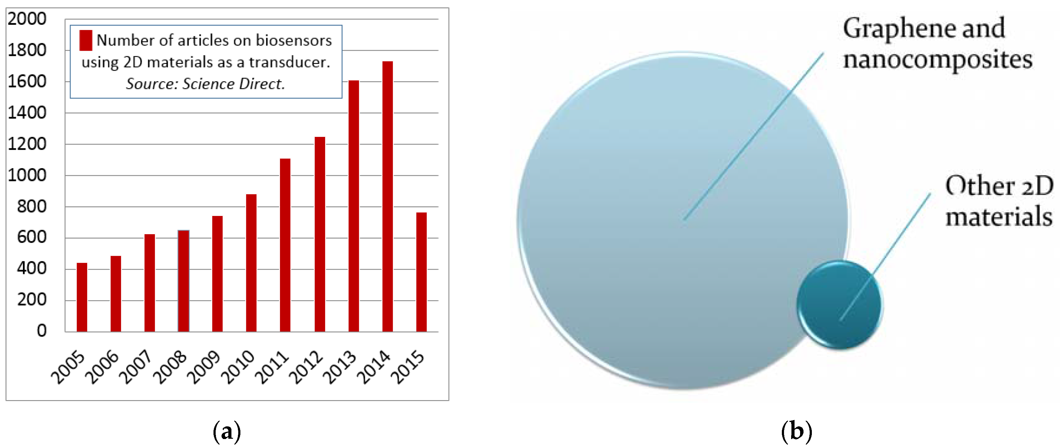
2.2. Current Trends in Biosensors
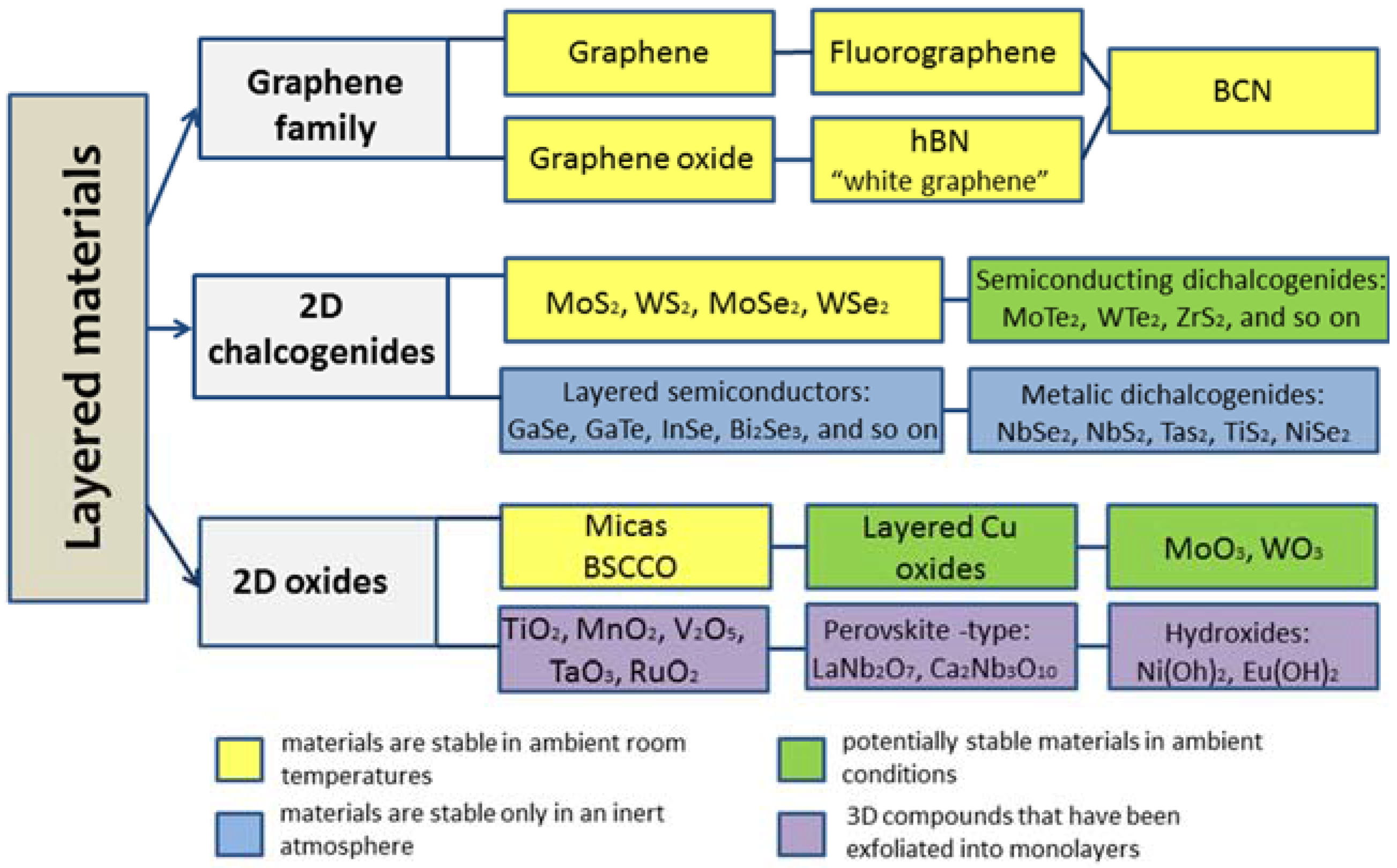
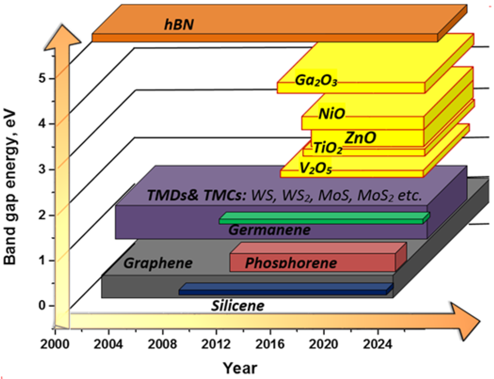
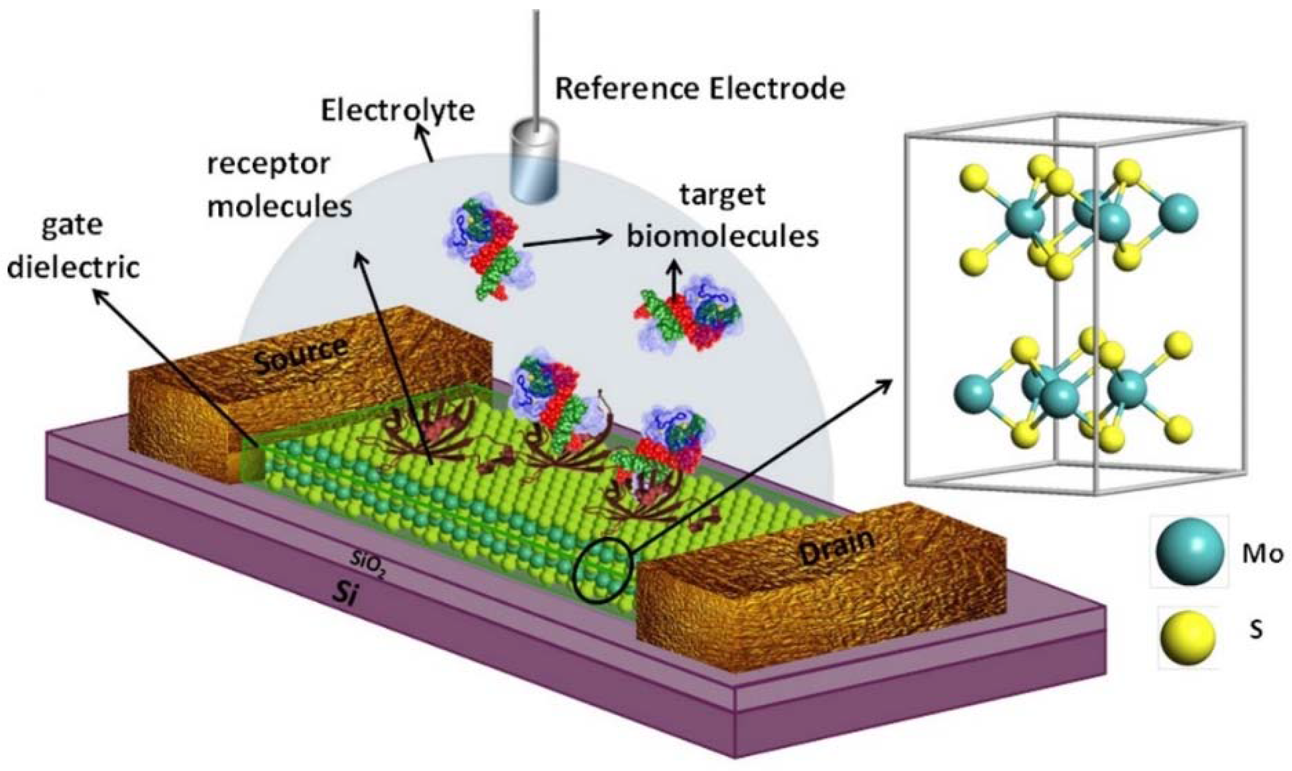
- Primarily, since graphene has a zero band gap, the transistors based on intrinsic graphene have a low on-to-off current ration, resulting in high standby power dissipation, which limits their real circuit application [51]. While 2D non-graphene materials have almost all of the necessary range of band gap values (Figure 2), they can be used for the design of a field effect transistor (FET) device. FET is characterized by high electron mobility and a high on-to-off ratio. Thus, integrating the 2D non-graphene material-based channel of FET with biosensing layers, one can expect the design of a complex biosensing device (FET biosensor). Such devices possess an extremely high sensitivity due to the enhancement of the interface-related phenomena and selectivity due to the immobilized biosensitive layers’ affinity.
- Another significant feature of 2D non-graphene materials is that unlike graphene or Si, many of them have either an intrinsic direct band gap in a bulk state or undergo the transition from indirect to direct semiconductors upon being scaled down to single layers [51]. This opens up their application as a transducer for biosensors of the optical type of detection, where their strong light-matter interaction can be influenced by the interface-related biological actions.
- Finally, it has to be noticed that among the various transducer materials that have been developed, nanostructured metal oxides are promising due to their exceptional optical and electrical properties that offer excellent prospects for the interfacing of biological recognition events with electronic or optical signal transduction and for the designing of a new generation of bioelectronics devices that may exhibit novel functions.
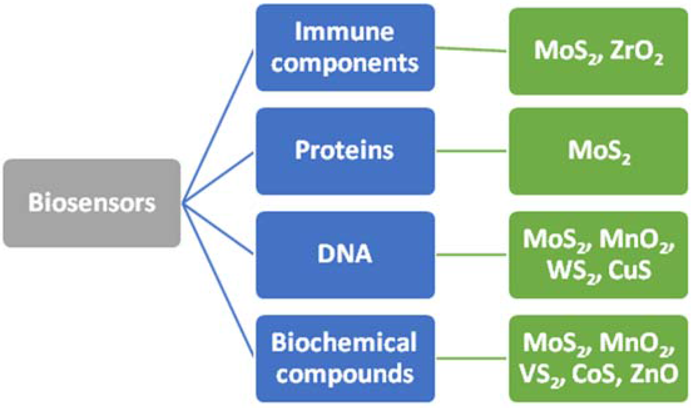
3. Application of 2D Non-Graphene Materials and 2D Nanostructures in Biosensor Design
3.1. MoS2 Material for Electrochemical and Optical Biosensors


3.2. WS2
3.3. VS2
3.4. CoS
3.5. CuS
3.6. g-C3N4
3.7. BN
4. Application of 2D Oxide Nanostructures as Transducers for Biosensors
4.1. MnO2
4.2. α-MoO3
4.3. ZnO
4.4. CuO
5. Conclusions and Outlook

| 2D | Detection Type | Purpose | Sensitivity: Detection Range and Threshold | Comment | Reference |
|---|---|---|---|---|---|
| MoS2 | electro-chemical | Determination of glucose | 2.8 μM–300 μM | Biosensor was developed by immobilizing glucose oxidase (GOx) on a glass carbon electrode that was modified with MoS2 nanosheets that were decorated with Au NPs | [60] |
| electro-chemical | Detection of dopamine | 1.0 mM DA/pH 7.4 | MoS2 sheet-based electrodes were employed for the electrochemical detection of an important neurotransmitter, namely dopamine (DA), in the presence of ascorbic acid (AA) | [54] | |
| FET | Detection of proteins | 713 for a pH change of 1 unit | Biosensors based on field-effect transistors (FETs); specific detection of protein is also demonstrated, and an extremely high sensitivity of 196 was achieved, even at a 100 femtomolar concentration | [53] | |
| fluorescent | Detection of Ag | 25 mg/mL | The developed sensor with high sensitivity and selectivity may be an alternative method for Ag ion detection in lake water samples and other applications | [59] | |
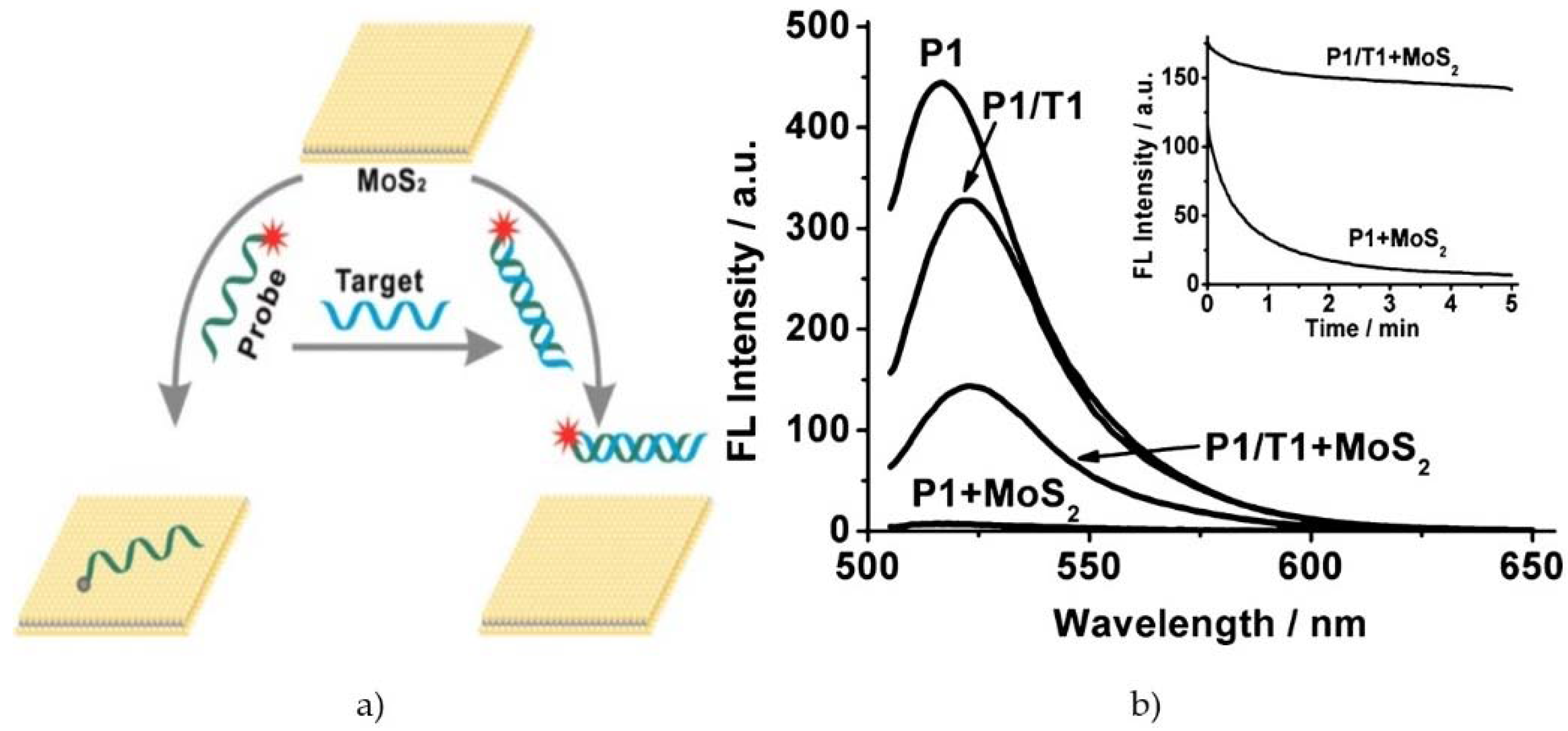
| fluorescent, microfluidic | Fluorescent DNA detection | 0.2 µL | MoS2 nanosheets are able to quench most of the fluorescence in a very short time (~min) and possess different affinities towards ssDNA versus dsDNA | [57] | |
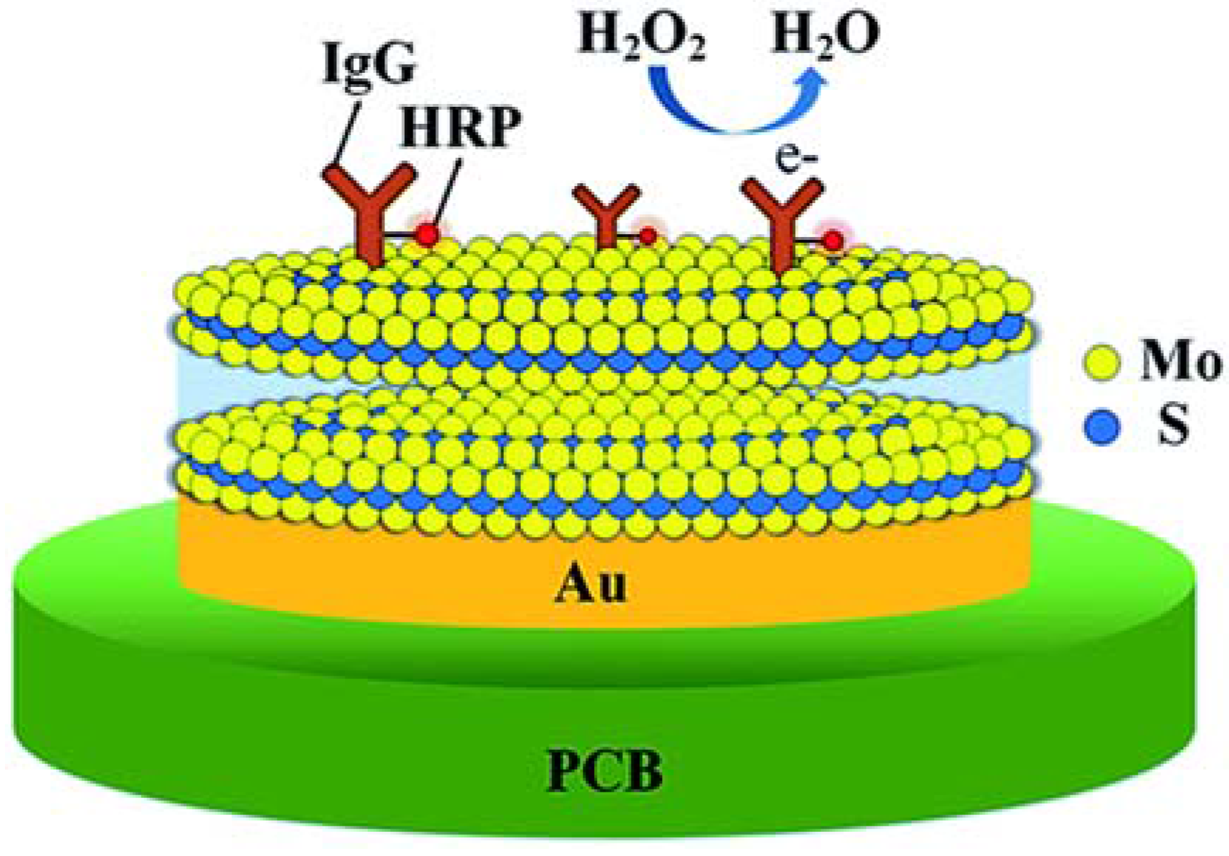
| electro-chemical | Immobilization horseradish peroxidase conjugated IgG | 0–20 ng/mL | The cyclic voltammetry results showed that the sensor of Au-MoS2 conjugated with IgG-HRP thus exhibited excellent analytical responses to H2O2 with a wide linear range | [62] | |
| fluorescent | Detection of prostate specific antigen | 0.2 ng/mL | The binding of the aptamer to the target PSA induces a rigid aptamer structure, which makes the integration with the MoS2 nanosheet very weak | [58] | |
| electro-chemical | DNA analysis | 1.0 × 10⁻¹⁶–1.0 × 10⁻¹⁰ M | The tlh gene sequence assay can be performed label-freely with a detection limit of 1.9 × 10⁻¹⁷ M | [55] | |
| electro-chemical | Determination of bisphenol A | 0.05–100 mM, (5.0 × 10⁻⁹ M) | Biosensor based on MoS2 and chitosan-gold nanoparticle composite-modified electrode | [63] | |
| MnO2 | fluorescent | In vivo sensing of ascorbic acid (AA) | 2.7–25.9 mM−1 | The authors investigate the mechanism of single-layer MnO2 nanosheets suppressing fluorescence of 7-β hydroxycoumarin | [89] |
| fluorescent | DNA hybridization | 0–5 nM | Probing DNA hybridization and aptamer-target interactions in a homogeneous solution | [90] | |
| VS2 | fluorescent | Detection of cytochrome c | 0.75 nM–50 mM | VS2 nanosheets with a high fluorescence quenching ability were synthesized by the solution route | [66] |
| electro-chemical | Determination of 17β-estradiol | 1.0 × 10⁻¹¹–1.0 × 10⁻⁸ M (1.0 × 10⁻¹² M) | VS2 nanoflowers and gold nanoparticle-modified glassy carbon electrode | [67] | |
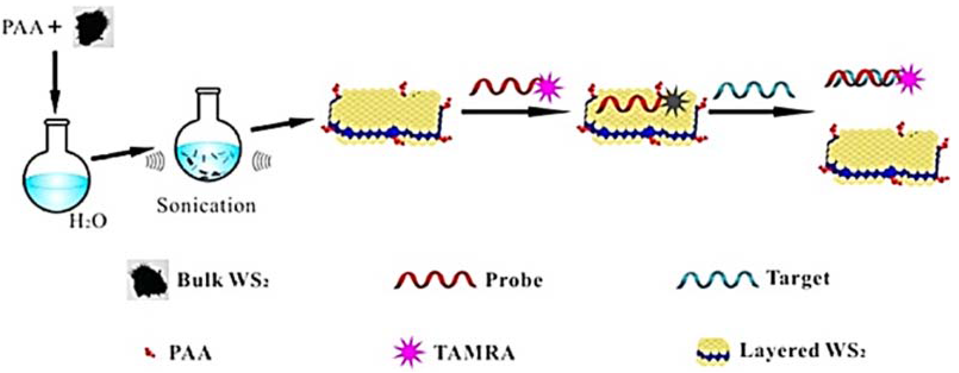
| WS2 | fluorescent | Platform for biosensing (ssDNA) | 1−80 ng/mL | The adsorbed ssDNA is detachable from the nanosheet upon the interaction with other biomolecules, resulting in the restoration of the fluorescence | [88] |
| electro-chemical | Determination of 17β-estradiol | 1 × 10⁻¹¹–5.0 × 10⁻⁹ M (2.0 × 10⁻¹² M) | Aptamers immobilized on the WS2 nanosheets/AuNP-modified glassy carbon electrode | [65] | |
| CoS | electro-chemical | Determination of 17β-estradiol | 1.0 × 10⁻⁹–1.0 × 10⁻¹² M (7.0 × 10⁻¹³ M) | Thiol group tagged 17β-estradiol aptamer on CoS and AuNP-modified electrode | [68] |
| CuS | electro-chemical | Detection of DNA | 0.1 pM−1 nM (20 fM) | DNA labeled at 5 end using 6-mercapto-1-hexhane immobilized on the CuS- acetylene black (AB)/Au-modified electrode | [69] |
| h-BN | electro-chemical | Detection of forchlorfenuron | 0.5 to 10 mM (0.07 μM) | The fabricated enzyme-based sensor demonstrated linear sensitivity for range 0.5–10 mM with a detection limit 0.07 μM | [81] |
| CuO | electro-chemical | Glucose | 2241 μA·mM−1·cm−2, 0.1–4 mM | Glucose level was detected by a fast (~2 s) and precise technique | [96] |
| ZnO | electro-chemical | Detection of cortisol | 7.74 mA/M | Immunosensor based on 2D ZnO nano-flakes synthesized on Au-coated substrates | [93] |
Acknowledgments
Conflicts of Interest
References
- Novoselov, K.S.; Geim, A.K.; Morozov, S.V.; Jiang, D.; Zhang, Y.; Dubonos, S.V.; Grigorieva, I.V.; Firsov, A.A. Electricfield Effect in Atomically Thin Carbon films. Science 2004, 306, 666–669. [Google Scholar] [CrossRef] [PubMed]
- Stoller, M.D.; Park, S.J.; Zhu, Y.W.; An, J.H.; Ruoff, R.S. Graphene-Based Ultracapacitors. Nano Lett. 2008, 8, 3498–3502. [Google Scholar] [CrossRef] [PubMed]
- Stankovich, S.; Dikin, D.A.; Piner, R.D.; Kohlhaas, K.A.; Kleinhammes, A.; Jia, Y.; Wu, Y.; Nguyen, S.T.; Ruoff, R.S. Synthesis of Graphene-Based Nanosheets via Chemical Reduction of Exfoliated Graphite Oxide. Carbon 2007, 45, 1558–1565. [Google Scholar] [CrossRef]
- Yoo, J.J.; Balakrishnan, K.; Huang, J.S.; Meunier, V.; Sumpter, B.G.; Srivastava, A.; Conway, M.; Reddy, A.L.M.; Yu, J.; Vajtai, R.; et al. Ultrathin Planar Graphene Supercapacitors. Nano Lett. 2011, 11, 1423–1427. [Google Scholar] [CrossRef] [PubMed]
- Wu, Z.S.; Ren, W.C.; Gao, L.B.; Liu, B.L.; Jiang, C.B.; Cheng, H.M. Synthesis of High-Quality Graphene with a Pre-Determined Number of Layers. Carbon 2009, 47, 493–499. [Google Scholar] [CrossRef]
- Lee, C.; Wei, X.D.; Kysar, J.W.; Hone, J. Measurement of the Elastic Properties and Intrinsic Strength of Monolayer Graphene. Science 2008, 321, 385–388. [Google Scholar] [CrossRef] [PubMed]
- Nair, R.R.; Blake, P.; Grigorenko, A.N.; Novoselov, K.S.; Booth, T.J.; Stauber, T.; Peres, N. Fine Structure Constant Defines Visual Transparency of Graphene. Science 2008, 320, 1308. [Google Scholar] [CrossRef] [PubMed]
- Ghosh, S.; Calizo, I.; Teweldebrhan, D.; Pokatilov, E.P.; Nika, D.L.; Balandin, A.A. Extremely High Thermal Conductivity of Graphene: Prospects for Thermal Management Applications in Nanoelectronic Circuits. Appl. Phys. Lett. 2008, 92, 151911–151913. [Google Scholar] [CrossRef]
- Chen, Z.; Yu, D.S.; Xiong, W.; Liu, P.P.; Liu, Y.; Dai, L.M. Graphene-Based Nanowire Supercapacitors. Langumuir 2014, 30, 3567–3571. [Google Scholar] [CrossRef] [PubMed]
- Qian, Y.; Lu, S.B.; Gao, F.L. Synthesis of Manganese Dioxide/Reduced Graphene Oxide Composites with Excellent Electrocatalytic Activity Toward Reduction of Oxygen. Mater. Lett. 2011, 65, 56–58. [Google Scholar] [CrossRef]
- Shao, Y.Y.; Wang, J.; Wu, H.; Liu, J.; Aksay, I.A.; Lin, Y.H. Graphene Based Electrochemical Sensors and Biosensors: A Review. Electroanalysis 2010, 22, 1027–1036. [Google Scholar] [CrossRef]
- Ferrari, A.C.; Bonaccorso, F.; Falko, V.; Novoselov, K.S.; Roche, S.; Bøggild, P.; Borini, S.; Koppens, F.; Palermo, V.; Pugno, N.; et al. Science and technology roadmap for graphene, related two-dimensional crystals, and hybrid systems. Nanoscale 2014, 7, 4598–4810. [Google Scholar] [CrossRef] [PubMed]
- Butler, S.Z.; Hollen, S.M.; Cao, L.; Cui, Y.; Gupta, J.; Gutiérrez, H.R.; Heinz, T.F.; Hong, S.S.; Huang, J.; Ismach, A.F.; et al. Progress, challenges, and opportunities in two-dimensional materials beyond graphene. ACS Nano 2013, 7, 2898–2926. [Google Scholar] [CrossRef] [PubMed]
- Li, L.; Yu, Y.; Ye, G.J.; Ge, Q.; Ou, X.; Wu, H.; Feng, D.; Chen, X.H.; Zhang, Y. Black phosphorus field-effect transistors. Nat. Nanotechnol. 2014, 9, 372–377. [Google Scholar] [CrossRef] [PubMed]
- Zhu, C.; Du, D.; Lin, Y. Graphene and graphene-like 2D materials for optical biosensing and bioimaging: A review. 2D Mater. 2015, 2, 032004. [Google Scholar] [CrossRef]
- Geim, A.K.; Grigorieva, I.V. Van der Waals heterostructures. Nature 2013, 499, 419–425. [Google Scholar] [CrossRef] [PubMed]
- Gupta, A.; Sakthivel, T.; Seal, S. Recent development in 2D materials beyond graphene. Prog. Mater. Sci. 2015, 73, 44–126. [Google Scholar] [CrossRef]
- Mas-Ballesté, R.; Gómez-Navarro, C.; Gómez-Herrero, J.; Zamora, F. 2D materials: To graphene and beyond. Nanoscale 2011, 3, 20–30. [Google Scholar] [CrossRef] [PubMed]
- North, S.H.; Lock, E.H. Critical aspects of biointerface design and their impact on biosensor development. Anal. Bioanal. Chem. 2010, 397, 925–933. [Google Scholar] [CrossRef] [PubMed]
- Solanki, P.R.; Kaushik, A. Nanostructured metal oxide-based biosensors. NPG Asia Mater. 2011, 3, 17–24. [Google Scholar] [CrossRef]
- Ronkainen, N.J.; Halsall, H.B.; Heineman, W.R. Electrochemical biosensors. Chem. Soc. Rev. 2010, 39, 1747–1763. [Google Scholar] [CrossRef] [PubMed]
- Zhu, C.; Yang, G.; Li, H.; Du, D.; Lin, Y. Electrochemical sensors and biosensors based on nanomaterials and nanostructures. Anal. Chem. 2015, 87, 230–249. [Google Scholar] [CrossRef] [PubMed]
- Walcarius, A.; Minteer, S.D.; Wang, J.; Lin, Y.; Merkoçi, A. Nanomaterials for bio-functionalized electrodes: Recent trends. J. Mater. Chem. B 2013, 1, 4878–4908. [Google Scholar] [CrossRef]
- Gea, X.; Asiri, A.M.; Du, D.; Wen, W.; Wang, S.; Lin, Y. Nanomaterial-enhanced paper-based biosensors. Trends Anal. Chem. 2014, 58, 31–39. [Google Scholar] [CrossRef]
- Ravindra, N.M.; Prodan, C. Advances in the manufacturing, types, and applications of biosensors. JOM 2007, 59, 37–43. [Google Scholar] [CrossRef]
- Turner, D.C.; Chang, C.Y.; Fang, K.; Brandow, S.L.; Murphy, D.B. Selective adhesion of functional microtubules to patterned silane surfaces. Biophys. J. 1995, 69, 2782–2789. [Google Scholar] [CrossRef]
- Clark, L.C.; Lyons, C. Electrode systems for continuous monitoring in cardiovascular surgery. Ann. N. Y. Acad. Sci. 1962, 102, 29–45. [Google Scholar] [CrossRef] [PubMed]
- Renneberg, R.; Pfeiffer, D.; Lisdat, F.; Wilson, G.; Wollenberger, U.; Ligler, F.; Turner, A.P.F. Frieder Scheller and the short history of biosensors. Adv. Biochem. Eng. Biotechnol. 2008, 109, 1–18. [Google Scholar] [PubMed]
- Scognamiglio, V.; Pezzotti, G.; Pezzotti, I.; Cano, J.; Buonasera, K.; Giannini, D.; Giardi, M.T. Biosensors for effective environmental and agrifood protection and commercialization: From research to market. Microchim. Acta 2010, 170, 215–225. [Google Scholar] [CrossRef]
- Scheller, F.W.; Wollenberger, U.; Warsinke, A.; Lisdat, F. Research and development in biosensors. Curr. Opin. Biotechnol. 2001, 12, 35–404. [Google Scholar] [CrossRef]
- Teng, Y.; Zhang, X.; Fu, Y.; Liu, H.; Wang, Z.; Jin, L.; Zhang, W. Optimized ferrocene-functionalized ZnO nanorods for signal amplification in electrochemical immunoassay of Escherichia Coli. Biosens. Bioelectron. 2011, 26, 4661–4666. [Google Scholar] [CrossRef] [PubMed]
- Queiros, R.B.; Noronha, J.P.; Marques, P.V.S.; Sales, M.G.F. Emerging (bio)sensing technology for assessing and monitoring freshwater contamination—Methods and applications. In Ecological Water Quality—Water Treatment and Reuse, 1st ed.; Voudouris, K., Ed.; InTech: Rijeka, Croatia, 2012; pp. 65–94. [Google Scholar]
- Ansari, A.A.; Kaushik, A.; Solanki, P.R.; Malhotr, B.D. Nanostructured zinc oxide platform for mycotoxin detection. Bioelectrochem 2010, 77, 75–81. [Google Scholar] [CrossRef] [PubMed]
- Soldatkin, A.P.; Volotovsky, V.; El’skaya, A.V.; Jaffrezic-Renault, N.; Martelet, C. Improvement of urease based biosensor characteristics using additional layers of charged polymers. Anal. Chim. Acta 2000, 403, 25–29. [Google Scholar] [CrossRef]
- Ilangovan, R.; Daniel, D.; Krastanov, A.; Zachariah, C.; Elizabeth, R. Enzyme based biosensor for heavy metal ions determination. Biotechnol. Biotechnol. Eq. 2006, 20, 184–189. [Google Scholar] [CrossRef]
- Domínguez-Renedo, O.; Alonso-Lomillo, M.A.; Arcos-Martínez, M.J. Determination of metals based on electrochemical biosensors. Crit. Rev. Env. Sci. Technol. 2013, 43, 1042–1073. [Google Scholar] [CrossRef]
- Dhull, V.; Gahlaut, A.; Dilbaghi, N.; Hooda, V. Acetylcholinesterase biosensors for electrochemical detection of organophosphorus compounds: A review. Biochem. Res. Int. 2013, 2013, 1–18. [Google Scholar] [CrossRef] [PubMed]
- Tusa, J.K.; He, H. Critical care analyzer with fluorescent optical chemosensors for blood analytes. J. Mater. Chem. 2005, 15, 2640–2647. [Google Scholar] [CrossRef]
- Osaka, T.; Komaba, S.; Seyama, M.; Tanabe, K. High-sensitivity urea sensor based on the composite film of electroinactive polypyrrole with polyion complex. Sens. Actuators B Chem. 1996, 36, 463–469. [Google Scholar] [CrossRef]
- Jijun, T.; Jie, H.; Zhongchao, H.; Min, P.; Yuquan, C. A novel lactate biosensor. In Proceedings of the 27th Annual International Conference of the IEEE Engineering in Medicine and Biology Society, Shanghai, China, 17–18 January 2006; pp. 252–254.
- Heinemann, L. Continuous glucose monitoring and clinical trials. J. Diabetes Sci. Technol. 2009, 3, 981–985. [Google Scholar] [CrossRef] [PubMed]
- Fojta, M. Electrochemical sensors for DNA interactions and damage. Electroanalysis 2002, 14, 1449–1463. [Google Scholar] [CrossRef]
- Hang, T.C.; Guiseppi-Elie, A. Frequency dependent and surface characterization of DNA immobilization and hybridization. Biosens. Bioelectron. 2004, 19, 1537–1548. [Google Scholar] [CrossRef] [PubMed]
- Hofmann, U.; Michaelis, S.; Winckler, T.; Wegener, J.; Feller, K.H. A whole-cell biosensor as in vitro alternative to skin irritation tests. Biosens. Bioelectron. 2013, 39, 156–162. [Google Scholar] [CrossRef] [PubMed]
- Monosik, R.; Stredansky, M.; Strurdik, E. Application of electrochemical biosensor in clinical diagnosis in clinical diagnosis. J. Clin. Lab. Anal. 2012, 26, 22–34. [Google Scholar] [CrossRef] [PubMed]
- Kaushik, A.; Tiwari, S.; Jayant, R.D.; Marty, A.; Nair, M. Towards detection and diagnosis of Ebola virus disease at point-of-care. Biosens. Bioelectron. 2016, 75, 254–272. [Google Scholar] [CrossRef] [PubMed]
- Kaushik, A.; Yndart, A.; Jayant, R.D.; Sagar, V.; Atluri, V.; Bhansali, S.; Nair, M. Electrochemical sensing method for point-of-care cortisol detection in human immunodeficiency virus-infected patients. Int. J. Nanomed. 2015, 10, 677–685. [Google Scholar]
- Kaushik, A.; Vasudev, A.; Arya, S.K.; Pasha, S.K.; Bhansali, S. Recent advances in cortisol sensing technologies for point-of-care application. Biosens. Bioelectron. 2015, 53, 499–512. [Google Scholar] [CrossRef] [PubMed]
- Yang, G.; Zhu, C.; Du, D.; Zhu, J.; Lin, Y. Graphene-like two-dimensional layered nanomaterials: Applications in biosensors and nanomedicine. Nanoscale 2015, 7, 14217–14231. [Google Scholar] [CrossRef] [PubMed]
- Chen, Y.; Tan, C.; Zhang, H.; Wang, L. Two-dimensional graphene analogues for biomedical applications. Chem. Soc. Rev. 2015, 44, 2681–2701. [Google Scholar] [CrossRef] [PubMed]
- Minoru, O.; Takayoshi, S. Two-Dimensional Dielectric Nanosheets: Novel Nanoelectronics from Nanocrystal Building Blocks. Adv. Mater. 2012, 2, 210–228. [Google Scholar]
- Late, D.J.; Rout, C.S. A Perspective on Atomically Thin 2D Inorganic Layered Materials for Biosensor. J. Nanomed. Res. 2015, 2, 15–18. [Google Scholar] [CrossRef]
- Sarkar, D.; Liu, W.; Xie, X.; Anselmo, A.C.; Mitragotri, S.; Banerjee, K. MoS2 Field-Effect Transistor for Next-Generation Label-Free Biosensors. ACS Nano 2014, 4, 3992–4003. [Google Scholar] [CrossRef] [PubMed]
- Narayanan, T.N.; Vusa, C.S.R.; Alwarappan, S. Erratum: Selective and Efficient Electrochemical Biosensing of Ultrathin Molybdenum Disulfide Sheets. Nanotechnology 2014, 25, 335702. [Google Scholar] [CrossRef] [PubMed]
- Wang, X.; Nan, F.; Zhao, J.; Yang, T.; Ge, T.; Jiao, T.A. Label-Free Ultrasensitive Electrochemical DNA Sensor Based on Thin-Layer MoS2 Nanosheets with high Electrochemical Activity. Biosens. Bioelectron. 2015, 64, 386–391. [Google Scholar] [CrossRef] [PubMed]
- Zhu, C.; Zeng, Z.; Li, H.; Li, F.; Fan, C.; Zhang, H. Single-Layer MoS2-Based Nanoprobes for Homogeneous Detection of Biomolecules. J. Am. Chem. Soc. 2013, 135, 5998–6001. [Google Scholar] [CrossRef] [PubMed]
- Huang, Y.; Shi, Y.; Yang, H.Y.; Ai, Y. A Novel Single-Layered MoS2 Nanosheet Based Microfluidic Biosensor for Ultrasensitive Detection of DNA. Nanoscale 2015, 7, 2245–2249. [Google Scholar] [CrossRef] [PubMed]
- Kong, R.-M.; Ding, L.; Wang, Z.; You, J.; Qu, F. A Novel Aptamer-Functionalized MoS2 Nanosheet Fluorescent Biosensor for Sensitive Detection of Prostate Specific Antigen. Anal. Bioanal. Chem. 2015, 407, 369–377. [Google Scholar] [CrossRef] [PubMed]
- Mao, K.; Wu, Z.; Chen, Y.; Zhou, X.; Shen, A.; Hu, J. A Novel Biosensor Based on Single-Layer MoS2 Nanosheets for Detection of Ag. Talanta 2015, 132, 658–663. [Google Scholar] [CrossRef] [PubMed]
- Su, S.; Sun, H.; Xu, F.; Yuwen, L.; Fan, C.; Wang, L. Direct Electrochemistry of Glucose Oxidase and a Biosensor for Glucose Based on a Glass Carbon Electrode Modified with MoS2 Nanosheets Decorated with Gold Nanoparticles. Microchim. Acta 2014, 181, 1497–1503. [Google Scholar] [CrossRef]
- Song, H.; Ni, Y.; Kokotc, S. Investigations of an electrochemical platform based on the layered MoS2-graphene and horseradish peroxidase nanocomposite for direct electrochemistry and electrocatalysis. Biosens. Bioelectron. 2014, 56, 137–143. [Google Scholar] [CrossRef] [PubMed]
- Kim, H.-U.; Kim, H.; Ahn, C.; Kulkarni, A.; Jeon, M.; Yeom, G.Y.; Lee, M.-H.; Kim, T. In Situ Synthesis of MoS2 on a Polymer Based Gold Electrode Platform and its Application in Electrochemical Biosensing. RSC Adv. 2015, 5, 10134–10138. [Google Scholar] [CrossRef]
- Huang, K.-J.; Liu, Y.-J.; Liu, Y.-M.; Wang, L.-L. Molybdenum Disulfide Nanoflower-Chitosan-Au Nanoparticles Composites Based Electrochemical Sensing Platform for Bisphenol A Determination. J. Hazard. Mater. 2014, 276, 207–215. [Google Scholar] [CrossRef] [PubMed]
- Yuan, Y.; Li, R.; Liu, Z. Establishing Water-Soluble Layered WS2 Nanosheet as a Platform for Biosensing. Anal. Chem. 2014, 86, 3610–3615. [Google Scholar] [CrossRef] [PubMed]
- Huang, K.-J.; Liu, Y.-J.; Shi, G.-W.; Zhang, J.-Z.; Liu, Y.-M. A Novel Aptamer Sensor Based on Layered Tungsten Disulfide Nanosheets and Au Nanoparticles Amplification for 17β-Estradiol Detection. Anal. Methods 2014, 6, 8011–8017. [Google Scholar] [CrossRef]
- Yin, X.; Cai, J.; Feng, H.; Wu, Z.; Zou, J.; Cai, Q. A Novel VS2 Nanosheet-Based Biosensor for Rapid Fluorescence Detection of Cytochrome C. New J. Chem. 2015, 39, 1892–1898. [Google Scholar] [CrossRef]
- Huang, K.-J.; Liu, Y.-J.; Shi, G.-W.; Yang, X.-R.; Liu, Y.-M. Label-Free Aptamer Sensor for 17β-Estradiol Based on Vanadium Disulfide Nanoflowers and Au Nanoparticles. Sens. Actuators B 2014, 201, 579–585. [Google Scholar] [CrossRef]
- Huang, K.J.; Liu, Y.J.; Zhang, J.Z.; Cao, T.; Liu, Y.M. Aptamer/Au Nanoparticles/Cobalt Sulfide Nanosheets Biosensor for 17β-Estradiol Detection Using a Guanine-Rich Complementary DNA Sequence for Signal Amplification. Biosens. Bioelectron. 2015, 67, 184–191. [Google Scholar] [CrossRef] [PubMed]
- Huang, K.J.; Liu, Y.J.; Zhang, J.Z.; Liu, Y.M. A Sequence-Specific DNA Electrochemical Sensor Based on Acetylene Black Incorporated Two-Dimensional CuS Nanosheets and Gold Nanoparticles. Sens. Actuators B Chem. 2015, 209, 570–578. [Google Scholar] [CrossRef]
- Zhang, X.D.; Xie, X.; Wang, H.; Zhang, J.-J.; Pan, B.-C.; Xie, Y. Enhanced Photoresponsive Ultrathin Graphitic-Phase C3N4 Nanosheets for Bioimaging. J. Am. Chem. Soc. 2013, 135, 18–21. [Google Scholar] [CrossRef] [PubMed]
- Wang, Q.B.; Wang, W.; Lei, J.P.; Xu, N.; Gao, F.L.; Ju, H.X. Fluorescence Quenching of Carbon Nitride Nanosheet through Its Interaction with DNA for Versatile Fluorescence Sensing. Anal. Chem. 2013, 85, 12182–12188. [Google Scholar] [CrossRef] [PubMed]
- Tang, Y.; Song, H.; Su, Y.; Lv, Y. Turn-on persistent luminescence probe based on graphitic carbon nitride for imaging detection of biothiols in biological fluids. Anal. Chem. 2013, 85, 11876–11884. [Google Scholar] [CrossRef] [PubMed]
- Ma, T.Y.; Tang, Y.; Dai, S.; Qiao, S.Z. Proton-Functionalized Two-Dimensional Graphitic Carbon Nitride Nanosheet: An Excellent Metal-/Label-Free Biosensing Platform. Small 2014, 10, 2382–2389. [Google Scholar] [CrossRef] [PubMed]
- Rong, M.; Lin, L.; Song, X.; Zhao, T.; Zhong, Y.; Yan, J.; Wang, Y.; Chen, X. A label-free fluorescence sensing approach for selective and sensitive detection of 2,4,6-trinitrophenol (TNP) in aqueous solution using graphitic carbon nitride nanosheets. Anal. Chem. 2015, 87, 1288–1296. [Google Scholar] [CrossRef] [PubMed]
- Ou, X.; Tan, X.; Liu, X.; Lu, Q.; Chen, S.; Wei, S. A signal-on electrochemiluminescence biosensor for detecting Con A using phenoxy dextran-graphite-like carbon nitride as signal probe. Biosens. Bioelectron. 2015, 70, 89–97. [Google Scholar] [CrossRef] [PubMed]
- Liu, Y.; Wang, Q.; Lei, J.; Hao, Q.; Wang, W.; Ju, H. Anodic electrochemiluminescence of graphitic-phase C3N4 nanosheets for sensitive biosensing. Talanta 2014, 122, 130–134. [Google Scholar] [CrossRef] [PubMed]
- Wang, Y.Z.; Hao, N.; Feng, Q.M.; Shi, H.W.; Xu, J.J.; Chen, H.Y. A ratiometric electrochemiluminescence detection for cancer cells using g-C3N4 nanosheets and Ag–PAMAM–luminol nanocomposites. Biosens. Bioelectron. 2016, 77, 76–82. [Google Scholar] [CrossRef] [PubMed]
- Zhi, C.Y.; Bando, Y.; Tang, C.C.; Huang, Q.; Golberg, D. Boron nitride nanotubes: Functionalization and composites. J. Mater. Chem. 2008, 18, 3900–3908. [Google Scholar] [CrossRef]
- Ciofani, G.; Danti, S.; Genchi, G.G.; Mazzolai, B.; Mattoli, V. Boron Nitride Nanotubes: Biocompatibility and Potential Spill-Over in Nanomedicine. Small 2013, 9, 1672–1685. [Google Scholar] [CrossRef] [PubMed]
- Uosaki, K.; Elumalai, G.; Noguchi, H.; Masuda, T.; Lyalin, A.; Nakayama, A.; Taketsugu, T. Boron Nitride Nanosheet on Gold as an Electrocatalyst for Oxygen Reduction Reaction: Theoretical Suggestion and Experimental Proof. J. Am. Chem. Soc. 2014, 136, 6542–6545. [Google Scholar] [CrossRef] [PubMed]
- Xu, Q.; Cai, L.; Zhao, H.; Tang, J.; Shen, Y.; Hu, X.; Zeng, H. Forchlorfenuron detection based on its inhibitory effect towards catalase immobilized on boron nitride substrate. Biosens. Bioelectron. 2015, 63, 294–300. [Google Scholar] [CrossRef] [PubMed]
- Yang, G.; Abulizi, A.; Zhu, J. Sonochemical fabrication of gold nanoparticles-boron nitride sheets nanocomposites for enzymeless hydrogen peroxide detection. Ultrason. Sonochem. 2014, 21, 1958–1963. [Google Scholar] [CrossRef] [PubMed]
- Sodzel, D.; Khranovskyy, V.; Beni, V.; Turner, A.P.F.; Viter, R.; Eriksson, M.O.; Holtz, P.-O.; Janot, J.-M.; Bechelany, M.; Balme, S.; et al. Continuous sensing of hydrogen peroxide and glucose via quenching of the UV and visible luminescence of ZnO nanoparticles. Michrochim. Acta 2015, 182, 1819–1826. [Google Scholar] [CrossRef]
- Peng, J.; Wang, S.; Zhang, P.; Jiang, L.; Shi, J.; Zhu, J. Fabrication of Graphene Quantum Dots and Hexagonal Boron Nitride Nanocomposites for Fluorescent Cell Imaging. J. Biomed. Nanotechnol. 2013, 9, 1679–1685. [Google Scholar] [CrossRef] [PubMed]
- Yang, G.; Shi, J.; Wang, S.; Xiong, W.; Jiang, L.; Burdab, C.; Zhu, J. Fabrication of a boron nitride–gold nanocluster composite and its versatile application for immunoassays. Chem. Commun. 2013, 49, 10757–10759. [Google Scholar] [CrossRef] [PubMed]
- Gomez, J.L. Zinc Oxide Nanostructures: From Growth to Application. J. Mater. Sci. 2013, 48, 612–624. [Google Scholar] [CrossRef]
- Yuan, Y.; Wu, S.; Shu, F.; Liu, Z. An MnO2 Nanosheet as a Label-Free Nanoplatform for Homogeneous Biosensing. Chem. Commun. 2014, 50, 1095–1097. [Google Scholar] [CrossRef] [PubMed]
- Zhai, W.; Wang, C.; Yu, P.; Wang, Y.; Mao, L. Single-Layer MnO2 Nanosheets Suppressed Fluorescence of 7-Hydroxycoumarin: Mechanistic Study and Application for Sensitive Sensing of Ascorbic Acid in Vivo. Anal. Chem. 2014, 86, 12206–12213. [Google Scholar] [CrossRef] [PubMed]
- He, D.; He, X.; Wang, K.; Yang, X.; Yang, X.; Li, X.; Zou, Z. Nanometer-Sized Manganese Oxide-Quenched Fluorescent Oligonucleotides: An Effective Sensing Platform for Probing Biomolecular Interactions. Chem. Commun. 2014, 50, 11049–11052. [Google Scholar] [CrossRef] [PubMed]
- Balendhran, S.; Walia, S.; Alsaif, M.; Nguyen, E.P.; Ou, J.Z.; Zhuiykov, S.; Sriram, S.; Bhaskaran, M.; Kalantarzadeh, K. Field Effect Biosensing Platform Basedon 2D α-MoO3. Acsnano 2013, 11, 9753–9760. [Google Scholar]
- Yakimova, R.; Selegard, L.; Khranovskyy, V.; Pearce, R.; Spetz, A.L.; Uvdal, K. ZnO materials and surface tailoring for biosensing. Front. Biosci. 2012, 4, 254–278. [Google Scholar] [CrossRef]
- Vabbina, P.K.; Kaushik, A.; Pokhrel, N.; Bhansali, S.; Pala, N. Electrochemical Cortisol Immunosensors Based on Sonochemically Synthesized Zinc Oxide 1D Nanorods and 2D Nanoflakes. Biosens. Bioelectron. 2015, 63, 124–130. [Google Scholar] [CrossRef] [PubMed]
- Sticker, D.; Rothbauer, M.; Charwat, V.; Steinkühler, J.; Bethge, O.; Bertagnolli, E.; Wanzenboeck, H.; Ertl, P. Zirconium Dioxide Nanolayer Passivated Impedimetric Sensors for Cell-Based Assays. Sens. Actuators B 2015, 213, 35–44. [Google Scholar] [CrossRef]
- Zhao, Y.; Zhao, J.; Li, Y.; Ma, D.; Hou, S.; Li, L.; Hao, X.; Wang, Z. Room temperature synthesis of 2D CuO nanoleaves in aqueous solution. Nanotechnology 2011, 22, 115604. [Google Scholar] [CrossRef] [PubMed]
- Sun, S.; Sun, Y.; Chen, A.; Zhang, X.; Yang, Z. Nanoporous copper oxide ribbon assembly of free-standing nanoneedles as biosensors for glucose. Analyst 2015, 140, 5205–5215. [Google Scholar] [CrossRef] [PubMed]
- Bhattacharjee, A.; Ahmaruzzaman, M. Facile synthesis of 2-dimensional CuO nanoleaves and their degradation behavior for Eosin Y. Mater. Lett. 2015, 16, 20–25. [Google Scholar] [CrossRef]
- Bhattacharjee, A.; Ahmaruzzaman, M. Green Synthesis of 2D CuO nanoleaves (NLs) and its application for the reduction of pnitrophenol. Mater. Lett. 2015, 161, 79–82. [Google Scholar] [CrossRef]
© 2016 by the authors; licensee MDPI, Basel, Switzerland. This article is an open access article distributed under the terms and conditions of the Creative Commons by Attribution (CC-BY) license (http://creativecommons.org/licenses/by/4.0/).
Share and Cite
Shavanova, K.; Bakakina, Y.; Burkova, I.; Shtepliuk, I.; Viter, R.; Ubelis, A.; Beni, V.; Starodub, N.; Yakimova, R.; Khranovskyy, V. Application of 2D Non-Graphene Materials and 2D Oxide Nanostructures for Biosensing Technology. Sensors 2016, 16, 223. https://doi.org/10.3390/s16020223
Shavanova K, Bakakina Y, Burkova I, Shtepliuk I, Viter R, Ubelis A, Beni V, Starodub N, Yakimova R, Khranovskyy V. Application of 2D Non-Graphene Materials and 2D Oxide Nanostructures for Biosensing Technology. Sensors. 2016; 16(2):223. https://doi.org/10.3390/s16020223
Chicago/Turabian StyleShavanova, Kateryna, Yulia Bakakina, Inna Burkova, Ivan Shtepliuk, Roman Viter, Arnolds Ubelis, Valerio Beni, Nickolaj Starodub, Rositsa Yakimova, and Volodymyr Khranovskyy. 2016. "Application of 2D Non-Graphene Materials and 2D Oxide Nanostructures for Biosensing Technology" Sensors 16, no. 2: 223. https://doi.org/10.3390/s16020223
APA StyleShavanova, K., Bakakina, Y., Burkova, I., Shtepliuk, I., Viter, R., Ubelis, A., Beni, V., Starodub, N., Yakimova, R., & Khranovskyy, V. (2016). Application of 2D Non-Graphene Materials and 2D Oxide Nanostructures for Biosensing Technology. Sensors, 16(2), 223. https://doi.org/10.3390/s16020223