Article

Two New Mesophotic Species of the Red Algal Genus Chrysymenia (Rhodymeniaceae, Rhodymeniales) from the Gulf of Mexico: C. stanlaurelii sp. nov. and C. oliverhardyi sp. nov.

by
William E. Schmidt
1,*,
Natalia Arakaki
2,
Carlos Frederico D. Gurgel
3,
Daniela Gabriel
4,
Thomas Sauvage
5,
James N. Norris
6 and
Suzanne Fredericq
1
1
Department of Biology, University of Louisiana at Lafayette, Lafayette, LA 70504-3602, USA
2
Banco de Germoplasma de Organismos Acuáticos, Área Funcional de Investigaciones en Acuicultura, Instituto del Mar del Perú, Esquina Gamarra y General Valle S/N, Chucuita, Callao 07021, Peru
3
Instituto de Biodiversidade e Sustentabilidade—NUPEM, Universidade Federal do Rio de Janeiro, Macaé, RJ 27965-045, Brazil
4
Research Center in Biodiversity and Genetic Resources (CIBIO), University of the Azores, 9500-321 Ponta Delgada, Azores, Portugal
5
Smithsonian Marine Station, Pierce, FL 34949, USA
6
Department of Botany, National Museum of Natural History, Smithsonian Institution, Washington, DC 20560, USA
*
Author to whom correspondence should be addressed.
Diversity 2026, 18(6), 320; https://doi.org/10.3390/d18060320
Submission received: 29 April 2026 / Revised: 13 May 2026 / Accepted: 18 May 2026 / Published: 28 May 2026
(This article belongs to the Special Issue Systematics, Ecology and Biodiversity of Marine Algae and Seagrasses)

Abstract

Two new mesophotic species of Chrysymenia are reported for the Gulf of Mexico. Chrysymenia oliverhardyii sp. nov. is a broadly foliose species growing attached to rhodoliths (free-living carbonate nodules predominantly accreted by crustose coralline algae) at 58–66 m depth offshore Louisiana (northwestern Gulf) and at ~64–68 m depth in the vicinity of the Dry Tortugas, Florida (southeastern Gulf). A multi-marker phylogenetic analysis based on chloroplast-encoded rbcL and UPA, as well as nuclear LSU rDNA sequences, indicates that this taxon is most closely related to C. stanlaurelii sp. nov., a slender, branched species from the vicinity of the Florida Middle Grounds, Florida (northeastern Gulf), growing at 60 m depth. These two species from the eastern Gulf of Mexico share a basal most recent common ancestor with respect to the other known species of Chrysymenia. Illustrations and discussion are provided for the new species, as well as for the other Gulf of Mexico members C. planifrons, C. littleriana, C. halymenioides and C. nodulosa. A nomenclatural list, morphological figures, a dichotomous key, and a phylogenetic tree of pertinent Chrysymenia are provided.

1. Introduction

With the finding that the mesophotic communities in the Gulf of Mexico (GoMx) are surprisingly rich in members of the red algal order Rhodymeniales Nägeli [1,2,3,4,5], increased taxon sampling throughout the region has enhanced our understanding of the phylogenetic relationships within the order. Though members of the family Rhodymeniaceae Harvey exhibit a range of morphologies, they are characterized by a pre-fertilization four-celled female carpogonial branch organized in a specialized procarp, the apparent lack of a tela arachnoidea associated with the post-fertilization carposporophyte, and the presence of intercalary positioned cruciately or irregular-cruciately divided tetrasporangia [6,7,8,9]. Taxonomic emphasis in the family is also placed on the gonimoblasts being composed of compact gonimolobes, with most of the gonimolobe cells becoming carposporangia, and with typically elongate and slender carposporophytic fusion products and enlarged pit connections between partly fused cells.
The Rhodymeniaceae currently contains approximately 23 accepted genera [10]. One genus, Chrysymenia J. Agardh, with type C. ventricosa (Lamouroux) J. Agardh [11] described from the French Mediterranean coast has been historically defined by an almost completely hollow thallus that lacks diaphragms and internal rhizoids, and in which the only solid portion of the thallus is limited to the stipe [4]. A genus with a similar thallus organization, but with the presence of internal rhizoids, is characteristic of Cryptarachne (Harvey) Kylin [6] with type C. agardhii (Harvey) Kylin [12], a species described from Key West, Atlantic Florida, and presently regarded as a synonym of Chrysymenia agardhii Harvey. Currently, a total of 23 species is recognized as Chrysymenia (including Cryptarachne), and two species in the genus Cryptarachne are recognized by some systematists [13]. Most of these species are tropical or subtropical in distribution and occur commonly subtidally at depths of >50 m. Norris and Ballantine [14] designated three Chrysymenia “form groups” based purely on habit shape. Form group-1 was characterized by erect thalli that are terete to moderately compressed; form group-2 comprised erect thalli that are blade-like; and form group-3 was composed of repent thalli with flattened or lobed forms. In the Gulf of Mexico, form group-1 is represented by C. pseudoventricosa W.E. Schmidt [4], C. halymenioides Harvey, and an undescribed species, all with cylindrical to moderately compressed axes. Form group-2 is represented by C. planifrons (Melvill) J. Agardh, and an undescribed species. The third group does not have representative taxa in the Gulf of Mexico. This morphological characterization based on habit shape has not been supported by molecular phylogenetic studies [4,5].
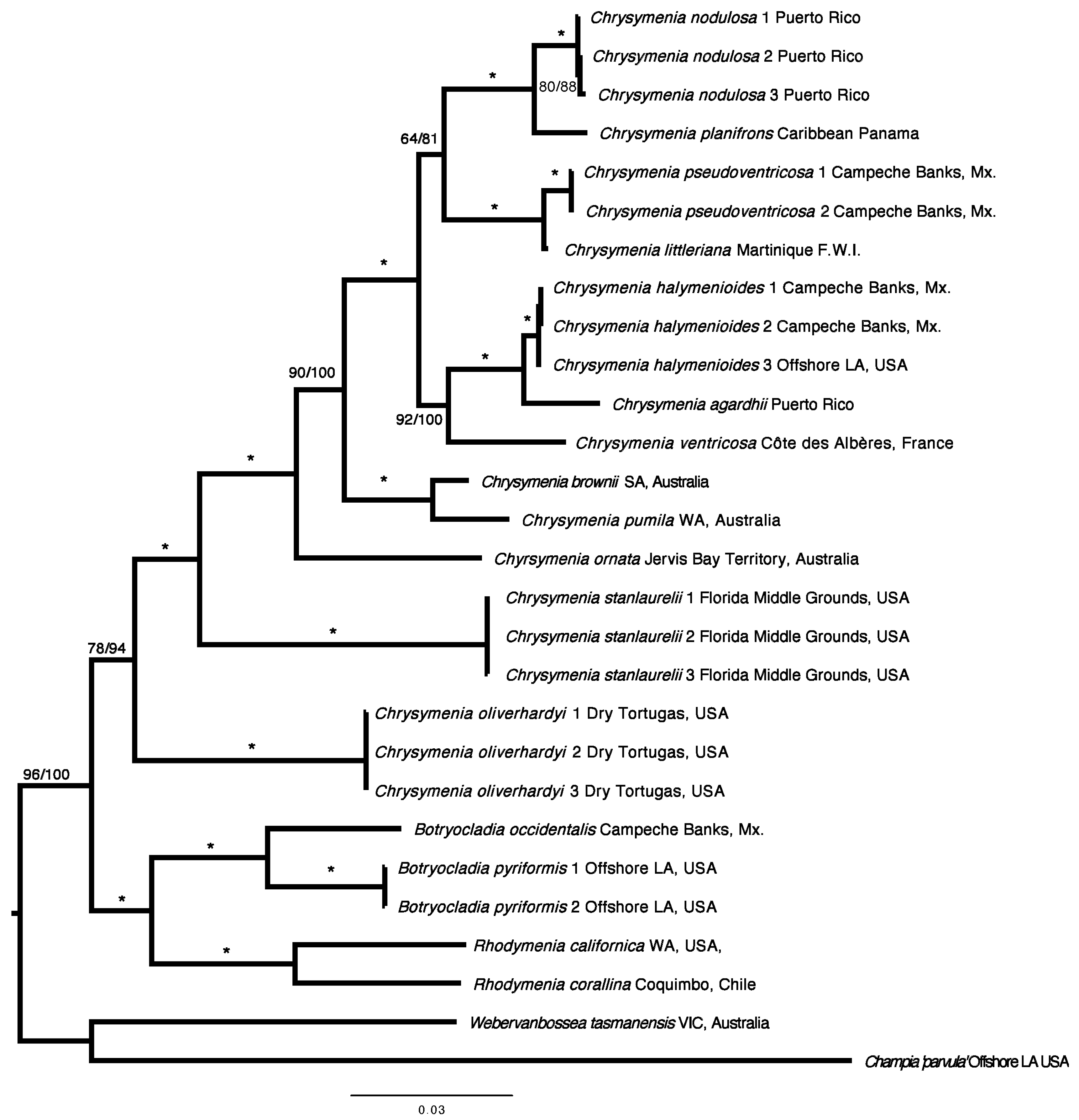
The two undescribed Gulf of Mexico species of Chrysymenia were collected growing on the surface of rhodoliths: unattached, benthic algal nodules of various sizes and origins that are predominantly accreted by non-geniculate (crustose) coralline red algae (CCA) precipitating CaCO3 within their cell walls [15,16]. These rhodoliths are autogenic rhodoliths, a type of nucleated rhodoliths sensu [17]) in which the core derives from an already existing calcium carbonate rubble as opposed to other materials, and which are established by differential erosion processes of salt dome (diapir) caprock [18,19]. Such nodules frequently become secondarily covered by various encrusting and fleshy algae [20,21,22,23,24], including the two undescribed species of Chrysymenia herein newly described on the basis of morphological and molecular evidence. A phylogenetic analysis of concatenated plastid-encoded rbcL (encodes the large subunit of the enzyme ribulose-1,5-bisphosphate carboxylase/oxygenase) and UPA (encodes photosystem II reaction center protein D1 gene), and nuclear LSU rDNA (partial 28S rDNA) sequences is provided for the genus (Figure 1). Morphological evidence is presented for six Chrysymenia species, including two new species from the Gulf of Mexico which are described here. Not illustrated in this paper are two species, Chrysymenia agardhii [14,15] and C. pseudoventricosa [4], which have been illustrated in separate papers.

2. Materials and Methods

2.1. Study Area and Sample Collection

Mesophotic specimens from the Gulf of Mexico used in this paper were collected during different expeditions (NSF-I, Dry Tortugas and offshore Louisiana, 29 May–7 June 2004; NSF-II, Campeche Banks and offshore Louisiana, 4–24 June 2005; NSF-III, Florida Middle Ground and offshore Louisiana, 28 June–29 July 2006; GOMRI-V, vicinity of the Dry Tortugas and offshore Louisiana, 7–14 September 2014, aboard the R/V Pelican, using an Hourglass design box dredge [21,22,25] with minimum tows (usually 10 min or less)) (Table 1). Samples from other locations worldwide are also listed in Table 1. Voucher specimens were liquid-preserved in 5% Formalin/seawater, pressed as herbarium sheets, desiccated in silica gel, and deposited in the Herbarium of the University of Louisiana at Lafayette (LAF). Silica gel-dried samples and herbarium-pressed vouchers were rehydrated for 20 min prior to observation. Chrysymenia samples were rinsed with seawater to remove excess mucilage. Additional information about the rhodolith habitats in the Gulf of Mexico is provided in the literature [19,20,21,22,23,24,26].

2.2. Morphological Examination

Vegetative and reproductive structures were sectioned manually using a stainless steel razor blade. Longitudinal, cross and oblique sections were stained either with 1% aqueous aniline blue acidified with 1% diluted acetic acid (the slides were then mounted in a 50% KaroTM (Karpokrafts, NY, USA) Syrup/water 50% solution [4,5] with phenol to retard microbial growth), or stained with a modified aceto-iron-chloral hydrate [27] stain [28], and mounted with 50% Hoyer’s dilution [29]. Photographs of the sections were taken on an Olympus BX 60 compound photomicroscope [Tokyo, Japan] with a Polaroid DMC Ie digital camera (Polaroid Inc., Cambridge, MA, USA) or a Canon Rebel EOS T2G (Tokyo, Japan] attached to an Olympus BX60 (Tokyo, Japan) microscope. Herbarium-pressed specimens were scanned using an all-in-one Epson (Nagano, Japan] Artisan 835 inkjet printer.

2.3. Molecular Data Acquisition

Total DNA was extracted from silica gel-dried specimens using a DNeasy Plant Mini Kit (Qiagen, Valencia, CA, USA) following the manufacturer’s instructions. Chloroplast-encoded rbcL and the Universal Plastid Amplicon 23S rRNA gene (UPA), as well as nuclear LSU rDNA nucleotide sequence data, are included for 21 vouchers of Chrysymenia comprising 13 distinct species, and from other members of the Rhodymeniaceae including two species of Botryocladia, two species of Rhodymenia, one species of Webervanbossea, and one species of Champia. Three vouchers each of the two new species were newly generated for this study.
PCR and sequencing of chloroplast-encoded rbcL were conducted using the methods and primers described in [4,5,30,31]. The rbcL alignment consisted of 1367 base pairs (bp), but 100 bps were excluded because the 5′ end of many sequences were incomplete. A portion of LSU rDNA and UPA was also amplified and sequenced using the primers and PCR protocol referenced in [32]. PCR products were gel-purified and sequenced in-house for both directions using the BigDyetm Terminator v. 3.1 (Life Technologies Grand Island NY, USA) on an ABI 3130xl Genetic Analyzer [Foster City, California, USA]. All DNA extractions are deposited in the Seaweeds Lab at the University of Louisiana at Lafayette.

2.4. Multigene Sequence Alignment

Resulting sequences were assembled with Sequencher v. 5.2 (Gene Codes Corporation [Ann Arbor, MI, USA]. The newly generated rbcL, LSU and UPA sequences (KT#, HQ#) (Table 1) were then aligned in Mega v. 6 [33].

2.5. Phylogenetic Analysis

A phylogenetic analysis was conducted on the concatenated rbcL, LSU and UPA sequences, and separately (not shown in this paper because separate trees were not significantly different in topology from the concatenated tree shown). The concatenated alignment was analyzed in Partitionfinder [34] to determine the best fitting model of evolution and data partition. The analyses each resulted in the selection of the General Time Reversible model plus gamma and a proportion of invariable sites with five partitions (UPA, LSU and the three codon positions of rbcL) on the basis of two information criteria, i.e., Akaike information criterion corrected (AICc) and Akaike information criterion (AIC). A distance matrix was also resolved from the branch lengths of the ML tree using the function cophenetic phylo of the APE Package in R [35] (not shown in this paper).
The alignments of the concatenated datasets were analyzed by maximum likelihood (ML) as implemented by RAXML v 2.4.4 [36] with the above models and partition scheme with 1000 restarts to find the tree with the highest likelihood score and 1000 Bootstrap (BS) replications. A Bayesian MCMC (Markov Chain Monte Carlo) was also applied to the aligned dataset using MrBayes v. 3.2.5 [37,38]. The Bayesian analysis consisted of two independent runs of five million generations with sampling every 1000 generations for a total of 10,002 trees. Convergence was visualized using Tracer v1.6 [39] and the first 10 percent of the trees of each run was discarded as burnin. The resulting posterior probabilities from Bayesian consensus trees were mapped on the ML tree.

3. Results

3.1. Phylogeny of Chrysymenia

An undescribed foliose taxon, Chrysymenia oliverhardyi sp. nov. from the vicinity of the Dry Tortugas, FL, USA, shares a basal most recent common ancestor with a clade comprising all other Chrysymenia species (Figure 1), and it does not resolve as sister to the other foliose taxa included in the dataset, namely C. agardhii from Puerto Rico or C. planifrons from Caribbean Panama. Each of these three foliose taxa are nested in separate clades. Chrysymenia agardhii is sister to C. halymenioides from the Gulf of Mexico with full support and belongs in the clade that also contains the type species, C. ventricose, from the Mediterranean. This C. agardhii-C. halymenioides-C. ventricosa clade in turn is sister to a clade comprising two subclades, i.e., a strongly supported C. planifrons-C. nodulosa clade and a C. pseudoventricosa-C. littleriana clade. Sister to these Gulf of Mexico–Caribbean clades is a clade of the Australian species, C. brownii and C. pumila, with C. ornata in a separate clade.
An undescribed, slender species from the Florida Middle Ground is here referred to as a new species, Chrysymenia stanlaurelii sp. nov. (Figure 1).
The alignment comprised 2343 base pairs (rbcL: 1369 bp; UPA: 369 bp; LSU: 603 bp).
The ML and the Bayesian trees correspond with each other in terms of topology and similar branch support (with Bayesian posterior possibilities slightly higher).

3.2. Two Undescribed Species

3.2.1. Chrysymenia stanlaurelii W.E.Schmidt, Arakaki and Fredericq sp. nov.

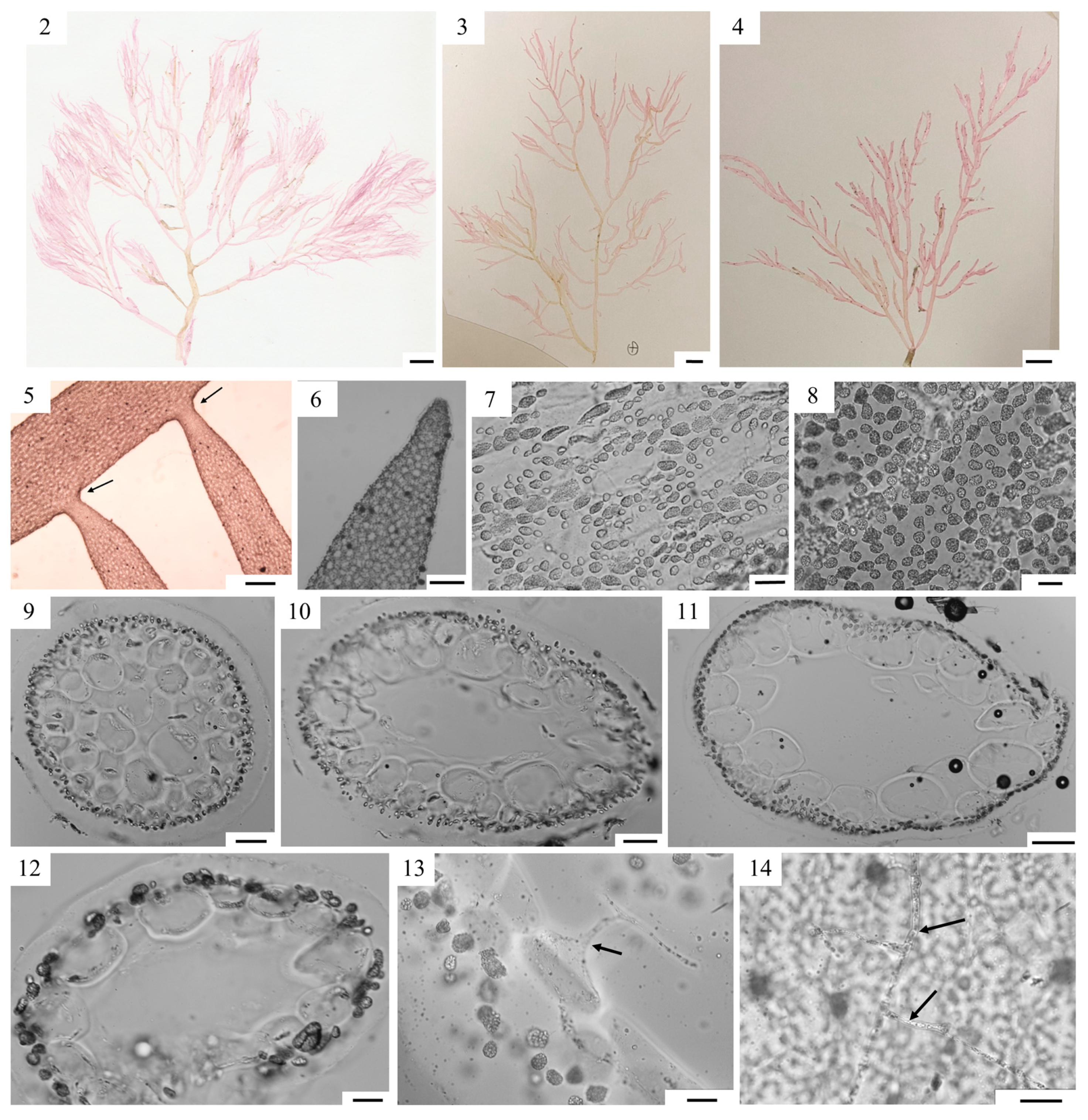
Figure 2(2–14).
HOLOTYPE: Cystocarpic specimen, LAF-NSF-III-7-5-06-3-7, Florida Middle Grounds, 28°10.20′ N, 84°01.83′ W, dredged from 38.9 to 42.6 m depth, coll. S. Fredericq, 4.vii.2006, attached to rhodolith, Figure 2(3).
PARATYPE: Tetrasporophyte, LAF-NSF-III-7-5-06-8-10, 28°05.20′ N, 83°46.16 W, dredged from 33-.4 to 38.5 m depth; coll. S. Fredericq, 5.vii.2006, attached to rhodolith, Figure 2(3,5–14).
TYPE LOCALITY: Florida Middle Grounds, West Florida, USA.
ETYMOLOGY: Named after the comic actor Stan Laurel, the “thin” partner of the Laurel and Hardy team.
ADDITIONAL MATERIAL EXAMINED: Florida Middle Grounds: LAF-NSF-III-7-4-06-4-5 (WES143), 28°34.24′ N, 84°28.77′ W, W Florida, 46–47 m depth; LAF-NSF-III-7-4-06-2-1 (WES146), 28°41.79′ N, 84°23.30′ W, 47–45 m depth.
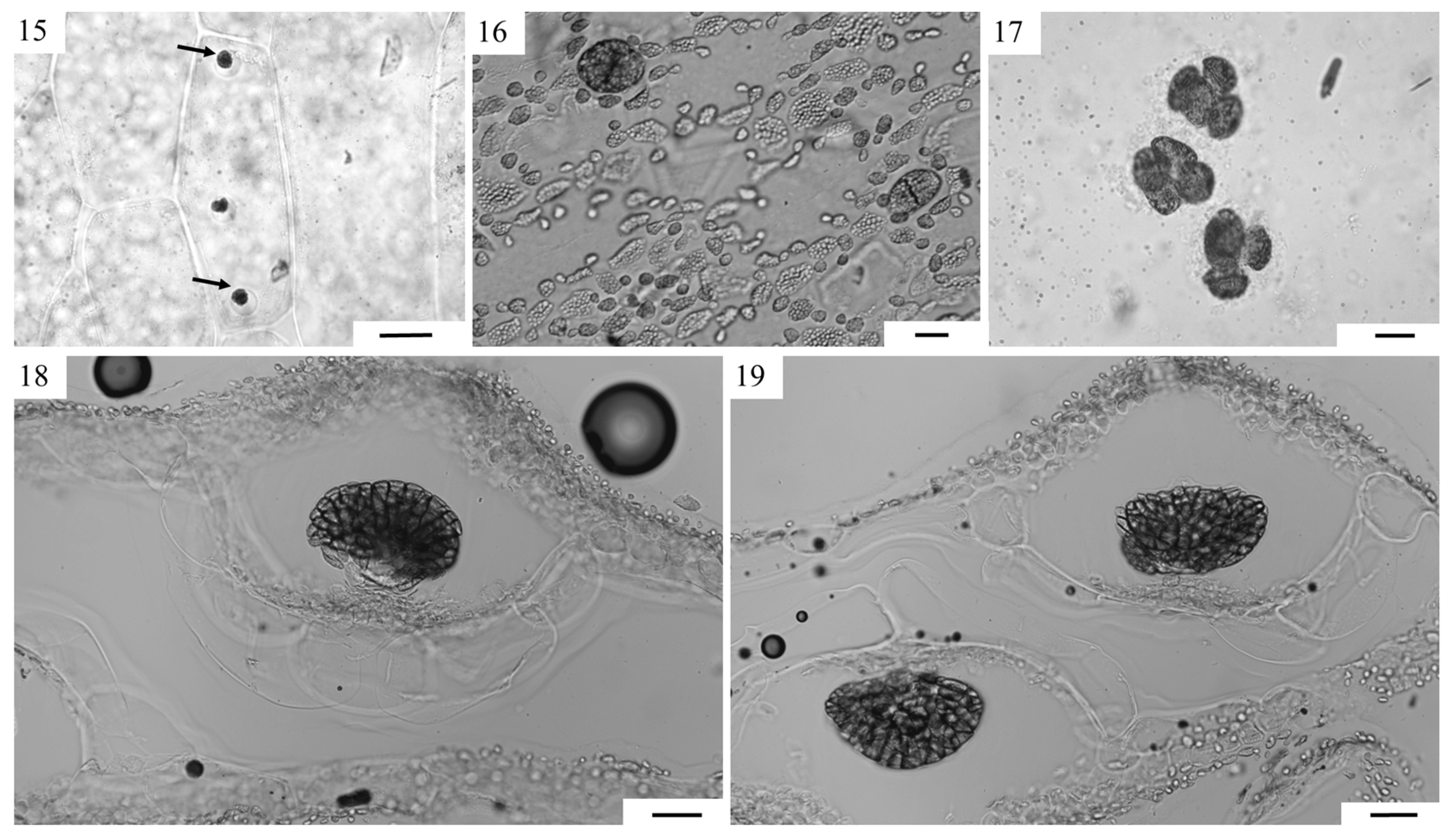
HABIT, VEGETATIVE AND REPRODUCTIVE STRUCTURES: Thalli pink, erect, up to 35 cm tall (Figure 2(2–4)) above a short stipe, composed of slightly gelatinous terete axes and branches up to 5 mm in diameter. Lateral branches (arrows) have a compressed base at point of insertion on axis (Figure 2(5)) and tapering branch tip (Figure 2(6)). Cortication in young specimens not complete (Figure 2(7)), with cortical cells found at the junction of the larger medullary cell layer (Figure 2(9)), eventually becoming nearly complete in older specimens (Figure 2(8)). Lower stipe showing solid center with cortical cells and medullary cells (Figure 2(9)), mid-stipe region showing hollow center with pigmented cortical cells and hyaline medullary cells ((Figure 2(10)) and upper part of stipe showing hollow center with pigmented cortical cells and fewer hyaline medullary cell layers (Figure 2(11)). Branches and axes composed of hyaline medullary cells, and one to two layers of pigmented cortical cells (Figure 2(12)). Medullary cells oval–circular to oval–rectangular in shape, 90–340 µm × 50–110 µm, loosely organized in younger specimens and becoming more tightly organized at maturity. Cortical cells ovoid to irregular, up to 19 µm diam (Figure 2(12,13)). Medullary filaments arising from medullary cells thallus-inward, observed only in mature specimens (Figure 2(13)). Gland cells pyriform, 12–25 µm wide, up to 25 µm long (Figure 3(15)), infrequent, originating from unspecialized medullary cells, projecting into central cavity, 2-(3)-6 per medullary cell. Tetrasporangia scattered throughout thallus, spherical to elliptical, cruciately divided, 35–60 × 25–40 µm (Figure 3(16,17)). Female gametophytes bearing compact carposporophytes on one (Figure 3(18)) or opposite sides of a branch (Figure 3(19)). Male gametophytes not observed.
REPRESENTATIVE SEQUENCES: HQ400588, HQ400589, HQ400590 (rbcL); KT154738, KT154737, KT154736 (LSU); KT154712, KT154713, KT154707 (UPA)

3.2.2. Chrysymenia oliverhardyi Arakaki, W.E.Schmidt and Fredericq sp. nov.

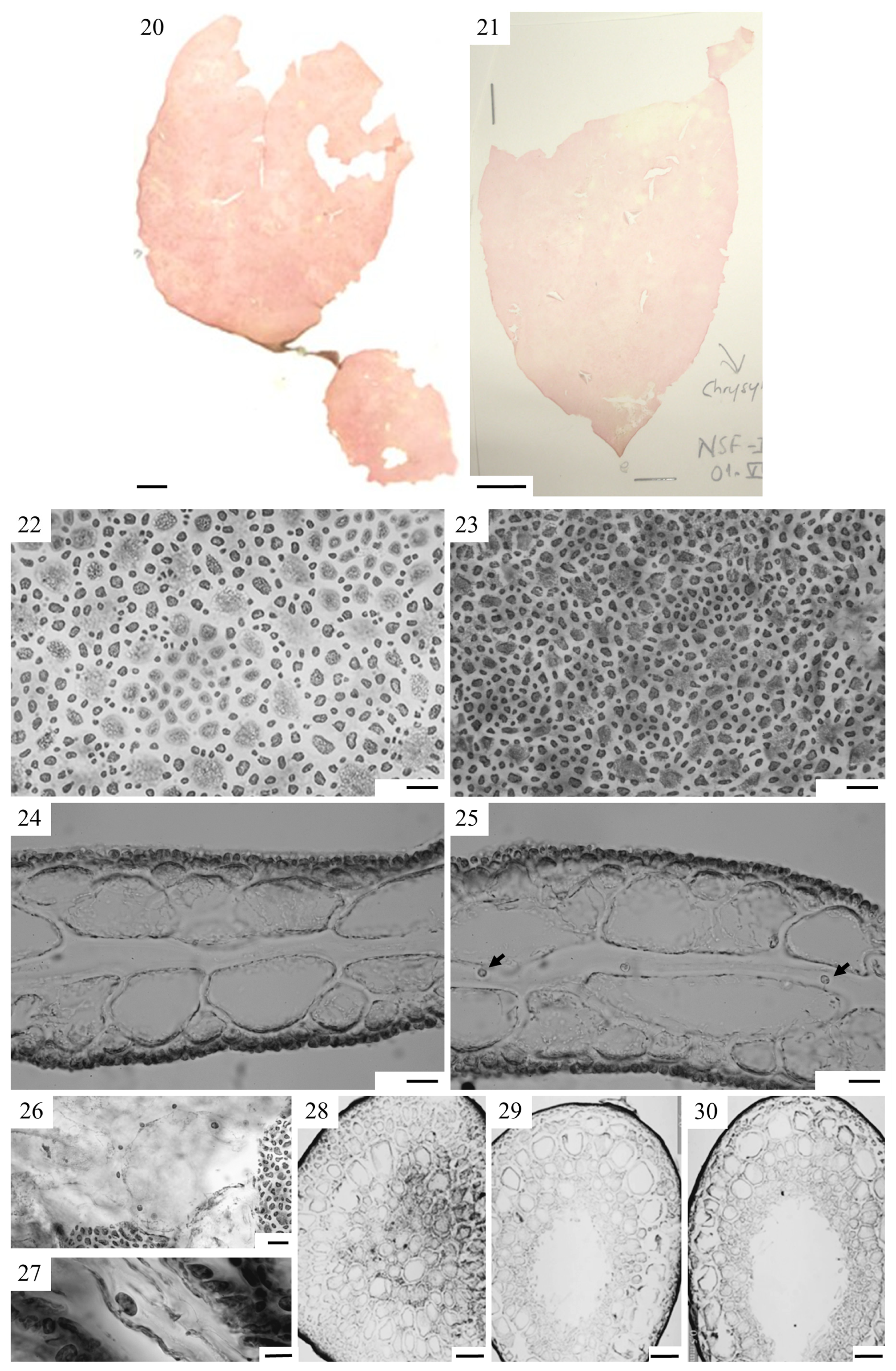
Figure 4(20–30), Figure 5(31–36)
HOLOTYPE: vicinity of Dry Tortugas, Florida: 1.vi.2004 (cystocarpic), 24°35.36′ N, 83°24.62′ W, N135, LAF-NSF-I-6-04-22-2, 64–64.1 m depth, Figs 20, 33-36.
PARATYPE: non-fertile, Figure 4(21), LAF-NSF-I
TYPE LOCALITY: Dry Tortugas, FL, Dry Tortugas, Florida: 1.vi.2004 (non-fertile), 24°36.08′ N, 83°29.95′ W
DISTRIBUTION: Dry Tortugas, NW Gulf of Mexico
ETYMOLOGY: Named after the comic Oliver Hardy, the “large” partner of the Laurel and Hardy team
ADDITIONAL MATERIAL EXAMINED: Dry Tortugas: LAF-NSF-1-6-04-18-3, 63.9–68.3 m depth. LAF-6981, GOMRI-V-25, 11 September 2014, 24°32.763′ N, 83°22.875′ W, 24°32.763′ N. LAF-6983, GOMRI-V-25, 11 September 2014, 24°32.763′ N, 83°22.875′ W, 24°32.763′ N. Offshore Louisiana: 23.viii.2008 (cystocarpic, tetrasporic), LAF-8-23-08-6-5, 28°03.89′ N, 92°27.60′ W, 63–66 m depth; LAF-8-23-08-10-3 (cystocarpic), 27°48.16′ N, 93°03.16′ W, 58–61 m depth, LAF-NSF-8-22-08-10-3.
HABIT, VEGETATIVE AND REPRODUCTIVE STRUCTURE: Thallus erect, 14 cm long and 450 μm thick, pink in color, with entire margins (Figure 4(20,21)), firmly gelatinous, soft. Discoid holdfast forming a short, robust, 2 cm long solid stipe that expands and extends into a blade with a cuneate base (Figure 4(20,21)).
Cortication in mature specimens becoming complete (Figure 4(22)) whereas cortication in younger parts of the thallus is incomplete (Figure 4(23)). Cortex of 1–2 layers of spherical cells 6.25 μm in diameter, subcortical cells ~7.5 μm × ~12.5 μm (Figure 4(24,25)). Medulla consisting of a central cavity and two layers of hyaline, thin-walled, irregularly arranged elongated subspherical cells (~124 μm × ~200 μm) lining the cavity (Figure 4(24,25)). Rhizoidal filaments not traversing the medulla (Figure 4(24,25)). Six to thirteen sessile gland cells, ~8.8 μm diameter, are borne on medullary cells (Figure 4(26,27)).
Thallus hollow, except for the lower stipe region which is solid (Figure 4(28)). Transverse sections through the middle and upper parts of the stipe showing a gradual formation of the central empty space (Figure 4(29,30)), with mucilage present and rhizoidal filaments absent.
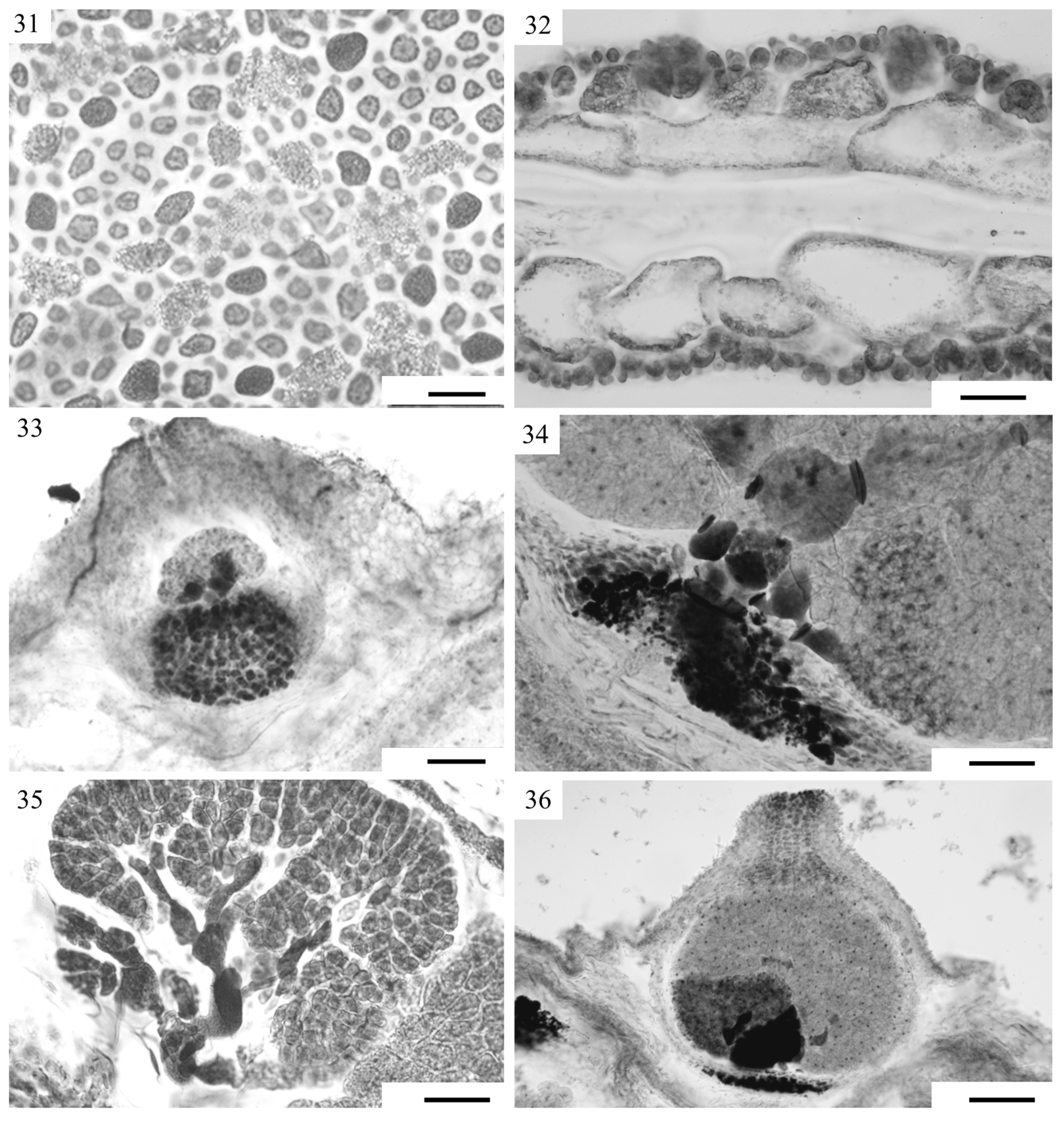
Cruciately divided tetrasporangia (19 μm × 14 μm diameter) (Figure 5(31,32)) are scattered over the blade surface (Figure 5(31)) and formed from cortical cells.
Thalli dioecious. After putative fertilization, a gonimoblast initial on the female gametophyte cuts off gonimoblasts (Figure 5(33)) outwardly, and a darkly staining nutritive tissue is formed below. Branched, darkly staining multinucleate cells partly fuse through enlarged and darkly staining pit connections (Figure 5(34), arrows) with nutritive cells from a basal group of darkly staining filaments. Carposporangia are transformed gonimoblast cells that are organized in three to four lobes of different ages (Figure 5(35,36)). All gonimoblast cells transform into carposporangia (Figure 5(34–36)). Cystocarps are subspherical, protuberant (450 μm diameter), uniostiolate (Figure 5(33,36)), surrounded by a cellular pericarp (Figure 5(36)), and scattered over the entire blade. Male gametophytes were not observed.
REPRESENTATIVE SEQUENCES: HQ400588, HQ400589, HQ400590 (rbcL); KT154736, KT154737, KT154738 (LSU); KT154712, KT154713, KT154707 (UPA).

3.2.3. Chrysymenia planifrons (Melvill) J. Agardh 1876, p. 319 [39]

Figure 6(37–43)
BASIONYM: Chrysymenia agardhii var. planifrons Melvill 1875: 263 [40]
SYNONYM: Cryptarachne planifrons (Melvill) Kylin 1931: 12 [6]
Chrysymenia curtissiana J. Agardh (1885) [41] fide Kylin (1931) [6]
TYPE LOCALITY: Key West, Florida
DISTRIBUTION: Dry Tortugas FL, Atlantic Florida, North Carolina, Antilles, Brazil [13,42].
SPECIMENS EXAMINED: Caribbean Panama: Long Bay Point, Bocas del Toro, BW1165, WES 312 (9°24.000′, N 65°20.533′ W), 10 m depth. Dry Tortugas, FL: SE Gulf of Mexico, LAF-NSF-1-20-04-1-1, 24°35.75, N 83°27.93′ W, 64.2 m depth.
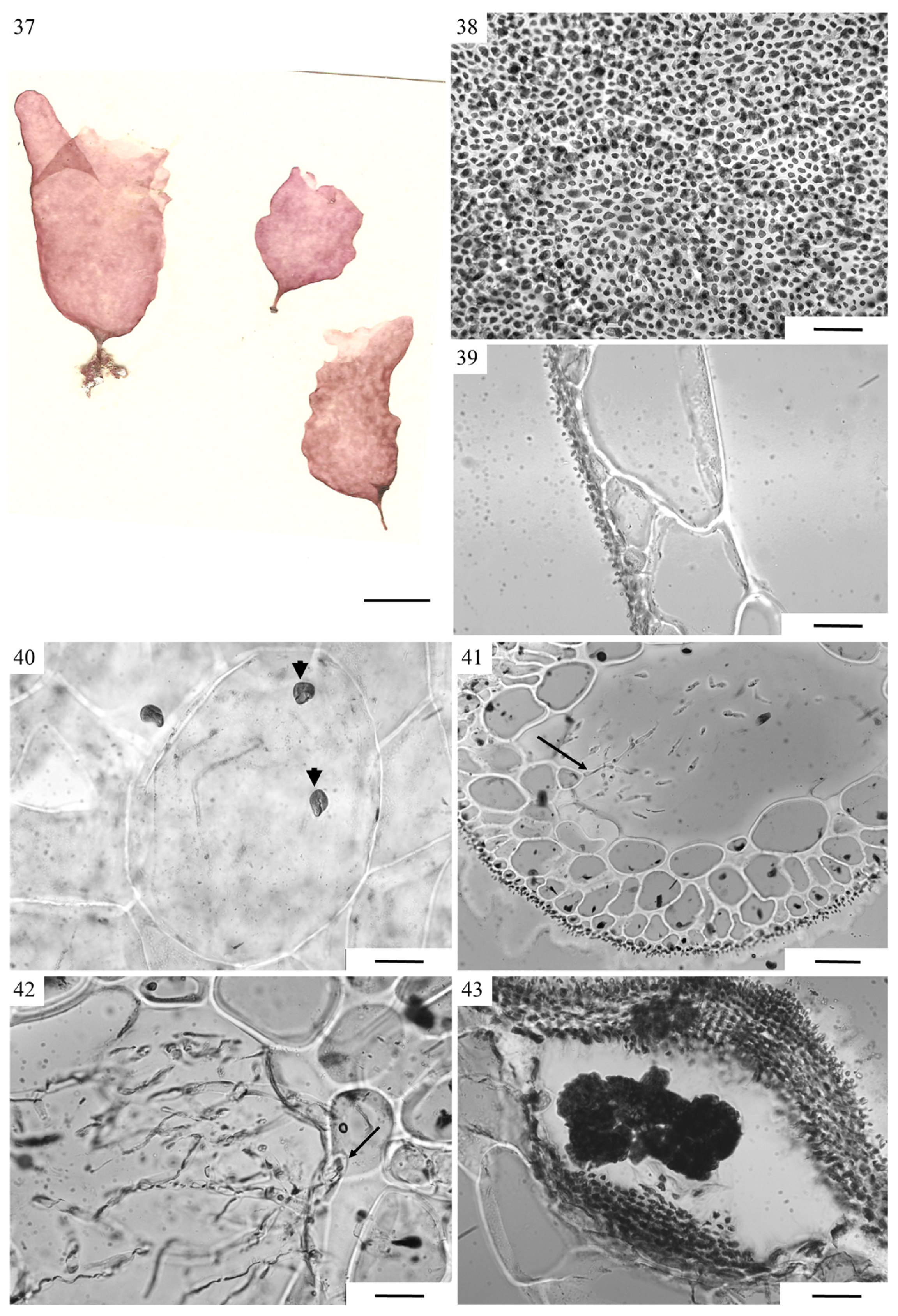
RESULTS: Plants flattened (Figure 6(37)) with or without protuberances. Thallus completely corticated (Figure 6(38)), composed of one to two layers of large, hyaline medullary cells, and two layers of smaller pigmented cortical cells (Figure 6(39)). Solitary pyriform gland cells borne on unmodifided glandiferous cells (Figure 6(40)). Middle portion of stipe hollow, with a few internal rhizoids (Figure 6(41)). Stipe cortex composed of two to three layers of pigmented cells similar to those in rest of thallus. Medullary cells composed of three to four layers of hyaline cells increasing in size toward thallus center (Figure 6(41)). Internal rhizoids developing from smaller cells cut off from unmodified medullary cells (Figure 6(42)). Cystocarps protruding inwardly and outwardly with nutritive cells forming mound not extending over entire width of base; carposporanial mass lobed in appearance (Figure 6(43)). Male gametophytes and tetrasporophytes were not observed.

3.2.4. Chrysymenia littleriana J.N.Norris and Ballantine 1995: 154 [14]

Figure 7(44–52).
TYPE LOCALITY: Diamond Rock, Martinique, French West Indies, Lesser Antilles, 14°27.0′ N, 61°02.83′ W, attached to rock, ~8 m depth.
DISTRIBUTION: Lesser Antilles [43,44]
SPECIMENS EXAMINED: Martinique, French West Indies, Lesser Antilles: DML30914 (WES136), Diamond Rock (14°26.94 ’N, 61°02.41′ W).
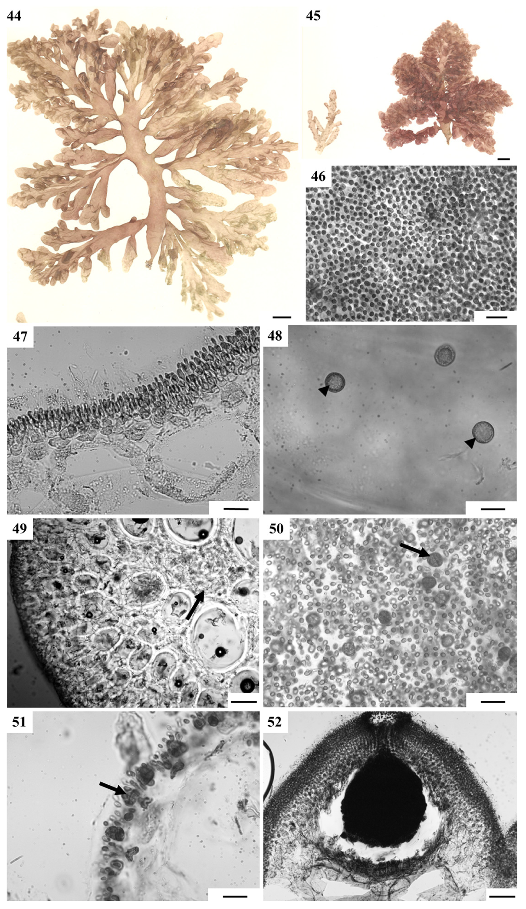
RESULTS: A full range of habit morphologies was observed, with three (Figure 7(44)) to five (Figure 7(45)) orders of branching. Thallus with complete cortication (Figure 7(46)), composed of a pigmented cortical layer of elongated cortical cells, a subcortical layer, and medullary layers of large hyaline cells (Figure 7(47)). Unmodified glandiferous cells in the innermost medullary cell layer bearing gland cells (Figure 7(48)). Middle portion of stipe solid (Figure 7(49)), with the outer cortex composed of two to three layers of small, elongated pigmented cells. Medulla composed of four to six layers of oval hyaline cells gradually enlarging and becoming less aggregated the closer to the central axis. Internal rhizoids cut off from medullary cells filling the intermedullary spaces, and completely filling the central cavity (Figure 7(49)).
Cruciately divided tetrasporangia scattered over the thallus (Figure 7(51)) in an unmodified cortical layer (Figure 7(51)). Female gametophytes bearing uniostiolate dome-shaped cystocarps; nutritive cells forming a crescent shape extending over the entire cystocarp floor; mass of carposporangia pyriform (Figure 7(52)).

3.2.5. Chrysymenia halymenioides Harvey 1853: 188, pl. XX.A. [12]

Figure 8(53–60).
TYPE LOCALITY: Key West, Florida (“thrown up from deep water”).
Distribution: North Carolina, Bermuda and the NE Gulf of Mexico [42,43,44,45,46,47,48,49]
Specimens examined: Campeche Banks, Mexico: SE Gulf of Mexico, LAF-NSF-II-18-6-05-106-3, 22°14.043′ N, 90°41.625′ W, 37–43 m depth; LAF-NSF-II-7-6-05-16-2 (WES 17), 22°16.020′ N, 90°43.230′ W, 51–52 m depth; LAF-NSF-II-7-6-05-17-5 (WES 19), 22°15.150′ N, 90°43.290′ W, 52–53 m depth; LAF-NSF-II-17-6-05-100-2 (WES 10), 22°11.330′ N, 91°08.710′ W, 49–53 m depth. Offshore LA, USA: LAF-NSF-II-22.96-123-2 (WES 23), 27°55.07′ N, 92°23′.08′ W, 58–71 m depth, 22.vi.2005
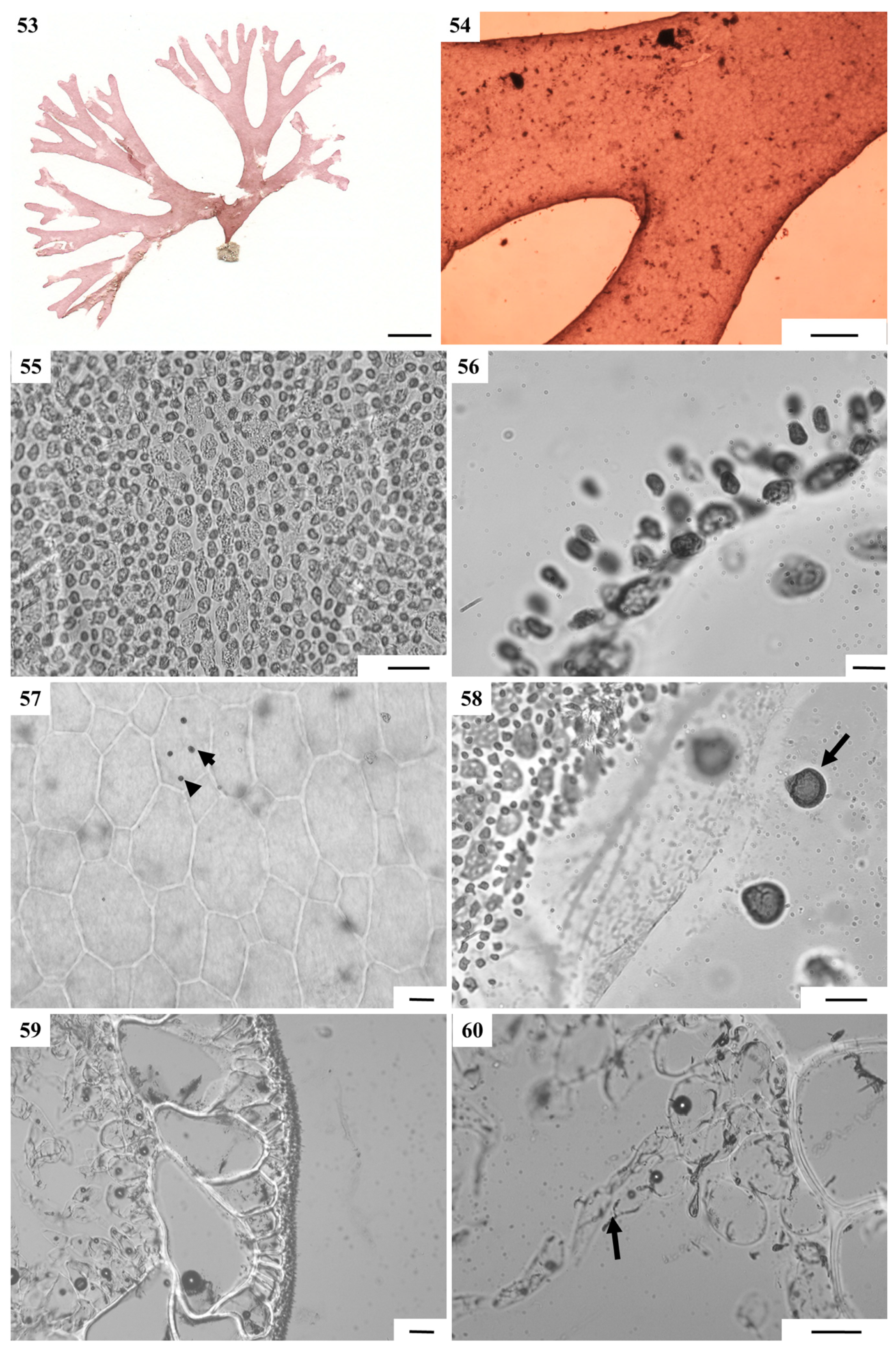
RESULTS: Plant regularly dichotomous (Figure 8(53)), lacking constrictions at base of branches (Figure 8(54)). Thallus completely corticated (Figure 8(55)), composed of a large hyaline medullary layer and a small two-celled cortical layer (Figure 8(56)). Pyriform gland cells borne on unmodified glandiferous medullary cells (Figure 8(57,58)). Middle portion of stipe hollow, with the outer cortex composed of two to three layers of small, pigmented cells. Medulla composed of only two layers of oval, hyaline cells comprising an outer layer ~60 µm diam., and an inner layer, 160–210 µm diam. (Figure 8(59)). Loose aggregations of small inner medullary cells, 40–90 µm diam., cut off filaments that grow towards central axis (Figure 8(60)). Reproductive plants were not observed.

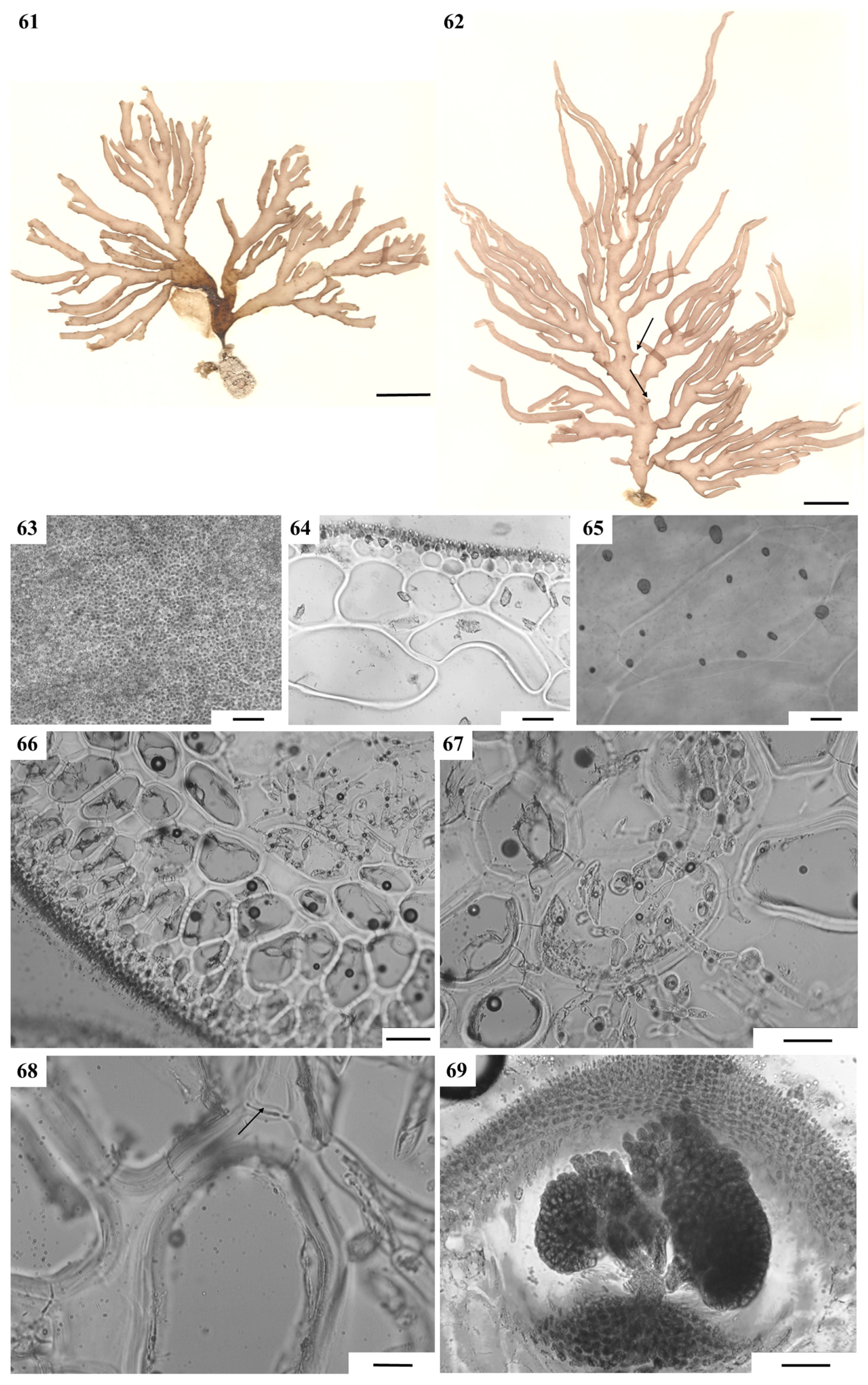
3.2.6. Chrysymenia nodulosa J.N. Norris and Ballantine 1995: 159, Figure 2a,b, 4, 5, 7, 8a,b, 9 [14]

Figure 9(61–69).
TYPE LOCALITY: Media Luna Reef, La Parguera, Puerto Rico, Greater Antilles, 17 m depth, 1.5 km seaward.
DISTRIBUTION: Greater Antilles, Bermuda [14,15].
SPECIMENS EXAMINED: Puerto Rico: Isla de Culebra, (18°20.263′ N, 65°20. 533′ W), 26 m depth, DML30319 (WES 148), DML30357 (WES142), DML30318 (WES96).
RESULTS: Characteristic surface nodules present (Figure 9(61,62)). Thallus hollow, completely corticated (Figure 9(63)), composed of two to three pigmented cortical cell layers and three layers of hyaline medullary cells increasing in size toward the interior (Figure 9(64)). Gland cells solitary, borne on unmodified glandiferous cells (Figure 9(65)). Middle portion of stipe hollow, containing internal rhizoids (Figure 9(66,67)). Stipe cortex reaching three to five layers of pigmented cells more elongated than cortical cells in rest of thallus. Stipe medulla reaching five to six layers of cells gradually increasing toward the center (Figure 9(66)). Common presence of rhizoids inside cell wall of innermost medullary cells devoid of cytoplasm (Figure 9(67)). Pit connections link rhizoids to neighboring cells (Figure 9(68)). Cystocarps uniostiolate, with nutritive cells organized in mound at base of cystocarp, carposporangial mass lobed (Figure 9(69)) Male gametophytes were not observed.

4. Discussion

Our integrative morpho-anatomical and molecular systematics study of the genus Chrysymenia from the Gulf of Mexico revealed the presence of two new species, C. stanlaurelii and C. oliverhardyi. Specimens of C. stanlaurelii may have been routinely mistaken by collectors for slender members of C. enteromorpha Harvey, a species that was recently transferred to the genus Botryocladia as B. enteromorpha W.E.Schmidt, Lozada–Troche, Ballantine and Fredericq by Schmidt et al. [7]. Likewise, specimens of Chrysymenia oliverhardyi sp. nov. superficially resembles blades of Halymenia spp. and this convergence in habit may be the reason this species was not recognized prior to our study. The characterization of the other mesophotic Gulf of Mexico taxa illustrated and discussed, i.e., C. planifrons, C. littleriana, C. halymenioides, and C. nodulosa, are consistent with the descriptions by [14,46].
Regarding the foliose species of Chrysymenia recorded in the Gulf of Mexico, so far, C. oliverhardyi has a rather restricted distribution in the vicinity of the Dry Tortugas, FL (SE GoMX), and offshore Louisiana (NW GoMX). C. oliverhardyi is distinguished by a simple, thin, entire frond reaching 14 cm in height, from C. agardhii, a species with a more widespread distribution in Atlantic FL, North Carolina, Caribbean Sea and Bermuda [13]. The latter is characterized by undulate thalli 10–20 cm high and composed of dichotomously to palmately laciniate distal lobes, with margins that are irregularly erose-dentate [12,42]. Chrysymenia planifrons are broadly foliose with deltoid lobes to 40 cm high, with undulate margins, growing in ~30 m depth [13]. Internally, the medulla of C. oliverhardyi contains six to thirteen gland cells per bearing cell, and a hollow cavity lacking rhizoidal filaments. In contrast, C. agardhii has a densely packed medulla with numerous rhizoidal filaments and two to four gland cells [12], and C. planifrons has few rhizoidal filaments, and one to few gland cells. In our dredged collections, we were fortunate to find tetrasporophyte and carposporophyte thalli of various species. Unfortunately, as there is not much information available about the female reproductive structure of C. agardhii and C. planifrons, comparison with reproductive specimens of C. oliverhardyi is not possible at this time. Our phylogenetic results also reveal that a flattened thallus arose multiple times in the Chrysymenia phylogeny.
Because Chrysymenia oliverhardyi has a foliose, compressed, erect thallus, it is included in the form group-2 of species according to the morphological classification by Norris and Ballantine [14]. Other species worldwide with flattened fronds include C. agardhii, C. planifrons, C. dickieana J. Agardh [50], C. lobata Howe [51], C. polyglandulosa Okamura [52], and C. ornata. C. planifrons is distributed from the east coast of Florida and extends south and eastwards to Key West, FL (the type locality), the Virgin Islands, the Netherlands Antilles, and Caribbean Panama [42,53].
The species historically referred to as “Chrysymeniaenteromorpha is currently known as Botryocladia enteromorpha [7] and may be confused with C. stanlaurelii which has a compressed, branched frond. However, C. stanlaurelii lacks a simple saccate base, has the tendency to not branch at its branch apices, and is also more slender in appearance, and the constrictions at the base of the branches are not as narrow as in B. enteromorpha. Although gland cells were not discussed in Harvey’s original report of “C.enteromorpha [42], this species was described as having crowded gland cells; however, in C. stanlaurelii, cells were never observed as being crowded and they were usually spaced equidistantly on their glandiferous bearing cells.
Schneider and Searles [44] observed that the medullary cells bearing gland cells in younger specimens of “Chrysymeniaenteromorpha are completely corticated, an observation that agrees with our findings; yet, this situation was not observed in C. stanlaurelii, in which medullary cells remain uncorticated in younger specimens. Littler and Littler [43] and Dawes and Mathieson [46] also observed that the number of gland cells on the bearing cell in B. enteromorpha (as C. enteromorpha) varies from three to 18; rarely were there more than three observed in C. stanlaurelii.
All species observed in this study included tetrasporic specimens, with one voucher of C. stanlaurelii found to bear irregularly divided tetrasporangia, closer to a tetrahedral arrangement instead of a typical cruciate arrangement. Such tetrasporangia are pyriform in shape with the tip pointing towards a central point, and each of the tetraspores was connected to the other. This type of tetrasporangia was found throughout the frond, from thallus base to apex, and was larger than the typical tetrasporangia in this genus. Since the tetrasporangia are haploid, this aberration may also represent an error in the meiotic process. It is highly unlikely that these tetraspores are functional given that they are still pit-connected to one another. This error or deviation in form may be a variant typical of this species.
Chrysymenia oliverhardyi is currently the sister of the other Chrysymenia in the phylogenetic tree and does not resolve with C. ornata, the only other Chrysymenia included in this study known to lack internal rhizoids. Chrysymenia littleriana can be confused with C. pseudoventricosa (formerly referred to as C. ventricosa (Lamouroux) J.Agardh sensu Taylor [42], a species from which C. littleriana was segregated by Norris and Ballantine [14] on the basis of position and order of branching (1–3 in C. littleriana vs. 3–5 in C. pseudoventricosa (as C. ventricosa)), apex shape (tapering to blunt vs. broadly obtuse), shape and size of tetrasporangia (smaller and more elongated in C. littleriana) and degree of thallus compression (terete vs. compressed).
The examined collection of Chrysymenia planifrons from Caribbean Panama represents a range extension into Central America. The specimens studied were much smaller than typically reported for this species (2–4 cm vs. 40 cm), but were similar to larger specimens with respect to vegetative and reproductive morphology.
Pit connections linking rhizoids to neighboring cells in Chrysymenia nodulosa may represent a transformation of inner cells into rhizoids, or, alternatively, may be the result of rhizoids growing secondarily into a living or cytoplasm-devoid medullary cell. This type of linkage was not seen in stipes of any of the other taxa examined in this study.
Most samples from the Gulf of Mexico and Caribbean Sea are nested together, with the southern hemisphere sample resolving outside the clade. The two exceptions are C. oliverhardyi and C. stanlaurelii.
Previous studies of Chrysymenia [8,9] were limited to C. ornata and C. wrightii, which led to an informal proposal to resurrect the genus Cryptarachne on the basis of lack of internal rhizoids in C. ornata and their presence in C. wrightii. However, with the increased taxon sampling presented here, it was found that the true Chrysymenia clade contained specimens that both possess and lack internal rhizoids. This finding corroborates with molecular evidence of the opinion of Okamura [53] and later that of Abbott and Littler [54] that the division of Chrysymenia and Cryptarachne is unfounded on the basis of internal filaments. Chrysymenia wrightii and C. enteromorpha were recently transferred to Botryocladia as B. wrightii (Harvey)W.E.Schmidt, Ballantine and Fredericq and B. enteromorpha W.E.Schmidt, Ballantine and Fredericq, respectively [7].
  • Key to known species of Chrysymenia from the Gulf of Mexico and the Caribbean Sea
  • 1a. Thallus flattened-------------------------------------------------------------------------------------2
  • 1b. Thallus terete to compressed---------------------------------------------------------------------4
  •   2a. Presence of medullary filaments-----------------------------------------------------------3
  •   2b. Absence of medullary filaments--------------------------Chrysymenia oliverhardyi
  • 3a. Thallus dichotomously or palmately lobed, cortical cell layers 2–3------Chrysymenia agardhii
  • 3b. Thallus irregularly lobed, cortical cell layers 1---------------------Chrysymenia planifrons
  •   4a. Cortication incomplete towards tips of branches-------Chrysymenia stanlaurelii
  •   4. Cortication complete----------------------------------------------------------------------------5
  • 5a. Thallus dichotomously branched -------------------------------Chrysymenia halymenioides
  • 5b. Thallus not dichotomously branched---------------------------------------------------------6
  •   6a. Thallus with nodules on surface----------------------------------Chrysymenia nodulosa
  •   6b. Thallus lacking nodules on surface-------------------------------------------------------7
  • 7a. Branches compressed at base, with broad round apices at tips--Chrysymenia littleriana
  • 7b. Branches not or barely compressed at base, terminal tips more tapered-------------------------------------------------------------------------------------------Chrysymenia pseudoventricosa

Author Contributions

Conceptualization, W.E.S., N.A. and S.F.; methodology, W.E.S., N.A. and S.F.; formal analysis, W.E.S., N.A. and T.S.; data curation, W.E.S. and D.G.; resources, S.F.; writing—original draft, W.E.S.; writing—review and editing, C.F.D.G. D.G., T.S., J.N.N. and S.F.; funding acquisition, S.F. All authors have read and agreed to the published version of the manuscript.

Funding

This research was funded by NSF research grants NSF DEB-0315995, NSF DEB-1045690. NSF DEB-1754504: and NSF DEB-1456674 (ARTS) to SF. CFDG received support from the National Council for Scientific and Technological Development—CNPq, grant 307603/2022-9.

Institutional Review Board Statement

Not applicable.

Data Availability Statement

All DNA sequences have been submitted to Genbank, https://www.ncbi.nlm.nih.gov/genbank/.

Acknowledgments

We greatly thank the crew of the R/V Pelican for their help with sampling protocols aboard ship. We also thank the collectors listed in Table 1. Many thanks to Darryl Felder, UL Lafayette, and Rachel Collin, Smithsonian Institute, Bocas del Toro, Caribbean Panama, for facilitating our collecting in, respectively, the Gulf of Mexico and Panama.

Conflicts of Interest

The authors declare no conflicts of interest. The funders had no role in the design of the study; in the collection, analyses, or interpretation of data; in the writing of the manuscript; or in the decision to publish the results.

Abbreviations

The following abbreviations are used in this manuscript:
CCAcrustose coralline algae
rbcLlarge subunit of the enzyme ribulose-1,5-bisphosphate carboxylase/oxygenase
UPAphotosystem II reaction center protein D1 gene
LSU rDNApartial 28S rDNA
GoMxGulf of Mexico

References

  1. Gavio, B.; Fredericq, S. Botryocladia caraibica sp. nov. (Rhodymeniales, Rhodophyta), a new species from the Caribbean. Cryptogam. Algol. 2003, 24, 93–106. [Google Scholar]
  2. Gavio, B.; Fredericq, S. New species and new records of offshore members of the Rhodymeniales (Rhodophyta) in the northern Gulf of Mexico. Gulf Mex. Sci. 2005, 23, 58–83. [Google Scholar]
  3. Fredericq, S.; Cho, T.O.; Earle, S.A.; Gurgel, C.F.; Krayesky, D.M.; Mateo Cid, L.E.; Mendoza Gonzáles, A.C.; Norris, J.N.; Suárez, A.M. Seaweeds of the Gulf of Mexico. In Gulf of Mexico: Its Origins, Waters, and Biota. I. Biodiversity; Felder, D.L., Camp, D.K., Eds.; Texas A&M University Press: College Station, TX, USA, 2009; pp. 187–259. [Google Scholar]
  4. Schmidt, W.E.; Gurgel, C.F.D.; Fredericq, S. Taxonomic transfer of the red algal genus Gloiosaccion to Chrysymenia (Rhodymeniaceae, Rhodymeniales), including the description of a new species, Chrysymenia pseudoventricosa, for the Gulf of Mexico. Phytotaxa 2016, 243, 54–70. [Google Scholar] [CrossRef]
  5. Schmidt, W.E.; Lozada-Troche, C.; Ballantine, D.L.; Arakaki, N.; Gabriel, D.; Norris, J.N.; Fredericq, S. Taxonomic Transfer of the red algae Chrysymenia enteromorpha and C. wrightii to the genus Botryocladia (Rhodymeniaceae, Rhodymeniales). Phytotaxa 2017, 434, 122–138. [Google Scholar] [CrossRef]
  6. Kylin, H. Die Florideenordung Rhodymeniales. Lunds Univ. Arsskrift N.F. Avd. 2 1931, 27, 1–48. [Google Scholar]
  7. Kylin, H. Die Gattungen der Rhodophyceen; C.W.K. Gleerups Förlag: Lund, Sweden, 1956; 673p. [Google Scholar]
  8. Saunders, G.W.I.; Strachan, M.; Kraft, G.T. The families of the order Rhodymeniales (Rhodophyta): A molecular-systematic investigation with a description of Faucheaceae fam. nov. Phycologia 1999, 38, 23–40. [Google Scholar] [CrossRef]
  9. Le Gall, L.; Dalen, J.L.; Saunders, G.W. Phylogenetic analyses of the red algal order Rhodymeniales support recognition of the Hymenocladiaceae fam. nov., Fryeellaceae fam. nov., and Neogastroclonium gen. nov. J. Phycol. 2008, 44, 1556–1571. [Google Scholar] [CrossRef]
  10. Guiry, M.D.; Guiry, G.M. AlgaeBase. World-Wide Electronic Publication, National University of Ireland, Galway. Available online: https://www.algaebase.org (accessed on 18 April 2026).
  11. Agardh, J.G. Algae Maris Mediterranei et Adriatici; Fortin, Masson et Cie: Paris, France, 1842; 264p. [Google Scholar]
  12. Harvey, W.H. Nereis boreali-americana Part II. Rhodospermeae. In Smithsonian Contributions to Knowledge; Smithsonian Institution: Washington, DC, USA, 1853; Volume 5, pp. 1–258. [Google Scholar]
  13. Wynne, M.J. Chrysymenia tigillis sp. nov. (Rhodymeniales, Rhodophyta) from the Sultanate of Oman, with a census of currently recognized species in the genus Chrysymenia. Phycol. Res. 2005, 53, 215–223. [Google Scholar] [CrossRef][Green Version]
  14. Norris, J.N.; Ballantine, D.L. Two new species of the red alga Chrysymenia Agardh (Rhodymeniales: Rhodymeniaceae) from the tropical western Atlantic. Proc. Bio. Soc. Wash. 1995, 108, 153–165. [Google Scholar]
  15. Ballantine, D.L.; Ruiz, H.; Norris, J.N. The benthic marine algal flora of Puerto Rico IIIB. Rhodophyta: Ceramiales and Rhodymeniales. Smithson. Contr. Bot. 2026, 121, 173. [Google Scholar]
  16. Foster, M.S. Rhodoliths: Between rocks and soft places—Minireview. J. Phycol. 2001, 37, 659–667. [Google Scholar] [CrossRef]
  17. Spalding, H.L.; Amado-Filho, G.M.; Bahia, R.G.; Ballantine, D.L.; Fredericq, S.; Leichter, J.J.; Nelson, W.A.; Slattery, M.; Tsuda, R.T. Macroalgae. In Mesophotic Coral Ecosystems; Coral Reefs of the World 12; Springer Nature: Cham, Switzerland, 2019; pp. 507–536. [Google Scholar]
  18. Freiwald, A.; Henrich, R. Reefal coralline algal build-ups within the Arctic Circle: Morphology and sedimentary dynamics under extreme environmental seasonality. Sediment 1994, 41, 963–984. [Google Scholar] [CrossRef]
  19. Rezak, R.; Bright, T.J.; McGrail, D.W. Reefs and Banks of the Northwestern Gulf of Mexico: Their Geological, Biological, and Physical Dynamics; Wiley: New York, NY, USA, 1985; 259p. [Google Scholar]
  20. Felder, D.L.; Camp, D.K. (Eds.) Gulf of Mexico Origin, Waters and Biota, Biodiversity; Texas A&M University Press: College Station, TX, USA, 2009; Volume 1. [Google Scholar]
  21. Gore, R.H. The Gulf of Mexico; Pineapple Press: Sarasota, FL, USA, 1992. [Google Scholar]
  22. Richards, J.L.; Vieira-Pinto, T.; Schmidt, W.E.; Sauvage, T.; Gabrielson, P.W.; Oliveira, M.C.; Fredericq, S. Molecular and morphological diversity of Lithothamnion spp. rhodoliths (Hapalidiaceae, Hapalidiales) from deepwater rhodolith beds in the northwestern Gulf of Mexico. Phytotaxa 2016, 278, 81–114. [Google Scholar] [CrossRef]
  23. Fredericq, S.; Arakaki, N.; Camacho, O.; Gabriel, D.; Krayesky, D.; Self-Krayesky, S.; Rees, G.; Richards, J.; Sauvage, T.; Venera-Ponton, D.; et al. A dynamic Approach to the study of rhodoliths: A case study for the northwestern Gulf of Mexico. Cryptogam. Algol. 2014, 35, 77–98. [Google Scholar] [CrossRef]
  24. Fredericq, S.; Krayesky-Self, S.; Sauvage, T.; Richards, J.; Kittle, R.; Arakaki, N.; Hickerson, E.; Schmidt, W.E. The critical importance of rhodoliths in the life cycle completion of both macro- and microalgae, and as holobionts for the establishment and maintenance of marine biodiversity. Front. Mar. Sci. 2019, 5, 502. [Google Scholar] [CrossRef]
  25. Felder, D.L.; Thoma, B.P.; Schmidt, W.E.; Sauvage, T.; Self-Krayesky, S.; Chistoserdov, A.; Bracken-Grissom, H.; Fredericq, S. Seaweeds and decapod crustaceans on Gulf deep banks after the Macondo Oil Spill. BioScience 2014, 64, 808–819. [Google Scholar] [CrossRef]
  26. Camacho, O.; Fredericq, S. Diversity of brown macroalgae (Phaeophyceae) emerging from deepwater rhodoliths collected in the Gulf of Mexico. Diversity 2026, 17, 860. [Google Scholar] [CrossRef]
  27. Wittmann, W. Aceto-iron-haematoxylin-chloral hydrate for chromosome staining. Stain Technol. 1965, 40, 161–164. [Google Scholar] [CrossRef]
  28. Hommersand, M.H.; Fredericq, S.; Cabioch, J. Developmental morphology of Gigartina pistillata (Gigartinaceae, Rhodophyta). Phycologia 1992, 31, 300–325. [Google Scholar] [CrossRef]
  29. Stevens, R.B. Mycology Guidebook; University of Washington Press: Seattle, WA, USA, 1981. [Google Scholar]
  30. Lin, S.M.; Fredericq, S.; Hommersand, M.H. Systematics of the Delesseriaceae (Ceramiales, Rhodophyta) based on LSU rDNA and rbcL sequences, including the Phycodryoideae, subfam. nov. J. Phycol. 2001, 37, 881–899. [Google Scholar] [CrossRef]
  31. Gavio, B.; Fredericq, S. Grateloupia turuturu (Halymeniaceae, Rhodophyta) is the correct identity for the non-native species in the Atlantic known as G. doryphora. Eur. J. Phycol. 2002, 37, 349–360. [Google Scholar] [CrossRef]
  32. Sherwood, A.R.; Sauvage, T.; Kurihara, A.; Conklin, K.; Presting, G. A comparative analysis of COI, LSU and UPA marker data for the Hawaiian florideophyte Rhodophyta: Implications for DNA barcoding of red algae. Crypt. Algol. 2010, 31, 451–465. [Google Scholar]
  33. Tamura, K.; Stecher, G.; Peterson, D.; Peterson, N.; Filipski, A.; Kumar, S. MEGA6: Molecular Evolutionary Genetics Analysis version 6.0. Mol. Biol. Evol. 2013, 30, 2725–2729. [Google Scholar] [CrossRef]
  34. Lanfear, R.; Calcott, B.; Ho, S.Y.W.; Guindon, S. PartitionFinder: Combined selection of partitioning schemes and substitution models for phylogenetic analyses. Mol. Biol. Evol. 2012, 29, 1695–1701. [Google Scholar] [CrossRef] [PubMed]
  35. Paradis, E.; Claude, J.; Strimmer, K. APE: Analyses of Phylogenetics and Evolution in R language. Bioinformatics 2004, 20, 289–290. [Google Scholar] [CrossRef]
  36. Stamatakis, A. RAxML-VI-HPC: Maximum Likelihood-based phylogenetic analyses with thousands of taxa and mixed models. Bioinformatics 2006, 22, 2688–2690. [Google Scholar] [CrossRef]
  37. Ronquist, F.; Huelsenbeck, J.P. MrBayes 3: Bayesian phylogenetic inference under mixed models. Bioinformatics 2003, 19, 1572–1574. [Google Scholar] [CrossRef]
  38. Ronquist, F.; Teslenko, M.; van der Mark, P.; Ayres, D.; Darling, A.; Höhna, S.; Larget, B.; Liu, L.; Suchard, M.A.; Huelsenbeck, J.P. MrBayes 3.2: Efficient Bayesian phylogenetic inference and model choice across a large model space. Syst. Biol. 2012, 61, 539–542. [Google Scholar] [CrossRef]
  39. Rambaut, A.; Drummond, A. Tracer v1.6. 2014. Available online: http://tree.bio.ed.ac.uk/software/tracer (accessed on 6 May 2017).
  40. Agardh, J. Species Genera et Ordines Algarum…; C.W.K. Gleerup: Lipsiae, Germany, 1876; Volume 3, Part 1. [Google Scholar]
  41. Melvill, J.C. Notes on the marine algae of South Carolina and Florida. J. Bot. 1875, 13, 258–265. [Google Scholar]
  42. Agardh, J.G. Till algernes systematic. Nya bidrag. VII. Florideae. Lunds Univ. Arsskr. 1885, 21, 117 pp. [Google Scholar]
  43. Taylor, W.R. Marine Algae of the Eastern Tropical and Subtropical Coasts of the America; University of Michigan Press: Ann Arbor, MI, USA, 1960; pp. 1–870. [Google Scholar]
  44. Littler, D.M.; Littler, M.M. Caribbean Reef Plants. An Identification Guide to the Reef Plants of the Caribbean, Bahamas, Florida and Gulf of Mexico; Offshore Graphics: Washington, DC, USA, 2000; pp. 1–542. [Google Scholar]
  45. Schneider, C.W.; Searles, R.B. North Carolina marine algae. II. New records and observations of the benthic offshore flora. Phycologia 1973, 12, 201–211. [Google Scholar] [CrossRef]
  46. Dawes, C.J.; Mathieson, A. The Seaweeds of Florida; University Press of Florida: Gainesville, FL, USA, 2008; pp. 1–591. [Google Scholar]
  47. Mateo-Cid, L.E.; Mendoza-González, A.C.; Fredericq, S. A checklist of subtidal seaweeds from Campeche Banks, Mexico. Acta Bot. Venez. 2013, 36, 92–108. [Google Scholar]
  48. Børgesen, F. The marine algae of the Danish West Indies. Part III. Rhodophyceae (6), with addenda to the Chlorophyceae, Phaeophyceae and Rhodophyceae. Dan. Bot. Ark. 1920, 3, 369–498. [Google Scholar]
  49. Wynne, M.J. A checklist of benthic marine algae of the tropical and subtropical Western Atlantic: Fourth revision. Nova Hedwig. Beih. 2017, 145, 1–202. [Google Scholar]
  50. Agardh, J.G. Analecta algologica. Acta Univ. Lund. 1892, 28, 1–82. [Google Scholar]
  51. Howe, M.A. The marine algae of Peru. Bull. Torrey Bot. Club 1914, 38, 489–514. [Google Scholar] [CrossRef]
  52. Okamura, K. On the Algae from the island Hatidyo. Rec. Ocean. Work. Jpn. Tokyo 1930, 2, 92–119. [Google Scholar]
  53. Okamura, K. Nippon Kaisô Shi [Marine Algae from Japan]; Uchida Rokakuho: Tokyo, Japan, 1936; 964p. [Google Scholar]
  54. Abbott, I.A.; Littler, M.M. Some Rhodymeniales from Hawaii. Phycologia 1969, 8, 165–169. [Google Scholar] [CrossRef]
Figure 1. Maximum likelihood phylogram showing 28 concatenated rbcL, UPA and LSU sequences representing 13 Chrysymenia species including two new taxa, C. stanlaurelii and C. oliverhardyi, seven sequences representing other genera of Rhodymeniaceae (Botryocladia, Rhodymenia, Webervanbossea), and one outgroup taxon in the Champiaceae (Champiaparvula’) inferred from RaxML analysis. Numbers indicate bootstrap values at right, and BP values at left; * denotes full support. Scale bar indicates the number of substitutions per site.
Diversity 18 00320 g001
Figure 2. Chrysymenia stanlaurelii W.E. Schmidt, Arakaki and Fredericq, sp. nov. from the Florida Middle Grounds. (2) LAF-NSF-III-7-5-06-6-8, vegetative specimen, 28°15.754′ N, 83°44.996′ W, ~33 m depth. (3,514). LAF-7-5-06-8-10, 28°05.20′ N, 83°46.16′ W, ~30 m depth. (3). Habit of tetrasporangial paratype, LAF-NSF-III-7-4-06-5-1, 28°30.50′ N, 84°28.188′, 47–45 m depth. Scale bar = 1 cm. (4). Habit of cystocarpic holotype, LAF-NSF-7-5-06-3-7, 28°10.20′ N, 84°01.83′ W, 38-9-42.6 m depth. Scale bar = 1 cm. (5). Compressed base of lateral branches (arrows) attached to axis. Scale bar 0.5 mm. (6). Tapering branch tip. Scale bar = 0.5 mm. (7). Surface view of immature thallus showing incomplete cortication. Scale bar = 40 µm. (8). Surface view of mature thallus showing complete cortication, pigmented cortical cells and hyaline medullary cells. Scale bar = 20 µm. (9). Cross-section through lower stipe, showing a solid center with cortical cells and medullary cells. Scale bar = 5 µm. (10). Cross-section through upper part of stipe showing hollow center with pigmented cortical cells and fewer hyaline medullary cell layers. Scale bar = 5 µm. (11). Cross-section through mid-thallus showing cortex, and medulla lacking narrow inner filaments. Scale bar = 5 mm. (12). Transverse section of thallus with pigmented cortical cells, hyaline medullary cells and decussate tetrasporangia. Scale bar = 25 µm. (13). Surface view showing medullary cells bearing inner filaments (arrow). Scale bar = 40 µm. (14). Surface view of tetrasporangial thallus with abnormal/decussate tetrasporangia, narrow inner filaments (arrows) and unmodified cortex. Scale bar = 100 µm.
Diversity 18 00320 g002
Figure 3. Chrysymenia stanlaurelii W.E.Schmidt, Arakaki and Fredericq, sp. nov. from the Florida Middle Grounds. (15). Surface view showing medullary cells and unmodified glandiferous cells with gland cells (arrows). Scale bar = 50 µm. (16,17) (LAF-NSF-III-7-5-06-6-8, WES146). (16). Surface view of tetrasporangial thallus with cruciate tetrasporangia, and unmodified cortex. Scale bar = 5 µm. (17). Abnormal tetrahedrally divided tetrasporangia. Scale bar = 20 µm. (18,19) (NSF-7-5-06-3-7, WES145). Female gametophyte bearing compact carposporophyte on one (18) or on opposite sides of a branch (19). Scale bars = 50 µm.
Diversity 18 00320 g003
Figure 4. Chrysymenia oliverhardyi Arakaki, W.E.Schmidt and Fredericq sp. nov. from the vicinity of the Dry Tortugas, Florida (NSF-I-22-2). (20). Holotype: Habit of female gametophyte bearing cystocarps from the Dry Tortugas, Florida (LAF-NSF-I-6-04-22-2, N135). Scale bar = 1 cm. (21). Habit of vegetative thallus paratype. Scale bar 1 cm. (22). View from the surface of vegetative thallus showing early complete cortication, with cortical cells arranged in rosettes. Scale bar = 40 µm. (23). Surface view of vegetative thallus showing mature complete cortication. Scale bar = 40 µm. (24). Longitudinal section showing pigmented cortex and hyaline medulla. Scale bar = 40 µm. (25). Longitudinal section showing medullary cells bearing gland cells (arrows). Scale bar = 40 µm. (26). Surface view of glandiferous cell. Scale bar = 40 µm. (27). Longitudinal section showing gland cell linked to medullary cell. Scale bar = 4.5 µm. (28). Cross-section through lower stipe, showing a solid center with cortical cells and medullary cells. Scale bar = 0.5 mm. (29). Cross-section through mid-stipe region showing hollow center with pigmented cortical cells and hyaline medullary cells decreasing in size towards center. Scale bar = 0.5 mm. (30). Cross-section through upper part of stipe showing hollow center with pigmented cortical cells and fewer hyaline medullary cell layers that decrease in size towards center. Scale bar = 0.5 mm.
Diversity 18 00320 g004
Figure 5. Chrysymenia oliverhardyi Arakaki, W.E.Schmidt and Fredericq sp. nov. (31). Surface view showing random distribution of tetrasporangial initials. Scale bar = 20 µm. (32). Cross-section showing cruciately divided tetrasporangia (LAF-8-23-08-6-5, 23.viii.2008, LA). Scale bar = 100 µm. (33,34). Female and cystocarp structures (LAF-8-22-08-10-3, 22.viii.2008, LA). (33). Young uniostiolate cystocarp. Outward production of carposporangia from gonimoblast initial surrounded by basal cushion of darkly staining nutritive cells. Scale bar = 50 µm. (34). Partly incorporation of multinucleate nutritive cells into gonimoblast fusion cell with large pit connections. Scale bar = 40 µm. (35). Carposporangia, fused gonimoblast cells and darkly staining gonimoblast cells. Scale bar = 40 µm. (36). Mature cystocarp filled with carposporangia of different ages. Scale bar = 100 µm.
Diversity 18 00320 g005
Figure 6. Chrysymenia planifrons from Long Bay Point, Panama (WES312). (37). Habit of non-reproductive specimens. Scale bar = 1 cm. (38). Surface view of thallus showing complete cortication. Scale bar = 50 µm. (39). Cross-section through main axis showing small pigmented cortical cells and hyaline medullary cells. Scale bar = 50 µm. (40). Surface view showing unmodified glandiferous cells with gland cells (arrowheads). Scale bar = 50 µm. (41). Cross-section through middle part of holdfast showing small-sized cortical cells, larger medullary cells and hollow interior with narrow medullary filaments (arrow). Scale bar = 100 µm. (42). Close-up of branched, narrow, internal filaments originating from large medullary cells (arrow). Scale bar = 50 µm. (43). Longitudinal section through cystocarp. Scale bar = 50 µm.
Diversity 18 00320 g006
Figure 7. Chrysymenia littleriana from Martinique (WES136). (44,45). Habits of individuals showing variation in degree and density of branching and size. (44). Scale bar = 1 cm. (45). (at left) depicts a small, cystocarpic specimen. Scale bar = 1 cm. (46). Surface view of thallus showing complete cortication. Scale bar = 20 µm. (47). Cross-section through main axis showing small, elongated surface cortical cells and enlarged, hyaline medullary cells. Scale bar = 20 µm. (48). Surface view showing glandiferous cells with spherical gland cells (arrowheads). Scale bar = 20 µm. (49). Cross-section through holdfast showing cell size gradation from cortex towards central medulla, and tightly appressed medullary filaments (arrow). Scale bar = 100 µm. (50). Surface view of tetrasporangial thallus, showing cortical cells and cruciately divided tetrasporangia (arrow). Scale bar = 20 µm. (51). Cross-section through tetrasporangial branch with tetrasporangia (arrow) scattered in an unmodified cortex. Scale bar = 20 µm. (52). Longitudinal section through a mature, dome-shaped, uniostiolate cystocarp. Scale bar = 100 µm.
Diversity 18 00320 g007
Figure 8. Chrysymenia halymenioides from the Campeche Banks (LAF-xx, WES17). (53). Habit of non-reproductive specimen. Scale bar = 2 cm. (54). Surface view at branch dichotomy showing lack of constrictions. Scale bar = 0.5 mm. (55). Surface view of thallus showing complete cortication. Scale bar = 20 µm. (56). Cross-section through main branch showing narrow, anticlinal surface cells borne on layer of periclinal cortical cells. Scale bar = 4.5 µm. (57). Surface view of mostly hexagonal medullary cells bearing unmodified glandiferous cells with gland cells (arrowheads). Scale bar = 50 µm. (58). Cross-section through mid-thallus showing spherical gland cells (arrow) linked to glandiferous cells. Scale bar = 20 µm. (59). Cross-section through the middle part of the holdfast showing a small-celled cortex, a large-celled outer medulla, and smaller inner medullary cells bearing narrow filaments. Scale bar =50 µm. (60) Barely visible narrow inner medullary filament (arrow). Scale bar = 50 µm.
Diversity 18 00320 g008
Figure 9. Chrysymenia nodulosa from Puerto Rico (WES142). (61). Habit of female gametophyte. Scale bar = 2 cm. (62). Cylindrical habit of non-reproductive specimen with characteristic cylindrical surface nodules (arrows). Scale bar = 2 cm. (63). Surface view of thallus showing complete cortication. Scale bar = 50 µm. (64). Cross-section through mid-thallus showing layer of pigmented cortical cells and hyaline medullary cells. Scale bar = 50 µm. (65). Surface view showing unmodified glandiferous cells with gland cells. Scale bar = 50 µm. (66). Cross-section through middle part of holdfast showing cortex, outer medulla, and inner medullary cells with narrow filaments. Scale bar = 100 µm. (67). Narrow inner filaments inside a large medullary cell. Scale bar = 50 µm. (68). Pit connection (arrow) between narrow internal filament and medullary cell. Scale bar = 20 µm. (69). Longitudinal section through cystocarp. Scale bar = 50 µm.
Diversity 18 00320 g009
Table 1. Collection information of samples used in this study. * Denotes sequences newly generated for this study. AM = Allan Millar; BW = Brian Wysor; MG = Michael Guiry; CFDG = Fred Gurgel; MHH = Max H. Hommersand; SF = Suzanne Fredericq; SL = Sandra Lindstrom; WN = Wendy Nelson.
TaxaCollection Information/Collector/VoucherGenBank Accession No.
rbcLLSUUPA
Botryocladia pyriformis (Børgesen) KylinOffshore LA, USA; 28°05.900′ N, 91°02.410′ W; 4.vi.05 57–59 m, SF (WES39)KT154691KT154739KT154718
Botryocladia pyriformis (Børgesen) KylinOffshore LA, USA; 27°56.429′ N, 92°00.460′ W; 9.vii.06 64–73 m, SF (WES58)KT154746KT154740KT154719
Botryocladia occidentalis (Børgesen) KylinCampeche Banks, Mexico, 22°16.020′ N, 90°43.230′ W; 7.vi.05, 51–52 m, SF (WES38)KT154690KT154741KT154717
Champia parvula var. prostrata L.G.WilliamsOffshore LA, USA; 28°02.510′ N, 92°26.880′ W; 23.vi.05, 60–74 m, SF (WES7)KT154748KT154744KT154721
Chrysymenia agardhii HarveyLa Parguera Puerto RicoEU715133--
Chrysymenia brownii (Harvey) De ToniPort McDonnell, SA, Australia; 21.ix.95, MHH (WES108)HQ400575KT154734KT154708
Chrysymenia halymenioides HarveyCampeche Banks, Mexico, 22°16.020′ N, 90°43.230′ W; 7.vi.05, 51–52 m, SF (WES17)HQ400584KT154730KT154705
Chrysymenia halymenioides HarveyCampeche Banks, Mexico, 22°15.150′ N, 90°43.290′ W; 7.vi.05, 52–53 m, SF (WES19)HQ400585--
Chrysymenia halymenioides HarveyCampeche Banks, Mexico, 22°11.330′ N, 91°08.710′ W; 17.vi.05, 49–53 m, SF (WES10)HQ400586KT154729KT154704
Chrysymenia halymenioides HarveyLouisiana, USA, 27°55.070′ N, 92°23.080′ W; 22.vi.05, 58–71 m, SF (WES23)HQ424470KT154731KT154706
Chrysymenia littleriana J.N. Norris & BallantineDiamond Rock, Martinique, 14°26.94′ N, 61°02.410′ W; 14.vi.95, SF (WES136)HQ400583KT154728KT154703
Chrysymenia nodulosa J.N. Norris & BallantineIsla de Culebra, Puerto Rico, 18°20.263′ N, 65°20.533′ W; 3.vi.95, SF (WES148)HQ400577KT154722KT154698
Chrysymenia nodulosa J.N. Norris & BallantineIsla de Culebra, Puerto Rico, 18°20.263′ N, 65°20.533′ W; 3.vi.95, SF (WES142)HQ400578KT154724-
Chrysymenia nodulosa J.N. Norris & BallantineIsla de Culebra, Puerto Rico, 18°20.263′ N, 65°20.533′ W; 3.vi.95, SF (WES96)HQ400579KT154723KT154699
Chrysymenia oliverhardyi sp. nov.Dry Tortugas, Florida, USA, 24°32.763′ N, 83°22.875′ W; 11.ix.2014, SF (LAF6981)PZ403344 *PZ395442 *-
Chrysymenia oliverhardyi sp. nov.Dry Tortugas, Florida, USA, 24°32.763′ N, 92°22.875′ W; 11.ix.2014, SF (LAF6983)PZ403343 *PZ395441 *-
Chrysymenia oliverhardyi sp. nov.Dry Tortugas, Florida, USA, 24°35.36′ N, 83°24.62′ W; 1.vi.2004, SF (N135)PZ403342 *PZ395440 *-
Chrysymenia ornata KylinJervis Bay, Jervis Bay Territory, Australia; 24.x.95, AM (WES311)HQ400587KT154735KT154711
Chrysymenia planifrons (Melvill) J. AgardhLong Bay Point, Panama, 9°24.00′ N, 82°13.30′ W; 17.x.98, BW (WES312)HQ400580KT154725KT154700
Chrysymenia pseudoventricosa W.E. Schmidt, Gurgel and FredericqCampeche Banks, Mexico, 22°10.420′ N, 91°09.550′ W; 14.vi.05, 42–43 m, SF (WES16)HQ400581KT154726KT154701
Chrysymenia pseudoventricosa W.E. Schmidt, Gurgel and FredericqCampeche Banks, Mexico, 21°48.048′ N, 91°54.962′ W; 13.vi.05, 30–38 m, SF (WES88)HQ400582KT154727KT154702
Chrysymenia pumila (J. Agardh) Weber-van BosseEdithburgh Pier, Yorke, (LAF1094) Peninsula, SA, Australia, 35°05.040′ N, 137°49.056′ E; 17.i.10, 2–3 m, CFDG (AD-A90892)HQ400576-KT154710
Chrysymenia stanlaurelii sp. nov.Florida Middle Grounds, FL, USA, 28°34.240′ N, 84°28.770′ W; 4.vii.06, 46–47 m, SF (WES143)HQ400588KT154738KT154712
Chrysymenia stanlaurelii sp. nov.Florida Middle Grounds, FL, USA, 28°30.500′ N, 84°28.188′ W; 4.vii.06, 45–47 m, SF (WES145)HQ400589KT154737KT154713
Chrysymenia stanlaurelii sp. nov.Florida Middle Grounds, FL, USA, 28°41.790′ N, 84°23.300′ W; 4.vii.06, 43–44 m, SF (WES146)HQ400590KT154736KT154714
Chrysymenia ventricosa (J. V. Lamouroux) J. AgardhCap Oullerstril, Côte des Albères, France, 17.vii.84, 24 m, MGKT154745KT154732KT154707
Rhodymenia californica KylinMosquito Pass, WA, USA; 2.vii.98, 30–40 m, BW (WES101)KT154747KT154743KT154716
Rhodymenia corallina (Bory de Saint Vincent) GrevilleLa Herradura, Coquimbo, Chile; 19.i.95, SF (WES119)HQ400593KT154742KT154715
Disclaimer/Publisher’s Note: The statements, opinions and data contained in all publications are solely those of the individual author(s) and contributor(s) and not of MDPI and/or the editor(s). MDPI and/or the editor(s) disclaim responsibility for any injury to people or property resulting from any ideas, methods, instructions or products referred to in the content.