Article

Untapped Potential of the Antarctic Strain Actinacidiphila fildesensis DEC002: Integrative Genome Analysis and Functional Profiling


by
Paris Lavin
1,2,*,
ZiAng Chen
3,
Clemente Michael Vui Ling Wong
3,
Chui Peng Teoh
3,
Natalia Fierro-Vásquez
1,
Romulo Oses
4,
Aparna Banerjee
5,
Gustavo Cabrera-Barjas
6 and
Cristina Purcarea
7,*
1
Departamento de Biotecnología, Facultad de Ciencias del Mar y Recursos Biológicos, Universidad de Antofagasta, Antofagasta 1240000, Chile
2
Centro de Investigación en Inmunología y Biotecnología Biomédica de Antofagasta (CIIBBA), Universidad de Antofagasta, Antofagasta 1240000, Chile
3
Biotechnology Research Institute, Universiti Malaysia Sabah, Jalan UMS, Kota Kinabalu 88400, Sabah, Malaysia
4
Centro Regional de Investigación y Desarrollo Sustentable de Atacama (CRIDESAT), Universidad de Atacama, Av. Copayapu N° 485, Copiapó 1530000, Chile
5
Functional Polysaccharides Research Group, Instituto de Ciencias Aplicadas, Facultad de Ingeniería, Universidad Autónoma de Chile, Talca 3467987, Chile
6
Facultad de Ciencias de la Rehabilitación y Calidad de Vida, Escuela de Nutrición y Dietética, Universidad San Sebastián, Campus Las Tres Pascualas, Concepción 4080871, Chile
7
Department of Microbiology, Institute of Biology Bucharest of the Romanian Academy, 060031 Bucharest, Romania
*
Authors to whom correspondence should be addressed.
Diversity 2026, 18(4), 236; https://doi.org/10.3390/d18040236
Submission received: 27 January 2026 / Revised: 13 April 2026 / Accepted: 14 April 2026 / Published: 20 April 2026
(This article belongs to the Special Issue Microbial Community Dynamics in Soil Ecosystems)

Abstract

The actinobacterial strain DEC002 was isolated recently from volcanic soils of Deception Island. Its taxonomic identity was resolved through a polyphasic strategy integrating morphology, physiological profiling, multilocus phylogeny, and genome-wide comparisons to resolve its identity. Concatenated core gene trees together with average nucleotide identity and digital DNA–DNA hybridization values place DEC002 within Actinacidiphila fildesensis with robust support. This is the first molecular confirmation of the species beyond King George Island and secures a second verified locality within the South Shetland Archipelago. Growth at low temperature with tolerance to moderate salinity indicates a psychrotolerant lifestyle. Cell-free supernatants inhibited representatives of foodborne Gram-negative and Gram-positive bacteria, including representatives of Enterobacteriaceae, Vibrio, Staphylococcus and Streptococcus. Genome analysis revealed enrichment in multiple biosynthetic gene clusters for nonribosomal peptides, polyketides, terpenes, and ribosomally synthesized and post-translationally modified peptides (RiPPs), supporting the biosynthetic potential of the strain. Functional annotations emphasize replication and repair modules, mobile element-associated proteins, helix–turn–helix regulators, and versatile transport systems, features coherent with cold stress and oligotrophic soils. Antibiotic susceptibility assays indicate a broad resistance phenotype under the experimental conditions tested, together with extracellular antimicrobial activity. These data refine the biogeography of A. fildesensis and indicate DEC002 as a credible Antarctic source of specialized metabolites with antimicrobial promise.

1. Introduction

Antarctica is recognized as one of the most extreme environments on Earth, hosting a wide diversity of microbial communities that are physiologically and genetically adapted to local conditions [1,2]. The severity of its environment not only makes it a promising setting for the discovery of new microbial natural products, but has also driven the evolution of indigenous species with unique biochemical adaptations and specialized-metabolic pathways [3,4]. For this reason, in recent years it has attracted substantial interest as an underexplored source of bioactive compounds, particularly secondary metabolites with antimicrobial activity and with potential biomedical and biotechnological applications [5]. Within this context, representatives of the phylum Actinomycetota, which are also prominent in Antarctic soils, include numerous taxa capable of producing a broad repertoire of secondary metabolites, among them antibiotics, antifungals, and other biologically active molecules [6,7,8].
Actinomycetota often persist in cold, nutrient-poor soils and sediments that are finely partitioned into microniches where many taxa compete for the same limited substrates; this sustained competition selects for adaptive traits and a diversified secondary metabolism [9,10]. Within this ecological setting, extremophilic Streptomyces and related lineages from polar environments display distinctive biosynthetic gene clusters documented by genomics and metabologenomics, patterns that align with the selection imposed by low temperature, chronic nutrient scarcity, and close microbial interaction; together, these factors raise the probability of discovering genuinely novel metabolites [11,12,13,14,15].
Although Antarctica is geographically isolated, surveys of its soils and other habitats have documented intrinsic and acquired antimicrobial resistance in resident microorganisms, including Actinomycetota [16,17,18]. The pattern is not readily explained by recent human inputs. Instead, much of the resistance reflects ancient evolutionary processes linked to secondary metabolism and to direct microbe-to-microbe antagonism, which together illuminate the baseline resistome of pristine settings [19,20,21]. This perspective also points to Antarctic soils as a practical source of antimicrobial leads for discovery efforts against antibiotic resistance [22,23,24].
The delimitation of the genus Actinacidiphila, defined only recently within the phylum Actinomycetota, is based on genomic studies carried out in the family Streptomycetaceae, which led to the reclassification of several species that had previously been placed in the genus Streptomyces [25]. This genus includes filamentous bacteria, with a high GC content, that remain phylogenetically close to Streptomyces. The first genomes examined from different environments show a notable potential for the biosynthesis of secondary metabolites. Actinacidiphila bryophytorum, for example, contains several dozen predicted biosynthetic clusters [26].
Taken together, these observations highlight the need to study Antarctic Actinomycetota to better understand microbial diversity and how antimicrobial resistance and activity are expressed in extreme environments, while also offering an unexplored opportunity to find new bioactive compounds.
Species of the genus Streptomyces are widely distributed across diverse ecosystems, including extreme environments such as Antarctica [24,27,28] where they are recognized as successful colonizers of soil habitats. This genus is the principal microbial source of bioactive secondary metabolites, accounting for nearly two-thirds of all known antibiotics, as well as producing immunosuppressive agents, antitumor compounds, and growth regulators [29,30,31]. Many Streptomyces isolates from Antarctic soils have been identified as prolific producers of secondary metabolites, several of which are potentially novel [24,32,33]. While microorganisms adapted to extreme environments are expected to have developed unique mechanisms to cope with different types of abiotic stress, Streptomyces species and related genera, including Actinacidiphila, that inhabit Antarctic habitats represent a promising reservoir of new biosynthetic gene clusters with applications in drug development [8,34].
In this context, the current study reports the isolation of the new strain DEC002 from Deception Island soil, NW Antarctica, belonging to the species Actinacidiphila fildensis. Corroborated by a polyphasic taxonomic and genomic characterization, the functional evaluation of this Antarctic bacterial strain revealed a multidrug-resistant (MDR) profile and antimicrobial activity against various pathogens as a promising representative of this recently defined bacterial genus for drug development applications.

2. Materials and Methods

2.1. Study Area and Strain Isolation

The bacterial strain DEC002 was isolated from a soil sample collected on Deception Island, Antarctica (62°57′30.6″ S; 60°42′53.5″ W), within the framework of the INACH RT_24-21 project (Figure 1). This area is a geothermal embayment on the southwestern shore of Port Foster, historically also referred to as Bahía Primero de Mayo, that hosts the Decepción Station (Argentina) and the Gabriel de Castilla Station (Spain) [35]. To selectively isolate Actinacidiphila while suppressing non-sporulating microorganisms, an adapted protocol was used [36]. Briefly, 1 g of soil collected at 0–5 cm depth was suspended in 10 mL of sterile water and heated at 40 °C for 30 min to favor sporulating bacteria. Serial dilutions (up to 10−3) were prepared, and 0.1 mL aliquots from each dilution were plated onto Actinomycete Isolation Agar (AIA) (Difco, Detroit, MI, USA) and incubated at 20 °C for seven days to allow colony formation.
Colonies producing a diffusible melanic pigment were purified through three successive streak plate isolation cycles, and the resulting strain was identified by 16S rRNA gene sequence, as described in Section 2.6.

2.2. Morphological Characterization

Morphological characteristics on ISP media (ISP 1–ISP 7) were recorded following the criteria of the International Streptomyces Project (ISP) [37]. Ultrastructural analysis of spore chain arrangement, spore dimensions, and surface morphology was performed on 14-day-old cultures grown on ISP medium 2 using a JSM-6610 variable-pressure scanning electron microscope (JEOL Ltd., Akishima, Tokyo, Japan). Mycelial samples were mounted on aluminum stubs with carbon tape and analyzed under variable-pressure conditions at a low beam intensity (3 kV). To optimize image clarity, the working distance was adjusted between 3 and 5 mm. ImageJ FIJI software (version 2.17.0) [38] was used for spore measurements. The statistical analyses were performed and visualized using R (version 4.4.3) [39] with the ggplot2 (version 4.0.0) package [40].

2.3. Physiological Characterization

Physiological capabilities of DEC002 were tested using API20NE and API ZYM strips (bioMérieux, Marcy-l’Étoile, France) according to manufacturer protocols. Salt tolerance was accessed by incubating the strain at 20 °C on R2A plates with different NaCl concentrations (1% to 10% w/v). To determine the growth pH range (2–13) the strain was incubated in R2A media (Difco, Detroit, MI, USA), buffered by citrate–phosphate (pH 3–6), phosphate (pH 6–8), or glycine-NaOH buffers (pH 8–13). In order to access the optimal growth temperature, the strain was incubated at temperatures ranging from 4 to 45 °C using R2A medium.

2.4. Antibiotic Susceptibility Testing

Antimicrobial susceptibility of DEC002 was evaluated by the disk-diffusion method against 28 antibiotics belonging to 17 classes, on R2A agar, at 28 °C. The antimicrobial activity assay was carried out using a modified Kirby–Bauer method [41]. Inocula were prepared from 48 to 72 h cultures grown at 28 °C on R2A; spores and vegetative cells were suspended in sterile 0.85% NaCl with 0.05% Tween-80, vortexed, allowed to settle to remove clumps, and adjusted to 0.5 McFarland (≈1–2 × 10⁸ CFU/mL). Incubations were aerobic at 28 °C for 48 h, with an additional reading at 72 h for slow growers. Inhibition zones were measured to the nearest 0.1 mm with confluent growth verified. Antimicrobial susceptibility results were interpreted using available breakpoints and recommendations from the Clinical and Laboratory Standards Institute (CLSI) and the European Committee on Antimicrobial Susceptibility Testing (EUCAST). Because CLSI/EUCAST breakpoints are not established for environmental Streptomyces, diameters were also analyzed as relative potency (RP = haloi/halo_ciprofloxacin) within-batch, normalizing to ciprofloxacin (10 µg) as an internal reference. Because assays were performed on R2A rather than standardized Mueller–Hinton (MH) (Difco, Detroit, MI, USA), halo diameters should be read as relative potency (not CLSI/EUCAST categories) in Table 2. Even so, a coherent pattern emerges.

2.5. Antimicrobial Activity

The antimicrobial activity of strain DEC002 was tested against the Gram-negative bacterial strains Vibrio parahaemolyticus, Vibrio cholerae, Escherichia coli, Enterobacter cloacae, Klebsiella pneumoniae, and the Gram-positive Enterobacter aerogenes, Staphylococcus aureus and Streptococcus pyogenes (the foodborne pathogens were provided by Prof. Fernando Silva, Universidad de Antofagasta, Antofagasta, Chile). This panel was selected to enable direct comparison with previously reported antimicrobial assays performed on the closely related strain INACH3013 of Actinacidiphila fildesensis, thereby allowing strain-level comparative interpretation within the same species framework. After cultivation in 150 mL of R2A broth at 12 °C with shaking (180 RPM) for 5 days, DEC002 cultures were centrifuged (8000× g, 10 min). Then, 10 µL of the harvested cell-free supernatant (CFS) was used for antimicrobial activity assays, allowing it to diffuse into MH agar (Difco, Detroit, MI, USA) plates previously inoculated with the target strains. The target strains were grown in MH broth at 37 °C for 6 h, with added 2% NaCl for V. parahaemolyticus. Cultures were adjusted to OD600 = 0.001, evenly spread on MH agar plates, and inoculated with 10 µL of DEC002 CFS. Plates were incubated at 37 °C for 24 h, and antimicrobial activity was determined by the presence of clear inhibition zones around the CFS spots.
The production of secondary metabolites in the CFS was evaluated by thin-layer chromatography (TLC), after ethyl acetate extraction, as previously described [8]. Specifically, comparative TLC and antimicrobial activity against Staphylococcus aureus of CFS extracts were performed for DEC002 and INACH3013 strains. For TLC, 30 µL of bacterial ethyl acetate extracts from 3-day cultures in R2A medium and half-diluted R2A medium were analyzed using a mobile phase of ethyl acetate:ethanol (9:1).

2.6. DNA Extraction, Sequencing, Genome Assembly, Quality Assessment and Digital DNA–DNA Hybridization

Genomic DNA from S. fildesensis strain DEC002 was extracted according to the described protocol [24]. The 16S rRNA gene, along with the housekeeping genes atpD, gyrB, recA, rpoB, and trpB used for multilocus phylogenetic analysis were amplified and sequenced using specific primers [42]. The corresponding gene sequences were deposited in the GenBank database under the following accession numbers: PX622582 and PX549428–PX549432.
Whole-genome sequencing was performed by the Integrated Microbiome Resource (IMR, Halifax, NS, Canada) using the PacBio Sequel platform. Genome assembly was conducted by IMR using hifiasm v0.22.0, a long-read assembler optimized for PacBio HiFi data. Assembly was performed with default parameters, and an average genome coverage of approximately 42× was obtained. Assembly quality was assessed using QUAST (version 5.3) to obtain standard metrics, including total genome size, number of contigs, N50 value, and GC content. Genome completeness and potential contamination were evaluated using BUSCO v5 with the appropriate bacterial lineage dataset. The draft genome sequence and raw long-read data are publicly available in the NCBI database under BioProject PRJNA1391833 and SRA accession SRR36542905.
Digital DNA–DNA hybridization (dDDH) was calculated using the Genome-to-Genome Distance Calculator (GGDC 3.0) implemented in the Type Strain Genome Server (TYGS), comparing the assembled genome against closely related type strains. Estimates were obtained using Formula 2, and species delineation was assessed using the 70% dDDH threshold. In parallel, average nucleotide identity values (ANIb and ANIm) were computed in TYGS, applying the standard species cutoff of 95–96%. Concordant dDDH and ANI results were used to support taxonomic assignment at the species level.

2.7. Genomic Functional Annotation

Functional annotation was performed using EggNOG-mapper v2, an orthology-based annotation platform. Gene functions were classified into Clusters of Orthologous Groups (COG) and evolutionary genealogy of genes: Non-supervised Orthologous Groups (eggNOG) categories. Assignment to Kyoto Encyclopedia of Genes and Genomes (KEGG) orthologs (KOs) was used to reconstruct metabolic pathways and cellular processes. Functional annotation was further refined using gene ontology (GO) terms, including biological process (BP), molecular function (MF), and cellular component (CC) categories, as well as conserved protein domains identified using the Protein Families (PFAM) database [43,44,45].
For each ontology, eggNOG/COG, KEGG (pathways aggregated from KOs), and GO (BP/MF/CC), we estimated the enrichment of strain DEC002 relative to the reference type strain GW25-5 using a 2 × 2 contingency table per term t: genes from DEC002 annotated to t (a), not annotated to t (b), genes from the reference annotated to t (c), and not annotated to t (d). Statistical significance was assessed with the two-sided Fisher’s exact test [46], and the false discovery rate (FDR) was controlled using a Benjamini–Hochberg test with a threshold of FDR q < 0.05 [47]. Genome structural annotation was carried out using Prokka, which was employed to identify protein-coding genes, ribosomal RNA genes, transfer RNA genes, and other genomic features using curated bacterial reference databases. In parallel, genome annotation was also performed using the Rapid Annotations using Subsystems Technology (RAST) server to assign genes to functional subsystems and metabolic categories. Secondary-metabolite biosynthetic gene clusters (BCGs) were annotated with the antiSMASH website (http://antismash.secondarymetabolites.org/ accessed on 11 January 2026), using default parameters for all options.

2.8. Phylogenetic Analysis

The multilocus sequence typing (MLST) phylogenetic analysis, based on the concatenated sequences of 16S rRNA, atpD, gyrB, recA, rpoB and trpB genes, was performed using Multiple Alignment using Fast Fourier Transform (MAFFT) v7 for sequence alignment [48] and Block Mapping and Gathering with Entropy (BMGE) v1.12 for trimming ambiguously aligned positions [49]. Maximum-likelihood inference was performed with IQ-TREE v1.6.12 [50] and the best-fit model (GTR+F+I+G4) was selected by ModelFinder (v1.0) implemented in IQ-TREE [51]. Node support was evaluated using 1000 ultrafast bootstrap replicates (UFBoot2) [52] and approximate Bayes test [53]. Trees were visualized using Interactive Tree Of Life (iTOL) v7 [54].
The MLST gene sequences of 283 Streptomyces strains were obtained from the Streptomyces MLST Databases (ID: 1 to 270; https://pubmlst.org/streptomyces/ accessed on 20 March 2025) and from genomes of S. fildesensis Ref. type strain GW25-5 (=CGMCC 4.5735T); genome accession [ASM4265218], S. fildesensis strain So13.3 [NZ_CP048835.1], S. fildesensis INACH3013 [NZ_JAAIKO000000000.1], A. soli [ASM399919v1], A. oryziradicis [GCF_005047355.1], A. alni [GCF_900112845.1], A. bryophytorum [GCF_016916835.1], A. paucisporeus [GCF_900142575.], A. acididurans [GCF_016918855.1], A. rubidus [GCF_900110255.1], A. guanduensis [GCF_900103985.1], A. yeochonensis CN732 [NZ_JQNR01000001], A. glaucinigra [GCA_900188405.1], and A. acididurans [GCF_016918855.1] from GenBank (http://www.ncbi.nlm.nih.gov/genbank/, accessed on 20 March 2025).
The genome-scale phylogenetic tree was constructed using Build Microbial SpeciesTree v1.6.0 on the KBase platform [55]. This workflow identifies universal single-copy bacterial marker genes, aligns each with MAFFT, infers gene trees with FastTree2, and integrates them into a consensus species tree. Bootstrap support is computed internally and represented in the final topology.

3. Results

3.1. Morphology, Identification, and Functional Characterization of Strain DEC002

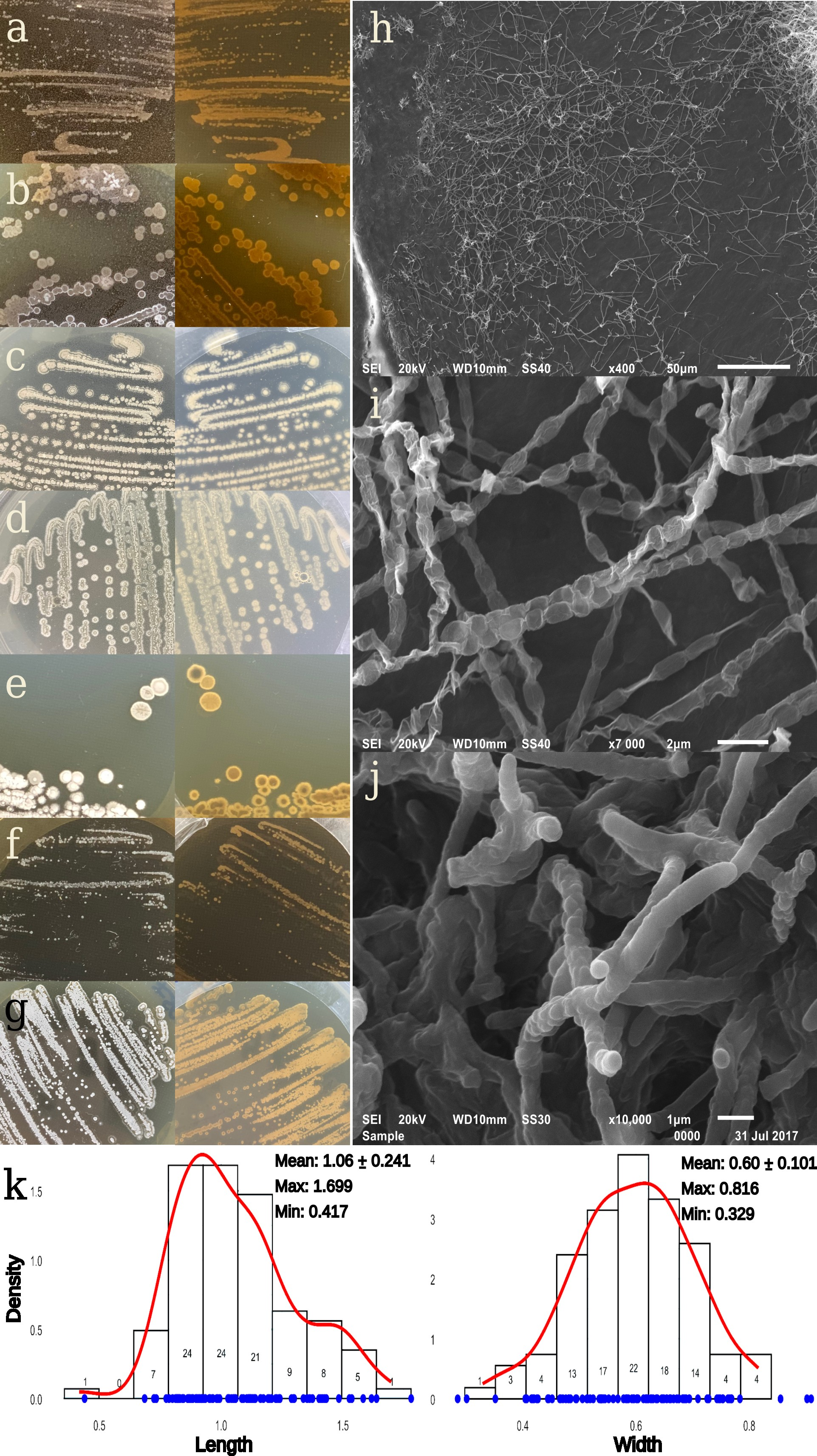
DEC002 strain colonies isolated from soil collected at Fumarole Bay (62°57′30.6″ S; 60°42′53.5″ W) on Deception Island, Antarctica (Figure 1a–c), displayed morphometric and ultrastructural characteristics typical of genus Streptomyces (Figure 2, Supplementary Table S1).
Based on cultivation on different media, the color of colony substrate varied from brown (Figure 2a,b) to yellow (Figure 2c,d) and brown–yellow (Figure 2e–g; Supplementary Table S1). The aerial mycelium color also varied from white to white/gray (Supplementary Table S1). At higher magnification, hyphae from strain DEC002 (Figure 2h–j) exhibited long, straight to slightly flexuous (rectiflexible) spore chains exceeding 20 μm in length (Figure 2i). Spore chains consisted of discrete, cylindrical units with smooth walls; individual spores measured 1.06 ± 0.24 µm in length and 0.596 ± 0.10 µm in width (Figure 2i). In culture, a clear petrichor-like odor consistent with geosmin release was evident, a trait widely associated with Streptomycetaceae [56]. Colony expansion was robust on ISP 2, 3, 4, 5, and 7 and on R2A (Supplementary Table S2), whereas growth on ISP 6 was only moderate. Across all media tested (ISP 2–7 and R2A), diffusible pigments were produced, with chromatic tones ranging from yellow to brown.
The strain DEC002 formed colonies with a consistently white aerial mycelium on all media surveyed (Supplementary Table S2). The substrate mycelium varied with the medium: brown on ISP2, yellow on ISP3–4, and mixed brown–yellow on ISP5–7 and R2A. Sporulation ranged from moderate to good. Histograms with kernel density estimates illustrate the distribution of spore dimensions, with mean spore length and width of 1.06 μm and 0.60 μm, respectively (Figure 2k).
The 16S rRNA amplicon analysis identified the DEC002 strain with 100% sequence identity to Streptomyces fildesensis INACH3013 [MK742730.1]. Importantly, this species is defined by the type strain GW25-5 (=CGMCC 4.5735T), which should be considered as the taxonomic reference for species-level assignment.
Functional characterization of DEC002 revealed phenotypic variations in comparison with other S. fildensens strains (Table S2) including colony color when cultivated on ISP 2, salt tolerance, temperature growth range, utilization of carbon (Table S2). On ISP 2, colonies were gray–white; spores were rod-shaped and arranged in straight to flexuous chains. DEC002 produced diffusible chromogens, among them a melanoid pigment, while starch hydrolysis was absent. DEC002 tolerated NaCl at 0–5% (w/v) and maintained growth at 4 °C, 10 °C, and 37 °C. Notably, the ability to grow at 4 °C, together with the complete salinity span, distinguishes the strain from at least one comparator (Table S2). Carbon assimilation was broad, with utilization of hexoses and pentoses (glucose, fructose, mannose, xylose, and arabinose), disaccharides (maltose, lactose, and sucrose), and additional substrates including galactose, raffinose, D-melibiose, and amygdalin. Bovine gelatin was hydrolyzed, and citrate was metabolized. Hydrogen sulfide production was not detected. Nitrite reduction was not assessed; the absence of color development in the nitrate reduction test after zinc addition in the API 20E system suggests nitrate reduction beyond nitrite (Table S2).
In addition, comparative screening of enzymatic and metabolic reaction profiles using API systems (Supplementary Table S3) revealed differences in specific activities relative to strain INACH3013. Notably, strain DEC0A2 exhibited lipase, cystine arylamidase, and N-acetyl-β-glucosaminidase activities, while esterase (C4) and alkaline phosphatase activities were absent. Substrate utilization patterns were largely similar between the two S. fildesensis strains, with the exception of citrate and sucrose metabolism (Supplementary Table S3).

3.1.1. Antimicrobial Activity

The CFS exhibited broad-spectrum antibacterial activity against foodborne pathogens (Table 1). The greatest inhibition was observed against V. cholerae (2.80 ± 0.154 cm), followed by S. pyogenes (1.90 ± 0.116 cm) and V. parahaemolyticus (1.81 ± 0.098 cm). Intermediate potency was noted against enteric bacilli and staphylococci: K. pneumoniae (1.67 ± 0.043 cm), E. coli (1.61 ± 0.068 cm), and S. aureus (1.58 ± 0.075 cm). The weakest responses were recorded for Enterobacter cloacae (1.39 ± 0.126 cm) and, most notably, Enterobacter aerogenes (1.08 ± 0.068 cm). The pattern indicates that the effect, attributable to secreted compounds in the medium, reflects greater susceptibility of Vibrio spp. and S. pyogenes, moderate efficacy against E. cloacae and S. aureus, and limited activity against r E. aerogenes (Table 1).

3.1.2. Antibiotic Resistance

The antimicrobial susceptibility test against 28 antibiotics belonging to 17 classes (Table 2) showed that DEC002 was resistant to 12 antibiotics, as indicated by the absence of inhibition zones, distributed among 13 different classes. Pronounced inhibitory activity is seen for protein-synthesis inhibitors, clarithromycin (≈44.5 mm), erythromycin (≈39.0 mm), spectinomycin (≈34.4 mm), streptomycin (≈29.6 mm), and tetracycline (≈41.3 mm), as well as for the tested to sulfonamide compounds (S3) (≈41.7 mm), while it was not susceptible to trimethoprim–sulfamethoxazole (SXT). Among DNA-targeting agents, ciprofloxacin is strong (≈34.0 mm) and novobiocin is outstanding (≈47.0 mm). β-Lactam responses are heterogeneous: imipenem yields very large halos (≈44.2 mm), whereas ampicillin (≈17.3 mm) and cephalothin (≈18.3 mm) are only moderate and other cephalosporins/carboxypenicillins show no inhibition. Minimal or absent halos occur with nalidixic acid, metronidazole, nitrofurantoin, mupirocin, clindamycin, trimethoprim, and vancomycin (~3.8 mm), and activity is limited for chloramphenicol, lincomycin, and rifampicin (~11–13 mm). Collectively, the profile suggests intrinsic or class-specific non-susceptibility to many cephalosporins and vancomycin, coupled with relative vulnerability to inhibitors of translation and DNA topoisomerases.

3.2. Draft Genome Sequencing and Annotation

Based on the functional properties of DEC002, the genome of this Antarctic bacterial strain was sequenced to identify structural elements associated with the natural antibiotic resistance of this Antarctic isolate from volcanic Deception Island. This genomic insight lays the groundwork for future studies on novel cold-active enzymes and bioactive molecules, while also highlighting the putative thermal-adaptation gene pool of this bacterium.
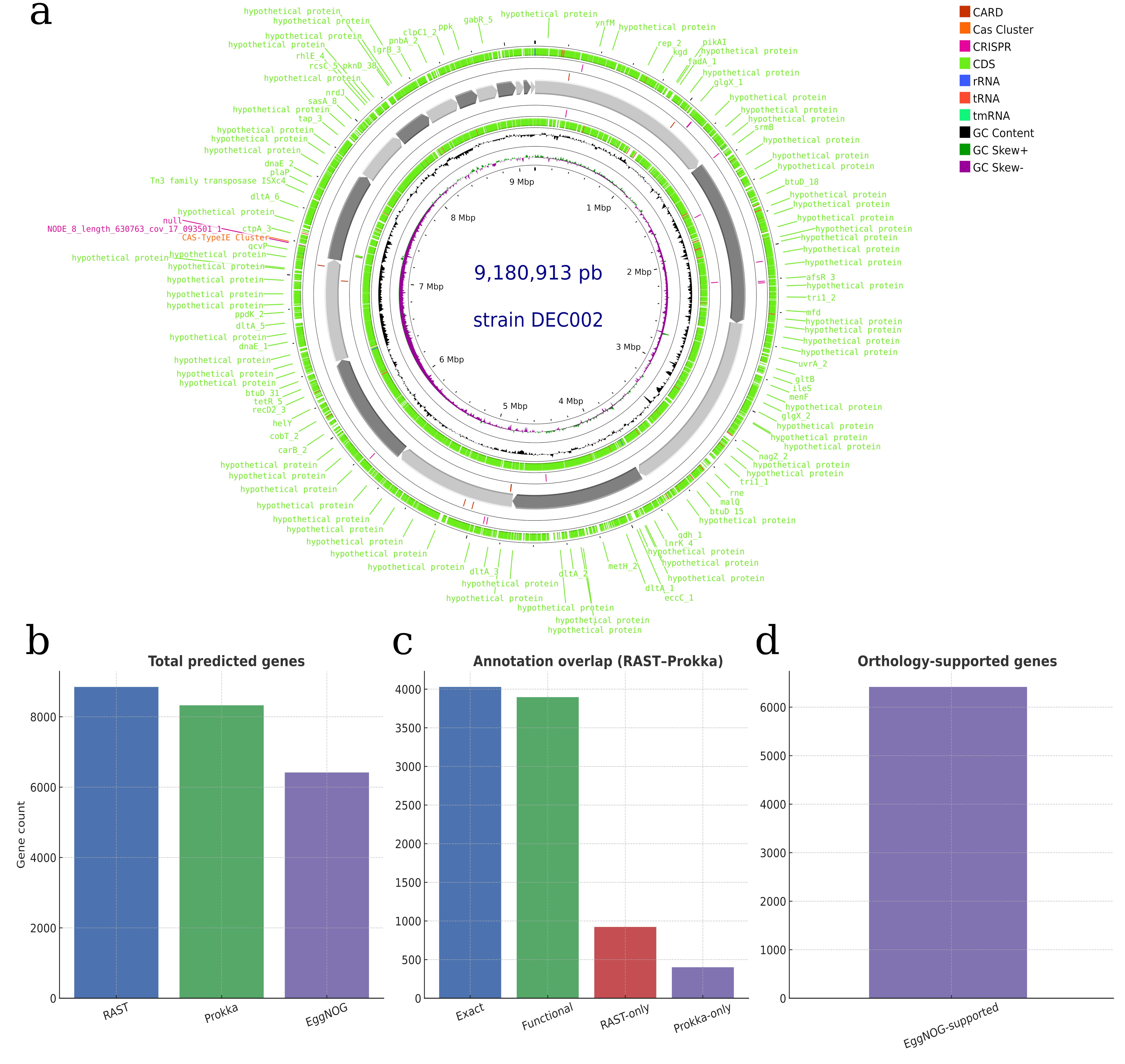
The genomic assembly of strain DEC002 generated by PacBio sequencing resulted in 17 contigs with a total length of 9,180,913 bp. Its average GC content was 70.63%, with a contig N50 of 948,363 bp and an L50 of 4 (Supplementary Table S4). No plasmids or closed chromosomes were detected. This configuration with few large contigs in a markedly GC-rich background provides a solid basis for structural and functional annotation. In concordance, the draft genome shows Prokka-derived feature rings for CDSs (8327 features), t/rRNAs (61/5 features), CRISPR arrays (24 features), and mobile elements (170 features: 89—integration/excision; 32—replication/recombination/repair; 21—phage; 9—stability/transfer/defense; 19—transfer), over which GC content and GC-skew profiles reveal local inflections that align with putative genomic islands, a pattern consistent with genomic dynamism in environmental Actinobacteria (Figure 3a). Gene-count summaries across gene-calling and annotation workflows reflect differences in structural gene prediction and downstream functional annotation. RASTtk returned the largest set of predicted open reading frames (ORFs), whereas Prokka produced a more conservative structural gene catalog. EggNOG-mapper was subsequently applied to the Prokka-predicted protein set to define the orthology-supported subset used for downstream functional and comparative analyses (Figure 3b). Cross-pipeline concordance further resolves into genes with identical labels, functionally equivalent assignments despite nomenclatural differences, and caller-specific ORFs, with a pronounced RAST-only fraction that reflects permissive ORF-calling and partial-signature matches, whereas the Exact/Functional blocks define a robust consensus core for downstream comparisons (Figure 3c). The orthology tier analytically consolidates these patterns by enumerating genes supported by EggNOG groups, thereby highlighting the evolutionarily corroborated portion of the genome that underpins pathway summaries, GO analyses, and enrichment tests throughout the study (Figure 3d).

3.3. Phylogenetical Analysis

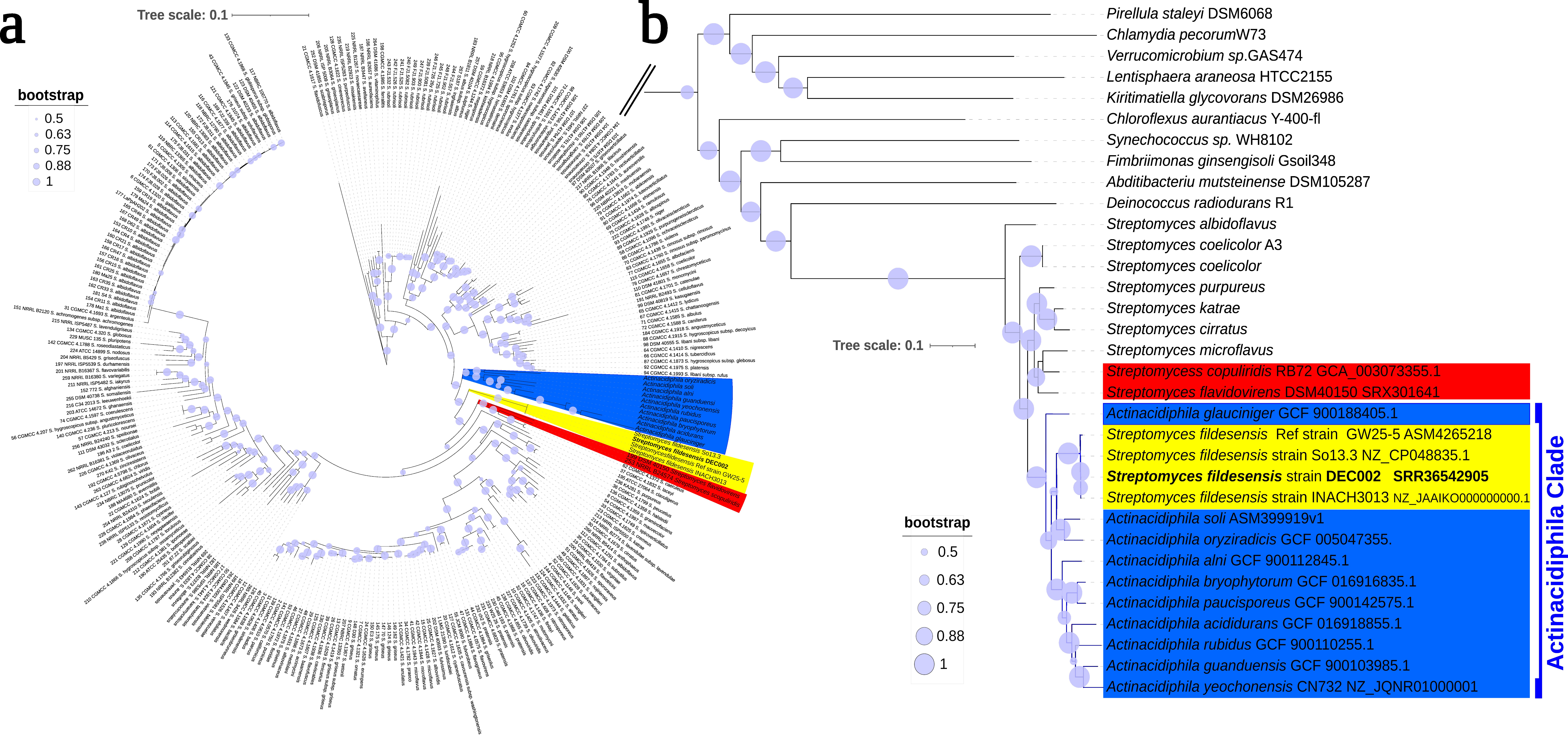
The phylogenetic reconstruction based on the concatenated gene markers 16S rRNA, atpD, gyrB, recA, rpoB, and trpB yields a well-resolved backbone for the genus and places DEC002 unequivocally within the Actinacidiphila lineage (Figure 4a). In that sector of the tree, DEC002 clusters with S. fildesensis as a discrete, exclusive unit. Short internal branches bind the members of this subcluster, while comparatively longer external branches partition it from neighboring groups, a pattern consistent with local cohesion and clear perimeter delimitation.
In the immediate neighborhood of DEC002, the terminal sister-branch is the isolate INACH3013, and the node joining DEC002-INACH3013 pair carries SH-aLRT/aBayes/UFBoot = 0/0.333/84 (Figure 4a). One tier deeper, the split that groups this pair with So13 is maximally supported (100/1/100). This pattern places DEC002 within the S. fildesensis block and documents strong local support for its immediate affinities. A nearby, more external division within the same block shows 97.7/1/84. Taken together, these values indicate that the DEC002-containing unit is both monophyletic and resilient to resampling. They also match the typical support observed on adjacent splits in this sector of the topology—SH-aLRT ≈ 96–100, aBayes ≈ 1.0, UFBoot ≈ 81–100—a profile consistent with a stable placement that is resistant to minor perturbations in branch order.
The concatenated matrix covers 283 Streptomyces accessions and 3852 aligned positions, of which 1301 are phylogenetically informative, yielding dense taxon coverage around the Actinacidiphila sector of the tree. Within this context, DEC002 falls immediately beside S. fildesensis entries yet remains clearly set apart from neighboring lineages by several internodes that carry strong support. To assess robustness, we inspected the ensemble of near-optimal IQ-TREE solutions; none placed DEC002 elsewhere, and the higher-level scaffold of the clade was retained throughout, indicating a stable, well-anchored position for the strain.
Visualization of the annotated tree in iTOL confirms these observations. The node labels printed on the ML topology reproduce the key supports (0/0.333/84 for DEC002 + 3013, 100/1/100 for (DEC002 + 3013) + So13, and 97.7/1/84 on a neighboring split) and the same pattern is evident in simplified outputs where only ultrafast bootstrap values are presented along the local path. Taken together, the explicit node-wise metrics, the consistent strength of adjacent splits, and the absence of plausible alternative placements converge on a single interpretation: DEC002 is firmly embedded within Actinacidiphila and forms a coherent subcluster with S. fildesensis strain, with high confidence in both its immediate affinities and its separation from nearby lineages (Figure 4a).
Extending this signal beyond the multilocus framework, a genome-scale reconstruction on KBase recovered the same neighborhood inferred by MLST and relationships (Figure 3b). In that species tree (Build Microbial SpeciesTree v1.6.0), DEC002 is grafted onto the stem already uniting INACH3013 and the curated S. fildesensis reference GW25-5 (genome GCF_042652185), with an internal bootstrap of 84% for the node placing DEC002 with this pair (Figure 4b). One tier deeper, the split that aggregates (DEC002 + INACH3013 + GW25-5) with So13 is fully supported (100%), yielding a locally saturated cluster that mirrors the multilocus result; additional internal divisions along this path register 92%, 81%, 89%, and 100%. The agreement in both topology and support magnitudes across MLST and genome-scale analyses consolidates the same conclusion: DEC002 belongs to Actinacidiphila and sits in immediate proximity to S. fildesensis isolates, while remaining clearly separated from adjacent lineages by strongly supported internodes.

3.4. Genome Comparison of Actinacidiphila fildesensis (Li et al., 2012) comb. nov.

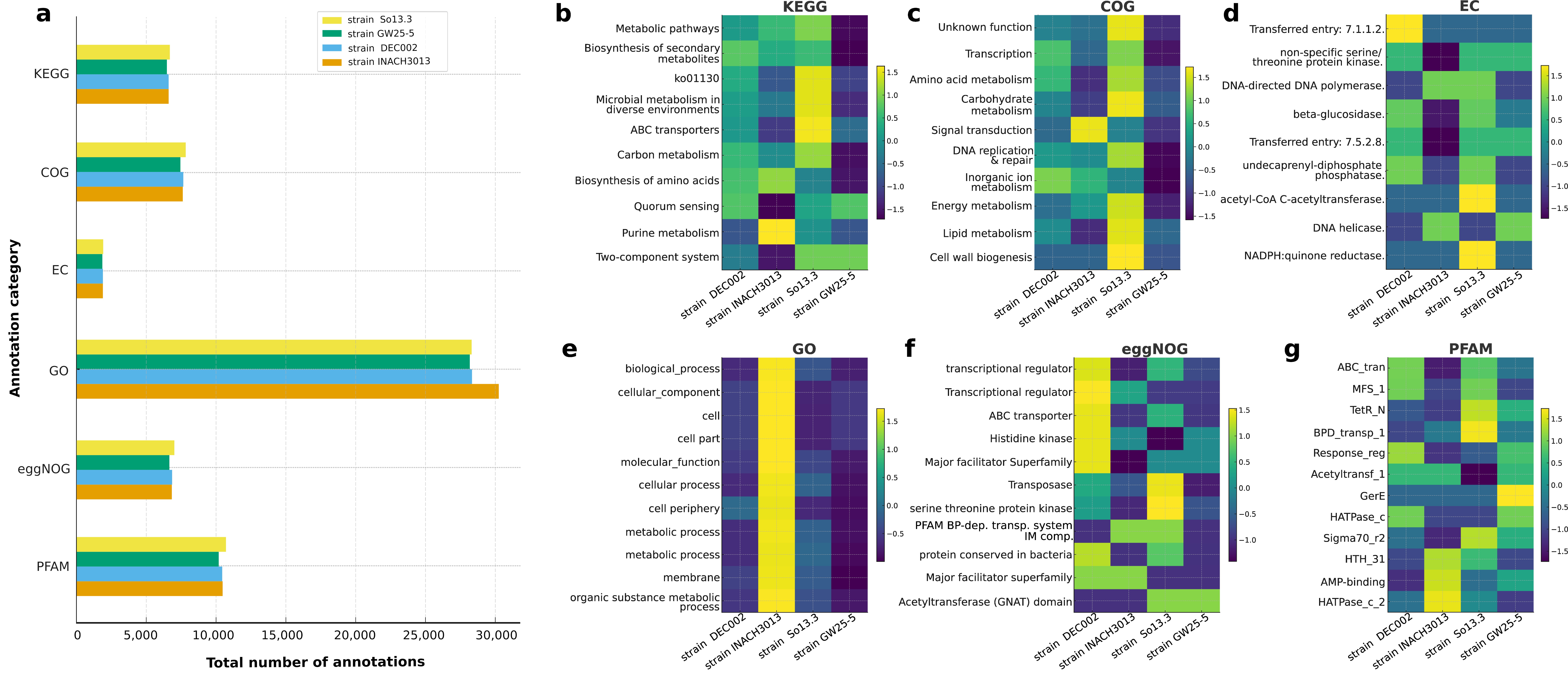
Across functional frameworks, the four A. fildesensis genomes display a conserved organization with only minor quantitative variation. KEGG analysis (Figure 5a) highlights ko01100 as dominant in all strains. This category corresponds to KO01100 (metabolic pathways), a global KEGG classification that groups genes involved in core cellular metabolism across multiple biochemical pathways. It was followed closely by secondary-metabolite biosynthesis and carbon metabolism, with nonribosomal peptide (NRPS) and polyketide (PKS) pathways, as well as terpenoid and post-translationally modified peptide (RiPP) pathways, which were consistently represented (Supplementary Figure S1). COG shows only minor variation, while GO counts for biological processes and cellular components remain similar across strains. Transport functions are widespread, including major facilitator, ABC, and compatible-solute permeases. Recurrent features related to cold adaptation and genome maintenance such as cold-shock proteins, fatty-acid desaturation, reactive oxygen species (ROS) detoxification, recombination and nucleotide-excision repair, and carbohydrate-active enzymes indicate the potential for gradual polysaccharide degradation in polar soils.
Within this shared genomic scaffold, the strains differ in their areas of functional profiles (Supplementary Figure S1 and Table S4). Strain DEC002 showed the most expansive transporter field suggestive of intensified uptake and export. INACH3013 balances transport with secondary metabolism and retains a co-localized set of carbohydrate-active enzymes, including β-1,4-glucanases, α-L-fucosidases, chitinases, and glycosyltransferases, supporting its capacity to incrementally depolymerize polysaccharides. So13.3 spans the broadest biosynthetic band, especially NRPS and PKS sectors, with several biosynthetic gene clusters showing high similarity to previously characterized clusters associated with known metabolites. These include two lanthipeptide clusters, as well as clusters putatively encoding actinomycin D (NRPS), antipain (NRPS), 2-methylisoborneol (terpene), spore pigment polyketides, pristinol (terpene), marineosin A/B (hybrid NRPS–PKS), ectoine, hopene (terpene), and ε-poly-L-lysine (NAPAA). Among these, actinomycin D, ectoine, hopene, and ε-poly-L-lysine BGCs are present in all strains, whereas antipain, spore pigment polyketides, and pristinol occur in the majority of strains. Strain GW25-5 preserves full cold-adaptation suites but shifts space toward genome integrity, oxidative-stress mitigation, and protein quality control. Figure 5b–g summarizes these functional contrasts. KEGG (Figure 5b) highlights carbon and amino-acid metabolism, energy metabolism, and secondary biosynthesis, with DEC002 showing the highest activity in microbial metabolism in diverse environments. COG (Figure 5c) emphasizes amino-acid metabolism, DNA repair and replication, carbohydrate processing, transcription, and post-translational modification, with modest elevations in defense and secondary metabolism for DEC002 and So13.3. EC annotations (Figure 5d) focus on redox and group-transfer chemistry and nucleic-acid processing, particularly NADH dehydrogenase, acetyl-CoA C-acetyltransferase, and serine–threonine kinases. GO terms (Figure 5e) highlight cellular processes, organic-substance metabolism, response to stimuli, membrane components, and catalytic activity, with deprecated terms updated. eggNOG (Figure 5f) overrepresents primary metabolism, transcriptional regulation, and ribosomal machinery, showing broadly similar profiles. PFAM (Figure 5g) ranks response regulators containing helix–turn–helix motifs, sigma-70 region 2, ABC-transporter components, and aldo–keto reductases, with So13.3 moderately enriched in regulatory domains. These data indicate common trends in cold adaptation and aerobic energy metabolism, while revealing strain-specific priorities, where DEC002 emphasizes trafficking, INACH3013 maintains functional balance, So13.3 favors specialized-metabolite biosynthesis with strong redox support, and GW25-5 prioritizes genome integrity and oxidative defenses, with comparatively lower transport activity.

3.4.1. Biosynthetic Gene Cluster of Actinacidiphila Strains

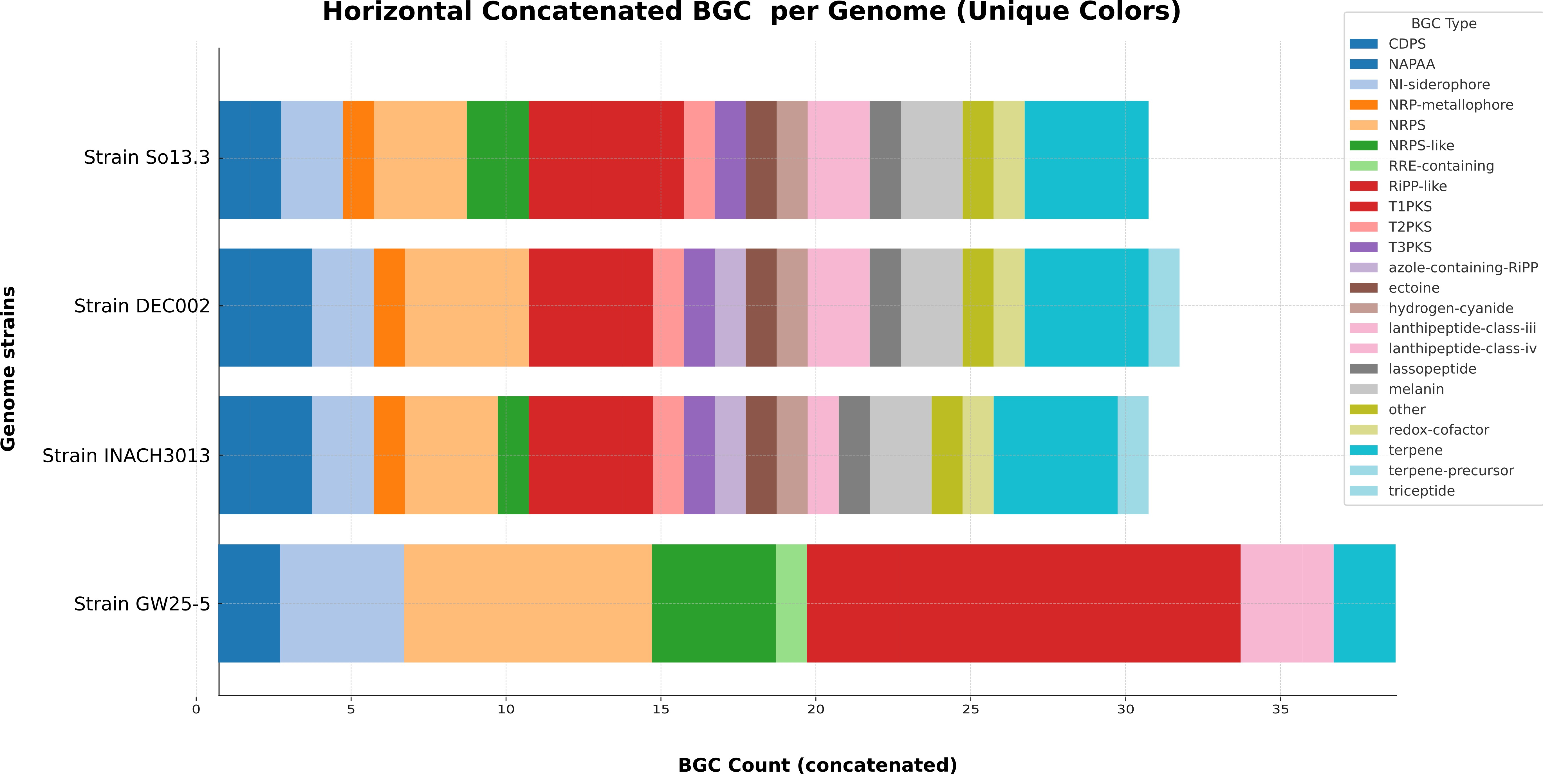
A comparison of the four Antarctic isolates genomes (Figure 6) revealed distinct quantitative differences in their biosynthetic gene cluster (BGC) repertoires, despite all strains belonging to the same species and originating from broadly similar polar soils (Fildes Peninsula and Deception Island). In the horizontal stacked display, strain So13.3 carries the most expansive inventory (≈31 BGCs) followed by strain DEC002 at ≈29. Strains GW25-5 and INACH3013 trail slightly with ≈27 and ≈26 clusters, respectively. The absolute spread, five clusters between extremes, is modest, yet once categories are concatenated the differences become visually immediate: contributions from NRPS, type-I PKS, hybrid systems, RiPPs, and smaller classes accumulate into distinct structural profiles for each genome (Figure 6). Strains So13.3 and DEC002 contain a larger fraction of NRPS and type-I PKS modules of the megasynthase system, strain GW25-5 shows an enrichment of RiPP biosynthetic gene clusters, particularly those encoding lantipeptides and other ribosomally synthesized peptides, whereas strain INACH3013 exhibits a more even but overall lower distribution across biosynthetic categories (Figure 6). In contrast, siderophore-, terpene-, and redox-associated clusters are consistently present across all four strains, indicating a conserved biosynthetic repertoire consistent with species-level classification in Actinacidiphila (Figure 6).

3.4.2. Thermal-Adaptation Gene Comparison

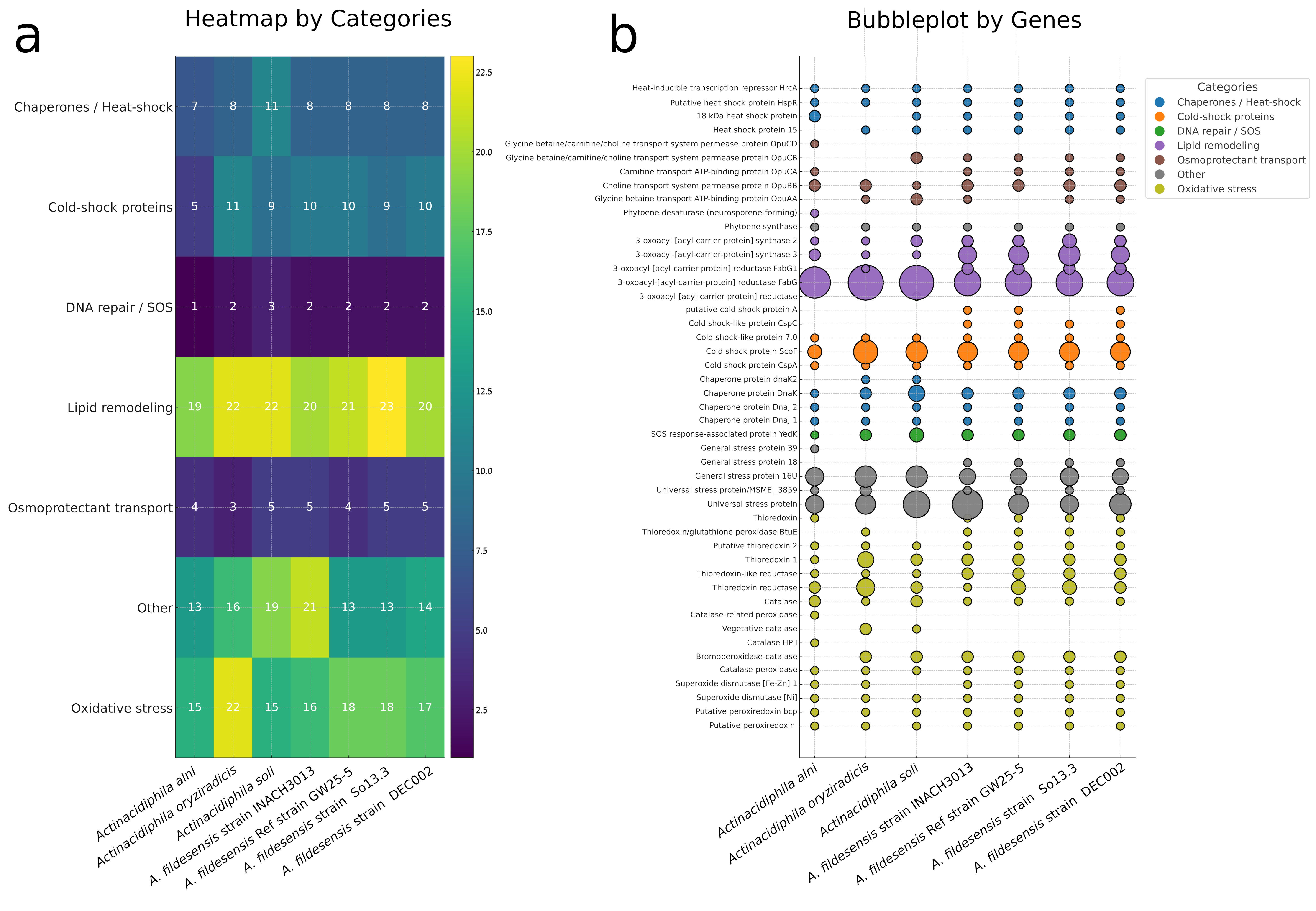
DEC002 concentrates defenses against oxidative and cold stress. It carries one peroxiredoxin, one bcp-type peroxiredoxin, one Ni–SOD, one Fe/Zn–SOD, four catalases (one catalase–peroxidase, two Bromoperoxidase-catalases and one catalase) and an expanded thioredoxin system with two thioredoxin reductases, two thioredoxin-like reductases, two Thioredoxin 1, one Putative thioredoxin 2, one Thioredoxin/glutathione peroxidase BtuE and one Thioredoxin. It encodes several universal stress proteins and four cold-shock genes including cspA, putative cspA, cspC, and six csh-like factors, plus membrane-remodeling enzymes such as FabG/FabG1 and 3-oxoacyl-ACP synthases. Genes for canonical heat-shock regulators (hsp15, hspR, hrcA) and OpuA–OpuE osmoprotectant transporters are not detected (Supplementary Table S5).
Across seven genomes, the heatmap separates three non-Antarctic Actinacidiphila strains from four Antarctic A. fildesensis isolates (Figure 7a), and the bubble diagram resolves the underlying counts (Figure 7b). Oxidative modules form a conserved core—single-copy peroxiredoxin, bcp-peroxiredoxin, and Ni–SOD in all genomes—while catalase patterns diverge: bromoperoxidase–catalase is absent in A. alni but duplicated in A. oryziradicis, A. soli, and all Antarctic strains; HPII is exclusive to A. alni; vegetative catalase is missing from Antarctic genomes. Redox buffering widens with clade: thioredoxin systems peak in A. oryziradicis, intermediate in non-Antarctic strains, lower in Antarctic genomes. General stress proteins show a binary marker: Gsp18 is present only in Antarctic isolates, whereas Usp counts surge in A. soli and INACH3013. Cold-shock capacity expands genus-wide via CspF, and CspC is Antarctic-specific and single-copy in every A. fildesensis genome (Figure 7a,b). Membrane biogenesis contrasts are pronounced: FabG is amplified in non-Antarctic genomes, FabG1 is duplicated in all Antarctic strains, and 3-oxoacyl-ACP synthases reach their highest copy numbers in Antarctica strains; OpuBB/OpuCB vary modestly, indicating distinct routes to osmotic and membrane homeostasis (Figure 7a,b).

4. Discussion

Streptomyces fildesensis, first isolated from the Fildes Peninsula on King George Island, NW Antarctica [57], exemplifies an Antarctic bacterium able to produce secondary metabolites, including actinomycin, which exhibits antimicrobial and anticancer activities under extreme conditions [8,24,28].
Strain DEC002 isolated from Deception Island (Figure 1c) showed typical morphometric and ultrastructure characteristics for the genus Streptomyces (Figure 2d–f). The main difference from the close relative strain INACH3013 was the white color of the aerial mycelium of the DEC002 colonies. Other phenotypic variations between strains included the positive sucrose utilization by strain DEC002 and the negative sodium citrate utilization by the same strain (Table 1). Regarding enzymatic activity (Supplementary Table S1), strain DEC002 exhibited positive activity for lipase, cystine arylamidase, and N-acetyl-β-glucosaminidase, while showing negative activity for alkaline phosphatase and esterase (C4). These enzymatic traits represent the main distinctions from strain INACH3013. According to the description of the species Streptomyces fildesensis [57], it tolerates salt concentrations of up to 3% NaCl, does not grow at 4 °C, and is only capable of utilizing arabinose, maltose, xylose, galactose, and sodium citrate. The description of the present strain, along with strain INACH3013 isolated from King George Island (NW Antarctica) [57], exemplifies that the range of salt tolerance, temperature, and carbon source utilization may be broader within the species. Bacteria that are slightly halotolerant, capable of surviving in NaCl concentrations of 6–8%, have often been found in Antarctic soils [58]. Studies of Antarctic Actinomycetota isolated from Deception Island and Galindez Island indicate that 92% of the strains were psychrotolerant, while 75% showed halotolerance [59]. Notably, all of these isolates could grow in environments with up to 10% NaCl [59]. Psychrotolerant bacteria are characterized by their ability to grow at 0 °C and have limited growth at 37 °C, with optimal growth between 10 and 28 °C [60]. Therefore, strains DEC002 and INACH3013 can be classified as psychrotrophs rather than psychrophiles. The observed psychrotolerance and halotolerance in strains DEC002 and INACH3013 likely reflect evolutionary adaptations to the extreme conditions of the Maritime Antarctic South Shetland Islands. This polar environment subjects the soil microbiota to constant osmotic stress and dehydration stress [61].

4.1. MLSA and Phylogenomic Evidence Supporting the Placement of Strain DEC002 Within Actinacidiphila

The concatenated-locus phylogeny provides a coherent backbone for Streptomyces and places strain DEC002 firmly within the Actinacidiphila clade (Figure 4a). Multilocus concatenation (typically atpD, gyrB, recA, rpoB, trpB and 16S rRNA genes) is well known to increase discriminatory power and topological stability relative to 16S-only analyses, thereby reducing the influence of locus-specific conflict and yielding higher support on internal nodes, exactly the pattern observed for the strain DEC002 subcluster, showing short intraclade branches, and longer external branches [42].
Within Actinacidiphila, DEC002 forms an exclusive unit with representatives of A. fildesensis, while remaining separated from neighboring lineages by well-supported internodes. The recovery of a monophyletic, strongly supported Actinacidiphila crown is consistent with recent genome-based taxonomic treatments that stabilize relationships across Streptomycetales and enumerate reference genomes for multiple Actinacidiphila species [25]. Dense sampling in the present analysis (283 Streptomyces entries) increases local coverage around Actinacidiphila, and inspection of near-optimal solutions showing no alternate placements for DEC002 aligns with prior observations that MLSA backbones resist minor topological perturbations when multiple protein-coding loci are concatenated [42].
Looking across the genome, the picture is consistent: the marker-based species tree reproduces the MLSA result and nests DEC002 within Actinacidiphila, with high support on the defining nodes and no mismatch with the multilocus topology. This alignment between locus-concatenated and genome-wide evidence is central to contemporary taxonomy in Streptomycetales, where classifications are interpreted alongside quantitative measures (Supplementary Tables S6 and S7) [25,62]. The proximity of DEC002 to A. fildesensis (formely S. fildesensis) is congruent with independent studies in which A. fildesensis appears as a recurrent neighbor in both 16S-based and polyphasic/MLSA contexts, and is routinely included among the closest Streptomyces references in comparative analyses, supporting the interpretation that DEC002 belongs within Actinacidiphila but is discretely delimited from adjacent species-level lineages [62,63].
The results presented should be interpreted in light of the current nomenclature and the pace at which it is updated. S. fildesensis isolated from Fildes Peninsula soil [57] as of the present date remains classified as Streptomyces taxa in LPSN [64] (accessed on 29 November 2025). Although the genus Actinacidiphila was established in 2022, S. fildesensis has not yet been formally transferred to this genus in the peer-reviewed literature [25,65]. In this context, the concordance observed between MLSA and genome-scale phylogenomic analyses suggests that S. fildesensis is phylogenetically affiliated with the genus Actinacidiphila. While a formal taxonomic transfer is beyond the scope of the present study, the evidence presented support the phylogenetic placement of S. fildesensis within the genus Actinacidiphila.

4.2. Functional Genome Annotation of Antarctic Actinacidiphila fildesensis

The internal concordance between the structural panorama in the six categories observed in Figure 5, Supplementary Figure S1 and Table S8, converge on a single message: the four A. fildesensis genomes preserve a broad and resilient metabolic core while modulating emphasis at the margins. KEGG and COG in Figure 5a–c stabilize carbon, amino-acid, and energy pathways, whereas EC, PFAM, and GO in Figure 5d–g register restrained quantitative shifts. The current data (Supplementary Figure S1 and Table S9) demonstrate that DEC002 harbors an extensive repertoire of NRPS/PKS biosynthetic gene clusters together with terpene- and RiPP-associated pathways, whereas the functional annotation profiles (Figure 5) indicate a highly conserved primary-metabolism backbone across strains. Taken together, these data support the dual strategy, expansive primary metabolism coupled to specialized small-molecule production, long recognized in Actinomycetota and exemplified by Streptomyces coelicolor [66,67,68,69].
The Antarctic setting supplies the selective scaffold. Cold imposes constraints on folding, membranes, and redox balance; the figures show conserved solutions, cold-shock proteins, desaturases, antioxidant systems, efficient electron transport, overlaid by fine-scale variation captured across EC, PFAM, and GO, likely reflecting freeze–thaw dynamics and nutrient pulses in polar soils [70,71,72,73,74,75]. Strain-resolved signals align with prior work on the Fildes Bay lineage that includes INACH3013: biosynthetic breadth and psychrotolerance dominate the overview and the heatmaps, with So13.3 accentuating specialized metabolism and DEC002 amplifying transport. This genomic architecture (Figure 5, Supplementary Figure S1) is consistent with draft genome data for A. fildesensis and with observed temperature-dependent trade-offs between antimicrobial production and growth. Similar patterns have been reported in phylogenomic analyses linking secondary-metabolite gene content with environmental resilience across clades [8,22,24,25,28].

4.3. Comparative Biosynthetic Potential and Antimicrobial Phenotypes

Although the four isolates belong to the same species, their biosynthetic portfolios differ in both composition and emphasis (Figure 6). The graphics make this evident: a shared toolkit is present, but the relative allocation to major BGC families diverges enough to yield strain-specific chemical fingerprints. That such divergence emerges among strains from similar polar soils accords with the genomic plasticity of Streptomyces and the rapid remodeling of specialized-metabolism loci [76,77]. No less notable is the stability of the overarching framework—NRPS, PKS, RiPPs, and several smaller classes recur in all four—while proportions shift in ways that plausibly matter ecologically. Modern genome-mining workflows are designed precisely to resolve and annotate these contrasts with increasing fidelity [78,79].
So13.3 and DEC002 sit at the richer end, with a clear emphasis on NRPS and type-I PKS pathways. Weighting toward large, modular assemblies often accompanies chemical competition, stress mitigation, and responses to episodic resource pulses—conditions expected under Antarctic seasonality and oligotrophy [76]. In such settings, maintaining an expanded, energetically costly suite of megasynthases may be advantageous when interactions compress into short favorable windows.
GW25-5 follows a different balance. It shows a tilt toward RiPPs—short, ribosomally encoded peptides that can be generated swiftly and diversified through post-translational tailoring. This allocation supports strategies centered on precise signaling or narrowly targeted antagonism while keeping biosynthetic costs comparatively low [80]. RiPP-biased repertoires are frequently reported for taxa in nutrient-poor or otherwise extreme habitats, where metabolic economy often outranks breadth [76].
The genome of INACH3013 represents the lower extreme in biosynthetic content. While the gene cluster complement is clearly Streptomyces-like, the overall number is reduced. These patterns are most consistent with genomic contraction rather than expansion, potentially mediated by homologous recombination, localized rearrangements, and gene loss within specialized-metabolism regions [76,77]. Genome streamlining may result from drift, local selection, or colonization history. Strain INACH3013 may represent a minimal version of the species’ biosynthetic capacity, while the other genomes retain additional specialized functions.
Despite strain-specific variation, a conserved core of biosynthetic gene clusters—including siderophores, terpenes, redox-associated clusters, and several minor families—is shared across strains. This pattern indicates a common genome, with divergence concentrated in flexible regions associated with rapidly evolving specialized metabolism [77,79]. Variation here is better interpreted not as noise but as the expected microevolutionary breadth of an adaptable Actinomycetota lineage under polar conditions. Practically, antiSMASH and the curated MIBiG framework provide the scaffolding to delineate this shared core and to situate lineage-specific elaborations with appropriate taxonomic and functional context [78,79].
Strain DEC002 in this study demonstrated promise in the inhibition of growth in many Gram-negative and Gram-positive bacteria such as Vibrio, Enterobacter, Klebsiella, E. coli, and Staphylococcus. Such similarities in activities towards both Gram-positive and Gram-negative bacteria have been documented before on strain INACH3013 and other Streptomyces species from Antarctica [22,23,24]. Inhibition across a wide range of bacterial groups by strain DEC002 suggests the production of multiple antimicrobial activities which collectively result in a broad inhibitory profile, as previously observed for the closely related strain INACH3013. Such findings are consistent with the presence of shared biosynthetic gene clusters between both strains. Comparative antiSMASH analysis indicates that DEC002 and INACH3013 harbor several BGCs with high similarity, including clusters associated with actinomycin D, ectoine, hopene, and ε-poly-L-lysine, supporting a partially conserved biosynthetic repertoire worthy of further genomic investigation [8,22,24]. A confirmatory assessment of secondary metabolite production was conducted by thin-layer chromatography, comparing strain DEC002 with the reference strain INACH3013 (Supplementary Figure S2). The analysis revealed comparable metabolite profiles, including two actinomycin compounds previously characterized for their antimicrobial and anticancer activities [8]. In polar environments where microbial competition has been documented as sufficiently stringent (nutrient-limited), the production by strain INACH3013 of cold-active antimicrobial compounds may represent a critical survival advantage [28]. Such adaptive features are in alignment with previous discoveries on antibiotic resistance in Antarctic psychrotolerant bacteria from varied habitats ranging from terrestrial and marine environments [81,82,83].
Strain DEC002 was resistant to 12 antibiotics, including three additional compounds (cefotaxime, clindamycin, and nalidixic acid) as compared with INACH3013. In contrast, DEC002 was susceptible to sulfonamides. Both DEC002 and INACH3013 were sensitive to ampicillin. DEC002 also exhibited resistance to multiple β-lactam antibiotics, including carbenicillin, ceftazidime, cefixime, cefamandole, cefotaxime, and cefpodoxime, suggesting the presence of either a narrow-spectrum β-lactamase or an alternative resistance mechanism [84]. Previous studies implicated anthropogenic enrichment in multidrug resistance in Antarctic microbes [85,86]. Based on the current data and BGC inventory for DEC002 (Table 1, Supplementary Table S4), it is possible to advance cautious inferences about the observed inhibition zones. For the Vibrio species and the Enterobacteriaceae, the patterns could be explained by secretion of ε-poly-L-lysine from a high-similarity cluster, since this cationic polymer is known to disrupt bacterial membranes under food-relevant conditions [87]. In the case of Staphylococcus aureus and Streptococcus pyogenes, the activity may involve class IV lanthipeptides that bind lipid II or form pores, together with an actinomycin-like nonribosomal peptide that intercalates DNA; additional nonribosomal and polyketide products, such as polycyclic tetramate macrolactams, could broaden the spectrum, and alkylated benzoquinone or phenol metabolites could intensify membrane stress in Gram-negative bacteria. By contrast, SapB is primarily morphogenetic, and terpene, melanin, or generic siderophore pathways are unlikely to account for centimeter-scale halos in aqueous supernatants under these conditions [88,89,90,91]. It is important to emphasize that the present study does not establish a direct causal relationship between specific biosynthetic gene clusters and the inhibition phenotypes observed in vitro. The bioactivity detected derives from crude culture supernatant and may reflect the combined or synergistic action of multiple metabolites or additional extracellular factors. No targeted metabolite extraction, purification, or structural elucidation was performed in this work. Therefore, the associations proposed between predicted BGCs and antimicrobial activity should be interpreted as hypothesis-generating rather than confirmatory. These predictions require experimental validation, including direct detection of ε-poly-L-lysine and the relevant lanthipeptides, and their association with the identified clusters (Supplementary Table S4) through bioactivity-guided fractionation, Liquid Chromatography–Tandem Mass Spectrometry (LC–MS/MS), and genome–metabolome correlation [92].

4.4. Comparative Analysis of Thermal-Adaptation Genes

The thermal-adaptation gene repertoire of strain DEC002 is consistent with strategies observed in cold-adapted and psychrotolerant Actinomycetota. The strong expansion of antioxidant systems (including multiple peroxiredoxins, catalase–peroxidases and both Ni- and Fe/Zn-superoxide dismutases) reflects a genomic investment in ROS detoxification, a hallmark of microorganisms exposed to fluctuating low-temperature and high-ROS environments [70,93]. DEC002 also encodes several cold-shock proteins (CspA/CspC/CspF) and universal stress proteins, which are widely associated with RNA stabilization, transcriptional reprogramming and survival under rapid temperature downshifts [94,95]. The presence of multiple 3-oxoacyl-ACP reductases suggests active remodeling of membrane lipid composition to maintain fluidity at low temperatures, a key adaptation reported in psychrophilic Actinomycetota and other cold-environment microbes [87,96]. In contrast, DEC002 lacks genes encoding canonical heat-shock regulators (HspR, HrcA) and osmoprotectant transporters (OpuA–OpuE), indicating an adaptive profile oriented predominantly toward cold and oxidative stress rather than heat or osmotic challenges. These characteristics are consistent with genomic patterns reported for cold-adapted Streptomyces and Antarctic Actinomycetota [97,98], suggesting that DEC002 is well adapted to the rapidly changing microhabitats of Deception Island.
The heatmap and the bubbleplot together delineate two strategies that separate the Antarctic A. fildesensis strain genomes isolates from the three non-Antarctic strains (Figure 7a,b). Antarctic genomes concentrate on rapid cold-shock response and broad stress protection, a pattern made explicit by the presence of general stress protein eighteen, by consistently elevated CspF, and by the Antarctic-specific CspC, all of which align with molecular constraints on folding, membranes, and redox balance in cold environments described for psychrophiles [70,99]. The same figures show a comparatively simple and stable thioredoxin and catalase architecture in the Antarctic group, with core enzymes retained and many catalase variants absent, consistent with compact repertoires favored under extreme oligotrophy [99].
The non-Antarctic genomes follow a contrasting reinforcement of membrane and redox modules. The heatmap highlights strong lipid-associated categories, while the bubbleplot attributes these signals to extensive FabG expansions in A. oryziradicis and A. soli together with a broadened thioredoxin network in A. oryziradicis, a configuration consistent with membrane remodeling and redox homeostasis in relatively buffered cold soils [71]. In sum, this analysis (Figure 7a,b) resolves the category-level partition and the bubbleplot specifies the gene-level levers that produce it, with Antarctic soils imposing fluctuating stresses such as freeze thaw, desiccation, and ultraviolet exposure that favor rapid response systems, and non-Antarctic settings selecting metabolic reinforcement instead [70,71,99,100].

4.5. Antibiotic Resistance Profiles Supported by Genomic Annotation

Because the assay was performed on R2A rather than standardized Mueller–Hinton, inhibition zones should be interpreted as relative potency rather than assigned CLSI/EUCAST S/I/R categories; nonetheless, the experimental pattern and the annotation-derived expectations align with an intrinsically conservative resistance profile characterized by broad non-susceptibility to several cephalosporins and relative vulnerability to inhibitors of translation and DNA topoisomerases [101,102]. The coexistence of multiple biosynthetic gene clusters (BGCs) and an expanded repertoire of transport systems in DEC002 is also consistent with intrinsic self-resistance mechanisms linked to secondary metabolite production. In Streptomycetales, resistance determinants are frequently embedded within or genetically associated with NRPS, PKS, and RiPP clusters, where ABC or MFS transporters mediate metabolite export and cellular protection [76,77]. Genome-mining studies and curated resources such as antiSMASH and MIBiG have repeatedly documented this co-localization pattern [78,79], supporting the concept that biosynthesis and self-protection co-evolve as integrated functional modules. Although the present data do not establish a direct mechanistic link, the genomic architecture of DEC002 aligns with established models of metabolite-associated intrinsic resistance in Actinomycetota [76,77,78,79]. Such genome–phenotype coherence is consistent with patterns widely documented in environmental Actinomycetota, where biosynthetic potential, intrinsic resistance, and ecological competition co-evolve as tightly coupled traits. Large zones with macrolides, tetracycline, spectinomycin, streptomycin, ciprofloxacin, and, most conspicuously, novobiocin are consistent with the absence, in the inspected annotations, of canonical determinants such as Erm-family 23S rRNA methylases and plasmid-mediated quinolone resistance, and with no quinolone resistance-determining region changes flagged at the annotation level, while acknowledging that sequence confirmation is pending; in actinomycetes, the high novobiocin susceptibility also concurs with the lack of novA/novR loci observed here [103,104,105]. In contrast, weak or absent inhibition by multiple cephalosporins together with large zones for imipenem is compatible with a chromosomal AmpC-like cephalosporinase background in the absence of carbapenemases, given the relative stability of imipenem to AmpC hydrolysis [106]. This pattern suggests that part of the resistance phenotype may reflect intrinsic structural or enzymatic features typical of environmental actinomycetes rather than recent acquisition of mobile resistance determinants. The diminished effect of vancomycin is consistent with a vanH–vanA–vanX module (with VanRS-like regulation) detected in the annotations, which fits glycopeptide self-resistance paradigms in actinomycetes; expression, however, has not been assessed [107]. The negligible activity of metronidazole and the limited effect of nitrofurantoin are consistent with their oxygen-dependent pharmacology. Resistance to mupirocin was observed despite the absence of canonical resistance genes (mupA/ileS2), suggesting an intrinsic tolerance potentially associated with membrane lipid remodeling, including the presence of multiple fabG copies and the desA3 desaturase, rather than target-specific resistance. The precise dfr/sul status underlying the trimethoprim–sulfonamide phenotype remains to be confirmed from the complete annotation set [108,109,110,111,112]. Finally, although a MacAB-like efflux module is annotated, macrolide halos remain large in R2A, consistent with the non-detection of erm/mph/msr.
The antimicrobial susceptibility profile presented by strain DEC002 reveals a resistance phenotype to several antibiotic classes, including beta lactams, chloramphenicol, nalidixic acid, metronidazole, and mupirocin. But it is only partially explained by the genomic annotation performed. For example, the presence of some resistance genes (Supplementary Table S10) partially explains some of the observed traits. Since susceptibility was observed in agents such as ciprofloxacin, macrolides, carbapenems, sulfonamides, tetracycline, and vancomycin. These inconsistencies indicate that the resistance observed could be modulated by other factors, such as gene regulation, differential expression, or even post-transcriptional mechanisms.
In bacteria, resistance to β-lactam antibiotics is commonly conferred by enzymes that hydrolyze the β-lactam ring or by reduced drug affinity due to alterations in penicillin-binding proteins (PBPs) [113,114]. In the present genomic annotation for DEC002, β-lactamase-like genes and PBP/cell-wall-modifying enzymes are annotated. While acquired clinical alleles may be absent, intrinsic β-lactamases and PBP variation remain plausible contributors alongside permeability and efflux to the complete resistance observed to carbenicillin and third-generation cephalosporins, and to the intermediate inhibition by ampicillin and cephalothin [113,114].
The reduced chloramphenicol susceptibility correlates with an annotated efflux determinant (cmlV), consistent with a partial-resistance phenotype mediated by decreased intracellular drug rather than enzymatic inactivation [115,116]. A similar efflux-mediated contribution could modulate the intermediate activity observed for some aminoglycosides; however, specific aminoglycoside-modifying enzymes should be confirmed before attributing the phenotype to defined AAC/APH/ANT families [117].
The fluoroquinolone pattern (resistance to nalidixic acid with preserved susceptibility to ciprofloxacin) suggests a selective mechanism. Stepwise mutations in gyrA/gyrB or parC/parE may reduce binding of older quinolones while preserving activity of newer agents, a hypothesis that should be confirmed by targeted sequencing and functional assays [118].
A similarly non-uniform response was observed among 30S ribosome-targeting antibiotics, with gentamicin and streptomycin showing lower activity than spectinomycin. This profile fits canonical mechanisms based on enzymatic modification, but in the absence of unambiguous genomic calls the linkage should be framed as hypothesis pending locus-level confirmation [117].
Susceptibility to vancomycin, despite annotation of van-cluster components (e.g., vanH/vanX and accessory/regulatory genes), is compatible with an incompletely functional or poorly expressed operon under the test conditions. Because annotations from different pipelines can disagree on the presence of vanA/vanB, the safest interpretation is that sequence evidence for a fully competent D-AlaD-Lac pathway is inconclusive, and phenotype should take precedence [119].
For trimethoprim, resistance in DEC002 would be explained by dfr/folA variants if present; however, where such calls are not unambiguously annotated, the mechanism should be reported as “putative,” with the observed susceptibility to sulfonamide combinations coherently attributed to the sequential blockade of the folate pathway [120,121]. Resistance to metronidazole could not be assigned to known genes; in aerobes, reduced activation via nitroreductases or altered redox metabolism often underlies decreased activity, but this remains a mechanistic inference without direct genomic support here [108].
Taken together, the genomic characterization of DEC002 explains part of the resistance phenotype, where classical determinants are explicitly annotated with robust genotype–phenotype correspondence. While noncanonical mechanisms, expression-dependent effects, or incomplete operons offer reasonable interpretations, we require future studies for a better understanding of the mechanism involved. This integrated, phenotype-anchored framing aligns best with current understanding of resistance in environmental actinomycetes and minimizes over-interpretation of sequence alone [108,113,114,115,116,117,118,119,121].

5. Conclusions

The phenotypic, phylogenetic, and genomic evidence collectively supports assigning strain DEC002 to the lineage previously described as Streptomyces fildesensis, and the same dataset consistently places it within the Actinacidiphila clade. Beyond taxonomy, DEC002 expands the known ecological and biogeographic context of the species because it is the first isolate recovered from an island other than the Fildes Peninsula (King George Island), being obtained instead from Deception Island. Morphology remains typical of the genus, yet DEC002 shows intra-specific variation compatible with local microdiversification (e.g., aerial mycelium color, differential sucrose/citrate utilization, and a distinctive enzymatic profile); together with tolerance to cold and salinity, these features support a psychrotrophic classification and suggest that the species’ physiological range is broader than initially reported. At the genome level, the isolates retain a robust metabolic core while adjusting peripheral functions related to stress response and transport, consistent with selection in Maritime Antarctic soils exposed to freeze–thaw cycling, desiccation, and episodic nutrient pulses. Meanwhile, genome mining reveals divergence in the composition and relative emphasis of biosynthetic gene clusters (NRPS/PKS/RiPPs), anticipating strain-specific chemical fingerprints and aligning with the observed broad-spectrum antimicrobial activity against both Gram-positive and Gram-negative bacteria, a trait that can be interpreted as a competitive advantage under conditions of intense resource competition characteristic of oligotrophic environments, where chemical interference represents a recurrent strategy for competitor exclusion [72,122]. Finally, the antibiotic resistance profile is consistent on a largely intrinsic and multifactorial basis, only partially explained by genomic annotation (e.g., putative β-lactamases, PBP variation, and efflux determinants), underscoring that causal attribution will require targeted functional and chemical validation to link gene clusters, metabolites, and bioactive phenotypes.

Supplementary Materials

The following supporting information can be downloaded at https://www.mdpi.com/article/10.3390/d18040236/s1. Table S1: Culture characteristics of strain DEC002 on various media after 7 days.; Table S2: Phenotypic characteristics of DEC002 and closest phylogenetic strain; Table S3: Enzymatic activity and metabolic reaction profile determined by API ZYM and API 20E Systems; Table S4: Assembly details and annotated genome features; Table S5: Comparative gene copy numbers for cold-shock response, oxidative-stress mitigation, and osmoprotection in Actinacidiphila spp. and S. fildesensis strains; Table S6: Whole-genome average nucleotide identity (ANIb and ANIm) for Actinacidiphila fildesensis DEC002 against conspecific and congeneric reference genomes; Table S7: Pairwise comparisons of user genomes vs. type strain genomes; Table S8: Top ten enrichments across annotation frameworks for Actinacidiphila fildesensis DEC002 relative to the remaining strains (eggNOG, COG, PFAM, KEGG Level 2, GO, EC); Table S9: Secondary metabolite BGCs in Actinacidiphila fildesensis strain DEC002; Table S10: Evidence-linked resistance candidates in DEC0A2 derived from genome annotations and disk-diffusion data; Figure S1: Top ten functional categories across Antarctic Actinacidiphila fildesensis strains Sol13.3, GW25-5, DEC0A2, and INACH 3013 based on EC, COG, KEGG, GO, eggNOG, and Pfam annotations; Figure S2: Comparative thin-layer chromatography (TLC) and antimicrobial activity against Staphylococcus aureus of cell-free supernatant extracts of the DEC002 and INACH3013 strains.

Author Contributions

Conceptualization, P.L. and C.P.; methodology, P.L., Z.C. and R.O.; software, N.F.-V.; formal analysis, Z.C., C.P.T. and N.F.-V.; investigation, P.L.; resources, P.L.; data curation, R.O. and G.C.-B.; writing—original draft preparation, P.L.; writing—review and editing, P.L., C.M.V.L.W., R.O. and C.P.; supervision, P.L.; project administration, A.B.; funding acquisition, P.L., C.M.V.L.W., A.B. and C.P. All authors have read and agreed to the published version of the manuscript.

Funding

This research was funded by MINEDUC-UA project, code ANT 22991—Universidad de Antofagasta—(to P.L.), the Yayasan Penyelidikan Antartika Sultan Mizan (YPASM), grant number LPS2309 (to C.M.W), the INACH RT_20-19 (to P.L.) and INACH RT_24-21 (to A.B.) projects (Instituto Antartico Chileno), and by the Romanian Academy, project RO1567–IBB13/2026 (to C.P.).

Institutional Review Board Statement

Not applicable.

Data Availability Statement

The data presented in this study are openly available in GENBANK at https://www.ncbi.nlm.nih.gov/, PX622582 and PX549428–PX549432, PRJNA1391833 and SRA accession SRR36542905.

Conflicts of Interest

The authors declare no conflicts of interest.

References

  1. Liu, J.T.; Lu, X.L.; Liu, X.Y.; Gao, Y.; Hu, B.; Jiao, B.H.; Zheng, H. Bioactive natural products from the antarctic and arctic organisms. Mini Rev. Med. Chem. 2013, 13, 617–626. [Google Scholar] [CrossRef] [Scilit] [PubMed]
  2. Cowan, D.A.; Tow, L.A. Endangered antarctic environments. Annu. Rev. Microbiol. 2004, 58, 649–690. [Google Scholar] [CrossRef] [Scilit] [PubMed]
  3. Nichols, D.; Sanderson, K.; Buia, A.D.; van de Kamp, J.L.; Holloway, P.E.; John Bowman Smith, M.; Mancuso Nichols, C.A.; Nichols, P.D.; McMeekin, T. Bioprospecting and biotechnology in Antarctica. In The Antarctic: Past, Present and Future; Antarctic Cooperative Research Centre: Hobart, Australia, 2001; pp. 85–105. [Google Scholar]
  4. O’Brien, A.; Sharp, R.; Russell, N.J.; Roller, S. Antarctic bacteria inhibit growth of food-borne microorganisms at low temperatures. FEMS Microbiol. Ecol. 2004, 48, 157–167. [Google Scholar] [CrossRef] [Scilit] [PubMed]
  5. Buzzini, P.; Branda, E.; Goretti, M.; Turchetti, B. Psychrophilic yeasts from worldwide glacial habitats: Diversity, adaptation strategies and biotechnological potential. FEMS Microbiol. Ecol. 2012, 82, 217–241. [Google Scholar] [CrossRef] [Scilit]
  6. Shing, Y.; Tan, G.Y.; Convey, P.; David, A.P.; Irene, K.P.T. Diversity and bioactivity of actinomycetes from Signy Island terrestrial soils, maritime Antarctic. Adv. Polar Sci. 2013, 24, 208. [Google Scholar] [CrossRef] [Scilit]
  7. Rego, A.; Raio, F.; Martins, T.P.; Ribeiro, H.; Sousa, A.G.G.; Séneca, J.; Baptista, M.S.; Lee, C.K.; Cary, S.C.; Ramos, V.; et al. Actinobacteria and Cyanobacteria Diversity in Terrestrial Antarctic Microenvironments Evaluated by Culture-Dependent and Independent Methods. Front. Microbiol. 2019, 10, 1018. [Google Scholar] [CrossRef] [Scilit]
  8. Astudillo-Barraza, D.; Oses, R.; Henríquez-Castillo, C.; Vui Ling Wong, C.M.; Pérez-Donoso, J.M.; Purcarea, C.; Fukumasu, H.; Fierro-Vásquez, N.; Pérez, P.; Lavin, P.L. Apoptotic Induction in Human Cancer Cell Lines by Antimicrobial Compounds from Antarctic Streptomyces fildesensis (INACH3013). Fermentation 2023, 9, 129. [Google Scholar] [CrossRef] [Scilit]
  9. Stubbendieck, R.M.; Vargas-Bautista, C.; Straight, P.D. Bacterial Communities: Interactions to Scale. Front. Microbiol. 2016, 7, 1234. [Google Scholar] [CrossRef] [Scilit]
  10. Hoskisson, P.A.; Fernández-Martínez, L.T. Regulation of specialised metabolites in Actinobacteria—Expanding the paradigms. Environ. Microbiol. Rep. 2018, 10, 231–238. [Google Scholar] [CrossRef] [Scilit]
  11. Du, Y.; Han, W.; Hao, P.; Hu, Y.; Hu, T.; Zeng, Y. A Genomics-Based Discovery of Secondary Metabolite Biosynthetic Gene Clusters in the Potential Novel Strain Streptomyces sp. 21So2-11 Isolated from Antarctic Soil. Microorganisms 2024, 12, 1228. [Google Scholar] [CrossRef] [Scilit]
  12. Hui, M.L.; Tan, L.T.; Letchumanan, V.; He, Y.W.; Fang, C.M.; Chan, K.G.; Law, J.W.; Lee, L.H. The Extremophilic Actinobacteria: From Microbes to Medicine. Antibiotics 2021, 10, 682. [Google Scholar] [CrossRef] [Scilit]
  13. Soldatou, S.; Eldjárn, G.H.; Ramsay, A.; van der Hooft, J.J.J.; Hughes, A.H.; Rogers, S.; Duncan, K.R. Comparative Metabologenomics Analysis of Polar Actinomycetes. Mar. Drugs 2021, 19, 103. [Google Scholar] [CrossRef] [Scilit] [PubMed]
  14. Waschulin, V.; Borsetto, C.; James, R.; Newsham, K.K.; Donadio, S.; Corre, C.; Wellington, E. Biosynthetic potential of uncultured Antarctic soil bacteria revealed through long-read metagenomic sequencing. ISME J. 2022, 16, 101–111. [Google Scholar] [CrossRef] [Scilit] [PubMed]
  15. Liao, L.; Su, S.; Zhao, B.; Fan, C.; Zhang, J.; Li, H.; Chen, B. Biosynthetic Potential of a Novel Antarctic Actinobacterium Marisediminicola antarctica ZS314(T) Revealed by Genomic Data Mining and Pigment Characterization. Mar. Drugs 2019, 17, 388. [Google Scholar] [CrossRef] [Scilit] [PubMed]
  16. Qu, J.; Lu, X.; Liu, T.; Qu, Y.; Xing, Z.; Wang, S.; Jing, S.; Zheng, L.; Wang, L.; Wang, X. Macrogenomic Analysis Reveals Soil Microbial Diversity in Different Regions of the Antarctic Peninsula. Microorganisms 2024, 12, 2444. [Google Scholar] [CrossRef] [Scilit]
  17. Van Goethem, M.W.; Pierneef, R.; Bezuidt, O.K.I.; Van De Peer, Y.; Cowan, D.A.; Makhalanyane, T.P. A reservoir of ‘historical’ antibiotic resistance genes in remote pristine Antarctic soils. Microbiome 2018, 6, 40. [Google Scholar] [CrossRef] [Scilit]
  18. Wang, F.; Stedtfeld, R.D.; Kim, O.S.; Chai, B.; Yang, L.; Stedtfeld, T.M.; Hong, S.G.; Kim, D.; Lim, H.S.; Hashsham, S.A.; et al. Influence of Soil Characteristics and Proximity to Antarctic Research Stations on Abundance of Antibiotic Resistance Genes in Soils. Environ. Sci. Technol. 2016, 50, 12621–12629. [Google Scholar] [CrossRef] [Scilit]
  19. Martinez, J.L. Natural Antibiotic Resistance and Contamination by Antibiotic Resistance Determinants: The Two Ages in the Evolution of Resistance to Antimicrobials. Front. Microbiol. 2012, 3, 1. [Google Scholar] [CrossRef] [Scilit]
  20. D’Costa, V.M.; King, C.E.; Kalan, L.; Morar, M.; Sung, W.W.L.; Schwarz, C.; Froese, D.; Zazula, G.; Calmels, F.; Debruyne, R.; et al. Antibiotic resistance is ancient. Nature 2011, 477, 457–461. [Google Scholar] [CrossRef] [Scilit]
  21. Davies, J.; Davies, D. Origins and evolution of antibiotic resistance. Microbiol. Mol. Biol. Rev. 2010, 74, 417–433. [Google Scholar] [CrossRef] [Scilit]
  22. Lavin, P.; Henríquez-Castillo, C.; Yong, S.T.; Valenzuela-Heredia, D.; Oses, R.; Frez, K.; Borba, M.P.; Purcarea, C.; Wong, C. Draft Genome Sequence of Antarctic Psychrotroph Streptomyces fildesensis Strain INACH3013, Isolated from King George Island Soil. Microbiol. Resour. Announc. 2021, 10, e01453-20. [Google Scholar] [CrossRef] [Scilit]
  23. Encheva-Malinova, M.; Stoyanova, M.; Avramova, H.; Pavlova, Y.; Gocheva, B.; Ivanova, I.; Moncheva, P. Antibacterial potential of streptomycete strains from Antarctic soils. Biotechnol. Biotechnol. Equip. 2014, 28, 721–727. [Google Scholar] [CrossRef] [Scilit] [PubMed]
  24. Lavin, P.; Yong, S.T.; Wong, C.; De Stefano, M. Isolation and characterization of Antarctic psychrotroph Streptomyces sp. strain INACH3013. Antarct. Sci. 2016, 28, 433–442. [Google Scholar] [CrossRef] [Scilit]
  25. Madhaiyan, M.; Saravanan, V.S.; See-Too, W.S.; Volpiano, C.G.; Sant’Anna, F.H.; Faria da Mota, F.; Sutcliffe, I.; Sangal, V.; Passaglia, L.M.P.; Rosado, A.S. Genomic and phylogenomic insights into the family Streptomycetaceae lead to the proposal of six novel genera. Int. J. Syst. Evol. Microbiol. 2022, 72, 005570. [Google Scholar] [CrossRef] [Scilit]
  26. Huang, Q.; Wu, L.; Xu, X.; Tian, G.; Zou, H.; Yang, X. Whole-Genome Sequence of the Endophytic Actinacidiphila bryophytorum Strain DS3, Isolated from the Roots of the Medicinal Plant Dysosma versipellis. Microbiol. Resour. Announc. 2023, 12, e0117122. [Google Scholar] [CrossRef] [Scilit]
  27. Şahin, S.; satıcıoğlu, I.; Duman, M.; Ay, H. Streptomyces antarcticus sp. nov., isolated from Horseshoe Island, Antarctica. Int. J. Syst. Evol. Microbiol. 2025, 75, 006856. [Google Scholar] [CrossRef] [Scilit]
  28. Lavin, P.; Yong, S.T.; Wong, C.M.V.L.; Gonzalez, A.; Dorador, C. The trade-off between antimicrobial production and growth of an Antarctic psychrotroph Streptomyces sp. strain INACH3013. Antarct. Sci. 2017, 29, 427–428. [Google Scholar] [CrossRef] [Scilit]
  29. Kuerec, A.H.; Maier, A.B. Why Is Rapamycin Not a Rapalog? Gerontology 2023, 69, 657–659. [Google Scholar] [CrossRef] [Scilit]
  30. Newman, D.J.; Cragg, G.M. Natural Products as Sources of New Drugs over the Nearly Four Decades from 01/1981 to 09/2019. J. Nat. Prod. 2020, 83, 770–803. [Google Scholar] [CrossRef] [Scilit]
  31. Genilloud, O. Actinomycetes: Still a source of novel antibiotics. Nat. Prod. Rep. 2017, 34, 1203–1232. [Google Scholar] [CrossRef] [Scilit]
  32. Ivanova, V.; Oriol, M.; Montes, M.; García, A.; Guinea, J. Secondary Metabolites from a Streptomyces Strain Isolated from Livingston Island, Antarctica. Z. Naturforschung C J. Biosci. 2014, 56, 1–5. [Google Scholar] [CrossRef] [Scilit] [PubMed]
  33. Ziang, C.; Peng, T.C.; Yin, F.H.; Paris, L.; Ling, W.C.M.V. Antimicrobial activity of two Antarctic Streptomyces strains. Malays. J. Microbiol. 2023, 19, 678–684. [Google Scholar] [CrossRef] [Scilit]
  34. Sivalingam, P.; Hong, K.; Pote, J.; Prabakar, K. Extreme Environment Streptomyces: Potential Sources for New Antibacterial and Anticancer Drug Leads? Int. J. Microbiol. 2019, 2019, 5283948. [Google Scholar] [CrossRef] [Scilit] [PubMed]
  35. The Deception Island Management Group. Deception Island Management Package (ASMA No. 4/ASPA No. 140). 2025. Available online: https://www.deceptionisland.aq/documents/asma.pdf (accessed on 20 December 2025).
  36. Kharel, M.; Shepherd, M.; Nybo, E.; Smith, M.; Bosserman, M.; Rohr, J. Isolation of Streptomyces Species from Soil. Curr. Protoc. Microbiol. 2010, 19, 10E.4.1–10E.4.5. [Google Scholar] [CrossRef] [Scilit]
  37. Shirling, E.B.; Gottlieb, D. Methods for characterization of Streptomyces species1. Int. J. Syst. Evol. Microbiol. 1966, 16, 313–340. [Google Scholar] [CrossRef] [Scilit]
  38. Schindelin, J.; Arganda-Carreras, I.; Frise, E.; Kaynig, V.; Longair, M.; Pietzsch, T.; Preibisch, S.; Rueden, C.; Saalfeld, S.; Schmid, B.; et al. Fiji: An Open-Source Platform for Biological-Image Analysis. Nat. Methods 2012, 9, 676–682. [Google Scholar] [CrossRef] [Scilit]
  39. R Core Team. R: A Language and Environment for Statistical Computing; R Foundation for Statistical Computing: Vienna, Austria, 2023; Available online: https://www.R-project.org/ (accessed on 20 December 2025).
  40. Wickham, H. ggplot2: Elegant Graphics for Data Analysis; Springer International Publishing: Cham, Switzerland, 2016. [Google Scholar]
  41. Matuschek, E.; Brown, D.F.; Kahlmeter, G. Development of the EUCAST disk diffusion antimicrobial susceptibility testing method and its implementation in routine microbiology laboratories. Clin. Microbiol. Infect. 2014, 20, O255–O266. [Google Scholar] [CrossRef] [Scilit]
  42. Guo, Y.; Zheng, W.; Rong, X.; Huang, Y. A multilocus phylogeny of the Streptomyces griseus 16S rRNA gene clade: Use of multilocus sequence analysis for streptomycete systematics. Int. J. Syst. Evol. Microbiol. 2008, 58, 149–159. [Google Scholar] [CrossRef] [Scilit]
  43. Cantalapiedra, C.P.; Hernández-Plaza, A.; Letunic, I.; Bork, P.; Huerta-Cepas, J. eggNOG-mapper v2: Functional Annotation, Orthology Assignments, and Domain Prediction at the Metagenomic Scale. Mol. Biol. Evol. 2021, 38, 5825–5829. [Google Scholar] [CrossRef] [Scilit]
  44. The Gene Ontology Consortium. The Gene Ontology resource: Enriching a GOld mine. Nucleic Acids Res. 2021, 49, D325–D334. [Google Scholar] [CrossRef] [Scilit]
  45. Kanehisa, M.; Sato, Y.; Kawashima, M. KEGG mapping tools for uncovering hidden features in biological data. Protein Sci. 2022, 31, 47–53. [Google Scholar] [CrossRef] [Scilit]
  46. Fisher, R.A. On the Interpretation of χ2 from Contingency Tables, and the Calculation of P. J. R. Stat. Soc. 1922, 85, 87–94. [Google Scholar] [CrossRef] [Scilit]
  47. Benjamini, Y.; Hochberg, Y. Controlling the False Discovery Rate—A Practical and Powerful Approach to Multiple Testing. J. R. Stat. Soc. Ser. B 1995, 57, 289–300. [Google Scholar] [CrossRef] [Scilit]
  48. Katoh, K.; Rozewicki, J.; Yamada, K.D. MAFFT online service: Multiple sequence alignment, interactive sequence choice and visualization. Brief. Bioinform. 2019, 20, 1160–1166. [Google Scholar] [CrossRef] [Scilit] [PubMed]
  49. Criscuolo, A.; Gribaldo, S. BMGE (Block Mapping and Gathering with Entropy): A new software for selection of phylogenetic informative regions from multiple sequence alignments. BMC Evol. Biol. 2010, 10, 210. [Google Scholar] [CrossRef] [Scilit]
  50. Nguyen, L.-T.; Schmidt, H.A.; von Haeseler, A.; Minh, B.Q. IQ-TREE: A Fast and Effective Stochastic Algorithm for Estimating Maximum-Likelihood Phylogenies. Mol. Biol. Evol. 2014, 32, 268–274. [Google Scholar] [CrossRef] [Scilit]
  51. Kalyaanamoorthy, S.; Minh, B.Q.; Wong, T.K.F.; von Haeseler, A.; Jermiin, L.S. ModelFinder: Fast model selection for accurate phylogenetic estimates. Nat. Methods 2017, 14, 587–589. [Google Scholar] [CrossRef] [Scilit]
  52. Hoang, D.T.; Chernomor, O.; von Haeseler, A.; Minh, B.Q.; Vinh, L.S. UFBoot2: Improving the Ultrafast Bootstrap Approximation. Mol. Biol. Evol. 2018, 35, 518–522. [Google Scholar] [CrossRef] [Scilit]
  53. Anisimova, M.; Gil, M.; Dufayard, J.-F.; Dessimoz, C.; Gascuel, O. Survey of Branch Support Methods Demonstrates Accuracy, Power, and Robustness of Fast Likelihood-based Approximation Schemes. Syst. Biol. 2011, 60, 685–699. [Google Scholar] [CrossRef] [Scilit]
  54. Letunic, I.; Bork, P. Interactive Tree of Life (iTOL) v6: Recent updates to the phylogenetic tree display and annotation tool. Nucleic Acids Res. 2024, 52, W78–W82. [Google Scholar] [CrossRef] [Scilit]
  55. Arkin, A.P.; Cottingham, R.W.; Henry, C.S.; Harris, N.L.; Stevens, R.L.; Maslov, S.; Dehal, P.; Ware, D.; Perez, F.; Canon, S.; et al. KBase: The United States Department of Energy Systems Biology Knowledgebase. Nat. Biotechnol. 2018, 36, 566–569. [Google Scholar] [CrossRef] [Scilit] [PubMed]
  56. Gerber, N.N.; Lechevalier, H.A. Geosmin, an earthly-smelling substance isolated from actinomycetes. Appl. Microbiol. 1965, 13, 935–938. [Google Scholar] [CrossRef] [PubMed]
  57. Li, J.; Tian, X.P.; Zhu, T.J.; Yang, L.L.; Li, W.J. Streptomyces fildesensis sp. nov., a novel streptomycete isolated from Antarctic soil. Antonie Van Leeuwenhoek 2011, 100, 537–543. [Google Scholar] [CrossRef] [Scilit]
  58. Doytchinov, V.V.; Dimov, S.G. Microbial Community Composition of the Antarctic Ecosystems: Review of the Bacteria, Fungi, and Archaea Identified through an NGS-Based Metagenomics Approach. Life 2022, 12, 916. [Google Scholar] [CrossRef] [Scilit]
  59. Tomova, I.; Gladka, G.; Tashyrev, A.; Vasileva-Tonkova, E. Isolation, identification and hydrolytic enzymes production of aerobic heterotrophic bacteria from two Antarctic islands. Int. J. Environ. Sci. 2014, 4, 614. [Google Scholar]
  60. Helmke, E.; Weyland, H. Psychrophilic versus psychrotolerant bacteria—Occurrence and significance in polar and temperate marine habitats. Cell. Mol. Biol. 2004, 50, 553–561. [Google Scholar]
  61. Vincent, W.; Pienitz, R.; Villeneuve, V.; Broady, P.; Hamilton, P.; Howard-Williams, C. Ice Shelf Microbial Ecosystems in the High Arctic and Implications for Life on Snowball Earth. Die Naturwissenschaften 2000, 87, 137–141. [Google Scholar] [CrossRef] [Scilit]
  62. Li, C.; Cao, P.; Jiang, M.; Sun, T.; Shen, Y.; Xiang, W.; Zhao, J.; Wang, X. Streptomyces oryziradicis sp. nov., a novel actinomycete isolated from rhizosphere soil of rice (Oryza sativa L.). Int. J. Syst. Evol. Microbiol. 2020, 70, 465–472. [Google Scholar] [CrossRef] [Scilit]
  63. Xing, J.; Jiang, X.; Kong, D.; Zhou, Y.; Li, M.; Han, X.; Ma, Q.; Tan, H.; Ruan, Z. Streptomyces soli sp. nov., isolated from birch forest soil. Arch. Microbiol. 2020, 202, 1687–1692. [Google Scholar] [CrossRef] [Scilit]
  64. Göker, M.; Christensen, H.; Fingerle, V.; Kostovski, M.; Margos, G.; Moore, E.R.B.; Oren, A.; Patrick, S.; Reischl, U.; Vázquez-Boland, J.A. List of Recommended Names for bacteria of medical importance: Report of the Ad Hoc Committee on Mitigating Changes in Prokaryotic Nomenclature. Int. J. Syst. Evol. Microbiol. 2025, 75, 006943. [Google Scholar] [CrossRef] [Scilit]
  65. Komaki, H. Recent Progress of Reclassification of the Genus Streptomyces. Microorganisms 2023, 11, 831. [Google Scholar] [CrossRef] [Scilit] [PubMed]
  66. Bentley, S.D.; Chater, K.F.; Cerdeño-Tárraga, A.M.; Challis, G.L.; Thomson, N.R.; James, K.D.; Harris, D.E.; Quail, M.A.; Kieser, H.; Harper, D.; et al. Complete genome sequence of the model actinomycete Streptomyces coelicolor A3(2). Nature 2002, 417, 141–147. [Google Scholar] [CrossRef] [Scilit] [PubMed]
  67. Hopwood, D.A. Streptomyces in Nature and Medicine: The Antibiotic Makers; Oxford University Press: Oxford, UK, 2007. [Google Scholar]
  68. Chater, K.F. Recent advances in understanding Streptomyces. F1000Research 2016, 5, 2795. [Google Scholar] [CrossRef] [Scilit] [PubMed]
  69. Barka, E.A.; Vatsa, P.; Sanchez, L.; Gaveau-Vaillant, N.; Jacquard, C.; Meier-Kolthoff, J.P.; Klenk, H.P.; Clément, C.; Ouhdouch, Y.; van Wezel, G.P. Taxonomy, Physiology, and Natural Products of Actinobacteria. Microbiol. Mol. Biol. Rev. 2016, 80, 1–43. [Google Scholar] [CrossRef] [Scilit]
  70. D’Amico, S.; Collins, T.; Marx, J.C.; Feller, G.; Gerday, C. Psychrophilic microorganisms: Challenges for life. EMBO Rep. 2006, 7, 385–389. [Google Scholar] [CrossRef] [Scilit]
  71. Casanueva, A.; Tuffin, M.; Cary, C.; Cowan, D.A. Molecular adaptations to psychrophily: The impact of ‘omic’ technologies. Trends Microbiol. 2010, 18, 374–381. [Google Scholar] [CrossRef] [Scilit]
  72. Cowan, D.A.; Makhalanyane, T.P.; Dennis, P.G.; Hopkins, D.W. Microbial ecology and biogeochemistry of continental Antarctic soils. Front. Microbiol. 2014, 5, 154. [Google Scholar] [CrossRef] [Scilit]
  73. Lambrechts, S.; Willems, A.; Tahon, G. Uncovering the Uncultivated Majority in Antarctic Soils: Toward a Synergistic Approach. Front. Microbiol. 2019, 10, 242. [Google Scholar] [CrossRef] [Scilit]
  74. Lebre, P.H.; Bosch, J.; Coclet, C.; Hallas, R.; Hogg, I.D.; Johnson, J.; Moon, K.L.; Ortiz, M.; Rotimi, A.; Stevens, M.I.; et al. Expanding Antarctic biogeography: Microbial ecology of Antarctic island soils. Ecography 2023, 2023, e06568. [Google Scholar] [CrossRef] [Scilit]
  75. Varliero, G.; Lebre, P.H.; Adams, B.; Chown, S.L.; Convey, P.; Dennis, P.G.; Fan, D.; Ferrari, B.; Frey, B.; Hogg, I.D.; et al. Biogeographic survey of soil bacterial communities across Antarctica. Microbiome 2024, 12, 9. [Google Scholar] [CrossRef] [Scilit]
  76. Chevrette, M.G.; Carlson, C.M.; Ortega, H.E.; Thomas, C.; Ananiev, G.E.; Barns, K.J.; Book, A.J.; Cagnazzo, J.; Carlos, C.; Flanigan, W.; et al. The antimicrobial potential of Streptomyces from insect microbiomes. Nat. Commun. 2019, 10, 516. [Google Scholar] [CrossRef] [Scilit]
  77. Doroghazi, J.R.; Buckley, D.H. Widespread homologous recombination within and between Streptomyces species. ISME J. 2010, 4, 1136–1143. [Google Scholar] [CrossRef] [Scilit]
  78. Blin, K.; Shaw, S.; Augustijn, H.E.; Reitz, Z.L.; Biermann, F.; Alanjary, M.; Fetter, A.; Terlouw, B.R.; Metcalf, W.W.; Helfrich, E.J.N.; et al. antiSMASH 7.0: New and improved predictions for detection, regulation, chemical structures and visualisation. Nucleic Acids Res. 2023, 51, W46–W50. [Google Scholar] [CrossRef] [Scilit]
  79. Medema, M.H.; Kottmann, R.; Yilmaz, P.; Cummings, M.; Biggins, J.B.; Blin, K.; de Bruijn, I.; Chooi, Y.H.; Claesen, J.; Coates, R.C.; et al. Minimum Information about a Biosynthetic Gene cluster. Nat. Chem. Biol. 2015, 11, 625–631. [Google Scholar] [CrossRef] [Scilit] [PubMed]
  80. Hetrick, K.J.; van der Donk, W.A. Ribosomally synthesized and post-translationally modified peptide natural product discovery in the genomic era. Curr. Opin. Chem. Biol. 2017, 38, 36–44. [Google Scholar] [CrossRef] [Scilit] [PubMed]
  81. De Souza, M.J.; Nair, S.; Loka Bharathi, P.A.; Chandramohan, D. Metal and antibiotic-resistance in psychrotrophic bacteria from Antarctic Marine waters. Ecotoxicology 2006, 15, 379–384. [Google Scholar] [CrossRef] [Scilit] [PubMed]
  82. Wong, C.M.V.L.; Tam, H.; Alias, S.; GonzÁLez, M.; González-Rocha, G.; Domínguez, M. Pseudomonas and Pedobacter isolates from King George Island inhibited the growth of foodborne pathogens. Pol. Polar Res. 2011, 32, 3–14. [Google Scholar] [CrossRef] [Scilit]
  83. Tomova, I.; Stoilova-Disheva, M.; Vasileva-Tonkova, E. Characterization of heavy metals resistant heterotrophic bacteria from soils in the Windmill Islands region, Wilkes Land, East Antarctica. Pol. Polar Res. 2014, 35, 593–607. [Google Scholar] [CrossRef] [Scilit]
  84. Poirel, L.; Corvec, S.; Rapoport, M.; Mugnier, P.; Petroni, A.; Pasteran, F.; Faccone, D.; Galas, M.; Drugeon, H.; Cattoir, V.; et al. Identification of the novel narrow-spectrum beta-lactamase SCO-1 in Acinetobacter spp. from Argentina. Antimicrob. Agents Chemother. 2007, 51, 2179–2184. [Google Scholar] [CrossRef] [Scilit]
  85. Miller, R.V.; Gammon, K.; Day, M.J. Antibiotic resistance among bacteria isolated from seawater and penguin fecal samples collected near Palmer Station, Antarctica. Can. J. Microbiol. 2009, 55, 37–45. [Google Scholar] [CrossRef] [Scilit]
  86. Hernández, J.; Stedt, J.; Bonnedahl, J.; Molin, Y.; Drobni, M.; Calisto-Ulloa, N.; Gomez-Fuentes, C.; Astorga-España, M.S.; González-Acuña, D.; Waldenström, J.; et al. Human-associated extended-spectrum β-lactamase in the Antarctic. Appl. Environ. Microbiol. 2012, 78, 2056–2058. [Google Scholar] [CrossRef] [Scilit]
  87. Chintalapati, S.; Kiran, M.D.; Shivaji, S. Role of membrane lipid fatty acids in cold adaptation. Cell. Mol. Biol. 2004, 50, 631–642. [Google Scholar]
  88. Arnison, P.G.; Bibb, M.J.; Bierbaum, G.; Bowers, A.A.; Bugni, T.S.; Bulaj, G.; Camarero, J.A.; Campopiano, D.J.; Challis, G.L.; Clardy, J.; et al. Ribosomally synthesized and post-translationally modified peptide natural products: Overview and recommendations for a universal nomenclature. Nat. Prod. Rep. 2013, 30, 108–160. [Google Scholar] [CrossRef] [Scilit] [PubMed]
  89. Cotter, P.D.; Ross, R.P.; Hill, C. Bacteriocins—A viable alternative to antibiotics? Nat. Rev. Microbiol. 2013, 11, 95–105. [Google Scholar] [CrossRef] [Scilit]
  90. Sikkema, J.; de Bont, J.A.; Poolman, B. Mechanisms of membrane toxicity of hydrocarbons. Microbiol. Rev. 1995, 59, 201–222. [Google Scholar] [CrossRef] [Scilit] [PubMed]
  91. Kodani, S.; Hudson, M.E.; Durrant, M.C.; Buttner, M.J.; Nodwell, J.R.; Willey, J.M. The SapB morphogen is a lantibiotic-like peptide derived from the product of the developmental gene ramS in Streptomyces coelicolor. Proc. Natl. Acad. Sci. USA 2004, 101, 11448–11453. [Google Scholar] [CrossRef] [Scilit] [PubMed]
  92. van der Hooft, J.J.J.; Mohimani, H.; Bauermeister, A.; Dorrestein, P.C.; Duncan, K.R.; Medema, M.H. Linking genomics and metabolomics to chart specialized metabolic diversity. Chem. Soc. Rev. 2020, 49, 3297–3314. [Google Scholar] [CrossRef] [Scilit]
  93. Krembs, C.; Eicken, H.; Junge, K.; Deming, J.W. High concentrations of exopolymeric substances in Arctic winter sea ice: Implications for the polar ocean carbon cycle and cryoprotection of diatoms. Deep Sea Res. Part I Oceanogr. Res. Pap. 2002, 49, 2163–2181. [Google Scholar] [CrossRef] [Scilit]
  94. Phadtare, S. Recent developments in bacterial cold-shock response. Curr. Issues Mol. Biol. 2004, 6, 125–136. [Google Scholar] [CrossRef] [Scilit]
  95. Horn, G.; Hofweber, R.; Kremer, W.; Kalbitzer, H.R. Structure and function of bacterial cold shock proteins. Cell. Mol. Life Sci. 2007, 64, 1457–1470. [Google Scholar] [CrossRef] [Scilit]
  96. Russell, N.J. Adaptation to temperature in bacterial membranes. Biochem. Soc. Trans. 1983, 11, 333–335. [Google Scholar] [CrossRef] [Scilit]
  97. Goordial, J.; Altshuler, I.; Hindson, K.; Chan-Yam, K.; Marcolefas, E.; Whyte, L.G. In Situ Field Sequencing and Life Detection in Remote (79°26′N) Canadian High Arctic Permafrost Ice Wedge Microbial Communities. Front. Microbiol. 2017, 8, 2594. [Google Scholar] [CrossRef] [Scilit] [PubMed]
  98. Selbmann, L.; Egidi, E.; Isola, D.; Onofri, S.; Zucconi, L.; Hoog, S.; Chinaglia, S.; Testa, L.; Tosi, S.; Balestrazzi, A.; et al. Biodiversity, evolution and adaptation of fungi in extreme environments. Plant Biosyst. 2013, 147, 237–246. [Google Scholar] [CrossRef] [Scilit]
  99. De Maayer, P.; Anderson, D.; Cary, C.; Cowan, D.A. Some like it cold: Understanding the survival strategies of psychrophiles. EMBO Rep. 2014, 15, 508–517. [Google Scholar] [CrossRef] [Scilit] [PubMed]
  100. Cary, S.C.; McDonald, I.R.; Barrett, J.E.; Cowan, D.A. On the rocks: The microbiology of Antarctic Dry Valley soils. Nat. Rev. Microbiol. 2010, 8, 129–138. [Google Scholar] [CrossRef] [Scilit]
  101. The European Committee on Antimicrobial Susceptibility Testing. Breakpoint Tables for Interpretation of MICs and Zone Diameters. 2025. Available online: http://www.eucast.org (accessed on 20 December 2025).
  102. CLSI M100; Performance Standards for Antimicrobial Susceptibility Testing. The Clinical & Laboratory Standards Institute (CLSI): Wayne, PA, USA, 2020.
  103. Jacoby, G.A. Plasmid-Mediated Quinolone Resistance. In Antimicrobial Drug Resistance: Mechanisms of Drug Resistance, Volume 1; Mayers, D.L., Sobel, J.D., Ouellette, M., Kaye, K.S., Marchaim, D., Eds.; Springer International Publishing: Cham, Switzerland, 2017; pp. 265–268. [Google Scholar]
  104. Minarini, L.A.; Darini, A.L. Mutations in the quinolone resistance-determining regions of gyrA and parC in Enterobacteriaceae isolates from Brazil. Braz. J. Microbiol. 2012, 43, 1309–1314. [Google Scholar]
  105. Steffensky, M.; Mühlenweg, A.; Wang, Z.X.; Li, S.M.; Heide, L. Identification of the novobiocin biosynthetic gene cluster of Streptomyces spheroides NCIB 11891. Antimicrob. Agents Chemother. 2000, 44, 1214–1222. [Google Scholar] [CrossRef] [Scilit]
  106. Jacoby, G.A. AmpC beta-lactamases. Clin. Microbiol. Rev. 2009, 22, 161–182. [Google Scholar] [CrossRef] [Scilit]
  107. Stegmann, E.; Frasch, H.J.; Kilian, R.; Pozzi, R. Self-resistance mechanisms of actinomycetes producing lipid II-targeting antibiotics. Int. J. Med. Microbiol. 2015, 305, 190–195. [Google Scholar] [CrossRef] [Scilit]
  108. Löfmark, S.; Edlund, C.; Nord, C.E. Metronidazole is still the drug of choice for treatment of anaerobic infections. Clin. Infect. Dis. 2010, 50, S16–S23. [Google Scholar] [CrossRef] [Scilit]
  109. Mahmoudi, S.; Mamishi, S.; Mohammadi, M.; Banar, M.; Ashtiani, M.T.H.; Mahzari, M.; Bahador, A.; Pourakbari, B. Phenotypic and genotypic determinants of mupirocin resistance among Staphylococcus aureus isolates recovered from clinical samples of children: An Iranian hospital-based study. Infect. Drug Resist. 2019, 12, 137–143. [Google Scholar] [CrossRef] [Scilit] [PubMed]
  110. Zhang, Y.M.; Rock, C.O. Membrane lipid homeostasis in bacteria. Nat. Rev. Microbiol. 2008, 6, 222–233. [Google Scholar] [CrossRef] [Scilit] [PubMed]
  111. Parsons, J.B.; Rock, C.O. Bacterial lipids: Metabolism and membrane homeostasis. Prog. Lipid Res. 2013, 52, 249–276. [Google Scholar] [CrossRef] [Scilit] [PubMed]
  112. Gago, G.; Diacovich, L.; Arabolaza, A.; Tsai, S.C.; Gramajo, H. Fatty acid biosynthesis in actinomycetes. FEMS Microbiol. Rev. 2011, 35, 475–497. [Google Scholar] [CrossRef] [Scilit]
  113. Munita, J.M.; Arias, C.A. Mechanisms of Antibiotic Resistance. Microbiol. Spectr. 2016, 4, 464–473. [Google Scholar] [CrossRef] [Scilit]
  114. Bush, K.; Bradford, P.A. β-Lactams and β-Lactamase Inhibitors: An Overview. Cold Spring Harb. Perspect. Med. 2016, 6, a025247. [Google Scholar] [CrossRef] [Scilit]
  115. Li, X.Z.; Nikaido, H. Efflux-mediated drug resistance in bacteria: An update. Drugs 2009, 69, 1555–1623. [Google Scholar] [CrossRef] [Scilit]
  116. Schwarz, S.; Kehrenberg, C.; Doublet, B.; Cloeckaert, A. Molecular basis of bacterial resistance to chloramphenicol and florfenicol. FEMS Microbiol. Rev. 2004, 28, 519–542. [Google Scholar] [CrossRef] [Scilit]
  117. Ramirez, M.S.; Tolmasky, M.E. Aminoglycoside modifying enzymes. Drug Resist. Updates 2010, 13, 151–171. [Google Scholar] [CrossRef] [Scilit]
  118. Hooper, D.C.; Jacoby, G.A. Mechanisms of drug resistance: Quinolone resistance. Ann. N. Y. Acad. Sci. 2015, 1354, 12–31. [Google Scholar] [CrossRef] [Scilit]
  119. Courvalin, P. Vancomycin resistance in gram-positive cocci. Clin. Infect. Dis. 2006, 42, S25–S34. [Google Scholar] [CrossRef] [Scilit]
  120. Huovinen, P. Trimethoprim resistance. Antimicrob. Agents Chemother. 1987, 31, 1451–1456. [Google Scholar] [CrossRef] [Scilit]
  121. Sköld, O. Sulfonamides and trimethoprim. Expert. Rev. Anti Infect. Ther. 2010, 8, 1–6. [Google Scholar] [CrossRef] [Scilit]
  122. Hibbing, M.E.; Fuqua, C.; Parsek, M.R.; Peterson, S.B. Bacterial competition: Surviving and thriving in the microbial jungle. Nat. Rev. Microbiol. 2010, 8, 15–25. [Google Scholar] [CrossRef] [Scilit]
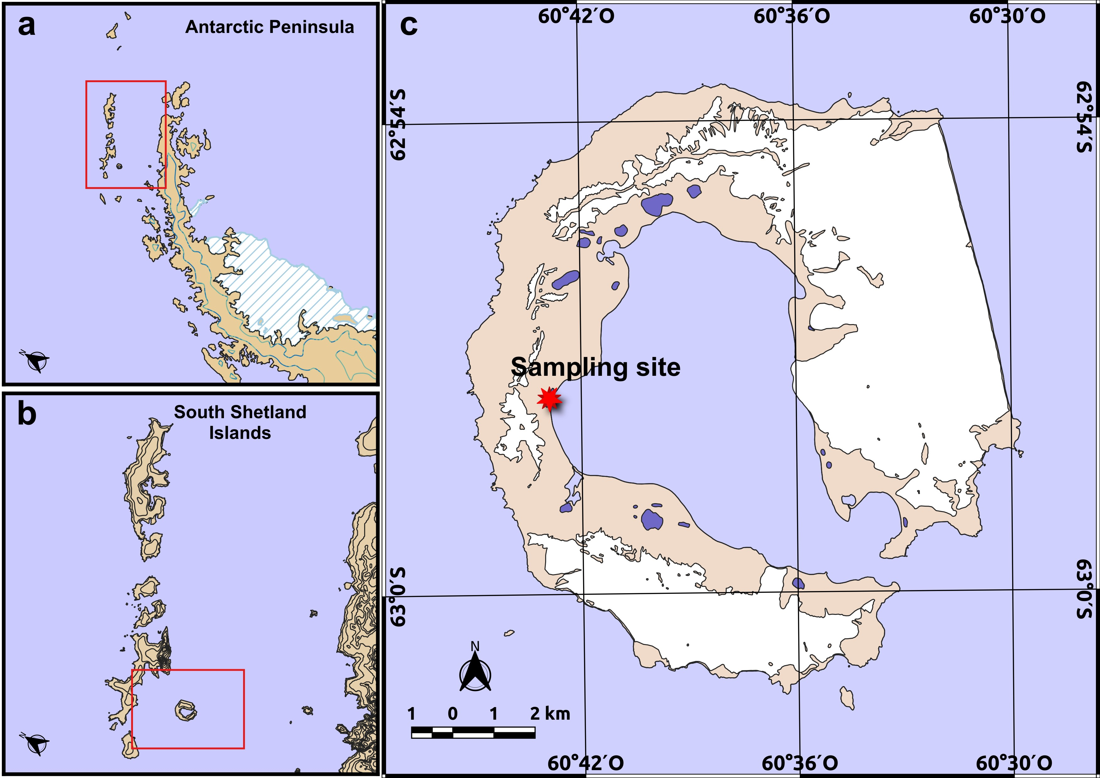
Figure 1. Sampling site. (a) Regional map of the northern Antarctic Peninsula; the red box indicates the area shown in panel (b). (b) South Shetland Islands highlighting the location of Deception Island; the red box indicates the area detailed in panel (c). (c) Detailed map of Deception Island showing the precise sampling site (red star) on the island surface; blue features represent inland water bodies and coastal lagoons, and shaded areas indicate ice cover. All maps are displayed in a standard polar stereographic projection. Spatial data were obtained from the Norwegian Polar Institute (Quantarctica dataset), and maps were generated using QGIS 3.16 Hannover.
Diversity 18 00236 g001
Figure 2. Colony morphology, mycelial differentiation and ultrastructural features of strain DEC002. (ag) Colony morphology of strain DEC002 on (a) ISP 1, (b) ISP 2, (c) ISP 3, (d) ISP 4, (e) ISP 5, (f) ISP 6, (g) ISP 7 media, showing the aerial mycelium (left panels) and the substrate mycelium (right panels). Scanning electron micrographs (SEM) of strain DEC002 displaying (h) filamentous vegetative mycelia, (i) mature spore chains, and (j) aerial hyphae undergoing sporulation. Spore morphometrics of strain DEC002 (k).
Diversity 18 00236 g002
Figure 3. Genome architecture and comparative functional annotation of strain DEC002. (a) Circular representation of the Prokka-annotated draft genome of strain DEC002, showing predicted coding sequences, RNA features, CRISPR elements, mobile genetic components, and GC content/GC-skew across the 9.18 Mb chromosome. (b) Total number of genes predicted by RASTtk, Prokka, and EggNOG-mapper, illustrating the broader ORF set produced by RASTtk relative to the more conservative predictions of Prokka and the orthology-supported subset defined by EggNOG. (c) Comparative agreement between RASTtk and Prokka annotations, indicating genes with identical assignments, those sharing equivalent biological functions despite differing nomenclature, and ORFs uniquely predicted by RASTtk. (d) Number of genes supported by EggNOG orthology, highlighting the subset of evolutionarily validated functions and the resolution of naming discrepancies between annotation pipelines.
Diversity 18 00236 g003
Figure 4. Multilocus and phylogenomic placement of Streptomyces fildesensis strain DEC002. (a) Maximum-likelihood phylogeny inferred with IQ-TREE v1.6.12 using a concatenated multilocus alignment (283 taxa, 3852 nucleotide sites; GTR+F+I+G4, 1000 ultrafast bootstraps). Node support is represented by circle size. (b) Genome-scale species tree generated with the KBase Build Microbial SpeciesTree v1.6.0 workflow using conserved single-copy bacterial markers. In both analyses, strain DEC002 forms a stable monophyletic cluster within S. fildesensis and is placed within the Actinacidiphila clade. Color coding highlights taxonomic groups: blue indicates members of the genus Actinacidiphila, yellow corresponds to S. fildesensis strains, and red denotes representatives of the genus Streptomyces. Both analyses consistently resolve DEC002 within S. fildesensis, confirming its taxonomic identity.
Diversity 18 00236 g004
Figure 5. Multi-framework comparative functional annotation profiles across four A. fildesensis genomes. (a) Horizontal barplot showing the total number of annotations assigned to each genome across the six functional annotation frameworks evaluated: KEGG, COG, EC, GO, eggNOG and PFAM. The relative distribution highlights the expected dominance of GO and PFAM terms due to their high granularity, while KEGG, COG, EC and eggNOG remain highly comparable across strains, reflecting a conserved global functional architecture. (bg) Z-score-scaled heatmaps summarizing functional enrichment patterns across genomes for each annotation system, presented in the following order: (b) KEGG, (c) COG, (d) EC, (e) GO, (f) eggNOG, and (g) PFAM. Rows correspond to the most represented categories within each framework (pathways, clusters, enzyme classes, gene ontology domains, orthologous groups, and protein families, respectively), and columns correspond to the four genomes. Warmer colors indicate relative overrepresentation, while cooler colors denote relative depletion within each strain after normalization. Together, the barplot and heatmaps reveal both the overall consistency of functional inventories and subtle framework-specific differences in category-level enrichment across the A. fildesensis lineage.
Diversity 18 00236 g005
Figure 6. Concatenated BGC repertoires per genome. Stacked horizontal bars depict the non-redundant BGC counts and composition for Antarctic Actinacidiphila fildesensis strains So13.3, DEC002, INACH3013, and GW25-5 (=CGMCC 4.5735T). Colors indicate antiSMASH categories (e.g., NRPS, PKS types I–III, RiPP classes, siderophore, terpene, redox-cofactor; full key at right). Bars summarize category balance and total repertoire size; they do not represent genomic order.
Diversity 18 00236 g006
Figure 7. Category-level and gene-level patterns of cold adaptation across seven Actinomycetota genomes. (a) The heatmap depicts the distribution of functional categories associated with oxidative-stress detoxification, redox balance, protein folding, osmotic protection and membrane remodeling, revealing expanded reductase and thioredoxin systems in the non-Antarctic Actinacidiphila sp. and characteristic enrichment of cold-shock and general stress elements in Antarctic Actinacidiphila fildesensis strain genomes. (b) The bubble plot shows the corresponding gene copy numbers, including the extensive amplification of FabG reductases in Actinacidiphila strains and the habitat-specific occurrence of CspC in Antarctic genomes. Bubble size is proportional to gene copy number. The underlying numerical data used to generate the bubble plot are provided in Supplementary Table S5. Together, both panels highlight distinct quantitative strategies for cold adaptation that differentiate temperate and Antarctic lineages.
Diversity 18 00236 g007
Table 1. Antibacterial activity of culture supernatant free of cell against bacterial foodborne pathogens.
Foodborne Pathogens StrainsActivity (cm ± Standard Deviation)
Vibrio parahaemolyticus1.81± 0.098
Vibrio cholerae2.80 ± 0.154
Enterobacter aerogenes1.08 ± 0.068
Enterobacter cloacae1.39 ± 0.126
Escherichia coli1.61 ± 0.068
Klebsiella pneumoniae1.67 ± 0.043
Staphylococcus aureus1.58 ± 0.075
Streptococcus pyogenes1.90 ± 0.116
Table 2. Antibiotic susceptibility of strain DEC002.
Antimicrobial ClassAntibiotic (μg)Diameter of Inhibition Zone (mm)Relative Potency (RP)
PenicillinAmpicillin (25)17.3 ± 1.30.51
PenicillinCarbenicillin (100)00
CephalosporinCeftazidime (35)00
CephalosporinCefamandole (35)00
CephalosporinCefixime (5)00
CephalosporinCefotaxime (30)00
CephalosporinCefpodoxime (10)00
CephalosporinCephalothin (30)18.3 ± 0.80.54
ChloramphenicolChloramphenicol (30)13.09 ± 0.180.39
FluoroquinolonesCiprofloxacin (10)33.98 ± 3.071
FluoroquinolonesNalidixic acid (30)00
MacrolidesClarithromycin (15)44.5 ± 1.61.31
MacrolidesErythromycin (15)39.01 ± 3.681.15
AminoglycosidesGentamicin (30)21.21 ± 0.30.62
AminoglycosidesSpectinomycin (25)34.4 ± 1.41.01
AminoglycosidesStreptomycin (25)29.55 ± 0.850.87
CarbapenemImipenem (10)44.15 ± 1.291.3
MetronidazoleMetronidazole (5)00
Fatty AcylsMupirocin (5)00
LincosamideClindamycin (2)00
LincosamideLincomycin (15)12.2 ± 0.210.36
NitrofurantoinNitrofurantoin (100)00
Coumarin glycosidesNovobiocin (5)47.04 ± 0.191.38
RifampinRifampicin (5)11.52 ± 0.690.34
Sulfonamide compounds S3Sulfonamide compounds (300)41.74 ± 3.351.23
TetracyclinesTetracycline hydrochloride (30)41.28 ± 4.791.22
Trimethoprim/sulfonamidesTrimethoprim–sulfamethoxazole (5)00
GlycopeptidesVancomycin (30)38.7 ± 0.721.14
Disclaimer/Publisher’s Note: The statements, opinions and data contained in all publications are solely those of the individual author(s) and contributor(s) and not of MDPI and/or the editor(s). MDPI and/or the editor(s) disclaim responsibility for any injury to people or property resulting from any ideas, methods, instructions or products referred to in the content.